evaluation of gate‐rtion (gate/geant4) monte carlo
TRANSCRIPT

HAL Id: hal-03039937https://hal.archives-ouvertes.fr/hal-03039937
Submitted on 4 Dec 2020
HAL is a multi-disciplinary open accessarchive for the deposit and dissemination of sci-entific research documents, whether they are pub-lished or not. The documents may come fromteaching and research institutions in France orabroad, or from public or private research centers.
L’archive ouverte pluridisciplinaire HAL, estdestinée au dépôt et à la diffusion de documentsscientifiques de niveau recherche, publiés ou non,émanant des établissements d’enseignement et derecherche français ou étrangers, des laboratoirespublics ou privés.
Evaluation of GATE-RTion (GATE/Geant4) MonteCarlo simulation settings for proton pencil beam
scanning quality assuranceCarla Winterhalter, Michael Taylor, David Boersma, Alessio Elia, Susanna
Guatelli, Ranald Mackay, Karen Kirkby, Lydia Maigne, Vladimir Ivanchenko,Andreas Resch, et al.
To cite this version:Carla Winterhalter, Michael Taylor, David Boersma, Alessio Elia, Susanna Guatelli, et al.. Evaluationof GATE-RTion (GATE/Geant4) Monte Carlo simulation settings for proton pencil beam scanningquality assurance. Medical Physics, American Association of Physicists in Medicine, 2020, 47 (11),pp.5817-5828. �10.1002/mp.14481�. �hal-03039937�

Evaluation of GATE-RTion (GATE/Geant4) Monte Carlo simulation settings forproton pencil beam scanning quality assurance
Carla Winterhalter†a) and Michael TaylorDivision of Cancer Sciences, University of Manchester, Manchester M13 9PL, UKThe Christie NHS Foundation Trust, Manchester M20 4BX, UK
David BoersmaACMIT Gmbh, Viktor Kaplan-Straße 2, Wiener Neustadt A-2700, AustriaEBG MedAustron GmbH, Marie Curie-Straße 5, Wiener Neustadt A-2700, Austria
Alessio EliaEBG MedAustron GmbH, Marie Curie-Straße 5, Wiener Neustadt A-2700, Austria
Susanna GuatelliCentre For Medical Radiation Physics, University of Wollongong, Wollongong, Australia
Ranald Mackay and Karen KirkbyDivision of Cancer Sciences, University of Manchester, Manchester M13 9PL, UKThe Christie NHS Foundation Trust, Manchester M20 4BX, UK
Lydia MaigneLaboratoire de Physique de Clermont, UMR 6533 CNRS - University Clermont Auvergne, Aubi�ere, France
Vladimir IvanchenkoCERN, Geneva 23 1211, SwitzerlandTomsk State University, Tomsk 634050, Russia
Andreas F. ReschDepartment of Radiation Oncology, Medical University of Vienna, W€ahringer G€urtel 18-20, Vienna 1090, Austria
David SarrutUniversit�e de Lyon, CREATIS, CNRS UMR5220, Inserm U1044, INSA-Lyon, Universit�e Lyon 1, Centre L�eon B�erard, Lyon, France
Peter SitchThe Christie NHS Foundation Trust, Manchester M20 4BX, UK
Marie VidalInstitut M�editerran�een de Protonth�erapie - Centre Antoine Lacassagne - F�ed�eration Claude Lalanne, Nice 06200, France
Lo€ıc Grevillot*EBG MedAustron GmbH, Marie Curie-Straße 5, Wiener Neustadt A-2700, Austria
Adam Aitkenhead*Division of Cancer Sciences, University of Manchester, Manchester M13 9PL, UKThe Christie NHS Foundation Trust, Manchester M20 4BX, UK
(Received 20 April 2020; revised 28 August 2020; accepted for publication 29 August 2020;published 17 October 2020)
Purpose: Geant4 is a multi-purpose Monte Carlo simulation tool for modeling particle transport inmatter. It provides a wide range of settings, which the user may optimize for their specific applica-tion. This study investigates GATE/Geant4 parameter settings for proton pencil beam scanning ther-apy.Methods: GATE8.1/Geant4.10.3.p03 (matching the versions used in GATE-RTion1.0) simulationswere performed with a set of prebuilt Geant4 physics lists (QGSP_BIC, QGSP_BIC_EMY,QGSP_BIC_EMZ, QGSP_BIC_HP_EMZ), using 0.1mm-10mm as production cuts on secondaryparticles (electrons, photons, positrons) and varying the maximum step size of protons (0.1mm, 1mm,none). The results of the simulations were compared to measurement data taken during clinicalpatient specific quality assurance at The Christie NHS Foundation Trust pencil beam scanning protontherapy facility. Additionally, the influence of simulation settings was quantified in a realistic patientanatomy based on computer tomography (CT) scans.Results: When comparing the different physics lists, only the results (ranges in water) obtained withQGSP_BIC (G4EMStandardPhysics_Option0) depend on the maximum step size. There is clinicallynegligible difference in the target region when using High Precision neutron models (HP) for dosecalculations. The EMZ electromagnetic constructor provides a closer agreement (within 0.35 mm) to
5817 Med. Phys. 47 (11), November 2020 0094-2405/2020/47(11)/5817/12© 2020 The Authors. Medical Physics published by Wiley Periodicals
LLC on behalf of American Association of Physicists in Medicine. Thisis an open access article under the terms of the Creative Commons
Attribution License, which permits use, distribution and reproductionin any medium, provided the original work is properly cited.
5817

measured beam sizes in air, but yields up to 20% longer execution times compared to the EMYelec-tromagnetic constructor (maximum beam size difference 0.79 mm). The impact of this on patient-specific quality assurance simulations is clinically negligible, with a 97% average 2%/2 mm gammapass rate for both physics lists. However, when considering the CT-based patient model, dose devia-tions up to 2.4% are observed. Production cuts do not substantially influence dosimetric results insolid water, but lead to dose differences of up to 4.1% in the patient CT. Small (compared to voxelsize) production cuts increase execution times by factors of 5 (solid water) and 2 (patient CT).Conclusions: Taking both efficiency and dose accuracy into account and considering voxel sizeswith 2 mm linear size, the authors recommend the following Geant4 settings to simulate patientspecific quality assurance measurements: No step limiter on proton tracks; production cuts of 1 mmfor electrons, photons and positrons (in the phantom and range-shifter) and 10 mm (world); bestagreement to measurement data was found for QGSP_BIC_EMZ reference physics list at the cost of20% increased execution times compared to QGSP_BIC_EMY.For simulations considering the patient CT model, the following settings are recommended: No step
limiter on proton tracks; production cuts of 1 mm for electrons, photons and positrons (phantom/range-shifter) and 10 mm (world) if the goal is to achieve sufficient dosimetric accuracy to ensure thata plan is clinically safe; or 0.1 mm (phantom/range-shifter) and 1 mm (world) if higher dosimetricaccuracy is needed (increasing execution times by a factor of 2); most accurate results expected forQGSP_BIC_EMZ reference physics list, at the cost of 10–20% increased execution times comparedto QGSP_BIC_EMY. © 2020 The Authors. Medical Physics published by Wiley Periodicals LLC onbehalf of American Association of Physicists in Medicine. [https://doi.org/10.1002/mp.14481]
Key words: dose calculation, Geant4, GATE-RTion, Monte Carlo, proton pencil beam scanningtherapy
1. INTRODUCTION
Geant4 is a C++ based, object-oriented, multi-purpose MonteCarlo (MC) tool to simulate particle transport for energiesranging from eV to TeV scale,1,2 with applications rangingfrom the modeling of high energy particle colliders to spaceand shielding simulations.3 Geant4 is extensively used formedical physics simulations, in particular to calculate thedose (energy deposited per unit mass) during particle therapy(e.g., see Refs. [4–6] for proton radiotherapy simulationsusing Geant4 or Geant4 wrappers). This study is focused onGATE/Geant4 simulations for patient dosimetry in protonpencil beam scanning.
Geant4 based Architecture for Medicine Oriented Simula-tion (GAMOS),7 TOol for PArticle Simulation (TOPAS)8 andGeant4 Application for Tomographic Emission (GATE)9-11
provide user-friendly interfaces to Geant4. This researchtakes place within a recent initiative called GATE-RTion,12
which is aimed at providing a validated long-term applica-tion, based on a GATE and Geant4 version, along with asso-ciated software tools for clinical dosimetric implementationto facilitate collaborations between hadron therapy institutes.Current collaborators include the Centre Antoine Lacassagne(Nice, France), the Christie NHS Foundation Trust (Manch-ester, UK) and MedAustron (Wiener Neustadt, Austria).
GATE/Geant4 offers a set of prebuilt physics lists whichthe user may adopt in his/her specific simulation scenario.Physics lists may use different models to describe a specificelectromagnetic or hadronic physics interaction and differentparameters such as the production cut and maximum stepsize. In order to support the Geant4 medical physics
community, the Geant4 Medical Physics BenchmarkingGroup13,14 has been recently established to benchmark Gean-t4 for medical physics and to provide recommendations interms of physics lists, production cuts and maximum stepsizes. For proton therapy (using proton energies up to250 MeV), these settings have been previously investigatedfor different Geant4 versions: Jarlskog and Paganetti com-pared simulations (Geant4 v.8.1) in water and a Faraday cupgeometry to measurements, and recommended a combinationof electromagnetic and nuclear models.15 That recommenda-tion was used as a basis for Grevillot et al., who comparedGATE/Geant4 (Geant4 v.9.2) simulations in water andPMMA to measurements and showed the importance of themaximum step size and production cut.16 Kurosu et al. simu-lated proton treatment nozzles for uniform17 and for spotscanning18 with different MC codes, including GATE/Geant4v.9.5, investigating the influence of both step size and pro-duction cut on depth dose curves. Fuchs et al. investigatedmulti-Coulomb scattering after a range of materials (GATE,Geant4 versions 9.5.02, 9.6.03, 10.0.02, 10.1, 10.2.02), con-cluding that agreement to measurements substantiallydepended on the multiple scattering model set in the specificGeant4 version used.19 Recently, Resch et al. compared simu-lated lateral dose profiles and field size factors to measure-ments to investigate the accuracy of electromagnetic andnuclear scattering models (GATE, Geant4 v.10.03.01), show-ing a high sensitivity of the elastic scattering cross sections.20
This study extends and complements the work above bycomparing a wide range of combinations of physics options,step limiter and production cuts to a comprehensive set ofmeasurements, aimed at clinical applications of Geant4/
Medical Physics, 47 (11), November 2020
5818 Winterhalter et al.: GATE/Geant4 settings for proton therapy 5818

GATE for proton therapy dosimetry. It takes the whole (clini-cal) process of beam characterization into account and allexperimental data were acquired as part of clinical commis-sioning or quality assurance. Additionally, the influence ofdifferent settings is quantified in simulations performed in arealistic CT-based patient model. This is therefore a compre-hensive validation of Geant4 v.10.3.3 in combination withGATE v.8.1, and therefore also of GATE-RTion v.1.0, whichis a long-term stable branch of GATE v.8.1 intended for clini-cal purposes. A set of parameter recommendations is pre-sented, taking both dose distributions and calculationefficiency into account.
The first part of the paper gives an overview of the simula-tion setup and the Geant4 settings under investigation. Next,the influence of the different settings on the beam-modelingprocess and on spot sizes in air is investigated by comparisonof simulations of individual pencil beams and measurementdata acquired during the commissioning of a proton therapyfacility. Then the effect of the settings on simulations in solidwater is evaluated in comparison to patient specific qualityassurance (PSQA) measurements. Finally, the effect of theMC settings on calculations in the CT-based patient model isassessed for a set of patients, which represent a range ofpotential dose calculation issues.
2. MATERIALS AND METHODS
2.A. AUTOMC simulations
In the following, MC settings are summarized accordingto Ref. [21]. A set of in-house developed scripts(AUTOMC22) written in GNU Octave23 (v.4.4.1), whichautomatically create GATE macro files, launch simulationsand analyse simulation results, have been developed for clini-cal proton therapy dose calculations at The Christie NHSFoundation Trust. This system performs all its MC calcula-tions using GATE v.8.1/ Geant4 v.10.3.3.2 These are the sameversions included in GATE-RTion v.1.0,12 and in keepingwith the goals of GATE-RTion, this work aims to providerecommendations in terms of simulation parameters to adoptfor proton pencil beam scanning.
The MC settings for simulations performed in this work aresummarized in Table I. Simulations and data analysis wereperformed on a Linux (based on Ubuntu 18.04 LTS) clusterconsisting of 1 master CPU (for data analysis) and 10 slaveCPUs (for simulations). Each CPU was a quad-core Intel XeonE3-1240 v6@ 3.70 GHz, with 16 GB RAM, leading to a totalof 40 cores. No variance reduction techniques were used.
2.A.1. Beam-model tuning
For proton pencil beam scanning MC calculations, pro-tons need not be tracked through the whole beam line. Insteada beam description upstream of the patient can be imple-mented,25–27 which parameterizes each spot by its number ofprotons (for absolute dose), mean energy and energy spread
(shape of the Bragg peak) and beam optics phase-space(beam sizes in air) at the “starting point” of the simulation (inAUTOMC: 89.5 cm upstream of iso-center). Mean energyand energy spread are automatically and iteratively adjustedsuch that depth dose curves measured with a Bragg peakchamber (diameter 84 mm) are reproduced (tuning process,see Ref. [22] for more details). For this study, error bars onthe mean energy were determined by repeating this tuningprocess with different initial energies. That is, for one setting,the tuning process was performed once with initial energiesabove the expected mean energy and once with initial ener-gies below the expected mean energy. The error bars showthe maximum difference between the obtained final meanenergies, and as such the error due to both statistics and thetolerances in the beam-modeling process.
2.A.2. Lateral spot profiles in air
Within a MC simulation, the beam optics phase-spaceconsisting of spot size, emittance and divergence of the pro-tons at the source plane defines the initial optics of the beam.Additionally, the Lexan “range-shifter,” which can be intro-duced into the beam to lower the proton energy, also influ-ences spot size and divergence. Table I summarizes therange-shifter options modeled within this study. Beam sizesin air (sigma, modeled with the beam spot following a Gaus-sian distribution) were simulated with and without range-shif-ter and compared to beam sizes measured with the Lynxdetector (IBA-dosimetry, Schwarzenbruck) at different dis-tances from iso-center.
2.A.3. PSQA measurements
PSQA measurements are routinely performed for eachradiation field before the first fraction is delivered to thepatient (e.g., Ref. [28]). At The Christie NHS FoundationTrust, doses are measured at multiple depths in a Solid Waterphantom using a the Octavius 1500 XDR array (PTW, Frei-burg) for relative dose distributions and a PTW 31021 Semi-flex 3D ion chamber for absolute dose values (see Ref. [22]for more details). Two example fields (one inhomogeneousfield with range-shifter and one homogeneous field withoutrange-shifter, patient 1 and patient 2 of Table II) were simu-lated to 0.25% statistical uncertainty at the 90–100% doselevel (see Ref. [24]) to demonstrate differences between thedifferent settings. 34 fields (six patients, all with range-shifterthicknesses 3 and 5 cm) were recalculated to 0.6% statisticaluncertainty at the 90–100% dose level, and compared to 200(relative) array planes and 74 (absolute) chamber measure-ment points, which were all taken as part of the clinicalPSQA. Place of measurement was at the shallow/central/deeppart of the high dose region (first three months of operationof The Christie NHS Foundation Trust proton beam therapycentre), shallow/deep part (next six months) and at the centralpart only (afterwards). Additionally, measurements werepartly repeated in different treatment rooms (see Ref. [22]).
Medical Physics, 47 (11), November 2020
5819 Winterhalter et al.: GATE/Geant4 settings for proton therapy 5819

2.A.4. Patient CT model
Finally, dose distributions are simulated in the patient CTmodel for six different patients (1 field per patient, seeTable II). The CT calibration was based on the stoichiometriccalibration by Schneider et al (Phys Med Biol 29), and Ref.[22] in detail describes the procedure to ensure matching CTcalibrations (based on same scanner parametrization, refer-ence tissues, and ionization values) between treatment plan-ning system and AUTOMC. Patients were chosen torepresent a range of several potential dose calculation issues,that is, different range-shifter options, heterogeneous anatom-ical sites, and treatments with metal implants.
2.B. Geant4 physics and simulation settings
The following prebuilt Geant4 physics lists, which areconsidered suitable for hadron therapy, were investigated:
• QGSP_BIC: QGSP_BIC with G4EMStandardPhysics(often referred as G4EMStandardPhysics_Option0,Wentzel multiple scattering model for protons).
• QGSP_BIC_EMY: QGSP_BIC with G4EMStan-dardPhysics_Option3, Urban multiple scattering modelfor protons.
• QGSP_BIC_EMZ: QGSP_BIC with G4EMStan-dardPhysics_Option4, Wentzel multiple scatteringmodel for protons.
• QGSP_BIC_HP_EMZ: Same electromagnetic interac-tions as QGSP_BIC_EMZ, with high precision (HP)neutron libraries for neutrons below 20 MeV.
The physics lists differ in terms of electromagnetic and hadro-nic physics models and processes. Details and parameters can befound at.2,14,30,31Multiple studies15,20 showed that the Binary Cas-cade model is adequate to model the intranuclear cascade in pro-ton therapy,14 and therefore the QGSP_BIC was chosen to modelhadronic interactions. The EM physics component was modeledusing one among G4EMStandardPhysics_Option0, G4EMStan-dardPhysics_Option3 (EMY), and G4EMStandardPhysics_Option4 (EMZ). G4EMStandardPhysics_Option4 is deemed tobe the most accurate EM physics constructor for medical physicsapplications, at the cost of longer computational times.14
QGSP_BIC_EMZ in combination with HP (QGSP_BIC_H-P_EMZ) neutron data libraries was also studied. For readability,these lists are in the following abbreviated as Option0, EMY,EMZ, HP_EMZ.
The dosimetric effects of varying the production cuts(threshold on the production of secondary photons, electronsand positrons) and step limiter (set by the user, restricting the
TABLE I. Overview of the AUTOMC simulation settings. First dimension is along the beam direction.
Beam-model generation Lateral spot size in air PSQA phantom Patient CT
Volume 400 mm 9 100 mm 9 100 mm,upstream surface at iso-center
0.1 mm 9 101 mm 9 101 mm,various positions between 40 cmup- and downstream of iso-center
According to phantom CT According to patient CT
Material Water (density = 1.0 g/cm3,I = 75.3 eV)
Air solid water (density = 1.057 g/cm3, I = 67.1 eV)
Hounsfield units converted toelemental composition &density
Scoring voxeldimensions
0.5 mm 9 1 mm 9 1 mm,laterally integrated over Braggpeak chamber area.
0.1 mm 9 1 mm 9 1 mm 2 mm 9 2 mm 9 2 mm 2 mm 9 2 mm 9 2 mm
Initial beam 19 energies between 70 and245 MeV, 4�107 protons per spot
90 MeV, 150 MeV, 230 MeV,2�107 initial protons per spot
Energies according to patientplan. Number of protons scaled toreach 0.25%/0.6% statisticaluncertainty at the 90–100%isodose24
Energies according to patientplan. Number of protons scaledto reach 0.25% statisticaluncertainty at the 90–100%isodose21
Lexan range-shifter
None None/2 cm/3 cm/5 cm.Downstream surface located46 cm upstream of iso-center
According to patient plan According to patient plan
TABLE II. Patient selection. 1 field has been recalculated per patient. Number of phases & fields, CTV volume and prescription are given for the whole treatment,range-shifter for the recalculated field
Treatment site Prescription [RBE.Gy] (#fractions) Number of phases & Fields CTV vol [cm3] Range-shifter Comment
Patient 1 Neck 50.4 (28); 9 (5) 2 phases, 3 + 2 fields 308; 56 5 cm —
Patient 2 Brainstem 52.2 (29) 1 phase, 2 fields 44 None —
Patient 3 Base of skull 54 (30); 16.2 (9) 2 phases, 3 fields each 29; 7 5 cm —
Patient 4 Paranasal 63; 70 (35) Simultaneous integrated boost, 4 fields 47; 28 None —
Patient 5 Spine 50.4 (28); 19.8 (11) 2 phases, 3 fields each 935; 37 None Titanium implant
Patient 6 Spine 18 (25); 21.6 (12); 9 (5) 3 phases, 4 + 3 + 3 fields 654; 202; 9 2 cm Titanium implant
Medical Physics, 47 (11), November 2020
5820 Winterhalter et al.: GATE/Geant4 settings for proton therapy 5820

maximum step length of protons) parameters were investi-gated as well. A maximum step length (step limiter) has beenset for protons. The step limiter was set to 0.1, 1 mm or wasnot set. In the case of no user-defined step limiter, the stepsize is calculated in Geant4 with consideration of either thedistance to the next geometrical boundary or the next interac-tion as dictated by the implemented physics processes. Pro-duction cuts set thresholds on the production of secondaryparticles. Only secondaries with a range exceeding the pro-duction cut are explicitly produced and tracked, otherwisetheir energy is considered local energy deposition. Produc-tion cuts were set for electrons, photons and positrons withinthe world (1 mm/10 mm) and within the phantom/range-shif-ter (0.1 mm/1 mm). These cuts were chosen to use one valuecomparable to calculation voxel size (see Table I), andanother value much smaller than the calculation voxel size.
2.C. Overview of study
Simulation results were compared to experimental data forthe combinations of production cuts and step limiters summa-rized in Table III. From here on, these will be referred to bythe scenario numbers shown in the table. Each of these sixscenarios was combined with the four prebuilt physics lists(Option0, EMY, EMZ, HP_EMZ) leading to a total of 24combinations.
First, the effect of these settings was evaluated for theenergy tuning process (Section 3.A) and for beam size simu-lations in air (Section 3.B). The effect of each physics list wasanalyzed while setting both production cuts and step limiterto the smallest values considered, which should provide themost accurate dosimetric results (reference scenario #1).Next, production cuts in phantom and world were variedwhile leaving the step limiter constant (scenarios 1, 2 and 3).Then, the effect of the step limiter was evaluated while keep-ing the cuts fixed at the smallest value considered (scenarios1, 4 and 5). Finally, the scenario with smallest cuts and step
limiter was compared to the combinations of no step limiterwith 0.1 mm/1 mm and 1 mm/10 mm production cuts (sce-narios 1, 5 and 6).
Based on these results, the three physics lists EMY, EMZand HP_EMZ in combination with scenario 5 (“small cuts”compared to 2 mm voxel linear size) and scenario 6 (“largecuts”) were further investigated and compared to measure-ment data acquired with the PSQA phantom (Section 3.C),and compared when considering realistic, CT scan basedpatient models (Section 3.D).
3. RESULTS
3.A. Beam-model definition for the Monte Carlosimulations
For the full proton energy range, the energy fine tuning(i.e., the mean energy and energy spread defined in the MCsimulation for each nominal TPS energy) does not substan-tially depend on the chosen physics lists [mean energy inFig. 1(a), agreement within 0.08 MeV and energy spread (notshown), agreement within 0.03 MeV]. Subsequently, the tun-ing process was repeated for a 245 MeV (nominal energy)spot with different production cuts [Fig. 1(b), scenario 1–3],step limiter [Fig. 1(c), scenarios 1,4 and 5] and combinationof production cuts and step limiter [Fig. 1(d), scenarios 1,5and 6]. Simulations performed with the EMY and EMZ didnot depend on the step limiter, whereas for simulations per-formed with Option0 energies varied by up to 0.5 MeV, indi-cating that for this physics option the range in water dependson the step limiter.
After the beam-modeling process, local dose differencesbetween MC based calculations and depth dose measurementwere analyzed for the 245 MeV spot [Fig. 2(a)]. There wasno substantial difference between different physics lists[Fig. 2(b), scenario 1]. It is worth noting that for Option0 thedifferences are dependent on the step limiter [Fig. 2(c), sce-nario 5], and that for all physics lists the pattern of differenceschanges with larger production cuts [Fig. 2(d), scenario 6].All observed differences between MC simulations and depthdose measurement lie within �3%.
The execution times for the 245 MeV spot decreased byfactors of 4.5–5.9 when deactivating the step limiter (scenario5 vs scenario 1) and by factors of 5.3–7.2 when increasingthe production cuts (scenario 6 vs scenario 5). Such resultsare to be expected because smaller steps and a higher numberof secondaries to track inevitably increases the executiontime. Compared to HP_EMZ (scenarios 5/6), shorter execu-tion times are achieved with Option0 (factors of 1.4/2.0), fol-lowed by EMY (factors of 1.2/1.5) and EMZ (factors of 1.0/1.1).
3.B. Lateral pencil beam profiles in air
For the 150 MeV beam without range-shifter, beam sizesin air (Fig. 3) agreed within 0.35 mm to measurements,
TABLE III. Overview of the combinations of production cuts and step limiterinvestigated in this study. In air, range cuts of 1 mm/10 mm correspond toproduction thresholds of 1 keV/1 keV (photons) 1 keV/16 keV (electrons)and 1 keV/16 keV (positrons). In water, range cuts of 0.1 mm/1 mm corre-spond to production thresholds of 1.1 keV/2.9 keV (photons), 85 keV/350 keV (electrons) and 83 keV/343 keV (positrons). The tracking cut forelectrons is 0.1 keV.
Scenario
Production cuts forelectrons, photons andpositrons (phantom andrange-shifter) [mm]
Production cuts forelectrons, photons andpositrons (world) [mm]
Steplimiter[mm]
1“Reference”
0.1 1 0.1
2 1 1 0.1
3 0.1 10 0.1
4 0.1 1 1
5 0.1 1 None
6 1 10 None
Medical Physics, 47 (11), November 2020
5821 Winterhalter et al.: GATE/Geant4 settings for proton therapy 5821

showing that the beam optics have been matched well. Withthe 5 cm range-shifter, simulations performed with EMZ andHP_EMZ [green/blue lines in Figs. 3(a)–3(c)] agreed within0.25 mm to measurements (magenta triangles), with negligi-ble differences when using the HP module. EMY lead tosmaller beam sizes compare to the other physics lists andmeasurements [up to 0.79 mm difference to measurements,red lines in Figs. 3(a)–3(c)]. As the beam optics have beendefined without range-shifter, and the range-shifter been sim-ulated as a physical component, these differences betweendifferent settings can be attributed to the differences in scat-tering in the range-shifter. Option0 simulated beam sizeswithout step limiter and with large cuts differed by up to0.51 mm from measurements [black line in Fig. 3(c)]. Simu-lations performed with the 5 cm range-shifter and a very lowenergy proton beam (90 MeV) also showed less scatteringwhen using EMY compared to EMZ/HP_EMZ [Figs. 3(d)–
3(e)]. For a high energy proton beam (230 MeV), less scat-tering occurs in the range-shifter and differences between thedifferent physics lists was therefore negligible.
3.C. PSQA measurements
The previous two sections showed that dose results dependon the step limiter for Option0. As the step limiter substan-tially increases simulation times, for the remainder of thestudy, we concentrate on EMY, EMZ and HP_EMZ withouta step limiter and in combination with “small” (0.1 mm/1 mm) and “large” (1 mm/10 mm) cuts (phantom/world, incomparison to the 2 mm calculation grid, scenarios 5 and 6).
Two example fields [patient 1, Figs. 4(a)–4(d) and patient2, Figs. 4(e)–4(h)] fields were simulated in solid water to0.25% statistical uncertainty at the 90–100% dose level todemonstrate the differences caused by the MC simulation
FIG. 1. Results for all energies and physics lists (a, scenario 1), and as a function of production cuts in phantom and world (b, scenario 1–3), as a function of steplimiter (c, scenario 1,4,5) and as a function of combinations of cuts and step limiter (d, scenario 1,5,6) for a 245 MeV (nominal energy) spot. EnergyNominal isthe energy reported by the TPS, EnergyMC is the mean energy used to define the source within Gate (i.e., EnergyMC is derived by the tuning process.) Errorbars are determined by using different starting conditions (initial energy) in the energy tuning process. [Color figure can be viewed at wileyonlinelibrary.com]
Medical Physics, 47 (11), November 2020
5822 Winterhalter et al.: GATE/Geant4 settings for proton therapy 5822

settings. For patient 1, an inhomogeneous field with a 5 cmrange-shifter, there was no substantial difference betweenEMZ and HP_EMZ [Fig. 4(b)]. EMZ and EMY simulateddose results differed by up to 1% of the prescription dose[Fig. 4(c)], whereas different production cuts did not substan-tially influence dose results [Fig. 4(d)]. For patient 2, ahomogenous field without range-shifter, magnitudes of differ-ences were comparable to statistical fluctuations [Figs. 4(e)–4(h)]. Execution times (Table IV) decreased by factors of upto 4.7 when changing from small to large cuts, and faster exe-cution times were achieved with EMY (up to factors of 1.6/1.3 faster than HP_EMZ/ EMZ).
Finally, 34 fields were recalculated in solid water to a0.6% statistical uncertainty at the 90-100% dose level andcompared to PSQA experimental data (see Section 2.A). Rel-ative measurements [Fig. 5(a)] show a mean 2%/2 mm localgamma agreement (10% low dose threshold) over all 34 fields(200 planes) of 97% for all three scenarios (EMZ, small cuts;EMZ, large cuts; EMY, large cuts). Absolute dose measure-ments [74 points, Fig. 5(b)] show a dose offset of �1.2%(EMZ, small cuts), �1.0% (EMZ, large cuts) and �0.9%(EMY, large cuts).
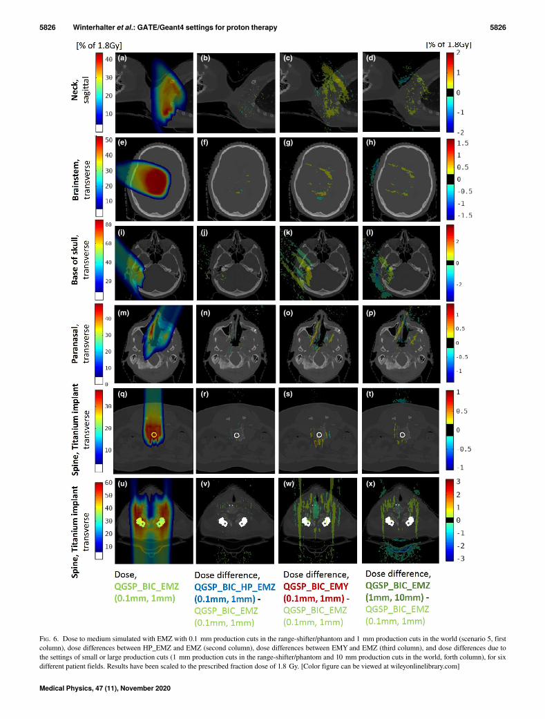
3.D. Patient CT
In patient CT (Fig. 6), there was no substantial differencein terms of dose calculation between EMZ and HP_EMZ inthe target region (Fig. 6, column 2). Differences betweenEMZ and EMY (Fig. 6, column 3) were substantial (within2.4%), especially when using a range-shifter [Figs. 6(c), 6(k),6(w)] and after tissue heterogeneities [e.g., implant inFig. 6(s)]. Production cuts (column 4) cause dose differencesof up to 4.1% at air-tissue interface [Fig. 6(l)]. Executiontimes were decreased by up to a factor of 2.2 when changingfrom small to large cuts, and faster execution times wereachieved with EMY (up to factors of 1.4/1.2 faster thanHP_EMZ/ EMZ, Table IV).
4. DISCUSSION
Comparison of simulated dose distributions in watershows that for QGSP_BIC with G4EMStan-dardPhysics_Option0 the results depend on the step limiter.In the Option0 configuration, steps are larger than in EMYand EMZ. If the step limiter is reduced in Option0, step sizes
FIG. 2. Measured integral depth dose curve for the 245 MeV spot (a), and differences between MC and measurements after the tuning process for the 245 MeVspot for all physics lists with range cuts and step limiter defined according to scenario 1(b), scenario 5(c) and scenario 6(d). [Color figure can be viewed at wileyonlinelibrary.com]
Medical Physics, 47 (11), November 2020
5823 Winterhalter et al.: GATE/Geant4 settings for proton therapy 5823

are therefore much more reduced compared to EMY andEMZ. As the step limiter substantially increases calculationtimes (factors of 4.5–5.9), EMY, EMZ or HP_EMZ withoutstep limiter are therefore better suited for dose simulations inproton therapy.
For the endpoints of this study (clinical measurement dataconsisting of depth dose curves, beam size in air, dose distri-butions in a solid water phantom and patient CT), there wasno substantial difference between HP_EMZ and EMZ in-field. This is expected since the in-field region is dominatedby the incident proton beam, while a bigger effect of the neu-tron distribution is expected out-of-field and beyond the distaledge of the Bragg peak. Using the high precision neutronlibraries, however, increased execution times by up to 30%.
It has been demonstrated that EMY (G4EMStan-dardPhysics_Option 3, Urban scattering model)
underestimates the beam sizes in air after a scattering mate-rial when compared to EMZ (G4EMStandardPhysics_Option4, Wentzel scattering model) and to measurements. This ismainly due to differences in multiple scattering modeling,and confirms in Geant4.10.3.3 the results of Fuchs et al.,19
who showed that multi-Coulomb scattering angles were bet-ter reproduced when using the Wentzel instead of the Urbanscattering model. Consequently, dose distributions simulatedin a solid water phantom differ (within 1% of prescriptiondose) when comparing EMYand EMZ, with the EMYoptionresulting in less lateral spread of the fields, just as it resultedin less lateral spread of single spots. However, as this lieswithin the measurement uncertainty of clinical PSQA mea-surements, and the calculation discrepancies are significantonly at very located areas of the dose map, no substantial dif-ference between physics lists is observed when comparing
FIG. 3. Beam size (sigma of the Gaussian distribution of the beam spot) of a 150 MeV (a–c), a 90 MeV and a 230 MeV (d,e) spot as a function of distance fromiso-center in air for all physics lists, (a) without range-shifter and with 2/3/5 cm Lexan range-shifter for small cuts and step limiter (scenario 1), (b,d) without andwith a 5 cm range-shifter for small cuts and step limiter default (scenario 5) and (c,e) for large cuts and step limiter default (scenario 6). Measurements are repre-sented with magenta triangles. [Color figure can be viewed at wileyonlinelibrary.com]
Medical Physics, 47 (11), November 2020
5824 Winterhalter et al.: GATE/Geant4 settings for proton therapy 5824

simulations to a wide range of relative array and absolutechamber measurements, with an average gamma index agree-ment (2%/2 mm) of 97% and absolute dose offset to
measurements of 1% (Fig. 5). Gamma analysis was chosento evaluate the agreement to measurements in solid water asthis is the standard clinical procedure to ensure treatmentsafety. When considering patient models based on CT acqui-sition, the same effect is observed, with dose differences ofup to 2.4% between EMZ and EMY, which are especiallypronounced when using a range-shifter. Execution times areup to 10–30% higher for EMZ when compared to EMY.
Production cuts influence the difference between simu-lated depth dose curves and measurements, and thereforehave to be chosen carefully to ensure correct absolute dosescaling. For PSQA in solid water, production cuts from 0.1 to1 mm do not substantially affect dose distributions and agree-ment to measurements, but increase execution times by up toa factor of 5. In contrast, in patient CT, production cuts causedose differences of up to 4%, especially at air-tissue
FIG. 4. Dose to water32 simulated with EMZ with 0.1 mm production cuts in the range-shifter/phantom and 1 mm production cuts in the world (scenario 5, a,e),dose differences between HP_EMZ and EMZ (b,f), dose differences between EMY and EMZ (c,g), and dose differences due to the settings of small (scenario 5,0.1 mm/1 mm) or large (scenario 6, 1 mm/10 mm) production cuts (d,h), for two different patient fields (a–d and e–h). Results have been scaled to the prescribedfraction dose of 1.8 Gy. [Color figure can be viewed at wileyonlinelibrary.com]
TABLE IV. Execution times for the fields displayed in Figs. 4(a)–4(d)/Figs. 6(a)–6(d) for each scenario (0.25% statistical uncertainty at the 90–100% dose level).
Physicslist
Production cuts inphantom/world
Patient 1, solidwater [CPU.h]
Patient 1, CT[CPU.h]
EMY 0.1 mm/1 mm 475.0 895.2
EMY 1 mm/10 mm 104.1 448.2
EMZ 0.1 mm/1 mm 605.8 1068.1
EMZ 1 mm/10 mm 127.9 491.0
HP_EMZ 0.1 mm/1 mm 644.9 1233.2
HP_EMZ 1 mm/10 mm 163.7 646.4
FIG. 5. Histograms of the gamma index values obtained when comparing simulations to relative measurements for 200 array planes (a) and histograms of abso-lute dose differences for 74 chamber measurements (b) for EMZ with small cuts (light green), EMZ with large cuts (dark green), EMY with large cuts (red). In(a), the percentage of planes which have <90%, <95% and <98% of pixels having c ≤ 1 are indicated in the figure legend. [Color figure can be viewed at wileyonlinelibrary.com]
Medical Physics, 47 (11), November 2020
5825 Winterhalter et al.: GATE/Geant4 settings for proton therapy 5825
(a) (b) (c) (d)
(e) (f) (g) (h)
(i) (j) (k) (l)
(m) (n) (o) (p)
(q) (r) (s) (t)
(u) (v) (w) (x)
FIG. 6. Dose to medium simulated with EMZ with 0.1 mm production cuts in the range-shifter/phantom and 1 mm production cuts in the world (scenario 5, firstcolumn), dose differences between HP_EMZ and EMZ (second column), dose differences between EMY and EMZ (third column), and dose differences due tothe settings of small or large production cuts (1 mm production cuts in the range-shifter/phantom and 10 mm production cuts in the world, forth column), for sixdifferent patient fields. Results have been scaled to the prescribed fraction dose of 1.8 Gy. [Color figure can be viewed at wileyonlinelibrary.com]
Medical Physics, 47 (11), November 2020
5826 Winterhalter et al.: GATE/Geant4 settings for proton therapy 5826

interfaces. This can be explained by the production of secon-daries with ranges between 0.1mm–1mm in the denser tissue,which have larger ranges in the surrounding air. Executiontimes in CT-based patient models are increased by a factor of2 when using small production cuts.
Physics settings selection are strongly dependent on theend-user application. Establishing recommendations for sim-ulation parameters for the GATE-RTion framework is ofparamount importance however, the final choice of simula-tion and physics settings remains the responsibility of theuser.
On the one hand, if using a full Geant4 based MC system,the user might decide for the settings providing the mostaccurate dosimetric calculations, independently from timeconstraints. On the other hand, quicker MC simulationsmight be essential for a clinical environment, and dependingon clinical tolerances and workload a compromise betweenspeed and dosimetric accuracy may be required to the user.
As such, the authors would recommend the followingGeant4 settings for PSQA dosimetry, when considering vox-els of 2 mm size:
- No step limiter for proton tracks.- Production cuts on electrons, photons and positrons of1 mm in the phantom and range-shifter, while adopting a10 mm value in the surrounding geometry (world).
- Best agreement to measurement data was found forQGSP_BIC_EMZ reference physics lists at the cost of 20%increased execution times compared to QGSP_BIC_EMY.
For simulations in CT-based patient models, the followingsettings are recommended:
- No step limiter on proton tracks.- Production cuts on electrons, photons and positrons of1 mm (phantom/range-shifter) and 10 mm (world) if thegoal is to achieve sufficient dosimetric accuracy to ensurethat a plan is clinically safe; or 0.1 mm (phantom/range-shifter) and 1 mm (world) if higher dosimetric accuracy isneeded. However, these more accurate simulations are sub-ject to a factor 2 increase in the execution time.
- Most accurate results are expected for QGSP_BIC_EMZreference physics list, at the cost of 10–20% increased exe-cution times compared to QGSP_BIC_EMY.
These recommendations are consistent with those of theGeant4 Medical Simulation Benchmarking Group,13 whichrecommends G4EMStandardPhysics_Option4 andQGSP_BIC_HP for hadron therapy.14 In addition, using therecommendations established in the present study increaseefficiency by omitting the HP neutron libraries for this appli-cation with no substantial impact on the simulated dose dis-tributions.
It is important to note that these results are specific to thegeometries and quantities of interest investigated in thisstudy. As such, the HP module might for example be relevantfor out-of-field dose and neutron contributions. However, no
measurements are available from our clinical facility to pro-vide a benchmark for this. Furthermore, there are additionalcombinations of parameters (for example setting cuts differ-ently in range-shifter and phantom) and Geant4 settingswhich have not been investigated within the scope of thestudy, as for example production cuts on protons and steplimiter for other particles (alpha, electrons).
This study has been performed using Geant4 v.10.3.3 asthis is the Geant4 version underlying the first GATE-RTionrelease. The results are applicable to any users of Geant4 v.10.3.3, including wrappers other than GATE.
5. CONCLUSIONS
GATE-RTion v.1.0 (GATE v.8.1/Geant4 v.10.3.p03) andmultiple settings of step limiter, production cuts and referencephysics lists have been evaluated against measurement dataand optimized for independent dose calculations for protontherapy. For this application, increasing production cuts cansubstantially decrease calculation times. When investigatingphysics lists, High Precision neutron models did not substan-tially influence the in-field dose. The Geant4 EMZ electro-magnetic physics list leads to most accurate dose results.Depending on the institute’s clinical tolerances and simula-tion workload however, EMY, which further reduces compu-tation time, might be an acceptable alternative for PSQApurposes. This study has provided recommendation in termsof physics-setting for clinical use of GATE-RTion 1.0 for pro-ton pencil beam scanning PSQA.
ACKNOWLEDGMENTS
This study was performed within the GATE-RTion frame-work, and we acknowledge the support of both the GATEand Geant4 community. We are thankful for the computationtime provided on the proton therapy development cluster, andthe computation support by Ian Porter. Furthermore, wethank Edward Smith for providing GATE support.
This work was funded by the Science and Technology Facili-ties Council (STFC) Advanced Radiotherapy Network, grantnumber ST/N002423/1 and the Engineering and PhysicalSciences Research Council, grant number EP/R023220/1 sup-ported by the NIHRManchester Biomedical Research Council.
AUTHORS’ CONTRIBUTIONS
CW performed the simulations and data analysis andwrote the manuscript draft. AA provided the AUTOMC sim-ulation and analysis framework and supported the data analy-sis. CW, MT, LM, PS, DS, MV, LG, and AA obtainedfunding for this study. MT, DB, AE, RM, KK, LM, AR, PS,DS, and MV regularly reviewed the simulation results andthe data interpretation. SG and VI reviewed the study andprovided knowledge on the underlying GEANT4 code. LGand AA supervised the project. All authors reviewed andapproved the final manuscript.
Medical Physics, 47 (11), November 2020
5827 Winterhalter et al.: GATE/Geant4 settings for proton therapy 5827

CONFLICT OF INTEREST
The authors have no relevant conflict of interest todisclose.
*Equal contributions.†Present address: Paul Scherrer Institut, Forschungsstrasse 111, WMSA/C27,Villigen PSI 5232, Schweiz.a)Author to whom correspondence should be addressed. Electronic mail:[email protected].
REFERENCES
1. Agostinelli S, Allison J, Amako K, et al. Geant4-a simulation toolkit.Nucl Instrum Methods Phys Res Sect A. 2003;506:250–303.
2. Allison J, Amako K, Apostolakis J, et al. Recent developments in Gean-t4. Nucl Instrum Methods Phys Res Sect A. 2016;835:186–225.
3. Allison J, Amako K, Apostolakis J, et al. Geant4 developments andapplications. IEEE Trans Nucl Sci. 2006;53:270–278.
4. Winterhalter C, Zepter S, Shim S, et al. Evaluation of the ray-castinganalytical algorithm for pencil beam scanning proton therapy. Phys MedBiol. 2019;64:065021.
5. Saini J, Maes D, Egan A, et al. Dosimetric evaluation of a commercialproton spot scanning Monte-Carlo dose algorithm: comparisons againstmeasurements and simulations. Phys Med Biol. 2017;62:7659.
6. Paganetti H, Jiang H, Lee S-Y, Kooy HM. Accurate Monte Carlo simula-tions for nozzle design, commissioning and quality assurance for a pro-ton radiation therapy facility. Med Phys. 2004;31:2107–2118.
7. Arce P, Lagaresa IJ, Harkness L,, et al. Gamos: a framework to do Gean-t4 simulations in different physics fields with an user-friendly interface.Nucl Instrum Methods Phys Res, Sect A. 2014;735:304–313.
8. Perl J, Shin J, Sch€umann J, Faddegon B, Paganetti H. TOPAS: an inno-vative proton Monte Carlo platform for research and clinical applica-tions.Med Phys. 2012;39:6818–6837.
9. Strulab D, Santin G, Lazaro D, Breton V, Morel C. GATE (Geant4Application for Tomographic Emission): a PET/SPECT general-purposesimulation platform. Nucl Phys B-Proc Suppl. 2003;125:75–79.
10. Jan S, Benoit D, Becheva E, et al. GATE V6: a major enhancement ofthe GATE simulation platform enabling modelling of CT and radiother-apy. Phys Med Biol. 2011;56:881.
11. Sarrut D, Bardi�es M, Boussion N, et al. A review of the use and potentialof the GATE Monte Carlo simulation code for radiation therapy anddosimetry applications.Med Phys. 2014;41:064301.
12. Grevillot L, Boersma DJ, Fuchs H, et al. Technical note: GATE-RTion: aGATE/Geant4 release for clinical applications in scanned ion beam ther-apy. Med Phys. 2020;47:3675–3681.
13. Twiki web page of the G4MSBG initiative: G4-Med, https://twiki.cern.ch/twiki/bin/view/Geant4/G4MSBG. Accessed on the 10/01/2020.
14. Arce P, Bolst D, Cutajar D, et al. Report on G4-Med, a Geant4 bench-marking system for medical physics applications developed by the Gean-t4 Medical Simulation Benchmarking Group. Med Phys. 2020. https://doi.org/10.1002/mp.14226
15. Jarlskog CZ, Paganetti H. Physics settings for using the Geant4 toolkit inproton therapy. IEEE Trans Nucl Sci. 2008;55:1018–1025.
16. Grevillot L, Frisson T, Zahra N, et al. Optimization of Geant4 settingsfor proton pencil beam scanning simulations using GATE. Nucl InstrumMethods Phys Res Sect B. 2010;268:3295–3305.
17. Kurosu K, Takashinaa M, Koizumia M, Dasc IJ, Moskvin VP. Optimiza-tion of GATE and PHITS Monte Carlo code parameters for uniformscanning proton beam based on simulation with FLUKA general-pur-pose code. Nucl Instrum Methods Phys Res Sect B. 2014;336:45–54.
18. Kurosu K, Das IJ, Moskvin VP. Optimization of GATE and PHITSMonte Carlo code parameters for spot scanning proton beam based onsimulation with FLUKA general-purpose code. Nucl Instrum MethodsPhys Res B. 2016;367:14–25.
19. Fuchs H, Vatnitsky S, Stock M, Georg D, Grevillot L. Evaluation ofGATE/Geant4 multiple Coulomb scattering algorithms for a 160 MeVproton beam. Nucl Instrum Methods Phys Res Sect B. 2017;410:122–126.
20. Resch AF, Elia A, Fuchs H, et al. Evaluation of electromagnetic andnuclear scattering models in GATE/Geant4 for proton therapy. MedPhys. 2019;46:2444–2456.
21. Sechopoulos I, Rogers DWO, Bazalova-Carter M, et al. RECORDS:improved reporting of Monte Carlo radiation transport studies: report ofthe AAPM Research Committee Task Group 268. Med Phys. 2018;45:e1–e5.
22. Aitkenhead A, Sitch P, Richardson JC, Winterhalter C, Patel I, MackayRI. Automated Monte-Carlo re-calculation of proton therapy plans usingGeant4/Gate: implementation and comparison to plan-specific qualityassurance measurements. Br J Radiol. 2020;93:20200228.
23. Eaton JW, Bateman D, Hauberg S, Wehbring R. GNU Octave version4.4.1 manual: a high-level interactive language for numerical computa-tions. https://www.gnu.org/software/octave/doc/v4.4.1/; 2018.
24. Chetty IJ, Rosu M, Kessler ML, et al. Reporting and analyzing statisticaluncertainties in Monte Carlo–based treatment planning. Int J RadiatOncol Biol Phys. 2006;65:1249–1259.
25. Grevillot L, Bertrand D, Dessy F, Freud N, Sarrut D. A Monte Carlopencil beam scanning model for proton treatment plan simulation usingGATE/Geant4. Phys Med Biol. 2011;56:5203–5219.
26. Grassberger C, Lomax A, Paganetti H. Characterizing a proton beamscanning system for Monte Carlo dose calculation in patients. Phys MedBiol. 2015;60:633–645.
27. Winterhalter C, Fura E, Tian Y, et al. Validating a Monte Carlo approachto absolute dose quality assurance for proton pencil beam scanning. PhysMed Biol. 2018;63:175001.
28. Trnkova P, Bolsi A, Albertini F, Weber DC, Lomax AJ. Factors influenc-ing the performance of patient specific quality assurance for pencil beamscanning IMPT fields.Med Phys. 2016;43:5998.
29. Schneider U, Pedroni E, Lomax A. The calibration of CT Hounsfieldunits for radiotherapy treatment planning. Physics in Medicine & Biol-ogy. 1996;41:111.
30. Physics Reference Manual, Version: geant4 10.3 (9 December 2016),http://geant4-userdoc.web.cern.ch/geant4-userdoc/UsersGuides/PhysicsReferenceManual/BackupVersions/V10.3/fo/PhysicsReferenceManual.pdf. Accessed on the 09/01/2020.
31. https://geant4.web.cern.ch/support/user_documentation. 09/01/2020.32. Paganetti H. Dose to water versus dose to medium in proton beam ther-
apy. Phys Med Biol. 2009;54:4399–4421.
Medical Physics, 47 (11), November 2020
5828 Winterhalter et al.: GATE/Geant4 settings for proton therapy 5828