evaluation of four methods for the assessment of joint...
TRANSCRIPT

Master degree thesis for the Veterinary program
ISSN 1652-8697 Master degree thesis 2011:57
Swedish University of Agricultural Sciences Faculty of Veterinary Medicine and Animal Science Department of Anatomy, Physiology and Biochemistry
Stina Bergfors
Uppsala
2012
Evaluation of four methods for the assessment of joint swelling in dogs

SLU Swedish University of Agricultural Sciences
Evaluation of four methods for the assessment of joint swelling in dogs
Stina Bergfors
Supervisor: Anna Bergh, Department of Anatomy, Physiology and Biochemistry
Examiner: Clarence Kvart, Department of Anatomy, Physiology and Biochemistry
Master degree thesis for the Veterinary program, Uppsala 2012 Faculty for Veterinary medicine and Animal science
Department of Anatomy, Physiology and Biochemistry Course code: EX0329, Level: X, 30 hp
Key words: joint swelling, dogs, tape measure, slide caliper, tonometry, rehabilitation, outcome assessment tools
Online publication of this work: http://epsilon.slu.se
ISSN 1652-8697 Master degree thesis 2011:57


CONTENT
SUMMARY .......................................................................................................................... 1
SAMMANFATTNING ...................................................................................................... 2
INTRODUCTION .............................................................................................................. 3
Objective ....................................................................................................................... 4
LITERATURE REVIEW .................................................................................................. 5
Clinical significance ................................................................................................ 5
The elbow joint – anatomical overview relevant to joint swelling .... 5
Joint swelling – pathogenesis ........................................................................... 5
Examination of joint swelling ............................................................................ 6
Palpation .................................................................................................................. 6
Tape measure ......................................................................................................... 6
Slide caliper ............................................................................................................. 7
Tonometer ............................................................................................................... 7
MATERIAL AND METHODS........................................................................................ 8
Study design ................................................................................................................ 8
Material ......................................................................................................................... 8
In vitro study .......................................................................................................... 8
In vivo study ............................................................................................................ 8
Method ........................................................................................................................... 9
Palpation .................................................................................................................. 10
Tape measure ......................................................................................................... 10
Slide caliper ............................................................................................................. 11
Tonometer ............................................................................................................... 11
Data analysis .............................................................................................................. 12
RESULT ................................................................................................................................ 13
In vitro study .............................................................................................................. 13
In vivo study ............................................................................................................... 13
Variance for numerical models........................................................................ 13
Inter-rater reliability ........................................................................................... 14
Intra-rater reliability ........................................................................................... 14
Correlation between measuring models ...................................................... 14
DISCUSSION ....................................................................................................................... 15
Sources of error ........................................................................................................ 15

Variance and correlation ..................................................................................... 16
Assessor variance and inter-rater reliability ............................................. 16
Variance between dogs and body sides ....................................................... 16
Intra-rater reliability ........................................................................................... 16
Accidental error ..................................................................................................... 17
Correlation between the methods .................................................................. 17
Tape measure ............................................................................................................ 17
Slide caliper ................................................................................................................ 17
Tonometer ................................................................................................................... 18
Conclusion ................................................................................................................... 18
ACKNOWLEDGMENTS .................................................................................................. 19
REFERENCES ..................................................................................................................... 20
APPENDIX ................................................................................................................... 22-30
I: Volumes injected saline solution ...................................................................... 22
II: Owners agreement of participation ............................................................... 23
III: Questionnaire ........................................................................................................ 24
IV: Measuring protocol ............................................................................................. 25
V: Measuring instructions ....................................................................................... 26
VI: Values from the measurements .................................................................... 27

1
SUMMARY
Evaluation of joint swelling is an important part of the orthopedic examination
and can be used to follow a patient’s progress during rehabilitation and therapy.
Usually a swelling is assessed by palpating the joint but a more objective, but still
easy to use, method would be preferable.
This study seeks to increase the knowledge in veterinary rehabilitation by
validating four different measurement tools (three recognized and one novel) for
the assessment of elbow joint swelling: palpation, tape measure (circumference
and figure eight measurement), slide caliper (craniocaudal and mediolateral
positioning) and tonometer. This was achieved by calculating the inter- and intra-
rater reliability and correlating the results from the different measurements with
each other.
The methods with best inter- and intra-rater reliability as well as good correlation
among themselves were shown to be slide caliper with craniocaudal positioning,
circumference and figure eight. Additional studies, with a larger and more diverse
material, ought to investigate these methods further.
Key words: joint swelling, dogs, tape measure, slide caliper, tonometry,
rehabilitation, outcome assessment tools

2
SAMMANFATTNING
Utvärdering av ledsvullnad är en viktig del av den ortopediska undersökningen
och kan användas för att följa en patients utveckling under rehabilitering och
behandling. Vanligen bedöms en svullnad genom palpation av leden men en mer
objektiv, fast ändå lätthanterlig, metod vore att föredra.
Denna studie söker att öka kunskapen inom veterinär rehabilitering genom att
validera fyra olika mätinstrument (tre erkända och en ny) för bedömning av
armbågsledssvullnad: palpation, måttband (mätningar av omkrets och i form av en
åtta), skjutmått (kraniokaudal och mediolateral positionering) samt tonometer.
Detta åstadkoms genom att beräkna inter- och intratestreliabilitet samt korrelera
resultaten från olika mätningar med varandra.
De metoder med bäst inter- och intratestreliabilitet så väl som god korrelation
sinsemellan visade sig vara skjutmått med kraniokaudal positionering, omkrets
samt mätning i form av en åtta. Ytterligare studier, med ett större och mer
varierande material, borde undersöka dessa metoder närmare.
Nyckelord: ledsvullnad, hundar, måttband, skjutmått, tonometri, rehabilitering,
verktyg för utfallsbedömning

3
INTRODUCTION
The assessment of joint function and swelling is an important part of the
orthopedic examination (Arthurs, 2011). A swollen joint could indicate an
inflammatory process in response to trauma, infection or such (Jacobson, et al.,
1998). The degree of swelling can be used to assess a prognosis, help choose
therapy and rehabilitation and to evaluate treatment (Hesbach Lamoreaux, 2007;
Lindley & Watson, 2010).
Swelling can be assessed subjectively by palpating the joint (Millis, 2004).
Sometimes a more objective evaluation is preferable since it is important that the
outcome is the same whatever method is used and that different clinicians achieve
the same result, in order to compare patients and follow progression (Bellamy,
2005). When assessing joints with small effusions imaging diagnostics, like
magnetic resonance (MR) and ultrasonography, are important instruments
(Jacobson, et al., 1998) and have both been studied in the dog (Lamb and Wong,
2005; Baeumlin et al., 2010). These devices are however not always available or
practical in the everyday clinical situation, furthermore they are relatively
expensive techniques. In everyday practice joint effusion is most often assessed
by palpation. Palpation is a fast, ready at hand and economic option but not an
objective one. The result of the method might differ between clinicians and for the
inexperienced it might be difficult to make a correct assessment.
A rise in rehabilitation of animals increases the use of alternative measuring tools,
previously used in human medicine but not yet validated for animals. For example
is the use of a tape measure (used for measuring joint swelling in humans
(Estersson, 1979)) or a slide caliper (used in rodent models for rheumatoid
arthritis to measure joint width (Bendele, 2001)) more objective alternatives,
without using the advanced techniques previously mentioned. The use of these
tools on dogs is examined in this study since validated outcome assessments tools
are vital to objectively evaluate rehabilitation.
In ophthalmology, a tonometer is used to measure intraocular pressure (Hessemer,
Rössler, & Jacobi, 1989) and it might be possible that this technique could be used
to estimate a joint swelling caused by excess fluid in the joint. Since the
tonometer is a rather expensive instrument a broader use would be valuable to
fully exploit its potential, wherefore this is investigated in this study. Furthermore
it would be a fast and simple technique to detect an effusion if it works.
The elbow is a common cause of pain and front leg lameness in dogs (Canapp et
al., 2009). Elbow dysplasia and osteoarthritis are frequent causes of lameness
localized to the joint of the elbow (Scott & Witte, 2011), both conditions that can
go unnoticed for a long period of time (Innes, 2009). According to an experienced
animal physiotherapist there is a high frequency of police dogs with joint swelling
localized to the elbows (personal communication: Pettersson, 2010). This is the
4
reason why the study used this particular patient group and joint since a parallel
study1 wanted to collect normal values for police dogs walking on a pressure mat.
OBJECTIVE
This study seeks to increase the knowledge in veterinary rehabilitation by
validating four different measurement tools (three recognized and one novel) to
assess elbow joint swelling: palpation, tape measure (circumference and figure
eight measurement), slide caliper (craniocaudal and mediolateral positioning) and
tonometer. This was achieved by calculating the inter- and intra-rater reliability
and correlating the results from the different measurements techniques with each
other. To the best of the author’s knowledge this comparison has not been made
before. The hypothesis of the study is that there is a difference in reliability and
correlation between the different assessment tools, wherefore one method will be
more preferable than the others. From the experiences made in the study
recommendations for the methods’ practical application can be given.
1 Master degree thesis for the Veterinary program by veterinary student Regina Lindberg
5
LITERATURE REVIEW
CLINICAL SIGNIFICANCE
In the clinical situation it is important to be able to measure the progress of a
disease and the outcome of treatments. Furthermore measurements can be used in
clinical research, in epidemiological studies and to standardize treatment policies.
The method for measuring must have validity; determine the factor of interest, as
well as reliability; yield the same result with repeated assessments, and
responsiveness; detect changes. The assessor is a source of error but training in
the method of measurement can maximize their reliability (Bellamy, 2005).
The elbow joint – anatomical overview relevant to joint swelling
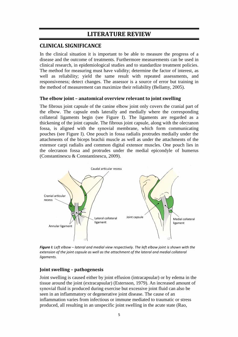
The fibrous joint capsule of the canine elbow joint only covers the cranial part of
the elbow. The capsule ends laterally and medially where the corresponding
collateral ligaments begin (see Figure I). The ligaments are regarded as a
thickening of the joint capsule. The fibrous joint capsule, along with the olecranon
fossa, is aligned with the synovial membrane, which form communicating
pouches (see Figure I). One pouch in fossa radialis protrudes medially under the
attachments of the biceps brachii muscle as well as under the attachments of the
extensor carpi radialis and common digital extensor muscles. One pouch lies in
the olecranon fossa and protrudes under the medial epicondyle of humerus
(Constantinescu & Constantinescu, 2009).
Figure I: Left elbow – lateral and medial view respectively. The left elbow joint is shown with the extension of the joint capsule as well as the attachment of the lateral and medial collateral ligaments.
Joint swelling – pathogenesis
Joint swelling is caused either by joint effusion (intracapsular) or by edema in the
tissue around the joint (extracapsular) (Estersson, 1979). An increased amount of
synovial fluid is produced during exercise but excessive joint fluid can also be
seen in an inflammatory or degenerative joint disease. The cause of an
inflammation varies from infectious or immune mediated to traumatic or stress
produced, all resulting in an unspecific joint swelling in the acute state (Rao,
6
2010). Degenerative joint diseases include a range of conditions where the joint
cartilage is injured leading to a degenerative cellular response resulting in an
increased production of proteolytic enzymes, with more damage to the cartilage as
an effect (Salter, 2002). One cause of degeneration is osteochondrosis, a common
development disorder of the growth cartilage in dogs (Ytrehus, Carlson, &
Ekman, 2007) and a part of elbow dysplasia (Innes, 2009).
EXAMINATION OF JOINT SWELLING
When examining a patient’s joints and limbs a comparison should always be made
with the contralateral extremity, to find both variations and resemblances
(Arthurs, 2011). When investigating lameness radiography is often used, this
readily shows bone lesions but will not always reveal soft tissue damage. An
alternative that visualizes soft tissue is ultrasonography, which has been studied
for the use in dogs (Lamb & Wong, 2005). Magnetic resonance imaging diagnosis
has also been studied in canines (Baeumlin et al., 2010). Goniometry, the
measurement of angles, is used to evaluate joint function by measuring the range
of motion. This is a well documented method in human medicine and has to some
extent been validated for the use on dogs (Jaegger, Marcellin-Little, & Levine,
2002).
Palpation
Joint effusion is most easily palpated in the standing dog, with full weight bearing
limbs (Millis, 2004). An effusion in the elbow joint can most readily be palpated
caudally to the lateral humeral condyle (see Figure I), where it is felt like a soft
swelling (Arthurs, 2011). Based on the anatomy of the elbow joint the synovial
recesses can be palpated both cranially and caudally to the lateral collateral
ligament of the elbow joint (Constantinescu & Constantinescu, 2009).
Tape measure
In human medicine joint swelling is sometimes measured with tape measure to
receive a more objective assessment of the swelling rather than just making a
subjective observation (Estersson, 1979). The use of tape measure has been
studied in assessing muscle mass as an indication of limb use in dogs (Millis,
2004) but to the best of the author’s knowledge there is no validation for its use in
assessing joint swelling, though the use has been suggested because of its
accuracy in human patients (Hesbach Lamoreaux, 2007).
Circumference
Human studies show that measuring the circumference of a joint gives
reproducible assessments of the joint swelling in certain joints when measured at
specific anatomical landmarks (Nicholas et al., 1976).
Figure eight
In human medicine joint swelling is sometimes measured using a tape measure
placed in the shape of a figure eight. This method is chosen in situation when a
measurement of the circumference would not include the affected part of the joint.
The measurement can be reproduced since easily recognizable anatomical
landmarks are established (Estersson, 1979).

7
Slide caliper
Joint width can be measured with a caliper and is used in rat models of
rheumatoid arthritis (Bendele, 2001). A slide caliper (Figure II) can be used to
determine the thickness of small objects. The instrument has one fixed point and
one flexible, enabling a correct adjustment to the space between one side of the
measured object to the other. The thickness can then be read on an analog scale or
a digital display, depending on the model.
Figure II: Digital slide caliper.
Tonometer
A tonometer is used in ophthalmology to measure intraocular pressure (IOP).
There are different kinds of tonometers, some which are penlike, handheld and
battery-driven, making them easy to manipulate. There are tonometers that have
to be in direct contact with the cornea to measure the IOP whilst others measure
the pressure through the eyelid (transpalpebral). The tonometer used in this study
(se Figure III) was a transpalpebral tonometer and the following description
concerns this model. When measuring the IOP a small rod is pressed against the
eyelid and depending on the elasticity of the eye the rod will be pushed back at
different speed. The tonometer uses the velocity at which the rod is pushed back
to calculate the IOP, which is then shown on a digital display (Lösch et al., 2005;
BiCOM, 2011).
Figure III: A Diaton tonometer, by courtesy of InnZ medical (BiCOM, 2011).

8
MATERIAL AND METHODS
STUDY DESIGN
The study consisted of two parts including different measurements of joint
swelling on euthanized dogs as well as living, here on referred to as the “in vitro”
and “in vivo” study respectively. Ethical permission was given by the Ethical
Committee on Animal Experiments in Uppsala, Sweden. The measuring methods
and protocols were almost the same for both groups of dogs; exceptions are
presented in the explanation of each study. Prior to the study instructions were
given on how to perform the measurements (both in writing and orally). Except
for palpation and tonometry the assessors where blinded to the result of their
measurements since a separate person performed the reading.
MATERIAL
In vitro study
Three canine carcasses, belonging to the Swedish University of Agricultural
Sciences, were used prior to their use in the veterinary education. The dogs were
of different breeds: two German Shepherds and one Drever (ages unknown). Both
elbow joints of each dog were injected with multiple standardized amounts of
sodium chloride solution (see Appendix I) to simulate joint swelling. After
injection measurements of the joints were made by assessors blinded to the
specific amount of sodium chloride solution. The assessors were two veterinary
students in their final year. Two of the dogs were injected twice to repeat the
measurement. During measurements a semi standing position was attained by
holding the dogs upright, two of the dogs by using a box between the front and
back legs and one dog by elevating the body with harness and ropes tied to it.
In vivo study
The dog unit of the Stockholm police force made their dogs available for the study
during two days when their ordinary training took place. During those days
interested handlers had the opportunity to join the study with their police dogs.
Before entering the handlers filled in an owner’s agreement of participation (see
Appendix II) and a questionnaire (see Appendix III). There were three different
assessors, all experienced veterinarians. Each dog was measured by two different
assessors (see Appendix VI for exact distribution).
A total of 12 dogs participated in the in vivo study, nine males and three females.
The dogs were of different breeds: German Shepherd (6 dogs), Malinois (4 dogs)
and Dutch Shepherd dog (2 dogs). The ages varied between one to nine years
(mean value 3.8 years, standard deviation 2.1). A number of the dogs had been in
service for some years whilst others were being tested if they could be used in the
police force. For summary and further information on the dogs see Table I.
9
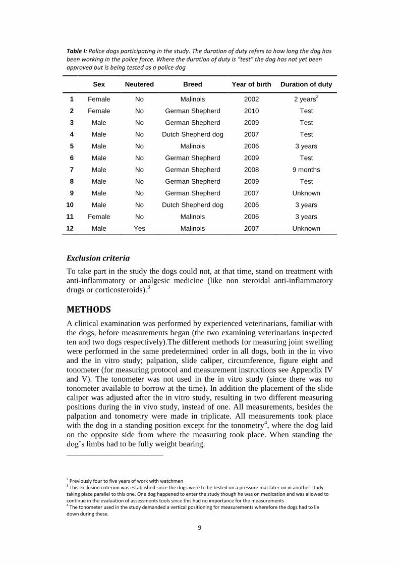
Table I: Police dogs participating in the study. The duration of duty refers to how long the dog has been working in the police force. Where the duration of duty is “test” the dog has not yet been approved but is being tested as a police dog
Sex Neutered Breed Year of birth Duration of duty
1 Female No Malinois 2002 2 years2
2 Female No German Shepherd 2010 Test
3 Male No German Shepherd 2009 Test
4 Male No Dutch Shepherd dog 2007 Test
5 Male No Malinois 2006 3 years
6 Male No German Shepherd 2009 Test
7 Male No German Shepherd 2008 9 months
8 Male No German Shepherd 2009 Test
9 Male No German Shepherd 2007 Unknown
10 Male No Dutch Shepherd dog 2006 3 years
11 Female No Malinois 2006 3 years
12 Male Yes Malinois 2007 Unknown
Exclusion criteria
To take part in the study the dogs could not, at that time, stand on treatment with
anti-inflammatory or analgesic medicine (like non steroidal anti-inflammatory
drugs or corticosteroids).3
METHODS
A clinical examination was performed by experienced veterinarians, familiar with
the dogs, before measurements began (the two examining veterinarians inspected
ten and two dogs respectively).The different methods for measuring joint swelling
were performed in the same predetermined order in all dogs, both in the in vivo
and the in vitro study; palpation, slide caliper, circumference, figure eight and
tonometer (for measuring protocol and measurement instructions see Appendix IV
and V). The tonometer was not used in the in vitro study (since there was no
tonometer available to borrow at the time). In addition the placement of the slide
caliper was adjusted after the in vitro study, resulting in two different measuring
positions during the in vivo study, instead of one. All measurements, besides the
palpation and tonometry were made in triplicate. All measurements took place
with the dog in a standing position except for the tonometry4, where the dog laid
on the opposite side from where the measuring took place. When standing the
dog’s limbs had to be fully weight bearing.
2 Previously four to five years of work with watchmen 3 This exclusion criterion was established since the dogs were to be tested on a pressure mat later on in another study taking place parallel to this one. One dog happened to enter the study though he was on medication and was allowed to continue in the evaluation of assessments tools since this had no importance for the measurements 4 The tonometer used in the study demanded a vertical positioning for measurements wherefore the dogs had to lie down during these.
10
Palpation
The synovial recesses where palpated caudally and cranially to the lateral
collateral ligament of the elbow joint. The joint was assessed as “without remark”
(Swedish: “utan anmärkning”: u.a.) or an effusion was estimated as “mild”
(Swedish: “lindrig”), “moderate” (Swedish: “måttlig”) or “severe” (Swedish:
“kraftig”). One measurement on each elbow joint was made and the assessor
noted the result themselves.
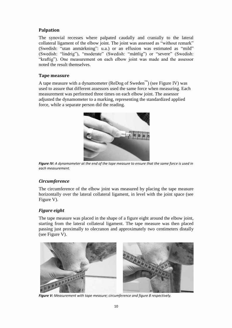
Tape measure
A tape measure with a dynamometer (ReDog of Sweden™
) (see Figure IV) was
used to assure that different assessors used the same force when measuring. Each
measurement was performed three times on each elbow joint. The assessor
adjusted the dynamometer to a marking, representing the standardized applied
force, while a separate person did the reading.
Figure IV: A dynamometer at the end of the tape measure to ensure that the same force is used in each measurement.
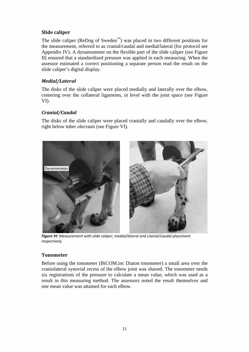
Circumference
The circumference of the elbow joint was measured by placing the tape measure
horizontally over the lateral collateral ligament, in level with the joint space (see
Figure V).
Figure eight
The tape measure was placed in the shape of a figure eight around the elbow joint,
starting from the lateral collateral ligament. The tape measure was then placed
passing just proximally to olecranon and approximately two centimeters distally
(see Figure V).
Figure V: Measurement with tape measure; circumference and figure 8 respectively.
11
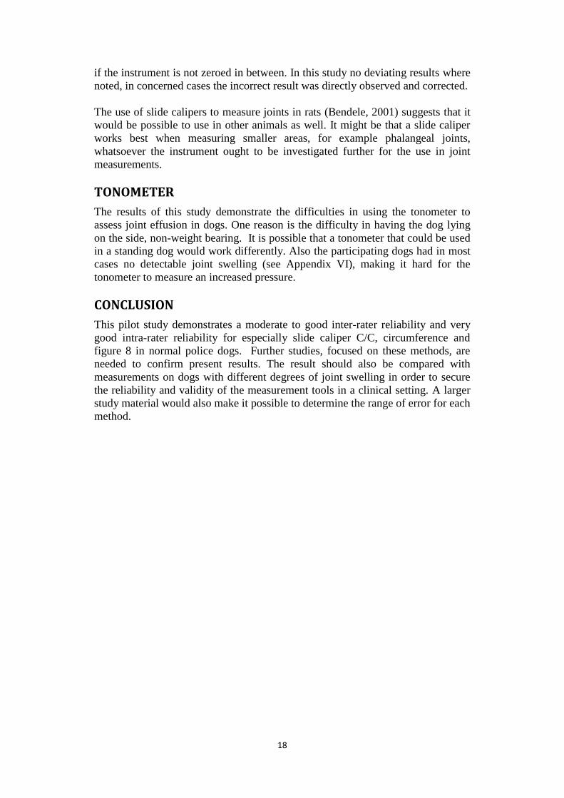
Slide caliper
The slide caliper (ReDog of Sweden™
) was placed in two different positions for
the measurement, referred to as cranial/caudal and medial/lateral (for protocol see
Appendix IV). A dynamometer on the flexible part of the slide caliper (see Figure
II) ensured that a standardized pressure was applied in each measuring. When the
assessor estimated a correct positioning a separate person read the result on the
slide caliper’s digital display.
Medial/Lateral
The disks of the slide caliper were placed medially and laterally over the elbow,
centering over the collateral ligaments, in level with the joint space (see Figure
VI).
Cranial/Caudal
The disks of the slide caliper were placed cranially and caudally over the elbow,
right below tuber olecranii (see Figure VI).
Figure VI: Measurement with slide caliper; medial/lateral and cranial/caudal placement respectively.
Tonometer
Before using the tonometer (BiCOM.inc Diaton tonometer) a small area over the
craniolateral synovial recess of the elbow joint was shaved. The tonometer needs
six registrations of the pressure to calculate a mean value, which was used as a
result in this measuring method. The assessors noted the result themselves and
one mean value was attained for each elbow.

12
DATA ANALYSIS
Statistical analysis of the data from the in vivo study was performed by an
experienced statistician5 (the data from the in vitro study was not analyzed, see
result for more information). The following description of the analysis is an
adapted translation of the information given to the author by the statistician.
Modified linear models where created for each of the numerical response
variables (slide caliper C/C and M/L, circumference, figure 8 and tonometer) with
regard to assessor, dog and left or right side as descriptive variables. Ten values
where considered as faulty measurements because of distinct deviation (so called:
“outliners”) and where not included (se Appendix VI for exact values). Tables for
analysis of variance were used to distribute the variance according to the different
factors.
Intra class correlation (ICC)6 was calculated to render values for inter- and intra-
rater reliability7. Inter-rater reliability could not be attained for the tonometer
because of too little data. ICC can be interpreted as follows:
0-0.2 indicates poor agreement
0.3-0.4 indicates fair agreement
0.5-0.6 indicates moderate agreement
0.7-0.8 indicates strong agreement
>0.8 indicates almost perfect agreement.
The models were also used to estimate the respective value for each dog and body
side, whereupon the correlation between the numerical variables was calculated.
The categorical variable, palpation, could not be analyzed due to lack of enough
material, especially dogs with different degrees of joint swelling8.
5 Statistician Mikael Andersson at the Swedish University of Agriculture. 6 ICC is the intraclass correlation, which describes how strong the resemblance is between units in the same group, for example how distinct the inter-rater-reliability is. 7 The inter-rater reliability describes how even the result of different assessors’ measurements are, while the intra-rater reliability shows how even the result is when the same assessor repeats a measurement. 8 Totally three dogs were considered, by one of the assessor, to perhaps have a mild swelling (see appendix VI).

13
RESULT
IN VITRO STUDY
The experimental situation could not satisfactory imitate a living dog, standing on
weight bearing limbs. Therefore the in vitro study served more as an opportunity
to practice the different measuring methods; to be able to make adjustments for
the in vivo study and to minimize sources of error. No analysis was made on the
data collected in the in vitro study (for additional information contact the author).
IN VIVO STUDY
The following result is an adapted translation of the information given to the
author by the statistician. Note that in contradiction to the instructions, 25 % of
the dogs (see Appendix VI) where cut for tonometrical measurements before the
second assessor had performed the other measurements.
Variance for numerical variables
Total variance for the five models of the numerical response variables (slide
caliper C/C and M/L, circumference, figure 8 and tonometer), with regard to
assessor, dog and left or right side, is described in Figure VII along with the
variance of accidental errors9.
Figure VII: Total variance (in percent) with regard to assessor, dog and left or right side is shown for the five models along with the variance of accidental errors. C/C and M/L refers to the different measurements with the slide caliper.
9 An accidental error is caused by misdistribution of the samples: Repeated measurements, rendering more values often reduce the accidental error (Andersson, 1984). Small variance in accidental error indicates good precision (George).

14
Inter-rater reliability
Interclass correlation (ICC) between the assessors (i.e. inter-rater reliability) is
described in Table II for the slide caliper and tape measure. No value was attained
for the tonometer.
Table II: ICC for assessors. C/C and M/L refers to the different measurements with the slide caliper
C/C M/L Circumference Figure 8 Tonometer
ICC 0.59 0.33 0.49 0.36 -
Intra-rater reliability
Interclass correlation (ICC) between measurements made by the same assessor
(i.e. intra-rater reliability) is described in Table III for the slide caliper, tape
measure and tonometer.
Table III: ICC between measurements. C/C and M/L refers to the different measurements with the slide caliper
C/C M/L Circumference Figure 8 Tonometer
ICC 0.95 0.61 0.94 0.96 0.52
Correlation between measuring models
Correlation between the different measuring models is described in Table IV to
give a notion of the relationship between the variables. Significant correlations
(three models are significantly correlated) are depicted in shaded, bold, italic
figures.
Table IV: Correlation between the five measuring models. C/C and M/L refers to the different measurements with the slide caliper. Shaded, bold, italic figures are significant correlations.
C/C M/L Circumference Figure 8 Tonometer
C/C 0.34 0.95 0.95 -0.15
M/L 0.34 0.37 0.32 -0.29
Circumference 0.95 0.37 0.98 -0.18
Figure 8 0.95 0.32 0.98 -0.12
Tonometer -0.15 -0.29 -0.18 -0.12

15
DISCUSSION
The study has evaluated four different measurement tools for assessing joint
swelling; the subjective palpation and the objective slide caliper, tape measure and
tonometer. Unfortunately the study material was too small to draw any conclusion
about the palpation but analysis of the data could determine inter- and intra-rater
reliability as well as correlation between the different methods for the other
instruments.
To optimize the study a comparison should also have been made with ultrasound,
a validated method for detecting joint swelling. When comparing the use of
ultrasonography with the clinical examination (including inspection, palpation and
range of motion) of human elbows, the examination can in most cases find a
swelling corresponding to joint effusion found with ultrasound, though
ultrasonography is more reliable (Luukkainen et al., 2005). To be able to quantify
a joint swelling more objectively and in a way that is practical in the everyday
clinical work is of great value in the striving to use evidence based methods in
veterinary medicine.
SOURCES OF ERROR
In the assessment of muscle mass an investigation establishes the importance of
limb position and points out the value of practice in the method and standardized
techniques to obtain reproducible results. The study also states the significance of
a dynamometer attached to the tape measure to ensure that a standardized tension
is applied in each measure (Millis, 2004).
Both tape measure and slide caliper were equipped with a dynamometer to ensure
the same tension was applied in each measure. All measurements took place in the
standing dog (except for tonometry) so that the measurements would be
performed with the same weight bearing on each leg.
To further minimize sources of error, strict instructions were given to the
assessors on how to perform the measuring. Sources of error that were not
excluded in the study were difference in hair coat and callosities in the skin
(though this was said to be noted if thought to interfere with the measurement).
According to the instructions the dogs’ hair would not be cut for the tonometry
measurements before all the other instruments had been used. These instructions
were not followed; 25 % of the dogs were cut before the second assessor had
performed the other measurements (see Appendix VI). This should however not
have had much influence on the result since the area in question is small
(maximum one square centimeter) and a majority of the dogs were uncut before
measurements. In case the cutting did interfere with the measurements it would
have had the same influence on both the left and right elbow (thus not rendering a
difference).
If the study is repeated non-sequential measuring should be considered. This was
done in a study comparing different instruments to measure limb circumference to
ensure that every new measure really was a new measurement (Baker et al.,
2010).

16
The assessment methods used in this study cannot differentiate between extra- and
intracapsular swelling. Preferable would have been to compare the measuring
result with an ultrasonographic evaluation of each dog’s elbow joints. The
ultrasound might also been able to detect small swellings, not noticed with the
other instruments. If the study is repeated an ultrasound can be used as a control
method (having the “correct answer”) that other instruments are compared with.
VARIANCE AND CORRELATION
There was a marked variance for the slide caliper, tape measure and tonometer
with regard to assessor and dog. The variance concerning the left or right body
side was much less except for the tonometer. Moderate to good inter-rater
reliability (see Table II) and very good intra-rater reliability (see Table III) was
attained for the instruments. The results, showing better intra- than inter- rater
reliability, are generally supported by human studies (Cleland, 2005).
Assessor variance and Inter-rater reliability
Despite strict instruction preceding the measurements there was quite a variance
between different assessors regarding the slide caliper, tape measure and
tonometer (see Figure VII) A reason for this might be that thought thorough
instructions were given, personal experience might have taken over, resulting in
inaccurate measurements. The observers noted, during the measurements, that the
assessor sometimes did not follow instructions as strictly as was hoped. If a
similar study is performed it is essential to even more strongly explain the
importance of a standardized technique. Despite the variance, the inter-rater
reliability was moderate to good for both slide caliper C/C and circumference.
The inter-rater reliability was only slightly lower for figure 8 and slide caliper
M/P (see Table II).
Variance between dogs and body sides
The marked variance between different dogs (see Figure VII) is expected due to
their difference in size. Variance between right and left elbow is small for both the
slide caliper techniques and the tape measure methods but not for tonometry (see
Figure VII). This is as it ought to be since there should not be a distinct difference
between the legs if the dog is not swollen in one of its elbows. That the tonometer
displays a large variance indicates that the instrument might not be suited for
these kinds of measurements. Another possible explanation might be that the
tonometer is very sensitive for external factors; that exact positioning of the joint
and small differences in skin thickness etc. might interfere with the measurement.
Intra-rater reliability
The intra-rater reliability was very good for slide caliper C/C, circumference and
figure 8. For the tonometer and slide caliper M/P the intra-rater reliability was
only slightly lower (see Table III). If the study is repeated, non-sequential
measurements with the same instruments ought to be considered, to ensure that
each measurement is made anew, guaranteeing no false high intra-rater reliability.
Non-sequential measuring was for example used in a study comparing different
ways of measuring limb circumference (Baker et al., 2010).

17
Accidental error
The accidental error is high for the slide caliper M/L and the tonometer (see
Figure VI). For the slide caliper this might be due to different positioning of the
instrument (the craniocaudal placement is more exact and easier to repeat than the
mediolateral). For the tonometer the explanation might be the heterogeneity of the
tissue, rendering very different circumstances for which the measurement takes
place (compared to when measuring intraocular pressure on homogenous eyelids).
Correlation between the methods
There is a significant correlation between the measurements circumference and
slide caliper C/C, circumference and figure 8 along with figure 8 and slide caliper
C/C, showing that these methods give roughly the same result.
TAPE MEASURE
The circumferential measurements gave the least variance with regard to the
person performing the measuring. It also correlates significantly to the techniques
using a figure 8 and the craniocaudal approach with the slide caliper.
Considering that circumferential measurements, of for instance the thigh, often is
used, by both veterinarian orthopedic surgeons and physical therapist, for
evaluation of limb use and function (Baker et al., 2010) the method seems
valuable. The use on human joints (Nicholas et al., 1979) indicates that it ought to
be able to practice this on animals too. Further studies with a larger material and
greater variety in joint swelling might give a better picture of how the tape
measure can be used in veterinary medicine for joint evaluation. Perhaps a tape
measure designed for circumferential measurements ought to be used though a
study comparing different instruments for measuring limb circumference does not
find these to be better than usual tape measures (Baker et al., 2010).
In view of the successful use of the figure 8 measure on for example human ankle
joints (Estersson, 1979) further studies ought to examine the use in canine
patients. The anatomical difficulties measuring a swelling in a human ankle
resemble the anatomy of the canine elbow joint (see Figure I).
SLIDE CALIPER
An issue when using the slide caliper is that there is a risk for variable results due
to prominent bone structures in the measuring area, interfering with the
measurement. This means that for a swollen joint to contribute to an increased
result it has to be swollen beyond the bone structures. This might be put right by
having a caliper with smaller tips (i.e. it can be placed so that it bypasses the bone
structures) but would also need other anatomical landmarks (e.g. measure
cranially or caudally to the collateral ligaments). When comparing the two
different positionings of the slide caliper, the craniocaudal had small variance (see
correlation between the methods and comparison of assessor variance, Table IV
and Figure VII).
With the particular slide caliper used in this study something to be observant
about is that a built in calculator gives the difference between two measurements
18
if the instrument is not zeroed in between. In this study no deviating results where
noted, in concerned cases the incorrect result was directly observed and corrected.
The use of slide calipers to measure joints in rats (Bendele, 2001) suggests that it
would be possible to use in other animals as well. It might be that a slide caliper
works best when measuring smaller areas, for example phalangeal joints,
whatsoever the instrument ought to be investigated further for the use in joint
measurements.
TONOMETER
The results of this study demonstrate the difficulties in using the tonometer to
assess joint effusion in dogs. One reason is the difficulty in having the dog lying
on the side, non-weight bearing. It is possible that a tonometer that could be used
in a standing dog would work differently. Also the participating dogs had in most
cases no detectable joint swelling (see Appendix VI), making it hard for the
tonometer to measure an increased pressure.
CONCLUSION
This pilot study demonstrates a moderate to good inter-rater reliability and very
good intra-rater reliability for especially slide caliper C/C, circumference and
figure 8 in normal police dogs. Further studies, focused on these methods, are
needed to confirm present results. The result should also be compared with
measurements on dogs with different degrees of joint swelling in order to secure
the reliability and validity of the measurement tools in a clinical setting. A larger
study material would also make it possible to determine the range of error for each
method.
19
ACKNOWLEDGMENT
The author would like to thank the Stockholm police’s dog unit for participating
with dogs and handlers in the study. Also thanks to ReDog for lending their slide
caliper and tape measure for use in the study and to InnZ medical for lending their
Diaton tonometer and pictures for use in the study. Thanks to Josefin Bergh Eklöf,
Sören Johansson and Ellinor Åberg who helped during the days of measurements
as well as the veterinarians from Bagarmossen.
Further the author would like to thank her fellow student; Regina Lindberg for
introducing her to this degree project and for discussing ideas and giving
constructive criticism all the way from brainstorming to finished paper. A special
thanks to Ila (Regina’s pointer) for standing model for the photographs of
measurements.
Of course a great thanks to this study’s supervisor; Anna Bergh, whom by giving
hints, suggesting articles and serving constructive criticism has managed to herd
the author in the right direction. In addition the statistician, Mikael Andersson,
deserves thanks for producing a result from the mess of data that was given to
him.

20
REFERENCES
Andersson, G. (1984). Litet statistiskt lexikon. Nämnaren , nr. 1, pp. 48-51.
Arthurs, G. (2011, March). Orthopaedic examination of the dog 1. Thoracic limb. In
practice , 33, pp. 126-133.
Baeumlin, Y., de Rycke, L., van Caelenberg, A., van Bree, H., & Gielin, I. (2010).
Magnetic resonance imaging of the canine elbow: an anatomic study. Veterinary
surgery , 39, pp. 566-573.
Baker, S., Roush, J., Unis, M., & Wodiske, T. (2010). Comparison of four commercial
devices to measure limb circumference in dogs. Veterinary and comparative
Orthopaedics and traumatology , 23, pp. 406-410.
Bellamy, N. (2005). Science of assessment. Annals of Rheumatic diseases , 64(Suppl II),
pp. ii42-ii45.
Bendele, A. (2001). Animal models of rheumatoid arthritis. Journal of musculoskeletal
and neuronal interaction , 1(4), pp. 377-385.
BiCOM, Diaton: Operation manual. Retrieved September 29, 2011, from Homepage
[online]: http://www.tonometerdiaton.com/index.php?do=home.manual
Canapp, S., Acciani, D., Hulse, D., Schultz, K., & Canapp, D. (2009). Rehabilitation
therapy for elbow disorders in dogs. Veterinary Surgery , 38, pp. 301-307.
Cleland, J. (2005). Orthopaedic Clinical Examination: An evidence based approach for
physical therapists (1st edition.). Saunders.
Constantinescu, G. M., & Constantinescu, I. A. (2009). A clinically oriented
comprehensive pictorial review of canine elbow anatomy. Veterinary Surgery , 38,
pp. 135-143.
Estersson, P. S. (1979). Measurment of ankle joint swelling using a figure 8. The journal
of orthopaedic and sports physical therapy , 1, pp. 51-52.
George, L. Fel. Retrieved 2012-12-08, from Nationalencyklopedin Homepage [online] :
http://www.ne.se/lang/fel/168082
Hesbach Lamoreaux, A. (2007). Techniques for objective outcome assessment. Clinical
techniques in small animal practice , 22, pp. 146-154.
Hessemer, V., Rössler, R., & Jacobi, K. W. (1989). Tono-Pen, a new tonometer.
International Ophthalmology , 13, pp. 51-56.
Innes, J. (2009, June). Getting the elbow: diagnosis and management of elbow disease in
dogs. Journal of Small animal practice , 50(6), pp. 18-20.
Jacobson, J., Andresen, R., Jaovisidha, S., Maeseneer, M. D., Foldes, K., Trudell, D., et
al. (1998). Detection of ankle effusions: Comparison study in cadavers using
radiography, sonography and MR imaging. American Journal of Roentgenology ,
170, pp. 1231-1238.
Jaegger, G., Marcellin-Little, D., & Levine, D. (2002, July). Reliability of goniometry in
Labrador retrievers. American Journal of Veterianry Research , 63, pp. 979-986.
Lamb, C., & Wong, K. (2005). Ultrasonographic anatomy of the canine elbow.
Veterinary Radiology & ultrasound , 46, pp. 319-325.
Lindley, S., & Watson, P. (2010). BSAVA manual of canine and feline rehabilitation
supportive and palliative care. British Small Animal Veterinary Association.

21
Lösch, A., Scheuerle, A., Rupp, V., Auffarth, G., & Becker, M. (2005). Transpalpebral
measurement of intraocularpressure using the TGDc-01 tonometer versus. Graefe's
Archive for Clinical and Experimental Ophthalmology , 243, pp. 313-316.
Luukkainen, R., Sanila, M., Saltyshev, M., Huhtala, H., & Koski, J. (2005). Relationship
between clinically detecte joint swelling and effusion diagnosed by ultrasonography
in elbow joints in patients with rheumatoid arthritis. Clinical Rheumatology , 24, pp.
228-231.
Millis, D. (2004). In D. Millis, D. Levine, & R. Taylor, Canine rehabilitation and
physical therapy (pp. 183; 190-191; 211-227). USA: Elsevier.
Nicholas, J. J., Taylor, F. H., Buckingham, R. B., & Otonello, D. (1976). Mesurment of
circumference of the knee with ordinary tape measure. Annals of the rheumatic
diseases , 35, pp. 282-284.
Pettersson, K. (2010). Animal Physiotherapist at Uppsala Djursjukhus, Personal
communication. (R. Lindberg, Interviewer)
Pollmeier, M., Toulmonde, C., Fleishman, C., & Hanson, P. D. (2006). Clinical
evaluation of firocoxiband carprofen for the treatment of dogs with osteoarthritis.
Veterinary record , 159, ss. 547-551.
Rao, D. G. (2010). Text Book on Systemic Pathology of Domestic Animals [online] pp
517-519. Lucknow, IND. Available from: Ebrary. [2011-09-20]: Global Media.
Salter, D. (2002). Degenerative joint disease. Current diagnostic pathology , 8, pp. 11-18.
Scott, H., & Witte, P. (2011, January). Investigaton of lameness in dogs 1. Forelimb. In
practice , 33, pp. 20-27.
Ytrehus, B., Carlson, C. S., & Ekman, S. (2007). Etiology and Pathogenesis of
Osteochondrosis. Veterinary Pathology , 44, pp. 429-448.
Figures and Tables
The tonometer depicted in Figure III and on the front page is used by courtesy of InnZ medical
(BiCOM, 2011). All other photographs, illustrations, diagrams and tables are the author's own
property.

Appendix I: Volumes injected saline solution
22
VOLUMES INJECTED SALINE SOLUTION
Saline solution of standardized volumes was injected in the elbow joints bilaterally to
simulate joint swelling in the in vitro study. After a first injection measuring took place after
which a second injection was performed to increase the measuring material.
First Injection (ml) Second Injection (ml)
Right Left Right Left
Drever 0 +2 0 +4
Black German Sheppard +4 0 +5 +2
Brown German Sheppard +2 0 +2 0

Appendix II: Owners agreement of participation
23
INFORMATION INFÖR DELTAGANDE I STUDIE AVSEENDE UTVÄRDERING AV OLIKA MÄTMETODER OCH EVENTUELLT SAMBAND MELLAN LEDSVULLNAD
I ARMBÅGSLED OCH BELASTNING SYFTE MED STUDIEN
Syftet med studien är att utvärdera fyra olika mätmetoder för ledsvullnad i armbågsled samt
undersöka eventuellt samband mellan en sådan svullnad och belastningsgrad mätt på en
tryckmätningsmatta. Tjänstehundar ska klara mycket i både arbete och träning, varför det är
viktigt att i ett tidigt skede kunna upptäcka eventuell ledpåverkan för att kunna sätta in
förebyggande träning/åtgärder. Tjänstehundar är tuffa och stoiska hundar som ofta dröjer med
att visa tecken på smärta. Det kan därför vara av vikt att på ett relativt snabbt och enkelt sätt
bedöma ledernas status. Det är även av värde att kunna jämföra hur resultat från olika
mätmetoder överensstämmer.
BESKRIVNING AV STUDIEN
Försöket tar ca 45-60 min/hund. Ni kommer att passera fyra stationer:
1. Information: Ifyllande av djurägarintyg och ett frågeformulär rörande hunden
2. Veterinärundersökning: En kortare klinisk undersökning
3. Registrering av ev ledsvullnad i armbåge: Hundens armbågsleder kommer att bedömas
avseende ledsvullnad med hjälp av fyra olika mätmetoder; skänkelmätare, palpation, mätning
med måttband samt Tonometer (en apparat som mäter tryck, används oftast för att mäta
trycket i ögat). Inför mätningen med Tonometer kommer ett litet område (ca 2x2 cm) att rakas
över armbågslederna.
4. Registrering med tryckmätningsmatta samt rörelsesensorer: En mjuk sele med
rörelsesensorer sätts på hunden som därefter får vänja sig vid sele och att gå över mattan
(cirka 5 minuter) . Efter uppvärmningen får hunden skritta och trava över en
tryckmätningsmatta som mäter hur hunden belastar sina tassar. Hunden kommer att filmas när
den går över mattan. När mätning erhållits från tryckmätningsmattan är studien avslutad för er
och hundens del.
Studien förväntas inte medföra några komplikationer, orsaka smärta eller på annat sätt
påverka de deltagande hundarna i negativ bemärkelse. Inga medicinska preparat eller andra
substanser kommer att tillföras hundarna under studiens gång. Studien är godkänd av etisk
nämnd, nummer C 62/11.
SAMTYCKE
Jag har muntligen informerats om studien och tagit del av och förstått ovanstående skriftliga
information. Jag är medveten om att deltagande i studien är frivilligt och att jag när som helst
kan avbryta deltagandet.
HUNDENS NAMN:
DATUM:
UNDERSKRIFT AV HUNDFÖRARE: NAMNFÖRTYDLIGANDE:
KONTAKTPERSON Leg. Veterinär Anna Bergh, [email protected]
Institutionen för anatomi, fysiologi och biokemi BOX 7011, 750 07 UPPSALA
MOBIL 070 3035997
Appendix III: Questionnaire
24
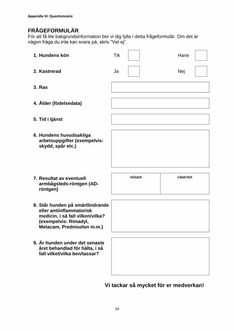
FRÅGEFORMULÄR För att få lite bakgrundsinformation ber vi dig fylla i detta frågeformulär. Om det är någon fråga du inte kan svara på, skriv ”Vet ej”.
1. Hundens kön Tik Hane
2. Kastrerad Ja Nej
3. Ras
4. Ålder (födelsedata)
5. Tid i tjänst
6. Hundens huvudsakliga arbetsuppgifter (exempelvis: skydd, spår etc.)
7. Resultat av eventuell armbågsleds-röntgen (AD-röntgen)
HÖGER VÄNSTER
8. Står hunden på smärtlindrande eller antiinflammatorisk medicin, i så fall vilken/vilka? (exempelvis: Rimadyl, Metacam, Prednisolon m.m.)
9. Är hunden under det senaste året behandlad för hälta, i så fall vilket/vilka ben/tassar?
Vi tackar så mycket för er medverkan!

Appendix IV: Measuring Protocol
25
MEASURING PROTOCOL
The following measuring protocol was used in the in vivo study. It was modified from the
protocol used in the in vitro study since the tonometer was not applied in that part and neither
were the different positions of the slide caliper. In the protocol C and M stands for the
different positions of the slide caliper (cranial/caudal and medial/lateral respectively),
explained in the material and methods section.
Mätperson: Hundnamn:
REGISTRERING 1 2 3
Höger Vänster Höger Vänster Höger Vänster
Palpation
Skänkelmätare
(mm)
C
M
Måttband horisontell
(cm)
Måttband 8:a (cm)
Tonometer (pa)
Kommentarer:10
10 If the dog for example had a callosity in the skin, showed pain during clinical examination etc.

Appendix V Values from the measurements
26
MEASURING INSTRUCTIONS
The following measuring instructions where give to the assessors before the in vivo study.
Note that the cranial/caudal measuring with the slide caliper is not mentioned, instructions
regarding this was instead given orally. The instructions for the tonometer were also slightly
changed in accordance to the requirements for the specific model that was used. Beskrivning av mätningars utförande Vid mätningarna hjälps två personer åt; en är ”mätperson” och den andre är ”avläsare”. Vid
den subjektiva mätningen (palpation) kommer mätpersonen själv att notera sitt resultat. Vid
övriga mätningar ska mätpersonen endast lägga an instrumentet korrekt och bedöma när det är
redo att avläsas, därefter noterar avläsaren resultatet. De objektiva mätningarna utförs totalt
tre gånger vardera på varje led. Alla mätningar utförs på belastat hund.
Palpation – Subjektivt mätning
Armbågsledens ledfickor palperas cranialt och caudalt om laterala kollateralligamentet. Leden
bedöms som ”u.a.” (utan anmärkning) alternativt bedöms en ledsvullnad som ”lindrig”,
”måttlig” eller ”kraftig”.
Måttband: Omkrets
Laterala kollateralligamentet palperas ut och måttbandet läggs an horisontellt över ligamentet,
i nivå med ledspringan. Kontrollera att måttbandet ligger i våg runt om leden. Drag i
momentnyckeln till markeringen och låt avläsaren notera resultatet. Upprepa mätningen
ytterligare två gånger (totalt tre mätningar).
Måttband: 8:a
Måttbandet ska läggas an i form av en åtta runt armbågsleden. Lämplig utgångspunkt är
armbågens laterala kollateralligament, därefter läggs måttbandet an så att det passerar precis
proximalt om armbågsspetsen och ca 2 cm distalt om densamma. Drag i momentnyckeln till
markeringen och låt avläsaren notera resultatet. Upprepa mätningen ytterligare två gånger
(totalt tre mätningar).
Skänkelmätare
Skänkelmätarens plattor läggs an medialt och lateralt över armbågen. Centrera plattorna över
armbågens kollateralligament, i nivå med ledspringan. Kontrollera att även den mediala
plattan ligger dikt an mot armbågen (håll gärna fast den där med en hand under mätningen
men iakttag försiktighet så att inget tryck påverkar leden). Mätpersonen bedömer när båda
plattorna på skänkelmätaren ligger dikt an mot armbågsleden och avläsaren noterar resultatet
på den digitala monitorn. Upprepa mätningen ytterligare två gånger (totalt tre mätningar).
Tonometer (Tonopen
™)
Inför mätningen med tonometer kommer ett område över armbågen att rakas. Vid mätningen
läggs instrumentet an så att den utskjutande delen träffar i det rakade området, över ledfickan.
Tonometern talar själv om när den har uppmätt ett användbart värde, vilket avläsaren noterar.
Upprepa mätningen ytterligare två gånger (totalt tre mätningar).
Appendix VI Values from the measurements
27
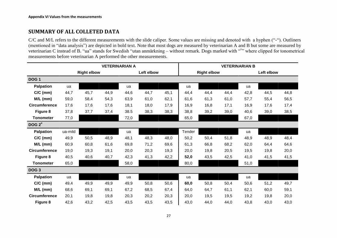
SUMMARY OF ALL COLLETED DATA
C/C and M/L refers to the different measurements with the slide caliper. Some values are missing and denoted with a hyphen (“-“). Outliners
(mentioned in “data analysis”) are depicted in bold text. Note that most dogs are measured by veterinarian A and B but some are measured by
veterinarian C instead of B. “ua” stands for Swedish “utan anmärkning – without remark. Dogs marked with “¤” where clipped for tonometrical
measurements before veterinarian A performed the other measurements.
VETERINARIAN A VETERINARIAN B
Right elbow Left elbow Right elbow Left elbow
DOG 1
Palpation ua
ua
ua
ua
C/C (mm) 44,7 45,7 44,9 44,6 44,7 45,1 44,4 44,4 44,4 42,8 44,5 44,8
M/L (mm) 59,0 58,4 54,3 63,9 61,0 62,1 61,6 61,3 61,0 57,7 55,4 56,5
Circumference 17,6 17,6 17,6 18,1 18,0 17,9 16,9 16,8 17,1 16,9 17,6 17,4
Figure 8 37,8 37,7 37,4 38.5 38,3 38,3 38,8 39,2 39,0 40,6 39,0 38,5
Tonometer 77,0
72,0
65,0
67,0
DOG 2¤
Palpation ua-mild
ua
Tender
ua
C/C (mm) 49,9 50,5 48,9 48,1 48,3 48,0 50,2 50,4 51,8 48,9 48,9 48,4
M/L (mm) 60,9 60,8 61,6 69,8 71,2 69,6 61,3 66,8 68,2 62,0 64,4 64,6
Circumference 19,0 19,3 19,1 20,0 20,3 19,3 20,0 19,8 20,5 19,5 19,8 20,0
Figure 8 40,5 40,6 40,7 42,3 41,3 42,2 52,0 43,5 42,5 41,0 41,5 41,5
Tonometer 65,0
58,0
80,0
51,0
DOG 3
Palpation ua
ua
ua
ua
C/C (mm) 49,4 49,9 49,9 49,9 50,8 50,6 60,0 50,8 50,4 50,6 51,2 49,7
M/L (mm) 68,6 69,1 69,1 67,2 68,5 67,4 64,0 64,7 61,1 62,1 60,0 59,1
Circumference 20,1 19,8 19,8 20,3 20,2 20,3 20,0 19,5 19,5 19,2 19,8 20,0
Figure 8 42,6 43,2 42,5 43,5 43,5 43,5 43,0 44,0 44,0 43,8 43,0 43,0

Appendix VI Values from the measurements
28
Tonometer 67,0
56,0
85,0
77,0
VETERINARIAN A VETERINARIAN B
Right elbow Left elbow Right elbow Left elbow
DOG 4
Palpation ua
ua-mild
ua
ua
C/C (mm) 46,3 46,7 48,4 49,5 51,5 48,4 51,4 58,2 51,6 48,7 52,6 52,1
M/L (mm) 62,8 60,5 59,4 58,7 58,1 59,5 62,4 61,9 62,0 64,8 62,1 63,1
Circumference 19,8 19,6 19,5 19,8 19,8 19,5 18,5 18,5 18,5 19,0 19,5 19,4
Figure 8 41,0 40,7 40,5 42,0 42,0 42,3 43,5 43,5 42,5 42,5 40,5 42,0
Tonometer 8?
8,0
-
83,0
DOG 5
Palpation ua
ua
ua
ua
C/C (mm) 43,2 43,7 43,5 42,6 42,6 42,6 42,3 42,3 41,3 42,8 42,7 42,9
M/L (mm) 55,2 56,2 56,6 60,2 62,8 62,4 50,3 49,7 50,5 52,2 49,0 44,5
Circumference 16,8 16,8 16,8 17,2 17,2 17,0 15,7 15,6 16,0 16,5 16,2 16,8
Figure 8 34,6 34,7 34,6 36,5 35,3 35,2 36,0 36,1 35,5 35,5 34,5 35,0
Tonometer 72,0
81,0
43,0
69,0
DOG 6¤
Palpation ua
ua
ua
ua
C/C (mm) 53,4 56,8 55,1 52,5 53,4 53,2 54,1 56,8 56,5 53,5 54,7 53,2
M/L (mm) 68,7 71,1 71,7 69,3 68,2 71,7 69,4 69,2 69,8 67,1 65,8 65,2
Circumference 21,0 21,1 21,6 21,5 21,3 21,6 21,0 20,5 20,5 21,0 21,3 21,5
Figure 8 44,6 45,2 44,5 44,4 45,0 44,5 45,0 46,5 46,0 44,5 44,5 45,0
Tonometer -
-
69,0
60,0
DOG 7
Palpation ua
ua
ua
ua
C/C (mm) 45,6 45,2 44,2 46,5 47,2 45,4 45,3 45,5 46,2 46,8 46,9 46,2

Appendix VI Values from the measurements
29
M/L (mm) 75,5 75,8 72,6 62,1 58,7 59,5 69,7 70,3 57,5 56,9 57,9 57,5
Circumference 19,5 19,6 19,3 19,5 19,1 19,5 19,0 18,7 18,5 18,7 18,0 18,5
Figure 8 39,4 39,4 39,1 40,2 39,6 39,7 41,0 42,0 42,5 43,0 42,5 42,5
Tonometer 73,0
74,0
81,0
67,0
VETERINARIAN A VETERINARIAN B
Right elbow Left elbow Right elbow Left elbow
DOG 8
Palpation ua
ua
ua
ua
C/C (mm) 50,0 50,4 51,3 50,6 51,0 52,4 15,5 14,9 15,5 15,0 15,7 15,0
M/L (mm) 61,8 64,2 63,5 69,9 65,9 67,4 33,0 32,5 33,1 27,2 26,8 28,1
Circumference 20,6 21,6 21,0 21,0 21,2 21,0 20,0 19,5 19,7 20,0 20,0 20,0
Figure 8 42,7 42,8 43,8 43,5 43,0 43,2 44,5 45,0 45,0 45,0 45,0 45,3
Tonometer 72,0
71,0
72,0
91,0
DOG 9
Palpation ua
ua-mild
ua
ua
C/C (mm) 54,1 54,7 54,5 50,3 50,4 50,9 52,9 52,5 51,8 52,1 52,1 52,3
M/L (mm) 69,1 73,0 67,7 20,5 65,3 64,0 41,2 41,5 55,4 59,7 62,5 61,2
Circumference 21,7 21,8 22,4 21,8 22,5 22,3 20,5 20,0 20,5 19,5 19,5 20,0
Figure 8 46,1 46,0 45,1 44,5 44,4 44,6 46,0 45,5 46,0 45,5 46,0 46,5
Tonometer -
-
-
DOG 10¤
Palpation ua
ua
ua
ua
C/C (mm) 47,2 48,4 48,9 48,6 46,8 46,8 47,3 48,4 48,0 47,2 48,8 47,9
M/L (mm) 63,4 64,8 65,2 66,4 65,7 62,2 47,2 47,8 48,1 56,1 56,3 57,7
Circumference 19,8 19,7 19,5 20,0 19,9 19,8 18,5 18,3 18,0 19,2 19,0 18,7
Figure 8 42,0 41,9 41,5 40,7 41,0 41,0 43,5 43,0 42,0 41,5 41,0 43,0
Tonometer 78,0
69,0
63,0
68,0

Appendix VI Values from the measurements
30
VETERINARIAN A VETERINARIAN C
Right elbow Left elbow Right elbow Right elbow
DOG 11
Palpation ua
ua
ua
ua
C/C (mm) 42,1 42,5 43,9 43,0 42,8 42,3 41,2 42,3 41,8 40,7 41,2 41,7
M/L (mm) 58,7 63,3 66,3 60,8 63,1 56,0 52,5 52,0 50,7 56,0 48,5 51,5
Circumference 17,0 17,2 17,1 17,3 17,3 17,1 17,4 17,3 17,0 16,9 17,0 16,5
Figure 8 36,6 36,5 36,2 36,2 36,1 36,1 39,0 36,0 36,3 37,0 37,0 37,3
Tonometer 78,0
73,0
90,0
94,0
DOG 12
Palpation ua
ua
ua
ua
C/C (mm) 46,7 48,9 47,8 46,1 46,1 46,2 44,1 44,4 44,4 44,5 44,6 44,4
M/L (mm) 60,2 59,8 59,0 61,9 60,6 61,3 58,1 58,0 58,0 63,7 64,1 62,8
Circumference 19,0 19,0 18,8 19,0 18,9 19,0 19,0 19,0 18,5 18,0 18,0 18,3
Figure 8 39,7 39,8 40,2 40,0 40,1 39,8 41,0 39,5 39,3 39,5 39,3 39,0
Tonometer 77,0
78,0
50,0
68,0