evaluation of digital optical density of bone repair in ... · pdf fileevaluation of digital...
TRANSCRIPT

Braz Dent J 16(3) 2005
Digital optical density of bone repair in rats 207
Evaluation of Digital Optical Density of BoneRepair in Rats Medicated with Ketoprofen
Márcia Valéria MARTINS1
Marcos André dos Santos da SILVA1
Edmundo MEDICI FILHO1
Luiz Cesar de MORAES1
Julio Cezar de Melo CASTILHO1
Rosilene Fernandes da ROCHA2
1Department of Surgery, Periodontology and Radiology and 2Department of Biosciences and Oral Diagnosis, Faculty ofDentistry of São José dos Campos, São Paulo State University (UNESP), São José dos Campos, SP, Brazil
The purpose of this study was to evaluate the influence of ketoprofen on bone repair process in tibiae of rats by means of analysis ofthe digital optical density. Twenty Wistar rats were assigned to two groups: an untreated control group and a group treated withketoprofen. The experimental procedures comprised the following stages: general anesthesia, preparation of a unicortical bone defecton the left tibia of each rat, medication with ketoprofen and radiographic examination. Digital radiographic images were acquired usingVisualix GX-S-HDI™ digital sensor and an x-ray equipment. Radiographs were taken at baseline, 7, 14, 21 and 30 days postoperativelyand the optical density (OD) was evaluated using the Vix winTM 1.4 system. The mean values of OD readings were analyzedstatistically by ANOVA and Tukey’s test with significance level set at á=5%. The control group showed a statistically significantcorrelation (p=0.001) between time and optical density, while the ketoprofen group exhibited a weak and not statistically significantcorrelation (p=0.100). The control group presented the smallest OD ratios at days 1 and 7, and the greatest OD ratios at days 14, 21and 30, with statistically significant difference (p=0.001). There was no significant differences (p=0.100) among the OD ratios in theketoprofen group, regardless of the evaluation period. The findings of this study suggest that ketoprofen influenced bone repair processbecause there was an increase in optical density during the first week and delayed new bone formation after the 21st day.
Key words: bone repair, ketoprofen, digital optical density, direct digital radiograph.
Correspondence: Dra. Márcia Valéria Martins, Av. Eng. Francisco José Longo, n. 153, apto 61, Jardim São Dimas, 12245-000 São José dosCampos, SP, Brasil. Tel: +55-12-3941-2831. e-mail: [email protected]
ISSN 0103-6440
INTRODUCTION
Anti-inflammatory drugs are employed to reduceor control an inflammatory process because they inhibitthe synthesis of inflammation mediators (prostaglandins)and are therefore largely useful in medical and dentalspecialties alike.
Non-steroidal anti-inflammatory drugs (NSAIDs)are competitive inhibitors of the cyclooxygenase enzyme(COX), which is in charge of the biosynthesis ofprostaglandins and thromboxanes from the arachidonicacid (1-3). Two forms of the cyclooxygenase enzymehave been described, COX-1 and COX-2. Ketoprofen isa specific inhibitor of COX-1 (3).
Current studies have associated the intake of
NSAIDs with bone repair process (2,4,5) and havedemonstrated that bone is one of the few tissues withremodeling capacity, being able to recover its structureand function even after a trauma.
Direct digital radiography has been largely usedin studies investigating bone defects because thismethodology is as efficient as conventional radiographictechniques for detection of incipient bone lesions andoffers technical resources and tools (softwares) thatassist in visualization, delimitation and measurement ofthese lesions (6,7).
The purpose of this study was to evaluate theinfluence of ketoprofen on the bone repair of unicorticaldefects created in tibiae of rats by analysis of the digitaloptical density (OD) at different periods of time.
Braz Dent J (2005) 16(3): 207-212

Braz Dent J 16(3) 2005
208 M.V. Martins et al.
MATERIAL AND METHODS
Twenty adult male rats from the same line(Rattus norvegicus albinus, Wistar) aged around 120days and weighing from 350 to 400 g were obtainedfrom the Animal Laboratory of the Faculty of Dentistryof São Jose dos Campos (UNESP). The animals werehoused in proper cages and environment and weremaintained on laboratory chow (Labina) and water adlibitum.
The animals were anesthetized with 2% aqueoussolution of 2-(2,6-xylidine)-5,6-dihydro-4H-1,3-thiazinehydrochloride (Rompum; Bayer do Brasil SA, SãoPaulo, SP, Brazil) and ketamine (Francotar; Virbac doBrasil Ind. Com Ltda, São Paulo, SP, Brazil.) at a 1:0.5
mL ratio and dose of 0.1 mL/100 mg. The rats werepreviously weighed to confirm the correct dose to beadministered. Hypodermic insulin syringes with intradermalneedles were used.
The skin over the left tibia was shaved and antisepsiswas performed with iodized alcohol. An incision wasmade on the proximal area of the tibia with aninterchangeable #15 blade mounted on a Bard-Parkerhandle and a #7 spatula was used to retract the soft tissuesand periosteum. A perforation was made on the bone witha 3-mm diameter trephine bur coupled to an electricengine (Kavo do Brasil S.A, Joinville, SC, Brazil) at 1,100rpm speed and under constant irrigation with saline (Fig.1 A and B). Skin was sutured with silk suture number 4(Ethicon-Johnson & Johnson, Somerville, NJ, USA) and
antisepsis was done withiodized alcohol
Ten animals weremedicated with ketoprofen(Merck & Co., Inc.,Whitehouse Station, NJ, USA)at a dose of 12.5 mg/day for30 days and 10 animals werenot medicated (control).
For the radiographicexamination, the rats in thecontrol and ketoprofengroups were initially submittedto general anesthesia byapplication of the same
anesthetics, syringes and needles used for surgery,although the anesthetic dose in this stage ranged from0.08 to 0.1 mL. Thereafter, each rat was positioned indorsal recumbency on a supporting table. Before thebaseline direct digital radiographs were obtained, animpression was taken from the left tibia with low fusionimpression compound (Godibar; Lysanda ProdutosOdontológicos, São Paulo, SP, Brazil), in a way that theinitial position could be reproduced in the further stages.Impression was taken on the lower portion of an individualmetallic clamping device shaped as a rectangular box,with 3 free ends and a groove for fixation on thesupporting table (Fig. 2). Visualix GX-S-HDI™ digitalsensor (Gendex Dental System, Dentsply International,Chicago, IL, USA) was placed on the lower portion (freefrom impression material) of each device. Because it is aCCD (Charge-Coupled Device) sensor, the image couldbe immediately transmitted to a computer screen.
Figure 1. A. Perforation being made on rat left tibia; B. Unicortical bone defect.
Figure 2. Acquisition of the radiographic image after placementof the digital sensor in the tibia of the rat.

Braz Dent J 16(3) 2005
Digital optical density of bone repair in rats 209
Thereafter, the tibia was introduced in the clampingdevice, positioned on the upper portion of the sensor andfixated to the device and impression mould with #9utility wax (Epoxiglass Ind. Com. de Produtos QuímicosLtda, Diadema, SP, Brazil). In this way, the bone defectwas also turned upwards and the initial position could bereproduced in all evaluation periods (Fig. 2). Afterwards,the left tibia of each rat was radiographed with an X-rayequipment (SPECTRO-70X™; Dabi Atlante, São Paulo,SP, Brazil) at 10 mA and 65 kVp, with 0.10 s exposuretime and 25 cm focus-object distance (Fig. 2).Radiographs were taken from rats in both control group(Fig. 3 A-E) and ketoprofen group (Fig. 4 A-E) atbaseline and 7, 14, 21 and 30 days after the unicorticalbone defects were created on the left tibia of the animals.
Optical density was evaluated using the Vixwin™ 1.4 system (Gendex Dental System). The meanvalues of OD readings were analyzed statistically byANOVA and Tukey’s test at 5% significance level.
RESULTS
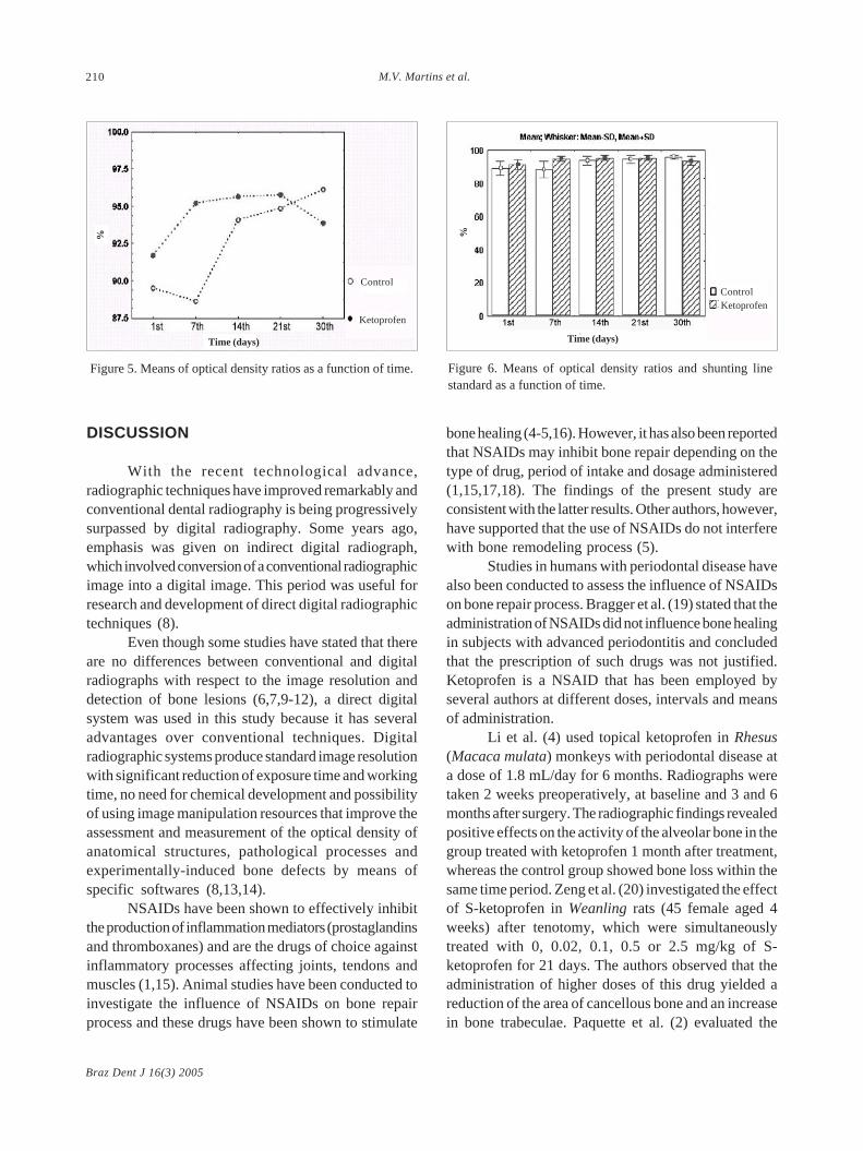
The control group had a moderate and statisticallysignificant correlation by ANOVA (p=0.001) betweentime and OD ratios (Fig. 5), while the ketoprofen groupexhibited a weak and not statistically significantcorrelation by ANOVA (p=0.100) (Fig. 6).
Tukey’s test (á=5%) showed that the controlgroup presented the smallest OD ratios (%) at days 1(0.89±0.04) and 7 (0.88±0.05) and the greatest ODratios (%) at days 14 (0.94±0.02), 21 (0.95±0.02) and30 (0.96±0.01) with a statistically significant difference(p=0.001) (Fig. 3).
For the ketoprofen group, there were nostatistically significant differences (p=0.100) amongthe evaluation periods. The ketoprofen group presentedgreater ratios than the control group up to 21st day, afterwhich, the values decreased until reach smaller rationsthan the control group at the 30th day.
Figure 3. Radiographic examination of the control group. A: Baseline; B: 7th day; C: 14th day; D: 21st day; E: 30th day.
Figure 4: Radiographic examination of the ketoprofen group. A: Baseline; B: 7th day; C: 14th day; D: 21st day; E: 30th day.
Braz Dent J 16(3) 2005
210 M.V. Martins et al.
DISCUSSION
With the recent technological advance,radiographic techniques have improved remarkably andconventional dental radiography is being progressivelysurpassed by digital radiography. Some years ago,emphasis was given on indirect digital radiograph,which involved conversion of a conventional radiographicimage into a digital image. This period was useful forresearch and development of direct digital radiographictechniques (8).
Even though some studies have stated that thereare no differences between conventional and digitalradiographs with respect to the image resolution anddetection of bone lesions (6,7,9-12), a direct digitalsystem was used in this study because it has severaladvantages over conventional techniques. Digitalradiographic systems produce standard image resolutionwith significant reduction of exposure time and workingtime, no need for chemical development and possibilityof using image manipulation resources that improve theassessment and measurement of the optical density ofanatomical structures, pathological processes andexperimentally-induced bone defects by means ofspecific softwares (8,13,14).
NSAIDs have been shown to effectively inhibitthe production of inflammation mediators (prostaglandinsand thromboxanes) and are the drugs of choice againstinflammatory processes affecting joints, tendons andmuscles (1,15). Animal studies have been conducted toinvestigate the influence of NSAIDs on bone repairprocess and these drugs have been shown to stimulate
bone healing (4-5,16). However, it has also been reportedthat NSAIDs may inhibit bone repair depending on thetype of drug, period of intake and dosage administered(1,15,17,18). The findings of the present study areconsistent with the latter results. Other authors, however,have supported that the use of NSAIDs do not interferewith bone remodeling process (5).
Studies in humans with periodontal disease havealso been conducted to assess the influence of NSAIDson bone repair process. Bragger et al. (19) stated that theadministration of NSAIDs did not influence bone healingin subjects with advanced periodontitis and concludedthat the prescription of such drugs was not justified.Ketoprofen is a NSAID that has been employed byseveral authors at different doses, intervals and meansof administration.
Li et al. (4) used topical ketoprofen in Rhesus(Macaca mulata) monkeys with periodontal disease ata dose of 1.8 mL/day for 6 months. Radiographs weretaken 2 weeks preoperatively, at baseline and 3 and 6months after surgery. The radiographic findings revealedpositive effects on the activity of the alveolar bone in thegroup treated with ketoprofen 1 month after treatment,whereas the control group showed bone loss within thesame time period. Zeng et al. (20) investigated the effectof S-ketoprofen in Weanling rats (45 female aged 4weeks) after tenotomy, which were simultaneouslytreated with 0, 0.02, 0.1, 0.5 or 2.5 mg/kg of S-ketoprofen for 21 days. The authors observed that theadministration of higher doses of this drug yielded areduction of the area of cancellous bone and an increasein bone trabeculae. Paquette et al. (2) evaluated the
Figure 6. Means of optical density ratios and shunting linestandard as a function of time.
Figure 5. Means of optical density ratios as a function of time.
Time (days) Time (days)
%%
Ketoprofen
Control
KetoprofenControl

Braz Dent J 16(3) 2005
Digital optical density of bone repair in rats 211
pharmacodynamic effect of ketoprofen (administeredeither topically or systemically during 22 days) on theprostaglandins of the gingival fluid of 42 individualsaged 35-57 years with moderate to severe periodontaldisease. The authors concluded that all dosages ofketoprofen pharmacologically reduced the prostaglandinlevels on the gingival fluid of the patients, thus inhibitingdisease progression.
The results of the present study do not agreewith those of the above-mentioned investigations(2,4,20). Unlike these authors, we have observed thatthe dosage and period of administration of a NSAIDdirectly interfered with its mechanism of action becauseOD values of the unicortical bone defects created intibiae of rats increased in both ketoprofen-treated (dailyoral dose of 12.5 mg/kg for 30 days) and control groupsup to the 21st day, with greater OD values for theketoprofen group. After this period, OD values in thetreated group decreased until reach smaller values thanthe control group at 30th day.
These findings indicate a negative influence ofketoprofen on bone repair process when administeredfor more than 21 days.
RESUMO
O objetivo deste estudo foi avaliar a influência do cetoprofenosobre o processo de reparação óssea em tíbias de ratos, por meioda análise da densidade óptica digital. Vinte ratos da linhagemWistar foram divididos em 2 grupos: um grupo controle (semtratamento) e um grupo tratado com cetoprofeno. Osprocedimentos experimentais consistiram de: anestesia, cirurgia,administração do cetoprofeno e exame radiográfico. As imagensradiográficas foram adquiridas empregando-se o sensor digitalVisualix GX-S-HDI™ e um aparelho de raios X. As radiografiasforam realizadas nos períodos baseline (inicial), 7, 14, 21 e 30dias pós-operatório, sendo a densidade óptica (DO) avaliada pormeio do sistema Vix winTM 1.4. Os valores médios da leitura daDO obtidos foram analisados estatisticamente por meio deANOVA e teste de Tukey com nível de significância de 5%. Nogrupo controle, houve diferença estatisticamente significante(p=0,001) entre o tempo e a DO, enquanto no grupo tratado comcetoprofeno a diferença não foi estatisticamente significante(p=0,100). O grupo controle apresentou as menores proporçõesde DO (%) no 1o e 7o dias e as maiores proporções de DO (%) no14o, 21o e 30o dias, com diferença estatisticamente significante(p=0,001). Não houve diferença estatisticamente significante(p=0,100) entre as proporções médias de DO (%) no grupotratado, independentemente do período de avaliação. Os achadosdeste trabalho sugerem que houve influência do cetoprofenosobre o processo de reparo ósseo, uma vez que na primeirasemana o medicamento proporcionou aumento na densidadeóptica e provocou atraso na neoformação óssea após o 21o dia.
REFERENCES
1 . Ho ML, Chang JK, Chuang LY, Hsu HK, Wang GJ. Effects ofnonsteroidal anti-inflamatory drugs and prostaglandins onosteoblastic functions. Biochem Pharmacol 1999;58:983-990.
2 . Paquette DW, Lawrence HP, McCombs GB, Wilder R, BinderTA, Troullos E, Annett M, Friedman M, Smith PC,Offenbacher S. Pharmacodynamic effects of ketoprofen oncrevicular fluid prostanoids in adult periodontitis. J ClinPeriodontol 2000;27:558-566.
3 . Llorens O, Perez JJ, Palomer A, Mauleon D. Differentialbinding mode of diversy cyclooxygenase inhibitors. J MolGraph Model 2002;20:359-371.
4 . Li KL, Vogel R, Jeffcoat MK, Alfano MC, Smith MA, CollinsJG, Offenbacher S. The effect of ketoprofen creams onperiodontal disease in rhesus monkeys. J Periodontal Res1996;31:525-532.
5 . de Lima V, Bezerra MM, de Menezes Alencar VB, Vidal FD, daRocha FA, de Castro Brito GA, de Albuquerque Ribeiro R.Effects of chlorpromazine on alveolar bone loss inexperimental periodontal disease in rats. Eur J Oral Sci2000;108:123-129.
6 . Holtzmann DJ, Johnson WT, Southard TE, Khademi JA,Chang PJ, Rivera EM. Storage-phosphor computedradiography versus film radiography in the detection ofpathologic periradicular bone loss in cadavers. Oral Surg OralMed Oral Pathol Oral Radiol Endod 1998;86:90-97.
7 . Kaeppler G, Vogel A, Axmann-Krcmar D. Intra-oral storagephosphor and conventional radiography in the assessment ofalveolar bone structures. Dentomaxillofac Radiol2000;29:362-367.
8 . Wenzel A, Grondahl HG. Direct digital radiography in thedental office. Int Dent J 1995;45:27-34.
9 . Thaete FL, Fuhrman CR, Oliver JH, Britton CA, CampbellWL, Feist JH, Straub WH, Davis PL, Plunkett MB. Digitalradiography and conventional imaging of the chest: acomparison of observer performance. AJR Am J Roentgenol1994;162:575-581.
10. Stassinakis A, Zeyer O, Bragger U. The diagnosis of bonelesions with conventional x-ray images and with a directdigital procedure (RVG). Scweiz Monatsschr Zahnmed 1995;105:1539-45.
11. Kullendorff B, Nilsson M, Rohlin M. Diagnostic accuracy ofdirect digital dental radiography for the detection of periapicalbone lesions: overall comparison between conventional anddirect digital radiography. Oral Surg Oral Med Oral PatholOral Radiol Endod 1996;82:344-350.
12. Mistak EJ, Loushine RJ, Primack PD, West LA, Runyan DA.Interpretation of periapical lesions comparing conventional,direct digital, and telephonically transmitted radiographicimages. J Endod 1998;24:262-266.
13. Meier AW, Brown CE, Miles DA, Analoui M. Interpretationof chemically created periapical lesions using direct digitalimaging. J Endod 1996;22:516-520.
14. Wenzel A. Effect of image enhancement for detectability ofbone lesions in digitized intraoral radiographs. Scand J DentRes 1988;96:149-60.
15. Yazdi M, Cheung DT, Cobble S, Nimni ME, Schonfeld SE.Effects of non-steroidal anti-inflammatory drugs ondemineralized bone-induced bone formation. J PeriodontalRes 1992;27:28-33.

Braz Dent J 16(3) 2005
212 M.V. Martins et al.
16. Offenbacher S, Williams RC, Jeffcoat MK, Howell TH, OdleBM, Smith MA, Hall CM, Johnson HG, Goldhaber P. Effectsof NSAIDs on beagle crevicular cyclooxygenase metabolitesand periodontal bone loss. J Periodontal Res 1992;27:207-213.
17. Allen HL, Wase A, Bear WT. Indomethacin and aspirin:effect of nonsteroidal anti-inflammatory agents on the rateof fracture repair in the rat. Acta Orthop Scand 1980;51:595-600.
18. Altman, R.D. Altman RD, Latta LL, Keer R, Renfree K,Hornicek FJ, Banovac K. Effect of nonsterioidal anti-inflammatory drugs on fracture healing: a laboratory study inrats. J Orthop Trauma 1995;9:392-400.
19. Bragger U, Muhle T, Fourmousis I, Lang NP, Mombelli A.Effect of the NSAID flurbiprofen on remodelling afterperiodontal surgery. J Periodontal Res 1997;32:575-582.
20. Zeng QQ, Jee WS, Ke HZ, Wechter WJ. S-ketoprofen inhibitstenotomy-induced bone loss and dynamics in weanling rats.Bone Miner 1993;21:203-218.
Accepted August 24, 2004