evaluation of condylar position by cbct after static and … · gothic arch tracing. the...
TRANSCRIPT

O
Ea
L
Á
a
A
R
A
A
K
I
C
M
T
C
S
h1a
r e v p o r t e s t o m a t o l m e d d e n t c i r m a x i l o f a c . 2 0 1 5;5 6(1):9–17
www.elsev ier .p t /spemd
Revista Portuguesa de Estomatologia,Medicina Dentária e Cirurgia Maxilofacial
riginal research
valuation of condylar position by CBCT after staticnd dynamic registration in edentulous patients
urdes Veloso ∗, Ricardo Dias, Ana Messias, Júlio Fonseca, Pedro Nicolau
rea de Medicina Dentária, Faculdade de Medicina, Universidade de Coimbra, Coimbra, Portugal
r t i c l e i n f o
rticle history:
eceived 7 August 2014
ccepted 6 February 2015
vailable online 23 March 2015
eywords:
ntermaxillary registration
entric relation
andibular condyle
emporomandibular joint
one beam computed tomography
tomatognatic system
a b s t r a c t
Objectives: The aims of this study are to: compare the condylar position in articular fossa
after static and dynamic registration; analyze symmetry between right and left condyles
and examine the relationship between articular eminence and condylar position.
Methods: Twenty completely edentulous patients were included in this study, after signing
a written informed consent. Static registration was obtained by mandibular manipulation
and dynamic registration was performed by Gothic Arch Tracing. Patients were submit-
ted to one cone beam in static registration, followed by another with dynamic registration.
Radiographic image measurements in lateral and frontal cuts were made.
Results: No statistically significant differences between the two methods were found. In
dynamic registration all the distances were smaller, more consistent and equidistant.
Condyles stayed in a closer position to the articular fossa and in a centred position. For this
registration a higher symmetry between left and right condyle exists, revealed by homoge-
neous results. Static registration had a higher heterogeneity of results, due to the fact that
it is dependent upon a number of factors.
Conclusions: Dynamic registration seems a reliable and an accurate method to use. With the
higher condylar symmetry and the centred position in articular fossa, it seems that this
registration reproduces a physiologic condylar position.
© 2015 Sociedade Portuguesa de Estomatologia e Medicina Dentária. Published by
Elsevier España, S.L.U. This is an open access article under the CC BY-NC-ND license
(http://creativecommons.org/licenses/by-nc-nd/4.0/).
∗ Corresponding author.E-mail address: lurdes [email protected] (L. Veloso).
ttp://dx.doi.org/10.1016/j.rpemd.2015.02.003646-2890/© 2015 Sociedade Portuguesa de Estomatologia e Medicina Dentária. Published by Elsevier España, S.L.U. This is an open accessrticle under the CC BY-NC-ND license (http://creativecommons.org/licenses/by-nc-nd/4.0/).
10 r e v p o r t e s t o m a t o l m e d d e n t c i r m a x i l o f a c . 2 0 1 5;5 6(1):9–17
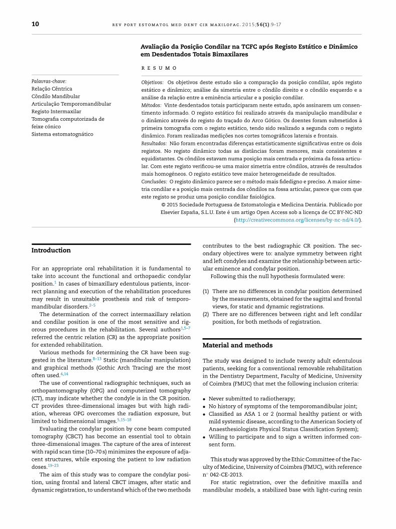
Avaliacão da Posicão Condilar na TCFC após Registo Estático e Dinâmicoem Desdentados Totais Bimaxilares
Palavras-chave:
Relacão Cêntrica
Côndilo Mandibular
Articulacão Temporomandibular
Registo Intermaxilar
Tomografia computorizada de
feixe cónico
Sistema estomatognático
r e s u m o
Objetivos: Os objetivos deste estudo são a comparacão da posicão condilar, após registo
estático e dinâmico; análise da simetria entre o côndilo direito e o côndilo esquerdo e a
análise da relacão entre a eminência articular e a posicão condilar.
Métodos: Vinte desdentados totais participaram neste estudo, após assinarem um consen-
timento informado. O registo estático foi realizado através da manipulacão mandibular e
o dinâmico através do registo do tracado do Arco Gótico. Os doentes foram submetidos à
primeira tomografia com o registo estático, tendo sido realizado a segunda com o registo
dinâmico. Foram realizadas medicões nos cortes tomográficos laterais e frontais.
Resultados: Não foram encontradas diferencas estatisticamente significativas entre os dois
registos. No registo dinâmico todas as distâncias foram menores, mais consistentes e
equidistantes. Os côndilos estavam numa posicão mais centrada e próxima da fossa articu-
lar. Com este registo verificou-se uma maior simetria entre côndilos, através de resultados
mais homogéneos. O registo estático teve maior heterogeneidade de resultados.
Conclusões: O registo dinâmico parece ser o método mais fidedigno e preciso. A maior sime-
tria condilar e a posicão mais centrada dos côndilos na fossa articular, parece que com que
este registo se produz uma posicão condilar fisiológica.
© 2015 Sociedade Portuguesa de Estomatologia e Medicina Dentária. Publicado por
Elsevier España, S.L.U. Este é um artigo Open Access sob a licença de CC BY-NC-ND
Introduction
For an appropriate oral rehabilitation it is fundamental totake into account the functional and orthopaedic condylarposition.1 In cases of bimaxillary edentulous patients, incor-rect planning and execution of the rehabilitation proceduresmay result in unsuitable prosthesis and risk of temporo-mandibular disorders.2–5
The determination of the correct intermaxillary relationand condilar position is one of the most sensitive and rig-orous procedures in the rehabilitation. Several authors1,5–7
referred the centric relation (CR) as the appropriate positionfor extended rehabilitation.
Various methods for determining the CR have been sug-gested in the literature.8–13 Static (mandibular manipulation)and graphical methods (Gothic Arch Tracing) are the mostoften used.4,14
The use of conventional radiographic techniques, such asorthopantomography (OPG) and computerized tomography(CT), may indicate whether the condyle is in the CR position.CT provides three-dimensional images but with high radi-ation, whereas OPG overcomes the radiation exposure, butlimited to bidimensional images.5,15–18
Evaluating the condylar position by cone beam computedtomography (CBCT) has become an essential tool to obtainthree-dimensional images. The capture of the area of interestwith rapid scan time (10–70 s) minimizes the exposure of adja-cent structures, while exposing the patient to low radiation
19–23
doses.The aim of this study was to compare the condylar posi-tion, using frontal and lateral CBCT images, after static anddynamic registration, to understand which of the two methods
(http://creativecommons.org/licenses/by-nc-nd/4.0/).
contributes to the best radiographic CR position. The sec-ondary objectives were to: analyze symmetry between rightand left condyles and examine the relationship between artic-ular eminence and condylar position.
Following this the null hypothesis formulated were:
(1) There are no differences in condylar position determinedby the measurements, obtained for the sagittal and frontalviews, for static and dynamic registrations.
(2) There are no differences between right and left condilarposition, for both methods of registration.
Material and methods
The study was designed to include twenty adult edentulouspatients, seeking for a conventional removable rehabilitationin the Dentistry Department, Faculty of Medicine, Universityof Coimbra (FMUC) that met the following inclusion criteria:
• Never submitted to radiotherapy;• No history of symptoms of the temporomandibular joint;• Classified as ASA 1 or 2 (normal healthy patient or with
mild systemic disease, according to the American Society ofAnaesthesiologists Physical Status Classification System);
• Willing to participate and to sign a written informed con-sent form.
This study was approved by the Ethic Committee of the Fac-
ulty of Medicine, University of Coimbra (FMUC), with referencen◦ 042-CE-2013.For static registration, over the definitive maxilla andmandibular models, a stabilized base with light-curing resin

t c i r m a x i l o f a c . 2 0 1 5;5 6(1):9–17 11
ppn
ipombwtolB
snpGdir
IP
eblii31u
nwip
Fw
Fig. 2 – Gothic Arch Tracing. The centric relationregistration was not considered correct until the apex of thetracing was sharp and thin (black arrow corresponds to theapex). This apex corresponds to the position of the
r e v p o r t e s t o m a t o l m e d d e n
late (Elite® LC tray, Italy) was made and prepared to sup-ort a wax rim (Modelling Wax, Anutex, Kemdent®, England)ecessary for obtaining static registration.
This registration was obtained by a pre-graduate studentn his/her last year, supervised by a tutor (an experiencedrosthodontist and clinical instructor). In this method theperator applied pressure against the chin area of theandible, pressing downwards and slightly backwards with
oth thumbs, to position the condyle in the articular fossa. Theax rims were simultaneously closed in contact, maintaining
he bases adapted to support tissues at the vertical dimensionf occlusion (VDO) required and blocked in the intermaxil-
ary position defined, with a rigid impression material (Tempond-NETM, Type I, Kerr®, Italy).
The static registration made in that consultation was repo-itioned in the mouth. Two facial landmarks on the tip of theose and on the tip of the chin were drawn using a blackermanent marker (Lumocolor®, medium – 1 mm, Staedtler®,ermany). A Willis gauge was used to register the verticalimension of occlusion established during the static reg-
stration, in order to assure that both dynamic and staticegistrations were done at the same VDO.
The radiographic examination was then made with CBCTS i-CAT® CBCT unit (Imaging Sciences International, Hatfield,A, USA). The device was operated at 5 mA and 120 kVcP.
A preview image was made before the final acquisition tovaluate sagittal orientation. Then, marks were made with alack permanent marker on the patient’s face, coinciding withaser marks (Fig. 1). These marks had the objective of allow-ng the reposition of the patient’s head, in the same position,n the next CBCT after dynamic registration. Then, a single60◦ rotation, 8.9 s scan was done for each patient, with a6 cm × 10 cm field of view, 0.3 voxel. All images were acquiredsing IS i-CAT VisionTM, Imaging Sciences International.
For Dynamic Registration, over the mandibular model aew stabilized base (light-curing resin plate: Elite® LC Tray)as prepared with an intraoral Gothic Arch tracer (Massad
ntra-oral Establisher®, Tulsa, USA), supporting the centralin. In the upper base a sliding platform of the same device
ig. 1 – Lateral view of the head. Laser marks coincidedith pen marks.
mandible in centric relation.
was placed and stabilized with impression compound (type I,KerrTM, Czech Republic).
The platform was painted with a blue permanent marker(Lumocolor®, medium – 1 mm, Staedtler®, Germany) and thetwo plates of intermaxillary registration were placed inthe mouth. The pin was placed at the height correspondingto the established VDO.
The patient was instructed to carry out extreme lateral andprotrusive mandibular movements and a Gothic Arch Tracingwas made, corresponding to the stability of CR position (Fig. 2).A transparent plastic disc with a small opening in the middlewas secured with sticky wax on the mandibular plate, so thatthe centre of the opening was superimposed on the apex ofthe Gothic Arch Tracing.
The patient’s mandible was then directed so that on clos-ing, the pin entered the plastic disc hole. Then, silicone biteregistration material (Jet Bite®, type 1, Coltène/WhaledentAG©, Switzerland) was injected into the interarch space. Forthe second scan, the patient’s head was placed in the sameposition as previously explained using the laser marked lines.
After registrations and cone beam tomographies, theimages were analyzed and selected in InVivo Dental Appli-cation, version 5.0 (Anatomage, USA).
To confirm that the images analyzed coincided, an overlap-ping procedure was followed.
In this sense, all the images and cuts selected previouslywere identified and converted to an image negative usingAdobe® Photoshop® CS6, version 13.0 × 64 and an impression
in acetate sheet was performed.Then a manual overlapping was done to confirm and vali-date that the image cuts selected were the same.

12 r e v p o r t e s t o m a t o l m e d d e n t c i r m a x i l o f a c . 2 0 1 5;5 6(1):9–17
Line 3
Line 2
Line 1
Fig. 3 – Sagittal view of the right condyle. A reference line 1was traced tangentially to the lowest posterior and anteriorextremities of the articular fossa. Reference line segment 2was then traced on a segment of line 1 overlapping thecondylar process and the middle point was recorded, based
Fig. 4 – Sagittal view of the right condyle. An angle tool wasthen used to form a 90◦ angle with reference line 1. Then,the distance was measured between the uppermost pointof the condyle and the closest internal point of the articularfossa overlapping the vertical line of the angle tool and thismeasurement was named “superior” (S). Anothermeasurement, named “anterior” (A) was obtained in asimilar fashion, except for an anterior variation of 45◦ anda final measurement, named “posterior” (P) was obtained
◦
on a reference line segment 3 over half of line 2.
An expert, blinded to the study, did a direct observation ofthe acetate sheets images and analyzed the condylar position.After which, the expert indicated the image (corresponding toone of the methods), that would be the registration of choiceto rehabilitate each patient. The results were registered on adigital table.
The measurements of distances were made using ImageJSoftware, 1.48v (Wayne Rasband, National Institutes of Health,
USA) with the original selected images.The measurements for sagittal and frontal cuts wereperformed based on the methodology followed by previous
Line 1
Line 2
M’
L’
Fig. 5 – Frontal view of the right condyle. The most medialand lateral points of the condylar head were identified. Theline measuring tool was used to connect these points toproduce line 1. A segment line was then traced overlappingthe line 1 up to exactly half of its length and this line wastermed the line 2. A point at the end of the line 2 wasnamed the middle point of reference for the frontal cut.
in the same way, except for a posterior variation of 45 withline 1.
authors.5 The same operator performed all the mea-surements. The entire measuring process was conductedidentically on the lateral cuts of the left side and the oper-ator recorded three measurements in three sagittal cuts
(Figs. 3 and 4). Measuring of the frontal cuts started with theRight Frontal cut (Figs. 5 and 6).Authors did an adaptation based on previous studies.24
for articular eminence inclination, axial views of the
Fig. 6 – Frontal view of the right condyle. The angle toolwas used to form a 90◦ angle, which was then placed at a45◦ angle placed to line 1 and the angle’s vertex wasadjusted to meet the middle point of reference, and thenthe “superior”, “medial” and “lateral” measurements wereobtained.

r e v p o r t e s t o m a t o l m e d d e n t c i r m a x i l o f a c . 2 0 1 5;5 6(1):9–17 13
Fig. 7 – Sagittal view of the right condyle. Reference Points:lowest point of the articular eminence (A); lowest point ofthe posterior wall of the articular fossa (B); highest point ofthe fossa (C).
cm
s
S
sf
msrbo
nP
˛
R
Taceo
oldrst
Line II
Line I
Fig. 8 – Sagittal view of the right condyle. Angle formedbetween a straight line, passing through A and B (line I); astraight line passing through C and A (line II).
ondylar processes were seen and measurements wereade (Figs. 7 and 8).The values were then introduced on a digital table for sub-
equent statistical analysis.Statistical analysis was carried out using IBM® SPSS®
tatistics version 20 for Windows.The comparison of sagittal and frontal measurements, for
tatic and dynamic registration was carried out using a t-testor paired samples.
The mean differences between right and left measure-ents in the sagittal plane, were determined to analyze the
ymmetry of the two condyles within the joint. Also, for eachegistration method, a Pearson correlation was establishedetween the posterior, anterior and superior measurementsf the left and right sides.
To determine the influence of the angle of articular emi-ence on the measurement obtained for superior space, aearson correlation was executed.
All analyses were performed at a significance level of = 0.05.
esults
wenty edentulous patients (9 males and 11 females) with mean age of 63.3 ± 9.0 years were enrolled in this clini-al trial. The patient enquiry determined a mean maxillarydentulism of 13.3 ± 11.16 years and mandibular edentulismf 7.35 ± 10.50 years.
The descriptive statistics of the three measurementsbtained for the sagittal and frontal planes for the right and
eft condyles, as well as the difference between static andynamic registration are summarized in Tables 1 and 2. For theight and left condyles, in both sagittal and frontal views, no
tatistically significant differences were determined betweenhe two methods of intermaxillary registration (p > 0.05).No statistically significant differences between the positionof the right and left condyles, for either form of registration,were found (Table 3).
In both condyles and for the two methods of determiningthe intermaxillary relationship, a strong positive correlationwas obtained between the posterior and superior measure-ments, meaning that larger posterior spaces correspond tolarger superior spaces. No statistically significant correlationswere established, with the anterior measurement (Tables 4–7).
There was a tendency to a moderate positive correlationbetween the angle of the articular eminence and its superiorspace, with statistical significance limited to the static regis-tration on the right side (r = 0.52, p = 0.019).
Discussion
According to several authors,25,26 when joint structures areanatomically correctly positioned, mandibular equilibriumis met. CR seems to be a correct position that ensuresspace for the articular disc and avoids temporomandibulardisorders.1,19,25,27
Comparing static with dynamic registration, the first oneis dependent upon a number of factors, for example: muscletone, tissue resiliency, guidance of the mandible and pressureapplied.4,9,28 It requires stabilized bases and tissue resiliencyor advanced alveolar ridge resorption could affect their sta-bility. Wax on the stabilized bases can undergo dimensionalinstability, leading to greater inaccuracy in the intermaxillaryregistration.13
In the dynamic method, to guarantee a correct registration,all procedures for the correct execution were followed withdemanding criteria. Stabilized bases, correct VDO and correctGothic Arch Tracing were considered fundamental for theiracceptance and validation. Patients with advanced alveolar
ridge resorption or with a macroglossia had more problemsdoing this registration.13,29 However, as seen in this study, this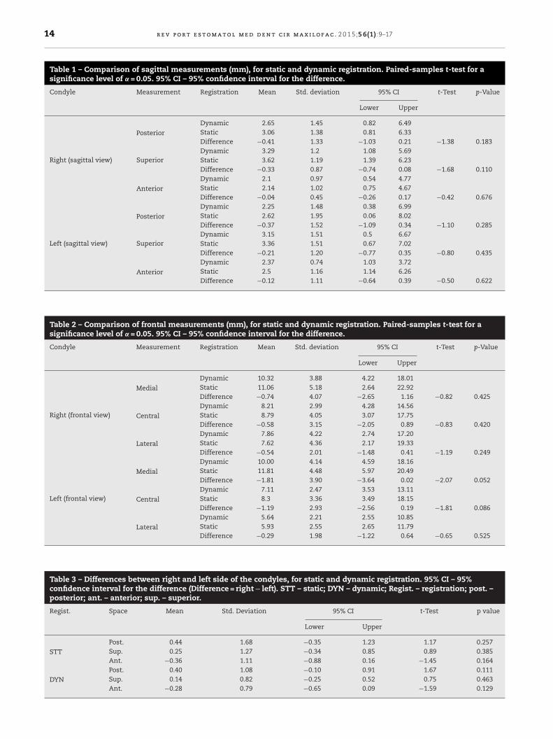
14 r e v p o r t e s t o m a t o l m e d d e n t c i r m a x i l o f a c . 2 0 1 5;5 6(1):9–17
Table 1 – Comparison of sagittal measurements (mm), for static and dynamic registration. Paired-samples t-test for asignificance level of = 0.05. 95% CI – 95% confidence interval for the difference.
Condyle Measurement Registration Mean Std. deviation 95% CI t-Test p-Value
Lower Upper
Right (sagittal view)
Posterior
Dynamic 2.65 1.45 0.82 6.49Static 3.06 1.38 0.81 6.33Difference −0.41 1.33 −1.03 0.21 −1.38 0.183
SuperiorDynamic 3.29 1.2 1.08 5.69Static 3.62 1.19 1.39 6.23Difference −0.33 0.87 −0.74 0.08 −1.68 0.110
Anterior
Dynamic 2.1 0.97 0.54 4.77Static 2.14 1.02 0.75 4.67Difference −0.04 0.45 −0.26 0.17 −0.42 0.676
Left (sagittal view)
Posterior
Dynamic 2.25 1.48 0.38 6.99Static 2.62 1.95 0.06 8.02Difference −0.37 1.52 −1.09 0.34 −1.10 0.285
SuperiorDynamic 3.15 1.51 0.5 6.67Static 3.36 1.51 0.67 7.02Difference −0.21 1.20 −0.77 0.35 −0.80 0.435
Anterior
Dynamic 2.37 0.74 1.03 3.72Static 2.5 1.16 1.14 6.26Difference −0.12 1.11 −0.64 0.39 −0.50 0.622
Table 2 – Comparison of frontal measurements (mm), for static and dynamic registration. Paired-samples t-test for asignificance level of = 0.05. 95% CI – 95% confidence interval for the difference.
Condyle Measurement Registration Mean Std. deviation 95% CI t-Test p-Value
Lower Upper
Right (frontal view)
Medial
Dynamic 10.32 3.88 4.22 18.01Static 11.06 5.18 2.64 22.92Difference −0.74 4.07 −2.65 1.16 −0.82 0.425
Central
Dynamic 8.21 2.99 4.28 14.56Static 8.79 4.05 3.07 17.75Difference −0.58 3.15 −2.05 0.89 −0.83 0.420
Lateral
Dynamic 7.86 4.22 2.74 17.20Static 7.62 4.36 2.17 19.33Difference −0.54 2.01 −1.48 0.41 −1.19 0.249
Left (frontal view)
Medial
Dynamic 10.00 4.14 4.59 18.16Static 11.81 4.48 5.97 20.49Difference −1.81 3.90 −3.64 0.02 −2.07 0.052
Central
Dynamic 7.11 2.47 3.53 13.11Static 8.3 3.36 3.49 18.15Difference −1.19 2.93 −2.56 0.19 −1.81 0.086
Lateral
Dynamic 5.64 2.21 2.55 10.85Static 5.93 2.55 2.65 11.79Difference −0.29 1.98 −1.22 0.64 −0.65 0.525
Table 3 – Differences between right and left side of the condyles, for static and dynamic registration. 95% CI – 95%confidence interval for the difference (Difference = right − left). STT – static; DYN – dynamic; Regist. – registration; post. –posterior; ant. – anterior; sup. – superior.
Regist. Space Mean Std. Deviation 95% CI t-Test p value
Lower Upper
STTPost. 0.44 1.68 −0.35 1.23 1.17 0.257Sup. 0.25 1.27 −0.34 0.85 0.89 0.385Ant. −0.36 1.11 −0.88 0.16 −1.45 0.164
DYNPost. 0.40 1.08 −0.10 0.91 1.67 0.111Sup. 0.14 0.82 −0.25 0.52 0.75 0.463Ant. −0.28 0.79 −0.65 0.09 −1.59 0.129

r e v p o r t e s t o m a t o l m e d d e n t c i r
Table 4 – Pearson correlation between measurements inleft condyle, in static registration (sagittal view).R = Pearson correlation; p value < 0.05; post – posterior;ant – anterior; sup – superior.
Sup Ant
LeftPost. R = 0.85; p < 0.001 R = −0.01; p = 0.985Sup. R = 1 R = 0.05; p = 0.850
Table 5 – Pearson correlation between measurements inright condyle, in static registration (sagittal view).R = Pearson correlation; p value < 0.05; post – posterior;ant – anterior; sup – superior.
Sup Ant
RightPost R = 0.47; p = 0.035 R = −0.07; p = 0.764Sup. R = 1 R = 0.27; p = 0.248
Table 6 – Pearson correlation between measurements inleft condyle, in dynamic registration (sagittal view).R = Pearson correlation; p value < 0.05; post – posterior;ant – anterior; sup – superior.
Sup Ant
LeftPost. R = 0.91; p < 0.001 R = −0.04; p = 0.885Sup. R = 1 R = 0.03; p = 0.913
Table 7 – Pearson correlation between measurements inright condyle, in dynamic registration (sagittal view).R = Pearson correlation; p value < 0.05; post – posterior;ant – anterior; sup – superior.
Sup Ant
Post. R = 0.74; p < 0.001 R = −0.22; p = 0.357
mi
irc
afam1vip
bscit
RightSup. R = 1 R = 0.07; p = 0.774
ethod seems more accurate and easier and all the patientsncluded were capable of doing it.
An expert, blinded to the study, observed the cone beammages of the two registrations and evaluated, at two sepa-ate times, which image corresponded to a theoretically betterondylar position.
According to Cohen’s Kappa, a statistical measurement ofgreement, there was a high variance in the selected imagesrom the first to the second observation (measurement ofgreement: 0.286, p = 0.199. Values of Cohen’s Kappa < 0.20eans poor strength of agreement. Values between 0.81 and
, means a very good strength of agreement). This higherariation means that only based on articular radiographicmages, it is almost impossible to define the rehabilitationosition.
Despite this, the results may be of clinical relevance,ecause in dynamic registration, all the distances weremaller, more consistent and equidistant. It seems that
ondyles stayed in a closer position to articular fossa, for sag-ttal and frontal view. Based on these results, we can suspecthat with dynamic registration the condyles were in a centredm a x i l o f a c . 2 0 1 5;5 6(1):9–17 15
position in the articular fossa and this fact can be observedin both sides simultaneously. Comparatively to static registra-tion, values were more homogeneous, when we consider thetwo, or each side separately.
Anterior and superior spaces are smaller for dynamicregistration, due to the fact that the static method was influ-enced by jaw manipulation and muscular symmetry. Condylarposition during dynamic registration may promote highermuscular symmetry.
In 2014, some authors18 analyzed in magnetic resonanceimaging (MRI) the effects of different registration positionson the condyle–disc position, in maximal intercuspation,Gothic Arch Tracing and retruded contact position (RCP).Dynamic Registration position ensures the widest posteriorspace for the retrodiscal tissues and the slightest sagittaldifference between condyle zenith and articular fossa. Theresults obtained in our study were in concordance with thesefindings. Gothic Arch Tracing seems to best fulfil this criterionof a physiologic or centric condylar position, with symmetrybetween condyles.
A strong correlation between the posterior and superiormeasurements was ascertained, for both condyles and the twomethods of determining the intermaxillary relationship. Thiscould be a sign of antero-inferior sliding of the condyles inthe joint. Thus, an inverse relationship would be expectedbetween the posterior and/or superior measurements withtheir corresponding anterior measurements. Despite the factthat there is a trend in this direction, it was not possible toestablish a statistically significant correlation.
Values for right and left condyles for dynamic registra-tion had a smaller variation and also a stronger correlationbetween them. This means that for Gothic Arch Tracing ahigher symmetry exists.
For static registration, a greater heterogeneity of resultswas seen. This could be possibly due to the fact that in staticregistration, wax rims could promote higher mucosal pressureon one side, resulting in a condylar displacement. In dynamicregistration, less pressure should be expected, because theforces are concentrated on a central pin and not on a waxblock.
For dynamic registration, only the patient participated inhis mandibular movement; left condyle reveals a tendency tostay more posterior, even for static and dynamic registration,but with a higher difference in static. This could be explainedduring static registration, because the operator exerts forceto the posterior and left direction when manipulating, for thereason that all the operators were right-handed.
Results demonstrated a correspondence between the angleof the articular eminence and the length of superior articularspace: the higher the angle the bigger this length is.
An author25 stated that the steeper the eminence, more thecondyle is forced to move inferiorly as it shifts anteriorly. Thiscould be a physiological position of comfort.
Another important fact ascertained in this clinical investi-gation was that a significant correlation between the anteriorposition of the right condyle and the period of edentulism wasfound, which was also in concordance with other authors.17
Regardless the promising results presented in the study,care should be taken in the interpretation in the results, dueto the limited number of patients.

n t c
r
1
1
1
1
1
1
1
1
1
1
2
2
2
2
2
2
16 r e v p o r t e s t o m a t o l m e d d e
Conclusion
From the results of this study, we can conclude that the suc-cess of these procedures was not dependent on the methodused, because no significant discrepancies were seen betweenCBCT images, when using static or dynamic registration meth-ods. Nevertheless, dynamic registration values showed morereliability and accuracy in defining a symmetric and equili-brated CR position. It seems that this method reproduces aphysiologic condylar position.
Ethical disclosures
Protection of human and animal subjects. The authorsdeclare that the procedures followed were in accordance withthe regulations of the relevant clinical research ethics commit-tee and with those of the Code of Ethics of the World MedicalAssociation (Declaration of Helsinki).
Confidentiality of data. The authors declare that they have fol-lowed the protocols of their work centre on the publication ofpatient data.
Right to privacy and informed consent. The authors haveobtained the written informed consent of the patients or sub-jects mentioned in the article. The corresponding author is inpossession of this document.
Conflict of interest
The authors have no conflicts of interest to declare.
e f e r e n c e s
1. Amorim VC, Lagana DC, de Paula Eduardo JV, Zanetti AL.Analysis of the condyle/fossa relationship before and afterprosthetic rehabilitation with maxillary complete dentureand mandibular removable partial denture. J Prosthet Dent.2003;89:508–14.
2. Dervis E. Changes in temporomandibular disorders aftertreatment with new complete dentures. J Oral Rehabil.2004;31:320–6.
3. Ilguy D, Ilguy M, Fisekcioglu E, Dolekoglu S, Ersan N. Articulareminence inclination, height, and condyle morphology oncone beam computed tomography. Sci World J. 2014;2014:1–6.
4. Thakur M, Jain V, Parkash H, Kumar P. A comparativeevaluation of static and functional methods for recordingcentric relation and condylar guidance: a clinical study. JIndian Prosthodont Soc. 2012;12:175–81.
5. Henriques JC, Neto AJ, Almeida GA, Machado NA, Lelis ER.Cone-beam tomography assessment of condylar positiondiscrepancy between centric relation and maximalintercuspation. Braz Oral Res. 2012;26:29–35.
6. Paixão F, Silva WA, Silva FA, Ramos GG, Cruz MV. Evaluationof the reproducibility of two techniques used to determineand record centric relation in angle’s class I patients. J Appl
Oral Sci. 2007;15:275–9.7. Utt TW, Meyers CE, Wierzba TF, Hondrum SO. Athree-dimensional comparison of condylar position changesbetween centric relation and centric occlusion using the
2
i r m a x i l o f a c . 2 0 1 5;5 6(1):9–17
mandibular position indicator. Am J Orthod DentofacialOrthop. 1995;107:298–308.
8. Shanahan TE. Physiologic jaw relations and occlusion ofcomplete dentures. J Prosthet Dent. 2004;91:203–5.
9. Keshvad A, Winstanley RB. Comparison of the replicability ofroutinely used centric relation registration techniques. JProsthodont. 2003;12:90–101.
0. Campos AA, Nathanson D, Rose L. Reproducibility andcondylar position of a physiologic maxillomandibular centricrelation in upright and supine body position. J Prosthet Dent.1996;76:282–7.
1. Zonnenberg AJ, Mulder J. Reproducibility of 2 methods tolocate centric relation in healthy individuals and TMDpatients. Eur J Prosthodont Restor Dent. 2012;20:151–8.
2. Alvarez MC, Turbino ML, Barros C, Pagnano VO, Bezzon OL.Comparative study of intermaxillary relationships of manualand swallowing methods. Braz Dent J. 2009;20:78–83.
3. Bansal S, Palaskar J. Critical evaluation of methods to recordcentric jaw relation. J Indian Prosthodont Soc. 2009;9:120–6.
4. Linsen SS, Stark H, Samai A. The influence of differentregistration techniques on condyle displacement andelectromyographic activity in stomatognathically healthysubjects: a prospective study. J Prosthet Dent. 2012;107:47–54.
5. Yanikoglu N, Guldag MU. Analysis of the condyle/fossarelationship in Kennedy class I and II partially edentuloussubjects. Oral Health Dent Manag. 2006;5:50–6.
6. Barghan S, Tetradis S, Mallya S. Application of cone beamcomputed tomography for assessment of thetemporomandibular joints. Aust Dent J. 2012;57:109–18.
7. Raustia AM, Pirttiniemi P, Salonen MA, Pyhtinen J. Effect ofedentulousness on mandibular size and condyle-fossaposition. J Oral Rehabil. 1998;25:174–9.
8. Linsen SS, Blattner F, Stratmann U. The influence of differentregistration positions on condyle displacement insymptomatic patients. Oral Surg Oral Med Oral Pathol OralRadiol. 2014;117:312–8.
9. Ferreira AF, Henriques JC, Almeida GA, Machado AR, MachadoNA, Neto AJ. Comparative analysis between mandibularpositions in centric relation and maximum intercuspation bycone beam computed tomography (CONE-BEAM). J Appl OralSci. 2009;17:27–34.
0. Hilgers ML, Scarfe WC, Scheetz JP, Farman AG. Accuracy oflinear temporomandibular joint measurements with conebeam computed tomography and digital cephalometricradiography. Am J Orthod Dentofacial Orthop.2005;128:803–11.
1. Miracle AC, Mukherji SK. Conebeam CT of the head and neck.Part 1: Physical principles. Am J Neuroradiol. 2009;30:1088–95.
2. Tsiklakis K, Syriopoulos K, Stamatakis HC. Radiographicexamination of the temporomandibular joint using conebeam computed tomography. Dentomaxillofac Radiol.2004;33:196–201.
3. Librizzi ZT, Tadinada AS, Valiyaparambil JV, Lurie AG, MallyaSM. Cone-beam computed tomography to detect erosions ofthe temporomandibular joint: effect of field of view and voxelsize on diagnostic efficacy and effective dose. Am J OrthodDentofacial Orthop. 2011;140:e25–30.
4. Sumbullu MA, Caglayan F, Akgul HM, Yilmaz AB. Radiologicalexamination of the articular eminence morphology usingcone beam CT. Dentomaxillofac Radiol. 2012;41:234–40.
5. Okeson JP. Management of temporomandibular disorders andocclusion. 6th ed. St. Louis: Mosby; 2008. p. 96–101.
6. Venturelli FA, Zuim PR, Antenucci RM, Garcia AR. Analysis ofmandibular position using different methods of location.Acta Odontol Latinoam. 2009;22:155–62.

t c i r
2
2
29. Raigrodski AJ, Sadan A, Carruth PL. A technique to stabilizerecord bases for Gothic arch tracings in patients with
r e v p o r t e s t o m a t o l m e d d e n
7. Milam SB, Schmitz JP. Molecular biology oftemporomandibular joint disorders: proposed mechanisms of
disease. J Oral Maxillofac Surg. 1995;53:1448–54.8. Wilson PH, Banerjee A. Recording the retruded contactposition: a review of clinical techniques. Br Dent J.2004;196:395–402.
m a x i l o f a c . 2 0 1 5;5 6(1):9–17 17
implant-retained complete dentures. J Prosthodont.1998;7:273–6.