evaluation of 18f-fluoride pet/mr and pet/ct in … nucl med-2015-rauscher-jnumed.114.15… · with...
TRANSCRIPT

Evaluation of 18F-Fluoride PET/MR and PET/CT
in patients with unclear foot pain
Isabel Rauscher1*, Ambros J. Beer1 #*, Christoph Schaeffeler2,3, Michael Souvatzoglou1, Moritz
Crönlein4, Chlodwig Kirchhoff4, Gunther Sandmann4, Sebastian Fürst1, Robert Kilger5, Michael
Herz1, Sybille Ziegler1, Markus Schwaiger1, Matthias Eiber1
1Department of Nuclear Medicine, Klinikum rechts der Isar, Technische Universität München,
Ismaninger Str. 22, 81675 Munich, Germany
2Musculoskeletal Imaging Department of Radiology, Kantonsspital Graubünden, Loëstrasse 170,
7000 Chur, Switzerland
3Department of Radiology, Klinikum rechts der Isar, Technische Universität München,
Ismaninger Str. 22, 81675 Munich, Germany
4Department of Trauma Surgery, Klinikum rechts der Isar, Technische Universität München,
Ismaninger Str. 22, 81675 Munich, Germany
5„University of applied science“ Fresenius, Lothstraße 34, 80335 Munich, Germany
#current address: Department of Nuclear Medicine, University Hospital Ulm, Albert-Einstein-
Allee 23, 89081 Ulm, Germany
* contributed equally
Corresponding author:
Dr. Isabel Rauscher
Email: [email protected]
Phone: +49-89-4140-6085
Fax: +49-89-4140-7431
Journal of Nuclear Medicine, published on February 12, 2015 as doi:10.2967/jnumed.114.150532by SNM headquarters on February 27, 2015. For personal use only. jnm.snmjournals.org Downloaded from

Short running title: 18F-Fluoride PET/MR and PET/CT
Word count: 5344
by SNM headquarters on February 27, 2015. For personal use only. jnm.snmjournals.org Downloaded from

ABSTRACT To evaluate the quality and diagnostic performance of 18F-Fluoride positron-emission
tomography/magnetic resonance imaging (18F-Fluoride PET/MR) compared to 18F-Fluoride
positron-emission tomography/computed tomography (18F-Fluoride PET/CT) imaging in patients
with unclear foot pain.
Methods: Twenty-two patients (9 men, 13 women; mean age 48±18 years, range: 20–78 years)
were prospectively included in this study and underwent a single injection dual imaging protocol
with 18F-Fluoride PET/CT (Siemens Biograph mCT) and PET/MR (Siemens Biograph mMR).
PET/MR protocol included at least T1w SE and PD fs sequences in two planes each with
simultaneous acquisition of PET over 20min. PET/CT included a native isotropic (0.6mm)
diagnostic CT (120kV, 90mAs) and a subsequent PET (2min per bed position (BP)). Two blinded
readers assessed in consensus randomly both PET datasets for image quality (three-point scale)
and for the presence of focal lesions with increased 18F-Fluoride uptake (maximum of 4 lesions).
For each dataset (PET/CT vs. PET/MR) the diagnoses were defined by using both PET and
morphological dataset. SUV values from the two devices were compared using linear correlation
and Bland-Altman plots. Moreover we estimated the potential for dose reduction for PET/MR
compared to PET/CT considering the longer acquisition time of PET/MR analyzing count rate
statistics.
Results: Image quality was rated diagnostic for both PET datasets. However, with a mean rating
of 3.0/3 for PET/MR and 2.3/3 for PET/CT, image quality was significantly superior for PET/MR
(p<0.0001). Sensitivity of the PET datasets in PET/MR and PET/CT were equivalent with the
same 42 lesions with focal 18F-Fluoride uptake. In PET/MR mean SUVmean was 10.4 [range 2.0-
67.7] and mean SUVmax 15.6 [range 2.9-94.1], corresponding mean SUVmean of PET/CT was 10.2
[range 1.8-55.6] and mean SUVmax 16.3 [range 2.5-117.5], resulting in a high linear correlation
by SNM headquarters on February 27, 2015. For personal use only. jnm.snmjournals.org Downloaded from

coefficient (correlation coefficient r=0.96, p<0.0001 for SUVmean and for SUVmax, respectively).
A final consensus reading revealed as most frequent main diagnoses, osteoarthritis, stress fracture
and bone marrow edema (BME). Hereby, PET/CT was more precise in visualizing osteoarthritis,
while PET/MR was more specific in non-degenerative pathologies due to the higher soft-tissue
and bone marrow contrast. The longer acquisition time of MR compared to CT would potentially
allow 18F-Fluoride dose reduction using hybrid 18F-Fluoride PET/MR imaging of at least 50%
according to the count rate analysis.
Conclusion:
In patients with unclear foot pain 18F-Fluoride PET/MR is technical feasible and robust in terms
of image quality and SUV quantification compared to 18F-Fluoride PET/CT. In a majority of
patients 18F-Fluoride PET/MR provided more diagnostic information at a higher diagnostic
certainty as compared to PET/CT. Thus PET/MR combines the high sensitivity of 18F-Fluoride
PET to pinpoint areas with the dominant disease activity and the specificity of MRI for the final
diagnosis with the potential for a substantial dose reduction compared to PET/CT.
Keywords: foot pain, PET/MR, 18F-Fluoride
by SNM headquarters on February 27, 2015. For personal use only. jnm.snmjournals.org Downloaded from
INTRODUCTION
Foot pain is a common problem in daily routine of orthopaedic surgeons. It can be a clinical
symptom of many different entities such as stress reactions or fractures, systemic disorders, foot
deformation and osteoarthritis, osteochondral lesions, interdigital neuroma, synovitis,
impingement, tendinopathy and tenosynovitis, or metatarsophalangeal joint instability. Incidence
and prevalence of metatarsalgia and foot pain vary depending upon the causative condition.
Multiple imaging modalities are available to evaluate foot pain including radiography, computed
tomography (CT), magnetic resonance imaging (MRI), bone scintigraphy, and ultrasound.
Radiography of the foot may reveal fractures, foot deformation, or arthritis whereas bone scans
are helpful with an earlier diagnosing of stress fractures as well as some types of infections and
tumors. MR imaging has shown to be useful and superior to CT in the assessment of soft tissue
pathologies and abnormalities of bone marrow.
Another option to evaluate areas of bone remodelling is 18F-Fluoride PET imaging which mainly
depicts osteoblastic activity. Several PET-only studies have shown that it is not possible to
differentiate benign from malignant lesions based on the intensity of 18F-fluoride uptake and that
diagnostic accuracy can be significantly improved by additional morphological CT and/or MR
imaging (1-3). A recent study of Fischer et al. revealed that 18F-Fluoride PET/CT has a
substantial therapeutic impact on management in patients with unclear foot pain (4). However, it
is well known that for most musculoskeletal pathologies, MRI is superior to CT due to its
increased soft tissue contrast and the possibility to image bone marrow edema (BME). In
combination with the excellent lesion-to-background ratio of 18F-Fluoride PET, 18F-Fluoride
PET/MR has the potential to further enhance the accuracy in the diagnosis of chronic foot pain.
Thus, the purpose of our study was to compare the performance of 18F-Fluoride PET/MR to 18F-
Fluoride PET/CT in 2 aspects: First, to evaluate the quality and performance of 18F-Fluoride PET
by SNM headquarters on February 27, 2015. For personal use only. jnm.snmjournals.org Downloaded from
using PET/MR vs. PET/CT and second, to analyse the diagnostic performance of combined 18F-
Fluoride PET/MR vs. PET/CT in patients with unclear foot pain.
MATERIAL AND METHODS
Patient Population
Twenty-two patients (9 men, 13 women; mean age 48 ±18 years, range, 20–78 years) with
unclear foot pain were prospectively enrolled in this study between February 2012 and August
2013, routinely referred to our institute for clinical 18F-Fluoride PET/CT imaging. For these
patients the specific diagnosis for this condition had remained inconclusive after clinical
examination and radiography. The study was approved by the local institutional review board.
Written informed consent was obtained from all patients. The inclusion criteria were: Informed
consent and the ability to undergo a PET/MR following the PET/CT examination. Exclusion
criteria were: Pregnancy, age under 18 years, and contraindications for MR imaging. All subjects
underwent a single-injection/ dual-imaging protocol. After completion of the PET/CT scan
patients were subsequently positioned on the PET/MRT scanner with the smallest possible
temporal delay to utilize the remaining activity of the initial 18F-Fluoride injection. 18F-Fluoride
was produced by proton irradiation of 18O-enriched water in a cyclotron (5).
18F-Fluoride PET/CT And PET/MR Imaging
Scanning was started approximately 75±18 min after the i.v. injection of a dose of 133±68 MBq
of 18F-Fluoride and performed on a clinical PET/CT system (Siemens Biograph mCT scanner,
Siemens Healthcare). This scanner has an axial FOV of 21.8cm and a ring diameter of 84.2cm.
The transverse spatial resolution of its PET detector assembly was measured to be 4.4mm near
the center of the FOV, whereas the sensitivity in the center was found to be 9.7kcps/MBq (6).
The patients were examined in a supine position. PET/CT included a native CT (field of view
by SNM headquarters on February 27, 2015. For personal use only. jnm.snmjournals.org Downloaded from

(FOV) 780mm, tube voltage 80kV, tube current 165mAs, care dose4D, rotation time 0.5s;
collimation 0.6mm) and a subsequent PET (2min per bed position (BP)). Attenuation maps were
obtained by bilinear transformation and were used for attenuation correction as previously
described (7). The acquired images were post-processed on the Siemens Biograph mCT scanner
providing multiplanar reformatted images for PET alone, CT alone (axial, coronal and sagittal
reformation with slice thickness 2mm and axial reconstruction with slice thickness 0.6mm) and
fused PET/CT.
All PET/MR examinations were performed using an integrated whole-body hybrid PET/MR
system (Biograph mMR; Siemens Healthcare). Compared to the Biograph mCT, PET/MR has a
larger axial FOV of 25.8cm and a smaller ring diameter of 65.6cm . The spatial resolution of
4.3mm is similar, but its sensitivity approximately 50% higher at 15.0kcps/MBq (8-10). PET data
acquired on the Biograph mCT was reconstructed without using time-of-flight information to
better match the reconstruction parameters of the Biograph mMR, which is not equipped with
such a capability. On average, the PET/MR scan was started 107±26 min p.i.. Patients were
positioned in the MR scanner as similar as possible to the PET/CT examination. Since the mean
acquisition time in PET/MR was longer than in PET/CT, the patient was immobilized using
cushions in various sizes around the coil to reduce motion artefacts during scan time. The
combined PET/MR protocol was as follows: First, a coronal 2-point Dixon 3D volumetric
interpolated examination (VIBE) T1 weighted (T1w) MR sequence was acquired, which was
used for the generation of attenuation-maps as recently published (11). Together with the start of
this Dixon MR sequence, the PET-acquisition (20 min) started simultaneously in the same BP,
thus ensuring optimal temporal and regional correspondence between MRI and PET data.
by SNM headquarters on February 27, 2015. For personal use only. jnm.snmjournals.org Downloaded from
Additionally, a dedicated MR protocol of the foot was defined depending on the localization of
the maximum pain with the following parameters: slice thickness 3mm, field of view (FoV) 120-
225 mm, matrix: 320x256-384x384. The protocol consisted of at least one intermediate-weighted
fat-saturated (PDfs) sequence in 2 planes and one T1- and T2- weighted turbo spin echo (TSE)
sequence. If clinically relevant, additional contrast-enhanced sequences were performed: T1-
weighted TSE sequence before and after application of Gadolinium (Gd) in the best suitable
plane and T1-weighted TSE fs sequence with Gd in a second plane.
Data Processing And Image Analysis
PET data obtained on the PET/CT and PET/MR scanners were processed with comparable
reconstruction and correction algorithms. For both modalities, emission data were corrected for
randoms, dead time, scatter, and attenuation. A 3-dimensional attenuation-weighted ordered-
subsets expectation maximization iterative reconstruction algorithm (OSEM 3D) was applied
with 3 iterations and 21 subsets, gaussian smoothing of 4mm in full width at half maximum, and
a zoom of 1.
For image analysis all datasets were transferred to a dedicated postprocessing workstation (Syngo
MMWP, Siemens Medical Solutions). PET/CT and PET/MR were analysed in consensus by a
dual board certified radiologist and nuclear physician with several years of experience in PET/CT
and PET/MR reading and a board certified radiologist with special training in musculoskeletal
radiology. The readers were aware of the patients´ history, results of prior radiography and
clinical examination. PET/MR and PET/CT were evaluated separately with at least 8 weeks
difference.
Visual Rating
by SNM headquarters on February 27, 2015. For personal use only. jnm.snmjournals.org Downloaded from

18F-Fluoride PET data from both CT and MRI were rated for every patient with regard to overall
image quality: 0–non diagnostic image quality, 1-low image quality (distinct artifacts, strong
image noise), 2-good image quality (little artifacts, moderate image noise), 3-excellent image
quality (no artifacts, low image noise).
Lesion Identification And Classification
Both PET datasets were evaluated for the presence of focal lesions with increased 18F-Fluoride
uptake compared to normal bone. Data of CT and MR imaging were used for exact anatomic
correlation with Fluoride uptake and were analysed for additional PET negative important
morphological findings. After describing both findings in PET and morphological imaging one
main diagnosis and up to two secondary diagnoses were determined for each PET/MR and
PET/CT dataset. Depending on the readers´ certainty they were either judged as diagnostic
(category 1) or suspicious (category 2).
The following criteria were used to define the most frequently observed pathologies:
- osteoarthritis: presence of osteophytes, joint space narrowing, subchondral cysts, subchondral
sclerosis, or a combination of them on both sides of the articular joint in PET/CT with the 18F-
Fluoride being centered along the joint space; additional finding in PET/MR:
- BME, which was defined as an ill-defined area of increased signal intensity on intermediate-
weighted images with fat suppression and corresponding hypointensity on T1w images not below
the intensity of skeletal muscle.
- stress reaction: BME and absence of a fracture line; 18F-Fluoride uptake and if adjacent to a
joint the absence of osteoarthritis on PET/CT
- stress fracture: BME with the presence of a fracture line on a T1-weighted MR image or a dense
sclerotic line on CT; 18F-Fluoride uptake
by SNM headquarters on February 27, 2015. For personal use only. jnm.snmjournals.org Downloaded from

Finally, a consensus reading was carried out based on all available data trying to define the
modality (PET/CT vs. PET/MR) that provided the most specific and precise diagnosis.
Quantitative Assessment
For quantitative comparison between the PET data acquired in PET/CT and in PET/MR, a SUV-
based analysis of mean and maximum tracer uptake in up to 4 focal lesions and in three
representative, not pathological osseous structures (calcaneus, distal tibia and calcaneus) was
performed. Volumes of interest (VOIs) were placed over matching corresponding Fluoride
positive lesions. To calculate SUVs, the axial slice with the maximum SUV of the lesion was first
located automatically, using standardized software for images of both scanners. An isocontour
VOI including all voxels above 50% of the maximum was then created to calculate SUVmean.
Within all VOI’s, mean and maximum standardized uptake values were measured.
Count Rate Analysis On PET/CT and PET/MR With Regard To Scan Duration
As image quality of PET/MR was suspected to be superior to that of PET/CT due to the longer
acquisition time in PET/MR (20min compared to 2min in PET/CT), the theoretical 18F-Fluoride
dose reduction in PET/MR was calculated that would yield equal numbers of detected true
coincidences on both scanners. For this purpose, count rate statistics were analyzed for all 7
patient data sets, the PET raw data of which had been acquired in listmode format, by
determining the number of trues in the first 120s of the PET/CT and PET/MR examinations as
well as of the entire 1200s of the PET/MR scan. The obtained information was then corrected for
decay to the start of the PET/CT scan. Since for one patient the PET/MR scan duration was only
600s, the number of trues in this case was extrapolated to an acquisition time of 1200s.
Statistical Analysis
by SNM headquarters on February 27, 2015. For personal use only. jnm.snmjournals.org Downloaded from
Statistical analysis was performed using the MedCalc 12.3.0.0 software package for Windows. P
values of less than 0.05 were considered statistically significant. First, descriptive statistical
evaluation was performed. Then, differences in image quality as well as SUVmean/SUVmax of
focal foot lesions/normal bone between PET/MR and PET/CT were compared by using the
Spearman rank correlation coefficient. The overall statistical differences in measured SUVs were
tested using the nonparametric Wilcoxon matched-pairs signed rank test. Because a high degree
of correlation does not necessarily imply good agreement between the 2 measurements, a Bland–
Altman plot was constructed to assess this agreement (12). A Bland–Altman plot displays the
difference between the 2 measurements versus their average as a scatterplot, on which each point
represents 1 measurement.
RESULTS
Visual Rating
The results of overall image quality showed that PET/MR was superior to PET/CT with an
overall excellent image quality score of 3.0/3 points in all PET/MR datasets while PET/CT
achieved 2.3 out of 3 possible points (2/3 points in 15/22 patients and 3/3 points in 7/22 patients).
Image quality of PET/MR was significantly superior to PET/CT (p<0.0001).
Quantitative Analysis
Both SUVmean and SUVmax of PET/CT and PET/MR showed a highly statistical significant linear
correlation (R=0.96; p<0.0001 for both SUVmean and SUVmax) for pathological foot lesions. Mean
SUVmean and SUVmax including standard deviation (SD) and range for pathological lesions and
regions of normal bone in PET/MR and PET/CT are presented in table 1 including p-value,
correlation coefficient (R) and 95% confidence interval (CI) for the correlation beetween
by SNM headquarters on February 27, 2015. For personal use only. jnm.snmjournals.org Downloaded from

PET/MR and PET/CT. Correlation analysis and Bland-Altman Plot for SUVmean and SUVmax can
be found in figure 1 and figure 1S, respectively.
Findings And Diagnoses In PET/MR And PET/CT
In total, the same 42 lesions with intense, focal 18F-Fluoride uptake were identified both in PET
datasets of PET/MR and PET of PET/CT showing an equivalent sensitivity. A detailed
presentation of patient characteristics including symptoms, findings and diagnoses in PET/MR
and PET/CT is shown in table 1S. Note that one patient did not show any abnormal findings.
Most Frequent Diagnoses The three most frequent diagnoses were osteoarthritis, stress fracture
and BME representing a stress reaction of bone. Osteoarthritis of one or several articular joints
was diagnosed in 9 out of 22 patients. Note that in patients with osteoarthritis but no other
concomitant findings PET/CT was favoured as modality of choice by the reading team. This was
related to the higher image resolution and thus better depiction of anatomical findings including
some additional minor findings like calcaneal spurs in CT. Nevertheless the certainty of
diagnosing osteoarthritis as the main final pathology was rated as diagnostic (category 1) in both
PET/CT and PET/MR.
Stress fractures were present in 4 out of 22 patients. Hereby, PET/MR was rated as modality of
choice in all but one case. However, in one patient the depiction of PET/CT as most conclusive
modality was only related to an additional sesamoid fracture whose displacement could be better
visualized in CT. Otherwise the reviewer regarded PET/MR more conclusive for stress fractures
due to the earlier depiction of the typical T1w hypointense line compared to a sclerotic line in CT
providing a higher diagnostic confidence.
by SNM headquarters on February 27, 2015. For personal use only. jnm.snmjournals.org Downloaded from

Six out of 22 patients showed stress reactions. Due to the visualization of BME, stress reactions
could be classified in all presented cases more precisely in PET/MR, which was chosen as
modality of choice by the reading team. In PET/CT this diagnosis could only be assumed
indirectly due to the absence of any sclerotic bone changes and signs of osteoarthritis in a region
of monofocal 18F-Fluoride uptake resulting in a lower diagnostic certainty (category 2).
Modality Of Choice (18F-Fluoride PET/MR Vs. 18F-Fluoride PET/CT) With regard to the
modality of choice PET/MR was rated in 13/22 patients to be more appropriate than PET/CT.
Hereby the better specificity of PET/MR was related to its capability to visualize bone marrow or
soft tissue pathologies. Besides stress reaction and stress fracture (see above) PET/MR enabled a
more precise diagnosis in cases with an aneurysmatic bone cyst, soft tissue edema, tenosynovitis,
large ganglion cysts or osteochondral lesions. Here, in 6 out of 22 patients the MR part of 18F-
Fluoride PET/MR revealed 6 complete new findings with no correlate both in 18F-Fluoride PET and CT. PET/CT was regarded as modality of choice in cases in which the better anatomical
depiction of morphological changes in the cortical bone (e.g. osteophytes, subchondral changes,
calcaneal spur) was crucial (6 out of 22 patients). In detail, this were the patients with only
osteoarthritis and no other pathological findings, a patient with an accessory navicular syndrome
and one patient with a fracture of the sesamoid bone.
Figure 2 shows representative images of a patient with a stress fracture at the base of OMT I and
figure 3 images of a patient with osteoarthritis and a ganglion originating from the dorsal upper
ankle joint, which could only be depicted in MRI. Additional 18F-Fluoride PET/MR and PET/CT
images of a patient with an aneurysmatic bone cyst and a stress reaction resolving on follow-up
imaging are shown in figure 2S and 3S.
by SNM headquarters on February 27, 2015. For personal use only. jnm.snmjournals.org Downloaded from
Assessment Of Potential Dose Reduction
The average number of true coincidences per injected dose detected in the first 120s of a scan
was measured to be (48928±31400) MBq-1 on the PET/CT and on the PET/MR (63599±44329)
MBq-1. The number of trues registered during the entire 1200s of the PET/MR scan was
(600405±419410) MBq-1, i.e. on average 12 times higher than that of the PET/CT scan with a
duration of 120s. This means that with equal scan duration approximately 80% of the dose
administered for the PET/CT scan would have been required on the PET/MR to yield the same
number of true coincidences. For a PET/MR scan duration of 20 minutes, only approximately
10% of the injected activity would have resulted in the same number of acquired trues.
Before conclusions can be drawn from count rate statistics with regard to image quality, effects
of additional hardware in the PET/MR field of view have to be evaluated in this clinical setting
(13). However, these first results hint at a possible dose reduction of at least 50% on the PET/MR
compared to the PET/CT, if the corresponding scan duration is 10 times longer.
DISCUSSION
The results of this study indicate that 18F-Fluoride PET/MR is equivalent to 18F-Fluoride PET/CT
concerning SUV-quantification and lesion detection in patients with unclear foot pain. Image
quality of PET/MR was superior to PET/CT potentially due to longer acquisition time in
PET/MR raising the possibility of 18F-Fluoride dose reduction. However, 18F-Fluoride PET/MR
was more conclusive than PET/CT due to the higher soft tissue contrast and the visualization of
bone marrow pathologies on MRI.
by SNM headquarters on February 27, 2015. For personal use only. jnm.snmjournals.org Downloaded from
Our study indicates that 18F-Fluoride PET/MR is technically feasible and robust despite
difference in attenuation correction which is concordant with other studies comparing 18F-FDG
PET/CT and PET/MR in malignant bone lesions (14, 15). I
However, in both cited whole-body studies SUVs of bone lesions were substantially lower in
PET/MR. In contrast, in our study, mean SUVmean/ SUVmax of PET/CT and PET/MR were nearly
similar. Two possible explanations could be the different radiotracer (18F-Fluoride vs. 18F-FDG)
and examination of a small peripheral part of the body instead of the central skeleton. Hereby,
amongst other MR FOV-restrictions can lead to significant quantification error in large compared
to small volumes (16). Furthermore, concerning attenuation correction in PET/MR other
problems (e.g. treating bone as soft tissue, increased attenuation by cortical bone and calcified
areas) are still unresolved. Compared to those potential confounders, our results indicate that for
18F-Fluoride PET/MR of the feet no relevant influence is present. From a clinical point-of-view
this is supported by the fact that in our study the identical lesions were identified in both
modalities by the reading team.
18F-Fluoride PET/MR would allow substantial reduction in the applied activity due to the
potential of longer PET-acquisition in parallel to the acquisition of MR. This would offer a
substantial dose reduction of at least 50% in patients with no history of malignant disease and
who often are of younger age (see table 1S). However, this has to be validated in further
prospective studies.
For diagnostic purposes 18F-Fluoride PET is a highly sensitive but not very specific tool for the
detection of metabolically active benign bone disease with the drawback of a low specificity (5).
Thus, additional morphological imaging is recommended either using sequentially CT/MRI or by
means of hybrid PET/CT and more recently PET/MR.
by SNM headquarters on February 27, 2015. For personal use only. jnm.snmjournals.org Downloaded from

The results from our study show that 18F-Fluoride PET/MR was regarded as the modality of
choice in a substantial higher number of cases (13/22) than 18F-Fluoride PET/CT. A majority of
these cases were patients with stress fractures or BME and here the superiority of MR in the
depiction of bone marrow pathologies is well known while the bony structures in these cases
were still without any detectable changes in CT leading to a less reliable diagnosis in PET/CT
(17).
18F-Fluoride PET/CT was only superior in cases with osteoarthritis showing no additional
findings/complications (4/22) and in cases (2/22) in which the superb image resolution of multi-
slice CT was crucial (e.g. assessment of small bony structures). Hereby, the clinical preference of
CT due to a more accurate anatomical depiction is related to an overall low signal of dense bony
structures on all MR-sequences leading to a less distinct delineation and the usual lower spatial
resolution of MR (18, 19). Nevertheless, even in complicated cases with osteoarthritis (5/9) by
means of stress reaction in adjacent bone or concomitant soft-tissue pathologies (e.g. ganglion
cyst) 18F-Fluoride PET/MR was regarded as equal or superior compared to 18F-Fluoride PET/CT.
A potential disadvantage of PET/MR is the use in patients with bilateral complaints as a
simultaneous examination of both feet would lead to a tremendous loss in diagnostic quality of a
PET/MR-study (use of body vs. dedicated surface coils). Thus, due to the natural history of
osteoarthritis in elderly people 18F-Fluoride PET/CT most likely would fulfil the diagnostic needs
in patients >60 years with pain in both feet and without a trauma history when primary
osteoarthritis is more likely.
Compared to morphological imaging alone the specific value of hybrid 18F-Fluoride PET/CT or
PET/MR imaging consists in the possibility to (semi)-quantitatively assess tracer uptake. Various
reports have described the value of 18F-Fluoride to help in the therapeutic management to the
by SNM headquarters on February 27, 2015. For personal use only. jnm.snmjournals.org Downloaded from

right regions due to a direct relation between the intensity of bone metabolism and the complaints
of the patient (4, 5). Especially in patients with multifocal disease the maximum of 18F-Fluoride
uptake can help the clinician to tailor the therapy to the region of most clinical relevance/highest
disease activity (e.g. the joint which is affected the most by osteoarthritis). A study by Fischer et
al. was showing that 18F-Fluoride PET in addition to sequentially performed MR offers additional
value information allowing a more specific therapy in 13 of 28 patients (4). Therefore, we
hypothesize that the use of 18F-Fluoride PET/MR can provide additional therapeutically relevant
information in patients with unclear foot pain. However, this has to be proven in prospective
studies also evaluating the contribution both of MR and 18F-Fluoride to the final diagnosis and
the therapeutic management in a large patient cohort.
Our study has some limitations. We did not examine the potential influence when performing
PET/CT and PET/MR in random order. However, despite reports for 18F-FDG PET leading to a
tendency to lower of SUV in benign lesions, such an effect is not known for 18F-Fluoride (20). In
addition, the rare number of studies comparing lesion-to-background-ratio of 18F-Fluoride and
skeletal scintigraphy both at an early and a later time-point do not indicate a relevant influence
(21). In addition, we have not specifically evaluated whether 18F-Fluoride PET/MR or PET/CT
lead to a difference in therapeutic management. However, as in many of our cases the final
diagnosis was the same in PET/MR and PET/CT differing only in the diagnostic certainty we
would not expect a substantial difference.
CONCLUSION
Despite different attenuation techniques 18F-Fluoride PET/MR can be regarded as technical
feasible and robust. In patients with unclear foot pain 18F-Fluoride PET/MR provided more
by SNM headquarters on February 27, 2015. For personal use only. jnm.snmjournals.org Downloaded from
diagnostic information at a higher diagnostic certainty as compared to 18F-Fluoride PET/CT.
Besides information on bone metabolism it provides additional diagnostic relevant findings from
soft-tissue and bone marrow pathology compared to PET/CT. A further advantage of 18F-Fluoride
PET/MR consists in the potential for additional dose reduction due to the longer acquisition time.
Thus, larger prospective studies exploring the use of 18F-Fluoride PET/MR in patients with
unclear foot pain when medical history, clinical, and radiographic examination remain
inconclusive are warranted. These should also focus on the impact of this technique on patient
management and cost-effectiveness.
by SNM headquarters on February 27, 2015. For personal use only. jnm.snmjournals.org Downloaded from

References
1. Cook GJ, Fogelman I. The role of positron emission tomography in skeletal disease.
Semin Nucl Med. 2001;31:50-61.
2. Even-Sapir E, Metser U, Flusser G, et al. Assessment of malignant skeletal disease: initial
experience with 18F-fluoride PET/CT and comparison between 18F-fluoride PET and
18F-fluoride PET/CT. J Nucl Med. 2004;45:272-278.
3. Even-Sapir E, Metser U, Mishani E, Lievshitz G, Lerman H, Leibovitch I. The detection
of bone metastases in patients with high-risk prostate cancer: 99mTc-MDP Planar bone
scintigraphy, single- and multi-field-of-view SPECT, 18F-fluoride PET, and 18F-fluoride
PET/CT. J Nucl Med. 2006;47:287-297.
4. Fischer DR, Maquieira GJ, Espinosa N, et al. Therapeutic impact of [(18)F]fluoride
positron-emission tomography/computed tomography on patients with unclear foot pain.
Skeletal Radiol. 2010;39:987-997.
5. Grant FD, Fahey FH, Packard AB, Davis RT, Alavi A, Treves ST. Skeletal PET with
18F-fluoride: applying new technology to an old tracer. J Nucl Med. 2008;49:68-78.
6. Jakoby BW, Bercier Y, Conti M, Casey ME, Bendriem B, Townsend DW. Physical and
clinical performance of the mCT time-of-flight PET/CT scanner. Phys Med Biol.
2011;56:2375-2389.
7. Kinahan PE, Hasegawa BH, Beyer T. X-ray-based attenuation correction for positron
emission tomography/computed tomography scanners. Semin Nucl Med. 2003;33:166-
179.
8. Delso G, Furst S, Jakoby B, et al. Performance measurements of the Siemens mMR
integrated whole-body PET/MR scanner. J Nucl Med. 2011;52:1914-1922.
by SNM headquarters on February 27, 2015. For personal use only. jnm.snmjournals.org Downloaded from

9. Delso G, Martinez-Moller A, Bundschuh RA, et al. Evaluation of the attenuation
properties of MR equipment for its use in a whole-body PET/MR scanner. Phys Med Biol.
2010;55:4361-4374.
10. Drzezga A, Souvatzoglou M, Eiber M, et al. First clinical experience with integrated
whole-body PET/MR: comparison to PET/CT in patients with oncologic diagnoses. J
Nucl Med. 2012;53:845-855.
11. Martinez-Moller A, Souvatzoglou M, Delso G, et al. Tissue classification as a potential
approach for attenuation correction in whole-body PET/MRI: evaluation with PET/CT
data. J Nucl Med. 2009;50:520-526.
12. Bland JM, Altman DG. Statistical methods for assessing agreement between two methods
of clinical measurement. Lancet. 1986;1:307-310.
13. Furst S, Souvatzoglou M, Martinez-Moller A, Schwaiger M, Nekolla SG, Ziegler SI.
Impact of flexible body surface coil and patient table on PET quantification and image
quality in integrated PET/MR. Nuklearmedizin. 2014;53:79-87.
14. Eiber M, Takei T, Souvatzoglou M, et al. Performance of whole-body integrated 18F-
FDG PET/MR in comparison to PET/CT for evaluation of malignant bone lesions. J Nucl
Med. 2014;55:191-197.
15. Beiderwellen K, Huebner M, Heusch P, et al. Whole-body [(1)(8)F]FDG PET/MRI vs.
PET/CT in the assessment of bone lesions in oncological patients: initial results. Eur
Radiol. 2014;24:2023-2030.
16. Delso G, Martinez-Moller A, Bundschuh RA, Nekolla SG, Ziegler SI. The effect of
limited MR field of view in MR/PET attenuation correction. Med Phys. 2010;37:2804-
2812.
by SNM headquarters on February 27, 2015. For personal use only. jnm.snmjournals.org Downloaded from
17. Choi J, Raghavan M. Diagnostic imaging and image-guided therapy of skeletal
metastases. Cancer Control. 2012;19:102-112.
18. Even-Sapir E. Imaging of malignant bone involvement by morphologic, scintigraphic, and
hybrid modalities. J Nucl Med. 2005;46:1356-1367.
19. Rybak LD, Rosenthal DI. Radiological imaging for the diagnosis of bone metastases. Q J
Nucl Med. 2001;45:53-64.
20. Lan XL, Zhang YX, Wu ZJ, Jia Q, Wei H, Gao ZR. The value of dual time point (18)F-
FDG PET imaging for the differentiation between malignant and benign lesions. Clin
Radiol. 2008;63:756-764.
21. Wong KK, Piert M. Dynamic bone imaging with 99mTc-labeled diphosphonates and 18F-
NaF: mechanisms and applications. J Nucl Med. 2013;54:590-599.
by SNM headquarters on February 27, 2015. For personal use only. jnm.snmjournals.org Downloaded from
A B
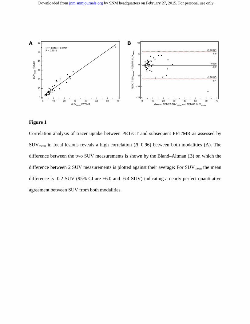
Figure 1
Correlation analysis of tracer uptake between PET/CT and subsequent PET/MR as assessed by
SUVmean in focal lesions reveals a high correlation (R=0.96) between both modalities (A). The
difference between the two SUV measurements is shown by the Bland–Altman (B) on which the
difference between 2 SUV measurements is plotted against their average: For SUVmean the mean
difference is -0.2 SUV (95% CI are +6.0 and -6.4 SUV) indicating a nearly perfect quantitative
agreement between SUV from both modalities.
by SNM headquarters on February 27, 2015. For personal use only. jnm.snmjournals.org Downloaded from

Figure 2
Simultaneously acquired 18F-Fluoride PET/CT and PET/MR images in a 49-year-old female
patient with pain over the left metatarsal foot since several months without history of trauma.
Sagittal MR images show bone marrow edema (BME) in fat-saturated (fs) proton-density (PD)
weighted images at the base of Os metatarsale I (OMT I) (red arrow A) with the presence of a
hypointense fracture line on the T1-weighted image (red arrow B). In the corresponding PET of
PET/MR (D), PET/MR (E) and PET/CT (F) an intense focal 18F-Fluoride uptake at the base of
OMT I is shown (red arrow). However, in the area of the PET positive region, the corresponding
CT scan shows only a slight sclerotic band (red arrow C).
by SNM headquarters on February 27, 2015. For personal use only. jnm.snmjournals.org Downloaded from

Figure 3
Sagittal 18F-Fluoride PET/CT and PET/MR images of a 55-year-old male patient with persistent
pain and swelling of the left foot since several years. There are advanced signs of osteoarthritis
particularly in the tarsometatarsal joints and in the subtalar joint which can be observed on both
MR imaging (sagittal T1 TSE sequence (A), sagittal PD fs sequence (B)) and sagittal CT (C)
including joint space narrowing, osteophytes, subchondral cysts and subchondral bone marrow
edema (only on MR imaging). Additionally, only MR imaging could detect the T1 hypointense,
PD fs hyperintense ganglion cyst originating from the posterior subtalar joint (red arrows).
Corresponding PET of PET/MR (D), PET/MR (E) and PET/CT (F) show intense focal 18F-
Fluoride uptake on both sides of the tarsometatarsal and the subtalar joint; however in this case
no additional information was provided by PET. Note the slight difference in slice positioning
leading to a different impression of 18F-Fluoride uptake.
by SNM headquarters on February 27, 2015. For personal use only. jnm.snmjournals.org Downloaded from
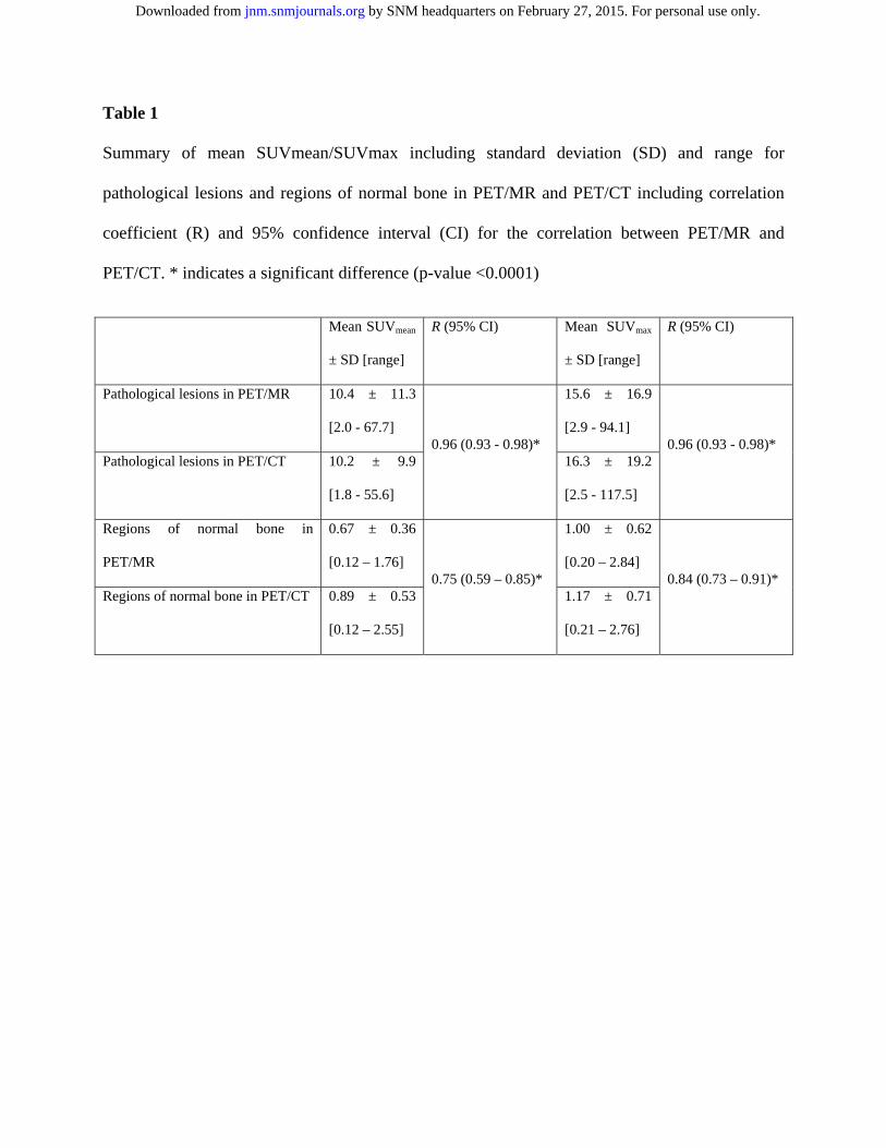
Table 1
Summary of mean SUVmean/SUVmax including standard deviation (SD) and range for
pathological lesions and regions of normal bone in PET/MR and PET/CT including correlation
coefficient (R) and 95% confidence interval (CI) for the correlation between PET/MR and
PET/CT. * indicates a significant difference (p-value <0.0001)
Mean SUVmean
± SD [range]
R (95% CI) Mean SUVmax
± SD [range]
R (95% CI)
Pathological lesions in PET/MR 10.4 ± 11.3
[2.0 - 67.7] 0.96 (0.93 - 0.98)*
15.6 ± 16.9
[2.9 - 94.1] 0.96 (0.93 - 0.98)*
Pathological lesions in PET/CT 10.2 ± 9.9
[1.8 - 55.6]
16.3 ± 19.2
[2.5 - 117.5]
Regions of normal bone in
PET/MR
0.67 ± 0.36
[0.12 – 1.76] 0.75 (0.59 – 0.85)*
1.00 ± 0.62
[0.20 – 2.84] 0.84 (0.73 – 0.91)*
Regions of normal bone in PET/CT 0.89 ± 0.53
[0.12 – 2.55]
1.17 ± 0.71
[0.21 – 2.76]
by SNM headquarters on February 27, 2015. For personal use only. jnm.snmjournals.org Downloaded from

Doi: 10.2967/jnumed.114.150532Published online: February 12, 2015.J Nucl Med. Gunther Sandmann, Sebastian Fürst, Robert Kilger, Michael Herz, Sybille Ziegler, Markus Schwaiger and Matthias EiberIsabel Rauscher, Ambros Beer, Christoph Schaeffeler, Michael Souvatzoglou, Moritz Crönlein, Chlodwig Kirchhoff,
F-Fluoride PET/MR and PET/CT in patients with unclear foot pain18Evaluation of
http://jnm.snmjournals.org/content/early/2015/02/11/jnumed.114.150532This article and updated information are available at:
http://jnm.snmjournals.org/site/subscriptions/online.xhtml
Information about subscriptions to JNM can be found at:
http://jnm.snmjournals.org/site/misc/permission.xhtmlInformation about reproducing figures, tables, or other portions of this article can be found online at:
and the final, published version.proofreading, and author review. This process may lead to differences between the accepted version of the manuscript
ahead of print area, they will be prepared for print and online publication, which includes copyediting, typesetting,JNMcopyedited, nor have they appeared in a print or online issue of the journal. Once the accepted manuscripts appear in the
. They have not beenJNM ahead of print articles have been peer reviewed and accepted for publication in JNM
(Print ISSN: 0161-5505, Online ISSN: 2159-662X)1850 Samuel Morse Drive, Reston, VA 20190.SNMMI | Society of Nuclear Medicine and Molecular Imaging
is published monthly.The Journal of Nuclear Medicine
© Copyright 2015 SNMMI; all rights reserved.
by SNM headquarters on February 27, 2015. For personal use only. jnm.snmjournals.org Downloaded from