euryarchaeota crenarchaeota
TRANSCRIPT


Hyperthermophiles
Euryarchaeota
Halophilicmethanogen
Methanosarcina
Methanospirillum
Thermoplasma
Halobacterium
Marine EuryarchaeotaMarine Crenarchaeota
Natronococcus
Methanobacterium Methanocaldococcus
Archaeoglobus
Thermococcus/Pyrococcus
Thermoproteus
Methanopyrus
Methanothermus
Pyrodictium
Desulfurococcus
Root
Sulfolobus
Crenarchaeota
Ferroplasma
Picrophilus
Halococcus
Extremehalophiles
Extremeacidophiles
Nanoarchaeum

GymnamoebasEntamoebas
Plasmodial slime molds
Cellular slime molds
Microsporidia
Fungi
Animals
Plants
Greenalgae
Redalgae
(Secondaryendosymbioses)
Foramin-iferans
Chlorarach-niophytes
Radio-larians
OomycetesDiatoms
Ciliates
Dinoflagellates
Apicomplexans
Parabasalids
Diplomonads
Kinetoplastids
Euglenids
Goldenalgae
Brownalgae
Amoebozoa
Fungi
Cercozoans
Stramenopiles
Alveolates
Euglenozoa
Chloroplast ancestor(primary endosymbiosis)
Mitochondrial ancestor(primary endosymbiosis)
Bacteria
Protists
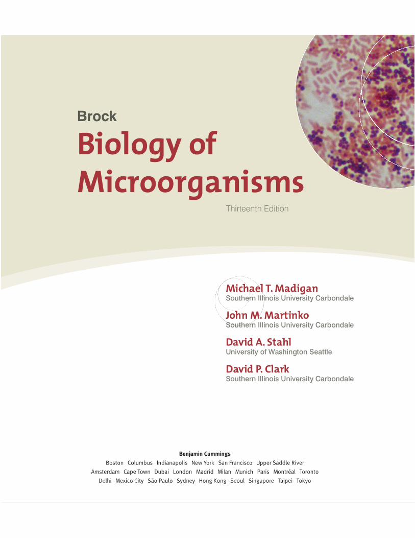
Brock
Biology of Microorganisms
Thirteenth Edition
Michael T. MadiganSouthern Illinois University Carbondale
John M. MartinkoSouthern Illinois University Carbondale
David A. StahlUniversity of Washington Seattle
David P. ClarkSouthern Illinois University Carbondale
Executive Editor: Deirdre EspinozaProject Editor: Katie CookAssociate Project Editor: Shannon CuttDevelopment Editor: Elmarie HutchinsonArt Development Manager: Laura SouthworthArt Editor: Elisheva MarcusManaging Editor: Deborah CoganProduction Manager: Michele MangelliProduction Supervisor: Karen GulliverCopyeditor: Anita Wagner
Art Coordinator: Jean LakePhoto Researcher: Maureen SpuhlerDirector, Media Development: Lauren FogelMedia Producers: Sarah Young-Dualan, Lucinda Bingham, and Ziki DekelArt: Imagineering Media Services, Inc.Text Design: Riezebos Holzbaur Design GroupSenior Manufacturing Buyer: Stacey WeinbergerSenior Marketing Manager: Neena BaliCompositor: Progressive Information TechnologiesCover Design: Riezebos Holzbaur Design Group
Cover Image: (front cover) Peter Siver/Visuals Unlimited/Corbis; (back cover) J.-H. Becking, Wageningen AgriculturalUniversity, Wageningen, Netherlands
Credits for selected images can be found on page P-1.
Copyright © 2012, 2009, 2006 Pearson Education, Inc., publishing as Benjamin Cummings, 1301 Sansome Street,San Francisco, CA 94111. All rights reserved. Manufactured in the United States of America. This publication isprotected by copyright and permission should be obtained from the publisher prior to any prohibited reproduction,storage in a retrieval system, or transmission in any form or by any means, electronic, mechanical, photocopying,recording, or likewise. To obtain permission(s) to use material from this work, please submit a written request toPearson Education, Inc., Permissions Department, 1900 E. Lake Ave., Glenview, IL 60025. For information regardingpermissions, call (847) 486-2635.
Many of the designations used by manufacturers and sellers to distinguish their products are claimed as trademarks.Where those designations appear in this book, and the publisher was aware of a trademark claim, the designationshave been printed in initial caps or all caps.
Library of Congress Cataloging-in-Publication Data
Brock biology of microorganisms / Michael T. Madigan . . . [et al.].—13th ed.p. cm.
Includes index.ISBN-13: 978-0-321-64963-8 (hardcover : alk. paper)ISBN-10: 0-321-64963-X (hardcover : alk. paper) 1. Microbiology. I. Madigan, Michael T., 1949–
II. Title: Biology of microorganisms. QR41.2.B77 2011579—dc22
2010044962
ISBN 10: 0-321-64963-X (student edition)ISBN 13: 978-0-321-64963-8 (student edition)ISBN 10: 0-321-72675-8 (professional copy)ISBN 13: 978-0-321-72675-9 (professional copy)1 2 3 4 5 6 7 8 9 10—CRK—14 13 12 11 10

About the Authors
iii
Michael T. Madigan re-ceived his B.S. in Biology andEducation from Wisconsin StateUniversity–Stevens Point (1971) andhis M.S. (1974) and Ph.D. (1976) inBacteriology from the University ofWisconsin–Madison. His graduateresearch was on the hot spring bac-terium Chloroflexus in the labora-tory of Thomas Brock. Following a
three-year postdoctoral in the Department of Microbiology,Indiana University, Mike moved to Southern Illinois University–Carbondale, where he has been a professor of microbiology for32 years. He has coauthored Biology of Microorganisms sincethe fourth edition (1984) and teaches courses in introductorymicrobiology, bacterial diversity, and diagnostic and appliedmicrobiology. In 1988 Mike was selected as the OutstandingTeacher in the College of Science and in 1993, the OutstandingResearcher. In 2001 he received the SIUC Outstanding ScholarAward. In 2003 he received the Carski Award for DistinguishedUndergraduate Teaching from the American Society for Micro-biology (ASM), and he is an elected Fellow of the AmericanAcademy of Microbiology. Mike’s research is focused on bacteriathat inhabit extreme environments, and for the past 12 years hehas studied the microbiology of permanently ice-covered lakes inthe McMurdo Dry Valleys, Antarctica. In addition to his researchpapers, he has edited a major treatise on phototrophic bacteriaand served for over a decade as chief editor of the journalArchives of Microbiology. He currently serves on the editorialboard of the journals Environmental Microbiology and Antonievan Leeuwenhoek. Mike’s nonscientific interests include forestry,reading, and caring for his dogs and horses. He lives beside apeaceful and quiet lake with his wife, Nancy, five shelter dogs(Gaino, Snuffy, Pepto, Peanut, and Merry), and four horses(Springer, Feivel, Gwen, and Festus).
John M. Martinko receivedhis B.S. in Biology from ClevelandState University. He then worked atCase Western Reserve University,conducting research on the serologyand epidemiology of Streptococcuspyogenes. His doctoral work at theState University of New York–Buffaloinvestigated antibody specificity andantibody idiotypes. As a postdoc-
toral fellow, he worked at Albert Einstein College of Medicinein New York on the structure of major histocompatibility com-plex proteins. Since 1981, he has been in the Department ofMicrobiology at Southern Illinois University–Carbondale wherehe was Associate Professor and Chair, and Director of the Molec-ular Biology, Microbiology, and Biochemistry Graduate Program.He retired in 2009, but remains active in the department as aresearcher and teacher. His research investigates structuralchanges in major histocompatibility proteins. He teaches anadvanced course in immunology and presents immunology andhost defense lectures to medical students. He also chairs theInstitutional Animal Care and Use Committee at SIUC. He hasbeen active in educational outreach programs for pre-universitystudents and teachers. For his educational efforts, he won the2007 SIUC Outstanding Teaching Award. He is also an avidgolfer and cyclist. John lives in Carbondale with his wife Judy, ahigh school science teacher.

iv About the Authors
David A. Stahl received hisB.S. degree in Microbiology from theUniversity of Washington–Seattle,later completing graduate studies inmicrobial phylogeny and evolutionwith Carl Woese in the Departmentof Microbiology at the University ofIllinois–Champaign-Urbana. Subse-quent work as a postdoctoral fellowwith Norman Pace, then at the
National Jewish Hospital in Colorado, focused on early applica-tions of 16S rRNA-based sequence analysis to the study of natu-ral microbial communities. In 1984 Dave joined the faculty at theUniversity of Illinois–Champaign-Urbana, holding appointmentsin Veterinary Medicine, Microbiology, and Civil Engineering. In1994 he moved to the Department of Civil Engineering at North-western University, and in 2000 returned to his alma mater, theUniversity of Washington–Seattle, as a professor in the Depart-ments of Civil and Environmental Engineering and Microbiol-ogy. Dave is known for his work in microbial evolution, ecology,and systematics, and received the 1999 Bergey Award and the2006 Procter & Gamble Award in Applied and EnvironmentalMicrobiology from the ASM; he is also an elected Fellow of theAmerican Academy of Microbiology. His main research interestsare the biogeochemistry of nitrogen and sulfur compounds andthe microbial communities that sustain these nutrient cycles. Hislaboratory was first to culture ammonia-oxidizing Archaea, agroup now believed to be the main mediators of this key processin the nitrogen cycle. He has taught several courses in environ-mental microbiology, is one of the co-founding editors of thejournal Environmental Microbiology, and has served on manyadvisory committees. Outside teaching and the lab, Dave enjoyshiking, bicycling, spending time with family, reading a good sci-ence fiction book, and, with his wife Lin, renovating an old farm-house on Bainbridge Island, Washington.
David P. Clark grew up inCroydon, a London suburb. He wona scholarship to Christ’s College,Cambridge, where he received hisB.A. degree in Natural Sciences in1973. In 1977 he received his Ph.D.from Bristol University, Departmentof Bacteriology, for work on theeffect of cell envelope compositionon the entry of antibiotics into
Escherichia coli. He then left England on a postdoctoral studyingthe genetics of lipid metabolism in the laboratory of John Cronanat Yale University. A year later he moved with the same labora-tory to the University of Illinois at Urbana-Champaign. Davidjoined the Department of Microbiology at Southern IllinoisUniversity–Carbondale in 1981. His research has focused on thegrowth of bacteria by fermentation under anaerobic conditions.He has published numerous research papers and graduated over20 Masters and Doctoral students. In 1989 he won the SIUCCollege of Science Outstanding Researcher Award. In 1991 hewas the Royal Society Guest Research Fellow at the Departmentof Molecular Biology and Biotechnology, Sheffield University,England. In addition to Brock Biology of Microorganisms, David isthe author of four other science books: Molecular Biology MadeSimple and Fun, now in its fourth edition; Molecular Biology:Understanding the Genetic Revolution; Biotechnology: Applyingthe Genetic Revolution; and Germs, Genes, & Civilization: HowEpidemics Shaped Who We Are Today. David is unmarried andlives with two cats, Little George, who is orange and very nosey,and Mr. Ralph, who is mostly black and eats cardboard.
DedicationsMichael T. Madigan dedicates this book to the memory of his children who rest on Boot Hill: Andy, Marcy, Willie, Plum, Teal, and Sugar. Whether in good times or bad, they always greeted him with tails a waggin’.John M. Martinko dedicates this book to his daughters Sarah,Helen, and Martha, and to his wife Judy. Thanks for all of yoursupport!David A. Stahl dedicates this book to his wife, Lin. My love, and one that helps me keep the important things in perspective.David P. Clark dedicates this book to his father, Leslie, who set him the example of reading as many books as possible.
Preface
The authors and Benjamin Cummings Publishers proudlypresent the 13th edition of Brock Biology of Microorganisms
(BBOM 13/e). This book is truly a milestone in the annals ofmicrobiology textbooks. Brock Biology of Microorganisms, and itspredecessor, Biology of Microorganisms, has introduced the fieldof microbiology to students for 41 years, more than any othertextbook of microbiology. Nevertheless, although this book goesback over four decades, its two main objectives have remainedfirm since the first edition was published in 1970: (1) to presentthe principles of microbiology in a clear and engaging fashion,and (2) to provide the classroom tools necessary for deliveringoutstanding microbiology courses. The 13th edition of BBOMfulfills these objectives in new and exciting ways.
Veteran textbook authors Madigan, Martinko, and Clark wel-come our new coauthor, Dave Stahl, to this edition of BBOM. Daveis one of the world’s foremost experts in microbial ecology and hasmasterfully crafted an exciting new view of the ecology material inBBOM, including a new chapter devoted entirely to microbialsymbioses, a first for any textbook of microbiology. Users will findthat the themes of ecology and evolution that have permeated thisbook since its inception reach new heights in the 13th edition.These fundamental themes also underlie the remaining contentof the book—the basic principles of microbiology, the molecularbiology and genetics that support microbiology today, the hugediversity of metabolisms and organisms, and the medical andimmunological facets of microbiology. It is our belief that out-standing content coupled with outstanding presentation havecome together to make BBOM 13/e the most comprehensive andeffective textbook of microbiology available today.
What’s New in the 13th Edition?In terms of content and pedagogy, instructors who have usedBBOM previously will find the 13th edition to be the same oldfriend they remember; that is, a book loaded with accurate, up-to-the-minute content that is impeccably organized and visuallyenticing. The 36 chapters in BBOM 13/e are organized into mod-ules by numbered head, which allows instructors to fine-tunecourse content to the needs of their students. In addition, studyaids and review tools are an integral part of the text. Our newMiniQuiz feature, which debuts in the 13th edition, is designed toquiz students’ comprehension as they work their way througheach chapter. Also new to this edition is the end-of-chapter reviewtool called “Big Ideas.” These capsule summaries pull together the
key concepts from each numbered section in a wrap-up style thatis certain to be a big hit with students, especially the night beforeexaminations! Our end-of-chapter key terms list, two detailedappendices, a comprehensive glossary, and a thorough indexcomplete the hard copy learning package. Many additional learn-ing resources are available online (see below).
In terms of presentation, BBOM 13/e will easily draw in andengage the reader. The book has been designed in a beautiful yetsimple fashion that gives the art and pedagogical elements thebreathing room they need to be effective and the authors thefreedom to present concepts in a more visually appealing way.Supporting the narrative are spectacular illustrations, with everypiece of art rendered in a refreshing new style. Moreover, the artcomplements, and in many cases integrates, the hundreds ofphotos in BBOM, many of which are new to the 13th edition.And, as users of BBOM have come to expect, our distinctive illus-trations remain the most accurate and consistent of those in anymicrobiology textbook today.
The authors are keenly aware that it is easy to keep piling onnew material and fattening up a textbook. In response to thistrend, BBOM 13/e went on a diet. With careful attention to con-tent and presentation, BBOM 13/e is actually a shorter book thanBBOM 12/e. The authors have carefully considered every topic toensure that content at any point in the book is a reflection of bothwhat the student already knows and what the student needs toknow in a world where microbiology has become the most excit-ing and relevant of the biological sciences. The result is a morestreamlined and exciting treatment of microbiology that bothstudents and instructors will appreciate.
Revision Highlights:Chapter 1• Find new coverage on the evolution and major habitats of
microorganisms—Earth’s most pervasive and extensive biomass.• A more visually compelling presentation of the impacts of
microorganisms on humans better emphasizes the importanceof microorganisms for the maintenance of all life on Earth.
Chapter 2• New coverage of cell biology and the nature of the chromo-
some in prokaryotic and eukaryotic cells is complemented by avisually engaging overview of the microbial world.
v
Prefacevi
Chapter 3• The cell chemistry chapter that previously held this position is
now available online (www.microbiologyplace.com). The newChapter 3 explores cell structure and function with strong newvisuals to carry the text and new coverage of the lipids and cellwalls of Bacteria and Archaea.
Chapter 4• Find updated coverage of catabolic principles along with an
overview of essential anabolic reactions.• Newly rendered and more instructive art makes mastering key
metabolic pathways and bioenergetic principles a more visualexperience.
Chapter 5• Updated coverage of the events in cell division and their
relation to medical microbiology connects basic science toapplications.
• Newly rendered art throughout makes the important conceptsof cell division and population growth more vivid, engaging,and interactive.
Chapter 6• The concise primer on molecular biology that every student
needs to know is updated and now includes an overview of thestructures of nucleic acids and proteins and the nature of chro-mosomes and plasmids.
Chapter 7• Find new coverage of the latest discoveries in the molecular
biology of Archaea and comparisons with related molecularprocesses in Bacteria.
• A new section highlights the emerging area of regulation bymicroRNA in eukaryotes.
Chapter 8• Review major updates on the regulation of gene expression—
one of the hottest areas in microbiology today—includingexpanded coverage of cell sensing capacities and signal trans-duction.
• Enjoy the new Microbial Sidebar featuring CRISPR, the newlydiscovered form of RNA-based regulation used by Bacteriaand Archaea to ward off viral attack.
Chapter 9• Major updates of the principles of virology are complemented
with an overview of viral diversity.• New art reinforces the relevance and importance of viruses as
agents of genetic exchange.
Chapter 10• The fundamental principles of microbial genetics are updated
and supplemented with new coverage that compares and con-trasts bacterial and archaeal genetics.
Chapter 11• Find “one-stop shopping” for coverage of molecular biological
methods, including cloning and genetic manipulations, as aprelude to the genomics discussion in the next chapter.
• Enjoy the colorful new Microbial Sidebar on new fluorescentlabeling methods that can differentiate even very closelyrelated bacteria.
Chapter 12• Extensive updates on microbial genomics and transcriptomics
will be found along with new coverage of the emerging relatedareas of metabolomics and interactomics.
• Readers will marvel at the diversity of prokaryotic genomes in thenew Microbial Sidebar “Record-Holding Bacterial Genomes.”
Chapter 13• The two chapters covering metabolic diversity have been
revised and moved up to Chapters 13 and 14 to precede ratherthan follow coverage of microbial diversity, better linking thesetwo important and often related areas.
• This chapter is loaded with reworked art and text that high-light the unity and diversity of the bioenergetics underlyingphototrophic and chemolithotrophic metabolisms.
Chapter 14• Restyled and impeccably consistent art showcases the compar-
ative biochemistry of the aerobic and anaerobic catabolism ofcarbon compounds.
Chapter 15• This retooled chapter combines the essentials of industrial
microbiology and biotechnology, including the production ofbiofuels and emerging green microbial technologies.
Chapter 16• Find new coverage of the origin of life and how the evolution-
ary process works in microorganisms.• Microbial phylogenies from small subunit ribosomal RNA
gene analyses are compared with those from multiple-gene andfull genomic analyses.

Preface vii
Chapters 17–19• Coverage of the diversity of Bacteria and Archaea better
emphasizes phylogeny with increased focus on phyla of partic-ular importance to plants and animals and to the health of ourplanet.
• Spectacular photomicrographs and electron micrographs carrythe reader through prokaryotic diversity.
Chapter 20• A heavily revised treatment of the diversity of microbial
eukaryotes is supported by many stunning new color photosand photomicrographs.
• Find an increased emphasis on the phylogenetic relationships ofeukaryotes and the “bacterial nature” of eukaryotic organelles.
Chapter 21• Viruses, the most genetically diverse of all microorganisms,
come into sharper focus with major updates on their diversity.• A new section describes viruses in nature and their abundance
in aquatic habitats.
Chapter 22• This chapter features a major new treatment of the latest
molecular techniques used in microbial ecology, includingCARD-FISH, ARISA, biosensors, NanoSIMS, flow cytometry,and multiple displacement DNA amplification.
• Find exciting new coverage of methods for functional analysesof single cells, including single-cell genomics and single-cellstable isotope analysis, and expanded coverage of methods foranalyses of microbial communities, including metagenomics,metatranscriptomics, and metaproteomics.
Chapter 23• A comparison of the major habitats of Bacteria and Archaea is
supported by spectacular new photos and by art that summa-rizes the phylogenetic diversity and functional significance ofprokaryotes in each habitat.
• Find broad new coverage of the microbial ecology of microbialmat communities and prokaryotes that inhabit the deep sub-surface.
Chapter 24• Revised coverage of the classical nutrient cycles is bolstered by
new art, while new coverage highlights the calcium and silicacycles and how these affect CO2 sequestration and global climate.
• Improved integration of biodegradation and bioremediationshows how natural microbial processes can be exploited for thebenefit of humankind.
Chapter 25• This new chapter focuses entirely on microbial symbioses,
including bacterial–bacterial symbioses and symbioses betweenbacteria and their plant, mammal, or invertebrate hosts. Findcoverage here of all of the established as well as more recentlydiscovered symbioses, including the human gut and how itsmicrobiome may control obesity, the rumen of animals impor-tant to agriculture, the hindgut of termites, the light organ ofthe squid, the symbioses between hydrothermal vent animalsand chemolithotrophic bacteria, the essential bacterial sym-bioses of insects, medicinal leeches, reef-building corals, andmore, all supported by spectacular new color photos and art.
• Learn how insects have shaped the genomes of their bacterialendosymbionts.
• Marvel at the new Microbial Sidebar that tells the intriguingstory of the attine ants and their fungal gardens.
Chapter 26• Key updates will be found on microbial drug resistance and are
supported by new art that reveals the frightening reality thatseveral human pathogens are resistant to all known antimicro-bial drugs.
Chapter 27• Extensively reworked sections on the normal microbial flora of
humans include new coverage of the human microbiome and amolecular snapshot of the skin microflora.
• Find revised coverage of the principles of virulence and patho-genicity that connect infection and disease.
Chapter 28• Here we present the perfect overview of immunology for instruc-
tors who wish to cover only the fundamental concepts and howthe immune system resists the onslaught of infectious disease.
• Find late-breaking practical information on the immuneresponse, including vaccines and immune allergies.
Chapter 29• Built on the shoulders of the previous chapter, here is a more
detailed probe of the mechanisms of immunity with emphasison the molecular and cellular interactions that control innateand adaptive immunity.
Chapter 30• This short chapter presents an exclusively molecular picture of
immunology, including receptor–ligand interactions (the “trig-gers” of the immune response), along with genetics of the keyproteins that drive adaptive immunity.

Prefaceviii
Chapter 31• Find revised and expanded coverage of molecular analyses in
clinical microbiology, including new enzyme immunoassays,reverse transcriptase PCR, and real-time PCR.
Chapter 32• Review major updates of the principles of disease tracking,
using 2009 pandemic H1N1 influenza as a model for hownewly emerging infectious diseases are tracked.
• Find updated coverage throughout, especially of the HIV/AIDSpandemic.
Chapter 33• Read all about the origins and history of pandemic H1N1
influenza and how the H1N1 virus is related to strains ofinfluenza that already existed in animal populations.
• Hot new coverage of immunization strategies for HIV/AIDS.
Chapter 34• Follow the emergence, rapid dispersal, and eventual entrench-
ment of West Nile virus as an endemic disease in North America.• Expanded coverage of malaria—the deadliest human disease of
all time—includes the promise of new antiparasitic drugs anddisease prevention methods.
Chapter 35• Find updates of water microbiology, including new rapid meth-
ods for detecting specific indicator organisms.
Chapter 36• Explore new methods of food processing, including aseptic and
high-pressure methods that can dramatically extend the shelf-life and safety of perishable foods and drinks.

ix
Archaea
CrenarchaeotaEuryarchaeota
Other ArchaeaAcidobacteria
ActinobacteriaBacteroidetes
Other
Other
CyanobacteriaFirmicutes
Planctomycetes
Verrucomicrobia
Unclassified andminor bacterialgroups
SAR11 group
RhodobacteralesBurkholderiales
Nitrosomonadales
Alteromonadales
OceanospirillalesPseudomonadales
Vibrionales
Other Proteobacteria
Proteobacteria
Figure 23.24 Ocean prokaryotic diversity. The results are pooled analyses of 25,975 sequences fromseveral studies of the 16S rRNA gene content of pelagic ocean waters. Many of these groups are covered in Chapters 17 and 18 (Bacteria) or 19 (Archaea). For Proteobacteria, major subgroups are indicated. Notethe high proportion of cyanobacterial and Gammaproteobacteria sequences. Data assembled and analyzedby Nicolas Pinel.
The 13th edition enhances the themes of ecology and evolution throughout, and is the only book on the market to include specialized coverage of archael and eukaryotic molecular biology. The book represents the most current research in the fi eld, with special attention paid to the microbial ecology chapters:
Chapter 22, Methods in Microbial Ecology, is heavily updated to present the latest molecular techniques used in microbial ecology, including CARD-FISH, ARISA, biosensors, NanoSIMS, fl ow cytometry, and multiple displacement DNA amplifi cation. It also includes exciting new coverage of methods for functional analyses of single cells, including single-cell genomics and single-cell stable isotope analysis, and expanded coverage of methods for analyses of microbial communities, including metagenomics, metatranscriptomics, and metaproteomics.
Chapter 23, Major Microbial Habitats and Diversity, compares the major habitats of Bacteria and Archaea and is supported by spectacular new photos and art that summarize the phylogenetic diversity and functional signifi cance of prokaryotes in each habitat.
Chapter 25, Microbial Symbioses, is a completely new chapter focused entirely on microbial symbioses,including bacterial–bacterial symbioses and symbioses between bacteria and their plant, mammal, or invertebrate hosts. Find coverage here of all the established as well as more recently discovered symbioses—including the human gut and how its microbiome may control obesity, the rumen of animals important to agriculture, the hindgut of termites, the light organ of the squid, the symbioses between hydrothermal vent animals and chemolithotrophic bacteria, and the essential bacterial symbioses of insects, medicinal leeches, reef-building corals, and more.
Yosh
itom
o K
ikuc
hi a
nd J
örg
Gra
f
Bacteroidetes
Betaproteobacteria
Ochrobactrum
Figure 25.40 Micrograph of a FISH-stained microbial communityin the bladder of Hirudo verbana. A probe (red) targeted at the 16SrRNA of Betaproteobacteria and a probe (green) targeted at the 16S rRNAof Bacteroidetes reveal distinct layers of different bacteria in the lumen ofthe bladder. Staining with DAPI (blue), which binds to DNA, reveals theintracellular alphaproteobacterium Ochrobactrum and host nuclei.
Cutting Edge Coverage Includes the Most Current Presentation of Microbial Ecology
For a detailed list of chapter-by-chapter updates, see page v of the Preface.
Chapter 24, Nutrient Cycles, Biodegradation, and Bioremediation. Exciting updates of all the nutrient cycles that form the heart of environmental microbiology and microbial ecology.
x
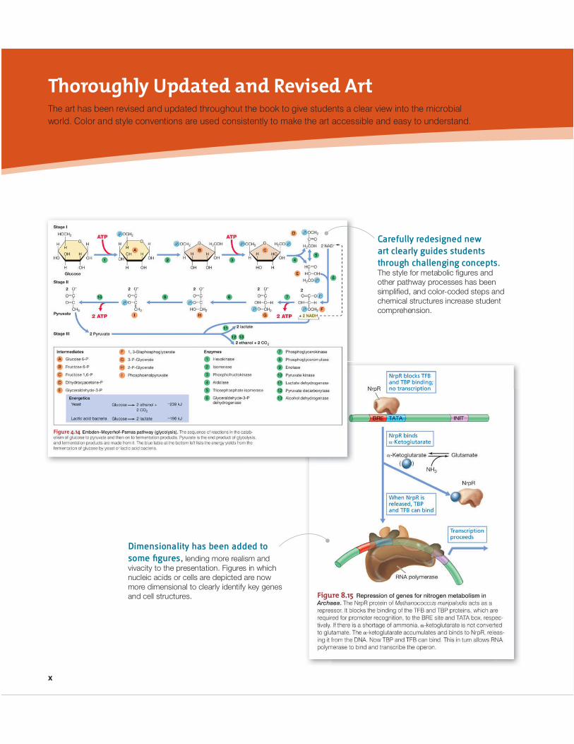
The art has been revised and updated throughout the book to give students a clear view into the microbial world. Color and style conventions are used consistently to make the art accessible and easy to understand.
Carefully redesigned new art clearly guides students through challenging concepts.The style for metabolic fi gures and other pathway processes has been simplifi ed, and color-coded steps and chemical structures increase student comprehension.
Dimensionality has been added to some fi gures, lending more realism and vivacity to the presentation. Figures in which nucleic acids or cells are depicted are now more dimensional to clearly identify key genes and cell structures.
Thoroughly Updated and Revised Art

xi
Illustrations and photos are often paired to give an idealized view next to a realistic view and to reinforce the connection between theory and practice.

xii
Brief Contents
UNIT I
Basic Principles of MicrobiologyChapter 1 Microorganisms and Microbiology 1Chapter 2 A Brief Journey to the Microbial World 24Chapter 3 Cell Structure and Function in Bacteria
and Archaea 47UNIT 2
Metabolism and GrowthChapter 4 Nutrition, Culture, and Metabolism
of Microorganisms 85Chapter 5 Microbial Growth 117
UNIT 3
Molecular Biology and Gene ExpressionChapter 6 Molecular Biology of Bacteria 150Chapter 7 Archaeal and Eukaryotic Molecular Biology 191Chapter 8 Regulation of Gene Expression 209
UNIT 4
Virology, Genetics, and GenomicsChapter 9 Viruses and Virology 236Chapter 10 Genetics of Bacteria and Archaea 263Chapter 11 Genetic Engineering 291Chapter 12 Microbial Genomics 313
UNIT 5
Metabolic Diversity and Commercial BiocatalysesChapter 13 Phototrophy, Chemolithotrophy, and Major
Biosyntheses 340Chapter 14 Catabolism of Organic Compounds 372Chapter 15 Commercial Products and Biotechnology 411
UNIT 6
Microbial Evolution and DiversityChapter 16 Microbial Evolution and Systematics 446Chapter 17 Bacteria: The Proteobacteria 475Chapter 18 Other Bacteria 517Chapter 19 Archaea 556
Chapter 20 Eukaryotic Cell Biology and EukaryoticMicroorganisms 584
Chapter 21 Viral Diversity 613
UNIT 7
Microbial EcologyChapter 22 Methods in Microbial Ecology 642Chapter 23 Major Microbial Habitats and Diversity 669Chapter 24 Nutrient Cycles, Biodegradation, and
Bioremediation 698Chapter 25 Microbial Symbioses 720
UNIT 8
Antimicrobial Agents and PathogenicityChapter 26 Microbial Growth Control 755Chapter 27 Microbial Interactions with Humans 787
UNIT 9
ImmunologyChapter 28 Immunity and Host Defense 816Chapter 29 Immune Mechanisms 838Chapter 30 Molecular Immunology 859
UNIT 10
Diagnosing and Tracking Microbial DiseasesChapter 31 Diagnostic Microbiology and Immunology 878Chapter 32 Epidemiology 913
UNIT 11
Human- and Animal-TransmittedInfectious DiseasesChapter 33 Person-to-Person Microbial Diseases 944Chapter 34 Vectorborne and Soilborne Microbial
Pathogens 981UNIT 12
Common-Source Infectious DiseaseChapter 35 Wastewater Treatment, Water Purification,
and Waterborne Microbial Diseases 1004Chapter 36 Food Preservation and Foodborne
Microbial Diseases 1022xviii
The fi rst twelve chapters cover the principles of microbiology. Basic principles are presented early on and then used as the foundation to tackle the material in greater detail later.
Information on metabolic diversity precedes the coverage of microbial diversity, better linking these important and often related areas.
Conceptual Framework Helps Students Focus on the Key Concepts
New chapter on symbiosis ties together the core concepts of the book—health, diversity, and the human ecosystem.
This newly revised chapter is the perfect overview for instructors who wish to cover immunology at a generalized level including the fundamental concepts of how the immune system resists the onslaught of infectious disease. Instructors who like to go into more detail can build on the core principles taught in Chapter 28 by covering Immune Mechanisms (Ch. 29) and Molecular Immunology (Ch. 30).

xiii
The new Big Ideas sections at the end of each chapter focus on the core concepts students need to know.
MiniQuiz critical thinking questions integrated throughout the text test student comprehension of core principles from each section.
MiniQuiz• What are the primary response regulator and the primary sensor
kinase for regulating chemotaxis?
• Why is adaptation during chemotaxis important?
• How does the response of the chemotaxis system to an attrac-tant differ from its response to a repellent?

xiv
The MyMicrobiologyPlace website is rich with media assets to give students extra practice. It includes chapter quizzes, new quantitative questions, animations, and additional tutorials. www.microbiologyplace.com
CourseCompass includes all of the assets from the MyMicrobiologyPlace website and all of the test questions from the computerized test bank. It also features class management tools, such as discussion boards and email functionality to help instructors easily teach online classes or give assignments. www.aw-bc.com/coursecompass
Instructor Resource DVD (IR-DVD)0-321-72086-5 / 978-0-321-72086-3
The IR-DVD offers a wealth of media resources including all the art from the book in both JPEG and PPT formats, PowerPoint lecture outlines, computerized test bank, and answer keys all in one convenient location. The animations help bring lectures to life, while the select step-edit fi gures help break down complicated processes.
Instructor Manual and Test Bank0-321-72021-0 / 978-0-321-72021-4
by W. Matthew Sattley and Christopher A. Gulvik
The Instructor Manual/Test Bank provides chapter summaries that help with class preparation as well as the answers to the end-of-chapter review and application questions. The test bank contains 3,000 questions for use in quizzes, tests, and exams.
Additional Resources
FOR
STU
DEN
TSFO
R I
NS
TRU
CTO
RS
Quantitative QuestionsNumber of genes in plasmid R100. The Esch-erichia coli plasmid R100 is a circular molecule of DNA containing 93.4 kbp. The average E. coli protein contains 300 amino acids; assume that the same is true for R100 proteins. With this assumption, calculate how many genes are in this plasmid.
Compare DNA polymerases. Escherichia coli contains at least fi ve different DNA polymerases. The three most characterized are DNA Pol I, Pol II, and Pol III. Polymerase I and II replicate DNA at about 20–40 nucleotides/sec whereas Pol III replicates at 250 to 1000 nucleotides/sec. The genome of E. coli strain K-12 is 4,639,221 bp. At the higher rates, how long does it take to reproduce the chromosome? How do these numbers agree with the roles of these DNA polymerases?
1
2

Acknowledgments
Abook of this stature is not the product of its authors alonebut instead is the collective effort of the many people who
comprise the book team. These include folks both inside andoutside of Benjamin Cummings. Executive editor DeirdreEspinoza and project editor Katie Cook, both of Benjamin Cum-mings, were the workhorses in editorial. Deirdre paved the wayfor the 13th edition and skillfully maneuvered the book aroundthe occasional roadblocks that accompany any major textbookproject. Katie ran the day-to-day operations of the BBOM teamin a highly professional manner, expertly managing reviews andmany other details and keeping all facets of the project on track.
The BBOM 13/e production and design team was headed upby Michele Mangelli (Mangelli Productions) who oversaw YvoRiezebos (Riezebos Holzbaur Design Group), and Laura South-worth (Benjamin Cummings). Michele managed the productionteam and did a great job of keeping everyone on mission and onbudget. The artistic magic of Yvo is clearly visible in the beautifultext and cover designs of BBOM 13/e. Laura created the new artlook for BBOM 13/e, one that readers should immediately appre-ciate for its clarity, consistency, and modern style. The authorsare extremely grateful to Michele, Yvo, and Laura, as well as tothe artist team at the studio of Imagineering (Toronto), for help-ing the authors produce such a beautiful book. Others in produc-tion included Karen Gulliver, Jean Lake, and Maureen Spuhler.Karen was our excellent production editor who ensured that apolished book emerged from a raw manuscript, while Jean wasour art coordinator, tracking and routing art and handling inter-actions with the art studio. Maureen was our photo researcherwho helped the authors locate photos that met the exacting stan-dards of BBOM. The authors are extremely grateful to Karen,Jean, and Maureen for transforming literally thousands of pagesof text and art manuscript into a superb learning tool.
The authors wish to give special thanks to four other membersof the production team: Elmarie Hutchinson, Anita Wagner, Eli-sheva (Ellie) Marcus, and Elizabeth McPherson. Our develop-mental editor Elmarie was a key contributor early in the project,helping the authors better link text and art and massaging thetext to improve readability. Anita was our absolutely spectacularcopyeditor; the authors could not have asked for a brighter ormore effective person in this key position on the book team.Anita improved the accuracy, clarity, and consistency of the textand rendered her editorial services in a style that the authorsfound both helpful and time saving. Ellie (Benjamin Cummings)was our art liaison on this project, translating for the art housethe intentions of the authors. Ellie has the unique gift of viewingart from both an artistic and a scientific perspective. Therefore,the consistency, clarity, and accuracy of the art in BBOM 13/e are
in large part due to her superb efforts. Elizabeth (University ofTennessee) was our manuscript accuracy checker; her eagle eye,extensive knowledge of all areas of microbiology, prompt service,and knack for editorial troubleshooting greatly improved theaccuracy and authority of the final product.
The authors also wish to acknowledge the excellent contribu-tions of Dr. Matt Sattley, Indiana Wesleyan University. Matt, aformer doctoral student of MTM, composed the Instructor’sManual that accompanies BBOM 13/e. The manual shouldgreatly assist instructors of any vintage to better organize theirmicrobiology courses and select review questions for studentassignments. We also thank Christopher Gulvik, University ofTennessee, for revising the test bank questions for this edition.
No textbook in microbiology could be published without thor-ough reviewing of the manuscript and the gift of new photosfrom experts in the field. We are therefore extremely grateful forthe kind help of the many individuals who provided general ortechnical reviews of the manuscript or who supplied new photos.They are listed below. And last but not least, the authors thankthe women in their lives—Nancy (MTM), Judy (JMM), Linda(DAS), and Donna (DPC)—for the sacrifices they have made thepast two years while this book was in preparation and for simplyputting up with them during the ordeal that is “a BBOM revision.”
F.C. Thomas AllnuttDaniel Arp, Oregon State UniversityMarie Asao, Ohio State UniversityTracey Baas, University of RochesterZsuzsanna Balogh-Brunstad, Hartwick CollegeTeri Balser, University of Wisconsin–MadisonTamar Barkay, Rutgers UniversityJohn Baross, University of WashingtonDouglas Bartlett, Scripps Institute of OceanographyCarl Bauer, Indiana UniversityDavid Bechhofer, Mount Sinai School of MedicineMercedes Berlanga, University of Barcelona (Spain)Werner Bischoff, Wake Forest University School of MedicineLuz Blanco, University of MichiganRobert Blankenship, Washington University–St. LouisAntje Boetius, Max Planck Institute for Marine Microbiology
(Germany)Jörg Bollmann, University of Toronto (Canada)Andreas Brune, Universität Marburg (Germany)Don Bryant, Penn State UniversityRichard Calendar, University of California–BerkeleyDonald Canfield, University of Southern DenmarkCenters for Disease Control and Prevention Public Health
Image Library, Atlanta, Georgia
xv

Acknowledgmentsxvi
Kee Chan, Boston UniversityJiguo Chen, Mississippi State UniversityRandy Cohrs, University of Colorado Health Sciences CenterMorris Cooper, Southern Illinois University School
of MedicineAmaya Garcia Costas, Penn State UniversityLluïsa Cros Miguel, Institut de Ciències del Mar (Spain)Laszlo Csonka, Purdue UniversityDiana Cundell, Philadelphia UniversityPhilip Cunningham, Wayne State UniversityCameron Currie, University of WisconsinHolger Daims, University of Vienna (Austria)Dayle Daines, Mercer University School of MedicineRichard Daniel, Newcastle University Medical SchoolEdward F. DeLong, Massachusetts Institute of TechnologyJames Dickson, Iowa State UniversityKevin Diebel, Metropolitan State College of DenverNancy DiIulio, Case Western Reserve UniversityNicole Dubilier, Max Planck Institute for Marine
Microbiology (Germany)Paul Dunlap, University of MichiganTassos Economou, Institute of Molecular Biology and
Biotechnology, Iraklio-Crete (Greece)Siegfried Engelbrecht-Vandré, Universität Osnabrück
(Germany)Jean Euzéby, École Nationale Vétérinaire de Toulouse (France)Tom Fenchel, University of Copenhagen (Denmark)Matthew Fields, Montana State UniversityJed Fuhrman, University of Southern CaliforniaDaniel Gage, University of ConnecticutHoward Gest, Indiana UniversitySteve Giovannoni, Oregon State UniversityVeronica Godoy-Carter, Northeastern UniversityGerhard Gottschalk, University of Göttingen, GermanyJörg Graf, University of ConnecticutDennis Grogan, University of CincinnatiRicardo Guerrero, University of Barcelona (Spain)Hermie Harmsen, University of Groningen (The Netherlands)Terry Hazen, Lawrence Berkeley National LaboratoryHeather Hoffman, George Washington UniversityJames Holden, University of Massachusetts–AmherstJulie Huber, Marine Biological Laboratories, Woods HoleMichael Ibba, Ohio State UniversityJohannes Imhoff, University of Kiel (Germany)Kazuhito Inoue, Kanagawa University (Japan)Rohit Kumar Jangra, University of Texas Medical BranchKen Jarrell, Queen’s University (Canada)Glenn Johnson, Air Force Research LaboratoryDeborah O. Jung, Southern Illinois UniversityMarina Kalyuzhnaya, University of WashingtonDeborah Kelley, University of WashingtonDavid Kehoe, Indiana UniversityStan Kikkert, Mesa Community CollegeChristine Kirvan, California State University–SacramentoKazuhiko Koike, Hiroshima University (Japan)Martin Konneke, Universität Oldenburg (Germany)
Allan Konopka, Pacific Northwest LaboratoriesSusan F. Koval, University of Western OntarioLee Krumholz, University of OklahomaMartin Langer, Universität Bonn (Germany)Amparo Latorre, Universidad de València (Spain)Mary Lidstrom, University of WashingtonSteven Lindow, University of California–BerkeleyWen-Tso Liu, University of IllinoisZijuan Liu, Oakland UniversityJeppe Lund Nielsen, Aalborg University (Denmark)John Makemson, Florida International UniversityGeorge Maldonado, University of MinnesotaLinda Mandelco, Bainbridge Island, WashingtonWilliam Margolin, University of Texas Health Sciences CenterWillm Matens-Habbena, University of WashingtonMargaret McFall-Ngai, University of WisconsinMichael McInerney, University of OklahomaElizabeth McPherson, University of TennesseeAubrey Mendonca, Iowa State UniversityWilliam Metcalf, University of IllinoisDuboise Monroe, University of Southern MaineKatsu Murakami, Penn State UniversityEugene Nester, University of WashingtonTullis Onstott, Princeton UniversityAharon Oren, Hebrew University, JerusalemVictoria Orphan, California Institute of TechnologyJörg Overmann, Universität Munich (Germany)Hans Paerl, University of North CarolinaVijay Pancholi, Ohio State University College of MedicineMatthew Parsek, University of WashingtonNicolas Pinel, University of WashingtonJörg Piper, Bad Bertrich (Germany)Thomas Pistole, University of New HampshireEdith Porter, California State University–Los AngelesMichael Poulsen, University of WisconsinJames Prosser, University of Aberdeen (Scotland)Niels Peter Revsbech, University of Aarhus (Denmark)Jackie Reynolds, Richland CollegeKelly Reynolds, University of ArizonaAnna-Louise Reysenbach, Portland State UniversityGary Roberts, University of WisconsinMelanie Romero-Guss, Northeastern UniversityVladimir Samarkin, University of GeorgiaKathleen Sandman, Ohio State UniversityW. Matthew Sattley, Indiana Wesleyan UniversityGene Scalarone, Idaho State UniversityBernhard Schink, Universität Konstanz (Germany)Tom Schmidt, Michigan State UniversityTimothy Sellati, Albany Medical CollegeSara Silverstone, Nazareth CollegeChristopher Smith, College of San MateoJoyce Solheim, University of Nebraska Medical CenterEvan Solomon, University of WashingtonJohn Spear, Colorado School of MinesNancy Spear, Murphysboro, IllinoisJohn Steiert, Missouri State University

Acknowledgments xvii
Selvakumar Subbian, University of Medicine and Dentistry of New Jersey
Karen Sullivan, Louisiana State UniversityJianming Tang, University of Alabama–BirminghamYi-Wei Tang, Vanderbilt UniversityRalph Tanner, University of OklahomaJ.H. Theis, School of Medicine University of California–DavisAbbas Vafai, Center for Disease Control and PreventionAlex Valm, Woods Hole Oceanographic InstitutionEsta van Heerden, University of the Free State (South Africa)Michael Wagner, University of Vienna (Austria)David Ward, Montana State UniversityGerhard Wanner, Universität Munich (Germany)Ernesto Weil, University of Puerto RicoDave Westenberg, Missouri University of Science and
TechnologyWilliam Whitman, University of GeorgiaFritz Widdel, Max Planck Institute for Marine Microbiology
(Germany)Arlene Wise, University of Pennsylvania
Carl Woese, University of IllinoisHoward Young Vladimir Yurkov, University of Manitoba (Canada)John Zamora, Middle Tennessee State UniversityDavide Zannoni, University of Bologna (Italy)Stephen Zinder, Cornell University
As hard as a publishing team may try, no textbook can ever becompletely error free. Although we are confident the reader willbe hard pressed to find errors in BBOM 13/e, any errors that doexist, either of commission or omission, are solely the responsi-bility of the authors. In past editions, users have been kindenough to contact us when they found an error. Users should feelfree to continue to do so and to contact the authors directlyabout any errors, concerns, or questions they may have about thebook. We will do our best to address them.
Michael T. Madigan ([email protected])John M. Martinko ([email protected])
David A. Stahl ([email protected])David P. Clark ([email protected])

Brief Contents
UNIT I
Basic Principles of MicrobiologyChapter 1 Microorganisms and Microbiology 1Chapter 2 A Brief Journey to the Microbial World 24Chapter 3 Cell Structure and Function in Bacteria
and Archaea 47UNIT 2
Metabolism and GrowthChapter 4 Nutrition, Culture, and Metabolism
of Microorganisms 85Chapter 5 Microbial Growth 117
UNIT 3
Molecular Biology and Gene ExpressionChapter 6 Molecular Biology of Bacteria 150Chapter 7 Archaeal and Eukaryotic Molecular Biology 191Chapter 8 Regulation of Gene Expression 209
UNIT 4
Virology, Genetics, and GenomicsChapter 9 Viruses and Virology 236Chapter 10 Genetics of Bacteria and Archaea 263Chapter 11 Genetic Engineering 291Chapter 12 Microbial Genomics 313
UNIT 5
Metabolic Diversity and Commercial BiocatalysesChapter 13 Phototrophy, Chemolithotrophy, and Major
Biosyntheses 340Chapter 14 Catabolism of Organic Compounds 372Chapter 15 Commercial Products and Biotechnology 411
UNIT 6
Microbial Evolution and DiversityChapter 16 Microbial Evolution and Systematics 446Chapter 17 Bacteria: The Proteobacteria 475Chapter 18 Other Bacteria 517Chapter 19 Archaea 556
Chapter 20 Eukaryotic Cell Biology and EukaryoticMicroorganisms 584
Chapter 21 Viral Diversity 613
UNIT 7
Microbial EcologyChapter 22 Methods in Microbial Ecology 642Chapter 23 Major Microbial Habitats and Diversity 669Chapter 24 Nutrient Cycles, Biodegradation, and
Bioremediation 698Chapter 25 Microbial Symbioses 720
UNIT 8
Antimicrobial Agents and PathogenicityChapter 26 Microbial Growth Control 755Chapter 27 Microbial Interactions with Humans 787
UNIT 9
ImmunologyChapter 28 Immunity and Host Defense 816Chapter 29 Immune Mechanisms 838Chapter 30 Molecular Immunology 859
UNIT 10
Diagnosing and Tracking Microbial DiseasesChapter 31 Diagnostic Microbiology and Immunology 878Chapter 32 Epidemiology 913
UNIT 11
Human- and Animal-TransmittedInfectious DiseasesChapter 33 Person-to-Person Microbial Diseases 944Chapter 34 Vectorborne and Soilborne Microbial
Pathogens 981UNIT 12
Common-Source Infectious DiseaseChapter 35 Wastewater Treatment, Water Purification,
and Waterborne Microbial Diseases 1004Chapter 36 Food Preservation and Foodborne
Microbial Diseases 1022xviii

Contents
xix
About the Authors iiiPreface vAcknowledgments xv
UNIT 1 Basic Principles of Microbiology
Chapter 1 Microorganisms and Microbiology 1
I Introduction to Microbiology 21.1 The Science of Microbiology 21.2 Microbial Cells 31.3 Microorganisms and Their Environments 51.4 Evolution and the Extent of Microbial Life 51.5 The Impact of Microorganisms on Humans 7
II Pathways of Discovery in Microbiology 10
1.6 The Historical Roots of Microbiology: Hooke, van Leeuwenhoek, and Cohn 11
1.7 Pasteur and the Defeat of Spontaneous Generation 12
1.8 Koch, Infectious Disease, and Pure CultureMicrobiology 15
1.9 The Rise of Microbial Diversity 181.10 The Modern Era of Microbiology 20
Microbial SidebarSolid Media, Pure Cultures, and the Birth ofMicrobial Systematics 17
Chapter 2 A Brief Journey to the MicrobialWorld 24
I Seeing the Very Small 252.1 Some Principles of Light Microscopy 252.2 Improving Contrast in Light Microscopy 262.3 Imaging Cells in Three Dimensions 292.4 Electron Microscopy 30
II Cell Structure and Evolutionary History 312.5 Elements of Microbial Structure 31
2.6 Arrangement of DNA in Microbial Cells 332.7 The Evolutionary Tree of Life 34
III Microbial Diversity 362.8 Metabolic Diversity 362.9 Bacteria 382.10 Archaea 412.11 Phylogenetic Analyses of Natural Microbial
Communities 432.12 Microbial Eukarya 43
Chapter 3 Cell Structure and Function in Bacteria and Archaea 47
I Cell Shape and Size 483.1 Cell Morphology 483.2 Cell Size and the Significance of Smallness 49
II The Cytoplasmic Membrane andTransport 51
3.3 The Cytoplasmic Membrane 513.4 Functions of the Cytoplasmic Membrane 543.5 Transport and Transport Systems 56
III Cell Walls of Prokaryotes 583.6 The Cell Wall of Bacteria: Peptidoglycan 583.7 The Outer Membrane 603.8 Cell Walls of Archaea 63
IV Other Cell Surface Structures and Inclusions 64
3.9 Cell Surface Structures 643.10 Cell Inclusions 663.11 Gas Vesicles 683.12 Endospores 69
V Microbial Locomotion 733.13 Flagella and Motility 733.14 Gliding Motility 773.15 Microbial Taxes 78
Microbial SidebarCan an Endospore Live Forever? 71

Contentsxx
UNIT 2 Metabolism and GrowthChapter 4 Nutrition, Culture, and Metabolism
of Microorganisms 85
I Nutrition and Culture ofMicroorganisms 86
4.1 Nutrition and Cell Chemistry 864.2 Culture Media 884.3 Laboratory Culture 90
II Energetics and Enzymes 924.4 Bioenergetics 924.5 Catalysis and Enzymes 93
III Oxidation–Reduction and Energy-RichCompounds 94
4.6 Electron Donors and Electron Acceptors 944.7 Energy-Rich Compounds and Energy Storage 97
IV Essentials of Catabolism 984.8 Glycolysis 984.9 Respiration and Electron Carriers 1014.10 The Proton Motive Force 1034.11 The Citric Acid Cycle 1054.12 Catabolic Diversity 106
V Essentials of Anabolism 1084.13 Biosynthesis of Sugars and Polysaccharides 1084.14 Biosynthesis of Amino Acids and Nucleotides 1094.15 Biosynthesis of Fatty Acids and Lipids 1104.16 Regulating the Activity of Biosynthetic Enzymes 111
Microbial SidebarYeast Fermentation, the Pasteur Effect, and theHome Brewer 99
Chapter 5 Microbial Growth 117
I Bacterial Cell Division 1185.1 Cell Growth and Binary Fission 1185.2 Fts Proteins and Cell Division 1185.3 MreB and Determinants of Cell Morphology 1205.4 Peptidoglycan Synthesis and Cell Division 121
II Population Growth 1235.5 The Concept of Exponential Growth 1235.6 The Mathematics of Exponential Growth 1245.7 The Microbial Growth Cycle 1255.8 Continuous Culture: The Chemostat 126
III Measuring Microbial Growth 1285.9 Microscopic Counts 1285.10 Viable Counts 1295.11 Turbidimetric Methods 131
IV Temperature and Microbial Growth 1325.12 Effect of Temperature on Growth 1345.13 Microbial Life in the Cold 1345.14 Microbial Life at High Temperatures 138
V Other Environmental Factors Affecting Growth 140
5.15 Acidity and Alkalinity 1405.16 Osmotic Effects 1415.17 Oxygen and Microorganisms 1435.18 Toxic Forms of Oxygen 146
Microbial SidebarMicrobial Growth in the Real World: Biofilms 133
UNIT 3 Molecular Biology and Gene Expression
Chapter 6 Molecular Biology of Bacteria 150
I DNA Structure and GeneticInformation 151
6.1 Macromolecules and Genes 1516.2 The Double Helix 1536.3 Supercoiling 1556.4 Chromosomes and Other Genetic Elements 156
II Chromosomes and Plasmids 1576.5 The Escherichia coli Chromosome 1576.6 Plasmids: General Principles 1596.7 The Biology of Plasmids 161
III DNA Replication 1626.8 Templates and Enzymes 1626.9 The Replication Fork 1636.10 Bidirectional Replication and the Replisome 1656.11 The Polymerase Chain Reaction (PCR) 169
IV RNA Synthesis: Transcription 1706.12 Overview of Transcription 1706.13 Sigma Factors and Consensus Sequences 1726.14 Termination of Transcription 1736.15 The Unit of Transcription 173
V Protein Structure and Synthesis 1746.16 Polypeptides, Amino Acids, and the Peptide
Bond 1746.17 Translation and the Genetic Code 1756.18 Transfer RNA 1786.19 Steps in Protein Synthesis 1806.20 The Incorporation of Selenocysteine
and Pyrrolysine 1836.21 Folding and Secreting Proteins 183

Contents xxi
Chapter 7 Archaeal and Eukaryotic Molecular Biology 191
I Molecular Biology of Archaea 1927.1 Chromosomes and DNA Replication in Archaea 1927.2 Transcription and RNA Processing in Archaea 1937.3 Protein Synthesis in Archaea 1957.4 Shared Features of Bacteria and Archaea 196
II Eukaryotic Molecular Biology 1977.5 Genes and Chromosomes in Eukarya 1977.6 Overview of Eukaryotic Cell Division 1987.7 Replication of Linear DNA 1997.8 RNA Processing 2007.9 Transcription and Translation in Eukarya 2037.10 RNA Interference (RNAi) 2057.11 Regulation by MicroRNA 205
Microbial SidebarInteins and Protein Splicing 203
Chapter 8 Regulation of Gene Expression 209
I Overview of Regulation 2108.1 Major Modes of Regulation 210
II DNA-Binding Proteins and Regulation of Transcription 210
8.2 DNA-Binding Proteins 2118.3 Negative Control of Transcription:
Repression and Induction 2128.4 Positive Control of Transcription 2148.5 Global Control and the lac Operon 2168.6 Control of Transcription in Archaea 217
III Sensing and Signal Transduction 2188.7 Two-Component Regulatory Systems 2188.8 Regulation of Chemotaxis 2208.9 Quorum Sensing 2218.10 The Stringent Response 2238.11 Other Global Control Networks 224
IV Regulation of Development in Model Bacteria 225
8.12 Sporulation in Bacillus 2268.13 Caulobacter Differentiation 227
V RNA-Based Regulation 2288.14 RNA Regulation and Antisense RNA 2288.15 Riboswitches 2308.16 Attenuation 231
Microbial SidebarThe CRISPR Antiviral Defense System 229
UNIT 4 Virology, Genetics,and Genomics
Chapter 9 Viruses and Virology 236
I Virus Structure and Growth 2379.1 General Properties of Viruses 2379.2 Nature of the Virion 2389.3 The Virus Host 2419.4 Quantification of Viruses 241
II Viral Replication 2439.5 General Features of Virus Replication 2439.6 Viral Attachment and Penetration 2449.7 Production of Viral Nucleic Acid and Protein 245
III Viral Diversity 2479.8 Overview of Bacterial Viruses 2479.9 Virulent Bacteriophages and T4 2509.10 Temperate Bacteriophages, Lambda and P1 2519.11 Overview of Animal Viruses 2549.12 Retroviruses 255
IV Subviral Entities 2579.13 Defective Viruses 2579.14 Viroids 2579.15 Prions 258
Microbial SidebarDid Viruses Invent DNA? 248
Chapter 10 Genetics of Bacteriaand Archaea 263
I Mutation 26410.1 Mutations and Mutants 26410.2 Molecular Basis of Mutation 26610.3 Mutation Rates 26810.4 Mutagenesis 26910.5 Mutagenesis and Carcinogenesis: The Ames Test 272
II Gene Transfer 27310.6 Genetic Recombination 27310.7 Transformation 27510.8 Transduction 27710.9 Conjugation: Essential Features 27910.10 The Formation of Hfr Strains and
Chromosome Mobilization 28110.11 Complementation 28410.12 Gene Transfer in Archaea 28510.13 Mobile DNA: Transposable Elements 286
Contentsxxii
Chapter 11 Genetic Engineering 291
I Methods for Manipulating DNA 29211.1 Restriction and Modification Enzymes 29211.2 Nucleic Acid Hybridization 29411.3 Essentials of Molecular Cloning 29511.4 Molecular Methods for Mutagenesis 29711.5 Gene Fusions and Reporter Genes 299
II Gene Cloning 30011.6 Plasmids as Cloning Vectors 30011.7 Hosts for Cloning Vectors 30211.8 Shuttle Vectors and Expression Vectors 30411.9 Bacteriophage Lambda as a Cloning Vector 30711.10 Vectors for Genomic Cloning and Sequencing 308
Microbial SidebarCombinatorial Fluorescence Labeling 301
Chapter 12 Microbial Genomics 313
I Genomes and Genomics 31412.1 Introduction to Genomics 31412.2 Sequencing and Annotating Genomes 31412.3 Bioinformatic Analyses and Gene Distributions 31812.4 The Genomes of Eukaryotic Organelles 32312.5 The Genomes of Eukaryotic Microorganisms 32512.6 Metagenomics 327
II Genome Function and Regulation 32712.7 Microarrays and the Transcriptome 32712.8 Proteomics and the Interactome 32912.9 Metabolomics 331
III The Evolution of Genomes 33212.10 Gene Families, Duplications, and Deletions 33212.11 Horizontal Gene Transfer and Genome Stability 33312.12 Transposons and Insertion Sequences 33412.13 Evolution of Virulence: Pathogenicity Islands 335
Microbial SidebarRecord-Holding Bacterial Genomes 320
UNIT 5 Metabolic Diversity andCommercial Biocatalyses
Chapter 13 Phototrophy, Chemolithotrophy,and Major Biosyntheses 340
I Phototrophy 34113.1 Photosynthesis 34113.2 Chlorophylls and Bacteriochlorophylls 34213.3 Carotenoids and Phycobilins 34513.4 Anoxygenic Photosynthesis 34613.5 Oxygenic Photosynthesis 350
II Chemolithotrophy 35313.6 The Energetics of Chemolithotrophy 35313.7 Hydrogen Oxidation 35413.8 Oxidation of Reduced Sulfur Compounds 35413.9 Iron Oxidation 35613.10 Nitrification 35813.11 Anammox 359
III Major Biosyntheses: Autotrophy and Nitrogen Fixation 361
13.12 The Calvin Cycle 36113.13 Other Autotrophic Pathways in Phototrophs 36213.14 Nitrogen Fixation and Nitrogenase 36313.15 Genetics and Regulation of N2 Fixation 367
Chapter 14 Catabolism of Organic Compounds 372
I Fermentations 37314.1 Energetic and Redox Considerations 37314.2 Lactic and Mixed-Acid Fermentations 37414.3 Clostridial and Propionic Acid Fermentations 37714.4 Fermentations Lacking Substrate-Level
Phosphorylation 37914.5 Syntrophy 381
II Anaerobic Respiration 38314.6 Anaerobic Respiration: General Principles 38314.7 Nitrate Reduction and Denitrification 38414.8 Sulfate and Sulfur Reduction 38614.9 Acetogenesis 38814.10 Methanogenesis 39014.11 Proton Reduction 39414.12 Other Electron Acceptors 39514.13 Anoxic Hydrocarbon Oxidation Linked
to Anaerobic Respiration 397
III Aerobic ChemoorganotrophicProcesses 400
14.14 Molecular Oxygen as a Reactant and AerobicHydrocarbon Oxidation 400
14.15 Methylotrophy and Methanotrophy 40114.16 Sugar and Polysaccharide Metabolism 40314.17 Organic Acid Metabolism 40614.18 Lipid Metabolism 406
Chapter 15 Commercial Products and Biotechnology 411
I Putting Microorganisms to Work 41215.1 Industrial Products and the Microorganisms
That Make Them 41215.2 Production and Scale 412

Contents xxiii
II Drugs, Other Chemicals, andEnzymes 415
15.3 Antibiotics: Isolation, Yield, and Purification 41515.4 Industrial Production of Penicillins
and Tetracyclines 41715.5 Vitamins and Amino Acids 41915.6 Enzymes as Industrial Products 420
III Alcoholic Beverages and Biofuels 42315.7 Wine 42315.8 Brewing and Distilling 42515.9 Biofuels 427
IV Products from Genetically EngineeredMicroorganisms 428
15.10 Expressing Mammalian Genes in Bacteria 42915.11 Production of Genetically Engineered
Somatotropin 43115.12 Other Mammalian Proteins and Products 43215.13 Genetically Engineered Vaccines 43315.14 Mining Genomes 43515.15 Engineering Metabolic Pathways 435
V Transgenic Eukaryotes 43715.16 Genetic Engineering of Animals 43715.17 Gene Therapy in Humans 43915.18 Transgenic Plants in Agriculture 439
Microbial SidebarSynthetic Biology and Microbial Photography 436
UNIT 6 Microbial Evolution and Diversity
Chapter 16 Microbial Evolution andSystematics 446
I Early Earth and the Origin andDiversification of Life 447
16.1 Formation and Early History of Earth 44716.2 Origin of Cellular Life 44816.3 Microbial Diversification: Consequences
for Earth’s Biosphere 45116.4 Endosymbiotic Origins of Eukaryotes 452
II Microbial Evolution 45416.5 The Evolutionary Process 45416.6 Evolutionary Analyses: Theoretical Aspects 45516.7 Evolutionary Analyses: Analytical Methods 45716.8 Microbial Phylogeny 45916.9 Applications of SSU rRNA Phylogenetic
Methods 462
III Microbial Systematics 46316.10 Phenotypic Analysis: Fatty Acid
Methyl Esters (FAME) 46316.11 Genotypic Analysis 46516.12 The Species Concept in Microbiology 46716.13 Classification and Nomenclature 470
Chapter 17 Bacteria: The Proteobacteria 475
I The Phylogeny of Bacteria 47617.1 Phylogenetic Overview of Bacteria 476
II Phototrophic, Chemolithotrophic, andMethanotrophic Proteobacteria 477
17.2 Purple Phototrophic Bacteria 47817.3 The Nitrifying Bacteria 48117.4 Sulfur- and Iron-Oxidizing Bacteria 48217.5 Hydrogen-Oxidizing Bacteria 48517.6 Methanotrophs and Methylotrophs 486
III Aerobic and Facultatively AerobicChemoorganotrophic Proteobacteria 488
17.7 Pseudomonas and the Pseudomonads 48917.8 Acetic Acid Bacteria 49117.9 Free-Living Aerobic Nitrogen-Fixing Bacteria 49117.10 Neisseria, Chromobacterium, and Relatives 49317.11 Enteric Bacteria 49417.12 Vibrio, Aliivibrio, and Photobacterium 49617.13 Rickettsias 498
IV Morphologically Unusual Proteobacteria 49917.14 Spirilla 50017.15 Sheathed Proteobacteria: Sphaerotilus and
Leptothrix 50217.16 Budding and Prosthecate/Stalked Bacteria 503
V Delta- and Epsilonproteobacteria 50717.17 Myxobacteria 50717.18 Sulfate- and Sulfur-Reducing Proteobacteria 51017.19 The Epsilonproteobacteria 512
Chapter 18 Other Bacteria 517
I Firmicutes, Mollicutes,and Actinobacteria 518
18.1 Nonsporulating Firmicutes 51818.2 Endospore-Forming Firmicutes 52118.3 Mollicutes: The Mycoplasmas 52518.4 Actinobacteria: Coryneform and
Propionic Acid Bacteria 52618.5 Actinobacteria: Mycobacterium 52818.6 Filamentous Actinomycetes: Streptomyces
and Relatives 529
Contentsxxiv
II Cyanobacteria and Prochlorophytes 53218.7 Cyanobacteria 53218.8 Prochlorophytes 536
III Chlamydia 53718.9 The Chlamydia 537
IV The Planctomycetes 53918.10 Planctomyces: A Phylogenetically
Unique Stalked Bacterium 539
V The Verrucomicrobia 54018.11 Verrucomicrobium and Prosthecobacter 540
VI The Flavobacteria and Acidobacteria 54118.12 Bacteroides and Flavobacterium 54118.13 Acidobacteria 541
VII The Cytophaga Group 54218.14 Cytophaga and Relatives 542
VIII Green Sulfur Bacteria 54318.15 Chlorobium and Other Green Sulfur Bacteria 543
IX The Spirochetes 54518.16 Spirochetes 545
X The Deinococci 54818.17 Deinococcus and Thermus 548
XI The Green Nonsulfur Bacteria:Chloroflexi 549
18.18 Chloroflexus and Relatives 549
XII Hyperthermophilic Bacteria 55018.19 Thermotoga and Thermodesulfobacterium 55018.20 Aquifex, Thermocrinis, and Relatives 551
XIII Nitrospira and Deferribacter 55218.21 Nitrospira and Deferribacter 552
Chapter 19 Archaea 556
I Diversity 55719.1 Phylogenetic and Metabolic Diversity of Archaea 557
II Euryarchaeota 55819.2 Extremely Halophilic Archaea 55819.3 Methanogenic Archaea 56219.4 Thermoplasmatales 56519.5 Thermococcales and Methanopyrus 56719.6 Archaeoglobales 56819.7 Nanoarchaeum and Aciduliprofundum 569
III Crenarchaeota 57019.8 Habitats and Energy Metabolism 57019.9 Crenarchaeota from Terrestrial Volcanic Habitats 57119.10 Crenarchaeota from Submarine Volcanic Habitats 57419.11 Crenarchaeota from Nonthermal Habitats and
Nitrification in Archaea 576
IV Evolution and Life at HighTemperatures 577
19.12 An Upper Temperature Limit for Microbial Life 57719.13 Molecular Adaptations to Life at High Temperature 57819.14 Hyperthermophilic Archaea, H2, and Microbial
Evolution 580
Chapter 20 Eukaryotic Cell Biology andEukaryotic Microorganisms 584
I Eukaryotic Cell Structure andFunction 585
20.1 Eukaryotic Cell Structure and the Nucleus 58520.2 The Mitochondrion and the Hydrogenosome 58620.3 The Chloroplast 58720.4 Endosymbiosis: Relationships of Mitochondria and
Chloroplasts to Bacteria 58820.5 Other Organelles and Eukaryotic Cell Structures 589
II Eukaryotic Microbial Diversity 59120.6 Phylogeny of the Eukarya 591
III Protists 59320.7 Diplomonads and Parabasalids 59320.8 Euglenozoans 59420.9 Alveolates 59420.10 Stramenopiles 59620.11 Cercozoans and Radiolarians 59820.12 Amoebozoa 598
IV Fungi 60120.13 Fungal Physiology, Structure, and Symbioses 60120.14 Fungal Reproduction and Phylogeny 60320.15 Chytridiomycetes 60420.16 Zygomycetes and Glomeromycetes 60420.17 Ascomycetes 60520.18 Basidiomycetes and the Mushroom Life Cycle 607
V Red and Green Algae 60720.19 Red Algae 60820.20 Green Algae 608
Chapter 21 Viral Diversity 613
I Viruses of Bacteria and Archaea 61421.1 RNA Bacteriophages 61421.2 Single-Stranded DNA Bacteriophages 615
Contents xxv
21.3 Double-Stranded DNA Bacteriophages 61821.4 The Transposable Phage Mu 62021.5 Viruses of Archaea 62221.6 Viral Genomes in Nature 623
II RNA Viruses of Eukaryotes 62321.7 Plant RNA Viruses 62421.8 Positive-Strand RNA Animal Viruses 62421.9 Negative-Strand RNA Animal Viruses 62721.10 Double-Stranded RNA Viruses: Reoviruses 62921.11 Retroviruses and Hepadnaviruses 630
III DNA Viruses of Eukaryotes 63321.12 Plant DNA Viruses 63321.13 Polyomaviruses: SV40 63521.14 Herpesviruses 63621.15 Pox Viruses 63721.16 Adenoviruses 638
Microbial SidebarMimivirus and Viral Evolution 634
UNIT 7 Microbial EcologyChapter 22 Methods in Microbial Ecology 642
I Culture-Dependent Analyses of Microbial Communities 643
22.1 Enrichment 64322.2 Isolation 647
II Culture-Independent Analyses of Microbial Communities 649
22.3 General Staining Methods 64922.4 Fluorescence In Situ Hybridization (FISH) 65122.5 PCR Methods of Microbial Community Analysis 65222.6 Microarrays and Microbial Diversity: Phylochips 65522.7 Environmental Genomics and Related Methods 656
III Measuring Microbial Activities in Nature 658
22.8 Chemical Assays, Radioisotopic Methods, and Microelectrodes 658
22.9 Stable Isotopes 66022.10 Linking Specific Genes and Functions
to Specific Organisms 662
Chapter 23 Major Microbial Habitats and Diversity 669
I Microbial Ecology 67023.1 General Ecological Concepts 67023.2 Ecosystem Service: Biogeochemistry
and Nutrient Cycles 671
II The Microbial Environment 67223.3 Environments and Microenvironments 67223.4 Surfaces and Biofilms 67423.5 Microbial Mats 677
III Terrestrial Environments 67823.6 Soils 67823.7 The Subsurface 681
IV Aquatic Environments 68323.8 Freshwaters 68323.9 Coastal and Ocean Waters: Phototrophic
Microorganisms 68523.10 Pelagic Bacteria, Archaea, and Viruses 68723.11 The Deep Sea and Deep-Sea Sediments 69023.12 Hydrothermal Vents 693
Chapter 24 Nutrient Cycles, Biodegradation,and Bioremediation 698
I Nutrient Cycles 69924.1 The Carbon Cycle 69924.2 Syntrophy and Methanogenesis 70124.3 The Nitrogen Cycle 70324.4 The Sulfur Cycle 70524.5 The Iron Cycle 70624.6 The Phosphorus, Calcium, and Silica Cycles 709
II Biodegradation and Bioremediation 71124.7 Microbial Leaching 71124.8 Mercury Transformations 71324.9 Petroleum Biodegradation and Bioremediation 71424.10 Xenobiotics Biodegradation and Bioremediation 715
Microbial SidebarMicrobially Wired 707
Chapter 25 Microbial Symbioses 720
I Symbioses between Microorganisms 72125.1 Lichens 72125.2 “Chlorochromatium aggregatum” 722
II Plants as Microbial Habitats 72325.3 The Legume–Root Nodule Symbiosis 72325.4 Agrobacterium and Crown Gall Disease 72925.5 Mycorrhizae 730
III Mammals as Microbial Habitats 73225.6 The Mammalian Gut 73225.7 The Rumen and Ruminant Animals 73425.8 The Human Microbiome 738
Contentsxxvi
IV Insects as Microbial Habitats 74125.9 Heritable Symbionts of Insects 74125.10 Termites 744
V Aquatic Invertebrates as Microbial Habitats 745
25.11 Hawaiian Bobtail Squid 74625.12 Marine Invertebrates at Hydrothermal
Vents and Gas Seeps 74725.13 Leeches 74925.14 Reef-Building Corals 750
Microbial SidebarThe Multiple Microbial Symbionts of Fungus-Cultivating Ants 743
UNIT 8 Antimicrobial Agents and Pathogenicity
Chapter 26 Microbial Growth Control 755
I Physical Antimicrobial Control 75626.1 Heat Sterilization 75626.2 Radiation Sterilization 75926.3 Filter Sterilization 760
II Chemical Antimicrobial Control 76226.4 Chemical Growth Control 76226.5 Chemical Antimicrobial Agents for External Use 763
III Antimicrobial Agents Used In Vivo 76726.6 Synthetic Antimicrobial Drugs 76726.7 Natural Antimicrobial Drugs: Antibiotics 77026.8 β-Lactam Antibiotics: Penicillins and
Cephalosporins 77126.9 Antibiotics from Prokaryotes 772
IV Control of Viruses and Eukaryotic Pathogens 774
26.10 Antiviral Drugs 77426.11 Antifungal Drugs 776
V Antimicrobial Drug Resistance and Drug Discovery 778
26.12 Antimicrobial Drug Resistance 77826.13 The Search for New Antimicrobial Drugs 782
Microbial SidebarPreventing Antimicrobial Drug Resistance 766
Chapter 27 Microbial Interactions withHumans 787
I Beneficial Microbial Interactions withHumans 788
27.1 Overview of Human–Microbial Interactions 78827.2 Normal Microflora of the Skin 79027.3 Normal Microflora of the Oral Cavity 79127.4 Normal Microflora of the Gastrointestinal Tract 79327.5 Normal Microflora of Other Body Regions 797
II Microbial Virulence and Pathogenesis 79827.6 Measuring Virulence 79827.7 Entry of the Pathogen into the Host—Adherence 79927.8 Colonization and Infection 80127.9 Invasion 80227.10 Exotoxins 80427.11 Endotoxins 807
III Host Factors in Infection 80827.12 Host Risk Factors for Infection 80927.13 Innate Resistance to Infection 811
Microbial SidebarProbiotics 796
Microbial SidebarVirulence in Salmonella 810
UNIT 9 ImmunologyChapter 28 Immunity and Host Defense 816
I Immunity 81728.1 Cells and Organs of the Immune System 81728.2 Innate Immunity 82028.3 Adaptive Immunity 82128.4 Antibodies 82228.5 Inflammation 824
II Prevention of Infectious Disease 82628.6 Natural Immunity 82628.7 Artificial Immunity and Immunization 82728.8 New Immunization Strategies 829
III Immune Diseases 83028.9 Allergy, Hypersensitivity, and Autoimmunity 83028.10 Superantigens: Overactivation of T Cells 834
Microbial SidebarThe Promise of New Vaccines 831
Chapter 29 Immune Mechanisms 838
I Overview of Immunity 83929.1 Innate Response Mechanisms 83929.2 Adaptive Response Mechanisms 842
II Antigens and Antigen Presentation 84329.3 Immunogens and Antigens 84329.4 Antigen Presentation to T Cells 844

Contents xxvii
III T Lymphocytes and Immunity 84729.5 T-Cytotoxic Cells and Natural Killer Cells 84729.6 T-Helper Cells 848
IV Antibodies and Immunity 84929.7 Antibodies 85029.8 Antibody Production 85229.9 Antibodies, Complement, and Pathogen
Destruction 855
Chapter 30 Molecular Immunology 859
I Receptors and Immunity 86030.1 Innate Immunity and Pattern Recognition 86030.2 Adaptive Immunity and the Immunoglobulin
Superfamily 862
II The Major Histocompatibility Complex (MHC) 864
30.3 MHC Protein Structure 86430.4 MHC Polymorphism and Antigen Binding 866
III Antibodies 86630.5 Antibody Proteins and Antigen Binding 86630.6 Antibody Genes and Diversity 867
IV T Cell Receptors 86930.7 T Cell Receptors: Proteins, Genes and Diversity 869
V Molecular Switches in Immunity 87130.8 Clonal Selection and Tolerance 87130.9 T Cell and B Cell Activation 87330.10 Cytokines and Chemokines 874
Microbial SidebarDrosophila Toll Receptors—An Ancient Response to Infections 861
UNIT 10 Diagnosing and TrackingMicrobial Diseases
Chapter 31 Diagnostic Microbiology and Immunology 878
I Growth-DependentDiagnostic Methods 879
31.1 Isolation of Pathogens from Clinical Specimens 87931.2 Growth-Dependent Identification Methods 88431.3 Antimicrobial Drug Susceptibility Testing 88831.4 Safety in the Microbiology Laboratory 888
II Immunology and Diagnostic Methods 89231.5 Immunoassays for Infectious Disease 89231.6 Polyclonal and Monoclonal Antibodies 894
31.7 In Vitro Antigen–Antibody Reactions: Serology 89531.8 Agglutination 89731.9 Immunofluorescence 89831.10 Enzyme Immunoassay and Radioimmunoassay 90031.11 Immunoblots 905
III Nucleic Acid–Based DiagnosticMethods 906
31.12 Nucleic Acid Hybridization 90631.13 Nucleic Acid Amplification 908
Chapter 32 Epidemiology 913
I Principles of Epidemiology 91432.1 The Science of Epidemiology 91432.2 The Vocabulary of Epidemiology 91432.3 Disease Reservoirs and Epidemics 91632.4 Infectious Disease Transmission 91932.5 The Host Community 921
II Current Epidemics 92232.6 The HIV/AIDS Pandemic 92232.7 Healthcare-Associated Infections 925
III Epidemiology and Public Health 92632.8 Public Health Measures for the Control of Disease 92632.9 Global Health Considerations 92932.10 Emerging and Reemerging Infectious Diseases 93132.11 Biological Warfare and Biological Weapons 93632.12 Anthrax as a Biological Weapon 939
Microbial SidebarSwine Flu—Pandemic (H1N1) 2009 Influenza 923
Microbial SidebarSARS as a Model of Epidemiological Success 938
UNIT 11 Human- and Animal-Transmitted InfectiousDiseases
Chapter 33 Person-to-Person Microbial Diseases 944
I Airborne Transmission of Diseases 94533.1 Airborne Pathogens 94533.2 Streptococcal Diseases 94633.3 Diphtheria and Pertussis 94933.4 Mycobacterium, Tuberculosis, and Hansen’s
Disease 95133.5 Neisseria meningitidis, Meningitis, and
Meningococcemia 95433.6 Viruses and Respiratory Infections 95433.7 Colds 95733.8 Influenza 958
Contentsxxviii
II Direct-Contact Transmission of Diseases 961
33.9 Staphylococcus 96133.10 Helicobacter pylori and Gastric Ulcers 96333.11 Hepatitis Viruses 964
III Sexually Transmitted Infections 96533.12 Gonorrhea and Syphilis 96633.13 Chlamydia, Herpes, Trichomoniasis,
and Human Papillomavirus 96933.14 Acquired Immunodeficiency Syndrome:
AIDS and HIV 971
Chapter 34 Vectorborne and SoilborneMicrobial Pathogens 981
II Animal-Transmitted Pathogens 98234.1 Rabies Virus 98234.2 Hantavirus 984
II Arthropod-Transmitted Pathogens 98634.3 Rickettsial Pathogens 98634.4 Lyme Disease and Borrelia 98934.5 Malaria and Plasmodium 99134.6 West Nile Virus 99534.7 Plague and Yersinia 996
III Soilborne Pathogens 99834.8 Fungal Pathogens 99834.9 Tetanus and Clostridium tetani 1000
Microbial SidebarSpecial Pathogens and Viral Hemorrhagic Fevers 985
UNIT 12 Common-Source Infectious Disease
Chapter 35 Wastewater Treatment, WaterPurification, and WaterborneMicrobial Diseases 1004
I Wastewater Microbiology and Water Purification 1005
35.1 Public Health and Water Quality 100535.2 Wastewater and Sewage Treatment 100735.3 Drinking Water Purification 1010
II Waterborne Microbial Diseases 101235.4 Sources of Waterborne Infection 101235.5 Cholera 101335.6 Giardiasis and Cryptosporidiosis 101535.7 Legionellosis (Legionnaires’ Disease) 101735.8 Typhoid Fever and Other Waterborne Diseases 1018
Chapter 36 Food Preservation and FoodborneMicrobial Diseases 1022
I Food Preservation and MicrobialGrowth 1023
36.1 Microbial Growth and Food Spoilage 102336.2 Food Preservation 102436.3 Fermented Foods and Mushrooms 1027
II Foodborne Disease, Microbial Sampling, and Epidemiology 1030
36.4 Foodborne Disease and Microbial Sampling 103136.5 Foodborne Disease Epidemiology 1032
III Food Poisoning 103336.6 Staphylococcal Food Poisoning 103336.7 Clostridial Food Poisoning 1034
IV Food Infection 103636.8 Salmonellosis 103636.9 Pathogenic Escherichia coli 103736.10 Campylobacter 103836.11 Listeriosis 103936.12 Other Foodborne Infectious Diseases 1040
Appendix 1 Energy Calculations in MicrobialBioenergetics A-1
Appendix 2 Bergey’s Manual of SystematicBacteriology, Second Edition: List of Genera and Higher-OrderTaxa A-5
Glossary G-1
Photo Credits P-1
Index I-1
Microorganisms and Microbiology
1
Bacteria, such as thesescraped from the surface of ahuman tongue, are indepen-dant microorganisms that liveand interact with othermicroorganisms in microbialcommunities.
I Introduction to Microbiology 21.1 The Science of Microbiology 21.2 Microbial Cells 31.3 Microorganisms and Their Environments 51.4 Evolution and the Extent of Microbial Life 51.5 The Impact of Microorganisms on Humans 7
II Pathways of Discovery in Microbiology 101.6 The Historical Roots of Microbiology: Hooke, van
Leeuwenhoek, and Cohn 111.7 Pasteur and the Defeat of Spontaneous Generation 121.8 Koch, Infectious Disease, and Pure Culture
Microbiology 151.9 The Rise of Microbial Diversity 181.10 The Modern Era of Microbiology 20

UNIT 1 • Principles of Microbiology2
Pau
l V. D
unla
p
Pau
l V. D
unla
p
(a)
(b)
90 mm 0.01 mm (10 µm)
2 mm
(c)
Figure 1.1 Microbial cells. (a) Bioluminescent (light-emitting) colonies of the bacterium Photobacteriumgrown in laboratory culture on a Petri plate. (b) A single colony can contain more than 10 million (107) individualcells. (c) Scanning electron micrograph of cells of Photobacterium.
Microbiology is the study of microorganisms. Microorganismsare all single-celled microscopic organisms and include the
viruses, which are microscopic but not cellular. Microbial cells dif-fer in a fundamental way from the cells of plants and animals inthat microorganisms are independent entities that carry out theirlife processes independently of other cells. By contrast, plant andanimal cells are unable to live alone in nature and instead exist onlyas parts of multicellular structures, such as the organ systems ofanimals or the leaves of plants.
What is the science of microbiology all about? Microbiology isabout microbial cells and how they work, especially the bacteria, avery large group of very small cells (Figure 1.1) that, collectively, haveenormous basic and practical importance. Microbiology is aboutdiversity and evolution of microbial cells, about how different kindsof microorganisms arose and why. It is also about what microorga-nisms do in the world at large, in soils and waters, in the human body,and in animals and plants. One way or another, microorganismsaffect and support all other forms of life, and thus microbiology canbe considered the most fundamental of the biological sciences.
This chapter begins our journey into the microbial world. Herewe discover what microorganisms are and their impact on planetEarth. We set the stage for consideration of the structure andevolution of microorganisms that will unfold in the next chapter.We also place microbiology in historical perspective, as a processof scientific discovery. From the landmark contributions of bothearly microbiologists and scientists practicing today, we can seethe effects that microorganisms have in medicine, agriculture,the environment, and other aspects of our daily lives.
I Introduction to Microbiology
In the first five sections of this chapter we introduce the field ofmicrobiology, look at microorganisms as cells, examine where
and how microorganisms live in nature, survey the evolutionaryhistory of microbial life, and examine the impact that microor-ganisms have had and continue to have on human affairs.
1.1 The Science of MicrobiologyThe science of microbiology revolves around two interconnectedthemes: (1) understanding the living world of microscopic orga-nisms, and (2) applying our understanding of microbial lifeprocesses for the benefit of humankind and planet Earth.
As a basic biological science, microbiology uses and developstools for probing the fundamental processes of life. Scientists haveobtained a rather sophisticated understanding of the chemicaland physical basis of life from studies of microorganisms becausemicrobial cells share many characteristics with cells of multicellu-lar organisms; indeed, all cells have much in common. But unlikeplants and animals, microbial cells can be grown to extremelyhigh densities in small-scale laboratory cultures (Figure 1.1), mak-ing them readily amenable to rapid biochemical and genetic study.Collectively, these features make microorganisms excellent exper-imental systems for illuminating life processes common to multi-cellular organisms, including humans.
As an applied biological science, microbiology is at the centerof many important aspects of human and veterinary medicine,agriculture, and industry. For example, although animal andplant infectious diseases are typically microbial, many microor-ganisms are absolutely essential to soil fertility and domestic ani-mal welfare. Many large-scale industrial processes, such as theproduction of antibiotics and human proteins, rely heavily onmicroorganisms. Thus microorganisms affect the everyday livesof humans in both beneficial and detrimental ways.
Although microorganisms are the smallest forms of life, collec-tively they constitute the bulk of biomass on Earth and carry outmany necessary chemical reactions for higher organisms. In theabsence of microorganisms, higher life forms would never haveevolved and could not now be sustained. Indeed, the very oxygenwe breathe is the result of past microbial activity (as we will see inFigure 1.6). Moreover, humans, plants, and animals are inti-mately tied to microbial activities for the recycling of key nutri-ents and for degrading organic matter. It is safe to say that no
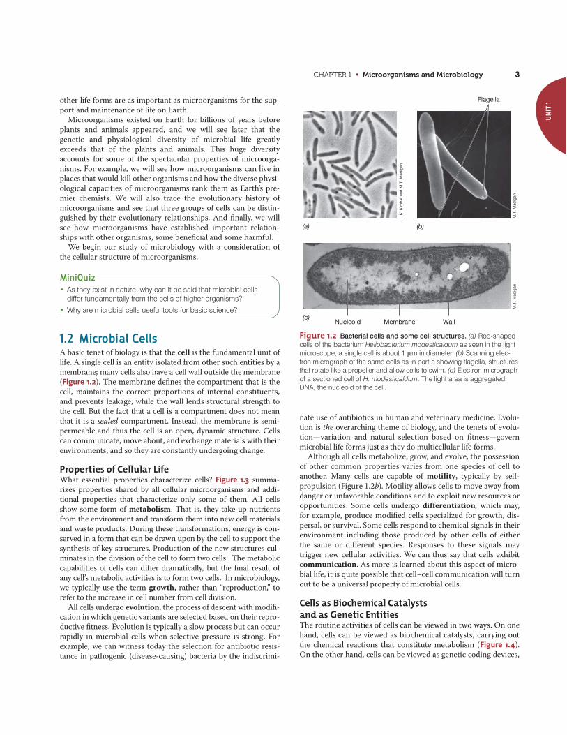
CHAPTER 1 • Microorganisms and Microbiology 3
other life forms are as important as microorganisms for the sup-port and maintenance of life on Earth.
Microorganisms existed on Earth for billions of years beforeplants and animals appeared, and we will see later that thegenetic and physiological diversity of microbial life greatlyexceeds that of the plants and animals. This huge diversityaccounts for some of the spectacular properties of microorga-nisms. For example, we will see how microorganisms can live inplaces that would kill other organisms and how the diverse physi-ological capacities of microorganisms rank them as Earth’s pre-mier chemists. We will also trace the evolutionary history ofmicroorganisms and see that three groups of cells can be distin-guished by their evolutionary relationships. And finally, we willsee how microorganisms have established important relation-ships with other organisms, some beneficial and some harmful.
We begin our study of microbiology with a consideration ofthe cellular structure of microorganisms.
MiniQuiz• As they exist in nature, why can it be said that microbial cells
differ fundamentally from the cells of higher organisms?
• Why are microbial cells useful tools for basic science?
1.2 Microbial CellsA basic tenet of biology is that the cell is the fundamental unit oflife. A single cell is an entity isolated from other such entities by amembrane; many cells also have a cell wall outside the membrane(Figure 1.2). The membrane defines the compartment that is thecell, maintains the correct proportions of internal constituents,and prevents leakage, while the wall lends structural strength tothe cell. But the fact that a cell is a compartment does not meanthat it is a sealed compartment. Instead, the membrane is semi-permeable and thus the cell is an open, dynamic structure. Cellscan communicate, move about, and exchange materials with theirenvironments, and so they are constantly undergoing change.
Properties of Cellular LifeWhat essential properties characterize cells? Figure 1.3 summa-rizes properties shared by all cellular microorganisms and addi-tional properties that characterize only some of them. All cellsshow some form of metabolism. That is, they take up nutrientsfrom the environment and transform them into new cell materialsand waste products. During these transformations, energy is con-served in a form that can be drawn upon by the cell to support thesynthesis of key structures. Production of the new structures cul-minates in the division of the cell to form two cells. The metaboliccapabilities of cells can differ dramatically, but the final result ofany cell’s metabolic activities is to form two cells. In microbiology,we typically use the term growth, rather than “reproduction,” torefer to the increase in cell number from cell division.
All cells undergo evolution, the process of descent with modifi-cation in which genetic variants are selected based on their repro-ductive fitness. Evolution is typically a slow process but can occurrapidly in microbial cells when selective pressure is strong. Forexample, we can witness today the selection for antibiotic resis-tance in pathogenic (disease-causing) bacteria by the indiscrimi-
nate use of antibiotics in human and veterinary medicine. Evolu-tion is the overarching theme of biology, and the tenets of evolu-tion—variation and natural selection based on fitness—governmicrobial life forms just as they do multicellular life forms.
Although all cells metabolize, grow, and evolve, the possessionof other common properties varies from one species of cell toanother. Many cells are capable of motility, typically by self-propulsion (Figure 1.2b). Motility allows cells to move away fromdanger or unfavorable conditions and to exploit new resources oropportunities. Some cells undergo differentiation, which may,for example, produce modified cells specialized for growth, dis-persal, or survival. Some cells respond to chemical signals in theirenvironment including those produced by other cells of eitherthe same or different species. Responses to these signals maytrigger new cellular activities. We can thus say that cells exhibitcommunication. As more is learned about this aspect of micro-bial life, it is quite possible that cell–cell communication will turnout to be a universal property of microbial cells.
Cells as Biochemical Catalysts and as Genetic EntitiesThe routine activities of cells can be viewed in two ways. On onehand, cells can be viewed as biochemical catalysts, carrying outthe chemical reactions that constitute metabolism (Figure 1.4).On the other hand, cells can be viewed as genetic coding devices,
UN
IT 1
Flagella
Nucleoid Membrane Wall
L.K
. Kim
ble
and
M.T
. Mad
igan
M.T
. Mad
igan
M.T
. Mad
igan
(a) (b)
(c)
Figure 1.2 Bacterial cells and some cell structures. (a) Rod-shapedcells of the bacterium Heliobacterium modesticaldum as seen in the lightmicroscope; a single cell is about 1 �m in diameter. (b) Scanning elec-tron micrograph of the same cells as in part a showing flagella, structuresthat rotate like a propeller and allow cells to swim. (c) Electron micrographof a sectioned cell of H. modesticaldum. The light area is aggregatedDNA, the nucleoid of the cell.
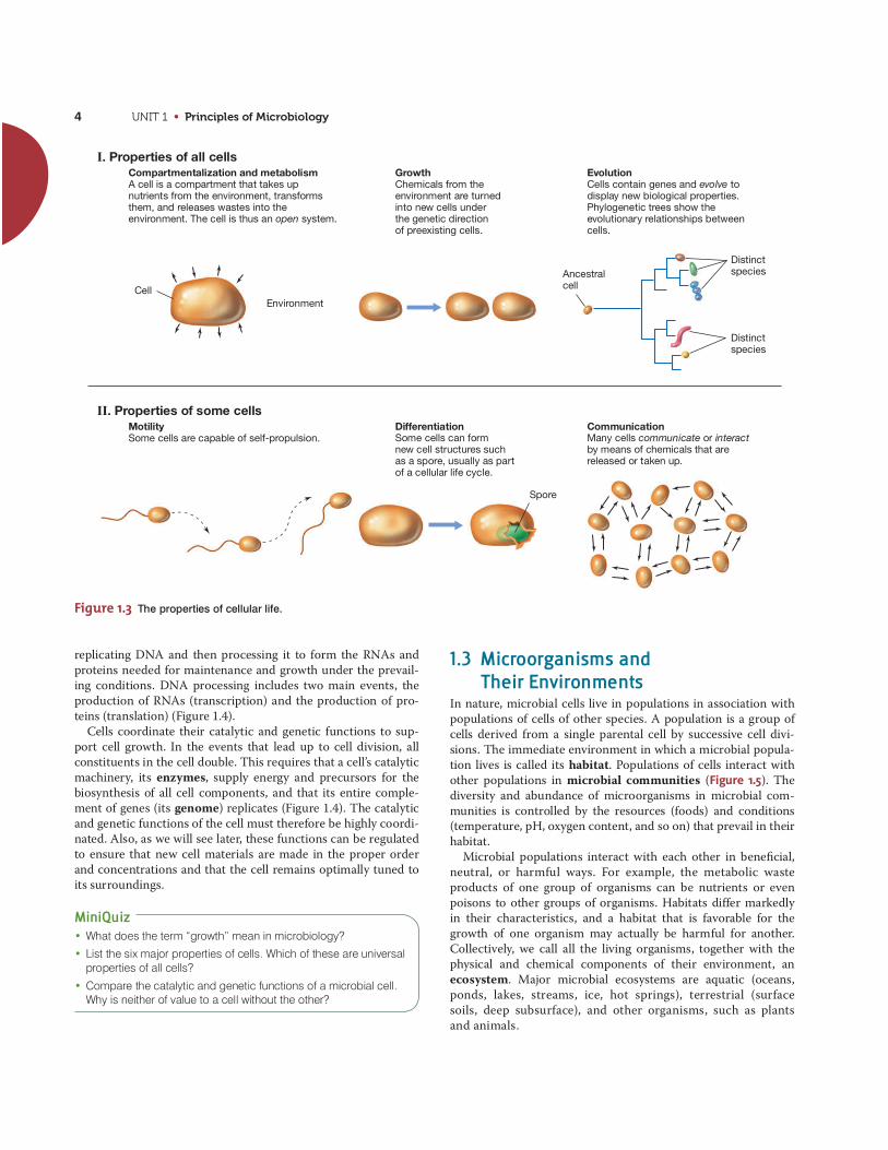
UNIT 1 • Principles of Microbiology4
replicating DNA and then processing it to form the RNAs andproteins needed for maintenance and growth under the prevail-ing conditions. DNA processing includes two main events, theproduction of RNAs (transcription) and the production of pro-teins (translation) (Figure 1.4).
Cells coordinate their catalytic and genetic functions to sup-port cell growth. In the events that lead up to cell division, allconstituents in the cell double. This requires that a cell’s catalyticmachinery, its enzymes, supply energy and precursors for thebiosynthesis of all cell components, and that its entire comple-ment of genes (its genome) replicates (Figure 1.4). The catalyticand genetic functions of the cell must therefore be highly coordi-nated. Also, as we will see later, these functions can be regulatedto ensure that new cell materials are made in the proper orderand concentrations and that the cell remains optimally tuned toits surroundings.
MiniQuiz• What does the term “growth” mean in microbiology?
• List the six major properties of cells. Which of these are universalproperties of all cells?
• Compare the catalytic and genetic functions of a microbial cell.Why is neither of value to a cell without the other?
Compartmentalization and metabolismA cell is a compartment that takes upnutrients from the environment, transformsthem, and releases wastes into theenvironment. The cell is thus an open system.
GrowthChemicals from the environment are turned into new cells under the genetic direction of preexisting cells.
DifferentiationSome cells can form new cell structures such as a spore, usually as part of a cellular life cycle.
CommunicationMany cells communicate or interact by means of chemicals that arereleased or taken up.
Distinctspecies
Distinctspecies
EvolutionCells contain genes and evolve to display new biological properties. Phylogenetic trees show the evolutionary relationships between cells.
CellEnvironment
Spore
MotilitySome cells are capable of self-propulsion.
Ancestralcell
I. Properties of all cells
II. Properties of some cells
Figure 1.3 The properties of cellular life.
1.3 Microorganisms and Their Environments
In nature, microbial cells live in populations in association withpopulations of cells of other species. A population is a group ofcells derived from a single parental cell by successive cell divi-sions. The immediate environment in which a microbial popula-tion lives is called its habitat. Populations of cells interact withother populations in microbial communities (Figure 1.5). Thediversity and abundance of microorganisms in microbial com-munities is controlled by the resources (foods) and conditions(temperature, pH, oxygen content, and so on) that prevail in theirhabitat.
Microbial populations interact with each other in beneficial,neutral, or harmful ways. For example, the metabolic wasteproducts of one group of organisms can be nutrients or evenpoisons to other groups of organisms. Habitats differ markedly in their characteristics, and a habitat that is favorable for thegrowth of one organism may actually be harmful for another.Collectively, we call all the living organisms, together with thephysical and chemical components of their environment, anecosystem. Major microbial ecosystems are aquatic (oceans,ponds, lakes, streams, ice, hot springs), terrestrial (surfacesoils, deep subsurface), and other organisms, such as plantsand animals.
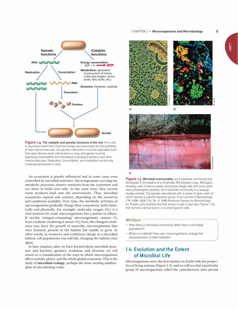
CHAPTER 1 • Microorganisms and Microbiology 5
MiniQuiz• How does a microbial community differ from a microbial
population?
• What is a habitat? How can microorganisms change thecharacteristics of their habitats?
1.4 Evolution and the Extent of Microbial Life
Microorganisms were the first entities on Earth with the proper-ties of living systems (Figure 1.3), and we will see that a particulargroup of microorganisms called the cyanobacteria were pivotal
UN
IT 1
Growth
Catalyticfunctions
Geneticfunctions
Replication Transcription
Translation
RNA
Proteins
DNA Energy conservation: ADP + Pi ATPMetabolism: generation of precursors of macro- molecules (sugars, amino acids, fatty acids, etc.)
Enzymes: metabolic catalysts
Figure 1.4 The catalytic and genetic functions of the cell. For a cellto reproduce itself there must be energy and precursors for the synthesisof new macromolecules, the genetic instructions must be replicated suchthat upon division each cell receives a copy, and genes must beexpressed (transcribed and translated) to produce proteins and othermacromolecules. Replication, transcription, and translation are the keymolecular processes in cells.
(a) (b)
D. E
. Cal
dw
ell
Jiri
Sna
idr
Ric
ard
o G
uer
rero
(c)
Figure 1.5 Microbial communities. (a) A bacterial community thatdeveloped in the depths of a small lake (Wintergreen Lake, Michigan),showing cells of various green and purple (large cells with sulfur gran-ules) phototrophic bacteria. (b) A bacterial community in a sewagesludge sample. The sample was stained with a series of dyes, each ofwhich stained a specific bacterial group. From Journal of Bacteriology178: 3496–3500, Fig. 2b. © 1996 American Society for Microbiology. (c) Purple sulfur bacteria like that shown in part a (see also Figure 1.7a)that formed a dense bloom in a small Spanish lake.
An ecosystem is greatly influenced and in some cases evencontrolled by microbial activities. Microorganisms carrying outmetabolic processes remove nutrients from the ecosystem anduse them to build new cells. At the same time, they excretewaste products back into the environment. Thus, microbialecosystems expand and contract, depending on the resourcesand conditions available. Over time, the metabolic activities ofmicroorganisms gradually change their ecosystems, both chem-ically and physically. For example, molecular oxygen (O2) is avital nutrient for some microorganisms but a poison to others.If aerobic (oxygen-consuming) microorganisms remove O2from a habitat, rendering it anoxic (O2 free), the changed condi-tions may favor the growth of anaerobic microorganisms thatwere formerly present in the habitat but unable to grow. Inother words, as resources and conditions change in a microbialhabitat, cell populations rise and fall, changing the habitat onceagain.
In later chapters, after we have learned about microbial struc-ture and function, genetics, evolution, and diversity, we willreturn to a consideration of the ways in which microorganismsaffect animals, plants, and the whole global ecosystem. This is thestudy of microbial ecology, perhaps the most exciting subdisci-pline of microbiology today.

UNIT 1 • Principles of Microbiology6
in biological evolution because oxygen (O2)—a waste product oftheir metabolism—prepared planet Earth for more complex lifeforms.
The First Cells and the Onsetof Biological EvolutionHow did cells originate? Were cells as we know them today thefirst self-replicating structures on Earth? Because all cells areconstructed in similar ways, it is thought that all cells havedescended from a common ancestral cell, the last universal com-mon ancestor (LUCA). After the first cells arose from nonliving
materials, a process that occurred over hundreds of millions ofyears, their subsequent growth formed cell populations, andthese then began to interact with other populations in microbialcommunities. Evolution selected for improvements and diversifi-cation of these early cells to eventually yield the highly complexand diverse cells we see today. We will consider this complexityand diversity in Chapters 2 and 17–21. We consider the topic ofhow life originated from nonliving materials in Chapter 16.
Life on Earth through the AgesEarth is 4.6 billion years old. Scientists have evidence that cellsfirst appeared on Earth between 3.8 and 3.9 billion years ago,and these organisms were exclusively microbial. In fact,microorganisms were the only life on Earth for most of its his-tory (Figure 1.6). Gradually, and over enormous periods of time,more complex organisms appeared. What were some of thehighlights along the way?
During the first 2 billion years or so of Earth’s existence, itsatmosphere was anoxic; O2 was absent, and nitrogen (N2), carbondioxide (CO2), and a few other gases were present. Only microor-ganisms capable of anaerobic metabolisms could survive underthese conditions, but these included many different types of cells,including those that produce methane, called methanogens. Theevolution of phototrophic microorganisms—organisms that har-vest energy from sunlight—occurred within a billion years of theformation of Earth. The first phototrophs were relatively simpleones, such as purple bacteria and other anoxygenic (non-oxygen-evolving) phototrophs (Figure 1.7a; see also Figure 1.5), which arestill widespread in anoxic habitats today. Cyanobacteria (oxygenic,or oxygen-evolving, phototrophs) (Figure 1.7b) evolved fromanoxygenic phototrophs nearly a billion years later and began theslow process of oxygenating the atmosphere. Triggered by in-creases in O2 in the atmosphere, multicellular life forms eventu-ally evolved and continued to increase in complexity, culminatingin the plants and animals we know today (Figure 1.6). We will
Humans
Bacteria
Archaea
Eukarya
Shellyinvertebrates
Vascular plants
Mammals
Origin of Earth(4.6 bya)
Origin of cellular life
Anoxygenicphototrophicbacteria
Origin ofcyanobacteria
AnoxicEarth
O2O2
~20% O2
Earth is slowlyoxygenated
LUCA
Moderneukaryotes
Algaldiversity
4bya
bya2 bya
1bya
3
Microbial life forms only
Present
(a)
(b)
4 3 2 1 0
bya
Figure 1.6 A summary of life on Earth through time and origin ofthe cellular domains. (a) Cellular life was present on Earth about 3.8 bil-lion years ago (bya). Cyanobacteria began the slow oxygenation of Earthabout 3 bya, but current levels of O2 in the atmosphere were not achieveduntil 500–800 million years ago. Eukaryotes are nucleated cells andinclude both microbial and multicellular organisms. (Shelly invertebrateshave shells or shell-like parts.) (b) The three domains of cellular organ-isms are Bacteria, Archaea, and Eukarya. The latter two lineages divergedlong before nucleated cells with organelles (labeled as “modern eukary-otes” in part a) appear in the fossil record. LUCA, last universal commonancestor. Note that 80% of Earth’s history was exclusively microbial.
(b)
Thom
as D
. Bro
ck
No
rber
t Pfe
nnig
(a)
Figure 1.7 Phototrophic microorganisms. (a) Purple sulfur bacteria(anoxygenic phototrophs). (b) Cyanobacteria (oxygenic phototrophs).Purple bacteria appeared on Earth long before oxygenic phototrophsevolved (see Figure 1.6a).

CHAPTER 1 • Microorganisms and Microbiology 7
explore the evolutionary history of life later, but note here that theevents that unfolded beyond LUCA led to the evolution of threemajor lineages of microbial cells, the Bacteria, the Archaea, andthe Eukarya (Figure 1.6b); microbial Eukarya were the ancestorsof the plants and animals.
How do we know that evolutionary events unfolded as sum-marized in Figure 1.6? The answer is that we may never knowthat all details in our description are correct. However, scientistscan reconstruct evolutionary transitions by using biomarkers,specific molecules that are unique to particular groups in pres-ent-day microorganisms. The presence or absence of a given bio-marker in ancient rocks of a known age therefore reveals whetherthat particular group was present at that time.
One way or the other and over enormous periods of time(Figure 1.6), natural selection filled every suitable habitat onEarth with one or more populations of microorganisms. Thisbrings us to the question of the current distribution of microbiallife on Earth. What do we know about this important topic?
The Extent of Microbial LifeMicrobial life is all around us. Examination of natural materialssuch as soil or water invariably reveals microbial cells. Butunusual habitats such as boiling hot springs and glacial ice arealso teeming with microorganisms. Although widespread onEarth, such tiny cells may seem inconsequential. But if we couldcount them all, what number would we reach?
Estimates of total microbial cell numbers on Earth are on theorder of cells. The total amount of carbon present inthis very large number of very small cells equals that of all plantson Earth (and plant carbon far exceeds animal carbon). But inaddition, the collective contents of nitrogen and phosphorus inmicrobial cells is more than 10 times that in all plant biomass.
Thus, microbial cells, small as they are, constitute the majorfraction of biomass on Earth and are key reservoirs of essentialnutrients for life. Most microbial cells are found in just a few verylarge habitats. For example, most microbial cells do not reside onEarth’s surface but instead lie underground in the oceanic andterrestrial subsurface (Table 1.1). Depths up to about 10 km underEarth’s surface are clearly suitable for microbial life. We will seelater that subsurface microbial habitats support diverse popula-tions of microbial cells that make their livings in unusual waysand grow extremely slowly. By comparison to the subsurface, sur-face soils and waters contain a relatively small percentage of thetotal microbial cell numbers, and animals (including humans),which can be heavily colonized with microorganisms (see Figure1.10), collectively contain only a tiny fraction of the total micro-bial cells on Earth (Table 1.1).
Because most of what we know about microbial life has comefrom the study of surface-dwelling organisms, there is obviouslymuch left for future generations of microbiologists to discoverand understand about the life forms that dominate Earth’s biol-ogy. And when we consider the fact that surface-dwelling orga-nisms already show enormous diversity, the hunt for newmicroorganisms in Earth’s unexplored habitats should yield someexciting surprises.
2.5 * 1030
MiniQuiz• What is LUCA and what major lineages of cells evolved from
LUCA? Why were cyanobacteria so important in the evolution oflife on Earth?
• How old is Earth, and when did cellular life forms first appear?How can we use science to reconstruct the sequence oforganisms that appeared on Earth?
• Where are most microbial cells located on Earth?
1.5 The Impact of Microorganisms on Humans
Through the years microbiologists have had great success in dis-covering how microorganisms work, and application of thisknowledge has greatly increased the beneficial effects ofmicroorganisms and curtailed many of their harmful effects.Microbiology has thus greatly advanced human health and wel-fare. Besides understanding microorganisms as agents of disease,microbiology has made great advances in understanding the roleof microorganisms in food and agriculture, and in exploitingmicrobial activities for producing valuable human products, gen-erating energy, and cleaning up the environment.
Microorganisms as Agents of DiseaseThe statistics summarized in Figure 1.8 show microbiologists’success in preventing infectious diseases since the beginning ofthe twentieth century. These data compare today’s leading causesof death in the United States with those of 100 years ago. At thebeginning of the twentieth century, the major causes of death inhumans were infectious diseases caused by microorganismscalled pathogens. Children and the aged in particular suc-cumbed in large numbers to microbial diseases. Today, however,infectious diseases are much less deadly, at least in developedcountries. Control of infectious disease has come from anincreased understanding of disease processes, improved sanitaryand public health practices, and the use of antimicrobial agents,such as antibiotics. As we will see from the next sections, thedevelopment of microbiology as a science can trace importantaspects of its roots to studies of infectious disease.
UN
IT 1Table 1.1 Distribution of microorganisms in and on Eartha
Habitat Percent of total
Marine subsurface 66
Terrestrial subsurface 26
Surface soil 4.8
Oceans 2.2
All other habitatsb 1.0
aData compiled by William Whitman, University of Georgia, USA; refer to total numbers(estimated to be about 2.5 1030 cells) of Bacteria and Archaea. This enormousnumber of cells contain, collectively, about grams of carbon.bIncludes, in order of decreasing numbers: freshwater and salt lakes, domesticated animals, sea ice, termites, humans, and domesticated birds.
5 * 1017*
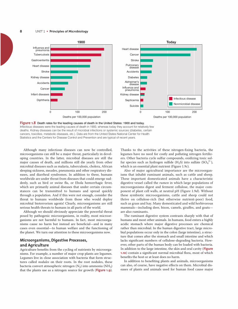
UNIT 1 • Principles of Microbiology8
Although many infectious diseases can now be controlled,microorganisms can still be a major threat, particularly in devel-oping countries. In the latter, microbial diseases are still themajor causes of death, and millions still die yearly from othermicrobial diseases such as malaria, tuberculosis, cholera, Africansleeping sickness, measles, pneumonia and other respiratory dis-eases, and diarrheal syndromes. In addition to these, humansworldwide are under threat from diseases that could emerge sud-denly, such as bird or swine flu, or Ebola hemorrhagic fever,which are primarily animal diseases that under certain circum-stances can be transmitted to humans and spread quicklythrough a population. And if this were not enough, consider thethreat to humans worldwide from those who would deploymicrobial bioterrorism agents! Clearly, microorganisms are stillserious health threats to humans in all parts of the world.
Although we should obviously appreciate the powerful threatposed by pathogenic microorganisms, in reality, most microor-ganisms are not harmful to humans. In fact, most microorga-nisms cause no harm but instead are beneficial—and in manycases even essential—to human welfare and the functioning ofthe planet. We turn our attention to these microorganisms now.
Microorganisms, Digestive Processes,and AgricultureAgriculture benefits from the cycling of nutrients by microorga-nisms. For example, a number of major crop plants are legumes.Legumes live in close association with bacteria that form struc-tures called nodules on their roots. In the root nodules, thesebacteria convert atmospheric nitrogen (N2) into ammonia (NH3)that the plants use as a nitrogen source for growth (Figure 1.9).
Thanks to the activities of these nitrogen-fixing bacteria, thelegumes have no need for costly and polluting nitrogen fertiliz-ers. Other bacteria cycle sulfur compounds, oxidizing toxic sul-fur species such as hydrogen sulfide (H2S) into sulfate (SO4
2-),which is an essential plant nutrient (Figure 1.9c).
Also of major agricultural importance are the microorgan-isms that inhabit ruminant animals, such as cattle and sheep.These important domesticated animals have a characteristicdigestive vessel called the rumen in which large populations ofmicroorganisms digest and ferment cellulose, the major com-ponent of plant cell walls, at neutral pH (Figure 1.9d). Withoutthese symbiotic microorganisms, cattle and sheep could notthrive on cellulose-rich (but otherwise nutrient-poor) food,such as grass and hay. Many domesticated and wild herbivorousmammals—including deer, bison, camels, giraffes, and goats—are also ruminants.
The ruminant digestive system contrasts sharply with that ofhumans and most other animals. In humans, food enters a highlyacidic stomach where major digestive processes are chemicalrather than microbial. In the human digestive tract, large micro-bial populations occur only in the colon (large intestine), a struc-ture that comes after the stomach and small intestine and whichlacks significant numbers of cellulose-degrading bacteria. How-ever, other parts of the human body can be loaded with bacteria.In addition to the large intestine, the skin and oral cavity (Figure1.10) contain a significant normal microbial flora, most of whichbenefits the host or at least does no harm.
In addition to benefiting plants and animals, microorganismscan also, of course, have negative effects on them. Microbial dis-eases of plants and animals used for human food cause major
Figure 1.8 Death rates for the leading causes of death in the United States: 1900 and today.Infectious diseases were the leading causes of death in 1900, whereas today they account for relatively fewdeaths. Kidney diseases can be the result of microbial infections or systemic sources (diabetes, certaincancers, toxicities, metabolic diseases, etc.). Data are from the United States National Center for HealthStatistics and the Centers for Disease Control and Prevention and are typical of recent years.
Deaths per 100,000 populationDeaths per 100,000 population
Kidney disease
Septicemia
Heart disease
Cancer
Stroke
Pulmonarydisease
Accidents
Diabetes
Alzheimer’sdisease
Influenza andpneumonia
Suicide
1900 Today
Influenza andpneumonia
Tuberculosis
Gastroenteritis
Heart disease
Stroke
Kidney disease
Accidents
Cancer
Infant diseases
Diphtheria
0 100 200 0 100 200
Infectious disease
Nonmicrobial disease

CHAPTER 1 • Microorganisms and Microbiology 9
economic losses in the agricultural industry every year. In somecases a food product can cause serious human disease, such aswhen pathogenic Escherichia coli or Salmonella is transmittedfrom infected meat, or when microbial pathogens are ingestedwith contaminated fresh fruits and vegetables. Thus microorga-nisms significantly impact the agriculture industry both positivelyand negatively.
Microorganisms and Food, Energy,and the EnvironmentMicroorganisms play important roles in the food industry,including in the areas of spoilage, safety, and production. Afterplants and animals are produced for human consumption, theproducts must be delivered to consumers in a wholesome form.Food spoilage alone results in huge economic losses each year.Indeed, the canning, frozen food, and dried-food industries werefounded as means to preserve foods that would otherwise easilyundergo microbial spoilage. Food safety requires constant moni-toring of food products to ensure they are free of pathogenicmicroorganisms and to track disease outbreaks to identify thesource(s).
However, not all microorganisms in foods have harmful effectson food products or those who eat them. For example, manydairy products depend on the activities of microorganisms,including the fermentations that yield cheeses, yogurt, and but-termilk. Sauerkraut, pickles, and some sausages are also productsof microbial fermentations. Moreover, baked goods and alcoholic
beverages rely on the fermentative activities of yeast, which gen-erate carbon dioxide (CO2) to raise the dough and alcohol as akey ingredient, respectively. Many of these fermentations are dis-cussed in Chapter 14.
UN
IT 1
(a) (c)
(b)
(d)
N2 + 8H 2NH3 + H2
N2
NH3
Rumen
Grass Cellulose Glucose Microbial fermentation
CO2 + CH4(Waste products)
Fatty acids(Nutrition for animal)
NO3–
H2S
S0SO4
2–
N-cycle S-cycle
Soybeanplant
Joe
Bur
ton
Figure 1.9 Microorganisms in modern agriculture. (a, b) Root nodules on this soybean plant containbacteria that fix molecular nitrogen (N2) for use by the plant. (c) The nitrogen and sulfur cycles, key nutrientcycles in nature. (d) Ruminant animals. Microorganisms in the rumen of the cow convert cellulose fromgrass into fatty acids that can be used by the animal.
Figure 1.10 Human oral bacterial community. The oral cavity ofwarm-blooded animals contains high numbers of various bacteria, asshown in this electron micrograph (false color) of cells scraped from ahuman tongue.

UNIT 1 • Principles of Microbiology10
Some microorganisms produce biofuels. Natural gas (methane)is a product of the anaerobic degradation of organic matter by methanogenic microorganisms (Figure 1.11). Ethyl alcohol(ethanol), which is produced by the microbial fermentation ofglucose from feedstocks such as sugarcane or cornstarch, is amajor motor fuel in some countries (Figure 1.11b). Waste materi-als such as domestic refuse, animal wastes, and cellulose can alsobe converted to biofuels by microbial activities and are more effi-cient feedstocks for ethanol production than is corn. Soybeansare also used as biofuel feedstocks, as soybean oils can be con-verted into biodiesel to fuel diesel engines. As global oil produc-tion is waning, it is likely that various biofuels will take on agreater and greater part of the global energy picture.
Microorganisms are used to clean up human pollution, aprocess called microbial bioremediation, and to produce com-mercially valuable products by industrial microbiology andbiotechnology. For example, microorganisms can be used to con-sume spilled oil, solvents, pesticides, and other environmentallytoxic pollutants. Bioremediation accelerates cleanup in either oftwo ways: (1) by introducing specific microorganisms to a pol-luted environment, or (2) by adding nutrients that stimulate pre-existing microorganisms to degrade the pollutants. In both casesthe goal is to accelerate metabolism of the pollutant.
In industrial microbiology, microorganisms are grown on alarge scale to make products of relatively low commercial value,such as antibiotics, enzymes, and various chemicals. By contrast,the related field of biotechnology employs genetically engineeredmicroorganisms to synthesize products of high commercialvalue, such as human proteins. Genomics is the science of theidentification and analysis of genomes and has greatly enhanced
biotechnology. Using genomic methods, biotechnologists canaccess the genome of virtually any organism and search in it forgenes encoding proteins of commercial interest.
At this point the influence of microorganisms on humansshould be apparent. Microorganisms are essential for life andtheir activities can cause significant benefit or harm to humans.As the eminent French scientist Louis Pasteur, one of thefounders of microbiology, expressed it: “The role of the infi-nitely small in nature is infinitely large.” We continue our intro-duction to the microbial world in the next section with anhistorical overview of the contributions of Pasteur and a fewother key scientists.
MiniQuiz• List two ways in which microorganisms are important in the food
and agricultural industries.
• Which biofuel is widely used in many countries as a motor fuel?
• What is biotechnology and how might it improve the lives ofhumans?
II Pathways of Discovery in Microbiology
The future of any science is rooted in its past accomplishments.Although microbiology claims very early roots, the science
did not really develop in a systematic way until the nineteenthcentury. Since that time, microbiology has expanded in a way
John
A. B
rezn
ak
(a) (b)
Figure 1.11 Biofuels. (a) Natural gas (methane) is collected in a funnel from swamp sediments where itwas produced by methanogens and then ignited as a demonstration experiment. (b) An ethanol plant in theUnited States. Sugars obtained from corn or other crops are fermented to ethanol for use as a motor fuelextender.

CHAPTER 1 • Microorganisms and Microbiology 11
unprecedented by any of the other biological sciences and hasspawned several new but related fields. We retrace these path-ways of discovery now and discuss a few of the major contribu-tors (Table 1.2).
1.6 The Historical Roots of Microbiology:Hooke, van Leeuwenhoek, and Cohn
Although the existence of creatures too small to be seen with thenaked eye had long been suspected, their discovery was linked tothe invention of the microscope. Robert Hooke (1635–1703), anEnglish mathematician and natural historian, was also an excel-lent microscopist. In his famous book Micrographia (1665), thefirst book devoted to microscopic observations, Hooke illus-trated, among many other things, the fruiting structures of molds(Figure 1.12). This was the first known description of microor-ganisms. The first person to see bacteria was the Dutch draperand amateur microscope builder Antoni van Leeuwenhoek(1632–1723). In 1684, van Leeuwenhoek, who was well aware ofthe work of Hooke, used extremely simple microscopes of hisown construction (Figure 1.13) to examine the microbial contentof natural substances.
Van Leeuwenhoek’s microscopes were crude by today’s stan-dards, but by careful manipulation and focusing he was able tosee bacteria, microorganisms considerably smaller than molds(molds are fungi). He discovered bacteria in 1676 while studyingpepper–water infusions. He reported his observations in a seriesof letters to the prestigious Royal Society of London, which pub-lished them in 1684 in English translation. Drawings of some ofvan Leeuwenhoek’s “wee animalcules,” as he referred to them, areshown in Figure 1.13b, and a photo taken through such a micro-scope is shown in Figure 1.13c.
As years went by, van Leeuwenhoek’s observations were con-firmed by many others. However, primarily because of the lack ofexperimental tools, little progress in understanding the natureand importance of the tiny creatures was made for almost 150
years. Only in the nineteenth century did improved microscopesand some simple tools for growing microoorganisms in the labo-ratory become available, and using these, the extent and nature ofmicrobial life became more apparent.
In the mid- to late nineteenth century major advances in thescience of microbiology were made because of the attention givento two major questions that pervaded biology and medicine at thetime: (1) Does spontaneous generation occur? and (2) What is thenature of infectious disease? Answers to these seminal questionsemerged from the work of two giants in the fledgling field ofmicrobiology: the French chemist Louis Pasteur and the Germanphysician Robert Koch. But before we explore their work, let usbriefly consider the groundbreaking efforts of a German botanist,Ferdinand Cohn, a contemporary of Pasteur and Koch, and thefounder of the field we now call bacteriology.
Ferdinand Cohn (1828–1898) was born in Breslau (now inPoland). He was trained as a botanist and became an excellentmicroscopist. His interests in microscopy led him to the study ofunicellular algae and later to bacteria, including the large sulfurbacterium Beggiatoa (Figure 1.14). Cohn was particularly inter-ested in heat resistance in bacteria, which led to his discoverythat some bacteria form endospores. We now know that bacterialendospores are formed by differentiation from the mother (vege-tative) cell (Figure 1.3) and that endospores are extremely heat-resistant. Cohn described the life cycle of the endospore-formingbacterium Bacillus (vegetative cell endospore vegetativecell) and showed that vegetative cells but not endospores werekilled by boiling.
Cohn is credited with many other accomplishments. He laidthe groundwork for a system of bacterial classification, includ-ing an early attempt to define a bacterial species, an issue stillunresolved today, and founded a major scientific journal ofplant and microbial biology. He strongly advocated use of thetechniques and research of Robert Koch, the first medicalmicrobiologist. Cohn devised simple but effective methods forpreventing the contamination of culture media, such as the use
SSU
NIT
1Table 1.2 Giants of the early days of microbiology and their major contributions
Investigator Nationality Datesa Contributions
Robert Hooke English 1664 Discovery of microorganisms (fungi)
Antoni van Leeuwenhoek Dutch 1684 Discovery of bacteria
Edward Jenner English 1798 Vaccination (smallpox)
Louis Pasteur French Mid- to late 1800s Mechanism of fermentation, defeat of spontaneous generation, rabies and other vaccines, principles of immunization
Joseph Lister English 1867 Methods for preventing infections during surgeries
Ferdinand Cohn German 1876 Discovery of endospores
Robert Koch German Late 1800s Koch’s postulates, pure culture microbiology, discovery of agents of tuberculosis and cholera
Sergei Winogradsky Russian Late 1800s to mid-1900s Chemolithotrophy and chemoautotrophy, nitrogen fixation, sulfur bacteria
Martinus Beijerinck Dutch Late 1800s to 1920 Enrichment culture technique, discovery of many metabolic groups of bacteria, concept of a virus
aThe year in which the key paper describing the contribution was published, or the date range in which the investigator was mostscientifically active.
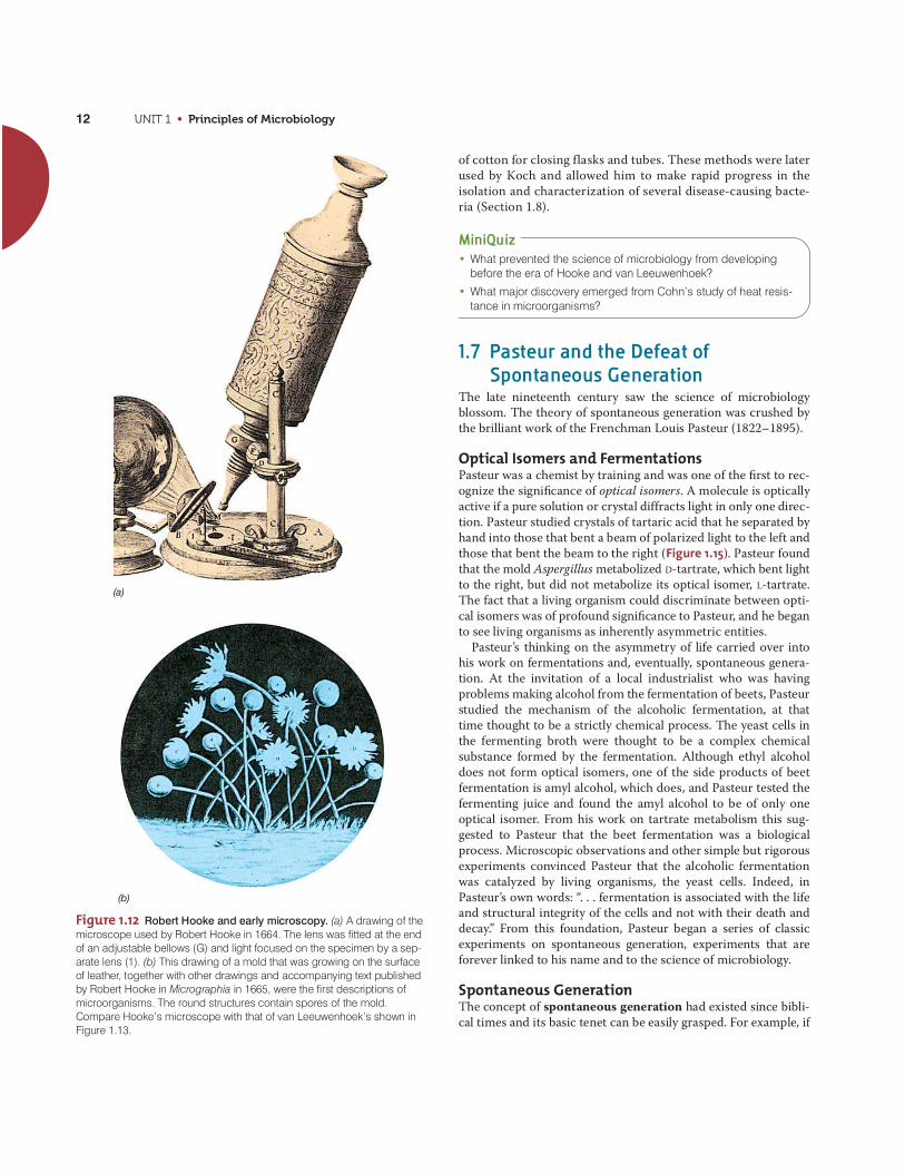
UNIT 1 • Principles of Microbiology12
of cotton for closing flasks and tubes. These methods were laterused by Koch and allowed him to make rapid progress in theisolation and characterization of several disease-causing bacte-ria (Section 1.8).
MiniQuiz• What prevented the science of microbiology from developing
before the era of Hooke and van Leeuwenhoek?
• What major discovery emerged from Cohn’s study of heat resis-tance in microorganisms?
1.7 Pasteur and the Defeat ofSpontaneous Generation
The late nineteenth century saw the science of microbiologyblossom. The theory of spontaneous generation was crushed bythe brilliant work of the Frenchman Louis Pasteur (1822–1895).
Optical Isomers and FermentationsPasteur was a chemist by training and was one of the first to rec-ognize the significance of optical isomers. A molecule is opticallyactive if a pure solution or crystal diffracts light in only one direc-tion. Pasteur studied crystals of tartaric acid that he separated byhand into those that bent a beam of polarized light to the left andthose that bent the beam to the right (Figure 1.15). Pasteur foundthat the mold Aspergillus metabolized D-tartrate, which bent lightto the right, but did not metabolize its optical isomer, L-tartrate.The fact that a living organism could discriminate between opti-cal isomers was of profound significance to Pasteur, and he beganto see living organisms as inherently asymmetric entities.
Pasteur’s thinking on the asymmetry of life carried over intohis work on fermentations and, eventually, spontaneous genera-tion. At the invitation of a local industrialist who was havingproblems making alcohol from the fermentation of beets, Pasteurstudied the mechanism of the alcoholic fermentation, at thattime thought to be a strictly chemical process. The yeast cells inthe fermenting broth were thought to be a complex chemicalsubstance formed by the fermentation. Although ethyl alcoholdoes not form optical isomers, one of the side products of beetfermentation is amyl alcohol, which does, and Pasteur tested thefermenting juice and found the amyl alcohol to be of only oneoptical isomer. From his work on tartrate metabolism this sug-gested to Pasteur that the beet fermentation was a biologicalprocess. Microscopic observations and other simple but rigorousexperiments convinced Pasteur that the alcoholic fermentationwas catalyzed by living organisms, the yeast cells. Indeed, inPasteur’s own words: “. . . fermentation is associated with the lifeand structural integrity of the cells and not with their death anddecay.” From this foundation, Pasteur began a series of classicexperiments on spontaneous generation, experiments that areforever linked to his name and to the science of microbiology.
Spontaneous GenerationThe concept of spontaneous generation had existed since bibli-cal times and its basic tenet can be easily grasped. For example, if
(b)
(a)
Figure 1.12 Robert Hooke and early microscopy. (a) A drawing of themicroscope used by Robert Hooke in 1664. The lens was fitted at the endof an adjustable bellows (G) and light focused on the specimen by a sep-arate lens (1). (b) This drawing of a mold that was growing on the surfaceof leather, together with other drawings and accompanying text publishedby Robert Hooke in Micrographia in 1665, were the first descriptions ofmicroorganisms. The round structures contain spores of the mold.Compare Hooke’s microscope with that of van Leeuwenhoek’s shown in Figure 1.13.

CHAPTER 1 • Microorganisms and Microbiology 13
T. D
. Bro
ck
Bria
n J.
Fo
rd
(a)
(b)
(c)
Lens
Figure 1.13 The van Leeuwenhoek microscope. (a) Areplica of Antoni van Leeuwenhoek’s microscope. (b) VanLeeuwenhoek’s drawings of bacteria, published in 1684.Even from these simple drawings we can recognize severalshapes of common bacteria: A, C, F, and G, rods; E, cocci;H, packets of cocci. (c) Photomicrograph of a human bloodsmear taken through a van Leeuwenhoek microscope. Redblood cells are clearly apparent.
Figure 1.14 Drawing by Ferdinand Cohn of large filamentoussulfur-oxidizing bacteria Beggiatoa mirabilis. The small granules insidethe cells consist of elemental sulfur, produced from the oxidation ofhydrogen sulfide (H2S). Cohn was the first to identify the granules assulfur in 1866. A cell of B. mirabilis is about 15 �m in diameter. Comparewith Figure 1.22b. Beggiatoa moves on solid surfaces by a gliding mech-anism and in so doing, cells often twist about one another.
food is allowed to stand for some time, it putrefies. When exam-ined microscopically, the putrefied food is seen to be teemingwith bacteria and perhaps even maggots and worms. From wheredo these organisms not apparent in the fresh food originate?Some people said they developed from seeds or germs thatentered the food from air. Others said they arose spontaneouslyfrom nonliving materials, that is, by spontaneous generation.Who was right? Keen insight was necessary to solve this contro-versy, and this was exactly the kind of problem that appealed toLouis Pasteur.
Pasteur became a powerful opponent of spontaneous genera-tion. Following his discoveries about fermentation, Pasteur pre-dicted that microorganisms observed in putrefying materials arealso present in air and that putrefaction resulted from the activi-ties of microorganisms that entered from the air or that had beenpresent on the surfaces of the containers holding the decayingmaterials. Pasteur further reasoned that if food were treated insuch a way as to destroy all living organisms contaminating it,that is, if it were rendered sterile and then protected fromfurther contamination, it should not putrefy.
UN
IT 1
(a) (b)
T T
P
L-form D-form
M M
Pb' b'
h
n n
h
Figure 1.15 Louis Pasteur’s drawings of tartaric acid crystals fromhis famous paper on optical activity. (a) Left-handed crystal (bends light to the left). (b) Right-handed crystal (bends light to the right). Note that the two crystals are mirror images of one another, a hallmark ofoptical isomers.

UNIT 1 • Principles of Microbiology14
Steam forced out open end
(a)
(c)
(b)
Short time
Long time
Dust and microorganismstrapped in bend Open end
Nonsterile liquidpoured into flask
Neck of flask drawn out in flame
Liquid sterilizedby extensive heating
Flask tipped somicroorganism-laden dust contacts sterile liquid
Liquid putrefies
Liquid cooled slowly Liquid remainssterile indefinitely
Figure 1.16 The defeat of spontaneous generation: Pasteur’s swan-necked flask experiment. In (c) the liquid putrefies because microorgan-isms enter with the dust.
M.T
. Mad
igan
(b)
(a)
Figure 1.17 Louis Pasteur and some symbols of his contributions tomicrobiology. (a) A French 5-franc note honoring Pasteur. The shepherdboy Jean Baptiste Jupille is shown killing a rabid dog that had attackedchildren. Pasteur’s rabies vaccine saved Jupille’s life. In France, the francpreceded the euro as a currency. (b) The Pasteur Institute, Paris, France.Today this structure, built for Pasteur by the French government, houses amuseum that displays some of the original swan-necked flasks used inhis experiments.
Pasteur used heat to eliminate contaminants. Killing all thebacteria or other microorganisms in or on objects is a process wenow call sterilization. Proponents of spontaneous generationcriticized such experiments by declaring that “fresh air” was nec-essary for the phenomenon to occur. In 1864 Pasteur counteredthis objection simply and brilliantly by constructing a swan-necked flask, now called a Pasteur flask (Figure 1.16). In such aflask nutrient solutions could be heated to boiling and sterilized.However, after the flask was cooled, air was allowed to reenter,but the bend in the neck prevented particulate matter (includingmicroorganisms) from entering the nutrient solution and causingputrefaction.
The teeming microorganisms observed after particulate matterwas allowed to enter at the end of this simple experiment (Figure1.16c) effectively settled the controversy, and microbiology wasable to bury the idea of spontaneous generation for good andmove ahead on firm footing. Incidentally, Pasteur’s work also ledto the development of effective sterilization procedures that wereeventually refined and carried over into both basic and applied
microbiological research. Food science also owes a debt toPasteur, as his principles are applied today in the preservation ofmilk and many other foods by heat treatment (pasteurization). www.microbiologyplace.com Online Tutorial 1.1: Pasteur’s Experiment
Other Accomplishments of Louis PasteurPasteur went on to many other triumphs in microbiology andmedicine. Some highlights include his development of vaccinesfor the diseases anthrax, fowl cholera, and rabies during a veryscientifically productive period from 1880 to 1890. Pasteur’swork on rabies was his most famous success, culminating in July1885 with the first administration of a rabies vaccine to a human,a young French boy named Joseph Meister who had been bittenby a rabid dog. In those days, a bite from a rabid animal wasinvariably fatal. News spread quickly of the success of Meister’svaccination, and of one administered shortly thereafter to ayoung shepherd boy, Jean Baptiste Jupille (Figure 1.17). Within a

CHAPTER 1 • Microorganisms and Microbiology 15
UN
IT 1
Figure 1.18 Robert Koch. The German physician and microbiologist iscredited with founding medical microbiology and formulating his famouspostulates.
year several thousand people bitten by rabid animals had traveledto Paris to be treated with Pasteur’s rabies vaccine.
Pasteur’s fame from his rabies research was legendary and ledthe French government to establish the Pasteur Institute in Parisin 1888 (Figure 1.17b). Originally established as a clinical centerfor the treatment of rabies and other contagious diseases, the Pas-teur Institute today is a major biomedical research center focusedon antiserum and vaccine research and production. The medicaland veterinary breakthroughs of Pasteur were not only highly sig-nificant in their own right but helped solidify the concept of thegerm theory of disease, whose principles were being developed atabout the same time by a second giant of this era, Robert Koch.
MiniQuiz• Define the term sterile. How did Pasteur’s experiments using
swan-necked flasks defeat the theory of spontaneous generation?
• Besides ending the controversy over spontaneous generation,what other accomplishments do we credit to Pasteur?
1.8 Koch, Infectious Disease, and PureCulture Microbiology
Proof that some microorganisms cause disease provided thegreatest impetus for the development of microbiology as an inde-pendent biological science. Even as early as the sixteenth centuryit was thought that something that induced disease could betransmitted from a diseased person to a healthy person. After thediscovery of microorganisms, it was widely believed that theywere responsible, but definitive proof was lacking. Improvementsin sanitation by Ignaz Semmelweis and Joseph Lister providedindirect evidence for the importance of microorganisms in caus-ing human diseases, but it was not until the work of a Germanphysician, Robert Koch (1843–1910) (Figure 1.18), that the con-cept of infectious disease was given experimental support.
The Germ Theory of Disease and Koch’s PostulatesIn his early work Koch studied anthrax, a disease of cattle and occa-sionally of humans. Anthrax is caused by an endospore-formingbacterium called Bacillus anthracis. By careful microscopy and byusing special stains, Koch established that the bacteria were alwayspresent in the blood of an animal that was succumbing to the dis-ease. However, Koch reasoned that the mere association of thebacterium with the disease was not proof of cause and effect. Hesensed an opportunity to study cause and effect experimentallyusing anthrax. The results of this study formed the standard bywhich infectious diseases have been studied ever since.
Koch used mice as experimental animals. Using appropriatecontrols, Koch demonstrated that when a small amount of bloodfrom a diseased mouse was injected into a healthy mouse, the lat-ter quickly developed anthrax. He took blood from this secondanimal, injected it into another, and again observed the character-istic disease symptoms. However, Koch carried this experiment acritically important step further. He discovered that the anthraxbacteria could be grown in nutrient fluids outside the host andthat even after many transfers in laboratory culture, the bacteriastill caused the disease when inoculated into a healthy animal.
On the basis of these experiments and others on the causativeagent of tuberculosis, Koch formulated a set of rigorous criteria,now known as Koch’s postulates, for definitively linking a spe-cific microorganism to a specific disease. Koch’s postulates statethe following:1. The disease-causing organism must always be present in ani-
mals suffering from the disease but not in healthy animals.2. The organism must be cultivated in a pure culture away from
the animal body.3. The isolated organism must cause the disease when inoculated
into healthy susceptible animals.4. The organism must be isolated from the newly infected ani-
mals and cultured again in the laboratory, after which it shouldbe seen to be the same as the original organism.Koch’s postulates, summarized in Figure 1.19, were a monu-
mental step forward in the study of infectious diseases. The pos-tulates not only offered a means for linking the cause and effect ofan infectious disease, but also stressed the importance oflaboratory culture of the putative infectious agent. With thesepostulates as a guide, Koch, his students, and those that followedthem discovered the causative agents of most of the important
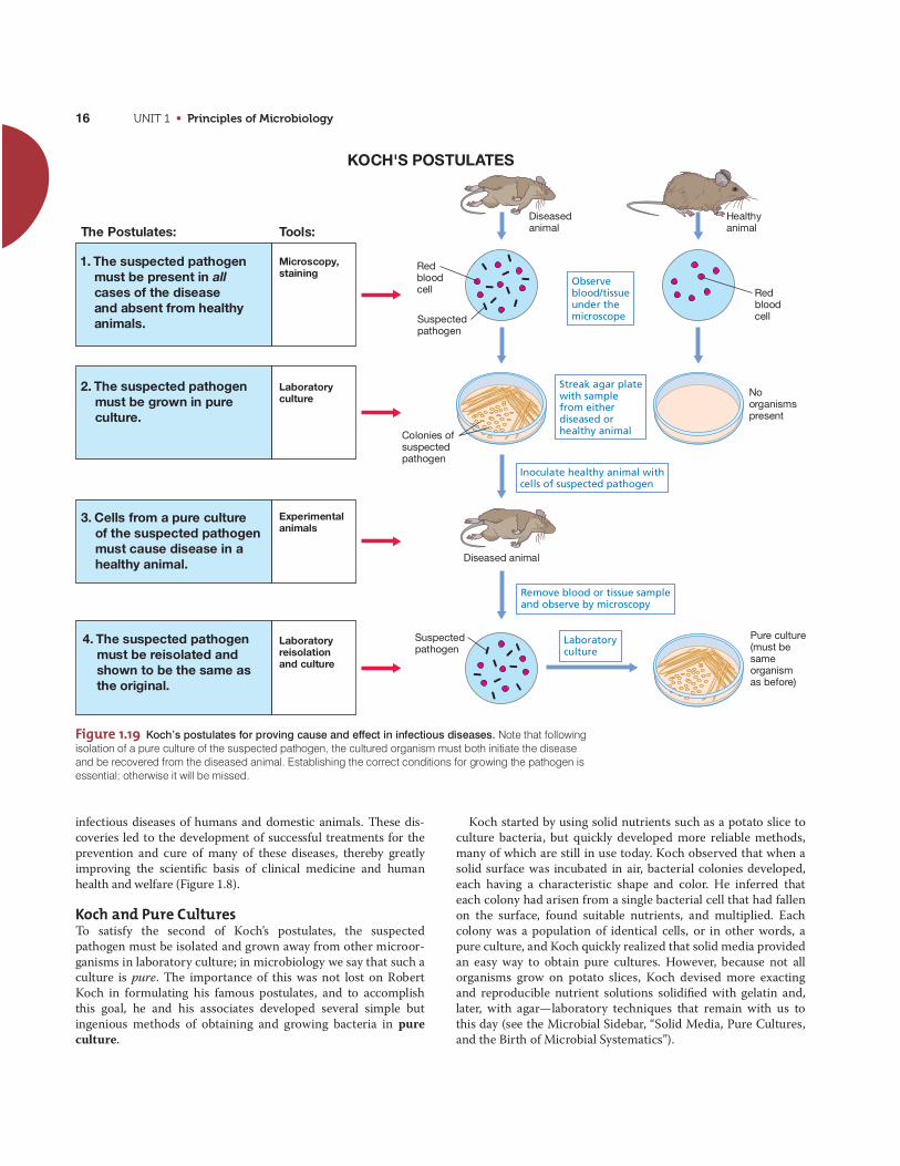
UNIT 1 • Principles of Microbiology16
infectious diseases of humans and domestic animals. These dis-coveries led to the development of successful treatments for theprevention and cure of many of these diseases, thereby greatlyimproving the scientific basis of clinical medicine and humanhealth and welfare (Figure 1.8).
Koch and Pure CulturesTo satisfy the second of Koch’s postulates, the suspectedpathogen must be isolated and grown away from other microor-ganisms in laboratory culture; in microbiology we say that such aculture is pure. The importance of this was not lost on RobertKoch in formulating his famous postulates, and to accomplishthis goal, he and his associates developed several simple butingenious methods of obtaining and growing bacteria in pureculture.
Koch started by using solid nutrients such as a potato slice toculture bacteria, but quickly developed more reliable methods,many of which are still in use today. Koch observed that when asolid surface was incubated in air, bacterial colonies developed,each having a characteristic shape and color. He inferred thateach colony had arisen from a single bacterial cell that had fallenon the surface, found suitable nutrients, and multiplied. Eachcolony was a population of identical cells, or in other words, apure culture, and Koch quickly realized that solid media providedan easy way to obtain pure cultures. However, because not allorganisms grow on potato slices, Koch devised more exactingand reproducible nutrient solutions solidified with gelatin and,later, with agar—laboratory techniques that remain with us tothis day (see the Microbial Sidebar, “Solid Media, Pure Cultures,and the Birth of Microbial Systematics”).
Diseasedanimal
Diseased animal
Observe blood/tissueunder the microscope
Streak agar platewith samplefrom eitherdiseased orhealthy animal
Suspectedpathogen
Suspectedpathogen
Laboratoryculture
Redbloodcell
Redbloodcell
Colonies ofsuspectedpathogen
Noorganismspresent
Pure culture(must be sameorganismas before)
Inoculate healthy animal withcells of suspected pathogen
Remove blood or tissue sampleand observe by microscopy
2. The suspected pathogenmust be grown in pure culture.
1. The suspected pathogenmust be present in allcases of the diseaseand absent from healthyanimals.
3. Cells from a pure culture of the suspected pathogenmust cause disease in a healthy animal.
4. The suspected pathogenmust be reisolated andshown to be the same asthe original.
KOCH'S POSTULATES
The Postulates: Tools:Healthyanimal
Microscopy,staining
Laboratory culture
Experimentalanimals
Laboratoryreisolationand culture
Figure 1.19 Koch’s postulates for proving cause and effect in infectious diseases. Note that followingisolation of a pure culture of the suspected pathogen, the cultured organism must both initiate the diseaseand be recovered from the diseased animal. Establishing the correct conditions for growing the pathogen isessential; otherwise it will be missed.

MICROBIAL SIDEBAR
Solid Media, Pure Cultures, and the Birth of Microbial Systematics
Robert Koch was the first to grow bacteriaon solid culture media. Koch’s early use
of potato slices as solid media was fraughtwith problems. Besides the problem that notall bacteria can grow on potatoes, the sliceswere frequently overgrown with molds. Kochthus needed a more reliable and repro-ducible means of growing bacteria on solidmedia, and he found the answer for solidify-ing his nutrient solutions in agar.
Koch initially employed gelatin as a solidi-fying agent for the various nutrient fluids heused to culture bacteria, and he kept hori-zontal slabs of solid gelatin free of contami-nation under a bell jar or in a glass box (seeFigure 1.20c). Nutrient-supplemented gelatinwas a good culture medium for the isolationand study of various bacteria, but it had sev-eral drawbacks, the most important of whichwas that it did not remain solid at 37°C, theoptimum temperature for growth of mosthuman pathogens. Thus, a different solidify-ing agent was needed.
Agar is a polysaccharide derived from redalgae. It was widely used in the nineteenthcentury as a gelling agent. Walter Hesse, anassociate of Koch, first used agar as a solidi-fying agent for bacteriological culture media(Figure 1). The actual suggestion that agarbe used instead of gelatin was made byHesse’s wife, Fannie. She had used agar tosolidify fruit jellies. When it was tried as asolidifying agent for microbial media, itssuperior gelling qualities were immediatelyevident. Hesse wrote to Koch about this dis-covery, and Koch quickly adapted agar to hisown studies, including his classic studies onthe isolation of the bacterium Mycobacteriumtuberculosis, the cause of the disease tuber-culosis (see text and Figure 1.20).
Agar has many other properties that makeit desirable as a gelling agent for microbialculture media. In particular, agar remainssolid at 37°C and, after melting during thesterilization process, remains liquid to about45°C, at which time it can be poured intosterile vessels. In addition, unlike gelatin,
agar is not degraded by most bacteria andtypically yields a transparent medium, mak-ing it easier to differentiate bacterial coloniesfrom inanimate particulate matter. For thesereasons, agar found its place early in theannals of microbiology and is still used todayfor obtaining and maintaining pure cultures.
In 1887 Richard Petri, a German bacteriol-ogist, published a brief paper describing amodification of Koch’s flat plate technique(Figure 1.20c). Petri’s enhancement, whichturned out to be amazingly useful, was thedevelopment of the transparent double-sideddishes that bear his name (Figure 2). Theadvantages of Petri dishes were immediatelyapparent. They could easily be stacked andsterilized separately from the medium, and,following the addition of molten culturemedium to the smaller of the two dishes, thelarger dish could be used as a cover to pre-vent contamination. Colonies that formed onthe surface of the agar in the Petri dishretained access to air without direct exposureto air and could easily be manipulated forfurther study. The original idea of Petri hasnot been improved on to this day, and thePetri dish, constructed of either glass orplastic, is a mainstay of the microbiologylaboratory.
Koch quickly grasped the significance ofpure cultures and was keenly aware of theimplications his pure culture methods had forclassifying microorganisms. He observedthat colonies that differed in color, morphol-ogy, size, and the like (see Figure 2) bredtrue and could be distinguished from oneanother. Cells from different colonies typicallydiffered in size and shape and often in theirtemperature or nutrient requirements as well.Koch realized that these differences amongmicroorganisms met all the requirements thatbiological taxonomists had established forthe classification of larger organisms, suchas plant and animal species. In Koch’s ownwords (translated from the German): “All bac-teria which maintain the characteristics whichdifferentiate one from another when they arecultured on the same medium and under thesame conditions, should be designated asspecies, varieties, forms, or other suitabledesignation.” Such insightful thinking wasimportant for the rapid acceptance of micro-biology as a new biological science, rootedas biology was in classification at the time ofKoch. It has since had a profound impact onthe diagnosis of infectious diseases and thefield of microbial diversity.
17
Pau
l V. D
unla
p
Figure 1 A hand-colored photograph taken by WalterHesse of colonies formed on agar. The colonies includethose of molds and bacteria obtained during Hesse’s studiesof the microbial content of air in Berlin, Germany, in 1882.From Hesse, W. 1884. “Ueber quantitative Bestimmung derin der Luft enthaltenen Mikroorganismen,” in Struck, H. (ed.),Mittheilungen aus dem Kaiserlichen Gesundheitsamte.August Hirschwald.
Figure 2 Photo of a Petridish containing colonies ofmarine bacteria. Each colonycontains millions of bacterialcells descended from a singlecell.

Figure 1.20 Robert Koch’s drawings of Mycobacterium tuberculosis.(a) Section through infected lung tissue showing cells of M. tuberculosis(blue). (b) M. tuberculosis cells in a sputum sample from a tubercularpatient. (c) Growth of M. tuberculosis on a glass plate of coagulated bloodserum stored inside a glass box to prevent contamination. (d) M. tubercu-losis cells taken from the plate in part c and observed microscopically;cells appear as long cordlike forms. Original drawings from Koch, R. 1884.“Die Aetiologie der Tuberkulose.” Mittheilungen aus dem KaiserlichenGesundheitsamte 2:1–88.
UNIT 1 • Principles of Microbiology18
Tuberculosis: The Ultimate Testof Koch’s PostulatesKoch’s crowning accomplishment in medical bacteriology washis discovery of the causative agent of tuberculosis. At the timeKoch began this work (1881), one-seventh of all reported humandeaths were caused by tuberculosis (Figure 1.8). There was astrong suspicion that tuberculosis was a contagious disease, butthe suspected agent had never been seen, either in diseased tis-sues or in culture. Koch was determined to demonstrate thecause of tuberculosis, and to this end he brought together all ofthe methods he had so carefully developed in his previous studieswith anthrax: microscopy, staining, pure culture isolation, and ananimal model system.
As is now well known, the bacterium that causes tuberculosis,Mycobacterium tuberculosis, is very difficult to stain because ofthe large amounts of a waxy lipid present in its cell wall. But Kochdevised a staining procedure for M. tuberculosis cells in tissuesamples; using this method, he observed blue, rod-shaped cells ofM. tuberculosis in tubercular tissues but not in healthy tissues(Figure 1.20). However, from his previous work on anthrax, Kochrealized that he must culture this organism in order to prove thatit was the cause of tuberculosis.
Obtaining cultures of M. tuberculosis was not easy, but eventu-ally Koch was successful in growing colonies of this organism ona medium containing coagulated blood serum. Later he usedagar, which had just been introduced as a solidifying agent (see
the Microbial Sidebar). Under the best of conditions, M. tubercu-losis grows slowly in culture, but Koch’s persistence and patienceeventually led to pure cultures of this organism from human andanimal sources.
From this point it was relatively easy for Koch to use his postu-lates (Figure 1.19) to obtain definitive proof that the organism hehad isolated was the cause of the disease tuberculosis. Guineapigs can be readily infected with M. tuberculosis and eventuallysuccumb to systemic tuberculosis. Koch showed that diseasedguinea pigs contained masses of M. tuberculosis cells in theirlungs and that pure cultures obtained from such animals trans-mitted the disease to uninfected animals. Thus, Koch success-fully satisfied all four of his postulates, and the cause oftuberculosis was understood. Koch announced his discovery ofthe cause of tuberculosis in 1882 and published a paper on thesubject in 1884 in which his postulates are most clearly stated.For his contributions on tuberculosis, Robert Koch was awardedthe 1905 Nobel Prize for Physiology or Medicine. Koch had manyother triumphs in medicine, including discovering the organismresponsible for the disease cholera and developing methods todiagnose exposure to M. tuberculosis (the tuberculin test).
Koch’s Postulates TodayFor human diseases in which an animal model is available, it isrelatively easy to use Koch’s postulates. In modern clinical medi-cine, however, this is not always so easy. For instance, thecausative agents of several human diseases do not cause diseasein any known experimental animals. These include many of thediseases associated with bacteria that live only within cells, suchas the rickettsias and chlamydias, and diseases caused by someviruses and protozoan parasites. So for most of these diseasescause and effect cannot be unequivocally proven. However, theclinical and epidemiological (disease tracking) evidence for virtu-ally every infectious disease of humans lends all but certain proofof the specific cause of the disease. Thus, although Koch’s postu-lates remain the “gold standard” in medical microbiology, it hasbeen impossible to satisfy all of his postulates for every humaninfectious disease.
MiniQuiz• How do Koch’s postulates ensure that cause and effect of a
given disease are clearly differentiated?
• What advantages do solid media offer for the isolation ofmicroorganisms?
• What is a pure culture?
1.9 The Rise of Microbial DiversityAs microbiology moved into the twentieth century, its initialfocus on basic principles, methods, and medical aspects broad-ened to include studies of the microbial diversity of soil andwater and the metabolic processes that organisms in these habi-tats carried out. Two giants of this era included the DutchmanMartinus Beijerinck and the Russian Sergei Winogradsky.
(a) (b)
(c) (d)

CHAPTER 1 • Microorganisms and Microbiology 19
UN
IT 1
Figure 1.21 Martinus Beijerinck and Azotobacter. (a) A page from thelaboratory notebook of M. Beijerinck dated 31 December 1900 describingthe aerobic nitrogen-fixing bacterium Azotobacter chroococcum (namecircled in red). Compare Beijerinck’s drawings of pairs of A. chroococcumcells with the photomicrograph of cells of Azotobacter in Figure 17.18a.(b) A painting by M. Beijerinck’s sister, Henriëtte Beijerinck, showing cells of Azotobacter chroococcum. Beijerinck used such paintings to illus-trate his lectures.
(a)
(b)
Lesl
ey R
obe
rtso
n an
d t
he K
luyv
er L
abor
ator
y M
useu
m,
Del
ft U
nive
rsity
of T
echn
olo
gyLe
sley
Rob
erts
on a
nd th
e K
luyv
er L
abor
ator
y M
useu
m, D
elft
Uni
vers
ity o
f Te
chno
log
y
Martinus Beijerinck and the EnrichmentCulture TechniqueMartinus Beijerinck (1851–1931), a professor at the Delft Poly-technic School in Holland, was originally trained in botany, so hebegan his career in microbiology studying plants. Beijerinck’sgreatest contribution to the field of microbiology was his clearformulation of the enrichment culture technique. In enrich-ment cultures microorganisms are isolated from natural samplesusing highly selective techniques of adjusting nutrient and incu-bation conditions to favor a particular metabolic group of orga-nisms. Beijerinck’s skill with the enrichment method was readilyapparent when, following Winogradsky’s discovery of the processof nitrogen fixation, he isolated the aerobic nitrogen-fixing bac-terium Azotobacter from soil (Figure 1.21).
Using the enrichment culture technique, Beijerinck isolated thefirst pure cultures of many soil and aquatic microorganisms,
including sulfate-reducing and sulfur-oxidizing bacteria, nitrogen-fixing root nodule bacteria (Figure 1.9), Lactobacillus species,green algae, various anaerobic bacteria, and many others. In hisstudies of tobacco mosaic disease, Beijerinck used selective filter-ing techniques to show that the infectious agent (a virus) wassmaller than a bacterium and that it somehow became incorpo-rated into cells of the living host plant. In this insightful work, Bei-jerinck not only described the first virus, but also the basicprinciples of virology, which we present in Chapters 9 and 21.
Sergei Winogradsky, Chemolithotrophy,and Nitrogen FixationSergei Winogradsky (1856–1953) had interests similar toBeijerinck’s—the diversity of bacteria in soils and waters—andwas highly successful in isolating several key bacteria from naturalsamples. Winogradsky was particularly interested in bacteria thatcycle nitrogen and sulfur compounds, such as the nitrifying bac-teria and the sulfur bacteria (Figure 1.22). He showed that thesebacteria catalyze specific chemical transformations in nature and
Figure 1.22 Sulfur bacteria. The original drawings were made bySergei Winogradsky in the late 1880s and then copied and hand-coloredby his wife Hèléne. (a) Purple sulfur phototrophic bacteria. Figures 3 and4 show cells of Chromatium okenii (compare with photomicrographs of C. okenii in Figures 1.5a and 1.7a). (b) Beggiatoa, a sulfur chemolithotroph(compare with Figure 1.14).
From
Mic
rob
iolo
gie
du
So
l, us
ed w
ith p
erm
issi
on
From
Win
ogra
dsky
, S. 1
949.
Mic
robi
olog
ie d
u S
ol.
Mas
son,
Par
is.
(a)
(b)

UNIT 1 • Principles of Microbiology20
proposed the important concept of chemolithotrophy, the oxida-tion of inorganic compounds to yield energy. Winogradsky furthershowed that these organisms, which he called chemolithotrophs,obtained their carbon from CO2. Winogradsky thus revealed that,like phototrophic organisms, chemolithotrophic bacteria wereautotrophs.
Winogradsky performed the first isolation of a nitrogen-fixingbacterium, the anaerobe Clostridium pasteurianum, and as justmentioned, Beijerinck used this discovery to guide his isolationof aerobic nitrogen-fixing bacteria years later (Figure 1.21).Winogradsky lived to be almost 100, publishing many scientificpapers and a major monograph, Microbiologie du Sol (Soil Micro-biology). This work, a milestone in microbiology, contains draw-ings of many of the organisms Winogradsky studied during hislengthy career (Figure 1.22).
MiniQuiz• What is meant by the term “enrichment culture”?
• What is meant by the term “chemolithotrophy”? In what way arechemolithotrophs like plants?
1.10 The Modern Era of MicrobiologyIn the twentieth century, the field of microbiology developed rap-idly in two different yet complementary directions—applied andbasic. During this period a host of new laboratory tools becameavailable, and the science of microbiology began to mature andspawn new subdisciplines. Few of these subdisciplines werepurely applied or purely basic. Instead, most had both discovery(basic) and problem-solving (applied) components. Table 1.3summarizes these major subdisciplines of microbiology thatarose in the twentieth century.
Several microbiologists are remembered for their key contri-butions during this period. In the early twentieth century manyremained focused on medical aspects of microbiology, and eventoday, many dedicated microbiologists grapple with the impactsof microorganisms on human, animal, and plant disease. But fol-lowing World War II, an exciting new emphasis began to takehold with studies of the genetic properties of microorganisms.From roots in microbial genetics has emerged “modern biology,”driven by molecular biology, genetic engineering, and genomics.This molecular approach has revolutionized scientific thinking inthe life sciences and has driven experimental approaches to themost compelling problems in biology. Some key Nobel laureatesand their contributions to the molecular era of microbiology arelisted in Table 1.4.
Many of the advances in microbiology today are fueled by thegenomics revolution; that is, we are clearly in the era of “molecu-lar microbiology.” Rapid progress in DNA sequencing technologyand improved computational power have yielded huge amountsof genomic information that have supported major advances inmedicine, agriculture, biotechnology, and microbial ecology. Forexample, to obtain the sequence of the entire genome of a bac-terium takes only a few hours (although sequence analysis is amuch more time-consuming process). The fast-paced field of
genomics has itself spawned highly focused new subdisciplines,such as transcriptomics, proteomics, and metabolomics, whichexplore, respectively, the patterns of RNA, protein, and meta-bolic expression in cells. The concepts of genomics, transcrip-tomics, proteomics, and metabolomics are all developed inChapter 12.
All signs point to a continued maturation of molecular micro-biology as we enter a period where technology is almost ahead ofour ability to formulate exciting scientific questions. In fact,microbial research today is very close to defining the minimalistgenome—the minimum complement of genes necessary for a liv-ing cell. When such a genetic blueprint is available, microbiolo-gists should be able to define, at least in biochemical terms, theprerequisites for life. When that day arrives, can the laboratorycreation of an actual living cell from nonliving components, thatis, spontaneous generation under controlled laboratory condi-tions, be far off? Almost certainly not. Stay tuned, as much excit-ing science is on the way!
MiniQuiz• For each of the following topics, name the subdiscipline of
microbiology that focuses on it: metabolism, enzymology,nucleic acid and protein synthesis, microorganisms and theirnatural environments, microbial classification, inheritance ofcharacteristics.
Table 1.3 The major subdisciplines of microbiology
Subdiscipline Focus
I. Basic emphasesa
Microbial physiology Nutrition, metabolism
Microbial genetics Genes, heredity, and genetic variation
Microbial biochemistry Enzymes and chemical reactions in cells
Microbial systematics Classification and nomenclature
Virology Viruses and subviral particles
Molecular biology Nucleic acids and protein
Microbial ecology Microbial diversity and activity in natural habitats; biogeochemistry
II. Applied emphasesa
Medical microbiology Infectious disease
Immunology Immune systems
Agricultural/soil microbiology Microbial diversity and processes in soil
Industrial microbiology Large-scale production of antibiotics, alcohol, and other chemicals
Biotechnology Production of human proteins by genetically engineered microorganisms
Aquatic microbiology Microbial processes in waters and wastewaters, drinking water safety
aNone of these subdisciplines are devoted entirely to basic science or applied science.However, the subdisciplines listed in I tend to be more focused on discovery and thosein II more focused on solving specific problems or synthesizing commercial productsfrom microbial sources.