etiology of intracerebral hemorrhage (ich): novel insights
TRANSCRIPT
Etiology of intracerebral hemorrhage (ICH): novel insights from Zebrafish embryos
SHAHRAM EISA-BEYGI* and MOHAMMAD REZAEI
Department of Stem Cells and Developmental Biology, Cell Science Research Center, Royan Institute for Stem Cell Biology and Technology, ACECR, Tehran, Iran
ABSTRACT Intracerebral hemorrhage (ICH) is the most severe subtype of stroke. Treatment op-tions are scarce and given the high morbidity and mortality, relatively ineffective. Since patients with ICH may have an unknown heritable component, the need to identify potential risk factors necessitates the use of animal models to elucidate the genetic underpinnings of neurovascular development and, thereby, identify candidate regulatory pathways that are likely to be disrupted in patients with ICH. Zebrafish (Danio rerio) exhibits the anatomical and physiological complex-ity of a closed circulatory system observed in all vertebrates (with arteries, veins and capillaries). Moreover, studies over the last decade, aided by the application of chemical mutagenesis screens, morpholino mediated knockdown approaches and tissue-specific transgenic markers, have paved the way for the identification of several genes and signaling pathways that regulate developmen-tal neurovascular stabilization. We hypothesize that mutations in these genes or pharmacological perturbations of these gene-products may account, at least in part, for the etiology of some forms of spontaneous ICH in humans.
KEY WORDS: intracerebral hemorrhage, neurovascular development, stroke etiology, zebrafish
Introduction
Despite the high morbidity associated with intracerebral hemor-rhage (ICH) and the appreciation that genetic factors during devel-opment contribute significantly to the disease onset, the etiological underpinnings remain elusive (Rost et al., 2008). The mechanisms contributing to the stabilization of nascent neurovascular architec-ture are far from being elucidated. Given the absence of effective treatment options to improve the outcome, the pressing need to characterize the potential risk factors for ICH is recognized by researchers and clinicians alike. One of the limited routes towards therapeutic applications is to identify and characterize the novel genetic variants that underlie ICH in vertebrate models of stroke.
Functional studies over the last decade have yielded valuable insights into the mechanisms regulating vascular development in zebrafish, which show a high degree of conservation with other vertebrates (Isogai et al., 2001; Ellertsdóttir et al., 2010; Gore et al., 2012). The processes of endothelial cell (EC) fate-specification and maturation in zebrafish are driven by a conserved set of genes (Detrich et al., 1995; Kabrun et al., 1997). Similarly, the processes of angiogenesis (Liang et al., 2001; Habeck et al., 2002), vasculo-genesis (Herbert et al., 2009) artery-vein specification (Swift and
Int. J. Dev. Biol. 60: 119-126 (2016)doi: 10.1387/ijdb.160136se
www.intjdevbiol.com
*Address correspondence to: Shahram Eisa-Beygi. Department of Stem Cells and Developmental Biology, Cell Science Research Center, Royan Institute for Stem Cell Biology and Technology, ACECR, Tehran, Iran. E-mail: [email protected]
Accepted: 10 May 2016.
ISSN: Online 1696-3547, Print 0214-6282© 2016 UPV/EHU PressPrinted in Spain
Abbreviations used in this paper: EC, endothelial cell; ICH, intracerebral hemorrhage.
Weinstein, 2009; Hong et al., 2008), vessel lumenization (vascular tube formation) (Kamei et al., 2006) and vascular stabilization (Eisa-Beygi et al., 2014) in zebrafish are under the control of a conserved set of genes, growth/transcription factors and regulatory pathways.
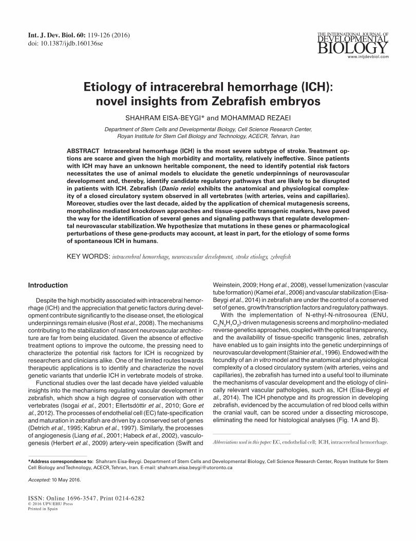
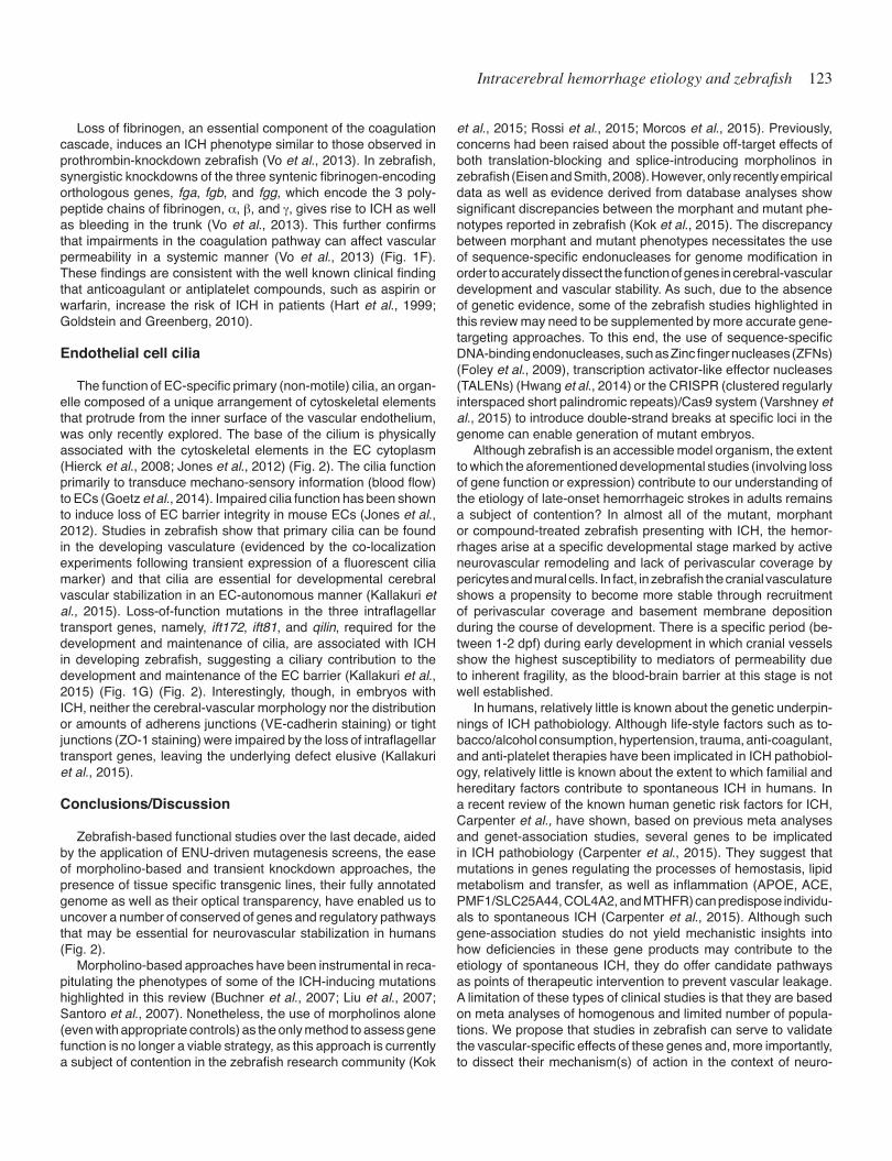
With the implementation of N-ethyl-N-nitrosourea (ENU, C3N3H7O2)-driven mutagenesis screens and morpholino-mediated reverse genetics approaches, coupled with the optical transparency, and the availability of tissue-specific transgenic lines, zebrafish have enabled us to gain insights into the genetic underpinnings of neurovascular development (Stainier et al., 1996). Endowed with the fecundity of an in vitro model and the anatomical and physiological complexity of a closed circulatory system (with arteries, veins and capillaries), the zebrafish has turned into a useful tool to illuminate the mechanisms of vascular development and the etiology of clini-cally relevant vascular pathologies, such as, ICH (Eisa-Beygi et al., 2014). The ICH phenotype and its progression in developing zebrafish, evidenced by the accumulation of red blood cells within the cranial vault, can be scored under a dissecting microscope, eliminating the need for histological analyses (Fig. 1A and B).
120 S. Eisa-Beygi and M. Rezaei
In this review, we provide a synthesis of the recent advances into the etiology of ICH, derived from zebrafish developmental stud-ies. By providing a synthesis of these findings, we also elucidate, where possible, the inter-dependence of these conserved signaling and regulatory pathways. It is predicted that loss-of-function muta-tions in these genes or pharmacological curtailment of these gene products may impair neurovascular stabilization and precipitate a hemorrhagic phenotype.
Genes regulating cell-cell and cell-matrix adhesions
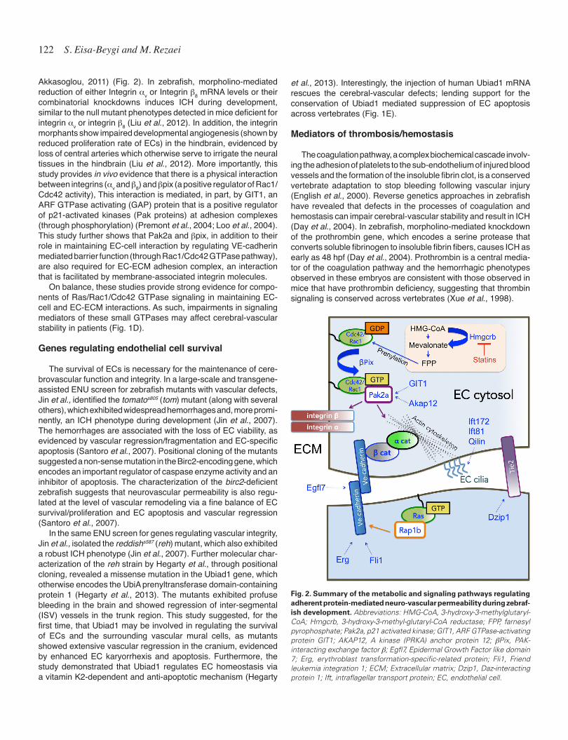
The trans-membrane adhesion proteins anchored to cytoskeletal elements near the plasma membranes of ECs confer structural rigidity and ensure vascular stability by maintaining the physical contact between neighboring ECs. The EC-specific vascular endo-thelial (VE)-cadherin is expressed at the adherens junctions of EC membranes and contributes to barrier integrity. Membrane-bound VE-cadherin (Cdh5) interacts via its C-terminal domain, with the actin cytoskeleton, through a number of intracellular binding part-ners (a/b-catenin). This association is required for the regulation of EC-cell adhesion, cell shape, cell motility and control of gene expression (Dejana et al., 2008; Dejana and Vestweber, 2013) (Fig. 2). Consistently, loss of VE-cadherin function in zebrafish impairs EC elongation due to disruption of actin fibers and defec-tive junction remodeling, which disrupts angiogenic sprouting (Sauteur et al., 2014). Hence, the adhesion between the ECs is not only maintained via the adhesive function of VE-cadherin, but also via the dynamic actin-based cytoskeletal structures that provide a scaffold for the VE-cadherin mediated EC-cell adhe-sions. This is evidenced by data showing that perturbations in the polymerization state of actin cytoskeleton (hyper-polymerization and/or de-polymerization) disrupt EC barrier function and increase permeability in vivo (Spindler et al., 2010).
The VE-cadherin-mediated EC-cell barrier integrity is, in part, under the positive control of Rac1 and Cdc42 GTPase activities (Daneshjou et al., 2015; Broman et al., 2006) and is negatively regulated by the hyper-activation of RhoA GTPase signaling (Gian-notta et al., 2013), as observed in vascular pathologies, such as cerebral aneurysms, endotoxin-induced sepsis, thrombin-induced EC hyper-permeability or viral-induced micro-vascular leak (Bor-ikova et al., 2010; Han et al., 2013; Parker et al., 2015; Han et al., 2013; van Nieuw Amerongen and Hinsbergh, 2007; Darwish and Liles, 2013).
In zebrafish, cdh5 transcripts exhibit a vascular-specific en-richment profile, detectable during early stages of development (Fig. 1C). Consistent with its role in EC-cell barrier integrity, morpholino-mediated depletion of cdh5 transcripts, results in ICH and defective vascular lumenization during zebrafish development (Montero-Balaguer et al., 2009).
The VE-cadherin mediated EC-cell stability is under the control of a conserved signaling pathway involving P21-activated kinase 2a (Pak2a), a kinase belonging to the Pak protein family (Gavard and Gutkind, 2006). Pak2a physically interacts with the membrane-bound (prenylated) Rho-family GTPases, Cdc42 and Rac1, in their active (GTP-bound) state. This interaction regulates actin organization at the EC membrane–cytosol boundary (Vidal et al., 2002; Renkema et al., 2002). The Pak proteins, through regulat-ing the organization of actin cytoskeleton, thus EC contractility, regulate changes in cell shape of ECs (Itakura et al., 2013) (Fig. 2). Whereas depletion of PAK2 function in embryonic mice induces developmental defects and mortality, loss of PAK2 function in adult mice disrupts vascular permeability (Radu et al., 2015). In zebrafish, both ENU-mediated truncation of pak2a and morpholino-induced
Fig. 1. Zebrafish model of developmental Intracerebral hemorrhage (ICH). (A,B) Bright-field photomicrographs of 48 bpf zebrafish embryos with and without ICH phenotypes. Black arrow denotes areas of blood extravasation. Ventral views are shown. Anterior is to top. (C) Bright-field image of a 48 hpf wild-type zebrafish embryo, showing cdh5 mRNA deposi-tion in the cerebral-vascular endothelium. Anterior is to left. (D-G) List of genes regulating cerebral vascular stabilization in zebrafish via modulating endothelial cell-cell and endothelial cell-extracellular matrix interactions (D), endothelial cell survival/proliferation (E), hemostatic processes (F), and endothelial cilia function (G).
G
B
C
D
E
F
A
Intracerebral hemorrhage etiology and zebrafish 121
depletion of its transcripts induce an ICH phenotype during de-velopment (Stainier et al., 1996; Buchner et al., 2007). Although neurovascular angiogenesis is slightly delayed in Pak2a-deficient embryos, the primary defects are due to loss of vascular stability (Buchner et al., 2007). Consistently, knockdown of the A kinase (PRKA) anchor protein 12 (Akap12) expression, an modulator of actin dynamics via pak2a-mediated signaling (Fig. 2), induces loosened EC-cell contacts, a discontinuous EC layer and frequent ICH in developing zebrafish (Kwon et al., 2012).
Similarly, a hypomorphic mutation in the bPix gene (redhead strain), which encodes a Pak-interacting guanine nucleotide ex-change factor (GEF), phenocopies the vascular defects observed in Pak2a or Akap12-deficient fish (Stainier et al., 1996; Liu et al., 2007). Interestingly, bpix acts as a Pak2a-interacting molecular switch by activating the Rho GTPases, Rac1, and Cdc42 (Fig. 2). In zebrafish with reduced bpix expression, the primary cause for ICH is shown to be defective EC-mural cell interactions (Liu et al., 2007). The findings in Pak2a and bpix-deficient zebrafish provide strong in vivo evidence for the involvement of Rac1 and Cdc42 GTPases in neurovascular development and support previous in-vitro evidence about the involvement of Rac1/Cdc42 in the maintenance of endothelial-cell barrier integrity and lumenization (Birukov et al., 2004; Kouklis et al., 2004, Broman et al., 2006; Koh et al., 2008). Consistently, in patients with cerebral cavernous malformation (CCM), an autosomal familial disorder, characterized by hemorrhage-prone vascular malformations, the Rac1/Cdc42 signaling pathway is significantly reduced, whereas, the RhoA pathway is hyper-activated (Liu et al., 2011; Storkebaum et al., 2011; Lant et al., 2015). These consistent results highlight the potential clinical utility of zebrafish-based chemical/genetic screens for the identification of disease-related genes in cerebrovascular pathologies.
The Rac1/Cdc42 signaling is also regulated at the level of subcellular localization. Inhibition of the 3-hydroxy-3-methylgl-utaryl-coenzyme A reductase (Hmgcr), a rate-limiting enzyme required for the biosynthesis of geranylgeranyl pyrophosphate, a 20-carbon lipid-tag for the membrane localization of Rac1/Cdc42 at their carboxyl terminus (CAAX terminus) (Itoh et al., 2002) (Fig. 2), induces loosened EC-cell interactions and ICH in developing zebrafish (Gjini et al., 2011; Eisa-Beygi et al., 2013). Interestingly, the zebrafish-based findings are consistent with emerging clinical studies, which have disclosed a link between cholesterol-lowering HMGCR inhibitors (statins) and increased risk of ICH. (Amarenco et al., 2006; Goldstein et al., 2008; Flaster et al., 2011; Lauer et al., 2015). Statins may impair vascular stability by decreasing the prenylation of Rac1/Cdc42 proteins and inhibit downstream path-ways that govern VE-cadherin mediated EC-cell barrier function (Eisa-Beygi et al., 2014; Eisa-Beygi et al., 2016) (Fig. 2).
The VE-cadherin mediated EC-cell barrier function is also regulated by the small GTP-binding protein, Ras GTPase. The activation of Ras GTPase enhances VE-cadherin mediated EC-cell barrier function (Sawada et al., 2012). This is consistent with data in zebrafish showing that knockdown of rap1b, which encodes a Ras GTPase effector, results in ICH, along with reduced expres-sion and membrane localization of VE-cadherin and its binding partner, b-catenin (Gore et al., 2008). When Rap1b morpholino oligonucleotides were co-injected in combination with morpholinos against the previously identified Rac1/Cdc42 signaling mediators, pak2a and bPix, the incidence of ICH increased significantly (Gore
et al., 2008) (Fig. 2). This is suggestive of a synergy through genetic cross-regulation of components of Ras, Rac1 and Cdc42 GTPases as positive regulators of VE-cadherin mediated vascular stability.
Studies in zebrafish have revealed that EC-cell junction integrity is also regulated at the level of VE-cadherin gene expression via the E26 transformation-specific (ETS) domain transcription factors. The conserved transcription regulators, erythroblast transformation-specific-related gene (erg) and Friend leukemia integration 1 (fli1), both of which are required for hematopoetic and angiogenic processes, are important in neurovascular stabilization, because they regulate VE-cadherin dynamics (via regulating VE-cadherin mRNA enrichment levels) (Liu and Patient, 2008). Both erg and fli1 transcripts are detected in endothelial precursors/hemangioblasts as well as mature blood vessels during zebrafish development. Injection of morpholino-oligonucleotides against either erg or fli1 induces ICH phenotypes in developing zebrafish (Liu and Patient, 2008). Consistently, the mRNA levels of VE-cadherin were signifi-cantly diminished in the morphants prior to the onset of ICH, sug-gesting that the hemorrhages arise, in part, due to downregulation of VE-cadherin levels (Liu and Patient, 2008) (Fig. 2).
In addition to VE-cadherin, another EC-specific cell surface receptor, angiopoietin receptor 1 (Tie2), is also shown to be im-portant for developmental neurovascular stabilization via sonic hedgehog (shh) signaling. A mutation in Daz-interacting protein 1 (Dzip1), a component of sonic-hedgehog signaling, results in ICH by 2 dpf, along with reduced mRNA levels of angiopoeitin 1 (Ang1), an inductive ligand of Tie2 (Lamont et al., 2010) (Fig. 2). Although the loss of Dzip1 function is not shown to affect the pat-terning of cerebral vessels or artery/vein specification, the stability of vascular endothelium is disrupted, evidenced by the weakened perivascular-EC contacts and extravasation of blood components into the brain parenchyma. The hemorrhages are due to impaired ang-1/Tie2 signaling (Lamont et al., 2010).
Studies in zebrafish have also uncovered a functional role for the epidermal growth factor like domain 7 (Egfl7), a conserved vascular-endothelial specific secreted growth factor in vertebrates (Parker et al., 2004). Egfl7 is required for the processes of tubu-logenesis, lumenization, EC migration and polarity (Parker et al., 2004; Schmidt et al., 2007; De Mazière et al., 2008). Recently, a functional role for Egfl7 in neurovascular stabilization has been shown via the use of TAL effector nucleases (TALENs) designed against egfl7. Injection of TALENs RNA into fertilized zebrafish embryos induces ICH in some of the embryos by 2 dpf (Rossi et al., 2015). The effects of Egfl7 knockdown on neurovascular stability are likely due to aberrantly distributed EC adherens and tight junc-tions, as evidenced by previous findings (De Mazière et al., 2007).
Neurovascular permeability is also maintained via the adherence of ECs to constituents of extracellular matrix (ECM). The ECM is composed of a non-cellular component made up of macromol-ecules (proteins and carbohydrates) that provide a scaffold for the development and maintenance of the nascent vascular endothelium into functional blood vessels (Davis and Senger, 2005). A strong EC-ECM interaction is required for cerebral-vascular stabilization (Baeten and Akkasoglou, 2011). The interaction between the intracellular cystoskeleton of the ECs and ECM components is mediated, in part, by the cell adhesion receptors, integrins, which form the focal adhesions (Davis and Senger, 2005). By enabling ECs to bind to ECM membrane components, integrins contribute to both vascular stabilization and angiogenesis (Baeten and
122 S. Eisa-Beygi and M. Rezaei
Akkasoglou, 2011) (Fig. 2). In zebrafish, morpholino-mediated reduction of either Integrin av or Integrin b8 mRNA levels or their combinatorial knockdowns induces ICH during development, similar to the null mutant phenotypes detected in mice deficient for integrin av or integrin b8 (Liu et al., 2012). In addition, the integrin morphants show impaired developmental angiogenesis (shown by reduced proliferation rate of ECs) in the hindbrain, evidenced by loss of central arteries which otherwise serve to irrigate the neural tissues in the hindbrain (Liu et al., 2012). More importantly, this study provides in vivo evidence that there is a physical interaction between integrins (av and b8) and bpix (a positive regulator of Rac1/Cdc42 activity), This interaction is mediated, in part, by GIT1, an ARF GTPase activating (GAP) protein that is a positive regulator of p21-activated kinases (Pak proteins) at adhesion complexes (through phosphorylation) (Premont et al., 2004; Loo et al., 2004). This study further shows that Pak2a and bpix, in addition to their role in maintaining EC-cell interaction by regulating VE-cadherin mediated barrier function (through Rac1/Cdc42 GTPase pathway), are also required for EC-ECM adhesion complex, an interaction that is facilitated by membrane-associated integrin molecules.
On balance, these studies provide strong evidence for compo-nents of Ras/Rac1/Cdc42 GTPase signaling in maintaining EC-cell and EC-ECM interactions. As such, impairments in signaling mediators of these small GTPases may affect cerebral-vascular stability in patients (Fig. 1D).
Genes regulating endothelial cell survival
The survival of ECs is necessary for the maintenance of cere-brovascular function and integrity. In a large-scale and transgene-assisted ENU screen for zebrafish mutants with vascular defects, Jin et al., identified the tomatos805 (tom) mutant (along with several others), which exhibited widespread hemorrhages and, more promi-nently, an ICH phenotype during development (Jin et al., 2007). The hemorrhages are associated with the loss of EC viability, as evidenced by vascular regression/fragmentation and EC-specific apoptosis (Santoro et al., 2007). Positional cloning of the mutants suggested a non-sense mutation in the Birc2-encoding gene, which encodes an important regulator of caspase enzyme activity and an inhibitor of apoptosis. The characterization of the birc2-deficient zebrafish suggests that neurovascular permeability is also regu-lated at the level of vascular remodeling via a fine balance of EC survival/proliferation and EC apoptosis and vascular regression (Santoro et al., 2007).
In the same ENU screen for genes regulating vascular integrity, Jin et al., isolated the reddishs587 (reh) mutant, which also exhibited a robust ICH phenotype (Jin et al., 2007). Further molecular char-acterization of the reh strain by Hegarty et al., through positional cloning, revealed a missense mutation in the Ubiad1 gene, which otherwise encodes the UbiA prenyltransferase domain-containing protein 1 (Hegarty et al., 2013). The mutants exhibited profuse bleeding in the brain and showed regression of inter-segmental (ISV) vessels in the trunk region. This study suggested, for the first time, that Ubiad1 may be involved in regulating the survival of ECs and the surrounding vascular mural cells, as mutants showed extensive vascular regression in the cranium, evidenced by enhanced EC karyorrhexis and apoptosis. Furthermore, the study demonstrated that Ubiad1 regulates EC homeostasis via a vitamin K2-dependent and anti-apoptotic mechanism (Hegarty
et al., 2013). Interestingly, the injection of human Ubiad1 mRNA rescues the cerebral-vascular defects; lending support for the conservation of Ubiad1 mediated suppression of EC apoptosis across vertebrates (Fig. 1E).
Mediators of thrombosis/hemostasis
The coagulation pathway, a complex biochemical cascade involv-ing the adhesion of platelets to the sub-endothelium of injured blood vessels and the formation of the insoluble fibrin clot, is a conserved vertebrate adaptation to stop bleeding following vascular injury (English et al., 2000). Reverse genetics approaches in zebrafish have revealed that defects in the processes of coagulation and hemostasis can impair cerebral-vascular stability and result in ICH (Day et al., 2004). In zebrafish, morpholino-mediated knockdown of the prothrombin gene, which encodes a serine protease that converts soluble fibrinogen to insoluble fibrin fibers, causes ICH as early as 48 hpf (Day et al., 2004). Prothrombin is a central media-tor of the coagulation pathway and the hemorrhagic phenotypes observed in these embryos are consistent with those observed in mice that have prothrombin deficiency, suggesting that thrombin signaling is conserved across vertebrates (Xue et al., 1998).
Fig. 2. Summary of the metabolic and signaling pathways regulating adherent protein-mediated neuro-vascular permeability during zebraf-ish development. Abbreviations: HMG-CoA, 3-hydroxy-3-methylglutaryl-CoA; Hmgcrb, 3-hydroxy-3-methyl-glutaryl-CoA reductase; FPP, farnesyl pyrophosphate; Pak2a, p21 activated kinase; GIT1, ARF GTPase-activating protein GIT1; AKAP12, A kinase (PRKA) anchor protein 12; bPix, PAK-interacting exchange factor b; Egfl7, Epidermal Growth Factor like domain 7; Erg, erythroblast transformation-specific-related protein; Fli1, Friend leukemia integration 1; ECM; Extracellular matrix; Dzip1, Daz-interacting protein 1; Ift, intraflagellar transport protein; EC, endothelial cell.
Intracerebral hemorrhage etiology and zebrafish 123
Loss of fibrinogen, an essential component of the coagulation cascade, induces an ICH phenotype similar to those observed in prothrombin-knockdown zebrafish (Vo et al., 2013). In zebrafish, synergistic knockdowns of the three syntenic fibrinogen-encoding orthologous genes, fga, fgb, and fgg, which encode the 3 poly-peptide chains of fibrinogen, a, b, and g, gives rise to ICH as well as bleeding in the trunk (Vo et al., 2013). This further confirms that impairments in the coagulation pathway can affect vascular permeability in a systemic manner (Vo et al., 2013) (Fig. 1F). These findings are consistent with the well known clinical finding that anticoagulant or antiplatelet compounds, such as aspirin or warfarin, increase the risk of ICH in patients (Hart et al., 1999; Goldstein and Greenberg, 2010).
Endothelial cell cilia
The function of EC-specific primary (non-motile) cilia, an organ-elle composed of a unique arrangement of cytoskeletal elements that protrude from the inner surface of the vascular endothelium, was only recently explored. The base of the cilium is physically associated with the cytoskeletal elements in the EC cytoplasm (Hierck et al., 2008; Jones et al., 2012) (Fig. 2). The cilia function primarily to transduce mechano-sensory information (blood flow) to ECs (Goetz et al., 2014). Impaired cilia function has been shown to induce loss of EC barrier integrity in mouse ECs (Jones et al., 2012). Studies in zebrafish show that primary cilia can be found in the developing vasculature (evidenced by the co-localization experiments following transient expression of a fluorescent cilia marker) and that cilia are essential for developmental cerebral vascular stabilization in an EC-autonomous manner (Kallakuri et al., 2015). Loss-of-function mutations in the three intraflagellar transport genes, namely, ift172, ift81, and qilin, required for the development and maintenance of cilia, are associated with ICH in developing zebrafish, suggesting a ciliary contribution to the development and maintenance of the EC barrier (Kallakuri et al., 2015) (Fig. 1G) (Fig. 2). Interestingly, though, in embryos with ICH, neither the cerebral-vascular morphology nor the distribution or amounts of adherens junctions (VE-cadherin staining) or tight junctions (ZO-1 staining) were impaired by the loss of intraflagellar transport genes, leaving the underlying defect elusive (Kallakuri et al., 2015).
Conclusions/Discussion
Zebrafish-based functional studies over the last decade, aided by the application of ENU-driven mutagenesis screens, the ease of morpholino-based and transient knockdown approaches, the presence of tissue specific transgenic lines, their fully annotated genome as well as their optical transparency, have enabled us to uncover a number of conserved of genes and regulatory pathways that may be essential for neurovascular stabilization in humans (Fig. 2).
Morpholino-based approaches have been instrumental in reca-pitulating the phenotypes of some of the ICH-inducing mutations highlighted in this review (Buchner et al., 2007; Liu et al., 2007; Santoro et al., 2007). Nonetheless, the use of morpholinos alone (even with appropriate controls) as the only method to assess gene function is no longer a viable strategy, as this approach is currently a subject of contention in the zebrafish research community (Kok
et al., 2015; Rossi et al., 2015; Morcos et al., 2015). Previously, concerns had been raised about the possible off-target effects of both translation-blocking and splice-introducing morpholinos in zebrafish (Eisen and Smith, 2008). However, only recently empirical data as well as evidence derived from database analyses show significant discrepancies between the morphant and mutant phe-notypes reported in zebrafish (Kok et al., 2015). The discrepancy between morphant and mutant phenotypes necessitates the use of sequence-specific endonucleases for genome modification in order to accurately dissect the function of genes in cerebral-vascular development and vascular stability. As such, due to the absence of genetic evidence, some of the zebrafish studies highlighted in this review may need to be supplemented by more accurate gene-targeting approaches. To this end, the use of sequence-specific DNA-binding endonucleases, such as Zinc finger nucleases (ZFNs) (Foley et al., 2009), transcription activator-like effector nucleases (TALENs) (Hwang et al., 2014) or the CRISPR (clustered regularly interspaced short palindromic repeats)/Cas9 system (Varshney et al., 2015) to introduce double-strand breaks at specific loci in the genome can enable generation of mutant embryos.
Although zebrafish is an accessible model organism, the extent to which the aforementioned developmental studies (involving loss of gene function or expression) contribute to our understanding of the etiology of late-onset hemorrhageic strokes in adults remains a subject of contention? In almost all of the mutant, morphant or compound-treated zebrafish presenting with ICH, the hemor-rhages arise at a specific developmental stage marked by active neurovascular remodeling and lack of perivascular coverage by pericytes and mural cells. In fact, in zebrafish the cranial vasculature shows a propensity to become more stable through recruitment of perivascular coverage and basement membrane deposition during the course of development. There is a specific period (be-tween 1-2 dpf) during early development in which cranial vessels show the highest susceptibility to mediators of permeability due to inherent fragility, as the blood-brain barrier at this stage is not well established.
In humans, relatively little is known about the genetic underpin-nings of ICH pathobiology. Although life-style factors such as to-bacco/alcohol consumption, hypertension, trauma, anti-coagulant, and anti-platelet therapies have been implicated in ICH pathobiol-ogy, relatively little is known about the extent to which familial and hereditary factors contribute to spontaneous ICH in humans. In a recent review of the known human genetic risk factors for ICH, Carpenter et al., have shown, based on previous meta analyses and genet-association studies, several genes to be implicated in ICH pathobiology (Carpenter et al., 2015). They suggest that mutations in genes regulating the processes of hemostasis, lipid metabolism and transfer, as well as inflammation (APOE, ACE, PMF1/SLC25A44, COL4A2, and MTHFR) can predispose individu-als to spontaneous ICH (Carpenter et al., 2015). Although such gene-association studies do not yield mechanistic insights into how deficiencies in these gene products may contribute to the etiology of spontaneous ICH, they do offer candidate pathways as points of therapeutic intervention to prevent vascular leakage. A limitation of these types of clinical studies is that they are based on meta analyses of homogenous and limited number of popula-tions. We propose that studies in zebrafish can serve to validate the vascular-specific effects of these genes and, more importantly, to dissect their mechanism(s) of action in the context of neuro-
124 S. Eisa-Beygi and M. Rezaei
vascular development. In addition to identifying novel genes required for cerebral-
vascular permeability and testing novel hypotheses regarding the genetic and mechanistic underpinnings of vascular development, we predict that stroke research in zebrafish, which is still in its phase of infancy, will pave the way to better understanding of the pathophysiology of ICH and enable us to test therapeutic strategies aimed at restoring the vascular defects using spontaneous models of ICH (such as the mutant strains generated via sequence-specific nucleases). The severity associated with ICH is chiefly due to the ensuing pathophysiological processes within the cranial vault, typi-fied by hematoma expansion, release of inflammatory mediators, and neurological deterioration (Aronowski and Zhao, 2011). Our knowledge of the entire suite of cellular and biochemical processes triggered by vascular rupture and hematoma expansion is limited due, in part, to the inherent limitations with present mammalian models. Mammalian models of ICH are generated via a number of interventions, including injection of bacterial collagenase into the basal ganglia or infusion of autologous blood into the brain pa-renchyma (Rosenberg et al., 1990; Belayev et al., 2003). Although these models serve as excellent paradigms for the assessment of neuro-behavioural changes following ICH (using well-established neurosensory and motor behaviour measurements), they have several shortcomings. These limitations include heterogeneity of the resulting hemorrhages, the non-specific inflammatory reac-tion elicited by the bacterial collagenase, variability in the arterial pressure, the lack of feasibility for real-time/in vivo imaging of hematoma expansion and edema formation, heavy reliance on immunohistological processing, lack of optical clarity, high cost, and high mortality.
A defining feature of zebrafish, and of particular importance to stroke research, is the presence of numerous tissue-specific transgenic lines, which enables real-time imaging pathophysiologi-cal processes after the onset of hemorrhage. Hence, improved understanding of the pathophysiological cascade of events and molecular mechanisms ensuing ICH in an optically transparent paradigm, like zebrafish, can be of clinical relevance in terms of identifying potential targets for new prognosis and therapeutic interventions to ameliorate the progression of the disease in vivo. The development, over the last decade, of stable transgenic mark-ers for thrombocytes, blood vessels, erythrocytes, macrophages and leukocytes, neuronal networks, and apoptotic cells enables for simultaneous and real-time fluorescent detection of the patho-physiological processes ensuing ICH. Hence, zebrafish embryos provide a relatively rapid screening system with the genomic and functional complexity of a vertebrate model organism.
References
AMARENCO, P., BOGOUSSLAVSKY, J., CALLAHAN, A. 3RD, GOLDSTEIN, L.B., HENNERICI, M., RUDOLPH, A.E., SILLESEN, H., SIMUNOVIC, L., SZAREK, M., WELCH, K.M., ZIVIN, J.A., STROKE PREVENTION BY AGGRESSIVE RE-DUCTION IN CHOLESTEROL LEVELS (SPARCL) INVESTIGATORS. (2006). High-dose atorvastatin after stroke or transient ischemic attack. N. Engl. J. Med. 355: 549-559.
ARONOWSKI, J., ZHAO, X. (2011). Molecular pathophysiology of cerebral hemor-rhage: Secondary brain injury. Stroke. 42: 1781–1786.
BAETEN, K.M., AKASSOGLOU, K. (2011). Extracellular matrix and matrix recep-tors in blood-brain barrier formation and stroke. Dev. Neurobiol. 71: 1018-1039.
BELAYEV, L., SAUL, I., CURBELO, K., BUSTO, R., BELAYEV, A., ZHANG, Y.,
RIYAMONGKOL, P., ZHAO, W., GINSBERG, M.D. (2003). Experimental intra-cerebral hemorrhage in the mouse: histological, behavioral, and hemodynamic characterization of a double-injection model. Stroke. 34: 2221-2227.
BIRUKOV, K.G., BOCHKOV, V.N., BIRUKOVA, A.A., KAWKITINARONG, K., RIOS, A., LEITNER, A., VERIN, A.D., BOKOCH, G.M., LEITINGER, N., GARCIA, J.G. (2004). Epoxycyclopentenone-containing oxidized phospholipids restore endo-thelial barrier function via Cdc42 and Rac. Circ. Res. 95: 892-901.
BORIKOVA, A.L., DIBBLE, C.F., SCIAKY, N., WELCH, C.M., ABELL, A.N., BENCHARIT, S., JOHNSON, G.L. (2010). Rho kinase inhibition rescues the endothelial cell cerebral cavernous malformation phenotype. J. Biol. Chem. 285: 11760-11764.
BROMAN, M.T., KOUKLIS, P., GAO, X., RAMCHANDRAN, R., NEAMU, R.F., MIN-SHALL, R.D., MALIK, A.B. (2006). Cdc42 regulates adherens junction stability and endothelial permeability by inducing alpha-catenin interaction with the vascular endothelial cadherin complex. Circ. Res. 98: 73-80.
BUCHNER, D.A., SU, F., YAMAOKA, J.S., KAMEI, M., SHAVIT, J.A., BARTHEL, L.K., MCGEE, B., AMIGO, J.D., KIM, S., HANOSH, A.W., JAGADEESWARAN, P., GOLDMAN, D., LAWSON, N.D., RAYMOND, P.A., WEINSTEIN, B.M., GINS-BURG, D., LYONS, S.E. (2007). pak2a mutations cause cerebral hemorrhage in redhead zebrafish. Proc. Natl. Acad. Sci. USA. 104: 13996-134001.
CARPENTER, A.M., SINGH, I.P., GANDHI, C.D., PRESTIGIACOMO, C.J. (2015). Genetic risk factors for spontaneous intracerebral haemorrhage. Nat. Rev. Neurol. 12: 40-49.
DANESHJOU, N., SIERACKI, N., VAN NIEUW AMERONGEN, G.P., CONWAY, D.E., SCHWARTZ, M.A., KOMAROVA, Y.A., MALIK, A.B. (2015). Rac1 functions as a reversible tension modulator to stabilize VE-cadherin trans-interaction. J. Cell Biol. 209: 181.
DARWISH, I., LILES, W.C. (2013). Emerging therapeutic strategies to prevent infection-related microvascular endothelial activation and dysfunction. Virulence. 4: 572-582.
DAVIS, G.E., SENGER, D.R. (2005). Endothelial extracellular matrix: biosynthesis, remodeling, and functions during vascular morphogenesis and neovessel stabi-lization. Circ. Res. 97: 1093-1107.
DE MAZIÈRE, A., PARKER, L., VAN DIJK, S., YE, W., KLUMPERMAN, J. (2008). Egfl7 knockdown causes defects in the extension and junctional arrangements of endothelial cells during zebrafish vasculogenesis. Dev. Dyn. 237: 580-591.
DEJANA, E., ORSENIGO, F., LAMPUGNANI, M.G. (2008). The role of adherens junctions and VE-cadherin in the control of vascular permeability. J. Cell Sci.121: 2115-2122.
DEJANA, E., VESTWEBER, D. (2013). The role of VE-cadherin in vascular mor-phogenesis and permeability controlProg. Mol. Biol. Transl. Sci. 116: 119–144.
DETRICH, H.W. 3RD1, KIERAN, M.W., CHAN, F.Y., BARONE, L.M., YEE, K., RUNDSTADLER, J.A., PRATT, S., RANSOM, D., ZON, L.I. (1995). Intraembryonic hematopoietic cell migration during vertebrate development. Proc. Natl. Acad. Sci. USA. 92: 10713-10717.
EISA-BEYGI, S., HATCH, G., NOBLE, S., EKKER, M., MOON, T.W. (2013). The 3-hydroxy-3-methylglutaryl-CoA reductase (HMGCR) pathway regulates devel-opmental cerebral-vascular stability via prenylation-dependent signalling pathway. Dev. Biol. 373: 258-266.
EISA-BEYGI, S., MACDONALD, R.L., WEN, X.Y. (2014). Regulatory pathways af-fecting vascular stabilization via VE-cadherin dynamics: insights from zebrafish (Danio rerio). J. Cereb. Blood Flow Metab. 34: 1430-1433.
EISA-BEYGI, S., WEN, X.Y., MACDONALD, R.L. (2014). A call for rigorous study of statins in resolution of cerebral cavernous malformation pathology. Stroke. 45: 1859-1861.
EISA-BEYGI, S. (2016). Statins and intracerebral hemorrhage: Still missing a mecha-nism? Mechanisms. Int. J. Stroke. 11: NP46-NP7.
EISEN, J.S., SMITH, J.C. (2008). Controlling morpholino experiments: don’t stop making antisense. Development 135: 1735-1743.
ELLERTSDÓTTIR, E., LENARD, A., BLUM, Y., KRUDEWIG, A., HERWIG, L., AF-FOLTER, M., BELTING, H.G. (2010). Vascular morphogenesis in the zebrafish embryo. Dev. Biol. 341: 56-65.
FLASTER, M., MORALES-VIDAL, S., SCHNECK, M.J., BILLER, J. (2011). Statins in hemorrhagic stroke. Expert. Rev. Neurother. 11: 1141-1149.
FOLEY, J.E., MAEDER, M.L., PEARLBERG, J., JOUNG, J.K., PETERSON, R.T., YEH, J.R. (2009). Targeted mutagenesis in zebrafish using customized zinc-finger nucleases. Nat. Protoc. 4: 1855-1867.
Intracerebral hemorrhage etiology and zebrafish 125
GAVARD, J., GUTKIND, J.S. (2006). VEGF controls endothelial-cell permeability by promoting the beta-arrestin-dependent endocytosis of VE-cadherin. Nat. Cell Biol. 8: 1223-1234.
GIANNOTTA, M., TRANI, M., DEJANA, E. (2013). VE-cadherin and endothelial adherens junctions: active guardians of vascular integrity. Dev. Cell. 26: 441-454.
GJINI, E., HEKKING, L.H., KÜCHLER, A., SAHARINEN, P., WIENHOLDS, E., POST, J.A., ALITALO, K., SCHULTE-MERKER, S. (2011). Zebrafish Tie-2 shares a redundant role with Tie-1 in heart development and regulates vessel integrity. Dis. Model. Mech. 4: 57-66.
GOETZ, J.G., STEED, E., FERREIRA, R.R., ROTH, S., RAMSPACHER, C., BOSELLI, F., CHARVIN, G., LIEBLING, M., WYART, C., SCHWAB, Y., VERMOT, J. (2014). Endothelial cilia mediate low flow sensing during zebrafish vascular development. Cell. Rep. 6: 799-808.
GOLDSTEIN, J.N., GREENBERG, S.M. (2010). Should anticoagulation be resumed after intracerebral hemorrhage? Cleve. Clin. J. Med. 77: 791-799.
GOLDSTEIN, L.B., AMARENCO, P., SZAREK, M., CALLAHAN, A., III, HENNERICI, M., SILLESEN, H., ZIVIN, J.A., WELCH, K.M., SPARCL INVESTIGATORS. (2008). Hemorrhagic stroke in the Stroke Prevention by Aggressive Reduction in Cholesterol Levels study. Neurology. 70: 2364-2370.
GORE, A.V., LAMPUGNANI, M.G., DYE, L., DEJANA, E., WEINSTEIN, B.M. (2008). Combinatorial interaction between CCM pathway genes precipitates hemorrhagic stroke. Dis. Model. Mech. 1: 275-281.
GORE, A.V., MONZO, K., CHA,Y.R., PAN, W., WEINSTEIN, B.M. (2012). Vascular development in the zebrafish. Cold Spring Harb Perspect Med. 2: a006684.
HABECK, H., ODENTHAL, J., WALDERICH, B., MAISCHEIN, H., SCHULTE-MERKER, S., TÜBINGEN 2000 SCREEN CONSORTIUM (2002). Analysis of a zebrafish VEGF receptor mutant reveals specific disruption of angiogenesis. Curr. Biol. 12: 1405-1412.
HAN, J., DING, R., ZHAO, D., ZHANG, Z., MA, X. (2013). Unfractionated heparin attenuates lung vascular leak in a mouse model of sepsis: role of RhoA/Rho kinase pathway. Thromb. Res. 132: e42-47.
HART, R.G., BENAVENTE, O., PEARCE, L.A. (1999). Increased risk of intracranial hemorrhage when aspirin is combined with warfarin: A meta-analysis and hypoth-esis. Cerebrovasc. Dis. 9: 215-217.
HERBERT, S.P., HUISKEN, J., KIM, T.N., FELDMAN, M.E., HOUSEMAN, B.T., WANG, R.A., SHOKAT, K.M., STAINIER, D.Y. (2009). Arterial-venous segrega-tion by selective cell sprouting: an alternative mode of blood vessel formation. Science. 326: 294-298.
HIERCK, B.P., VAN DER HEIDEN, K., ALKEMADE, F.E., VAN DE PAS, S., VAN THIENEN, J.V., GROENENDIJK, B.C., BAX, W.H., VAN DER LAARSE, A., DERUITER, M.C., HORREVOETS, A.J., POELMANN, R.E. (2008). Primary cilia sensitize endothelial cells for fluid shear stress. Dev Dyn. 237: 725-735.
HONG, C.C., PETERSON, Q.P., HONG, J.Y., PETERSON, R.T. (2008). Artery/vein specification is governed by opposing phosphatidylinositol-3 kinase and MAP kinase/ERK signaling. Curr. Biol. 16: 1366-1372.
HWANG, W.Y., PETERSON, R.T., YEH, J.R. (2014). Methods for targeted mutagenesis in zebrafish using TALENs. Methods. 69: 76-84.
ISOGAI, S., HORIGUCHI, M., WEINSTEIN, B.M. (2001). The vascular anatomy of the developing zebrafish: an atlas of embryonic and early larval development. Dev. Biol. 230: 278-301.
ITAKURA, A., ASLAN, J.E., KUSANTO, B.T., PHILLIPS, K.G., PORTER, J.E., NEW-TON, P.K., NAN, X., INSALL, R.H., CHERNOFF, J., MCCARTY, O.J. (2013). p21-Activated kinase (PAK) regulates cytoskeletal reorganization and directional migration in human neutrophils. PLoS ONE. 8: e73063.
ITOH, R.E., KUROKAWA, K., OHBA, Y., YOSHIZAKI, H., MOCHIZUKI, N., MATSUDA, M. (2002). Activation of rac and cdc42 video imaged by fluorescent resonance energy transfer-based single-molecule probes in the membrane of living cells. Mol. Cell. Biol. 22: 6582-6591.
JIN, S.W., HERZOG, W., SANTORO, M.M., MITCHELL, T.S., FRANTSVE, J., JUNG-BLUT, B., BEIS, D., SCOTT, I.C., D’AMICO, L.A., OBER, E.A., VERKADE, H., FIELD, H.A., CHI, N.C., WEHMAN, A.M., BAIER, H., STAINIER, D.Y. (2007). A transgene-assisted genetic screen identifies essential regulators of vascular development in vertebrate embryos. Dev. Biol. 307: 29-42.
JONES, T.J., ADAPALA, R.K., GELDENHUYS, W.J., BURSLEY, C., ABOUALAIWI, W.A., NAULI, S.M., THODETI, C.K. (2012). Primary cilia regulates the directional
migration and barrier integrity of endothelial cells through the modulation of hsp27 dependent actin cytoskeletal organization. J. Cell. Physiol. 227: 70-76.
KABRUN, N., BUHRING, H.J., CHOI, K., ULLRICH, A., RISAU, W., KELLER, G. (1997). Flk-1 expression defines a population of early embryonic hematopoietic precursors. Development. 124: 2039–2048.
KALLAKURI, S., YU, J.A., LI, J., LI, Y., WEINSTEIN, B.M., NICOLI, S., SUN, Z. (2015). Endothelial cilia are essential for developmental vascular integrity in zebrafish. J. Am. Soc. Nephrol. 26: 864-875.
KAMEI, M., SAUNDERS, W.B., BAYLESS, K.J., DYE, L., DAVIS, G.E., WEINSTEIN, B.M. (2006). Endothelial tubes assemble from intracellular vacuoles in vivo. Nature. 442: 453-456.
KOH, W., MAHAN, R.D., DAVIS, G.E. (2008). Cdc42- and Rac1-mediated endothelial lumen formation requires Pak2, Pak4 and Par3, and PKC-dependent signaling. J. Cell Sci. 121: 989-1001.
KOK, F.O., SHIN, M., NI, C.W., GUPTA, A., GROSSE, A.S., VAN IMPEL, A., KIRCH-MAIER, B.C., PETERSON-MADURO, J., KOURKOULIS, G., MALE, I., DESANTIS, D.F., SHEPPARD-TINDELL, S., EBARASI, L., BETSHOLTZ, C., SCHULTE-MERKER, S., WOLFE, S.A., LAWSON, N.D. (2015). Reverse genetic screening reveals poor correlation between morpholino-induced and mutant phenotypes in zebrafish. Dev. Cell. 32: 97-108.
KOUKLIS, P., KONSTANTOULAKI, M., VOGEL, S., BROMAN, M., MALIK, A.B. (2004). Cdc42 regulates the restoration of endothelial barrier function. Circ. Res. 94: 159-166.
KWON, H.B., CHOI, Y.K., LIM, J.J., KWON, S.H., HER, S., KIM, H.J., LIM, K.J., AHN, J.C., KIM, Y.M., BAE, M.K., PARK, J.A., JEONG, C.H., MOCHIZUKI, N., KIM, K.W. (2012). AKAP12 regulates vascular integrity in zebrafish. Exp. Mol. Med. 44: 225-235.
LAMONT, R.E., VU, W., CARTER, A.D., SERLUCA, F.C., MACRAE, C.A., CHILDS, S.J. (2010). Hedgehog signaling via angiopoietin1 is required for developmental vascular stability. Mech. Dev. 127: 159-168.
LANT, B., YU, B., GOUDREAULT, M., HOLMYARD, D., KNIGHT, J.D., XU P., ZHAO, L., CHIN, K., WALLACE, E., ZHEN, M., GINGRAS, A.C., DERRY, W.B. (2015). CCM-3/STRIPAK promotes seamless tube extension through endocytic recycling. Nat. Commun. 6: 6449.
LAUER, A., GREENBERG, S.M., GUROL, M.E. (2015). Statins in Intracerebral Hemorrhage. Curr. Atheroscler. Rep. 17: 526.
LIANG, D., CHANG, J.R., CHIN, A.J., SMITH, A., KELLY, C., WEINBERG, E.S., GE, R. (2001). The role of vascular endothelial growth factor (VEGF) in vasculogenesis, angiogenesis, and hematopoiesis in zebrafish development. Mech. Dev. 108: 29-43.
LIU, F., PATIENT, R. (2008). Genome-wide analysis of the zebrafish ETS family identifies three genes required for hemangioblast differentiation or angiogenesis. Circ. Res. 103: 1147-1154.
LIU, J., FRASER, S.D., FALOON, P.W., ROLLINS, E.L., VOM BERG, J., STAROVIC-SUBOTA, O., LALIBERTE, A.L., CHEN, J.N., SERLUCA, F.C., CHILDS, S.J. (2007). A betaPix Pak2a signaling pathway regulates cerebral vascular stability in zebrafish. Proc. Natl. Acad. Sci. USA. 104: 13990-13995.
LIU, J., ZENG, L., KENNEDY, R.M., GRUENIG, N.M., CHILDS, S.J. (2012). bPix plays a dual role in cerebral vascular stability and angiogenesis, and interacts with integrin avb8. Dev. Biol. 363: 95-105.
LIU, H., RIGAMONTI, D., BADR, A., ZHANG, J. (2011). Ccm1 regulates microvascular morphogenesis during angiogenesis. J. Vasc. Res. 48: 130-140.
LOO, T.H., NG, Y.W., LIM, L., MANSER, E. (2004). GIT1 activates p21-activated kinase through a mechanism independent of p21 binding. Mol. Cell. Biol. 24: 3849-3859.
MONTERO-BALAGUER, M., SWIRSDING, K., ORSENIGO, F., COTELLI, F., MIONE, M., DEJANA, E. (2009). Stable vascular connections and remodeling require full expression of VE-cadherin in zebrafish embryos. PLoS One. 4: e5772.
MORCOS, P.A., VINCENT, A.C., MOULTON, J.D. (2015). Gene Editing Versus Morphants. Zebrafish. 12: 319.
PARKER, L.H., SCHMIDT M., JIN, S.W., GRAY, A.M., BEIS, D., PHAM, T., FRANTZ, G., PALMIERI, S., HILLAN, K., STAINIER, D.Y., DE SAUVAGE, F.J., YE, W. (2004). The endothelial-cell-derived secreted factor Egfl7 regulates vascular tube formation. Nature. 428: 754-758.
PARKER, W.H., QU, Z.C., MAY, J.M. (2015). Intracellular Ascorbate Prevents Endo-thelial Barrier Permeabilization by Thrombin. J. Biol Chem. 290: 21486-21497.
PREMONT, R.T., PERRY, S.J., SCHMALZIGAUG, R., ROSEMAN, J.T., XING, Y.,
126 S. Eisa-Beygi and M. Rezaei
CLAING, A. (2004). The GIT/PIX complex: an oligomeric assembly of GIT family ARF GTPase-activating proteins and PIX family Rac1/Cdc42 guanine nucleotide exchange factors. Cell Signal. 16: 1001-1011.
RADU, M., LYLE, K., HOEFLICH, K.P., VILLAMAR-CRUZ, O., KOEPPEN, H., CHER-NOFF, J. (2015). p21-Activated Kinase 2 Regulates Endothelial Development and Function through the Bmk1/Erk5 Pathway. Mol. Cell. Biol. 35: 3990-4005.
RENKEMA, G.H., PULKKINEN, K., SAKSELA, K. (2002). Cdc42/Rac1-mediated activation primes PAK2 for superactivation by tyrosine phosphorylation. Mol. Cell. Biol. 22: 6719-6725.
ROSENBERG, G.A., MUN-BRYCE, S., WESLEY, M., KORNFELD, M. (1990). Collagenase-induced intracerebral hemorrhage in rats. Stroke. 21: 801-807.
ROSSI, A., KONTARAKIS, Z., GERRI, C., NOLTE, H., HÖLPER, S., KRÜGER, M., STAINIER, D.Y. (2015). Genetic compensation induced by deleterious mutations but not gene knockdowns. Nature. 524: 230-233.
ROST, N.S., SMITH, E.E., CHANG, Y., SNIDER, R.W., CHANDERRAJ, R., SCHWAB, K., FITZMAURICE, E., WENDELL, L., GOLDSTEIN, J.N., GREENBERG, S.M., ROSAND, J. (2008). Prediction of functional outcome in patients with primary intracerebral hemorrhage: the FUNC score. Stroke. 39, 2304-2309.
SANTORO, M.M., SAMUEL, T., MITCHELL, T., REED, J.C., STAINIER, D.Y. (2007). Birc2 (cIap1) regulates endothelial cell integrity and blood vessel homeostasis. Nat. Genet. 39: 1397-1402.
SAUTEUR, L., KRUDEWIG, A., HERWIG, L., EHRENFEUCHTER, N., LENARD, A., AFFOLTER, M., BELTING, H.G. (2014). Cdh5/VE-cadherin promotes endothelial cell interface elongation via cortical actin polymerization during angiogenic sprout-ing. Cell Rep. 9: 504-513.
SAWADA, J., LI, F., KOMATSU, M. (2015). R-Ras protein inhibits autophosphorylation of vascular endothelial growth factor receptor 2 in endothelial cells and suppresses receptor activation in tumor vasculature. J. Biol. Chem. 290, 8133-8145.
SPINDLER, V., SCHLEGEL, N., WASCHKE, J. (2010). Role of GTPases in control of microvascular permeability. Cardiovasc. 87: 243-253.
SCHMIDT, M., PAES, K., DE MAZIÈRE, A., SMYCZEK, T., YANG, S., GRAY, A., FRENCH, D., KASMAN, I., KLUMPERMAN, J., RICE, D.S., YE, W. (2007). EGFL7 regulates the collective migration of endothelial cells by restricting their spatial distribution. Development. 134: 2913-2923.
STAINIER, D.Y., FOUQUET, B., CHEN, J.N., WARREN, K.S., WEINSTEIN, B.M., MEILER, S.E., MOHIDEEN, M.A., NEUHAUSS, S.C., SOLNICA-KREZEL, L., SCHIER, A.F., ZWARTKRUIS, F., STEMPLE, D.L., MALICKI, J., DRIEVER, W., FISHMAN, M.C. (1996). Mutations affecting the formation and function of the cardiovascular system in the zebrafish embryo. Development.123: 285-292.
STORKEBAUM, E., QUAEGEBEUR, A., VIKKULA, M., CARMELIET, P. (2011). Cerebrovascular disorders: molecular insights and therapeutic opportunities. Nat. Neurosci. 14: 1390-1397.
SWIFT, M.R., WEINSTEIN, B.M. (2009). Arterial-venous specification during develop-ment. Circ. Res. 104: 576-588.
VAN NIEUW AMERONGEN, G.P., BECKERS, C.M., ACHEKAR, I.D., ZEEMAN, S., MUSTERS, R.J., VAN HINSBERGH, V.W. (2007). Involvement of Rho kinase in endothelial barrier maintenance. Arterioscler. Thromb. Vasc. Biol. 27: 2332-2339.
VARSHNEY, G.K., PEI, W., LAFAVE, M.C., IDOL, J., XU, L., GALLARDO, V., CAR-RINGTON, B., BISHOP, K., JONES, M., LI, M., HARPER, U., HUANG, S.C., PRAKASH, A., CHEN, W., SOOD, R., LEDIN, J., BURGESS, S.M. (2015). High-throughput gene targeting and phenotyping in zebrafish using CRISPR/Cas9. Genome Res. 25: 1030-1042.
VIDAL, C., GENY, B., MELLE, J., JANDROT-PERRUS, M., FONTENAY-ROUPIE, M. (2002). Cdc42/Rac1-dependent activation of the p21-activated kinase (PAK) regulates human platelet lamellipodia spreading: implication of the cortical-actin binding protein cortactin. Blood. 100: 4462-4469.
XUE, J., WU, Q., WESTFIELD, L.A., TULEY, E.A., LU, D., ZHANG, Q., SHIM, K., ZHENG, X., SADLER, J.E. (1998). Incomplete embryonic lethality and fatal neonatal hemorrhage caused by prothrombin deficiency in mice. Proc Natl Acad Sci USA. 95: 7603–7607.
Further Related Reading, published previously in the Int. J. Dev. Biol.
Nervous vascular parallels: axon guidance and beyondMarco Arese, Guido Serini and Federico BussolinoInt. J. Dev. Biol. (2011) 55: 439-445http://dx.doi.org/10.1387/ijdb.103242ma
5 yr ISI Impact Factor (2013) = 2.879
Current concepts of blood-brain barrier developmentStefan Liebner, Cathrin J. Czupalla and Hartwig WolburgInt. J. Dev. Biol. (2011) 55: 467-476http://dx.doi.org/10.1387/ijdb.103224sl
The role of angiogenic growth factors in organogenesisEnrico CrivellatoInt. J. Dev. Biol. (2011) 55: 365-375http://dx.doi.org/10.1387/ijdb.103214ec
Evolution of angiogenesisRamón Muñoz-ChápuliInt. J. Dev. Biol. (2011) 55: 345-351http://dx.doi.org/10.1387/ijdb.103212rm
Neural stem cells at the crossroads: MMPs may tell the wayGaetana A. Tonti, Ferdinando Mannello, Emanuele Cacci and Stefano BiagioniInt. J. Dev. Biol. (2009) 53: 1-17http://dx.doi.org/10.1387/ijdb.082573gt