estrogen receptor binding to dna is not required for its ... · classical pathway involves er...
TRANSCRIPT

Estrogen Receptor Binding to DNA is Not Required for its Activity Through the Non-classical AP1 Pathway
Monika Jakacka, Masafumi Ito, Jeffrey Weiss, Pei-Yu Chien, Barry D. Gehm, and J. Larry Jameson
Division of Endocrinology, Metabolism, and Molecular Medicine, Northwestern University Medical School, Chicago, IL
Running Title: Non-classical Pathway of Estrogen Receptor Action
Correspondence: J. Larry Jameson, M.D., Ph.D.Department of MedicineNorthwestern University Medical School250 East Superior StreetWeasley 296Chicago, IL 60611Tel: 312-503-0469Fax: 312-503 0474Email: [email protected]
1
Copyright 2001 by The American Society for Biochemistry and Molecular Biology, Inc.
JBC Papers in Press. Published on January 18, 2001 as Manuscript M008384200 by guest on M
arch 31, 2019http://w
ww
.jbc.org/D
ownloaded from
Abstract
In the classical signaling pathway, the estrogen receptor (ER) binds directly to estrogen
response elements (EREs) to regulate gene transcription. To test the hypothesis that the non-
classical pathway involves ER interactions with other proteins rather than direct binding to DNA,
mutations were introduced into the DNA binding domain (DBD) of the mouse ERα. The effects
of these DBD mutations were examined in DNA binding assays and using reporter constructs
containing either EREs (classical) or AP1 (non-classical) response elements. Using the AP1
reporter, there was a reversal of ER action relative to that seen with the ERE reporter. Estradiol
induced suppression, and ICI 182,780 stimulated, transcription of the AP1 reporter. DBD
mutations in the proximal (P-box) of the first zinc finger of the ER (E207A/G208A and
E207G/G208S) eliminated ERE binding. These mutants were inactive using the ERE reporter but
retained partial or full activity with the AP1 reporter. The DBD mutant ERs interacted with Jun
when tested in mammalian cell two-hybrid assays. Two mutations (K366D and I362R) in the
ER ligand binding domain known to alter coactivator interactions impaired transcriptional
responses using either the ERE or AP1 reporters. We conclude that ER action through the AP1
response element involves interactions with other promoter-bound proteins instead of, or in
addition to, direct binding to DNA. Interactions with coactivators are required for both pathways.
These data support a model in which ER-mediated transcriptional activation or repression is
dependent on the ligand and the nature of the response element in the target gene.
2
by guest on March 31, 2019
http://ww
w.jbc.org/
Dow
nloaded from

Introduction
Estrogen has a wide range of physiologic activities, including the control of development,
reproduction, and metabolism as well as effects on cell growth and differentiation. Most, if not
all, actions of estrogen occur through its receptors, ERα and ERβ. The functional domains of the
ER are relatively well-defined. These domains include the amino-teminal domain (A/B
regions), DNA binding domain (C), hinge (D), ligand binding domain (E) and the carboxy-
terminal domain (F). A ligand-dependent activation function 2 (AF2) in the C-terminal region
of the ligand binding domain (LBD) and a ligand-independent activation function 1 (AF1) in the
N-terminal domain have also been characterized (1,2).
In the traditional model of ER action, the receptor binds as homo- (3) or hetero-dimers
(4-7) to estrogen response elements (EREs) in the promoters of many, though not all, estrogen-
responsive genes. Similar to other nuclear receptors, the ER recruits an array of transcriptional
cofactors (coactivators and corepressors) that bind to the receptor and also interact with other
transcription factors, including components of the general transcription factor apparatus. Some of
the cofactors also possess chromatin-remodeling activities or recruit additional proteins to the
complex to mediate transcription (reviewed in (8)).
It is now recognized that the type of ligand bound to the ER influences its interaction
with cofactors. The crystal structures of the ER LBD when bound to an agonist (estradiol) or an
antagonist (raloxifene) have been solved. Comparison of these structures suggests a molecular
basis for the differential ligand-dependent cofactor binding (9). The binding of 17-β-estradiol
induces a major shift in the position of helix 12, one of several helices that form the coactivator
interaction surface. Substitution of raloxifene or 4-hydroxy-tamoxifen (10) for estradiol changes
3
by guest on March 31, 2019
http://ww
w.jbc.org/
Dow
nloaded from
the orientation of helix 12 in a manner that partially obscures the residues involved in the
coactivator interaction. Antagonist-bound ER binds to corepressors in vitro (11,12), but these
interactions are not as strong as those seen with certain other nuclear receptors, such as the
thyroid hormone or retinoic acid receptors. The region of cofactor binding has been localized to a
hydrophobic surface of the LBD.
Not all genes that are regulated by the ER contain an ERE. The mechanism for estrogen
action through this “non-classical” pathway (or pathways) is not clear. However, several lines of
evidence suggest that the ER interacts with other transcription factors bound to their response
elements (e.g. NFκB, SP1, electrophile response element, AP1) in these target genes. Repression
exerted through the NFκB site has been examined in the context of the human interleukin 6 (IL-
6) promoter (13,14). In this case, repression is dependent on two transcription factors, NFκB and
C/EBPβ. A direct interaction of NFκB and ER has been demonstrated and requires the DBD and
the D region of ER (13). This direct protein binding contributes to IL-6 promoter repression by
estrogen (14).
The ER has also been shown to affect gene expression from promoters containing an AP1
site. In some cases, such as the collagenase (15-17), human insulin growth factor 1 (18), or
chicken ovalbumin (19) promoters, estrogen activates expression. Of interest, in the context of
the collagenase promoter, ER antagonists also stimulate expression (15,17). Other genes
containing an AP1 site in their promoters are negatively regulated by estrogen, including the
ovine follicle-stimulating hormone β (FSHβ) (20) and human choline acetyltransferase gene
(21).
In this report, we further examine the mechanism by which the ER acts through the non-
4
by guest on March 31, 2019
http://ww
w.jbc.org/
Dow
nloaded from

classical pathway, using the AP1 response element as a model. We demonstrate, using selective
DBD mutations, that DNA binding by the ER is not necessary for its activity through this non-
classical pathway.
5
by guest on March 31, 2019
http://ww
w.jbc.org/
Dow
nloaded from
Experimental procedures
Plasmids
The mouse ERα expression vector was provided by Malcolm Parker (Imperial Cancer Research
Fund, London, U.K.) and subcloned into pcDNA3.1(-) (Invitrogen). Point mutations were
introduced using overlapping PCR and the sequence of the mutated cDNA was confirmed by
DNA sequencing. The Gal4-jun expression vector contains the Gal4 DNA binding domain fused
to a fragment of human c-Jun lacking its DNA binding domain (amino acid 332 to the C-
terminus) in pSG424 (22). The Gal4 DNA binding domain in pSG424 was used as a control. The
reporter plasmid ERE2-tk109-luc has been described previously (23). The AP1-luc reporter
contains 7 AP1 sites linked to a basal promoter (Stratagene); the 73col-luc reporter contains a
fragment of collagenase promoter (-73 to +63) containing one AP1 site (24). The UAS-E1b-
TATA-luc reporter contains five copies of the upstream activating sequence (UAS) upstream of
E1bTATA in the pA3-luc vector (25).
Cell Culture
TSA-201 cells, derived from estrogen receptor negative human embryonic kidney 293 cells (26),
were cultured in DMEM supplemented with 5% fetal bovine serum. MCF-7 cells, (estrogen
receptor positive, subclone WS8, derived from human breast adenocarcinoma), provided by V.
Craig Jordan (Nothwestern University Medical School, Chicago), were cultured in MEM
supplemented with nonessential amino acids, 10mM Hepes and 5% calf serum. Four days before
transfection, cells were harvested using phenol red-free trypsin-EDTA and cultured in
6
by guest on March 31, 2019
http://ww
w.jbc.org/
Dow
nloaded from
estrogen-depleted media (prepared without phenol red and supplemented with sera extracted
three times with dextran-coated charcoal).
Transfections and Luciferase Assays
Cells were transferred to 12 or 24-well plates in estrogen-depleted medium one day prior to
transfection. TSA-201 cells were transfected with calcium phosphate as previously described
(27) and MCF-7 cells were transfected with liposomes as previously described (23). ERE, AP1,
and 73col reporter plasmids (500 ng/well) were transfected together with 10ng/well of receptor
expression vector or empty vector used as a control. Mammalian cell two-hybrid experiments
used the UAS-E1b-TATA-luc reporter (500ng/well) to detect Gal4-jun (50ng/well) activity
and its interaction with various ER mutants (1ng/well). 17β-estradiol was purchased from
Sigma and ICI 182,780 was provided by Alan Wakeling (Zeneca Pharmaceuticals). Estradiol
(1nM) and ICI 182,780 (100nM) were added to treatment media as stock solutions in absolute
ethanol. Ethanol was added to control media in the same final solvent concentration (typically
0.1%). Luciferase activity was determined 24 or 48 hours after transfection by using an
AutoLumat LB953 luminometer (EG&G) and expressed as relative light units (RLUs). The mean
and standard errors of triplicate or quadruplicate samples are shown for representative
experiments. All transfection experiments were repeated 3 or more times with similar results.
Electrophoretic Mobility Shift Assays
The ERE probe and the conditions for electrophoretic mobility shift assays (EMSAs) were
previously described (28). Protein samples were prepared by in vitro translation (TNT, T7
coupled in vitro translation kit, Promega), preincubated with a binding buffer containing 10mM
7
by guest on March 31, 2019
http://ww
w.jbc.org/
Dow
nloaded from
Hepes, pH 7.9, 50mM KCl, 5% glycerol, 50 ng/µl herring testes DNA, and 1nM 17β-estradiol at
4°C for 30min and then incubated with labeled ERE probe at 4°C for 30min, in a total volume of
20µl. The samples were loaded and subjected to electrophoresis through 4% non-denaturing
polyacrylamide gels, and radioactivity was visualized by autoradiography.
Western Blots
TSA-201 cells were transfected with the indicated expression vectors and cultured in media
supplemented with regular (unextracted) serum. Nuclear extracts were prepared as described
elsewhere (29). The extracts were fractionated using 10% SDS-PAGE gels and transferred onto
HybondTM-P transfer membranes (Amersham). Immunodetection was performed using mouse
monoclonal ER antibody D-12 (Santa Cruz Biotechnology) and anti-mouse HRP-conjugated
IgG (Promega). Proteins were visualized using an ECL+Plus kit (Amersham) according to the
manufacturer’s instructions.
8
by guest on March 31, 2019
http://ww
w.jbc.org/
Dow
nloaded from

Results
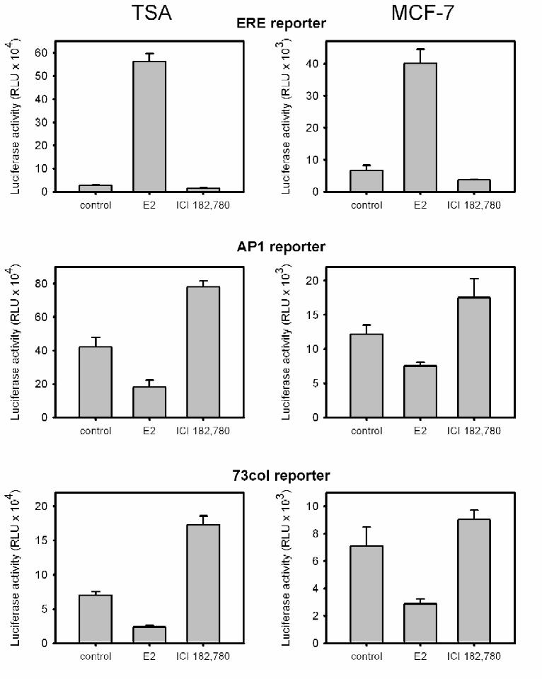
ER agonists repress and antagonists stimulate transcription mediated by the non-classical AP1
pathway
The response to ER agonists and antagonists was examined using classical and non-
classical pathway reporter constructs. For the classical pathway, ERE2-tk109-luc, which
contains two copies of the vitellogenin gene ERE upstream of the 109 base pair fragment of
thymidine kinase promoter, was used. For the non-classical pathway, two different AP1
luciferase reporters were used: 73col-luc, which contains a fragment of the collagenase promoter
(-73 to +63) and includes a single AP1 site (24), and AP1-luc, which contains seven AP1 sites
upstream of a basal promoter (Stratagene). Transfections were performed in ER-negative TSA
201 cells.
In the absence of transfected ER, hormone treatments did not alter the activity of any of
these reporter constructs, confirming the absence of endogenous ER or other estrogen-
responsive pathways (data not shown). When ER was co-transfected with the ERE reporter,
estradiol activated and the antiestrogen ICI 182,780 repressed transcription (Fig 1, TSA, ERE
reporter). The responses to agonists and antagonists were reversed when the AP1-luc reporter
was used. Estradiol suppressed transcription, and the antiestrogen ICI 182,780 stimulated
promoter activity (Fig. 1, TSA, AP1 reporter). These effects were specific for the presence of
AP1 sites; no response was observed using a reporter lacking the AP1 sites but retaining the
remainder of the promoter and the vector backbone (data not shown). A reporter containing a
9
by guest on March 31, 2019
http://ww
w.jbc.org/
Dow
nloaded from

fragment of the native collagenase promoter (-73 to +63), which contain a single AP1 site,
displayed a response pattern similar to the artificial AP1-luc reporter (Fig. 1, TSA, 73 col
reporter), and all subsequent experiments were performed with AP1-luc. Consistent with the
effects of estradiol and ICI 182,780, DES repressed whereas raloxifene, tamoxifen, and 4-
hydroxy-tamoxifen stimulated AP1-luc transcription (data not shown).
Reporter constructs were also transfected into ER-positive MCF-7 cells in the absence
of the ER expression vector. Responses mirrored those observed in TSA cells. Estrogen
activated the ERE reporter and repressed the AP1-luc and 73col-luc reporters, whereas ICI
182,780 repressed the ERE reporter and activated the AP1-luc and 73col-luc reporters (Fig. 1,
MCF-7). The ligand effects on the non-classical pathway were enhanced when exogenous ER
was transfected into MCF-7 cells along with the AP1-luc reporter (data not shown).
Mutations that abolish ER binding to the ERE do not disrupt activity through the non-classical
AP1 pathway
To determine whether the non-classical pathway requires ER binding to DNA, four
mutations were introduced into the ER DBD. The mutant receptors were examined for DNA
binding and functional activity using the ERE and AP1 reporters. These mutations were
hypothesized to preclude ER action through the classical pathway, but to retain the ability to
interact with other transcription factors, thereby potentially mediating actions through the non-
classical pathway. The structure of the zinc fingers and the location of introduced mutations are
depicted in Fig. 2A. The first 3 mutants are all within the “P-box” of the first zinc finger, a
region known to mediate interaction with DNA (30). The E207G/G208S mutant (a double
10
by guest on March 31, 2019
http://ww
w.jbc.org/
Dow
nloaded from

mutant) has been demonstrated previously to disrupt binding to the ERE (31). The
E207A/G208A mutant was created with the intent of better preserving the protein structure by
substituting with alanine residues. In the third mutant, K210 was changed to A, disrupting the
direct interaction of the positively charged lysine with DNA (32). The fourth mutant, A277T, is
in the “D-box” of the second zinc finger, a region that has been implicated in homodimerization
and thereby DNA binding (32).
The DNA-binding properties of these mutants were tested in gel mobility shift assays
(Fig. 2B). E207G/G208S had no detectable binding and E207A/G208A exhibited minimal
residual binding (0.8%) relative to the WT ER. The mutation in the dimerization domain
(A227T) retained a small amount of ERE binding (2.4% of WT). The K210A mutation was
substantially less effective than the other mutants at disrupting DNA binding (8.1% of WT).
The ability of these mutants to act through the classical and non-classical pathways was
examined in transient transfection assays (Fig. 3A,B). Three of the mutants (E207G/G208S,
E207A/G208A, K210A) showed little or no estrogen responsiveness through the ERE-mediated
classical pathway (Fig. 3A). The A227T D-box mutant retained significant estrogen
responsiveness through the ERE reporter. The loss of activity with the E207G/G208S and
E207A/G208A mutants was confirmed using artificial reporters containing EREs from other
estrogen responsive genes such as human pS2 and human oxytocin (data not shown). Western
blot analysis of nuclear extracts demonstrated equal expression of the WT and mutant proteins
(Fig. 3C).
In the AP1 reporter assay (Fig. 3B), the E207G/G208S, E207A/G208A, and A227T
mutants each retained the non-classical pattern of response. The E207A/G208A mutant was the
11
by guest on March 31, 2019
http://ww
w.jbc.org/
Dow
nloaded from

most active, with responses similar to those of the WT ER. Thus, E207A/G208A exhibits
selective loss of ERE binding and transcriptional control by the classical pathway, but retains full
regulation by the non-classical pathway. The K210A mutant lost the ability to act through the
non-classical pathway.
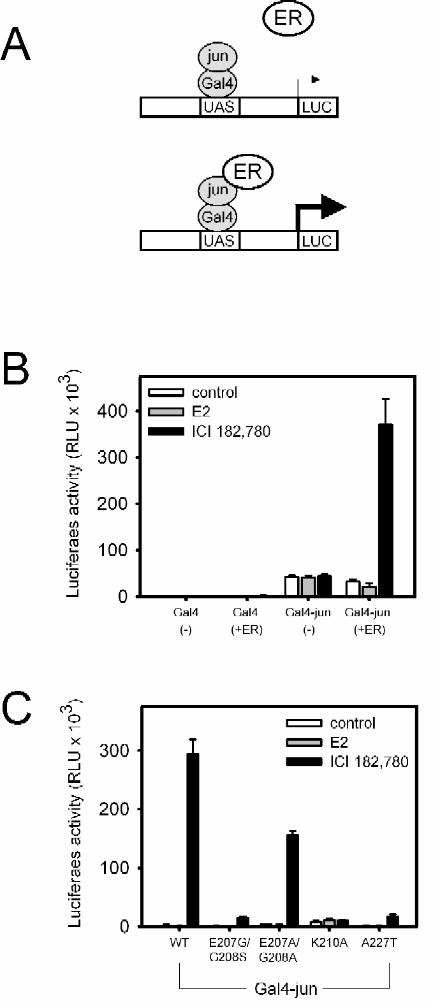
Functional interaction between ER and the AP1 protein Jun
A mammalian two-hybrid assay was used to test the hypothesis that ER interacts with
AP1 proteins in the non-classical pathway. WT or mutant ER was cotransfected with a Gal4-jun
fusion protein, using UAS-E1b-TATA-luc as a reporter (Fig. 4B). In control experiments, the
Gal4 DBD alone did not activate the UAS-E1b-TATA-luc reporter, either in the absence or
presence of ER. Gal4-jun increased basal expression but did not confer responsiveness to
estradiol or ICI 182,780. When WT ER was coexpressed, estradiol repressed Gal4-jun mediated
transcription, whereas ICI 182,780 activated transcription, a pattern that mimics the activity seen
in the AP1 reporter assays.
The activities of the DBD mutants were also tested in this assay. E207A/G208A, a
mutant that preserved the non-classical response pattern for AP1 (Fig. 3B), strongly activated
Gal4-jun mediated transcription in the presence of ICI 182,780 (Fig. 4C). The E207G/G208S
and A227T mutants, which exhibit less robust activity in the AP1 reporter assay (Fig. 3B),
activated Gal4-jun to a lesser extent than E207A/G208A or WT ER (Fig. 4C). K210A lost all
activity in both the AP1 reporter assay and in the interaction assay. Thus, the transcriptional
properties of the ER DBD mutants are similar in the Gal4-jun and AP1 reporter assays.
12
by guest on March 31, 2019
http://ww
w.jbc.org/
Dow
nloaded from

ER mutations that abolish interactions with coactivators disrupt signaling through the classical
and non-classical pathways
The LBD of ER plays an important role in cofactor interactions, which are central to ER
activity through the ERE. Two point mutations (I362R and K366D) were introduced into ER
helix 3, which together with helices 4, 5 and 12 form a hydrophobic cavity that is involved in
cofactor interactions. The specific substitutions were chosen based on their high degree of
conservation when compared to related nuclear receptors (Fig. 5A), and because similar
mutations have been reported to disrupt interactions with specific coactivators, including SRC-1
(33,34). Using a mammalian two-hybrid system, these mutant ERs were confirmed to lose most
(I362R), or all (K366D), of their interaction with Gal4-SRC-1 and Gal4-GRIP-1 in the
presence of estradiol (data not shown).
The K366D mutation largely eliminated the effects of estradiol and ICI 182,780 (Fig. 5B,
C). With the ERE reporter, basal activity was greatly reduced, as were estradiol activation and
ICI suppression. With the AP1 reporter, estradiol suppression was eliminated and ICI stimulation
was markedly decreased. The effect of the I362R mutation was more dependent on the ligand
(estradiol versus ICI). Estradiol stimulation of the ERE reporter and suppression of the AP1
reporter was retained. ICI 182,780 stimulated the ERE reporter and elicited a small decrease with
the AP1 reporter. These results indicate that ER mutations that alter binding to transcriptional
cofactors impair ER action through both the classical and non-classical pathways.
The effects of the helix 3 mutants in the mammalian two-hybrid assays with Gal4-jun
(Fig. 5D) were similar to those seen with the AP1 reporter, consistent with the idea that ER
actions on the AP1 reporter are mediated through interactions with Jun.
13
by guest on March 31, 2019
http://ww
w.jbc.org/
Dow
nloaded from
Discussion
In the classical pathway, ER action is mediated by direct receptor binding to EREs.
Agonist binding induces an ER conformation that favors interactions with coactivators and
general transcription factors, resulting in increased transcription. When bound to an antagonist,
the ER does not interact with coactivators and, in turn, does not activate transcription. In
contrast, the mechanism by which ER regulates non-classical pathways is less well established.
We hypothesized that this mechanism involves ER interactions with other proteins, rather than
the direct binding to DNA. AP1-regulated genes were used as a model system for the non-
classical pathway. With these reporter genes, agonists repress and antagonists activate
transcription in the presence of ER. We identified a DBD mutant of ER (E207A/G208A) that lost
transactivation through the classical pathway, but retained regulation of the non-classical
pathway. These data provide new insights into mechanisms by which non-classical ER signaling
occurs. First, ligands traditionally considered estrogen receptor agonists or antagonists have an
opposite effect on the non-classical AP1 pathway, where agonists repress and antagonists
activate transcription. Such a reversal of activity suggests that a novel mechanism mediates ER
14
by guest on March 31, 2019
http://ww
w.jbc.org/
Dow
nloaded from
activity through the non-classical AP1 pathway. Second, it is possible to abolish the ability of
ER to bind to an ERE, and still preserve activity through the non-classical pathway. However, as
discussed below, the ER DBD likely interacts with other proteins, such as Jun, and this
interaction requires a structurally intact DBD. Third, the interaction between ER and cofactors
has functional significance both in the classical and the non-classical pathways.
In the non-classical pathway, we found that estradiol represses and ICI 182,780 activates
transcription via the AP1 reporter, a pattern which is the opposite of the ligands’ effects on the
ERE reporter. Though the activation of transcription by an antagonist is consistent with other
reports (15-17), the repressive effect of estradiol on the collagenase promoter has not been
observed previously, perhaps due to differences in experimental conditions. However, estrogen-
mediated suppression of gene expression is a common physiologic phenomenon and includes
genes such as IL-6 (13,14), TNFα (35), FSHβ (20), choline acetyltransferase (21), quinone
reductase (36), and lipoprotein lipase (37). In many cases, negative regulation involves the AP1
site (20,21,37).
ER action through the non-classical pathway does not involve ER binding to DNA, and
appears to be mediated by protein-protein interactions. For example, the P-box mutation
E207A/G208A in the first zinc finger eliminates DNA binding and ERE activation but preserves
activity through the non-classical pathway. A possible candidate for the protein-protein
interaction in the non-classical pathway is Jun, a member of the AP1 protein family. This idea is
supported by evidence for ER interaction with Gal4-jun in mammalian two-hybrid assays and
by the observation that ER mutants have similar effects when tested in Gal4-jun interaction
assays or in AP1 reporter assays. The ICI-induced interaction between Jun and ER detected in a
15
by guest on March 31, 2019
http://ww
w.jbc.org/
Dow
nloaded from

two-hybrid assay may not be direct, as other cellular proteins (e.g. coactivators) may participate
in the Gal4-jun-ER interaction. Attempts to supershift AP1-bound Jun with ER in EMSAs, or
to co-immunoprecipitate a Jun-ER complex, did not detect direct interactions (data not shown).
It is possible that these interactions are not strong enough to withstand the experimental
conditions. Alternatively, cofactors such as SRC-1, or other proteins, may bridge or stabilize the
Jun-ER complex.
Previous studies have demonstrated a direct interaction between ER and c-jun using GST
pull-down assays (16,38). It is notable, however, that in the mammalian two-hybrid assays, the
ER-jun interaction was induced by ICI 182,780 but not by estradiol, suggesting that
conformational changes may influence the protein interactions. The mechanism of estradiol-
induced repression remains unknown, but may be influenced by the promoter context of the
estrogen-dependent regulatory sites. For example, activation of interleukin 6 (IL-6) promoter
requires the synergistic activity of two transcription factors--CAAT enhancer binding protein β
(C/EBPβ) and NFκB. In this case, estradiol-induced repression appears to involve interactions
with each of these factors (13,14). Another possible repression mechanism could involve
estrogen-mediated inhibition of the Jun amino-terminal kinase (JNK) pathway (39). For
example, estrogen-dependent repression of RANK (receptor activator of NFκB ligand) appears
to involve downregulation of c-jun expression and a decrease in Jun phosphorylation by JNK
(40).
As with the AP1 reporter, WT ER and the DBD mutants activate Gal4-jun dependent
transcription in the presence of ICI 182,780. We found that a construct containing only the DEF
domains of ER does not affect transcription from the AP1 reporter despite hormonal treatment,
16
by guest on March 31, 2019
http://ww
w.jbc.org/
Dow
nloaded from

whereas the construct lacking the A/B domain, but containing the DBD, alters transcription in
the same manner as WT ER (data not shown). These findings are consistent with several
published reports (16,18,38), and raise the possibility that ER interactions with Jun involve a
region within the DBD. It is also notable that the K210A mutation eliminates activity through the
non-classical pathway, as well as interaction with Jun, even though this mutant retains partial
activity through the ERE. Because neither ER zinc finger is embedded within the protein
structure, the zinc fingers may participate in protein-protein interactions.
It is well established that coactivators are involved in the classical pathway. Using
selective ER mutants (I362R and K366D) that alter interactions with coactivators, we found that
these mutants not only affect the classical pathway, but also impair ER action through the non-
classical AP1 pathway. The hydrophobic pocket where these mutations were introduced interacts
with both coactivators and corepressors. For example, the mutation of residues analogous to I362
and K366 in TRα diminishes T3 activation, but also impairs basal repression and the interaction
with corepressors NCoR and SMRT (41). It is therefore possible that the I362R and K366D
mutations affect interactions with corepressors, although we did not detect any interactions
between ER and corepressors in two hybrid assays (data not shown), perhaps because these
interactions are weaker than the ER-coactivator interactions.
One can speculate that the reversal of the ligands’ effects on the non-classical pathway is
due to the reversal of cofactors bound to liganded ER. It is somewhat counterintuitive that the
antagonist ICI 182,780 might cause an interaction between the ER and coactivators, and that
estradiol would induce an interaction between the ER and corepressors. However, it is possible
that when the ER is involved in a protein-protein interaction instead of binding to an ERE, the
17
by guest on March 31, 2019
http://ww
w.jbc.org/
Dow
nloaded from

hydrophobic cofactor-binding pocket may assume a different shape, shifting the interactions of
associated proteins. As mentioned above, the same region of the LBD recognizes both
coactivators and corepressors (41), increasing the likelihood that subtle changes in the tertiary
protein structure could change a subset of proteins recognized by the ER.
The I362R mutant provides an additional argument in support of the hypothesis that
subtle changes in protein structure can have a profound effect on the recognition of coactivators.
This mutation does not completely eliminate the interaction with coactivators, and therefore still
stimulates transcription through the classical pathway in the presence of estradiol. However, this
mutant also activates transcription in the presence of an antagonist. In this respect, the I362R
mutation resembles some mutations of helix 12 that “switch” an antagonist to an agonist.
Examples include the mutant L540Q of hERα (42) and the double mutants L543A/L544A and
M547A/L548A of mERα (43), which are activated by ICI 164,384 and 4-hydroxy-tamoxifen.
We conclude that for the AP1 response element, ER interacts with other proteins instead
of, or in addition to, DNA to exert its transcriptional effects. The non-classical pathway retains
the requirement for cofactors. However, the patterns of cofactor binding change, as demonstrated
by the reversal of estradiol/ICI activities through the AP1 reporter. These data confirm the
sensitivity of ER activity to subtle changes in its structure, whether caused by artificial mutations
or an altered set of associated proteins.
18
by guest on March 31, 2019
http://ww
w.jbc.org/
Dow
nloaded from
Fig. 1. Estradiol and ICI 182,780 have opposite effects on the classical (ERE reporter) and the
non-classical pathways (AP1 reporters).
Transient transfection of TSA cells (TSA column) with mouse estrogen receptor alpha (mERα)
expression vector and reporters (ERE, ERE2-tk109-luc; AP1, AP1-luc; 73col, 73col–luc) and
transient transfection of MCF-7 cells (MCF-7 column) with the indicated reporters.
Fig. 2. Mutations introduced into the DBD of mERα abolish or diminish ER binding to DNA.
A. Schematic illustration of the mERα zinc fingers in the DBD. Amino acids that constitute P-
box and D-box are in bold letters. Residues E207 and K210 within the P-box make direct
contacts with the DNA base pairs. Residues P226, A227, and Q230 within the D-box directly
participate in the formation of a dimer. Mutated amino acids are circled.
19
by guest on March 31, 2019
http://ww
w.jbc.org/
Dow
nloaded from
B. Electrophoretic mobility shift assay using the ERE vitellogenin probe and WT or mutant
mERα proteins.
Fig. 3. Mutations that eliminate ERE binding preserve activity through the non-classical AP1
pathway.
A. Transient transfection of TSA cells using the ERE2-tk109-luc reporter and the indicated WT
and mutant mERα constructs.
B. Transient transfection of TSA cells using the AP1-luc reporter.
C. Western blot analysis of nuclear extracts of TSA cells transiently transfected with the
indicated constructs of mERα, using anti-ERα antibody.
Fig.4. Functional interaction between Jun and ER.
A. Schematic illustration of the experimental design.
B. Transient transfection of TSA cells using the UAS-E1b-TATA-luc reporter, Gal4 alone, or
the Gal4-jun fusion protein with full length mERα or empty vector pcDNA3.1.
C. Transient transfection of TSA cells using UAS-E1b-TATA-luc, Gal4-jun fusion and the
indicated mERα constructs.
Fig. 5. Evidence for transcriptional cofactor interaction in the classical and non-classical
pathways.
A. Alignment of the sequence of helix 3 among mERα and other members of the nuclear
receptor family. Mutated residues are shown in bold.
20
by guest on March 31, 2019
http://ww
w.jbc.org/
Dow
nloaded from

B. Transient transfection of TSA cells using the ERE2-tk109-luc reporter and the indicated
mERα constructs.
C. Transient transfection of TSA cells using the AP1-luc reporter and the indicated mERα
constructs.
D. Transient transfection of TSA cells with UAS-E1b-TATA-luc reporter, Gal4-jun and
expression vectors containing wt mERα or its mutants I362R or K366D.
Acknowledgements
The authors would like to thank Malcolm Parker for providing mERα cDNA, Alan Wakeling for providing ICI 182,780 and Craig Jordan for providing advice and the human pS2 and human oxytocin ERE luciferase reporter constructs. The work was supported by SPORE grant IP50 CA89018-01.References
1. Tsai, M. J., and O’Malley, B. W. (1994) Annu Rev Biochem 63, 451-862. Ribeiro, R. C., Kushner, P. J., and Baxter, J. D. (1995) Annu Rev Med 46, 443-533. Kumar, V., and Chambon, P. (1988) Cell 55(1), 145-564. Tremblay, G. B., Tremblay, A., Labrie, F., and Giguere, V. (1999) Mol Cell Biol 19(3),
1919-275. Ogawa, S., Inoue, S., Watanabe, T., Hiroi, H., Orimo, A., Hosoi, T., Ouchi, Y., and
Muramatsu, M. (1998) Biochemical & Biophysical Research Communications 243(1), 122-6
6. Pace, P., Taylor, J., Suntharalingam, S., Coombes, R. C., and Ali, S. (1997) Journal of Biological Chemistry 272(41), 25832-8
7. Cowley, S. M., Hoare, S., Mosselman, S., and Parker, M. G. (1997) Journal of Biological Chemistry 272(32), 19858-62
8. McKenna, N. J., Lanz, R. B., and O’Malley, B. W. (1999) Endocr Rev 20(3), 321-449. Brzozowski, A. M., Pike, A. C., Dauter, Z., Hubbard, R. E., Bonn, T., Engstrom, O.,
Ohman, L., Greene, G. L., Gustafsson, J. A., and Carlquist, M. (1997) Nature 389(6652), 753-8
10. Shiau, A. K., Barstad, D., Loria, P. M., Cheng, L., Kushner, P. J., Agard, D. A., and Greene, G. L. (1998) Cell 95(7), 927-37
11. Smith, C. L., Nawaz, Z., and O’Malley, B. W. (1997) Mol Endocrinol 11(6), 657-66
21
by guest on March 31, 2019
http://ww
w.jbc.org/
Dow
nloaded from

12. Lavinsky, R. M., Jepsen, K., Heinzel, T., Torchia, J., Mullen, T. M., Schiff, R., Del-Rio, A. L., Ricote, M., Ngo, S., Gemsch, J., Hilsenbeck, S. G., Osborne, C. K., Glass, C. K., Rosenfeld, M. G., and Rose, D. W. (1998) Proc Natl Acad Sci U S A 95(6), 2920-5
13. Stein, B., and Yang, M. X. (1995) Molecular & Cellular Biology 15(9), 4971-914. Ray, P., Ghosh, S. K., Zhang, D. H., and Ray, A. (1997) FEBS Letters 409(1), 79-8515. Paech, K., Webb, P., Kuiper, G. G., Nilsson, S., Gustafsson, J., Kushner, P. J., and
Scanlan, T. S. (1997) Science 277(5331), 1508-1016. Webb, P., Lopez, G. N., Uht, R. M., and Kushner, P. J. (1995) Molecular Endocrinology
9(4), 443-5617. Webb, P., Nguyen, P., Valentine, C., Lopez, G. N., Kwok, G. R., McInerney, E.,
Katzenellenbogen, B. S., Enmark, E., Gustafsson, J. A., Nilsson, S., and Kushner, P. J. (1999) Mol Endocrinol 13(10), 1672-85
18. Umayahara, Y., Kawamori, R., Watada, H., Imano, E., Iwama, N., Morishima, T., Yamasaki, Y., Kajimoto, Y., and Kamada, T. (1994) Journal of Biological Chemistry 269(23), 16433-42
19. Gaub, M. P., Bellard, M., Scheuer, I., Chambon, P., and Sassone-Corsi, P. (1990) Cell 63(6), 1267-76
20. Miller, C. D., and Miller, W. L. (1996) Endocrinology 137(8), 3437-4621. Schmitt, M., Bausero, P., Simoni, P., Queuche, D., Geoffroy, V., Marschal, C., Kempf, J.,
and Quirin-Stricker, C. (1995) Journal of Neuroscience Research 40(2), 152-6422. Sadowski, I., and Ptashne, M. (1989) Nucleic Acids Res 17(18), 753923. Gehm, B. D., McAndrews, J. M., Chien, P. Y., and Jameson, J. L. (1997) Proc Natl Acad
Sci U S A 94(25), 14138-4324. Angel, P., Imagawa, M., Chiu, R., Stein, B., Imbra, R. J., Rahmsdorf, H. J., Jonat, C.,
Herrlich, P., and Karin, M. (1987) Cell 49(6), 729-3925. Tagami, T., Lutz, W. H., Kumar, R., and Jameson, J. L. (1998) Biochem Biophys Res
Commun 253(2), 358-6326. Margolskee, R. F., McHendry-Rinde, B., and Horn, R. (1993) Biotechniques 15(5), 906-
1127. Nagaya, T., Madison, L. D., and Jameson, J. L. (1992) J Biol Chem 267(18), 13014-928. Chien, P. Y., Ito, M., Park, Y., Tagami, T., Gehm, B. D., and Jameson, J. L. (1999) Mol
Endocrinol 13(12), 2122-3629. Johnson, W., Albanese, C., Handwerger, S., Williams, T., Pestell, R. G., and Jameson, J.
L. (1997) J Biol Chem 272(24), 15405-1230. Kumar, V., Green, S., Stack, G., Berry, M., Jin, J. R., and Chambon, P. (1987) Cell 51(6),
941-5131. Mader, S., Kumar, V., de Verneuil, H., and Chambon, P. (1989) Nature 338(6212), 271-
432. Schwabe, J. W., Chapman, L., Finch, J. T., and Rhodes, D. (1993) Cell 75(3), 567-7833. Henttu, P. M., Kalkhoven, E., and Parker, M. G. (1997) Mol Cell Biol 17(4), 1832-934. Mak, H. Y., Hoare, S., Henttu, P. M., and Parker, M. G. (1999) Mol Cell Biol 19(5),
3895-90335. Srivastava, S., Weitzmann, M. N., Cenci, S., Ross, F. P., Adler, S., and Pacifici, R.
(1999) J Clin Invest 104(4), 503-1336. Montano, M. M., and Katzenellenbogen, B. S. (1997) Proc Natl Acad Sci U S A 94(6),
22
by guest on March 31, 2019
http://ww
w.jbc.org/
Dow
nloaded from
2581-637. Homma, H., Kurachi, H., Nishio, Y., Takeda, T., Yamamoto, T., Adachi, K., Morishige,
K., Ohmichi, M., Matsuzawa, Y., and Murata, Y. (2000) J Biol Chem 275(15), 11404-1138. Sabbah, M., Courilleau, D., Mester, J., and Redeuilh, G. (1999) Proc Natl Acad Sci U S
A 96(20), 11217-2239. Caelles, C., Gonzalez-Sancho, J. M., and Munoz, A. (1997) Genes & Development
11(24), 3351-6440. Shevde, N. K., Bendixen, A. C., Dienger, K. M., and Pike, J. W. (2000) Proceedings of
the National Academy of Sciences of the United States of America 97(14), 7829-3441. Hu, X., and Lazar, M. A. (1999) Nature 402(6757), 93-642. Montano, M. M., Ekena, K., Krueger, K. D., Keller, A. L., and Katzenellenbogen, B. S.
(1996) Mol Endocrinol 10(3), 230-4243. Mahfoudi, A., Roulet, E., Dauvois, S., Parker, M. G., and Wahli, W. (1995) Proc Natl
Acad Sci U S A 92(10), 4206-10
23
by guest on March 31, 2019
http://ww
w.jbc.org/
Dow
nloaded from


JamesonMonika Jakacka, Masafumi Ito, Jeffrey Weiss, Pei-Yu Chien, Barry D. Gehm and J. Larry
Non-classical AP1 PathwayEstrogen Receptor Binding to DNA is Not Required for its Activity Through the
published online January 18, 2001J. Biol. Chem.
10.1074/jbc.M008384200Access the most updated version of this article at doi:
Alerts:
When a correction for this article is posted•
When this article is cited•
to choose from all of JBC's e-mail alertsClick here
by guest on March 31, 2019
http://ww
w.jbc.org/
Dow
nloaded from