estimated plasma volume status in heart failure: clinical
TRANSCRIPT

HAL Id: hal-03109061https://hal.univ-lorraine.fr/hal-03109061
Submitted on 13 Jan 2021
HAL is a multi-disciplinary open accessarchive for the deposit and dissemination of sci-entific research documents, whether they are pub-lished or not. The documents may come fromteaching and research institutions in France orabroad, or from public or private research centers.
L’archive ouverte pluridisciplinaire HAL, estdestinée au dépôt et à la diffusion de documentsscientifiques de niveau recherche, publiés ou non,émanant des établissements d’enseignement et derecherche français ou étrangers, des laboratoirespublics ou privés.
Estimated plasma volume status in heart failure: clinicalimplications and future directions
Masatake Kobayashi, Nicolas Girerd, Kevin Duarte, Tahar Chouihed, TaishiroChikamori, Bertram Pitt, Faiez Zannad, Patrick Rossignol
To cite this version:Masatake Kobayashi, Nicolas Girerd, Kevin Duarte, Tahar Chouihed, Taishiro Chikamori, et al..Estimated plasma volume status in heart failure: clinical implications and future directions. ClinicalResearch in Cardiology, Springer Verlag, 2021, �10.1007/s00392-020-01794-8�. �hal-03109061�
1
Estimated Plasma Volume Status in Heart Failure:
Clinical Implications and Future Directions
Masatake Kobayashi 1 , Nicolas Girerd
1 , Kevin Duarte
1, Tahar Chouihed
1, Taishiro Chikamori
2,
Bertram Pitt,3 Faiez Zannad,
1 Patrick Rossignol
1
1. Université de Lorraine, INSERM, Centre d'Investigations Cliniques 1433, CHRU de Nancy,
Inserm 1116 and INI-CRCT (Cardiovascular and Renal Clinical Trialists) F-CRIN Network,
Nancy, France.
2. Department of Cardiology, Tokyo Medical University, Tokyo, Japan.
3. University of Michigan School of Medicine, Ann Arbor, Michigan, USA.
Corresponding Author:
Pr. Patrick Rossignol
Centre d'Investigation Clinique Pierre Drouin -INSERM - CHRU de Nancy, Institut lorrain du cœur et
des, vaisseaux Louis Mathieu, Nancy, France
Address : 4, rue du Morvan. 54500 Vandoeuvre-Les-Nancy
E-mail: [email protected]
Short title: Monitoring congestion with hemoconcentration/hemodilution in HF
Keywords: Plasma volume, congestion, hemoglobin, hematocrit, heart failure
Tables and figures: 2 tables

2
Abbreviations and Acronyms
PV=plasma volume
ePVS=estimated plasma volume status
HF=heart failure
CHF=chronic heart failure
AHF=acute heart failure
ADHF=acutely decompensated heart failure
HFrEF=heart failure with reduced ejection fraction
HFpEF=heart failure with preserved ejection fraction
HFmrEF=heart failure with mid-range ejection fraction

3
Abstract
Congestion is one of the main predictors of poor outcome in patients with heart failure (HF).
Assessing and monitoring congestion is essential for optimizing HF therapy. Among the various
available methods, serial measurements of estimated plasma volume (ePVS) using routine blood count
and/or body weight (e.g. the Strauss, Duarte, Hakim formulas) may be useful in HF management.
Further prospective study is warranted to determine whether ePVS can help optimize decongestion
therapy (loop diuretics, mineralocorticoid receptor antagonists, SGLT2i) in various HF settings. This
narrative review summarizes the recent evidence supporting the association of ePVS with clinical
congestion and outcome(s) and discusses future directions for monitoring ePVS in HF.
4
Introduction
Congestion is one of the main mechanisms underlying worsening heart failure (HF) and is associated
with poor outcome. Insufficient decongestion is associated with higher rates of HF re-admission and
death.1 Therefore, it is of paramount importance to better detect and monitor congestion before
progression to decompensation 2 and to assess the intensity of decongestion for an optimal tailoring of
HF therapy. International guidelines have long recommended assessing “volume status” in order to
adapt diuretic doses 3. However, congestion can be difficult to assess, especially when extrapulmonary
signs of congestion are mild, or in patients with residual congestion at discharge from a HF
hospitalization 2.
According to the principle of total body volume distribution, all fluid compartments are
balanced, and plasma volume (PV) would interact with the interstitial and intracellular compartments
4-7. PV expansion in patients with HF is associated with increases in all fluid compartments. In
particular, PV in patients with decompensated HF may increase by 40% compared with that in healthy
controls 8. In addition to accumulated fluid volume, the fluid redistribution from splanchnic venous
system to pulmonary circulation also contributes to expand PV, subsequently leading to urgent HF
hospitalization 2, 4, 9
. Decongestion therapy reduced greatly body weight/net fluid balance, but had less
impact on PV owing to plasma refilling from interstitial space to intravascular space 10, 11
. This
therefore warrants a reliable and actionable assessment of PV.
Hemoconcentration (or hemodilution) gauged by a change in hemoglobin or hematocrit
concentrations as an indirect marker of changes in PV has been shown to trend with clinical
assessments of congestion 12-14
. For instance, an analysis of the ESCAPE (Evaluation Study of
Congestive Heart Failure and Pulmonary Artery Catheterization Effectiveness) trial investigated
baseline-to-discharge increases in hematocrit, albumin and total protein values. Patients with 2 or
more of the 3 aforementioned variables with values in the top tertile were considered to have evidence
of hemoconcentration, which was found to be associated with greater net weight or fluid loss and
5
greater reduction in right atrial pressure and pulmonary capillary wedge pressure, along with a
substantially lower risk of mortality 12
. More recently, hemoglobin was found to be significantly
negatively correlated with all tested biological and ultrasound measurements of congestion in a
chronic HF population across a large spectrum of ejection fractions 15
. In a meta-analysis from a
systematic review including 18 studies and a total of 368 patients with HF, hemoglobin was found
negatively associated with measured PV (Beta=-6.66 ; SE=2.53, p=0.008)16
. Direct quantification of
PV accurately depicts the severity of congestion in patients with chronic HF 10, 17
. However, clinical
utility of direct PV measurement-guided management has not been well documented 18
. The
procedure, in addition, requires specialized equipment with relatively high cost and exceedingly high
logistical issues to implement, which may not readily available for clinicians. Therefore, PV estimated
from a routine blood count and/or body weight may be more clinically relevant for repeat use given its
low cost and practicability. Estimated PV status (ePVS) might provide a phenotypic characteristic to
tailor personalized therapy in the different settings of HF.
This narrative review summarizes the recent evidence supporting the association of ePVS with
clinical congestion and outcome and discusses future directions for monitoring ePVS in HF.
Estimated and Measured Plasma Volume Status
Formulae for Estimating Plasma Volume
Among recent reports in HF settings, two representative methods are available for estimating PV. Both
express relative values of PV and use routinely available variables (i.e., hemoglobin and body weight).
Strauss Formula
The concept of estimating PV was first reported by Strauss et al. in 1951 19
. This seminal research of
the Strauss formula demonstrated the association between the infusion of saline into human bodies and
rapid changes in their estimated PV. In 1970’s, Dill et al. reported the association of a decrease in
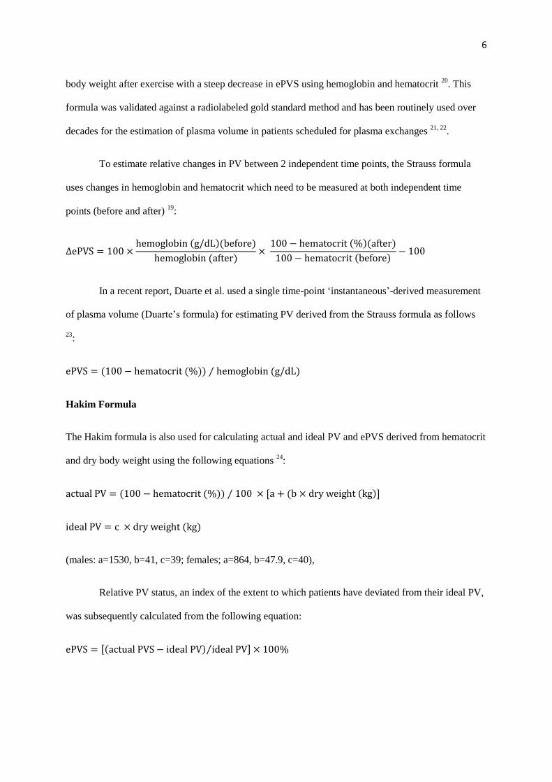
6
body weight after exercise with a steep decrease in ePVS using hemoglobin and hematocrit 20
. This
formula was validated against a radiolabeled gold standard method and has been routinely used over
decades for the estimation of plasma volume in patients scheduled for plasma exchanges 21, 22
.
To estimate relative changes in PV between 2 independent time points, the Strauss formula
uses changes in hemoglobin and hematocrit which need to be measured at both independent time
points (before and after) 19
:
In a recent report, Duarte et al. used a single time-point ‘instantaneous’-derived measurement
of plasma volume (Duarte’s formula) for estimating PV derived from the Strauss formula as follows
23:
Hakim Formula
The Hakim formula is also used for calculating actual and ideal PV and ePVS derived from hematocrit
and dry body weight using the following equations 24
:
(males: a=1530, b=41, c=39; females; a=864, b=47.9, c=40),
Relative PV status, an index of the extent to which patients have deviated from their ideal PV,
was subsequently calculated from the following equation:

7
One caveat with the Hakim formula is that it warrants dry body weight which is a major
potential confounder in the HF setting, where there is a complex interplay between congestion and loss
in body weight (“cachexia”), both associated with poor prognosis 25.
Measured Plasma Volume
Radioisotope assays are considered as the gold-standard, as they accurately assess PV status along
with red blood cell and total blood volume, using the concomitant determination of hematocrit. The
latter provides relevant information, since red blood cell abnormalities (non-hemodilutional anemia or
polycythemia) may coexist with PV abnormalities in patients with HF and may challenge its isolated
interpretation 7, 18
. Notwithstanding, the radioisotope assays are expensive and impractical for serial
monitoring, due to the need of frequent venous blood sampling and processing delays 26
.
131I-labeled albumin method 18
The 131I-labeled albumin method (Volumex, Daxor Corporation, New York) is a validated and
standardized clinically available technique. Low-dose iodinated I-131-labeled albumin is injected, and
from a contralateral forearm venous catheter, blood samples are collected at time 0 (pre-injection), and
at 12, 18, 24, 30, and 36 min post-injection. This technique may measure PV more quickly than 125 I-
HSA method 27
. Hematocrit is determined from each sample, and the plasma radioactivity of each
sample is measured (in duplicate) in a semi-automated counter (Food and Drug Administration
approved BVA-100 Blood Volume Analyzer, Daxor Corporation). Subsequent to an initial assessment
in patients with acutely decompensated HF (ADHF), it was proposed that the readily available
peripheral hematocrit test results may provide an updated view of the patients’ volume status, such
that, in those cases where it could be assumed that the red blood cell volume was reasonably stable
(absent bleeding or interventions to correct anemia), an increase in peripheral hematocrit could be
interpreted as a true (decongestive) decrease in PV 18
.

8
125I-HSA method 28
The International Committee for Standardization in Hematology recommends the 125I-HSA method
28. A 125I-labeled albumin is injected peripherally, and blood is collected from a different vein
(usually the opposite arm) at 10-, 20- and 30-min post-injection. The radioactivity of the plasma
samples and the standard are determined in a scintillation counter. PV is calculated by the following
equation:
99mTc-HSA method 7, 26
Technetium (99Tc)-labeled red blood cells is injected peripherally after 60 min in supine position.
Thereafter, blood is collected at 10-min intervals for 30minutes. Radioactivity is measured in an
automated counter. PV is calculated from the measured blood volume, venous hematocrit, corrected
for the trapped plasma and mean body hematocrit.
IV-administered visible fluorescent injectate (VFI)™ (FAST BIOMedical) 29
Recently, a non-isotopic (fluorescent) method to rapidly quantify PV and renal function was
developed. The method consists of 12mg of a 150-kDa carboxy-methylated dextran, conjugated to a
rhodamine dye (plasma volume marker), and 35mg of a 5-kDa carboxy-methylated dextran,
conjugated to fluorescein (renal clearance marker), in a total volume of 3.0 ml. As the large molecule
is retained in the intravascular space, its concentration reflects real-time plasma volume, and since the
small molecule is freely filtered across the glomerulus, its change represents the glomerular filtration
rate. The injectate is infused intravenously over 30s. Dye concentrations are quantified at only 1-time
point, 15 min after the injections for initial PV measurements using the indicator-dilution principle,
and this timing may have less influence on the value of PV 29
. Blood volumes are calculated based on
PV and the subject’s hematocrit. Samples are also taken at 60 and 170 min for determination of
glomerular filtration rate by the FAST BioMedical technique 29
. Validation results were recently

9
published using data from 16 healthy subjects and 16 subjects with chronic kidney disease.
Additionally, this technique accurately tracked dynamic changes in PV induced by a volume challenge
following an intravenous bolus of 350 mL of a 5% albumin solution in normal saline in 8 healthy
subjects 29
. The ongoing EMPAKT-CHF (Estimating versus Measuring Plasma Volume and Kidney
Function in Acute Decompensated Congestive Heart Failure) study is a one-armed, phase 2 clinical
trial using (VFI)™ to assay and assess measured PV and measured glomerular filtration rate in
hospitalized patients with acute decompensated congestive HF (NCT03808948).
Estimated Plasma Volume Status as a Congestion Marker
Comparison between Estimated Plasma Volume Status and Direct Plasma Volume
Several reports have assessed the correlation between ePVS and conventional radioisotope-labeled
albumin or red blood cell assays. Ling et al. compared ePVS derived from the Hakim formula and
actual PV using the gold standard 125Iodine labeled human serum albumin method 30
. Results showed
a moderate correlation between measured PV and estimated PV (r=0.51, p=0.006) in 30 patients with
CHF (r=0.68, p<0.001 in 119 healthy subjects). Martens et al. also compared ePVS derived from
Hakim and PV expansion using Technetium (99Tc)-labeled red blood cells in stable patients with
HFrEF, showing moderate to good correlation (r=0.71, p<0.001) and a good agreement between
measured and calculated PV 26
. Furthermore, Fudim et al. compared ePVS and measured PV in 110
stable CHF patients with NYHA class III or IV 31
. The authors used both the Hakim formula and
Strauss formulae (change and instantaneous), compared to low-dose 131
I-labeled albumin
intravenously. They reported moderate-to-strong correlation of the Hakim formula with the measured
quantitative PV (r=0.51, p=0.006 and r=0.64, p<0.001), while the Strauss formula ePVS was weakly
correlated with measured PV (r=0.29, p=0.003). A recent report comparing Strauss-derived ePVS and
PV measured by 125
I-human serum albumin in 10 patients with type 2 diabetes showed good
correlation (Lin's concordance index of 0.6 (p< 0.01) between both PV measurements) 32
.

10
Association between Clinical Congestion Markers and Estimated Plasma Volume Status
Clinical predictors of higher estimated or measured PV, including demographics, comorbidities,
ultrasound/hemodynamic parameters and biomarkers are presented in Table 1. Similar clinical
characteristics have been reported between ePVS derived from the Strauss and Hakim formulae.
Patients with higher ePVS were older, had more frequent ischemic heart disease, more severe clinical
congestion, poorer renal function, higher natriuretic peptide and were less likely to be on HF
medications 30, 33-35
. Recent published data showed the Duarte-derived ePVS was associated with left-
sided congestion such as E/e’ 36
, and with pulmonary artery pressure as assessed by an implanted
home monitoring device 37
. In addition, other data showed the significant association between ePVS
and echocardiographic parameters (i.e. tricuspid regurgitation) during decongestion therapy 38
. Of
note, some available data in the HF cohort studies demonstrated good correlations between
instantaneous and/or changes in ePVS and natriuretic peptide. Using data from the Eplerenone Post-
Acute Myocardial Infarction Heart Failure Efficacy and Survival Study (EPHESUS) trial, Rossignol et
al. showed a relatively weak correlation between baseline to 1-month changes in ePVS and BNP
(r=0.284; p<0.001), independently of study drug and renal function variations 39
. Not surprisingly,
significant associations between ePVS and congestion markers such as natriuretic peptide were also
observed in HFpEF 35
.
These findings therefore strengthen the evidence of ePVS as a clinically relevant congestion
marker.
Association of Estimated Plasma Volume Status with Clinical Outcomes in the Different Settings
of Heart Failure
Patients with acute heart failure (AHF) have more interstitial fluid volume due to fluid
accumulation/re-distribution compared to those with chronic heart failure (CHF) 9, 40, 41
, whereas fluid
volume profiles show a different distribution between patients with HFpEF and HFrEF 42, 43
. Androne

11
et al. showed that, in patients with CHF with absence of clinical congestion, increased measured blood
volume, as assessed by intravenous administration of iodine 131-labeled albumin, was associated with
an increased risk of death or urgent cardiac transplantation 17
. A number of other reports also found
independent associations between an increase in estimated PV level and a higher risk of clinical
outcomes, consistently and across a wide HF spectrum (acute/chronic, HFpEF/HFrEF) (Table 2).
Furthermore, across these HF phenotypes, ePVS improved consistently patient risk stratification on
top of established clinical-based models 23, 44, 45
. Both formulas incorporated hemoglobin and/or
hematocrit. Several underlying diseases (inflammation and renal impairment) may lead to anemia and
influence the value of ePVS. However, the prognostic value of ePVS persists regardless of the
presence of anemia in patients with de novo HF following acute myocardial infarction or in those with
acute HF 23, 33, 44
. Importantly, HFpEF manifested more often with anemia compared with HFrEF or
HFmrEF 46, 47
; therefore, further research is needed to explore whether prognostic value of ePVS
would be influenced by anemia or anemia-related diseases in HFpEF.
Chronic Heart Failure
The associations of ePVS with clinical outcomes have been well documented in patients with CHF
irrespective of LV systolic function.
Rossignol et al. investigated the prognostic value of changes in PV using the Strauss formula
in 6,820 patients with HFrEF in the Gruppo Italiano per lo Studio della Sopravvivenza
nell’Insufficienza Cardiaca-Heart Failure (GISSI-HF trial) 48. During a median follow-up of 3.9 years,
increased PV (>0%) from baseline was independently associated with a higher incidence of all-cause
mortality (adjusted HR [95%CI]=1.010 [1.002-1.018], p=0.017) and cardiovascular mortality
(adjusted HR [95%CI]=1.012 [1.003-1.020], p=0.007), independent of a significant decrease in body
weight. A post-hoc analysis of the Valsartan Heart Failure Trial (Val-HeFT) assessed ePVS derived
from the Hakim formula in 5,002 patients with HFrEF 30
. After a median follow-up of 716 days
(during which 20.0% of patients died), the highest ePVS quartiles were associated with increased all-
12
cause mortality after adjusting for clinical covariates including BNP (adjusted HR [95%CI]=1.26
[1.05-1.52], p=0.01). Similar prognostic implication of Strauss-derived or Hakim-derived ePVS is in
line with reports in HFpEF 35, 49
. In a post-hoc analysis of Treatment of Preserved Cardiac Function
Heart Failure with an Aldosterone Antagonist (TOPCAT), ePVS derived from the Hakim formula was
assessed in 3,414 patients with HFpEF, in which increased ePVS was associated with a higher risk of
the composite of all-cause death or HF re-hospitalization 35
.
Heart Failure with Reduced Ejection Fraction and Post-myocardial Infarction
In 6,080 patients with HFrEF following acute myocardial infarction from the EPHESUS study
database, after a mean follow-up of 16 months, a significant decrease in ePVS as assessed by the
Strauss-formula (delta ePVS) from baseline to 1 month was associated with a lower risk of clinical
outcomes 39
. Furthermore, in another report of the EPHESUS, Duarte et al. assessed the prognostic
value of instantaneous ePVS at 1 month as well as ePVS change from baseline to 1 month 23
. They
found that instantaneous ePVS at 1 month was a better predictor of cardiovascular events than change
in ePVS from baseline to 1 month over a follow-up period of 6 months. The fact that the Hakim
formula was not associated with outcomes in this analysis may arise from the integration of body
weight in this formula. Indeed, Hakim-derived ePVS increases when hematocrit decreases and
conversely decreases when weight decreases, whereas patients with events displayed lower weight and
hemoglobin. Ideally, dry weight (i.e., the body weight measured in non-congested patients), which
was not assessed in this latter study and is difficult to estimate in routine practice because of
frequently persisting edema in HF patients, should have been used to run this formula 23
.
Acute Heart Failure
A study assessing change in Strauss-derived ePVS from admission to discharge in 712 patients with
AHF 44
found that an increase in ePVS at discharge was associated with poor prognosis, and provided
a significant improvement in prognostic reclassification, on top of standard clinical prognostic models.
This is in keeping with findings of several smaller studies in AHF, showing the prognostic value of

13
discharge ePVS and of ePVS change from admission and discharge, likely depicting residual
congestion 50-52
. Other reports showed that patients with increased in admission ePVS also have worse
prognosis 33, 53
. In a recent report from the Effect of Nesiritide in Patients with Acute Decompensated
Heart Failure (ASCEND-HF) trial including 7,141 patients with decompensated HF, Duarte- and
Hakim-derived ePVS at baseline was associated with cardiovascular mortality or HF rehospitalization
at 30 days.
Non-Heart Failure
PV may manifest subclinical congestion, and its expansion may play crucial role of the perioperative
or emergency management even in patients without HF. In a retrospective cohort study including
1,887 patients undergoing coronary artery bypass graft surgery, preoperative assessment of Hakim-
derived ePVS ≥5.6% was associated with worse prognosis in a post-operative period 54
. Similarly,
worse prognostic value of higher ePVS was reported in patients undergoing transaortic valvular
intervention or mitral valvular intervention55, 56
. In addition, in 1,369 acute dyspneic patients admitted
to the emergency department, the highest tertiles of admission Duarte-derived ePVS was associated
with AHF diagnosis (adjusted OR [95%CI]=1.64 [1.16–2.33], p=0.005) as the main cause of dyspnea.
A value of ePVS >5.12 ml/g identified patients who, concomitant with infection or chronic renal
failure, were diagnosed with AHF in the emergency department. In this study, higher Duarte-derived
ePVS in dyspneic patients was associated with a higher risk of in-hospital mortality irrespective of the
presence of HF 57
.
Hemoconcentration and Worsening Renal Function
Aggressive decongestion as documented by a decrease in PV and hemoconcentration, improves
clinical outcomes in patients with HF 12-14, 58, 59
. Incident hemoconcentration, across a large range of
definitions, is frequently associated with worsening renal function 12-14, 58, 60-63
(Supplementary table
1), which has little negative prognostic influence 61, 64, 65
. In contrast, worsening renal function without

14
hemoconcentration, i.e. with persistent congestion, is associated with worse outcomes 66, 67
.
Importantly, in most of the aforementioned studies, hemoconcentration was derived from changes in
hemoglobin or hematocrit.
Plasma Volume as an Actionable Biomarker to Guide (De)-congestion Therapeutic Strategies in
Heart Failure
Half a century ago, furosemide infusion was shown to consistently increase hematocrit and decrease
estimated (Strauss formula) and measured (isotopic) plasma volume 68
. In the 1990’s, venovenous
ultrafiltration in chronic HF was found to decrease estimated PV (hematocrit-based formula) 69
along
with pulmonary wedge pressure and right atrial pressure, two established biomarkers of congestion.
Importantly, immediately after ultrafiltration, mean pulmonary wedge pressure decreased by 29% and
right atrial pressure decreased by 46%. Both variables remained unchanged in the next two days. PV
was reduced by 22% immediately after the procedure and by 10% after 24 hours, and subsequently
fully recovered in the next 24 hours. This differential kinetics may reflect a modest correlation
between ePVS changes and pulmonary wedge pressure changes. However, more importantly, changes
in ePVS and pulmonary wedge pressure were directionally and consistently concordant and were
simultaneously reduced by the ultrafiltration process. Ultimately, in HF, hemoconcentration, which is
easily and continuously measured by an in-line hematocrit sensor during ultrafiltration therapy, is
considered as an appropriate surrogate indicating when the plasma refill rate is exceeded by the rate of
fluid removal 70
. More recently, several contemporary HF cohort studies showed correlations between
volume depletion due to diuretic response and decreases in ePVS 30, 43, 44
.
The EPHESUS data in HF patients post-MI showed that the mineralocorticoid receptor
antagonist (MRA) eplerenone induced a diuretic-like effect compared to placebo, as assessed by
decreased ePVS (Strauss Formula) after one month, an effect associated with improved clinical
outcomes 39
. In patients with chronic HF, a higher dose of renin-angiotensin-antagonist-blocker

15
(RAS-blocker; ACE-I or ARB) and a higher dose of a MRA have moreover been associated with an
independent higher odds of having an optimal ePVS in HFrEF and HFmrEF 26
. In addition,
Sacubitril/Valsartan generally increases endogenous peptides with natriuretic effects, which may lower
circulating levels of aldosterone 71, 72
. Sacubitril/Valsartan, therefore, may theoretically decrease the
value of ePVS. Further research is warranted.
SGLT2 inhibitors are likely to become part of the life-saving drug armamentarium in HF,
downstream of the DAPA-HF trial in HFrEF 73
, while HFpEF trials are ongoing 74
. In a randomized,
placebo-controlled, double-blind trial, 75 subjects with type 2 diabetes were assigned either placebo,
the SGLT2 inhibitor dapagliflozin 10mg/day, or hydrochlorothiazide 25mg/day. SGLT2 inhibitor
dapagliflozin treatment resulted in a 7.3% reduction in measured (isotopic) PV from baseline, with
parallel consistent increases in hematocrit and hemoglobin 75
. In a pooled database of 13 phase 2b/3
placebo-controlled clinical trials involving 4,533 patients with type 2 diabetes and randomized to
dapagliflozin 10mg daily or matched placebo, dapagliflozin decreased estimated plasma (Strauss
Formula) by 9.6% (95% confidence interval 9.0 to 10.2) compared to placebo after 24 weeks 32
. Of
note, the increase in hematocrit, consistent with the rise in serum albumin levels, mediated a major
part of the beneficial effect in the EMPA-REG cardiovascular outcome trial 75, 76
. Interestingly, SGLT2
was reported to suppress sympathetic hyperactivity, leading to less hemodynamic changes despite a
significant increase in hematocrit 77, 78
.
Whether monitoring PV, hematocrit or hemoglobin may enable optimization of HF therapy
and therefore improve cardiovascular outcomes warrants dedicated randomized clinical trials.
Interestingly, a provocative retrospective analysis of a single center database supported both the short
(30-day readmission and mortality) and long-term benefit (365-day mortality), unrestricted by ejection
fraction category, of a volume-guided HF therapy versus propensity-matched controls in patients
hospitalized for HF. The HF treatment strategy was individualized and guided by initial blood volume
analysis with DAXOR. Their goal was to normalize measured total blood volume and/or red blood cell
volume disturbance, using a rise in peripheral hematocrit in relation to the patients’ peripheral

16
hematocrit. Both blood compositions were considered as a surrogate marker of interstitial fluid volume
decongestion or increase in red blood cell volume, depending on the primary treatment strategy. As
rightly pointed out by W. Tang in an accompanying editorial 79
, “Such hypothesis-generating insights
require independent validation with a randomized controlled trial design to demonstrate rigor and
reproducibility of the findings. Translating in clinical practice, the blood volume analysis is not an
inexpensive test, and requires logistic solutions for its administration. Hence, beyond demonstrating
incremental benefits by a blood volume analysis-guided approach in ADHF management, clinical
adaptability and practical use of such a strategy will also depend on its cost-benefit assessment at a
time when there is growing concerns regarding the increasing costs of health care”.
Clinical Perspectives. Monitoring Plasma Volume for Therapy Optimization
The CardioMEMS Heart Sensor Allows Monitoring of Pressure to Improve Outcomes in NYHA Class
III Heart Failure Patients (CHAMPION) trial highlighted the benefit of HF management according to
pulmonary pressure data derived from wireless devices on HF re-hospitalization in ambulatory
patients with HF 80. Importantly, in this trial, changes in treatment prompted by persistent
hemodynamic congestion were overwhelmingly dominated by an increase in diuretic doses 80
. This
invasive outpatient monitoring, however, may not be widely available in all patients with HF in the
context of elderly, high comorbidity burden and economic concerns. Monitoring congestion status,
using plasma volume estimation, based on changes in serial hemoglobin/hematocrit measurements
could hence represent a low-cost and easy-to-use alternative, which may enable to its applicability in
routine clinical practice.
On the basis of the available retrospective data, specific target hemoconcentration goals have
yet to be defined, and it is unclear whether the same targets should be applied to different patient
subsets 81
. Based on the latest published data with the Duarte’s Strauss-derived instantaneous
assessment of ePVS (Strauss-Duarte) in acutely decompensated or chronic HF, an actionable threshold
17
of >5.5ml/g may be proposed to define an excessive congestive status, associated with poor outcomes
44, 49, 57, which may allow prospective evaluation as a trigger for therapeutic action. Interestingly, in a
recent study, capillary blood samples were drawn by study nurses at home 5 days a week for two
months after an acute decompensation in 15 patients with HFrEF. Hematocrit and hemoglobin (along
with creatinine and potassium) were measured using an approved home-based device (ABOTT
iSTAT). An ePVS >5.5ml/g, suggestive of undiagnosed residual congestion, was observed at
discharge in 3 out the 15 patients. It was possible to document a number of episodes of re-congestion
(mean ± standard deviation; 1.4 ± 1.5 for an ePVS rise>10%, 2.3 ± 2.4 for an ePVS rise>15%, 1.8 ±
2.6 for an ePVS>5.5ml/g)82
. Adequately powered prospective clinical trials using
hematocrit/hemoglobin and ePVS are however needed to investigate this hypothesis.

18
References
1. Rubio-Gracia J, Demissei BG, Ter Maaten JM, Cleland JG, O'Connor CM, Metra M, Ponikowski P, Teerlink
JR, Cotter G, Davison BA, Givertz MM, Bloomfield DM, Dittrich H, Damman K, Perez-Calvo JI and Voors AA.
Prevalence, predictors and clinical outcome of residual congestion in acute decompensated heart failure. Int J Cardiol.
2018;258:185-191.
2. Girerd N, Seronde MF, Coiro S, Chouihed T, Bilbault P, Braun F, Kenizou D, Maillier B, Nazeyrollas P, Roul
G, Fillieux L, Abraham WT, Januzzi J, Jr., Sebbag L, Zannad F, Mebazaa A, Rossignol P, Ini-Crct GN and the EFHFG.
Integrative Assessment of Congestion in Heart Failure Throughout the Patient Journey. JACC Heart Fail. 2018;6:273-
285.
3. Yancy CW, Jessup M, Bozkurt B, Masoudi FA, Butler J, McBride PE, Casey DE, Jr., McMurray JJ, Drazner
MH, Mitchell JE, Fonarow GC, Peterson PN, Geraci SA, Horwich T, Januzzi JL, Johnson MR, Kasper EK, Levy WC,
Riegel B, Sam F, Stevenson LW, Tang WH, Tsai EJ and Wilkoff BL. 2013 ACCF/AHA Guideline for the Management
of Heart Failure: A Report of the American College of Cardiology Foundation/American Heart Association Task Force
on Practice Guidelines. J Am Coll Cardiol. 2013;62:e147-e239.
4. Miller WL. Fluid Volume Overload and Congestion in Heart Failure: Time to Reconsider Pathophysiology and
How Volume Is Assessed. Circ Heart Fail. 2016;9:e002922.
5. Francis GS, Benedict C, Johnstone DE, Kirlin PC, Nicklas J, Liang CS, Kubo SH, Rudin-Toretsky E and Yusuf
S. Comparison of neuroendocrine activation in patients with left ventricular dysfunction with and without congestive
heart failure. A substudy of the Studies of Left Ventricular Dysfunction (SOLVD). Circulation. 1990;82:1724-9.
6. Kalra PR, Anagnostopoulos C, Bolger AP, Coats AJS and Anker SD. The regulation and measurement of
plasma volume in heart failure. J Am Coll Cardiol. 2002;39:1901-1908.
7. Nijst P, Verbrugge FH, Bertrand PB, Martens P, Dupont M, Drieskens O, Penders J, Tang WH and Mullens W.
Plasma Volume Is Normal but Heterogeneously Distributed, and True Anemia Is Highly Prevalent in Patients With
Stable Heart Failure. J Card Fail. 2017;23:138-144.
8. Kaplan E, Puestow RC, Baker LA and Kruger S. Blood volume in congestive heart failure as determined with
iodinated human serum albumin. Am Heart J. 1954;47:824-38.
9. Fudim M, Hernandez AF and Felker GM. Role of Volume Redistribution in the Congestion of Heart Failure. J
Am Heart Assoc. 2017;6.
10. Miller WL and Mullan BP. Understanding the heterogeneity in volume overload and fluid distribution in
decompensated heart failure is key to optimal volume management: role for blood volume quantitation. JACC Heart
Fail. 2014;2:298-305.
11. Marenzi G, Lauri G, Grazi M, Assanelli E, Campodonico J and Agostoni P. Circulatory response to fluid
overload removal by extracorporeal ultrafiltration in refractory congestive heart failure. J Am Coll Cardiol. 2001;38:963-
8.
12. Testani JM, Chen J, McCauley BD, Kimmel SE and Shannon RP. Potential effects of aggressive decongestion
during the treatment of decompensated heart failure on renal function and survival. Circulation. 2010;122:265-72.
13. Greene SJ, Gheorghiade M, Vaduganathan M, Ambrosy AP, Mentz RJ, Subacius H, Maggioni AP, Nodari S,
Konstam MA, Butler J, Filippatos G and investigators ET. Haemoconcentration, renal function, and post-discharge

19
outcomes among patients hospitalized for heart failure with reduced ejection fraction: insights from the EVEREST trial.
Eur J Heart Fail. 2013;15:1401-11.
14. van der Meer P, Postmus D, Ponikowski P, Cleland JG, O'Connor CM, Cotter G, Metra M, Davison BA,
Givertz MM, Mansoor GA, Teerlink JR, Massie BM, Hillege HL and Voors AA. The predictive value of short-term
changes in hemoglobin concentration in patients presenting with acute decompensated heart failure. J Am Coll Cardiol.
2013;61:1973-81.
15. Pellicori P, Shah P, Cuthbert J, Urbinati A, Zhang J, Kallvikbacka-Bennett A, Clark AL and Cleland JGF.
Prevalence, pattern and clinical relevance of ultrasound indices of congestion in outpatients with heart failure. Eur J
Heart Fail. 2019;21:904-916.
16. Montero D, Lundby C, Ruschitzka F and Flammer AJ. True Anemia-Red Blood Cell Volume Deficit-in Heart
Failure: A Systematic Review. Circ Heart Fail. 2017;10.
17. Androne AS, Hryniewicz K, Hudaihed A, Mancini D, Lamanca J and Katz SD. Relation of unrecognized
hypervolemia in chronic heart failure to clinical status, hemodynamics, and patient outcomes. Am J Cardiol.
2004;93:1254-9.
18. Strobeck JE, Feldschuh J and Miller WL. Heart Failure Outcomes With Volume-Guided Management. JACC
Heart Fail. 2018;6:940-948.
19. Strauss MB, Davis RK, Rosenbaum JD and Rossmeisl EC. Water diuresis produced during recumbency by the
intravenous infusion of isotonic saline solution. J Clin Invest. 1951;30:862-8.
20. Dill DB and Costill DL. Calculation of percentage changes in volumes of blood, plasma, and red cells in
dehydration. J Appl Physiol. 1974;37:247-8.
21. Buffaloe GW and Heineken FG. Plasma volume nomograms for use in therapeutic plasma exchange.
Transfusion. 1983;23:355-7.
22. Sprenger KB, Huber K, Kratz W and Henze E. Nomograms for the prediction of patient's plasma volume in
plasma exchange therapy from height, weight, and hematocrit. J Clin Apher. 1987;3:185-90.
23. Duarte K, Monnez JM, Albuisson E, Pitt B, Zannad F and Rossignol P. Prognostic Value of Estimated Plasma
Volume in Heart Failure. JACC Heart Fail. 2015;3:886-93.
24. RM. H. Plasmapheresis. In: I. D. JT, B. PG and I. TS, eds. Handbook of dialysis, 3rd ed Philadelphia:
Lippincott. Williams and Wilkins; 2001: 236.
25. Rossignol P, Masson S, Barlera S, Girerd N, Castelnovo A, Zannad F, Clemenza F, Tognoni G, Anand IS, Cohn
JN, Anker SD, Tavazzi L, Latini R, on the behalf of G-H and Val-He FTI. Loss in body weight is an independent
prognostic factor for mortality in chronic heart failure: insights from the GISSI-HF and Val-HeFT trials. Eur J Heart
Fail. 2015.
26. Martens P, Nijst P, Dupont M and Mullens W. The optimal plasma volume status in heart failure in relation to
clinical outcome. Journal of cardiac failure. 2018.
27. Dworkin HJ, Premo M and Dees S. Comparison of red cell and whole blood volume as performed using both
chromium-51-tagged red cells and iodine-125-tagged albumin and using I-131-tagged albumin and extrapolated red cell
volume. The American journal of the medical sciences. 2007;334:37-40.
28. Recommended methods for measurement of red-cell and plasma volume: International Committee for
Standardization in Haematology. Journal of nuclear medicine : official publication, Society of Nuclear Medicine.
1980;21:793-800.
20
29. Molitoris BA, George AG, Murray PT, Meier D, Reilly ES, Barreto E, Sandoval RM, Rizk DV, Shaw AD and
Peacock WF. A Novel Fluorescent Clinical Method to Rapidly Quantify Plasma Volume. Cardiorenal Med. 2019;9:168-
179.
30. Ling HZ, Flint J, Damgaard M, Bonfils PK, Cheng AS, Aggarwal S, Velmurugan S, Mendonca M, Rashid M,
Kang S, Papalia F, Weissert S, Coats CJ, Thomas M, Kuskowski M, Cohn JN, Woldman S, Anand IS and Okonko DO.
Calculated plasma volume status and prognosis in chronic heart failure. Eur J Heart Fail. 2015;17:35-43.
31. Fudim M and Miller WL. Calculated Estimates of Plasma Volume in Patients With Chronic Heart Failure-
Comparison With Measured Volumes. Journal of cardiac failure. 2018.
32. Dekkers CCJ, Sjostrom CD, Greasley PJ, Cain V, Boulton DW and Heerspink HJL. Effects of the sodium-
glucose co-transporter-2 inhibitor dapagliflozin on estimated plasma volume in patients with type 2 diabetes. Diabetes,
obesity & metabolism. 2019.
33. Yoshihisa A, Abe S, Sato Y, Watanabe S, Yokokawa T, Miura S, Misaka T, Sato T, Suzuki S, Oikawa M,
Kobayashi A, Yamaki T, Kunii H, Saitoh SI and Takeishi Y. Plasma volume status predicts prognosis in patients with
acute heart failure syndromes. Eur Heart J Acute Cardiovasc Care. 2017:2048872617690889.
34. Kataoka H. Vascular expansion during worsening of heart failure: Effects on clinical features and its
determinants. Int J Cardiol. 2017;230:556-561.
35. Grodin JL, Philips S, Mullens W, Nijst P, Martens P, Fang JC, Drazner MH, Tang WHW and Pandey A.
Prognostic implications of plasma volume status estimates in heart failure with preserved ejection fraction: insights from
TOPCAT. Eur J Heart Fail. 2019.
36. Kobayashi M, Huttin O, Donal E, Duarte K, Hubert A, Le Breton H, Galli E, Fournet M, Mabo P, Schnell F,
Leclercq C, Rossignol P and Girerd N. Association of estimated plasma volume status with hemodynamic and
echocardiographic parameters. Clin Res Cardiol. 2020.
37. Almufleh A, Desai AS, Fay R, Ferreira JP, Buckley LF, Mehra MR, Rossignol P and Zannad F. Correlation of
laboratory haemoconcentration measures with filling pressures obtained via pulmonary arterial pressure sensors in
ambulatory heart failure patients. Eur J Heart Fail. 2020.
38. Hayasaka K, Matsue Y, Kitai T, Okumura T, Kida K, Oishi S, Akiyama E, Suzuki S, Yamamoto M, Mizukami
A, Yoshioka K, Kuroda S, Kagiyama N, Yamaguchi T and Sasano T. Tricuspid regurgitation pressure gradient identifies
prognostically relevant worsening renal function in acute heart failure. European heart journal cardiovascular Imaging.
2020.
39. Rossignol P, Menard J, Fay R, Gustafsson F, Pitt B and Zannad F. Eplerenone survival benefits in heart failure
patients post-myocardial infarction are independent from its diuretic and potassium-sparing effects. Insights from an
EPHESUS (Eplerenone Post-Acute Myocardial Infarction Heart Failure Efficacy and Survival Study) substudy. J Am
Coll Cardiol. 2011;58:1958-66.
40. Hamilton RW and Buckalew VM, Jr. Sodium, water, and congestive heart failure. Ann Intern Med.
1984;100:902-4.
41. Ghali JK and Tam SW. The critical link of hypervolemia and hyponatremia in heart failure and the potential role
of arginine vasopressin antagonists. J Card Fail. 2010;16:419-31.
42. Miller WL and Mullan BP. Volume Overload Profiles in Patients With Preserved and Reduced Ejection
Fraction Chronic Heart Failure: Are There Differences? A Pilot Study. JACC Heart Fail. 2016;4:453-9.

21
43. Takei M, Kohsaka S, Shiraishi Y, Goda A, Izumi Y, Yagawa M, Mizuno A, Sawano M, Inohara T, Kohno T,
Fukuda K, Yoshikawa T and West Tokyo Heart Failure Registry I. Effect of estimated plasma volume reduction on renal
function for acute heart failure differs between patients with preserved and reduced ejection fraction. Circulation Heart
failure. 2015;8:527-32.
44. Kobayashi M, Rossignol P, Ferreira JP, Aragao I, Paku Y, Iwasaki Y, Watanabe M, Fudim M, Duarte K,
Zannad F and Girerd N. Prognostic value of estimated plasma volume in acute heart failure in three cohort studies. Clin
Res Cardiol. 2018.
45. Kobayashi M, Girerd N, Duarte K, Preud'homme G, Pitt B and Rossignol P. Prognostic Impact of Plasma
Volume Estimated from Hemoglobin and Hematocrit in Heart Failure with Preserved Ejection Fraction. Clin Res
Cardiol. 2020;Accepted.
46. Ergatoudes C, Schaufelberger M, Andersson B, Pivodic A, Dahlström U and Fu M. Non-cardiac comorbidities
and mortality in patients with heart failure with reduced vs. preserved ejection fraction: a study using the Swedish Heart
Failure Registry. Clin Res Cardiol. 2019;108:1025-1033.
47. Streng KW, Nauta JF, Hillege HL, Anker SD, Cleland JG, Dickstein K, Filippatos G, Lang CC, Metra M, Ng
LL, Ponikowski P, Samani NJ, van Veldhuisen DJ, Zwinderman AH, Zannad F, Damman K, van der Meer P and Voors
AA. Non-cardiac comorbidities in heart failure with reduced, mid-range and preserved ejection fraction. Int J Cardiol.
2018;271:132-139.
48. Rossignol P, Masson S, Barlera S, Girerd N, Castelnovo A, Zannad F, Clemenza F, Tognoni G, Anand IS, Cohn
JN, Anker SD, Tavazzi L, Latini R, Gissi HF and Val-He FTI. Loss in body weight is an independent prognostic factor
for mortality in chronic heart failure: insights from the GISSI-HF and Val-HeFT trials. Eur J Heart Fail. 2015;17:424-
33.
49. Huang CY, Lin TT, Wu YF, Chiang FT and Wu CK. Long-term Prognostic Value of Estimated Plasma Volume
in Heart Failure with Preserved Ejection Fraction. Sci Rep. 2019;9:14369.
50. Balderston JR, Shah KB, Paciulli SC and Gertz ZM. Usefulness of Estimated Plasma Volume at Postdischarge
Follow-Up to Predict Recurrent Events in Patients With Heart Failure. Am J Cardiol. 2018;122:1191-1194.
51. Hudson SR, Chan D and Ng LL. Change in plasma volume and prognosis in acute decompensated heart failure:
an observational cohort study. Journal of the Royal Society of Medicine. 2016;109:337-46.
52. Bilchick KC, Chishinga N, Parker AM, Zhuo DX, Rosner MH, Smith LA, Mwansa H, Blackwell JN,
McCullough PA and Mazimba S. Plasma Volume and Renal Function Predict Six-Month Survival after Hospitalization
for Acute Decompensated Heart Failure. Cardiorenal medicine. 2017;8:61-70.
53. Kobayashi M, Bercker M, Huttin O, Pierre S, Sadoul N, Bozec E, Chouihed T, Ferreira JP, Zannad F, Rossignol
P and Girerd N. Chest X-ray quantification of admission lung congestion as a prognostic factor in patients admitted for
worsening heart failure from the ICALOR cohort study. Int J Cardiol. 2019.
54. Maznyczka AM, Barakat MF, Ussen B, Kaura A, Abu-Own H, Jouhra F, Jaumdally H, Amin-Youssef G, Nicou
N, Baghai M, Deshpande R, Wendler O, Kolvekar S and Okonko DO. Calculated plasma volume status and outcomes in
patients undergoing coronary bypass graft surgery. Heart. 2019;105:1020-1026.
55. Adlbrecht C, Piringer F, Resar J, Watzal V, Andreas M, Strouhal A, Hasan W, Geisler D, Weiss G,
Grabenwöger M, Delle-Karth G and Mach M. The impact of subclinical congestion on the outcome of patients
undergoing transcatheter aortic valve implantation. European journal of clinical investigation. 2020;50:e13251.

22
56. Schaefer AK, Poschner T, Andreas M, Kocher A, Laufer G, Wiedemann D and Mach M. Impact of Subclinical
Congestion on Outcome of Patients Undergoing Mitral Valve Surgery. Biomedicines. 2020;8.
57. Chouihed T, Rossignol P, Bassand A, Duarte K, Kobayashi M, Jaeger D, Sadoune S, Buessler A, Nace L,
Giacomin G, Hutter T, Barbe F, Salignac S, Jay N, Zannad F and Girerd N. Diagnostic and prognostic value of plasma
volume status at emergency department admission in dyspneic patients: results from the PARADISE cohort. Clin Res
Cardiol. 2018.
58. Testani JM, Brisco MA, Chen J, McCauley BD, Parikh CR and Tang WH. Timing of hemoconcentration during
treatment of acute decompensated heart failure and subsequent survival: importance of sustained decongestion. J Am Coll
Cardiol. 2013;62:516-24.
59. Parrinello G, Greene SJ, Torres D, Alderman M, Bonventre JV, Di Pasquale P, Gargani L, Nohria A, Fonarow
GC, Vaduganathan M, Butler J, Paterna S, Stevenson LW and Gheorghiade M. Water and sodium in heart failure: a
spotlight on congestion. Heart Fail Rev. 2015;20:13-24.
60. Davila C, Reyentovich A and Katz SD. Clinical correlates of hemoconcentration during hospitalization for acute
decompensated heart failure. J Card Fail. 2011;17:1018-22.
61. Darawsha W, Chirmicci S, Solomonica A, Wattad M, Kaplan M, Makhoul BF, Abassi ZA, Azzam ZS and
Aronson D. Discordance Between Hemoconcentration and Clinical Assessment of Decongestion in Acute Heart Failure.
J Card Fail. 2016;22:680-8.
62. Rao VS, Ahmad T, Brisco-Bacik MA, Bonventre JV, Wilson FP, Siew ED, Felker GM, Anstrom KK, Mahoney
DD, Bart BA, Tang WHW, Velazquez EJ and Testani JM. Renal Effects of Intensive Volume Removal in Heart Failure
Patients With Preexisting Worsening Renal Function. Circ Heart Fail. 2019;12:e005552.
63. Ferreira JP, Chouihed T, Nazeyrollas P, Levy B, Seronde MF, Bilbault P, Braun F, Roul G, Kenizou D, Zannad
N, Girerd N and Rossignol P. Practical management of concomitant acute heart failure and worsening renal function in
the emergency department. European journal of emergency medicine : official journal of the European Society for
Emergency Medicine. 2018;25:229-236.
64. Metra M, Davison B, Bettari L, Sun H, Edwards C, Lazzarini V, Piovanelli B, Carubelli V, Bugatti S, Lombardi
C, Cotter G and Dei Cas L. Is worsening renal function an ominous prognostic sign in patients with acute heart failure?
The role of congestion and its interaction with renal function. Circ Heart Fail. 2012;5:54-62.
65. Damman K, Tang WH, Testani JM and McMurray JJ. Terminology and definition of changes renal function in
heart failure. Eur Heart J. 2014;35:3413-6.
66. Zannad F and Rossignol P. Cardiorenal Syndrome Revisited. Circulation. 2018;138:929-44.
67. Davison BA, Metra M, Cotter G, Massie BM, Cleland JGF, Dittrich HC, Edwards C, Filippatos G, Givertz MM,
Greenberg B, Ponikowski P, Voors AA, O'Connor CM and Teerlink JR. Worsening Heart Failure Following Admission
for Acute Heart Failure: A Pooled Analysis of the PROTECT and RELAX-AHF Studies. JACC Heart Fail. 2015;3:395-
403.
68. Davidov M, Kakaviatos N and Finnerty FA, Jr. Intravenous administration of furosemide in heart failure. JAMA.
1967;200:824-9.
69. Guazzi MD, Agostoni P, Perego B, Lauri G, Salvioni A, Giraldi F, Matturri M, Guazzi M and Marenzi G.
Apparent paradox of neurohumoral axis inhibition after body fluid volume depletion in patients with chronic congestive
heart failure and water retention. British heart journal. 1994;72:534-9.
23
70. Boyle A and Sobotka PA. Redefining the therapeutic objective in decompensated heart failure:
hemoconcentration as a surrogate for plasma refill rate. J Card Fail. 2006;12:247-9.
71. Packer M and Kitzman DW. Obesity-Related Heart Failure With a Preserved Ejection Fraction: The
Mechanistic Rationale for Combining Inhibitors of Aldosterone, Neprilysin, and Sodium-Glucose Cotransporter-2.
JACC: Heart Failure. 2018;6:633-639.
72. Nakagawa H, Oberwinkler H, Nikolaev VO, Gaßner B, Umbenhauer S, Wagner H, Saito Y, Baba HA, Frantz S
and Kuhn M. Atrial Natriuretic Peptide Locally Counteracts the Deleterious Effects of Cardiomyocyte Mineralocorticoid
Receptor Activation. Circulation: Heart Failure. 2014;7:814-821.
73. McMurray JJV, Solomon SD, Inzucchi SE, Kober L, Kosiborod MN, Martinez FA, Ponikowski P, Sabatine MS,
Anand IS, Belohlavek J, Bohm M, Chiang CE, Chopra VK, de Boer RA, Desai AS, Diez M, Drozdz J, Dukat A, Ge J,
Howlett JG, Katova T, Kitakaze M, Ljungman CEA, Merkely B, Nicolau JC, O'Meara E, Petrie MC, Vinh PN, Schou M,
Tereshchenko S, Verma S, Held C, DeMets DL, Docherty KF, Jhund PS, Bengtsson O, Sjostrand M, Langkilde AM,
Committees D-HT and Investigators. Dapagliflozin in Patients with Heart Failure and Reduced Ejection Fraction. N Engl
J Med. 2019.
74. Rossignol P, Hernandez AF, Solomon SD and Zannad F. Heart failure drug treatment. Lancet. 2019;393:1034-
1044.
75. Lambers Heerspink HJ, de Zeeuw D, Wie L, Leslie B and List J. Dapagliflozin a glucose-regulating drug with
diuretic properties in subjects with type 2 diabetes. Diabetes Obes Metab. 2013;15:853-62.
76. McMurray JJV, Solomon SD, Inzucchi SE, Køber L, Kosiborod MN, Martinez FA, Ponikowski P, Sabatine MS,
Anand IS, Bělohlávek J, Böhm M, Chiang CE, Chopra VK, de Boer RA, Desai AS, Diez M, Drozdz J, Dukát A, Ge J,
Howlett JG, Katova T, Kitakaze M, Ljungman CEA, Merkely B, Nicolau JC, O'Meara E, Petrie MC, Vinh PN, Schou M,
Tereshchenko S, Verma S, Held C, DeMets DL, Docherty KF, Jhund PS, Bengtsson O, Sjöstrand M and Langkilde AM.
Dapagliflozin in Patients with Heart Failure and Reduced Ejection Fraction. N Engl J Med. 2019;381:1995-2008.
77. Sano M and Goto S. Possible Mechanism of Hematocrit Elevation by Sodium Glucose Cotransporter 2
Inhibitors and Associated Beneficial Renal and Cardiovascular Effects. Circulation. 2019;139:1985-1987.
78. Heerspink HJL, Kosiborod M, Inzucchi SE and Cherney DZI. Renoprotective effects of sodium-glucose
cotransporter-2 inhibitors. Kidney international. 2018;94:26-39.
79. Tang WHW, Telukuntla KS and Mayuga KA. Can Blood Volume Analysis-Guided Acute Heart Failure
Therapy Improve Clinical Outcomes? JACC Heart Fail. 2018;6:949-950.
80. Abraham WT, Adamson PB, Bourge RC, Aaron MF, Costanzo MR, Stevenson LW, Strickland W, Neelagaru S,
Raval N, Krueger S, Weiner S, Shavelle D, Jeffries B and Yadav JS. Wireless pulmonary artery haemodynamic
monitoring in chronic heart failure: a randomised controlled trial. Lancet. 2011;377:658-66.
81. Vaduganathan M, Greene SJ, Fonarow GC, Voors AA, Butler J and Gheorghiade M. Hemoconcentration-
guided diuresis in heart failure. Am J Med. 2014;127:1154-9.
82. Rossignol P, Fay R, Girerd N and Zannad F. Daily home monitoring of potassium, creatinine, and estimated
plasma volume in heart failure post-discharge. ESC Heart Failure. 2020;7:1257-1263.
83. Kobayashi M, Girerd N, Duarte K, Preud'homme G, Pitt B and Rossignol P. Prognostic impact of plasma
volume estimated from hemoglobin and hematocrit in heart failure with preserved ejection fraction. Clin Res Cardiol.
2020.

24
84. Fudim M, Lerman JB, Page C, Alhanti B, Califf RM, Ezekowitz JA, Girerd N, Grodin JL, Miller WL, Pandey
A, Rossignol P, Starling RC, Tang WHW, Zannad F, Hernandez AF, O'Connor CM and Mentz RJ. Plasma Volume
Status and its Association with In-Hospital and Post-Discharge Outcomes in Decompensated Heart Failure. J Card Fail.
2020.
85. Okonko DO, Jouhra F, Abu-Own H, Filippatos G, Colet JC, Suki C, Mori C, Ponikowski P and Anker SD.
Effect of ferric carboxymaltose on calculated plasma volume status and clinical congestion: a FAIR-HF substudy. ESC
Heart Fail. 2019;6:621-628.
86. Kobayashi M, Huttin O, Donal E, Duarte K, Hubert A, Le Breton H, Galli E, Fournet M, Mabo P, Schnell F,
Leclercq C, Rossignol P and Girerd N. Association of Estimated Plasma Volume Status with Hemodynamic and
Echocardiographic Parameters. Clin Res Cardiol. 2020 (accepted).
87. Massari F, Scicchitano P, Iacoviello M, Passantino A, Guida P, Sanasi M, Piscopo A, Romito R, Valle R,
Caldarola P and Ciccone MM. Multiparametric approach to congestion for predicting long-term survival in heart failure.
J Cardiol. 2019.
88. Chouihed T, Rossignol P, Bassand A, Duarte K, Kobayashi M, Jaeger D, Sadoune S, Buessler A, Nace L,
Giacomin G, Hutter T, Barbe F, Salignac S, Jay N, Zannad F and Girerd N. Diagnostic and prognostic value of plasma
volume status at emergency department admission in dyspneic patients: results from the PARADISE cohort. Clin Res
Cardiol. 2019;108:563-573.
89. Tamaki S, Yamada T, Morita T, Furukawa Y, Iwasaki Y, Kawasaki M, Kikuchi A, Kawai T, Seo M, Abe M,
Nakamura J, Yamamoto K, Kayama K, Kawahira M, Tanabe K, Ueda K, Kimura T, Sakamoto D and Fukunami M.
Prognostic Value of Calculated Plasma Volume Status in Patients Admitted for Acute Decompensated Heart Failure ―
A Prospective Comparative Study With Other Indices of Plasma Volume ―. Circulation Reports. 2019;1:361-371.

25
Graphic abstract

26
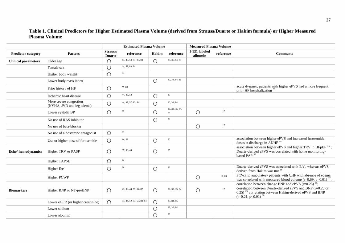
Table 1. Clinical Predictors for Higher Estimated Plasma Volume (derived from Strauss/Duarte or Hakim
formula) or Higher Measured Plasma Volume
NYHA, New York Heart Association; JVD, jugular venous distention; BP, blood pressure; RAAS, renin-angiotensin-aldosterone-system;
ADHF, acutely decompensated heart failure; TRV, tricuspid regurgitation velocity; PASP, pulmonary artery systolic pressure; EF, ejection
fraction; TAPSE, tricuspid annular plane systolic excursion; PCWP, pulmonary capillary wedge pressure; CHF, chronic heart failure; BNP,
brain natriuretic peptide; NT-proBNP, N-terminal pro-brain natriuretic peptide; eGFR, estimated glomerular filtration rate.
Table 2. Prognostic Value of Estimated Plasma Volume Status
BS: baseline; M: month; HF, heart failure; ePVS, estimated plasma volume status; NYHA, New York Heart Association;
LVEF, left ventricular ejection fraction; CHF, chronic heart failure; ADHF, acutely decompensated heart failure; HFrEF, heart
failure with reduced ejection fraction; HFpEF, heart failure with preserved ejection fraction; MI, myocardial infarction; ACM,
all-cause mortality; CVM, cardiovascular mortality; LVAD, left ventricular assistance device.
27
Table 1. Clinical Predictors for Higher Estimated Plasma Volume (derived from Strauss/Duarte or Hakim formula) or Higher Measured
Plasma Volume
Estimated Plasma Volume Measured Plasma Volume
Predictor category Factors Strauss/
Duarte reference Hakim reference
I-131 labeled
albumin reference Comments
Clinical parameters Older age ⃝ 44, 49, 53, 57, 83, 84 ⃝ 33, 35, 84, 85
Female sex ⃝ 44, 57, 83, 84
Higher body weight ⃝ 34
Lower body mass index
⃝ 30, 33, 84, 85
Prior history of HF ⃝ 57 83
acute dyspneic patients with higher ePVS had a more frequent
prior HF hospitalization 57
Ischemic heart disease ⃝ 44, 49, 52 ⃝ 35
More severe congestion
(NYHA, JVD and leg edema) ⃝ 44, 49, 57, 83, 84 ⃝ 30, 33, 84
Lower systolic BP ⃝ 57 ⃝
30, 33, 35, 84,
85 ⃝ 17
No use of RAS inhibitor
⃝ 33
No use of beta-blocker
⃝ 17
No use of aldosterone antagonist ⃝ 44
Use or higher dose of furosemide ⃝ 44, 57 ⃝ 30
association between higher ePVS and increased furosemide
doses at discharge in ADHF 44
Echo/ hemodynamics Higher TRV or PASP ⃝ 37, 38, 44 ⃝ 35
association between higher ePVS and higher TRV in HFpEF 35 ;
Duarte-derived ePVS was correlated with home monitoring-
based PAP 37
Higher TAPSE ⃝ 53
Higher E/e' ⃝ 86 ⃝ 33
Duarte-derived ePVS was associated with E/e’, whereas ePVS
derived from Hakim was not 86
Higher PCWP
⃝ 17, 69
PCWP in ambulatory patients with CHF with absence of edema
was correlated with measured blood volume (r=0.69, p=0.01) 17.
Biomarkers Higher BNP or NT-proBNP ⃝ 23, 39, 44, 57, 84, 87 ⃝ 30, 33, 35, 84 ⃝ 17
correlation between change BNP and ePVS (r=0.28) 39;
correlation between Duarte-derived ePVS and BNP (r=0.23 or
0.25) 23; correlation between Hakim-derived ePVS and BNP
(r=0.21, p<0.01) 30
Lower eGFR (or higher creatinine) ⃝ 34, 44, 52, 53, 57, 83, 84 ⃝ 35, 84, 85
Lower sodium
⃝ 33, 35, 84
Lower albumin
⃝ 85

28
Table 2. Prognostic Value of Estimated Plasma Volume Status
Author/year n HF phenotype ePVS timing Age
(yearrs) NYHA
III/IV(%) LVEF
(%) Follow-up Outcome Comments
Strauss/Duarte (change or instantaneous)
Rossignol 201548 6,820 CHF with HFrEF Change from BS to follow-up visit (within 1yr)
67 36 33 3.9 years (median) ACM, CVM PVS change>0 was associated with a higher incidence of outcomes
Rossignol 201139 6,080 HFrEF and post-MI Change from BS to 1M 64 ― 33 16 months (median)
ACM, CVM or HF hospitalization etc
decreased ePVS was associated with 11% to 19% to better outcomes
Duarte 201523 4,957 HFrEF and post-MI BS, 1 mo. and change from BS to 1 mo.
65 17 35 within 6 months CVM or HF hospitalization ePVS at 1 month was a better predictor than ePVS change
Kobayashi 202083 1,747 CHF with HFpEF BS 72 35 58 2.5 years (median) ACM or HF hospitalization ePVS was associated with congestion status, study-outcomes, and improved risk stratification regardless of renal function.
Kobayashi 201844 712 ADHF Admission, discharge and change from admission to discharge
73 69 41 1 year ACM or HF hospitalization higher ePVS at discharge had worse prognosis in three independent cohorts from three countries (Japan, France and Portugal)
Kobayashi 201953 117 ADHF Admission 74 55 40 90 days ACM or HF hospitalization combination of higher ePVS and higher congestion score assessed by chest X-ray had 5-fold higher risk of outcome
Fudim 2020 84 6,373 ADHF Admission 67 76 28 30 days & 180 days CVM or HF hospitalization
Duarte-ePVS tended to be associated with with 30-day, but not 180-day outcomes. Continuous KH-ePVS>0 (per 10 unit increase) was associated with improved 30-day outcomes.
Balderston 201850 218 ADHF Discharge and 1st visit at outpatient clinic
68 ― 34 90 days ACM or HF hospitalization ePVS at post-discharge had prognostic value of outcome
Hudson 201651 967 ADHF Change from admission to discharge
74 93 ― from 4.9 to 8.7 yrs ACM, ACM or HF hospitalization
highest ePVS tertile was associated with a higher risk of outcomes
Bilchick 201752 324 ADHF Change from admission to discharge
56 ― ― within 6 months ACM, cardiac transplantation or LVAD
increased ePVS was associated with a higher risk of outcomes
Massari 201987 436 184 AHF + 252 CHF Admission 75 56 42 463 days (median) ACM ePVS>5.3dl/g had worse prognosis
Chouihed 201888 1,369 Acute dyspnea
(including 288 AHF) Admission 77 ― ― in-hospital In-hospital mortality
higher ePVS tertile was associated with higher incidence of in-hospital mortality
Huang 201949 449 CHF with HFpEF
BS and change from BS to date of event (or end of follow-up period)
66 42 69 10.7 yrs (median) ACM or HF hospitalization ePVS>5.5dl/g at baseline or an increase in ePVS was associated with a higher risk of outcomes
Hakim
Ling HZ 201530 5,002 CHF with HFrEF BS 61 36 27 716 days ACM or HF re-hospitalization
ePVS>-4% was associated with a higher risk of primary outcome
Grodin 201935 3,414 CHF with HFpEF BS 69 33 61
CVM, HF re-hospitalization or aborted cardiac arrest
higher ePVS was associated with a higher risk of HF hospitalization
Martens 201826 1,173 CHF BS 70 58 32 33 months (mean) ACM or HF re- ePVS>0% was associated with a higher risk
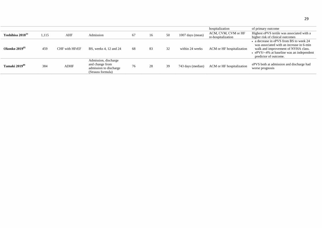
29
hospitalization of primary outcome
Yoshihisa 201833 1,115 AHF Admission 67 16 50 1007 days (mean) ACM, CVM, CVM or HF re-hospitalization
Highest ePVS tertile was associated with a higher risk of clinical outcomes
Okonko 201985 459 CHF with HFrEF BS, weeks 4, 12 and 24 68 83 32 within 24 weeks ACM or HF hospitalization
a decrease in ePVS from BS to week 24 was associated with an increase in 6-min walk and improvement of NYHA class.
ePVS>-4% at baseline was an independent predictor of outcome.
Tamaki 201989 384 ADHF
Admission, discharge and change from admission to discharge (Strauss formula)
76 28 39 743 days (median) ACM or HF hospitalization ePVS both at admission and discharge had worse prognosis

30
Supplementary table 1. Association between Hemoconcentration and Worsening Renal Function
Baseline Hemoconcentration Baseline WRF (or renal impairment)
Comments Author/year N
Age (yrs)
LVEF (%)
Follow-up period for
Hemoconcentration/WRF Hb
(g/dl) Hct (%)
Definition Incidence
(%) eGFR
(ml/min/1.73m²) Definition
Incidence
(%)
Testani 2010 1 336 56 19 during hospital index 12.5 37.8
the highest tertile of
changes in ≥2 of 3
markers (total protein,
ALB or Hct)
32.5 58 ≥20% decrease in
eGFR ―
Hemoconcentration was
associated with 5-fold higher risk
of WRF
van der Meer
2013 2 1,969 70 32 from admission to day 7 12.7 40.2 increase in Hb level 69.4 51 ― ―
Hemoconcentration was
significantly associated with an
increase in Cr level
Grenne 2013 3 1,684 65 28
from baseline to day 7 or
discharge (whichever
occurred first)
― 42.0 Hct change ≥ 3% 26.4 55
≥0.3mg/dl increase in
Cr, ≥25% increase in
BUN or ≥25%
decrease in eGFR
11.6 (by
worsening
eGFR)
Highest quartile of absolute
change in Hct was associated
with worsening Cr, BUN and
eGFR
Davila 2011 4 295 79 46 during hospital index 12.1 ― the upper quartile of
changes in Hb 25.4
1.27
(Cr, mg/dl)
≥0.3mg/dl increase in
Cr 20.0
Hemoconcentration was
associated with a higher risk of
WRF after adjustment for
covariates including Hb and Cr at
baseline
Darawsha 2016 5 704 76 45 during hospital index 12.1 37.1 increase in both Hb
and Hct levels 39.2 49 ― ―
Hemoconcentration was
significantly associated with an
increase in Cr level
Testani 2013 6 845 63 28 during hospital index 12.1 36.6 increase in both Hb
and Hct levels 49.9 60
≥20% decrease in
eGFR (at any time
during the
hospitalization)
―
Incidence of WRF was similar
between patients with early
hemoconcentration (<a mean of
LOS) and late hemoconcentration
(<a mean of LOS).
Rao 2019 7 105 69 37 from admission to day 4 10.8 ― increase in Hb level 54.2 35
≥median of renal
tubular injury
biomarker score 44
50.5
Increase in renal tubular injury
biomarkers was associated with a
higher incidence of
hemoconcentration
(references)
1. Testani JM, Chen J, McCauley BD, Kimmel SE and Shannon RP. Potential effects of aggressive decongestion during the treatment of decompensated heart failure on renal
function and survival. Circulation. 2010;122:265-72.
2. van der Meer P, Postmus D, Ponikowski P, Cleland JG, O'Connor CM, Cotter G, Metra M, Davison BA, Givertz MM, Mansoor GA, Teerlink JR, Massie BM, Hillege HL
and Voors AA. The predictive value of short-term changes in hemoglobin concentration in patients presenting with acute decompensated heart failure. J Am Coll Cardiol.
2013;61:1973-81.
31 3. Greene SJ, Gheorghiade M, Vaduganathan M, Ambrosy AP, Mentz RJ, Subacius H, Maggioni AP, Nodari S, Konstam MA, Butler J, Filippatos G and investigators ET.
Haemoconcentration, renal function, and post-discharge outcomes among patients hospitalized for heart failure with reduced ejection fraction: insights from the EVEREST trial. Eur J
Heart Fail. 2013;15:1401-11.
4. Davila C, Reyentovich A and Katz SD. Clinical correlates of hemoconcentration during hospitalization for acute decompensated heart failure. J Card Fail. 2011;17:1018-22.
5. Darawsha W, Chirmicci S, Solomonica A, Wattad M, Kaplan M, Makhoul BF, Abassi ZA, Azzam ZS and Aronson D. Discordance Between Hemoconcentration and Clinical
Assessment of Decongestion in Acute Heart Failure. J Card Fail. 2016;22:680-8.
6. Testani JM, Brisco MA, Chen J, McCauley BD, Parikh CR and Tang WH. Timing of hemoconcentration during treatment of acute decompensated heart failure and
subsequent survival: importance of sustained decongestion. J Am Coll Cardiol. 2013;62:516-24.
7. Rao VS, Ahmad T, Brisco-Bacik MA, Bonventre JV, Wilson FP, Siew ED, Felker GM, Anstrom KK, Mahoney DD, Bart BA, Tang WHW, Velazquez EJ and Testani JM.
Renal Effects of Intensive Volume Removal in Heart Failure Patients With Preexisting Worsening Renal Function. Circ Heart Fail. 2019;12:e005552.