esophageal rupture in an 89- year-old female · esophageal rupture in an 89-year-old female ......
TRANSCRIPT

Esophageal Rupture in an 89-year-old female
Ali Khoobehi, MDSUNY Downstate Medical Center

Presentation
HPI: EM is an xx-year-old female hx of HTN, osteoarthritis who presented to the ED c/o chest pain following dinner. Pt denied nausea, vomiting. Denied prior episodes of pain.
PMHx: HTN, OAMeds: Lopressor, FosamaxAll: NKDAPSurgHx: noneSocHx: denies smoking & drugs, social EtOH, no
recent travel
Physical Exam
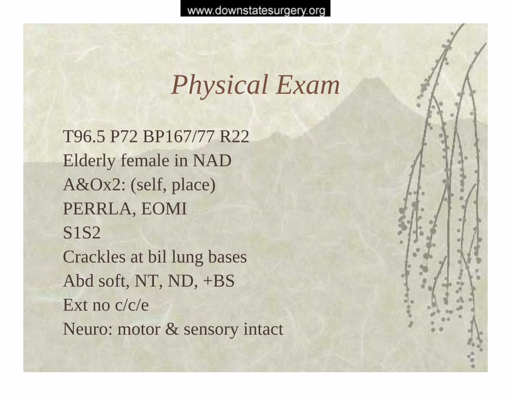
T96.5 P72 BP167/77 R22Elderly female in NADA&Ox2: (self, place)PERRLA, EOMIS1S2Crackles at bil lung basesAbd soft, NT, ND, +BSExt no c/c/eNeuro: motor & sensory intact

Labs
WBC 10.6 H/H 13.9/43.4 Plt 264Na 142 K 3.5Cl 97 CO2 29BUN 10 Crea 0.7 Glu 189TP 6.5 Alb 4.1 Tbili 0.5AST 47 ALT 46 AlkP 91Troponin neg, CK-MB 2.1

ER Studies: CXR

ER Studies: CT A&P

ER Studies: CT A&P

ER Course
Pneumomediastinum and contrast in the left thoracic cavity was noted on CT scan and thoracic surgical consult was placed. Head CT showed no abnormalities.Shortly thereafter, pt developed worsening SOB and was intubated in the ED.Pt had UGI series that showed extravasation of gastrograffin into the left chest. Perforation was localized to the distal thoracic esophagus.

Operative ExplorationPt was taken emergently to the OR for surgical exploration. As the OR was being prepared, a CVC and arterial line were placed, and was aggressively fluid resuscitated.Pt underwent esophagoscopy and left thoracotomy posterolaterally in the 7th ICS.Pt was found to have a longitudinal 8cm lac in the distal esophagus with necrotic wound edges and necrotic debris in the left chest.Mediastinal debridement and primary two-layered esophageal repair, mediastinal washout, placement of bilateral chest tubes, placement of feeding jejunostomy were then performed.

Operative Exploration
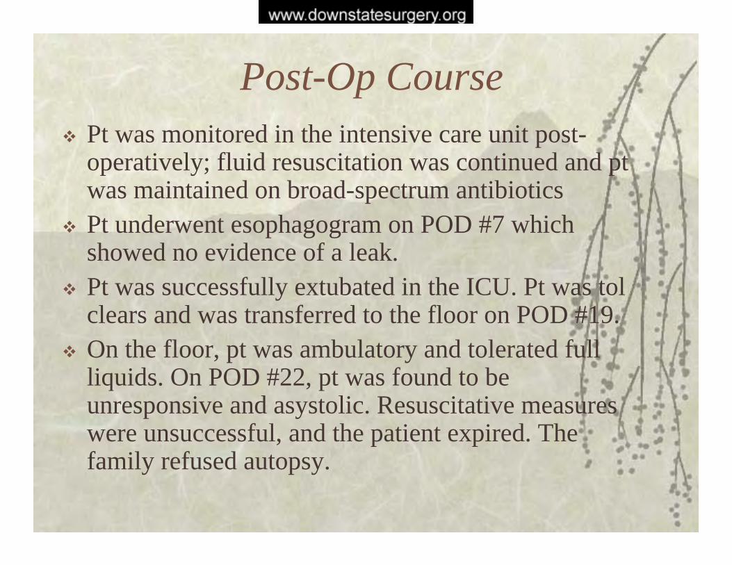
Post-Op CoursePt was monitored in the intensive care unit post-operatively; fluid resuscitation was continued and pt was maintained on broad-spectrum antibioticsPt underwent esophagogram on POD #7 which showed no evidence of a leak.Pt was successfully extubated in the ICU. Pt was tol clears and was transferred to the floor on POD #19.On the floor, pt was ambulatory and tolerated full liquids. On POD #22, pt was found to be unresponsive and asystolic. Resuscitative measures were unsuccessful, and the patient expired. The family refused autopsy.
Management of Esophageal Rupture
Presentation
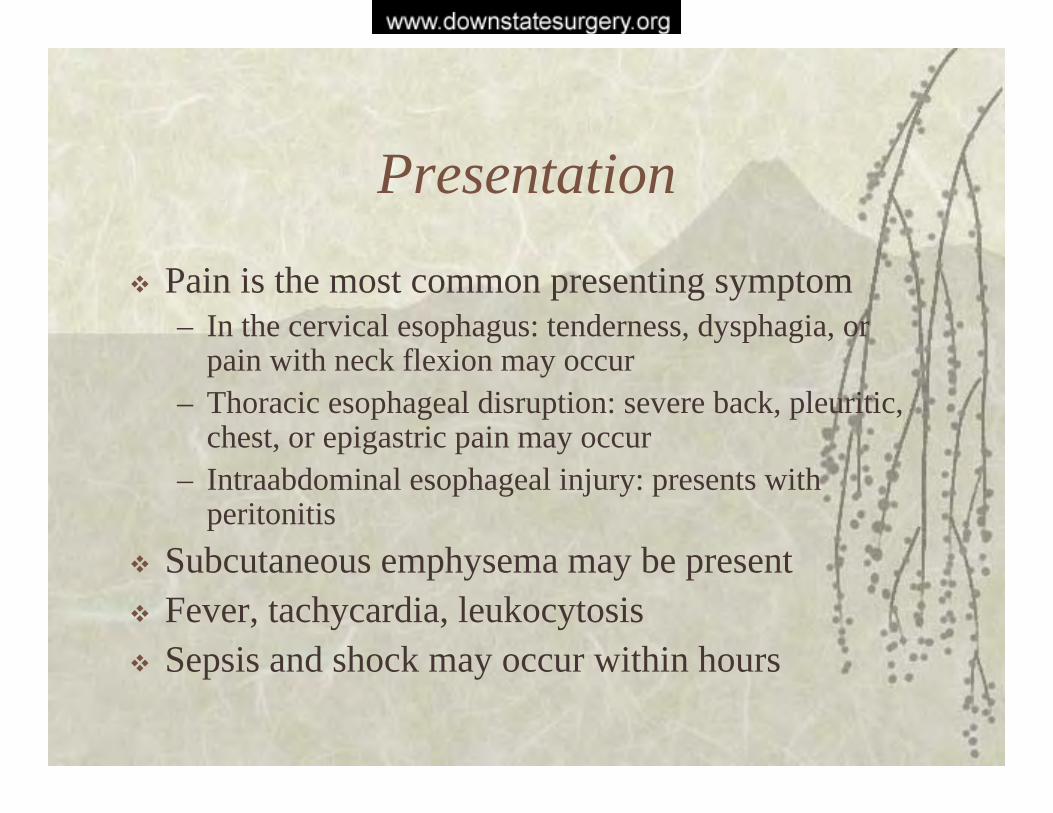
Pain is the most common presenting symptom– In the cervical esophagus: tenderness, dysphagia, or
pain with neck flexion may occur– Thoracic esophageal disruption: severe back, pleuritic,
chest, or epigastric pain may occur– Intraabdominal esophageal injury: presents with
peritonitisSubcutaneous emphysema may be presentFever, tachycardia, leukocytosisSepsis and shock may occur within hours

DiagnosisPlain chest radiograph– May show mediastinal
emphysema, pneumothorax, hydropneumothorax, pneumopericardium, or subdiaphragmatic air
CXR showing free mediastinal air along esophageal contour
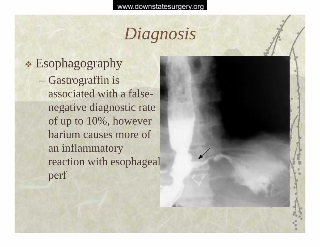
DiagnosisEsophagography– Gastrograffin is
associated with a false-negative diagnostic rate of up to 10%, however barium causes more of an inflammatory reaction with esophageal perf

Diagnosis
Esophagography
– Water-soluble contrast should be used initially; if this study is negative, it may be followed by a barium esophagogram if clinical suspicion is high
DiagnosisFlex Esophagoscopy– May be used to diagnose and localize perforations. It
can be used to determine the extent of injury, whether mucosal or transmural.
– May show concomitant disease, such as esophageal cancer or stricture
– May potentially exacerbate the injury via insufflation or direct contact with the injury upon intubation
– Rigid esophagoscopy is less commonly used, as visualization is inferior and the chance of worsening the injury is thought to be greater.

DiagnosisCT scan– May show mediastinal air,
abscess cavities in the periesophageal space, loculated collections in the pleural space, gross leakage of contrast into mediastinum or pleural space
– May be used to guide drainage in patients who are treated nonoperatively
Nonoperative TreatmentSelected patients may be treated nonoperatively, including patients which are elderly with multiple comorbidities who do not exhibit signs of sepsis.Generally, nonoperative management is undertaken in cases of instrumental perforation, especially in the cervical esophagus; cases of perf following sclerosis or dilation procedures in the esophagus; perforation being diagnosed several days after inciting event, and with minimal symptoms.

Nonoperative Management Criteria
Well circumscribedNot in abdominal cavityMinimal pleural soilageNo drainage into adjacent body cavitiesNo malignancy, obstruction, or strictureNo enteral intake since injuryMinimal symptomsNo signs of sepsis

Nonoperative Treatment
Pt remains NPO for 7 to 10 days, followed by a Gastrograffin swallow study to eval for resolution of leakIf swallow study is positive pt may be continued on parenteral nutrition with weekly swallow studies; leak may take weeks to resolveOperative treatment is mandated by worsening clinical course

Operative Management
Selection of therapy depends on location of injury, time interval since the injury, presence of underlying esophageal diseaseOptions include simple drainage of he contaminated space, debridement with primary repair, esophageal diversion and delayed repair, and esophagectomy
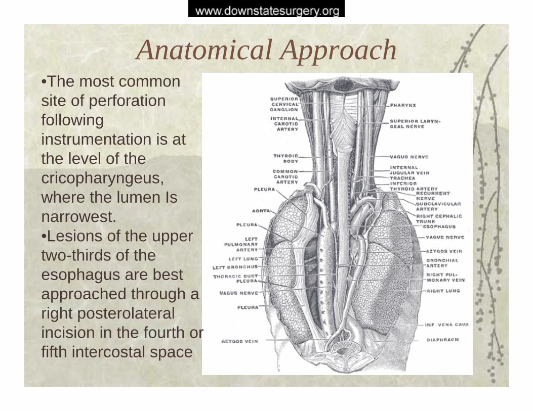
Anatomical Approach•The most common site of perforation following instrumentation is at the level of the cricopharyngeus, where the lumen Is narrowest.•Lesions of the upper two-thirds of the esophagus are best approached through a right posterolateral incision in the fourth or fifth intercostal space

Anatomical Approach
A left-sided posterolateral thoracotomy in the seventh intercostal space is recommended for perforations of the distal third of the esophagusIntraabdominal esophageal perforations are approached with an upper midline abdominal incision

Anatomical Approch to Repair
The layers of the esophagus include the mucosa, submucosa, and muscularis externa
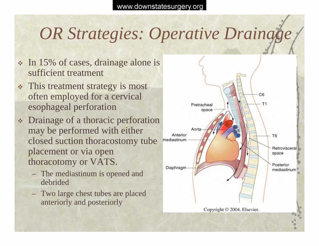
OR Strategies: Operative DrainageIn 15% of cases, drainage alone is sufficient treatmentThis treatment strategy is most often employed for a cervical esophageal perforationDrainage of a thoracic perforation may be performed with either closed suction thoracostomy tube placement or via open thoracotomy or VATS.– The mediastinum is opened and
debrided– Two large chest tubes are placed
anteriorly and posteriorly
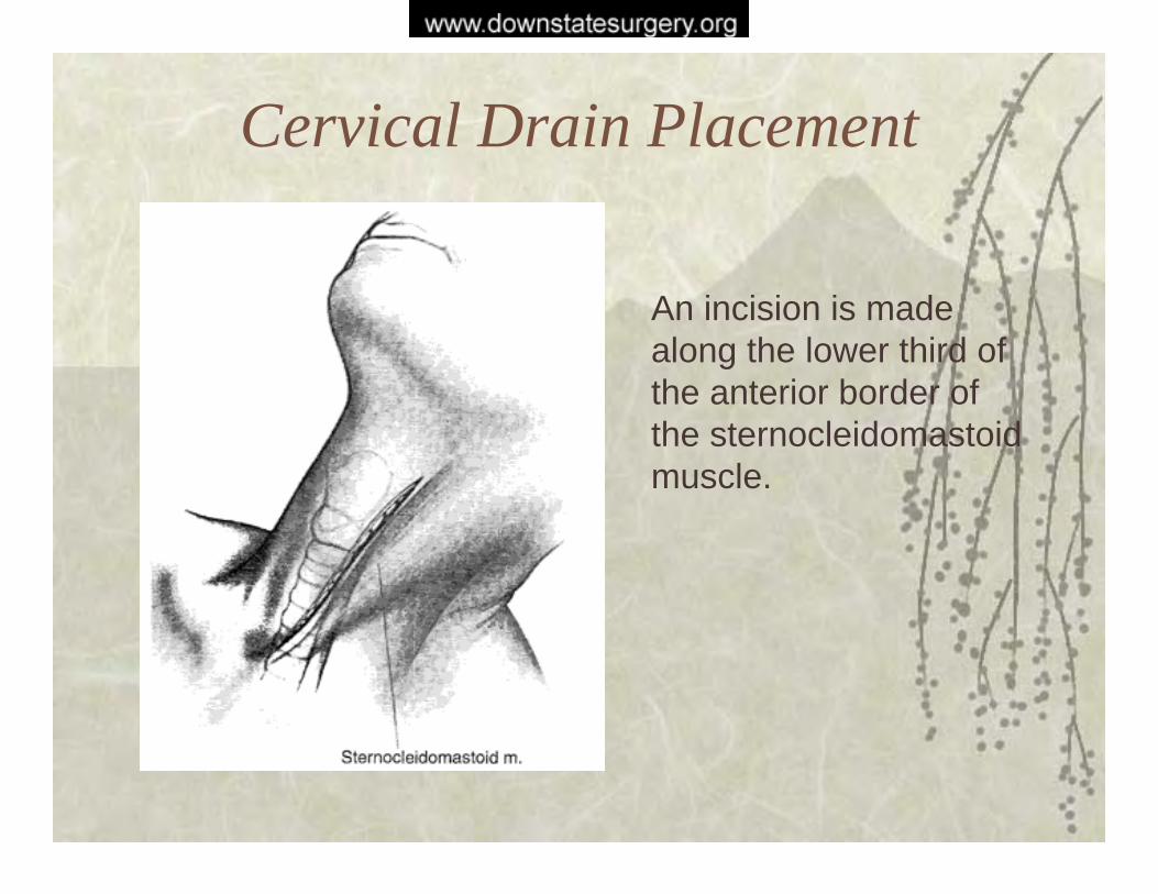
Cervical Drain Placement
An incision is made along the lower third of the anterior border of the sternocleidomastoid muscle.

Cervical Drain Placement
Cervical fascia is exposed then divided.

Cervical drain placement
The Carotid sheath and internal jugular vein are retracted laterally, and the middle thyroid vein is divided.

Cervical drain placement
Blunt dissection leads to the retrovisceral space and the prevertebral fascia posteriorly. Finger dissection is continued to the posterior mediastinum and suction drainage is placed.
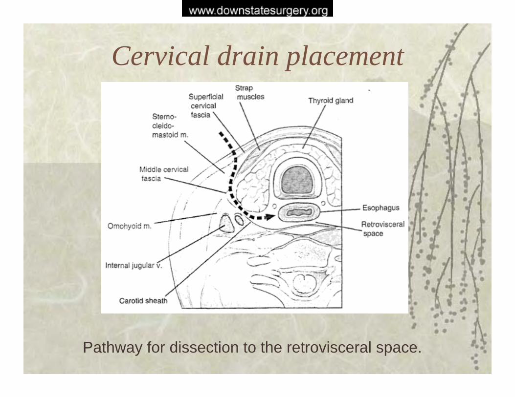
Cervical drain placement
Pathway for dissection to the retrovisceral space.

Primary RepairOperative debridement with primary repair is traditionally advised in patients presenting soon after their injury, however the recent literature supports primary repair even with a time period of more than 24 hours since the injury.Primary repair is contraindicated in the presence of carcinoma or megaesophagus from achalasia

Primary Repair
Left posterolatral thoracotomy is performed in the 7th ICS. A 7th rib resection may also be performed. Necrotic mediastinal pleura is excised and the esophagus is esposed.

Primary Repair
The esophagus may be elevated on a penrose drain to allow for debridement of the right mediastinal pleura if necessary.
Primary Repair
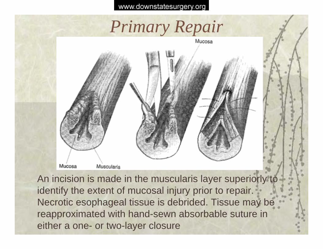
An incision is made in the muscularis layer superiorly to identify the extent of mucosal injury prior to repair. Necrotic esophageal tissue is debrided. Tissue may be reapproximated with hand-sewn absorbable suture in either a one- or two-layer closure

Time Interval to Primary RepairThe classic teaching is that primary repair should be considered if it has been 24 hours since the inciting event; recent studies challenge that notionLawrence et al. (Ann of Thoracic Surgery, 1999) advocate extending the time period for primary repair to 72 hours.– They present a series of 21 patients, 12 of which were
referred 24 hours after the inciting event.– All patients had single-layered, non-buttressed repairs
combined with mediastinal drainage– Mortality rate was 14.3%, with no difference in patients
who presented before 24 hours following injury and those who presented afterwards

Time Interval to Primary Repair
Jougen et al. (Eur J of CT Surg, 2003) advocate primary esophageal repair “whatever the time interval between perforation and treatment”– Operated on 21 patients with primary esophageal repair,
with and without buttressing of the repair– Mortality rate was 24%, with no statistically significant
difference in the group repaired before 24 hours and the group repaired afterwards

Primary Repair: Tissue Flap Reinforcement
Tissue flaps may be obtained from adjacent pleura, pericardium, intercostal muscle, or diaphragm
A Thal patchutilizes a tongue of gastric fundus to reinforce repair of a distal esophageal perf
Tissue Flap Reinforcement
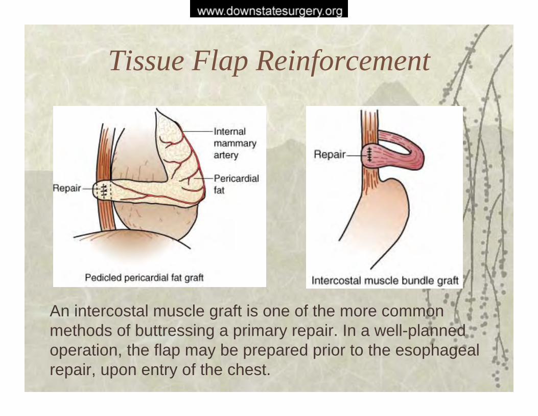
An intercostal muscle graft is one of the more common methods of buttressing a primary repair. In a well-planned operation, the flap may be prepared prior to the esophageal repair, upon entry of the chest.
Tissue Flap Reinforcement
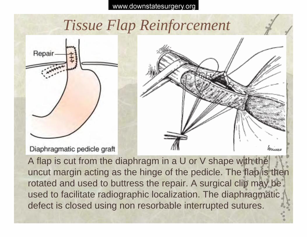
A flap is cut from the diaphragm in a U or V shape with the uncut margin acting as the hinge of the pedicle. The flap is then rotated and used to buttress the repair. A surgical clip may be used to facilitate radiographic localization. The diaphragmatic defect is closed using non resorbable interrupted sutures.
Tissue Flap Reinforcement
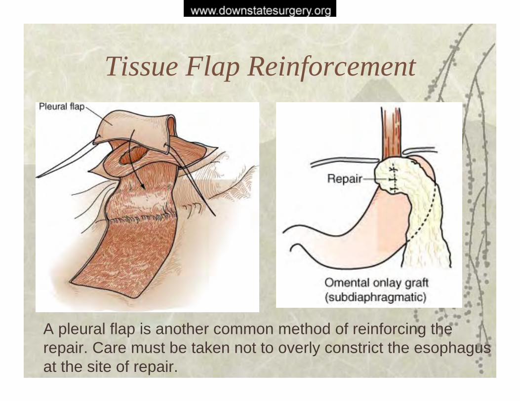
A pleural flap is another common method of reinforcing the repair. Care must be taken not to overly constrict the esophagusat the site of repair.

Esophageal ExclusionEsophagus may be diverted as part of a two-stage procedureCervical esophagostomy is createdOpen gastrostomy or feeding jejunostomy is placedA second, more difficult procedure is required to restore esophageal continuity. Also, complications may arise from the creation of a blind-loop of esophagus.
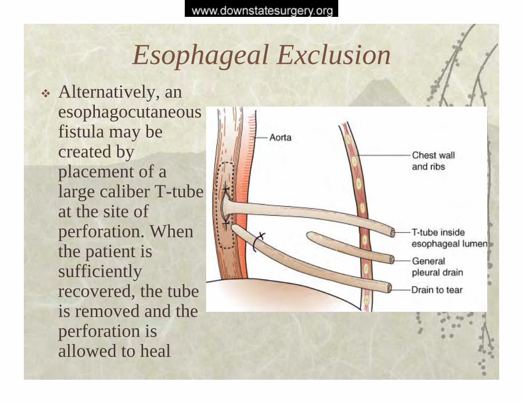
Esophageal ExclusionAlternatively, an esophagocutaneous fistula may be created by placement of a large caliber T-tube at the site of perforation. When the patient is sufficiently recovered, the tube is removed and the perforation is allowed to heal

Esophageal Resection
Esophagectomy with primary or delayed reconstruction is sometimes indicated– Obstruction due to malignancy, stricture, or neuromotor
dysfunction– Multiple strictures– Severe intrinsic disease such as esophagitis or reflux– Severe mediastinal contamination and inflammatory
reaction– Failure of primary repair

Esophageal Resection
Transhiatal esophagectomy may be performed if there is minimal pleural contaminationWith a more chronic perforation, a transthoracic approach is recommendedIf the patient is critically ill, a gastrostomy or jejunostomy should be placed, and the reconstruction should be performed 6 to 8 weeks later via placement of a gastric tube or colonic interposition

Esophageal StentsThere are case reports of esophageal stent endoprostheses used successfully to manage esophageal ruptureThese are generally employed when a patient is a high operative risk or is too unstable to withstand prolonged anesthesiaWhile promising, these minimally invasive treatments are currently considered experimental and controversial

Complications
Sepsis is associated with a 50% mortality rateThe most common late complication is stricture at the site of repair, which may be managed with dilatationEmpyema or loculated pneumothorax are treated with tube thoracostomy or percutaneous drainage; decortication may be required if pt does not improve

Aledronate induced esophagitis
Aledronate sodium (Fosamax) acts as a selective inhibitor of osteoclast mediated bone resorptionStandard dosage is 10 mg/day, though a more recent 70 mg weekly dosage is available.
Aledronate and esophagitisIn early dose-ranging studies of aledronate, it was found that a 40mg daily dose was associated with a small increase in UGI events, including esophagitis and gastritisCurrent instructions for alendronate administration include:– It should be taken first thing in the morning with 180-
240 mL of water– 30 minutes before the first food or beverage of the day– Patient should avoid lying down for at least 30 minutes
following medication ingestion
Aledronate esophagitis in the literature
There are a number of case reports of esophagitis that appear to be associated with aledronate ingestion; commonly causes a long, linear ulcer.Groen et al report a series of three cases of severe esophagitis which began immediately after starting aledronate therapy. The esophagitis responded to Fosamax cessation and PPI therapy.

Aledronate esophagitis in the literature
Groen et al report post-marketing data for 475,000 patients between October 1996 and March 1996.– 51 had adverse
esophageal events– 17 had esophageal events
classified as severe; no perforations reported

Aledronate esophagitis in the literature
Fosamax clinical trials show no difference in adverse UGI events when compared to placebo when properly used.Adverse UGI events, including perforation, increase with preexisting esophageal disease and if patients fail to follow the instructions when taking FosamaxOnce weekly dosing decreases rate of UGI events

Algorithm for Operative Strategy
One author’s algorithm for management of esophageal perforation (Sabiston et al.)

ReferencesDavies, Andrew P., et al. “Expanding Mesh Stent in the
Emergency Treatment of Boerhaave’s Syndrome.” Ann Thorac Surg 1999;67:1482-83.
De Groen, PC., et al. “Esophagitis Associated with the use of Aledronate.” New England J of Med 1996;335(14):1016-1021.
Famularo, G., et al. “Fatal Esophageal Perforation with Aledronate.” Am J Gastroenterology 2001;96(11):3212-13.
Jougon, Jacques., et al. “Primary esophageal repair for Boerhaave’s syndrome whatever the free interval between perforation and treatment.” Eur J CT Surg 2004;25:475-479.

ReferencesLawrence, David R., et al. “Primary Esophageal Repair for
Boerhaave’s Syndrome.” Ann Thorac Surg 1999;67:818–20.
Mineo, TC., et al. “The Diaphragmatic Flap: A Multiuse Material in Thoracic Surgery.” J Thor and CV Surg 1999;118(6):1084-89.
Vogel, Stephen B., et al. “Esophageal Perforation in Adults Aggressive, Conservative Treatment Lowers Morbidity and Mortality.” Ann of Surg 2005;241(6):1016-23.
Zwischenberger, Joseph B., et al. “Surgical Aspects of Esophageal Disease: Perforation and Caustic Injury.” Am J Respir Crit Care Med 2001;164:1037-1040.