escherichia coli templated iron oxide biomineralization
TRANSCRIPT
RSC Advances
PAPER
Ope
n A
cces
s A
rtic
le. P
ublis
hed
on 2
1 A
pril
2021
. Dow
nloa
ded
on 1
2/12
/202
1 5:
38:2
0 A
M.
Thi
s ar
ticle
is li
cens
ed u
nder
a C
reat
ive
Com
mon
s A
ttrib
utio
n-N
onC
omm
erci
al 3
.0 U
npor
ted
Lic
ence
.
View Article OnlineView Journal | View Issue
Escherichia coli t
aSchool of Chemistry, Chemical Engineering
Technology, Wuhan, 430070, China. E-mai
edu.cnbState Key Laboratory of Advanced Technolo
Wuhan, 430070, China. E-mail: zyfu@whutcLaboratory of Inorganic Materials Chemist
Belgium
† Electronic supplementary informa10.1039/d1ra00847a
Cite this: RSC Adv., 2021, 11, 15010
Received 31st January 2021Accepted 5th April 2021
DOI: 10.1039/d1ra00847a
rsc.li/rsc-advances
15010 | RSC Adv., 2021, 11, 15010–150
emplated iron oxidebiomineralization under oscillation†
Panpan He,a Junhui Guo,*a Liwen Lei,b Jiafeng Jiang,a Qichang Li,a Zhiyi Hu,b
Baolian Su, bc Zhengyi Fu *b and Hao Xie *a
Motility is significant in organisms. Studying the influence of motility on biological processes provides a new
angle in understanding the essence of life. Biomineralization is a representative process for organisms in
forming functional materials. In the present study, we investigated the biomineralization of iron oxides
templated by Escherichia coli (E. coli) cells under oscillation. The formation of iron oxide minerals with
acicular and banded morphology was observed. The surface charge of E. coli cells contributed to the
biomineralization process. The surface components of E. coli cells including lipids, carbohydrates and
proteins also have roles in regulating the formation and morphology of iron oxide minerals. As-prepared
mineralized iron oxide nanomaterials showed activity in photocatalytic degradation of methylene blue as
well as in electrocatalytic hydrogen evolution reaction. This study is helpful not only in understanding
motility in biological processes, but also in developing techniques for fabricating functional nanomaterials.
Introduction
Motility is one of the most signicant characteristics in organ-isms, especially animals or planktonic microbes. There arenumerous studies and reports concerning mechanisms ofvarious types of motility in organisms. However, few reportshave been on the inuence of motility on biomolecular statusand biological processes.
Biomineralization is one of the most abundant and essentialbiological processes in nature for organisms in forming func-tional materials. More than 60 biominerals have been foundincluding calcium carbonate, calcium oxalate, silica, titaniumdioxide, alumina, and iron oxide systems. Biomineralizationrelies on organisms1–3 and occurs in two ways,4 that is, bio-inducedmineralization and bio-controlled mineralization. Bio-inducedmineralization (BIM) is a response to changes in mineral saturationin uids caused by cellular metabolic activity. Bio-impact minerali-zation or organic matrix-mediated mineralization have import rolesin BIM since cells and related organic debris act as templates inregulating the formation of bio-induced minerals with distinctmorphologies and structures. Applications of BIM are mostly inecological restoration and cement-based material restoration.5,6 Bio-
and Life Sciences, Wuhan University of
l: [email protected]; h.xie@whut.
gy for Materials Synthesis and Processing,
.edu.cn
ry, University of Namur, B-5000 Namur,
tion (ESI) available. See DOI:
16
controlled mineralization (BCM) usually occurs in specic struc-tures of cells, with strictly controlled composition andmorphology ofbiominerals.4 Typical examples of BCM are the formation of mag-netosomes in magnetotactic bacteria as well as structural formationof bones and teeth.4–7 BCM has found wide applications in electrodematerials, sewage treatments, ecological restoration, cultural relicsrestoration, nano-drug carriers and cancer treatments.8–13
Due to the importance of biomineralization in both basictheories and practical applications, biomimetic mineralizationattracted attentions of scientists from a broad range of disci-plines including biology, chemistry, and materials science.These researches are mainly focused on roles of cell compo-nents or compositions in templating or regulating biomimeticmineralization14 and fall into two categories of mineralizationsystems, that is, in vitro mineralization under functions ofspecic biomolecules or in vivo mineralization promoting byintact cells or organisms. Biomolecules precisely template orregulate mineralization with their molecular structures or surfacegroups, which is advantageous in deliberately producing mineralswith specic structures, morphologies, or functions. For example,silk broin has been used for regulating the superstructure ofhemispherical CaCO3 crystals that has potential applications inpreparing inorganic materials with new morphologies and specialtextures.15 Formation of cuprous carbonate on immune complexeswas promoted by urease and could be used for detecting colori-metric signals.16 Intact cells or organisms are more efficient inproducing biominerals since essential supplies of ions and energyduring mineralization are accomplished by the organism that isalso a self-regulating system for mineralization. For example,formation of iron ore was observed in iron bacteria and adsorptionand transfer of rare earth elements were observed in Saccharomyces
© 2021 The Author(s). Published by the Royal Society of Chemistry

Paper RSC Advances
Ope
n A
cces
s A
rtic
le. P
ublis
hed
on 2
1 A
pril
2021
. Dow
nloa
ded
on 1
2/12
/202
1 5:
38:2
0 A
M.
Thi
s ar
ticle
is li
cens
ed u
nder
a C
reat
ive
Com
mon
s A
ttrib
utio
n-N
onC
omm
erci
al 3
.0 U
npor
ted
Lic
ence
.View Article Online
cerevisiae cells.17,18 Bacterial cell surface interacts with environ-mental ions and has multiple roles in bacterial-mediated miner-alization. Chemical groups such as hydroxyl, carboxyl, amine andhalide on bacterial surface are involved in adsorption and depo-sition of heavy metal ions as well as morphogenesis of bio-minerals.19–23 It inspired researchers in using bacterial-mediatedmineralization for synthesizing materials with applications inecological restoration, sewage treatment, drug carrier, etc.24–26
Factors such as temperature, pH, ion strength, biologicaltemplates and regulators have been extensively explored inbiomineralization. However, there is few reports on effects ofmotility on biomineralization. Since motility occurs to animalsor planktonic microbes all the time, it calls for the need ofexploring motility effects on biomineralization.
The present study aims to investigate effects of motility onbiomineralization by studying Escherichia coli templated bio-mineralization of iron oxides under oscillation. E. coli isa model microbe that is broadly used in studying biochemicalissues. Natural mineralization was not observed on E. colisurface although it tolerates and adsorbs heavy metal ions.27
In the present study, deposition and mineralization of ironions was induced on E. coli cell surface and compared betweenstatic or oscillatory conditions. Formation of needle-like orband-like nanomaterials of ferric oxide and ferric oxide wasobserved and characterized on E. coli surface under oscilla-tion. Roles of chemical compositions of E. coli cell surface wereexplored onmorphogenesis of ironminerals during oscillatorymineralization. Both photocatalytic and electro-catalyticperformances were evaluated of as-prepared mineralizediron oxide nanomaterials. The present study calls attention tobiochemical changes especially biomineralization undermotility conditions.
ExperimentalBacteria cultivation
Two bacterial strains were investigated including E. coli DH5aand Bacillus subtilis B168. Bacteria were cultured as previouslydescribed.20,21 Briey, single bacterial colony was inoculated in3–5 ml of Luria-Bertani (LB) medium, followed by shakingovernight at 37 �C at 220 rpm. Cell suspension was inoculatedin LB medium at a ratio of 1 : 50, followed by shaking at220 rpm at 37 �C for 12–18 hours. Cells were harvested bycentrifugation at 6000g for 5 minutes at 4 �C. Cells were thenwashed three times with Tris-buffered saline (TBS) buffer, pH7.0, before later use.
Surface treatment of E. coli cells
To explore inuence of specic chemical compositions(proteins, peptidoglycan, lipids) from bacterial cell surface onbiomineralization, E. coli cells were subjected to treatment indifferent fashions: (1) in 1% trypsin, 50 mM Tris–HCl, pH 7.0,and incubated at 37 �C for 5 hours; or (2) in 2mgml�1 lysozyme,10 mM Tris–HCl, pH 8.0, 20% sucrose, with or without 10 mMEDTA and incubated at 37 �C for 1 hour; or (3) in 95% ethanolsolution and incubated at room temperature for 5 minutes; or
© 2021 The Author(s). Published by the Royal Society of Chemistry
(4) in 0.5 M Tris–HCl, pH 8.0, 0.1% SDS, 50% (v/v) chloroform,and incubated at room temperature for 20 minutes. Cells werethen sedimented by centrifugation at 6000g for 3 minutes andwashed three times with deionized water before later use. In thecase of fashion (3) treatment, 20% sucrose was included in thewash.
Bacteria templated biomineralization of iron oxides underoscillation
Fe3+ solution was prepared by dissolving ferric sulfate or ferricchloride in deionized water at a nal concentration of 1 mgml�1 with the pH adjusting to 2.40. Biomineralization of ironoxide was initiated by supplying with bacterial cells. Typically,20 ml Fe3+ solution was supplied with 100 mg bacterial cells(wet weight). Mineralization mixtures were immediately sub-jected to oscillation (110 rpm or 220 rpm) at 37 �C. Minerali-zation was terminated by removing Fe3+ ions frommineralization mixtures via centrifugation at 5000g, 3 minutes.Mineralized bacterial cell precipitation was washed three timeswith deionized water and vacuum freeze-dried for 12 hoursbefore later characterization.
Liposomes templated biomineralization under oscillation
Soybean lecithin/cholesterol (5 : 1 w/w) were dissolved inchloroform/ethanol (7 : 1 v/v) and evaporated at 37 �C for 3hours on a rotary evaporator. The lipid was re-suspended in30 ml deionized water and hydrated by occasionally sonicationat 55 �C for 4 hours. Suspension of soybean lecithin/cholesterol(20 mg ml�1) was extruded twice through 0.22 micron lter togenerate unilamellar vesicles and stored at 4 �C. Bio-mineralization was initiated by mixing liposome solution(20 mg ml�1) with Fe3+ solution (1 mg ml�1) at a volume rationof 1 : 4 and subjected to oscillation (110 rpm or 220 rpm) at37 �C. Mineralization was terminated by removing Fe3+ frommineralization mixtures via concentration at 10 000g, 15minutes. Mineralized liposome precipitation was washed threetimes with deionized water and vacuum freeze-dried for 12hours before characterization.
Characterization of iron biominerals
Biominerals were characterized by scanning electron micro-scope (SEM), transmission electron microscopy (TEM), X-rayphotoelectron spectroscopy (XPS), vibrating sample magne-tometry (VSM), thermogravimetric analysis (TGA). Structuresand morphologies of biominerals were characterized by theeld-emission scanning electron microscopy (FESEM, HitachiS-4800), most of which were performed using an acceleratingvoltage of 5 kV and a working distance of 8 mm. Transmissionelectron microscopy (TEM, JEM-2010HT) and high-resolutiontransmission electron microscope (HRTEM, JEM-2010FEF)were used to identify the crystalline structure. Chemical statesand species of elements on biominerals surface were measuredby XPS on an X-ray photoelectron spectrometer (VG Multilab2000). TGA was performed in a LABSYS evo TGA (Setaram,France) at a heating rate of 5 �C min�1 from 40 �C to 900 �C.
RSC Adv., 2021, 11, 15010–15016 | 15011
RSC Advances Paper
Ope
n A
cces
s A
rtic
le. P
ublis
hed
on 2
1 A
pril
2021
. Dow
nloa
ded
on 1
2/12
/202
1 5:
38:2
0 A
M.
Thi
s ar
ticle
is li
cens
ed u
nder
a C
reat
ive
Com
mon
s A
ttrib
utio
n-N
onC
omm
erci
al 3
.0 U
npor
ted
Lic
ence
.View Article Online
Magnetic properties were analyzed by using a vibrating samplemagnetometer (Lake Shore 7400 Series VSM).
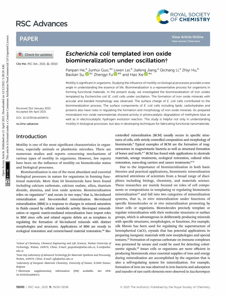
Fig. 2 TEM image (Panel A) and HR-TEM image (Panel B, the area indi-cated by the red box in Panel A). Panel C shows FFT pattern of Panel B.
Catalytic measurements of iron biominerals
Photocatalytic degradation of methylene blue was based on ref.28. Typically, 0.5 g biominerals was added into 50 ml of 0.02 mMmethylene blue, pH 3.5, and stirred in dark for 20 minutes, fol-lowed by the addition of 0.1 ml hydrogen peroxide. Degradation ofmethylene blue were initiated by exposing to natural light andassayed bymeasuring absorbance at 665 nm aer centrifugation ata time interval of 5 minutes. Degradation rate (R) was determinedby the formula R ¼ (A0 � At)/A0 � 100%, where A0 and At are theabsorbance at 625 nm initially and aer time t.
Electrocatalytic hydrogen evolution was based on ref. 29. Aplatinum wire was used as a counter electrode and a reversiblehydrogen electrode was used as a reference electrode. Theworking electrode was a glassy-carbon Rotating Disk Electrode(RDE, diameter: 5 mm, area: 0.196 cm2). The Pt loading of allsamples on glassy-carbon was 2.0 mg cm�2. Polarization curveswere collected in 1 M KOH solutions at a rotation rate of1600 rpm with a sweep rate of 5 mV s�1.
Results and discussionIron biomineralization on E. coli cell surface under oscillation
Previous work showed that E. coli surface interacts with variousmetals ions30 which may facilitate subsequent mineralization. ThepHwas adjusted to 2.54 (ref. 31) to promote ironmineralization onE. coli surface. Bacterial metabolism and biological processes relyon substance exchanges between bacterial cells and environment,which are greatly improved by oscillation. Bacterial growth in
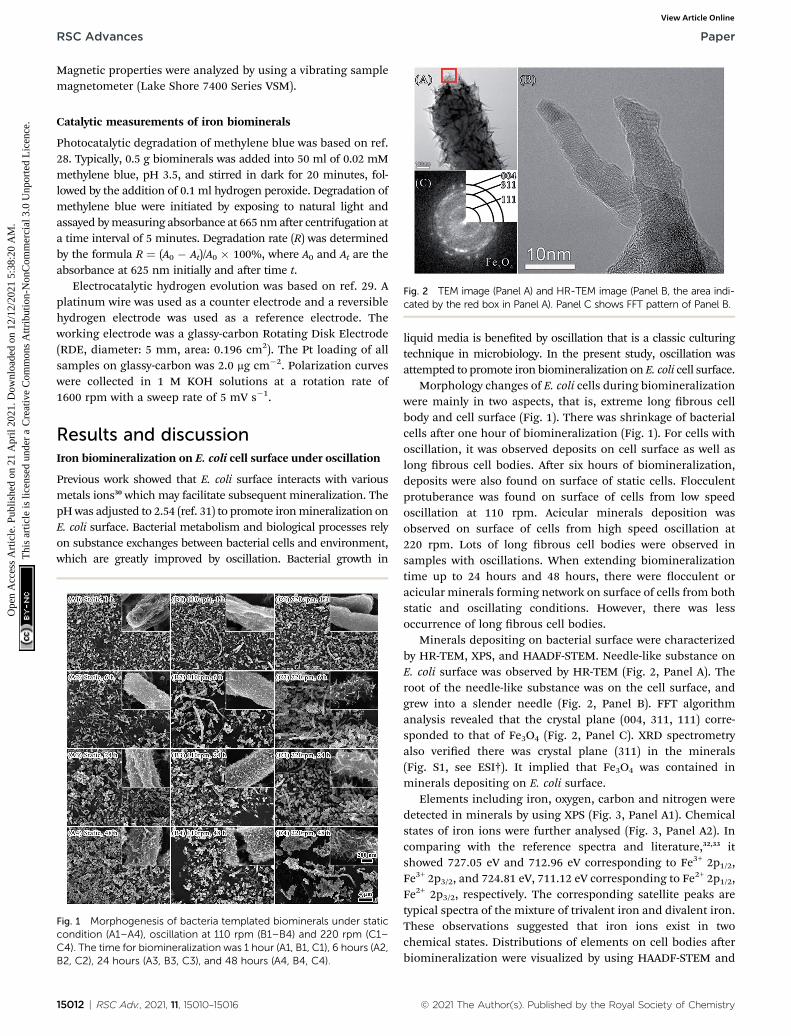
Fig. 1 Morphogenesis of bacteria templated biominerals under staticcondition (A1–A4), oscillation at 110 rpm (B1–B4) and 220 rpm (C1–C4). The time for biomineralization was 1 hour (A1, B1, C1), 6 hours (A2,B2, C2), 24 hours (A3, B3, C3), and 48 hours (A4, B4, C4).
15012 | RSC Adv., 2021, 11, 15010–15016
liquid media is beneted by oscillation that is a classic culturingtechnique in microbiology. In the present study, oscillation wasattempted to promote iron biomineralization on E. coli cell surface.
Morphology changes of E. coli cells during biomineralizationwere mainly in two aspects, that is, extreme long brous cellbody and cell surface (Fig. 1). There was shrinkage of bacterialcells aer one hour of biomineralization (Fig. 1). For cells withoscillation, it was observed deposits on cell surface as well aslong brous cell bodies. Aer six hours of biomineralization,deposits were also found on surface of static cells. Flocculentprotuberance was found on surface of cells from low speedoscillation at 110 rpm. Acicular minerals deposition wasobserved on surface of cells from high speed oscillation at220 rpm. Lots of long brous cell bodies were observed insamples with oscillations. When extending biomineralizationtime up to 24 hours and 48 hours, there were occulent oracicular minerals forming network on surface of cells from bothstatic and oscillating conditions. However, there was lessoccurrence of long brous cell bodies.
Minerals depositing on bacterial surface were characterizedby HR-TEM, XPS, and HAADF-STEM. Needle-like substance onE. coli surface was observed by HR-TEM (Fig. 2, Panel A). Theroot of the needle-like substance was on the cell surface, andgrew into a slender needle (Fig. 2, Panel B). FFT algorithmanalysis revealed that the crystal plane (004, 311, 111) corre-sponded to that of Fe3O4 (Fig. 2, Panel C). XRD spectrometryalso veried there was crystal plane (311) in the minerals(Fig. S1, see ESI†). It implied that Fe3O4 was contained inminerals depositing on E. coli surface.
Elements including iron, oxygen, carbon and nitrogen weredetected in minerals by using XPS (Fig. 3, Panel A1). Chemicalstates of iron ions were further analysed (Fig. 3, Panel A2). Incomparing with the reference spectra and literature,32,33 itshowed 727.05 eV and 712.96 eV corresponding to Fe3+ 2p1/2,Fe3+ 2p3/2, and 724.81 eV, 711.12 eV corresponding to Fe2+ 2p1/2,Fe2+ 2p3/2, respectively. The corresponding satellite peaks aretypical spectra of the mixture of trivalent iron and divalent iron.These observations suggested that iron ions exist in twochemical states. Distributions of elements on cell bodies aerbiomineralization were visualized by using HAADF-STEM and
© 2021 The Author(s). Published by the Royal Society of Chemistry
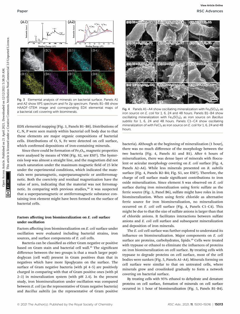
Fig. 4 Panels A1–A4 show oscillating mineralization with Fe3(SO4)2 asiron source on E. coli for 1, 6, 24 and 48 hours. Panels B1–B4 showoscillating mineralization with Fe3(SO4)2 as iron source on Bacillussubtilis for 1, 6, 24 and 48 hours. Panels C1–C4 show oscillatingmineralization of with FeCl3 as iron source on E. coli for 1, 6, 24 and 48hours.
Fig. 3 Elemental analysis of minerals on bacterial surface. Panels A1and A2 show XPS spectrum and Fe 2p spectrum. Panels B1–B8 showHAADF-STEM image and corresponding EDX elemental maps ofa bacterial cell covering with biominerals.
Paper RSC Advances
Ope
n A
cces
s A
rtic
le. P
ublis
hed
on 2
1 A
pril
2021
. Dow
nloa
ded
on 1
2/12
/202
1 5:
38:2
0 A
M.
Thi
s ar
ticle
is li
cens
ed u
nder
a C
reat
ive
Com
mon
s A
ttrib
utio
n-N
onC
omm
erci
al 3
.0 U
npor
ted
Lic
ence
.View Article Online
EDX elemental mapping (Fig. 3, Panels B1–B8). Distributions ofC, N, P were seen mainly within bacterial cell body due to thatthese elements are major organic compositions of bacterialcells. Distributions of O, S, Fe were detected on cell surface,which conrmed depositions of iron-containing minerals.
Since there could be formation of Fe3O4, magnetic propertieswere analysed by means of VSM (Fig. S2, see ESI†). The hyster-esis loop was almost a straight line, and the magnetism did notreach saturation under the maximum magnetic eld of 25 kOeunder the experimental conditions, which indicated the mate-rials were paramagnetic, superparamagnetic or antiferromag-netic. Both the coercivity and residual magnetization have thevalue of zero, indicating that the material was not ferromag-netic. In comparing with previous studies,34 it was suspectedthat a superparamagnetic or antiferromagnetic substance con-taining iron element might have been formed on the surface ofbacterial cells.
Factors affecting iron biomineralization on E. coli surfaceunder oscillation
Factors affecting iron biomineralization on E. coli surface underoscillation were evaluated including bacterial strains, ironsources, and surface components of E. coli cells.
Bacteria can be classied as either Gram negative or positivebased on Gram stain and bacterial cell wall.35 The signicantdifference between the two groups is that a much larger pepti-doglycan (cell wall) present in Gram positives than that innegatives which have more lipoglycans on the surface. Thesurface of Gram negative bacteria (with pI 4–5) are positivelycharged in comparing with that of Gram positive ones (with pI2–3) in mineralization system (with pH 2.4). In the presentstudy, iron biomineralization under oscillation was comparedbetween E. coli (as the representative of Gram negative bacteria)and Bacillus subtilis (as the representative of Gram positive
© 2021 The Author(s). Published by the Royal Society of Chemistry
bacteria). Although at the beginning of mineralization (1 hour),there was no much difference of the morphology between thetwo bacteria (Fig. 4, Panels A1 and B1). Aer 6 hours ofmineralization, there was dense layer of minerals with occu-lent or acicular morphology covering on E. coli surface (Fig. 4,Panels A2–A4). While less minerals presented on B. subtilissurface (Fig. 4, Panels B2–B4; Fig. S3, see ESI†). Therefore, thecharge of cell surface made signicant contributions to ironoxide mineralization. Since element S was observed on E. colisurface during iron mineralization using ferric sulfate as theferric source (Fig. 3, Panel B6), sulfate might have roles in ironbiomineralization. When using ferric chloride as alternativeferric source for iron biomineralization, no mineralizationoccurred on E. coli cell surface (Fig. 4, Panels C1–C4). Thismight be due to that the size of sulfate anions is larger than thatof chloride anions. It facilitates interactions between sulfateanions and E. coli cell surface and subsequent mineralizationand deposition of iron minerals.
The E. coli cell surface was further explored to understand itsinuence on biomineralization. Major components on E. colisurface are proteins, carbohydrates, lipids.35 Cells were treatedwith trypsase or ethanol to eliminate the inuences of proteinson iron biomineralization on cell surface. By treating cells withtrypsase to degrade proteins on cell surface, most of the cellbodies were sunken (Fig. 5, Panels A1–A4). Minerals forming oncell surface were similar to that on untreated cells, whereminerals grew and crosslinked gradually to form a networkcovering on bacterial surface.
By treating cells with 95% ethanol to dehydrate and denatureproteins on cell surface, formation of minerals on cell surfaceoccurred in 1 hour of biomineralization (Fig. 5, Panels B1–B4).
RSC Adv., 2021, 11, 15010–15016 | 15013
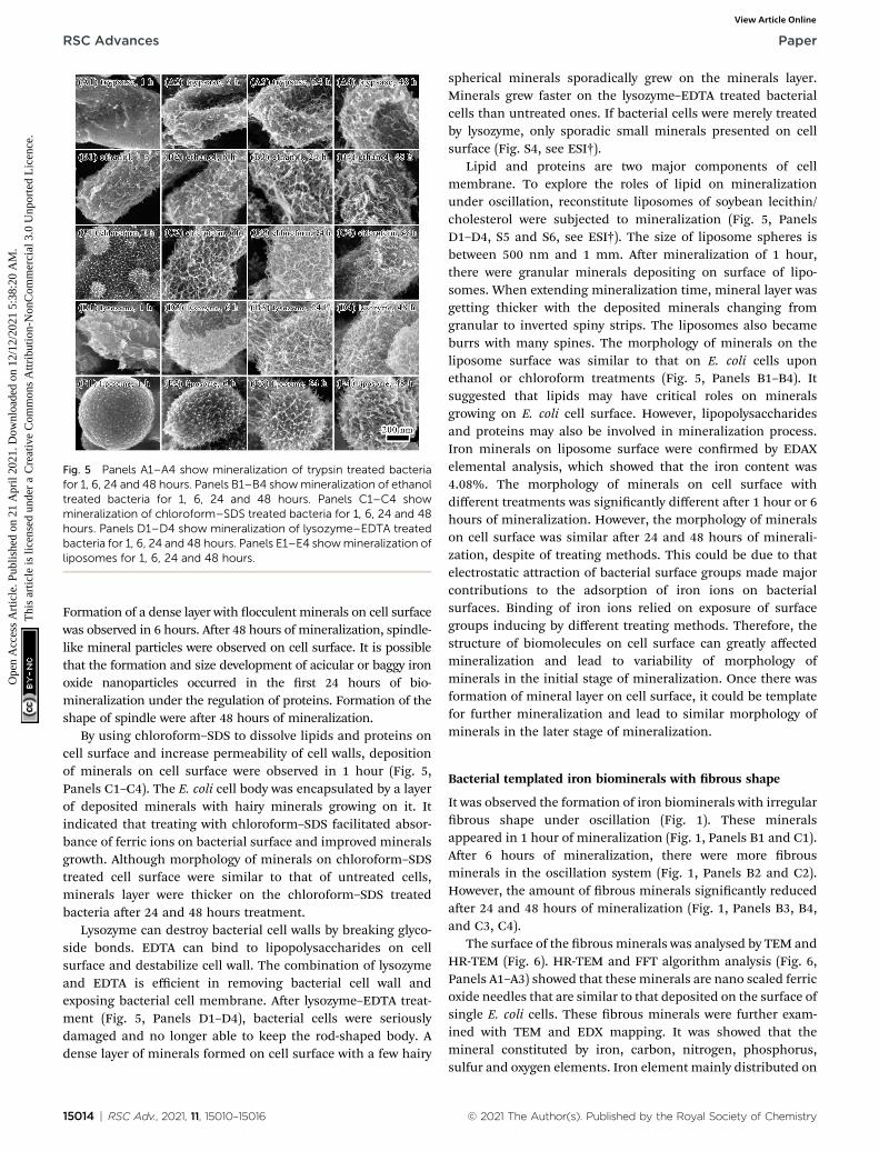
Fig. 5 Panels A1–A4 show mineralization of trypsin treated bacteriafor 1, 6, 24 and 48 hours. Panels B1–B4 showmineralization of ethanoltreated bacteria for 1, 6, 24 and 48 hours. Panels C1–C4 showmineralization of chloroform–SDS treated bacteria for 1, 6, 24 and 48hours. Panels D1–D4 show mineralization of lysozyme–EDTA treatedbacteria for 1, 6, 24 and 48 hours. Panels E1–E4 showmineralization ofliposomes for 1, 6, 24 and 48 hours.
RSC Advances Paper
Ope
n A
cces
s A
rtic
le. P
ublis
hed
on 2
1 A
pril
2021
. Dow
nloa
ded
on 1
2/12
/202
1 5:
38:2
0 A
M.
Thi
s ar
ticle
is li
cens
ed u
nder
a C
reat
ive
Com
mon
s A
ttrib
utio
n-N
onC
omm
erci
al 3
.0 U
npor
ted
Lic
ence
.View Article Online
Formation of a dense layer with occulent minerals on cell surfacewas observed in 6 hours. Aer 48 hours of mineralization, spindle-like mineral particles were observed on cell surface. It is possiblethat the formation and size development of acicular or baggy ironoxide nanoparticles occurred in the rst 24 hours of bio-mineralization under the regulation of proteins. Formation of theshape of spindle were aer 48 hours of mineralization.
By using chloroform–SDS to dissolve lipids and proteins oncell surface and increase permeability of cell walls, depositionof minerals on cell surface were observed in 1 hour (Fig. 5,Panels C1–C4). The E. coli cell body was encapsulated by a layerof deposited minerals with hairy minerals growing on it. Itindicated that treating with chloroform–SDS facilitated absor-bance of ferric ions on bacterial surface and improved mineralsgrowth. Although morphology of minerals on chloroform–SDStreated cell surface were similar to that of untreated cells,minerals layer were thicker on the chloroform–SDS treatedbacteria aer 24 and 48 hours treatment.
Lysozyme can destroy bacterial cell walls by breaking glyco-side bonds. EDTA can bind to lipopolysaccharides on cellsurface and destabilize cell wall. The combination of lysozymeand EDTA is efficient in removing bacterial cell wall andexposing bacterial cell membrane. Aer lysozyme–EDTA treat-ment (Fig. 5, Panels D1–D4), bacterial cells were seriouslydamaged and no longer able to keep the rod-shaped body. Adense layer of minerals formed on cell surface with a few hairy
15014 | RSC Adv., 2021, 11, 15010–15016
spherical minerals sporadically grew on the minerals layer.Minerals grew faster on the lysozyme–EDTA treated bacterialcells than untreated ones. If bacterial cells were merely treatedby lysozyme, only sporadic small minerals presented on cellsurface (Fig. S4, see ESI†).
Lipid and proteins are two major components of cellmembrane. To explore the roles of lipid on mineralizationunder oscillation, reconstitute liposomes of soybean lecithin/cholesterol were subjected to mineralization (Fig. 5, PanelsD1–D4, S5 and S6, see ESI†). The size of liposome spheres isbetween 500 nm and 1 mm. Aer mineralization of 1 hour,there were granular minerals depositing on surface of lipo-somes. When extending mineralization time, mineral layer wasgetting thicker with the deposited minerals changing fromgranular to inverted spiny strips. The liposomes also becameburrs with many spines. The morphology of minerals on theliposome surface was similar to that on E. coli cells uponethanol or chloroform treatments (Fig. 5, Panels B1–B4). Itsuggested that lipids may have critical roles on mineralsgrowing on E. coli cell surface. However, lipopolysaccharidesand proteins may also be involved in mineralization process.Iron minerals on liposome surface were conrmed by EDAXelemental analysis, which showed that the iron content was4.08%. The morphology of minerals on cell surface withdifferent treatments was signicantly different aer 1 hour or 6hours of mineralization. However, the morphology of mineralson cell surface was similar aer 24 and 48 hours of minerali-zation, despite of treating methods. This could be due to thatelectrostatic attraction of bacterial surface groups made majorcontributions to the adsorption of iron ions on bacterialsurfaces. Binding of iron ions relied on exposure of surfacegroups inducing by different treating methods. Therefore, thestructure of biomolecules on cell surface can greatly affectedmineralization and lead to variability of morphology ofminerals in the initial stage of mineralization. Once there wasformation of mineral layer on cell surface, it could be templatefor further mineralization and lead to similar morphology ofminerals in the later stage of mineralization.
Bacterial templated iron biominerals with brous shape
It was observed the formation of iron biominerals with irregularbrous shape under oscillation (Fig. 1). These mineralsappeared in 1 hour of mineralization (Fig. 1, Panels B1 and C1).Aer 6 hours of mineralization, there were more brousminerals in the oscillation system (Fig. 1, Panels B2 and C2).However, the amount of brous minerals signicantly reducedaer 24 and 48 hours of mineralization (Fig. 1, Panels B3, B4,and C3, C4).
The surface of the brousminerals was analysed by TEM andHR-TEM (Fig. 6). HR-TEM and FFT algorithm analysis (Fig. 6,Panels A1–A3) showed that these minerals are nano scaled ferricoxide needles that are similar to that deposited on the surface ofsingle E. coli cells. These brous minerals were further exam-ined with TEM and EDX mapping. It was showed that themineral constituted by iron, carbon, nitrogen, phosphorus,sulfur and oxygen elements. Iron element mainly distributed on
© 2021 The Author(s). Published by the Royal Society of Chemistry

Fig. 6 TEM image (Panel A1) and HR-TEM image (Panel A2, the areaindicated by the green box in Panel A1). Panel A3 shows FFT pattern ofyellow zone of Panel A2.
Fig. 7 Panel A shows degradation of methylene blue. Inset of Panel Ashows linear relationship between ln(C/C0) and irradiation time. PanelB shows polarization curve of iron biominerals for HER in 1 M KOHwitha potential scan rate of 5 mV s�1. Inset of Panel B shows durableoperation test of iron biominerals electrodes operated at designed h
(10 mA cm�2).
Paper RSC Advances
Ope
n A
cces
s A
rtic
le. P
ublis
hed
on 2
1 A
pril
2021
. Dow
nloa
ded
on 1
2/12
/202
1 5:
38:2
0 A
M.
Thi
s ar
ticle
is li
cens
ed u
nder
a C
reat
ive
Com
mon
s A
ttrib
utio
n-N
onC
omm
erci
al 3
.0 U
npor
ted
Lic
ence
.View Article Online
the surface with carbon element inside. Therefore, it is possiblethat aggregated E. coli cells formed the main body of the brouswith iron mineralization on the surface. At the early stage ofmineralization, iron oxide deposited on E. coli surface mayfacilitate the ordered aggregation of cells under oscillation andlead to the formation of brous minerals. Along with the bio-mineralization process, more iron minerals deposited on thecell surface and increased the fragility of brous minerals. Itmade it easy for the brous minerals to break under oscillation.Therefore, less brous minerals were observed aer 24 and 48hours of mineralization.
Catalytic performance of iron biominerals
Photocatalytic performance of iron biominerals was evaluatedby measuring degradation of methylene blue under visibleirradiation (Fig. 7, Panel A). Percentage of degradation (C/C0) ¼(1 � Ct/C0) � 100, where C0 and Ct in Fig. 7 (Panel A) representthe initial concentration aer the adsorption–desorption equi-librium for t minute and the real-time concentration of meth-ylene blue at time t minute, respectively. The degradation ofmethylene blue reached 92.11% in 15minutes and 98.11% in 30minutes. These results demonstrated the reasonable photo-catalytic activity of as prepared iron biominerals. The linearrelationship between ln(C/C0) and irradiation time (Fig. 7, PanelA, inset) indicated that photocatalytic degradation of methyleneblue followed the pseudo rst-order law. In the present study,the apparent rate constant was calculated as 0.17 � 0.05 min�1.
© 2021 The Author(s). Published by the Royal Society of Chemistry
The hydrogen evolution reaction (HER) is promising inproducing clean and renewable hydrogen resources. In thepresent study, the HER performance of iron minerals as anefficient, durable, and inexpensive hydrogen evolution elec-trode was investigated. Polarization curves were collected in 1MKOH solutions at a rotation rate of 1600 rpm with a sweep rateof 5 mV s�1 at room temperature. Before subjecting to electro-catalytic measurement, iron biominerals were calcinated at700 �C for 4 hours (Fig. S7, see ESI†). The iron oxide mineralelectrode achieved the best activity with the overpotential of235 mV at the current of 10 mA cm�2 (Fig. 7, Panel B). Thecatalytic stability of iron oxide minerals was also measured at 10mA cm�2 in the same electrolyte, which showed that the stablecurrent with a small deviation aer one hour at overpotential of�20 mV was presented (Fig. 7, Panel B, inset).
In future, more investigation concerning catalytic perfor-mances of acquired biominerals will be carried out to provide
RSC Adv., 2021, 11, 15010–15016 | 15015
RSC Advances Paper
Ope
n A
cces
s A
rtic
le. P
ublis
hed
on 2
1 A
pril
2021
. Dow
nloa
ded
on 1
2/12
/202
1 5:
38:2
0 A
M.
Thi
s ar
ticle
is li
cens
ed u
nder
a C
reat
ive
Com
mon
s A
ttrib
utio
n-N
onC
omm
erci
al 3
.0 U
npor
ted
Lic
ence
.View Article Online
detailed information and data including the UV-vis spectra,ROS generation, CV curves.
Conclusions
This article reported effects of motility on biomineralization ofiron oxide on E. coli surface. Under oscillation, the bacterial E.coli templated the formation of iron oxide minerals with acic-ular and banded morphology. The surface charge of E. coli cellscontributed to the biomineralization process. The surfacecomponents of E. coli cells, that is, lipids, carbohydrates andproteins, regulated the formation and morphology of iron oxideminerals. The morphology of iron oxide minerals formed onliposome surface was similar to that on E. coli cell surface,implying that lipids on E. coli cell surface played a critical role inmorphogenesis of iron oxide minerals. As-prepared mineralizediron oxide nanomaterials showed activity in photocatalyticdegradation of methylene blue as well as in electrocatalytichydrogen evolution reaction, implying potential uses of thismaterial in environment and energy.
Conflicts of interest
There are no conicts to declare.
Acknowledgements
Supported by National Natural Science Foundation of China(31771032, 51911530153, 51832003).
Notes and references
1 I. Aksay, M. Traus, S. Manne, I. Honma, N. Yao, L. Zhou,P. Fenter, P. Eisenberger and S. Gruner, Science, 1996, 273,892.
2 S. Stupp and P. Braun, Science, 1997, 277, 1242.3 X. Zhao, J. Yang, L. McCormick and J. Fendler, J. Phys. Chem.,1992, 96, 9933.
4 D. Bazylinski, Encyclopedia of Materials: Science & Technology,2001, p. 441.
5 W. Muynck, D. Belie and W. Verstraete, Ecol. Eng., 2010, 36,118.
6 A. Amiri and Z. Basaran, Construct. Build. Mater., 2018, 165,655.
7 S. Yao, B. Jin, Z. Liu, C. Shao, R. Zhao, X. Wang and R. Tang,Adv. Mater., 2017, 29, 1605903.
8 S. Shankar, A. Ahmad, R. Pasricha and M. Sastry, J. Mater.Chem., 2003, 13, 1822.
9 L. Li, X. Yang, A. Li, T. Zhang and Y. Liu, Appl. Mech. Mater.,2011, 71–78, 2831–2835.
10 D. Tamayo-Figueroa, E. Castillo and P. Brandao, World J.Microbiol. Biotechnol., 2019, 35, 58.
15016 | RSC Adv., 2021, 11, 15010–15016
11 T. Li, Y. Hu and B. Zhang, Front. Microbiol., 2018, 9, 1884.12 S. Xie, G. Yin, X. Pu, S. Xie, G. Yin, X. Pu, Y. Hu, Z. Huang,
X. Liao, Y. Yao and X. Chen, Curr. Drug Delivery, 2017, 13,999.
13 S. Kim, L. Palanikumar, H. Choi, M. Jeena, C. Kim and J. Ryu,Chem. Sci., 2018, 9, 2474.
14 I. Weiss and F. Marin, Biomineralization: From Nature toApplication, 2008, vol. 4, p. 71.
15 T. Chen, P. Shi, Y. Li, T. Duan, Y. Yu, X. Li and W. Zhu,CrystEngComm, 2018, 20, 2366.
16 B. Li, G. Lai, B. Lin, A. Yu and N. Yang, Sens. Actuators, B,2018, 262, 789.
17 J. Miot, K. Benzerara, G. Morin, A. Kappler, S. Bernard,M. Obst, C. Ferard, F. Skouri-Panet, J. Guigner, N. Posth,M. Galvez, G. Brown Jr and F. Guyot, Geochim. Cosmochim.Acta, 2009, 73, 696.
18 M. Jiang, T. Ohnuki and S. Utsunomiya, Geomicrobiol. J.,2018, 35, 375.
19 K. Saranya, A. Sundaramanickam, S. Shekhar, M. Meena,R. Sathishkumar and T. Balasubramanian, J. Environ.Manage., 2018, 222, 396.
20 P. Dhanwal, A. Kumar, S. Dudeja, H. Badgujar, R. Chauhan,A. Kumar, P. Dhull, V. Chhokar and V. Beniwal, WaterEnviron. Res., 2018, 90, 424.
21 M. Sahmoune, Microchem. J., 2018, 141, 87.22 A. Choinska-Pulit, J. Sobolczyk-Bednarek and W. Łaba,
Ecotoxicol. Environ. Saf., 2018, 149, 275.23 R. Rani and M. Saharay, RSC Adv., 2019, 9, 1653.24 F. Li, W. Wang, C. Li, R. Zhu, F. Ge, Y. Zheng and Y. Tang, J.
Hazard. Mater., 2018, 358, 178.25 M. Li, X. Cheng andH. Guo, Int. Biodeterior. Biodegrad., 2013,
76, 81.26 M. Ozaki, S. Sakashita, Y. Hamada and K. Usui, Protein Pept.
Lett., 2018, 25, 15.27 T. Beveridge and S. Koval, Appl. Environ. Microbiol., 1981, 42,
325.28 A. Lassoued, M. Lassoued, B. Dkhil, S. Ammar and A. Gadri,
J. Mater. Sci.: Mater. Electron., 2018, 29, 8142.29 J. Ying, G. Jiang, Z. Cano, L. Han, X. Yang and Z. Chen, Nano
Energy, 2017, 40, 88.30 T. Beveridge and S. Fyfe, Can. J. Earth Sci., 1985, 22, 1893.31 S. Langley and T. Beveridge, Appl. Environ. Microbiol., 1999,
65, 489.32 T. Yamashita and P. Hayes, Appl. Surf. Sci., 2008, 254, 2441.33 G. Bhargava, I. Gouzman, C. Chun, T. Ramanarayanan and
S. Bernasek, Appl. Surf. Sci., 2007, 253, 4322.34 A. Darbeheshti, M. Ghazi and M. Izadifard, Mater. Res.
Express, 2017, 4, 106110.35 G. Tortora, B. Funke, C. Case, D. Weber and W. Bair III,
Microbiology: An Introduction, Pearson, 13th edn, 2018.
© 2021 The Author(s). Published by the Royal Society of Chemistry