ergot alkaloids produced by claviceps cyperi
TRANSCRIPT

57
5. ERGOT ALKALOIDS PRODUCED BY CLAVICEPS CYPERI
Abstract
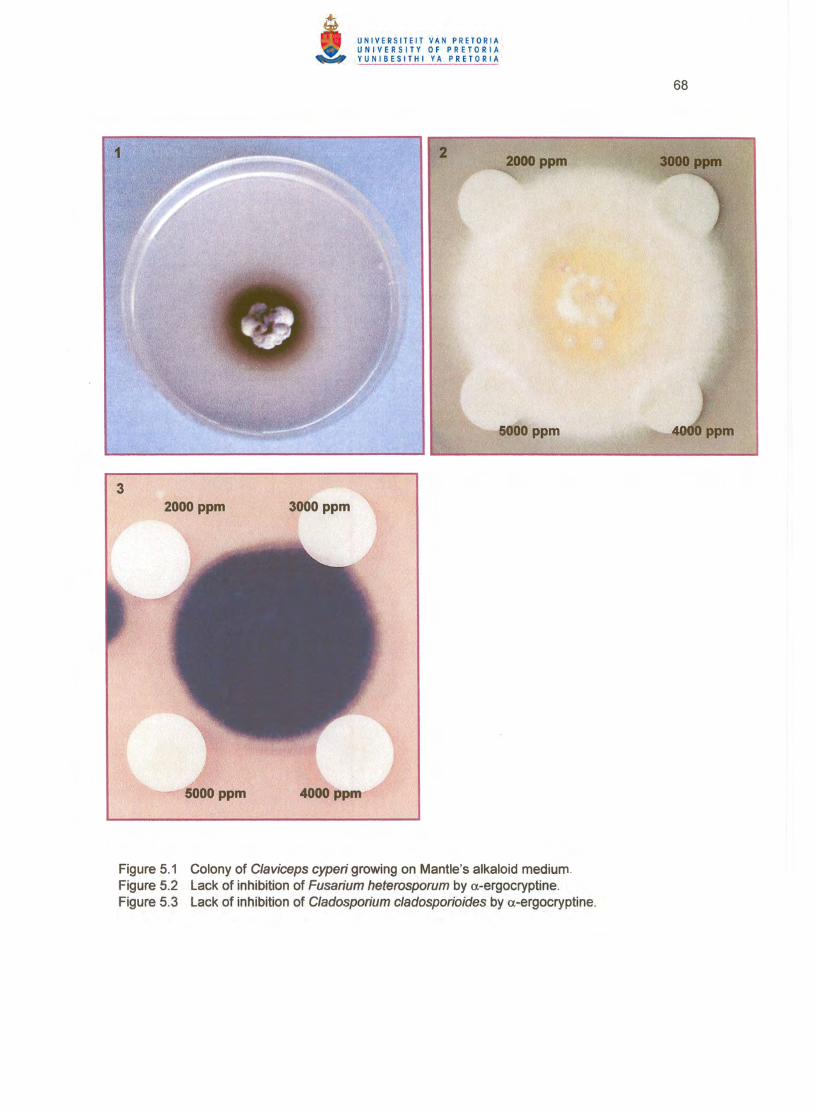
The main ergopeptine alkaloid in sclerotia of C/aviceps cyperi collected from ergotised nut sedge implicated in outbreaks of bovine ergotism in South Africa was identified by HPLC and tandem mass spectroscopy as a.-ergocryptine. All sclerotial samples tested also yielded ergosine, whereas ergocornine and ergocrystine were detected at low concentrations in a freshly collected sample. Trace amounts of 0.
ergocryptine was produced by C. cyperi on solid medium, but all attempts at inducing an isolate of the species to synthesise ergopeptines in liquid culture have failed . a.-Ergocryptine showed no antibiotic activity against Cladosporium cladosporioides and Fusarium heterosporum, two fungi commonly associated with C. cyperi honeydew.
5.1 INTRODUCTION
Various cases of bovine ergotism have recently been reported from the eastern Highveld
Region in South Africa (Van der Linde & Naude 2000). Examination of the fodder implicated in
the outbreaks showed that it was extensively contaminated with yellow nut sedge (Cyperus
esculentus L.) ergotised by Claviceps cyperi Loveless, whereas chemical analysis indicated
the presence of the ergot alkaloid a-ergocryptine, as well as traces of ergosine, ergocornine,
ergocrystine and ergotamine (Naude et a/. 2005).
Besides being important from a diagnostic/prognostic perspective, verification of the alkaloid or
alkaloids produced by a Claviceps species is of value in confirming the identity of the species,
defining chemoraces or chemotypes, and classifying intraspecific variability (Pazoutova et al.
2000), hence assisting in the elucidation of phylogenetic relationships and habitat
specialisation. The potential pharmaceutical application of the particular alkaloid/s is also of
considerable importance. a-Ergocryptine, for instance, is the source of 2-bromo-a
ergocryptine, a dopaminergic agent marketed world-wide under the trade name Parlodel by
the firm Novartis. The drug is used, among others, for puerperal prevention or suppression of
lactation, treatment of hyperprolactinaemia and prolactin-related menstrual and fertility
disorders, and as an adjunct in the treatment of Parkinsonism (Snyman 2001).
2-Bromo-a-ergocryptine is presently produced semi-synthetically from wheat (Triticum
aestivum L.) artificially infected with Claviceps purpurea (Fr.:Fr. ) Tul.
58
(http://www.dimok.de/ergotiharvest.html). However, C. purpurea is not a prolific producer of a
ergocryptine (Taber & Vining 1957), and C. cyperi may therefore be a more suitable source.
This report provides data on the ergot alkaloids present in sclerotia of C. cyperi and describes
various attempts at inducing sphacelial isolates of the species to produce a-ergocryptine in
culture.
5.2 MATERIALS AND METHODS
Sclerotia of C. cyper; collected between 1997 and 2000 from ergotised nut sedge at localities
where bovine ergotism occurred (Table 5. 1) were analysed for ergot alkaloids by Meadows
Cape in Paarl, Western Cape Province, according to the HPLC method of Rottinghaus et al.
(1 993), following extraction of the sclerotia as described by Scott et al. (1992). Sclerotia
collected in 1997, 1999, 2002 and 2003 were tested in 2003 for ergopeptines as described in
5.2.3 and 5.2.4. A preliminary experiment was also done in which C. cyper; PREM 56618 was
grown for 8 weeks at 22 °C in the dark on Mantle's alkaloid medium (Mantle 1973) solidified
with 1.5 % agar, and the cultures analysed by HPLC for a-ergocryptine at the Council for
Scientific and Industrial Research in Pretoria.
Following the above, in vitro production of alkaloids by C. cyper; in liquid medium was
attempted according to the procedures described below.
5.2.1 Preparation of inoculum
Pure cultures of C. cyper; PREM 57392, subsequently deposited as PPRI 7196, were obtained
by surface-disinfecting sclerotia in 1.75 % sodium hypochlorite for 5 minutes and rinsing them
three times in sterile distilled water. The sclerotia were dried on sterile tissue paper and
transferred to a microscope slide cleaned with 70 % ethanol. The cortex of each sclerotium
was removed with a sterile surgical blade, and the inside cut into sections which were plated
on potato-dextrose agar and glucose yeast extract agar. When the colonies were about 25
mm in diameter, plugs with mycelium cut from them were used to inoCUlate Erlenmeyer flasks
with glucose yeast extract broth. The flasks were incubated stationary in the dark for 2 weeks
at 22 °C and the cultures used to inoculate the media in 5.2.2.
59
5.2.2 Culturing
The following liquid media were prepared (concentrations are per litre):
Mantle's alkaloid medium (Mantle 1973) (MAM):
Sucrose 150 9
L-Asparagine 15 9
KH2P04 25mg
MgS04 ·7H20 25mg
FeS04 ·7H20 33mg
ZnS04 27mg
(pH adjusted to 5.5 with 1 N NaOH)
Bacon's alkaloid medium (Bacon et al. 1979) (BAC):
Sorbitol 1 00 9
Glucose 40 9
Succinic acid 10 9
KH2P04 1 9
MgS04·7H2O 0.3 9
FeS04 ·7H2O 1 mg
Yeast Extract 1 9
(pH adjusted to 5.5 with 1 N NaOH)
Molasses medium (MOL):
Molasses 150 9
Yeast extract 5 9
(pH adjusted to 5.2 with 1 N NaOH)
Glucose yeast extract (Fuentes et s/. 1964) (GVE):
Glucose 109
Yeast Extract 1 0 9
(pH adjusted to 5.2 with 1 N NaOH)
Each medium was dispensed into sixty-four 250 ml capacity Erlenmeyer flasks at 100 ml per
60
flask, and the flasks with medium were autoclaved for 15 minutes at 121 cC. When cooled,
each flask was inoculated with 1 ml of the above inoculum of C. cyperi and sixteen flasks of
each medium were incubated stationary in the dark at 15, 22, 27 and 34 cc, respectively.
5.2.3 Extraction of alkaloids
Extractions from cultures were done weekly for 8 weeks with duplicate flasks of each medium
from each incubation temperature. The content of each flask was filtered through Whatman
No. 1 paper. Two aliquots of 5 ml of the fi ltrate were each transferred to a 15 ml screw-cap
vial. Drops of 0.1 N sodium hydroxide were added to increase the pH to between 9 and 10.
Nine millilitres of chloroform was added to each vial and the vials agitated on a rotary shaker
for 30 minutes, after which they were centrifuged for 3 minutes at 4000 rpm. The upper
aqueous layer was discarded and the remaining chloroform evaporated to dryness with
nitrogen in a Reacti-Vap at 50 cC.
The mycelial mass was rinsed with distilled water, divided in two portions, and each portion
was transferred to a pre-weighed 15 ml screw-cap vial. The fungal material was dried for 2- 3
hours at 40 cC, the vials were weighed again, and the dry mycelial mass was calculated. Five
millilitres of methanol was added to each vial, the contents sonicated for 1 hour, and the tubes
then centrifuged for 3 minutes at 4000 rpm. Five millilitres of the liquid in each vial was
transferred to a clean vial and concentrated to dryness with nitrogen in a Reacti-Vap at 50 cC.
The entire experiment was conducted three times.
5.2.4 Alkaloid analysis
Vials with dried extracts were submitted on a weekly basis for alkaloid analysis to the
Veterinary Medical Diagnostic Laboratory at the University of Missouri, Columbia, USA.
Ergopeptine analysis was done by the HPLC technique of Rottinghaus et al. (1993). Samples
were extracted with alkaline chloroform, filtered and applied to Ergosil cleanup columns
(Analtech, Newark, Denmark). Following elution of pigments with acetone:chloroform (4:1),
alkaloids were eluted with methanol and analysed by HPLC with fluorescence detection. The
presence of ergopeptines was confirmed by treating the samples with 0.2 % acetic acid and
re-analysis by HPLC for the -ine isomers. The HPLC system consisted of a Perkin Elmer LC
250 pump and ISS 200 auto sampler, with detection on a Hitachi F-1200 fluorescence
61
detector. A Phenomenex Luna C18 column (150 mm x 4.6 mm) was used with a mobile phase
of acetonilrile:water (35:65) and a 200 mg 1-1 solution of ammonium carbonate in distilled
water.
The identity of the ergopeptine alkaloids was verified by tandem mass spectroscopy
(Finnigan/MAT TSQ 70 Tandem Mass Spectrometer (MS/MS») according to Rottinghaus et al.
(1993) at the USDA National Veterinary Services Laboratory, Ames, Iowa. A portion of each of
the chloroform extracts processed through the above Ergosil cleanup columns was applied to
a direct exposure probe, the solvent allowed to evaporate, and the probe inserted into the
mass spectrometer. The MS/MS was operated in the negative chemical ionisation mode with
methane as reagent gas and argon as collision gas. Daughter ions were collected for the 0..
ergocryptine parent ion m/Z 308. Typical daughter spectra were obtained with base peak mlZ
209, in accordance with Plattner et a'- (1983) and Rottinghaus et al. (1993).
5.2.5 Antimycotic activity of o..~ergocryptine
Colonies of the two fungal species most commonly associated with honeydew of C cyperi, viz.
Fusarium heterosporum Nees and Cladosporium c/adosporioides (Fresen.) G.A. de Vries,
were established centrally on potato-dextrose agar plates. When about 1 cm in diameter, four
antibiotic assay discs saturated with chloroform solutions containing 2000, 3000, 4000 and
5000 ppm pure a-ergocryptine (Sigma, Johannesburg), respectively, were placed equidistantly
around each colony on the agar. Plates were incubated at 18 °C in the dark and inspected
regularly.
5.3 RESULTS
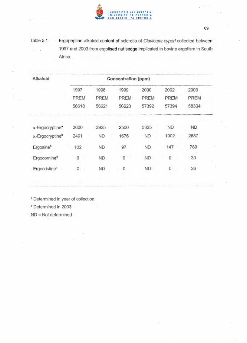
The main ergopeptine alkaloid detected in sclerotia of C. cyperi was a-ergocryptine, with
concentrations in freshly collected samples ranging from 2500 to 5325 ppm (Table 5.1). When
tested again in 2003, the a-ergocryptine content of the sclerotia on average was about 50 %
less than in the initial analysis. All sclerotia I samples tested in 2003 also contained ergosine at
concentrations 3.4-24 times lower than that of o..-ergocryptine. Small quantities of
ergocornine and ergocristine were present in sclerotia collected and tested in 2003.
Cultures of PREM 56618 grown on solid MAM produced a dark brown diffusable pigment in

62
the agar (Figure 5.1 ). The concentration of a-ergocryptine in these cultures after 8 weeks was
7 ppb .
. No growth of PREM 57392 occurred in any of the liquid media at 34 DC. Some growth was
evident in MOL at 15 DC. but not in the other three media. Cultures in MAM and BAC at 22 and
27 DC started to produce a brown pigment in the medium after about 4 weeks. Best growth in
terms of biomass produced at the latter temperatures (data not presented) occurred in GYE.
No ergopeptine alkaloid could be detected in any of the media at any time at any temperature
in any of the three runs of the experiment.
Colonies of F. heterosporum and C. cladosporioides grew unobstructedly over the antibiotic
assay discs impregnated with a-ergocryptine (Figures 5.2.5.3).
5.4 DISCUSSION
Environmental conditions play an important role in Claviceps fermentations (Mantle 1978;
Arora et al. 1992; Wainwright 1992). With most strains. the metal ions Mg. Fe and Zn in trace
amounts enhance alkaloid production. pH optima for growth are around 5.2- 5.5. and
temperature optima usually near 24 DC. Induction of alkaloid biosynthesis requires osmotic
pressures between 10 and 20 bar. attained by high sugar concentrations which can be
partially replaced by sodium chloride. The high osmotic pressure inhibits conidiogenesis.
induces differentiation into sclerotium-like cells. and enhances the entrance of nutrients into
the cells. As is common for most secondary metabolites, alkaloid synthesis follows rather than
accompanies active growth. Maximum alkaloid yield therefore usually occurs only after
prolonged incubation (12-50 days) (Abe 1948; Taber & Vining 1957; Mantle 1973). By
adhering to these principles, alkaloid synthesis in culture has successfully been achieved with
ergot species such as C. purpurea (Michener &Snell 1950; Taber &Vining 1957; Castagnoli &
Mantle 1966). C/aviceps paspa/i F. Stevens & J.G. Hall (Pacifici et a/. 1962), Claviceps
gigantea SF Fuentes. Isla. Ullstrup & Rodriques (Fuentes et al., 1964), Sphace/ia sorghi
McRae (Mantle 1973). C/aviceps fusiformis Loveless (Mantle 1978). Claviceps africana
Freder., P.G. Mantle & W.A.J. de Milliano, Claviceps sorghicola Tsukib., Shiman. & Uematsu
(Pazoutova 2001) and Claviceps zizaniae (Fyles) Pantidou (Kantorova et al. 2002).
Despite adhering to the above guidelines, and allowing for possible deviations in
63
environmental requirements, the present attempts at inducing C. cyperi to respond accordingly
have failed, except for the trace amounts of a-ergocryptine produced on solid medium. The
lack of success could probably be ascribed to an inherent inability of C. cyper; to synthesise
ergopeptines in liquid culture or to the isolates used having lost the capacity to produce
alkaloids. Von Bekesy (1940) showed that cultures of C. purpurea obtained from sclerotia with
. a high alkaloid content yielded the highest concentration of alkaloids. The two isolates of C.
cyperi used in the present study, particularly PPRI 7196, were selected for that reason.
Although alkaloid-producing cultures have an acute tendency to loose the ability to yield
alkaloids upon repeated transfer (Mantle 1978), PPRI 7196, when inoculated into the various
liquid media, had been subcultured only twice since being obtained in culture from PREM
57392 sclerotia. A more feasible explanation for the absence of ergopeptines in cultures of
PPRI 7196 therefore seems to be that cultural conditions were not conducive to alkaloid
production by this isolate of C. cyperi. Observations by Ramstad & Gjerstad (1955) indicated
that mycelial tissue of C. purpurea, in the same way as conidia, is incapable of producing
alkaloids, and that failure to produce alkaloids in culture is due to sclerotial tissue not
developing under these conditions. Mantle & Tonolo (1 968) similarly showed that improved in
vitro alkaloid yields by C. purpurea is associated with a plectenchymatic growth form. Enlarged
hyphal cells, probably sclerotial primordia, have been observed in cultures of C. cyperi on solid
medium in Chapter 3, but it is not known if such cells were formed in liquid medium in the
present study. Growth of the cultures certainly did not appear plectenchymatous. Regarding
the above it is interesting to note that Pazoutova (2001) reported sclerotia and cultures of C.
zizaniae, the Claviceps species phenotypically the closest related to C. cyperi (Chapter 6) and
the only other ergot species producing a-ergocryptine as main alkaloid, to be void of alkaloids.
In a subsequent study, however, Kantorova et al. (2002) found one wild strain of C. zizaniae to
synthesise a-ergocryptine at concentrations of up to 1 mg g-1 in the same medium used by
Pazoutova (2001), whereas a second strain did not produce any detectable alkaloids at all.
Contrary to the culture experiments, ergopeptine analysis of sclerotia of C. cyperi provided
vital information regarding the chemistry of C. cyperi. More than 80 ergot alkaloids are known
(Bock & Parberry, www.tacethno.com/info/claviceps Internet access 13.01.2005), but peptide
ergot alkaloids (ergotamine, ergosine, ergOCristine, ergocornine, ergostine, a-ergocryptine, ~
ergocryptine and derivatives) have thus far been detected only in C. purpurea (various
ergopeptines), C. zizaniae (a-ergocryptine) and C. africana (dihydroergosine) (Mantle 1968;
Flieger et al. 1997; Kantorova et al. 2002). C. cyperi is therefore the fourth Clav;ceps species
64
found capable of producing ergopeptines. This alkaloid profile corresponds with the alkaloid
analysis of the fodder implicated in the outbreaks of bovine ergotism (Naude et al. 2005) and
is typically associated with "summer syndrome" symptoms, e.g. hyperthermia, reduced food
intake, lethargy, drop in milk production, loss of body mass, increased respiratory rate, open
mouthed breathing, seeking shade and wading into water (Ross et al. 1989). One of the
fodder samples tested by Naude et al. (2005), however, contained ergotamine, which was not
detected in sclerotia of C. cyperi. The presence of ergotamine in the above sample can
probably be ascribed to contamination of the fodder by a Lolium sp. ergotised by C. purpurea,
also occurring in the area.
Besides elucidating the alkaloid profile of C. cyperi, results of the present study verified the
relatively narrow mesophilic temperature growth range of this ergot species (Chapter 3) and
also showed that a-ergocryptine does not possess antimycotic activity against F.
heterosporum and C. cladosporioides. The inhibition of growth of the latter two fungi by C.
cyperi observed in dual culture in Chapter 3 must therefore have been due to a different
metabolite produced by C. cyperi in the medium.
5.5 REFERENCES
ABE, M. 1948. Ergot fungus. IX. Separation of an active substance and its properties. Journal
of the Agricultural Chemical Society ofJapan 22:2.
ARORA, D.K, ELANDER, R.P. & MUKERJI, KG. (eds.). 1992. Handbook of applied
mycology. Vol 4. Fungal biotechnology. Marcel Dekker, New York.
BACON, C.W., PORTER, J.K & ROBBINS, J.D. 1979. Laboratory production of ergot
alkaloids by species of Balansia. Journal of General Microbiology 113: 119- 126.
CASTAGNOLl, N. & MANTLE, P.G. 1966. Occurrence of O-Iysergic acid and 6-methyl-ergol
8-ene-8-carboxylic acid in cultures of C/aviceps purpurea. Nature (London) 211 :859--860.
FLIEGER, M., WURST, M. & SHELBY, R.A. 1997. Ergot alkaloids: sources, structure and
analytical methods. Folia Microbiologica 42:3- 20.
65
FUENTES, S.F. , DE LA ISLA, M. DE L., ULLSTRUP, AJ. & RODRIQUES, AE. 1964 .
Claviceps gigantea, a new pathogen of maize in Mexico. Phytopathology 54:379- 381.
KANTOROVA, M., KOLiNSKA, R. , PAZOUTOVA, S., HONzATKO, A , HAVLICEK, V. &
FLiEGER, M. 2002. Ergot alkaloids produced by submerged cultures of Claviceps zizaniae .
. Journal of Natural Products 65: 1039---1040.
MANTLE, P.G. 1968. Studies on Sphacelia sorghi McRae, an ergot of Sorghum vulgare Pers.
Annals ofApplied Biology 62:443-449.
MANTLE, P.G. 1973. Production of ergot alkaloids in vitro by Sphacelia sorghi. Journal of
General Microbiology 75:275-281 .
MANTLE, P.G. 1978. The genus Claviceps. Pages 421 - 426. In: T.D. Wyllie & L.G.
Morehouse (eds.). Mycotoxic fungi, mycotoxins, mycotoxicoses: An encyclopedic handbook.
Vol. I. Mycotoxic fungi and chemistry of mycotoxins. Marcel Dekker, New York.
MANTLE, P.G. & TONOLO A 1968. Relationship between the morphology of Claviceps
purpurea and the production of alkaloids. Transactions of the British Mycological Society
51 :499- 505.
MICHENER, H.D. & SNELL, N. 1950. Studies on cultural requirements of C/aviceps purpurea
and inactivation of ergotamine. American Journal of Botany 37:52- 59.
NAUDE, T.W., BOTHA, C.J., VORSTER, J.H., ROUX, C., VAN DER LINDE, E.J., VAN DER
WALT, S.I., ROTTINGHAUS, G.E. , VAN JAARSVELD, L. & LAWRENCE, AN. 2005.
C/aviceps cyperi, a new cause of serious ergotism in dairy cattle consuming maize silage and
teff hay contaminated with ergotised Cyperus esculentus (nut sedge) on the Highveld of South
Africa. Onderstepoort Journal of Veterinary Research 72:23- 37.
PACIFICI, L.R., KELEHER, W.J. & SCHWARTING, A E. 1962. Production of lysergic acid
derivates in submerged culture. I. Fermentation studies. Uoydia 25:37- 45.
PAiOUTOvA, S. 2001. The phylogeny and evolution of the genus Claviceps. Mycological
66
Research 105:275-283.
PAZOUTOVA, S., OLSOVSKA, J., LlNKA, M. , KoLiNSKA, R. & FLiEGER, M. 2000.
Chemoraces and habitat specialisation of Claviceps purpurea populations. Applied and
Environmental Microbiology 66:541 9-6425.
PLATTNER, R.D., YATES, S.G. & POWER, J.K. 1983. Quadrapole mass spectrometry/mass
spectrometry of ergot cyclol alkaloids. Journal of Agriculture and Food Chemistry 65 :785
789.
RAMSTAD, E. & GJERSTAD, G. 1955. The parasitic growth of Claviceps purpurea (Fries)
Tulasne on rye and its relation to alkaloid formation. Journal of the American Pharmaceutical
Association 44:741 - 743.
ROSS, A.D., BRYDEN, W.L:; BAKAU, W. & BURGESS, L.W. 1989. Induction of heat stress in
beef cattle by feeding ergots of Claviceps purpurea. Australian Veterinary Journal 66:247
249.
ROTTINGHAUS, G.E. , SCHULTZ, L.M. , ROSS, P.F. & HILL, N.S. 1993. An HPLC technique
for the determination of ergot contamination in ground feed stuffs. Journal of Veterinary
Diagnostic Investigation 5:242- 247.
SCOTT, P.M., LOMBAERT, G.A., PELLAERS, P., BACHLER, S. & LAPPI , J. 1992. Ergot
alkaloids in grain foods sold in Canada. Journal of the Association of Official Analytical
Chemists International 75:773--779.
SNYMAN, J.R. 2001. (ed.). Monthly Index ofMedical Specialities 41 :1- 366.
TABER, W.A. & VINING, L.C. 1957. In vitro production of ergot alkaloids by cultures of
Claviceps purpurea. Canadian Journal ofMicrobiology 3:55-60.
VAN DER LINDE, E.J. & NAUDE T.W. 2000. Swam nuwe bedreiging vir melkbedryf.
Landbouweekblad 1131:24-27.

67
VON BEKESY, N. 1940. Untersuchungen uber den Alkaloidgehalt des Mutterkomes. II.
Mitteilung: Ober den Alkaloidgehalt des parasitisch kultivierten Mutterkornes. Biochemische
Zeitschrift 303:368- 382.
WAINWRIGHT, M. 1992. An introduction to fungal biotechnology. John Wiley, Chichester.
69
Table 5. 1 Ergopeptine alkaloid content of sclerotia of Claviceps cyperi collected between
1997 and 2003 from ergotised nut sedge implicated in bovine ergotism in South
Africa.
Alkaloid _._- --_._._
1997
PREM
56618
Concentration (ppm) - -----_. __..__.__... __..---- --_.._ ------_...--.-.- - - ---------.--... ~ .
1998 1999
PREM PREM 56621 56623
2000 2002 2003
PREM PREM PREM 57392 57394 58304
a.-Ergocryptinea
a.-Ergocryptineb
Ergosineb
Ergocomineb
Ergocristineb
3600
2491
102
0
0
3925
ND
ND
NO
ND
2500
1676
97
0
0
5325
ND
ND
NO
ND
ND
1902
147
0
0
ND
2687
789
30
26
a Determined in year of collection.
b Determined in 2003
NO =Not determined
70
6. MOLECULAR SYSTEMATICS OF CLAVICEPS CYPERI AND OTHER SOUTH
AFRICAN CLAVICEPS SPECIES
Abstract
Two South African isolates of Claviceps cyperi, one of C. purpurea, and a Canadian strain of C. grohii (CBS 124.47) were characterised with the aid of three primers to produce multilocus fingerprints. The internal transcribed spacers 1 and 2, the 5.8S region, as well as the ~-tubu lin
intron 3 region were sequenced. All available sequence data for several Claviceps species deposited in the GenBank nucleotide database were compared and optimally aligned with the South African isolates. K-means clustering and two-dimensional discriminant analysis as well as phylogenetic relationships were determined on these data sets. C. cyperi and C. zizaniae formed a distinct cluster showing little similarity with the other C/aviceps species which clustered in two main groups. The South African isolate of C. purpurea and C. grohii (CBS 124.47) showed high similarity with the GenBank C. purpurea strains and clustered with C. sulcata, C. fusiformis and C. paspa/i. C. africana was included in the second major cluster with C. viridis, C. pusi/la, C. sorghi, C. gigantea, C. maximensis, C. pha/aridis, C. sorghico/a and C. citrina. It was difficult to place C. citrina and C. paspa/i in either of the major clusters using the phylogenetic analysis. Sequence data from the ~-tubulin intron 3 region revealed a similar pattern and placed the two C. cyperi isolates in a distinct outgroup cluster at a large genetic distance from the other Claviceps species.
6.1 INTRODUCTION
C/aviceps species have traditionally been identified according to morphological
characteristics of the teleomorph and the anamorph, as well as the hosts from which
they were recorded. However, morphological features within species tend to be variable
(Pazoutova et af. 2000b), whereas description of a new species based solely on the host
is inclusive due to the polygeneric host range of the majority of ergot fungi (Paioutova et
a/. 2000b). Attempts to circumvent the problem have led to the establishment of
varieties, special forms or races, and host-specific groups (Pazoutova et al. 2000b;
Pazoutova 2001). Other means of distinguishing between species, such as alkaloid
profiles and the ability to float on water, have also been utilised to identify chemoraces
and habitat specialisation (Pafoutova et al. 2000b, 2002).
71
In recent years, much emphasis has been placed on the application of molecular
methods for verifying and detecting genetic variability in Claviceps. Most of the studies
concerned Cfaviceps purpurea (Fr.:Fr. ) Tul. and relied on random amplified polymorphic
DNA (RAPD) analysis utilising polymerase chain reaction (peR) techniques
(Jungehulsing &Tudzynski 1997; Pazoutova & Tudzynski 1999; Pazoutova et al. 2000b,
2002; Duncan et al. 2002). Application of RAPD, random amplified microsatellite (RAM),
amplified fragment length polymorphism (AFLP) and sequence analysis of the ~-tubulin
gene intron 3 region and EF-1-a gene intron 4 also enabled Pazoutova et al. (2000a)
and Tooley et al. (2000, 2001) to separate the Clav;ceps species associated with
Sorghum species. Although some of the above investigations included additional ergot
species as outgroups, the only comprehensive phylogenetic study was by Pazoutova
(2001), who compared 16 Claviceps species by means of peR amplification and
alignment of 5.BS rDNA and the adjacent internal transcribed spacers (ITS) 1 and 2.
One species that has thus far not been included in any molecular study is Claviceps
cyperi Loveless, causal agent of ergot in yellow and purple nut sedge (Cyperus
escufentus L. and Cyperus ratundus L. ), which has recently been implicated in several
cases of bovine ergotism in South Africa (Van der Linde & Naude 2000; Naude et al.
2005). Observations in Chapter 2 confi rmed that C. cyperi is morphologically distinct
from C/aviceps grahii J.W. Groves and C/aviceps nigricans Tul., the other two ergot
species recorded on Cyperaceae. This report describes the molecular characterisation
of C. cyperi and its separation from other C/aviceps species available in the GenBank
sequence database with the aid of multilocus peR fingerprinting of genomic DNA and
sequence analysis of the ITS1- 5.B rDNA-ITS2 and I3-tubulin gene intron 3 regions.
6.2 MATERIALS AND METHODS
6.2.1 Strains examined
Details of the C/aviceps and outgroup strains included in the study are summarised in
Table 6.1.
72
6.2.2 Extraction and purification of DNA
The CTAB method of Ausubel at a/. (1 989) was used for the extraction and purification
of genomic DNA of the four South African isolates included in this study (Table 6.1).
Purified DNA was quantified with fluorometry using Hoescht (H 33258) dye and a Hoefer
Dyna Quant 200 mini-fluorometer (Hoefer, San Francisco, CA). The final DNA
concentration was adjusted to between 25- 50 ng 111-1 at which all rep-PCR and RAPD
fingerprinting was performed.
6.2.3 peR fingerprinting of genomic DNA
All reactions were performed in a total volume of 10 III in 0.2 ml microtubes. Repetitive
sequence-based (rep-PCR) typing was performed with BOXA 1 Rand ERIC2 single
primers according to Versalovic et al. (1994). PCR buffer for rep-PCR was changed to
10 mM Tris-HCI; 50 mM KCI; 3.5 mM MgCI2; 0.1 % Triton X-100; pH 9.0 (Promega,
Madison , Wisconsin) . A GC-rich arbitrary primer ARP-7 was used according to Mathis &
McMillan (1996). PCR products were separated in 1.5 % agarose at 80 V constant in
TBE 1x buffer (89 mM Tris-base; 89 mM borate; 2 mM EDTA, pH 8.0). Amplification
products were stained with ethidium bromide (1 mg ml-1) for 15 minutes at 25°C and
destained for 10 minutes. DNA relative molecular mass marker (Roche Diagnostics, no
VI; GmbH, Mannheim) was loaded in lane one of each gel. Gel images were captured
with a CCD camera and stored as TIFF-uncompressed graphic files. Gel images were
processed and a similarity matrix and maximum parsimony tree created using
Bionumerics ver. 3.0 (Applied-Maths, BVBA, St.-Martens-Latem). Cluster and
discriminant analysis of fingerprints were performed on the similarity matrix with Ward's
clustering algorithm with Euclidean distances. (Statistica ver. 6.0; Statsoft, Tulsa,
Oklahoma). K-means clustering of group means was performed based on user-specified
clusters as derived from the dendrogram.
This clustering method attempts to minimise the distances within each cluster and to
maximise between-cluster distances. The K-means method tests the validity of user
specified clusters based on the topology of the dendrogram. In addition to K-means,

73
Multivariate Analysis of Variance (MANOVA) was used to test the validity of user
delineated groups (clusters). MANOVA is a statistical method which proves that the
likelihood of obtaining equally good separations and discrimination when groups are
chosen at random, approaches zero. In addition, MANOVA will determine which
characters are responsible for the discrimination between delineated groups. Parameter
L (Wi lkinson's likel ihood for normal distributions) predicts the likelihood that the groups
as delineated by the user, were drawn from the same population. When L is low to zero,
the strains or entries most likely were drawn from different populations confirming the
discrimination between user-delineated groups. Parameter p is the probability that a
random subdivision into different groups would produce equally good discrimination
between groups. Once again a low p value will confirm the val idity of user-delineated
groups.
6.2.4 Sequence analysis of the ITS1/2 and 5.85 regions (rONA operon)
This region of the rONA operon was chosen because sequence data of several
Claviceps species has been submitted to the GenBank database, which makes it
convenient for comparative purposes when new strains are sequenced. The complete
ITS 1, 5.B S and ITS 2 regions were amplified using primers ITS 5 (5' - GGA AGT AAA
AGT CGT AAC AAG G -3') and ITS 4 (5' - TCC TCC GCT TAT TGA TAT GC-3') which
are complementary to conserved sequences flanking the ITS 1 and 2 spacers. A
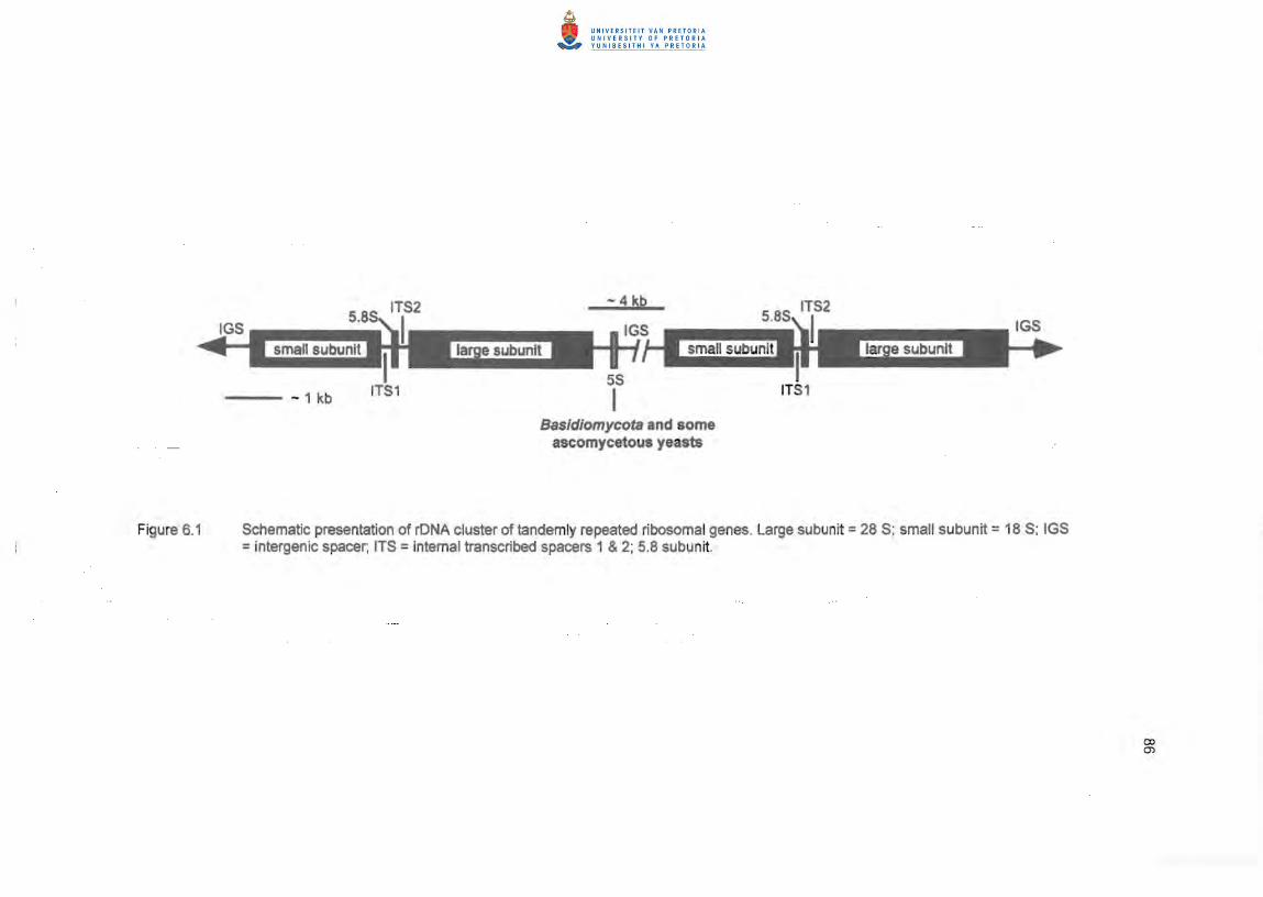
schematic presentation of the rONA gene cluster amplified is shown in Figure 6.1.
Amplification conditions were as follows: initial denaturation 95 DC for 3 minutes;
annealing at 60 DC for 1 minute; extension at 72 DC for 2 minutes; 30 cycles performed
at 94 DC for 1 minute, 60 DC for 1 minute and 72 DC for 2 minutes; final extension at 72
°C for 10 minutes. The reaction mixture consisted of 10 mM Tris-HCI; 1.5 mM MgCI2; 50
mM KCI; 0.1 % Triton X-100; pH 9.0; Taq polymerase 0.133 units per 10 f.!1 reaction
mixture (Promega, Madison, Wisconsin).
PCR amplified ITS 1/2 and 5.B S regions were purified using the High Pure PCR Product
Purification kit (Roche Molecular Diagnostics, Johannesburg, South Africa) or Qiagen
PCR Purification kit (Southern Cross Biotechnologies, Johannesburg) according to the
74
manufacturer's instructions. To assess the purity and concentration of the purified
product, 1.0 1-11 was subjected to electrophoresis on a 1 % agarose gel (Promega,
Madison, Wisconsin).
The purified PCR products were sequenced directly, without any additional cloning
procedures, using the ABI Prism BigDyeTM Terminator Cycle Sequencing Ready
Reaction kit (with AmpliTaqR DNA Polymerase, FS) (PE Applied Biosystems, Foster
City, California). Each sequencing reaction was carried out in a 5 IJI volume containing
approximately 100 ng template DNA, 12.5 pmol primer and 2 1-11 ready reaction premix
(supplied with the sequencing kit, containing the dye terminators, dNTP's, AmpliTaq
DNA Polymerase, MgCI2 and Tris-HCI buffer, pH 9.0).
Sequences were aligned in accordance with DNAMAN ver. 5.1 (Lynnon BioSoft,
Quebec) using optimal alignment and a dynamic method according to Feng & Doolittle
(1987) and Thompson et al. (1994). The following optimal alignment parameters were
applied: gap open penalty (10.00), gap extension penalty (5.00), percentage delay
divergent (40). Distance matrices were calculated using a) Observed Divergence in
combination with the Kimura 2-parameter correction (Kimura 1980), and b) Maximum
Likelihood (Hasegawa et al. 1985). A rooted phylogenetic tree was then constructed
using the Neighbour-joining method of Saitou & Nei (1987). Bootstrap confidence values
were determined using 500 permutations of the data set to establish confidence at
branching points (Felsenstein 1985). Sequence data sets were submitted to GenBank.
Accession numbers are listed in Table 6.1.
Distance matrix spreadsheets generated by DNAMAN software were analysed by
Statistica ver. 6.0 (Statsoft, Tulsa, Oklahoma). Dendrograms depicting grouping of
species were drafted using Ward's clustering algorithm with Euclidean distances. The K
means clustering procedure was used to place species in well-defined clusters or
groups showing distances between clusters and the distance from the cluster centre of
each strain within each cluster. Between-cluster distances were maximised and within
cluster variance minimised.
75
Non-metric multidimensional scaling was applied to detect meaningful hidden
dimensions that reveal observed similarities and dissimilarities (distances) between
strains and species. A two-dimensional scatterplot is constructed to show clusters of
data points and other particular patterns of relatedness between strains and species in a
two-dimensional plane. Principal component analysis is performed (standard Guttman
Lingoes) as a first step, followed by measures of goodness-ot-fit or the raw stress value,
which measure how well a particular configuration reproduces the observed distance
matrix. Stress values are presented as D-hat, D-star, alienation and a final stress value.
The lower the stress value, the better the fit of the reproduced distance matrix to the
observed distance matrix. The Shepard diagram is used to test the goodness-of-fit of
the data points (D-hat values) on the step-function of the diagram. The closer the fit of
the data points to the step function, the better the reproduction of the distances in the
input data as applied to the different dimensions. Deviations from this step-function
ind icate lack of fit. In order to arrive at an interpretable solution, six to nine dimensions
were computed followed by comparison of dimension one with dimensions two to five.
The final configuration placing the strains in distinct clusters and showing the optimal
separation between strains was selected .
6.2.5 Amplification and sequencing of the j3-tubulin gene intron 3 region
The j3-tubulin (tub2) intron 3 region was amplified using primers complementary to
conserved exonic sequences (Annis & Panaccione 1998). Forward primer BT5: 5'-GCT
CTA GAC TGC TIT CTG GCA GAC C-3'; reverse primer BT3: 5'-CGT CTA GAK GTR
CCC ATA CCG GCA-3'; redundancies K=GfT; R=AlG. A schematic presentation of the
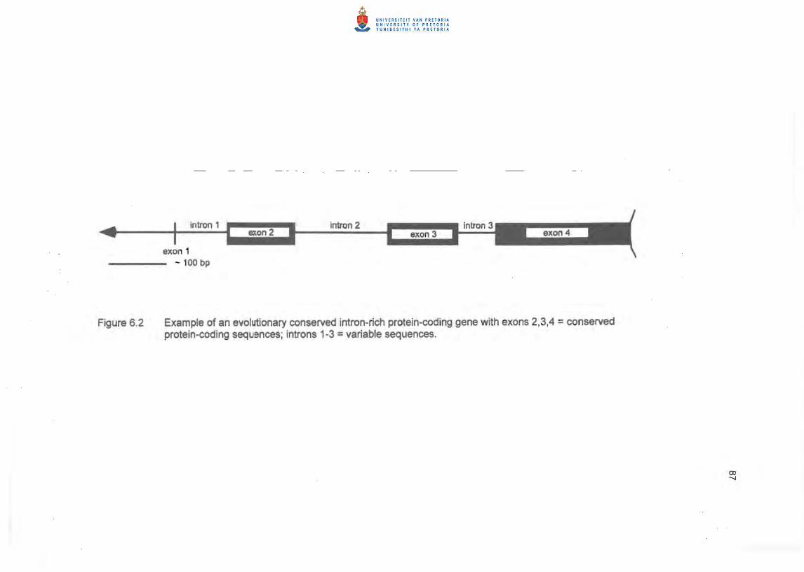
intron 3 region amplified is shown in Figure 6.2. Amplification conditions were as follows:
initial denaturation 95°C for 1 minute; 30 cycles of 94 °C for 15 seconds; 55 °C for 15
seconds and extension at 72 °C for 15. seconds; final extension at 72°C for 6 minutes.
PCR products were gel-purified and sequenced with an automated sequencer as
described for the ITS1 /2 5.8S region. Sequences were aligned and data presented as
described for the ITS 1/2 region spacers.
Distance matrix spreadsheets generated by DNAMAN software were analysed by
Statistica ver. 6.0 (Statsoft, Tulsa, Oklahoma). Dendrograms depicting grouping of
76
species were drafted using Ward's clustering algorithm with Euclidean distances. The K
means clustering procedure was used to place species in well-defined clusters or
groups showing distances between clusters and the distance from the cluster centre of
each strain within each cluster. Between-cluster distances were maximised and within
cluster variance minimised. Multidimensional scaling was performed to produce two
dimensional scatterplots as described for the analysis of the ITS 1,2 spacers.
6.3 RESULTS
6.3.1 Multilocus peR fingerprinting of genomic DNA
Fingerprinting with all three primers clearly distinguished between C. cyperi, C. grohii
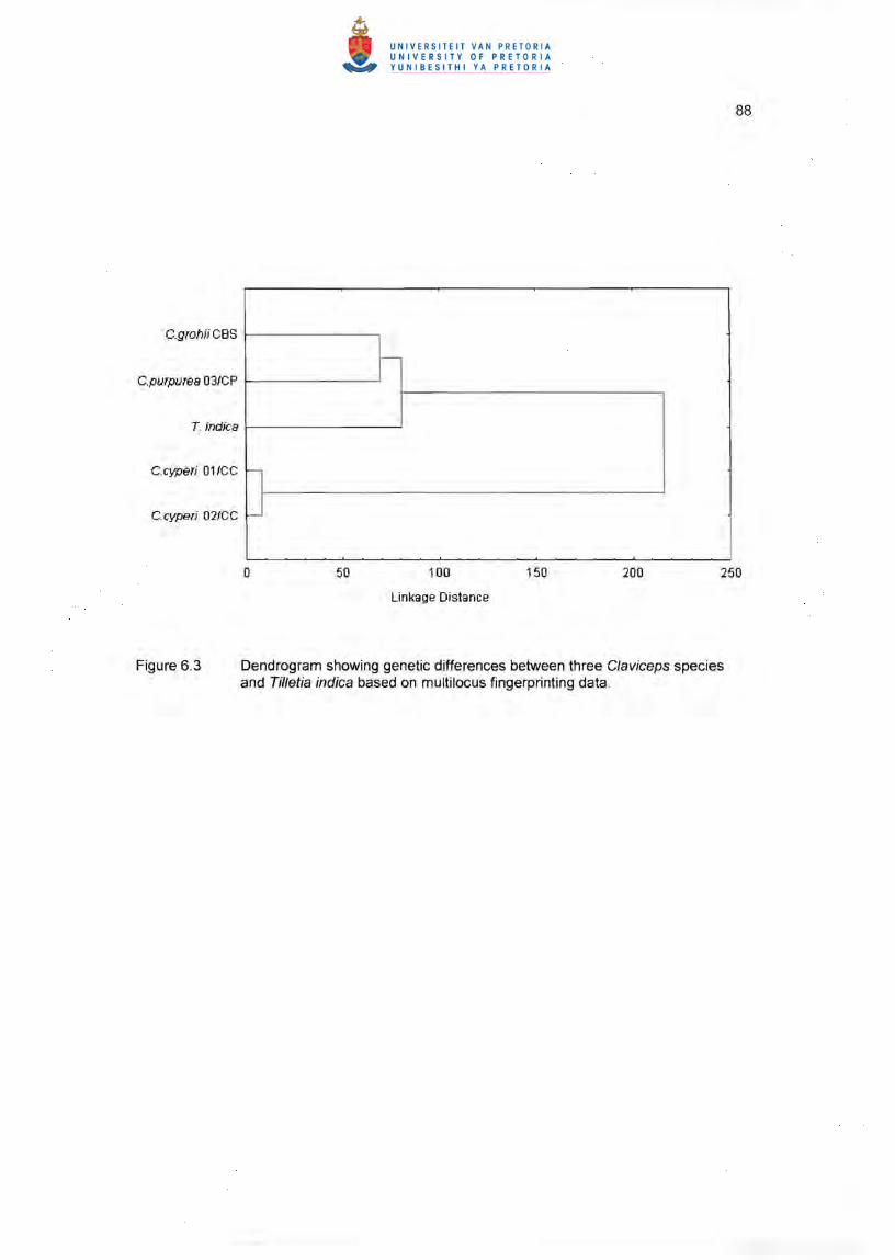
(CBS 124.47) and C. purpurea. The dendrogram (Figure 6.3) and similarity matrix
(Table 6.2) indicated a low level of similarity between these C/aviceps species. Using
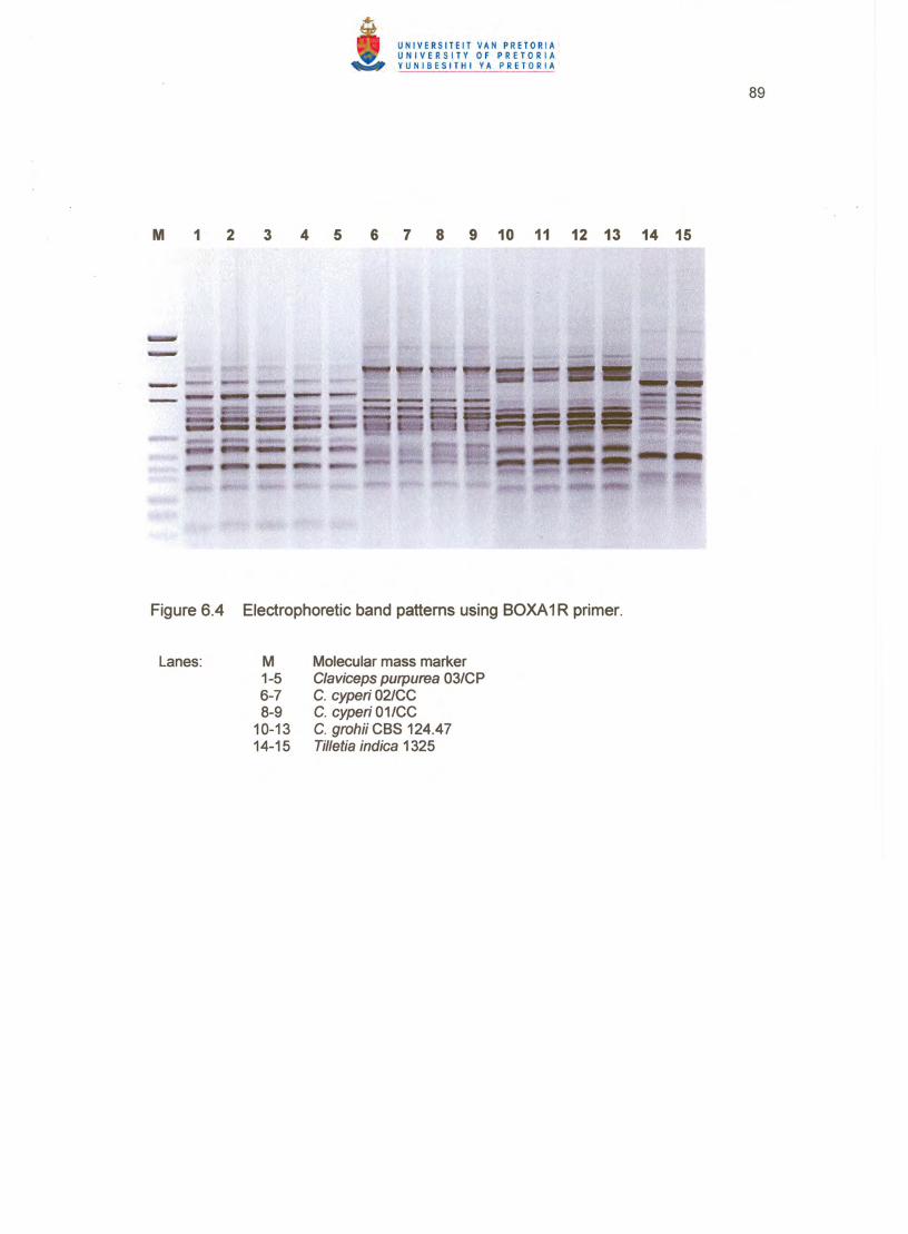
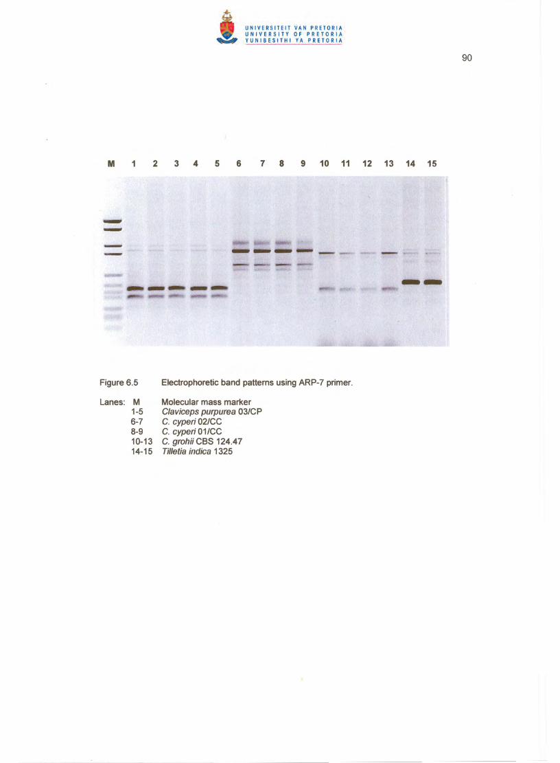
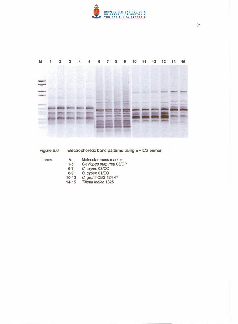
BOXA1R (Figure 6.4), ARP7 (Figure 6.5) and ERIC2 (Figure 6.6) primers, C. grohii
(CBS 124.47) clustered closer to C. pupurea (53 %) and showed 44 % similarity with
Tilletia indica Mitra, while the two C. cyperi isolates clustered separately from both C.
grohii (CBS 124.47) and C. purpurea, showing 96 % Similarity between them (Table
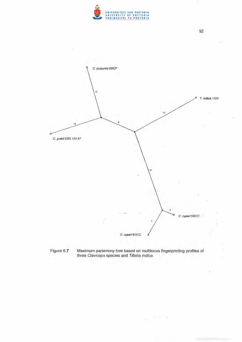
6.2). The maximum parsimony tree (Figure 6.7) shows the branching distances between
the different strains and in particular the longer distance between C. cyperi and the rest
of the tree. All strains showed unique patterns for each primer except the two c. cyperi
strains, which proved the usefulness of repetitive sequence-based PCR and GC-rich
arbitrary primers for grouping and distinguishing between Claviceps species up to strain
level (Figures 6.4 - 6.6).
MANOVA discriminant analysis confirmed the validity of the dendrogram clusters (Figure
6.3, Table 6.2). The likelihood that the strains of C/aviceps species were drawn from the
same population, indicating no discrimination between the strains, was very low as
indicated by parameter L. The probability that a random subdivision in groups will yield
the same degree of discrimination as the user-selected groups, was negligible as
indicated by P<O.001 (Table 6.2).
77
6.3.2 Sequence analysis of ITS1/2 and 5.8 S regions
Strains used to construct a phylogram are listed in Table 6.3. Amplification of the
complete ITS 1 and ~ spacers with the 5.8S gene produced only one PCR product for
the isolates of C. cyperi (01 /CC; 02/CC) of size 656 bp and 654 bp respectively. C.
grohii (CBS 124.47) showed a product of 585 bp size, whereas a product of size 584 bp
was amplified from C. purpurea (03/CP) . Both C. cyperi strains possessed an extended
ITS 1 spacer in comparison with C. purpurea 03/CP and C. grohii (CBS 124.47). No
polymorph isms or other non-specific fragments were amplified. When C. cyperi (01 /CC;
02/CC) and C. purpurea (03/CP) strains were compared to other Claviceps species
deposited in the GenBank sequence database, a dendrogram (Figure 6.8) placed the
C/aviceps species in four major clusters. The related teleomorphic species of
Atkinsonella , Epichloe and Echinodothis were included in a cluster (no. 4), together with
C/aviceps citrina Pazoutova, Fuik. , Leyva-Mir & Flieger, even though the distance from
the cluster centre of the latter species was larger than the distances of the other genera
in this cluster (Table 6.3). C/aviceps zizaniae (Fyles) Pantidou shows 93 % similarity in
the similarity matrix with the two C. cyperi isolates and was consequently included in a
cluster (no. 3), together with the C. cyperi isolates which were clearly distanced from all
the other clusters according to the table of Euclidean distances (Table 6.3). C. grohii
(CBS 124.47) and the four C. purpurea strains were included in a second major cluster.
Claviceps paspa/i F. Stevens & J.G. Hall did not fit well in any of the four clusters
(Figure 6.8, Table 6.3), because of a much larger distance from the centre of cluster 2.
C/aviceps fusiformis Loveless, C/aviceps africana Freder., P.G. Mantle & W.A.J. de
Milliano, C/aviceps viridis Padwick & Azmatullah, C/aviceps gigantea S.F. Fuentes, Isla,
Ullstrup & Rodriquez, C/aviceps sorghi B.G.P. Kulk., Seshadri & Hegde, C/aviceps
sorghicola Tsukib., Shiman. & T. Uematsu, C/aviceps maximensis T. Theis, Claviceps
pusil/a Ces. and C/aviceps phalaridis J. Walker formed the largest major cluster. In the
dendrogram (Figure 6.8), C. gigantea, C. sorghi and C. fusiformis formed a smaller sub
cluster within this major cluster.
The two-dimensional plot (Figure 6.9) also revealed four major clusters with
multidimensional scaling. Six dimensions were computed showing low stress values
indicating goodness-of-fit of data points to the step function of the Shepard diagram
78
(Table 6.3). The first major cluster included the same nine Claviceps species as
revealed by the dendrogram. All species in this cluster were grouped in a separate
cluster (Figure 6.9). The second major cluster contained C. purpurea, C. grahii (CBS
124.47) and C. paspali, with C. paspa/i and one C. purpurea strain slightly further
distanced from the cluster centre (Table 6.3). The teleomorphic species formed a less
closely spaced cluster in the two-dimensional plot while C. citrina did not fit well in any
cluster (Figure 6.9) as confirmed by the distance from the cluster centre (Table 6.3). The
C. cyperi isolates and C. zizaniae formed a separate cluster well distanced from the
other clusters with C. zizaniae further distanced from this cluster centre. The groupings
generated by multidimensional scaling were similar to the clustering pattern of the
dendrogram and supported the K-means partitioning of clusters .
A similar clustering pattern of four major clusters is depicted in the phylogenetic tree
(Figure 6.10) which places the species in the same major clusters, except C. paspa/i
which forms an outlier and occupied an intermediate position distanced from the major
clusters. This positioning of C. paspa/i was more pronounced in the phylogenetic tree
than in the dendrogram (Figure 6.8), the K-means table of distances (Table 6.3) and the
two-dimensional plot (Figure 6.9). C. cyperi together with C. zizaniae formed a distinct
separate cluster at a large distance from the other major clusters. It was apparent from
the phylogenetic tree (Figure 6.10) that the related teleomorphic species, as well as C.
citrina , were included in a single cluster, clearly separated from the other clusters as
well as from C. paspali. All branches linking major clusters were well supported by
bootstrapping.
6.3.3 Sequence analysis of the ~-tubulin gene Intron 3 region
Using the described amplification conditions, the ~tubulin (tub2) gene of similar size
was amplified from all isolates. The isolates of C. cyperi 01 ICC and 02/CC amplified
genes of 477 bp and 473 bp size, respectively. C. grahii (CBS 124.47) amplified a tub2
gene of 464 bp and C. purpurea 03/CP a tub2 gene of 475 bp. A dendrogram
presentation indicates five distinct clusters, with the two related species of Echinodothis
and Epichloe, not fitting well in any of the major clusters (Figure 6.11). C. grahii (CBS
124.47), C. purpurea 03/CP and other GenBank C. purpurea strains formed one cluster
79
(no. 4). The three C. africana strains formed a separate cluster (no. 3). and the C.
sorghicola strains grouped in cluster no. 5 together with C. Fusiformis which clustered at
a larger distance from this cluster centre (Table 6.4). The two C. paspa/i strains were
placed in cluster no. 2. Epichloe did not fit well in any cluster but linked at a larger
distance with cluster no. 2. The C. cyperi isolates were placed in cluster no. 7. with
Echinodothis linking at a much larger distance with this cluster (Figure 6.11). Similar to
the ITS1 /2 spacers sequence data, the intron 3 region also placed the C. cyperi isolates
at the largest distance from the other species clusters as indicated by Euclidean
distances (Table 6.4).
The two-dimensional plot (Figure 6.12) confirmed the clustering pattern of the
dendrogram and phylogram showing the same clusters clearly distanced (Table 6.4).
The C. cyperi isolates formed a distinct separate cluster at a large distance from the
other clusters (Table 6.4). The Echinodothis and Epichloe species formed outliers and
remained unclustered (Figure 6.12).
The phylogenetic tree (Figure 6.13) corresponded with the cluster pattern of the
dendrogram (Figure 6.11 ), although the Epichloe and Echinodothis species did not fit in
any cluster. As expected, the C. cyperi strains formed a distinct outgroup cluster (94 %
sequence similarity between strains) while C. purpurea 03/CP and C. grahii (CBS
124.47) (96 % sequence similarity), clustered with other C. purpurea strains. The
different Claviceps species grouped in separate clusters whereas C. Fusiformis was only
linked at a distance to the larger cluster containing C. africana and C. sorghicola. All
branches linking major clusters were well supported by bootstrapping.
6.4 DISCUSSION ·
In this study, multilocus fingerprinting clearly differentiated between strains of C. purpurea,
C. grahii (CBS 124.47) and C. cyperi, with the two C. cyperi isolates showing identical
band pattems. However, due to the limited number of isolates available, variation within
the species could not be established. Previous studies revealed considerable intraspecific
genetic diversity within various species of Claviceps (JungehOising & Tudzynski 1997;
Paioutov8 & Tudzynski 1999; Paioutov8 et al. 2000a,b, 2002; Tooley et af. 2000). The
80
above investigations indicated that multilocus fingerprinting could differentiate up to strain
level, rendering it appropriate for the study of genetic characteristics of populations of
ergot fungi. The lower similarity between C. purpurea and C. grahii (CBS 124.47) shown
by multi locus fingerprinting in the present study, contrasted with sequence analysis of the
ITS1/2 spacers and J3-tubulin gene, where higher sequence similarity was established
between these species. This observation is in agreement with the study by Tooley et al.
(2000) where multilocus genotyping revealed greater genetic variation at the intraspecific
level than sequence data of different genes. The random method of fragment amplification
by single primers comlementary to repeated sequence motifs that are spread over the
whole of the genome, should explain this intraspecific diversity.
The phylogenetic relationships indicated by the ITS 112 spacers corresponded with those
described by Pa.zoutova (2001), in which two distinct clades were observed , one
comprising C. citrina, C. phalaridis, C. sorghico/a, C. sorghi, C. gigantea, C. africana, C.
viridis and C. pusilla, and the other C. paspali, C. zizaniae, C. grahii, C. sulcata, C.
fusiformis and C. purpurea. In the present study, however, C. zizaniae showed a higher
sequence similarity to the two C. cyperi isolates, with which it formed a distinct separate
cluster based on ITS spacers sequence data. It is significant that the extended ITS1
spacer present in the C. cyperi isolates was also present in C. zizaniae. The evolutionary
significance of this sequence insertion in ITS spacer 1 indicates either a common ancestor
(monophyletic origin) or a para phyletic Origin of C. zizaniae and C. cyperi, though a
process of convergent evolution or a speciation event could also have been involved.
Physiologically, C. cyperi and C. zizaniae share a common alkaloid profile (Kantovora et
a/. 2002; Chapter 5). Both also have hosts that prefer a moist habitat, viz. yellow nut sedge
(Cyperus esculentus L.) and wild rice (Zizania palustris L. and Zizania aquatica L.),
respectively (Loveless 1967; Pantidou 1959). Morphologically, however, the two species
are quite distinct, with C. zizaniae producing considerably larger sclerotia than C. cyperi,
and a differently coloured capitulum without a collar-like appendage surrounding the base
(Pantidou 1959; Loveless 1967; Chapter 2). The above differences and similarities support
the view of Pa.zoutova et a/. (2000b, 2002), Pa.zoutova (2001) and Duncan et al. (2002 )
that the phylogenetic positions of Claviceps species tend to correspond with their alkaloid
profiles and ecological specialisation rather than their morphological features and host
81
specificity. Further evidence regarding the unrelatedness of morphology and phylogeny in
Claviceps can be derived from the prominent ranunculoid papilla present in C. cyperi, a
feature it shares with Claviceps ranunculoides A. Moller recorded from Setaria sp. in Brazil
(Moller 1901), that distinguishes them from all other known Claviceps species. No type
material or GenBalik ~lrain of C. ranunculoldes was available for Inclusion in the present
study. However, the species was included, together with C. africana, C. fusiformis, C.
paspa/i and C. purpurea, in a phylogenetic analysis of the 26S large subunit rDNA of
graminicolous Clavicipitaceae by Sullivan et a/. (2001). According to the above analysis, C.
ranunculoides clustered closest to C. africana followed by C. fusiformis, with C. paspali
and C. purpurea forming a separate cluster. This pattern implies that C. ranunculoides
probably would have clustered with C. africana, C. fusiformis, C. gigantea, C. maximensis,
C. phafaridis, C. pusilla, C. sorghi, C. sorgicola and C. viridis, at a far distance from C.
cyperi, had it been included in the present study.
Besides confirming C. cyperi to be a distinct species, the present investigation also
provided evidence regarding the phylogeny of two "dubious" Claviceps species, viz. C.
citrin a and C. paspali. As indicated above, Pazoutova (2001 ) placed C. citrina in a cluster
which included C. africana and C. sorghicola, whereas C. paspa/i grouped in a separate
cluster with C. fusiformis, C. grohii, C. purpurea, C. sulcata and C. zizaniae. However, in a
study using PCR amplification, Duncan et al. (2002) found C. citrina to cluster separately
from C. africana and C. sorghicola. This grouping corresponds with the present results
according to which C. citrina was not properly associated with any cluster, but only
grouped with Echinodothis tuberiformis (Berk. & Ravenel) G.F. Atk. , Epichloe typhina
(Pers.) Tul. & C. Tul. and Atkinsonella hypoxylon (Peck) Diehl at a much larger distance.
The present study also showed C. paspa/i to cluster separately from C. fusiformis and C.
purpurea, which is in agreement with the sequence analysis of the ~tubulin intron 3 and
EF-1 a-gene intron 4 regions reported for Claviceps species on sorghum by Tooley et al.
(2001 ). However, in the latter study C. fusiformis grouped with C. sorghicola and C.
africana with a bootstrap value of 82 %, possibly because resequencing of the original
RAPD and ITS1 sequences reported by Pazoutova et al. (1998) for the strain of C.
fusiformis referred to by Tooley et al. (2001) indicated that it is actually C. purpurea (S.
Pazoutova, pers. com.). All publications based on the incorrect sequence should therefore
be reconsidered.
82
In conclusion, although this study elucidated the phylogeny of the nut sedge ergot
pathogen, extensive genetic diversity studies on South African isolates of C. cypert still
need to be done to establish the genetic profi le of local populations of the species. The
most perplexing observation was the outgroup cluster of C. cypert that linked at a large
distance from the other Claviceps species. The presence of the extended ITS1 spacer not
present in any other Claviceps species except C. zizaniae, may explain this outgroup
placement insufficiently. The true natural phylogenetic relationship of C. cyperi and C.
zizaniae will only be resolved after a comprehensive study of many isolates from various
geographic areas.
6.5 REFERENCES
ANNIS, S.L. & PANACCIONE, D.G. 1998. The presence of peptide synthetase gene
transcripts and accumulation of ergopeptines in Claviceps purpurea and Neotyphodium
coenophialum. Canadian Journal of Microbiology 44:80- 86 .
AUSUBEL, F.M., BRENT, R, KINGSTON, R E. , MOORE, D.O., SEIDMAN, J.G.,
SMITH, J.A. & STRUHL, K. 1989. Preparation and analysis of DNA, Pages 215- 222.
In: F.M. Ausubel , R Brent, R E. Kingston, D.O. Moore, J.G. Seidman, J.A. Smith, & K.
Struhl (eds.). Current protocols in molecular biology. Vol. 1. John Wiley, New York.
DUNCAN, RA. JR, SULLIVAN, R, ALDERMAN, S.C., SPATAFORA, J.W. & WHITE,
J.F. JR 2002. Claviceps purpurea var. spartinae var. nov.: an ergot adapted to the
aquatic environment. Mycotaxon 81: 11 - 25.
FELSENSTEIN, J. 1985. Confidence limits on phylogenies: an approach using the
bootstrap. Evolution 39: 783- 791.
FENG. D.F. & DOOLITTLE, RF. 1987. Progressive sequence alignment as a
prerequisite to correct phylogenetic trees. Journal of Molecular Evolution 25:351 - 360.
83
HASEGAWA, M. , KISHINO, H. &YANO, T. 1985. Dating of the human-ape splitting by a
molecular clock of mitochondrial DNA. Journal of Molecular Evolution 22: 160- 174.
JUNGEHOLSING, U.T.A. & TUDZYNSKI, P. 1997. Analysis of genetic diversity in
Claviceps purpurea by RAPD markers. Mycological Research 101:1-6.
KANTOVoRA, M., KOLiNSKA, R., PAZOUTOVA, S. , HONzATKO, A. , HAVLICEK, V. &
FLiEGER, M. 2002. Ergot alkaloids produced by submerged cultures of Claviceps
zizaniae. Journal of Natural Products 65:1039- 1 040.
KIMURA, M. 1980. A simple method for estimating evolutionary rates of base
substitutions through comparative studies of nucleotide sequences. Journal of Molecular
Evolution 16:111- 120.
LOVELESS, A.R. 1967. A new species of Claviceps on Cyperaceae. Transactions of the
British Mycological Society 47:205- 213.
MATHIS, J.N. AND MCMILLAN, D.E. 1996. Detection of genetic variation in
Bradyrhizobium japonicum USDA 110 variants using DNA fingerprints generated with
GC rich arbitrary PCR primers. Plant and Soil 186:81- 85.
MOLLER, A. 1901. Phycomyceten und Ascomyceten. Untersuchungen aus Brazilien. In:
A.F.A. Schimper (ed.). Botanische Mitteilungen aus den Tropen. Vol. 9. G. Fischer,
Jena.
NAUDE, T.W., BOTHA, C.J., VORSTER, J.H., ROUX, C., VAN DER LINDE, E.J., VAN
DER WALT, S.I., ROTTINGHAUS, G.E., VAN JAARSVELD, L. & LAWRENCE, A.N.
2005. Claviceps cyperi, a new cause of serious ergotism in dairy cattle consuming
maize silage and teft hay contaminated with ergotised Cyperus esculentus (nut sedge)
on the Highveld of South Africa. Onderstepoort Journal of Veterinary Research 72:23
37.

84
PANTIDOU, M.E. 1959. C/aviceps from Zizania. Canadian Journal of Botany 37:1233
1236.
PAiOUTOVA, S. 2001. The phylogeny and evolution of the genus Claviceps.
Mycological Research 105:275- 283.
PAiOUTOVA, S., BANDYOPADHYAY, R , FREDERICKSON, D.E. , MANTLE, P.G. &
FREDERIKSEN, RA. 2000a. Relations among sorghum ergot isolates from the
Americas, Africa, India and Australia. Plant Disease 84:437- 442.
PAiOUTOVA, S., FUCIKOVSKY, L., LEYVA-MIR, S.G. & FLiEGER, M. 1998. Claviceps
citrina sp. nov., a parasite of the halophytic grass Distichlis spicata from Mexico.
Mycological Research 102:850-854.
PAiOUTOVA, s, OLSOVSKA, J. , LlNKA M., KoLiNSKA, R & FLiEGER, M. 2000b.
Chemoraces and habitat specialisation of Claviceps purpurea populations. Applied and
Environmental Microbiology 66:5419- 6425.
PAZOUTovA, S, RAYBOULD, A.F., HONzATKO, A. & KOLiNSKA, R 2002.
Specialised population of Claviceps purpurea from salt marsh Spartina species.
Mycological Research 106:210- 214.
PAZOUTOVA, S. & TUDZYNSKI, P. 1999. Claviceps sp. PRL 1980 (ATCC 26245),59
and Pepty 695/ch-l: their true story. Mycological Research 103:1044- 1048.
SAITOU, N. & NEI, M. 1987. The neighbor-joining method: a new method for
reconstructing phylogenetic trees. Molecular Biology and Evolution 4:406- 425.
SULLIVAN, R, BERGEN, M.S., BILLS, G.F., ALDERMAN, S.C., SPATAFORA, J.W. &
WHITE, J.F. 2001 . Features and phylogenetic status of an enigmatic clavicipitalean
fungus Neoclaviceps monostipa. Mycologia 93:90- 99.
85
THOMPSON, J.D., HIGGINS, D.G. & GIBSON, T.J. 1994. ClustalW: Improving the
sensitivity of progressive multiple sequence alignment through sequence weighing,
position, specific gap penalties, and weight matrix choice. Nucleic Acids Research
22:4673- 4680.
TOOLEY, P.W., O'NEILL, N.R., GOLEY, E.D. & CARRAS, M.M. 2000. Assessment of
diversity in Claviceps africana and other Claviceps species by RAM and AFLP analyses.
Phytopathology 90: 1126- 1130.
TOOLEY, P.W., GOLEY, E.D ., CARRAS, M.M., FREDERICK, R.D. & WEBER, E.L.
2001. Characterization of Claviceps species pathogenic on sorghum by sequence
analysis of the f3-tubulin gene intron 3 region and EF-1 -a. intron 4. Myco/ogia 93:541
551 .
VAN DER LINDE, E.J. & NAUDE, T.W. 2000. Swam nuwe bedreiging vir melkbedryf.
Landbouweekblad 1131:24-27.
VERSALOVIC, J. , SCHNEIDER, M., DE BRUIJN, F.J . & LUPSKI, J.R. 1994. Genomic
fingerprinting of bacteria using repetitive sequence-based polymerase chain reaction.
Methods in Molecular and Cellular Biology 5:25-40.
-- 4 kb
Basldiomycota and some ascomycetous yeasts
Figure 6.1 Schematic presentation of rONA cluster of tandemly repeated ribosomal genes. Large subunit = 28 S; small subunit = 18 S; IGS =intergenic spacer, ITS =internal transcribed spacers 1 & 2; 5.8 subun it.
ex> (J)
intron 1 Intron 2 intron 3
exon 1 - 100 bp
Figure 6.2 Example of an evolutionary conserved intron-rich protein-coding gene with exons 2,3,4 =conserved protein-coding sequences; introns 1-3 =variable sequences.
0) -.J
88
C.grohii CBS
r--Cpurpurea 03/CP
T. indica
C.cyperi 01/CC -
C.cyperi 02/CC -
o 50 100 150 200 250
Linkage Distance
Figure 6.3 Dendrogram showing genetic differences between three Claviceps species and Til/etia indica based on multilocus fingerprinting data.
92
C purpurea 03/CP
r indica 1325
17
C. grohii CBS 124.47
C. cyperi O2ICC
C. cyperi 01/CC
Figure 6.7 Maximum parsimony tree based on muttilocus fingerprinting profiles of three C/aviceps species and Tilletia indica.

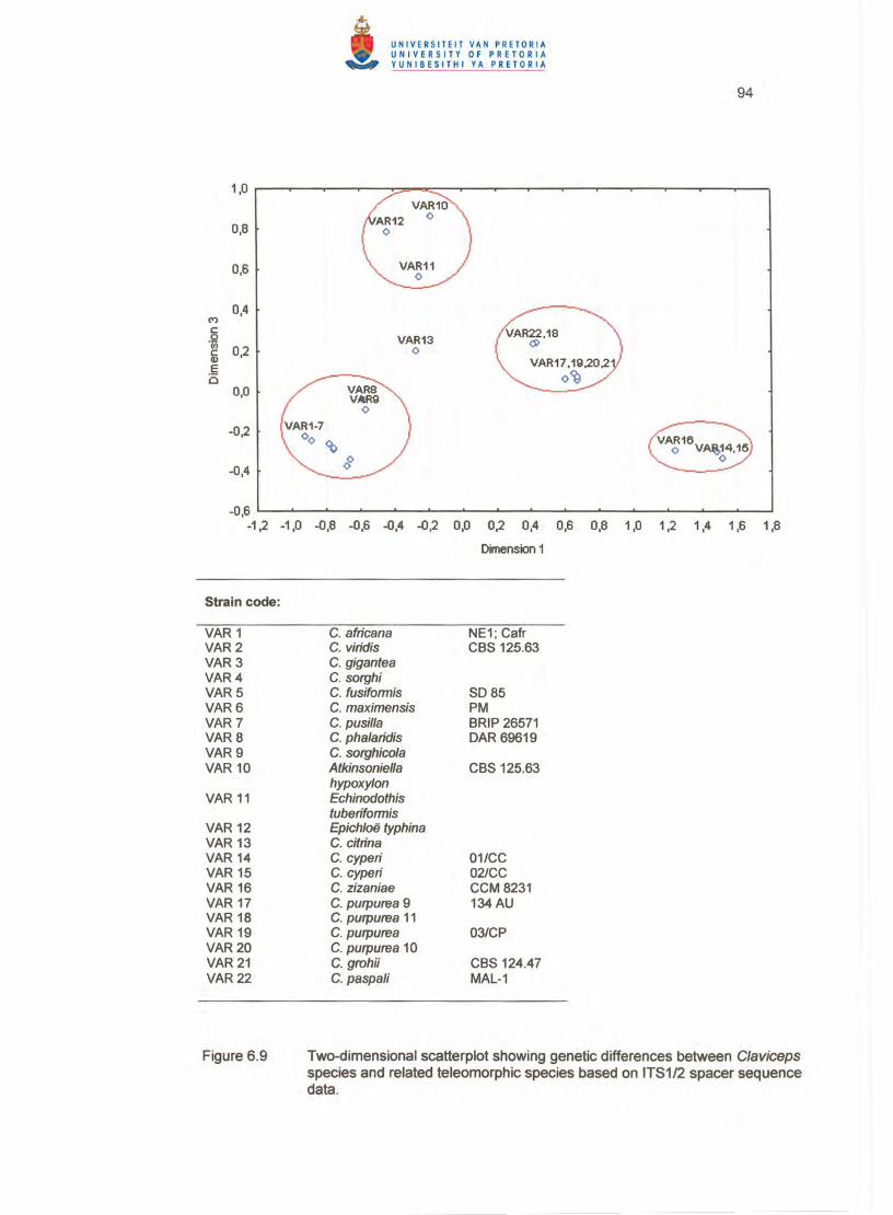
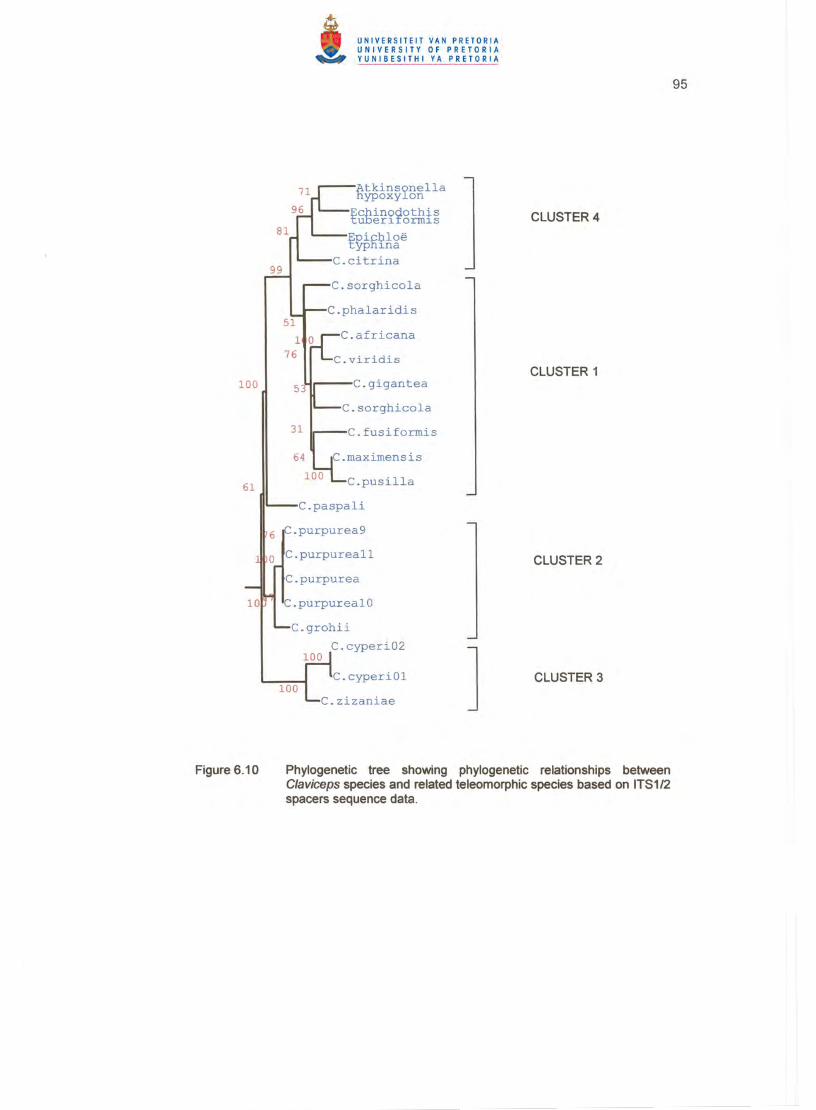
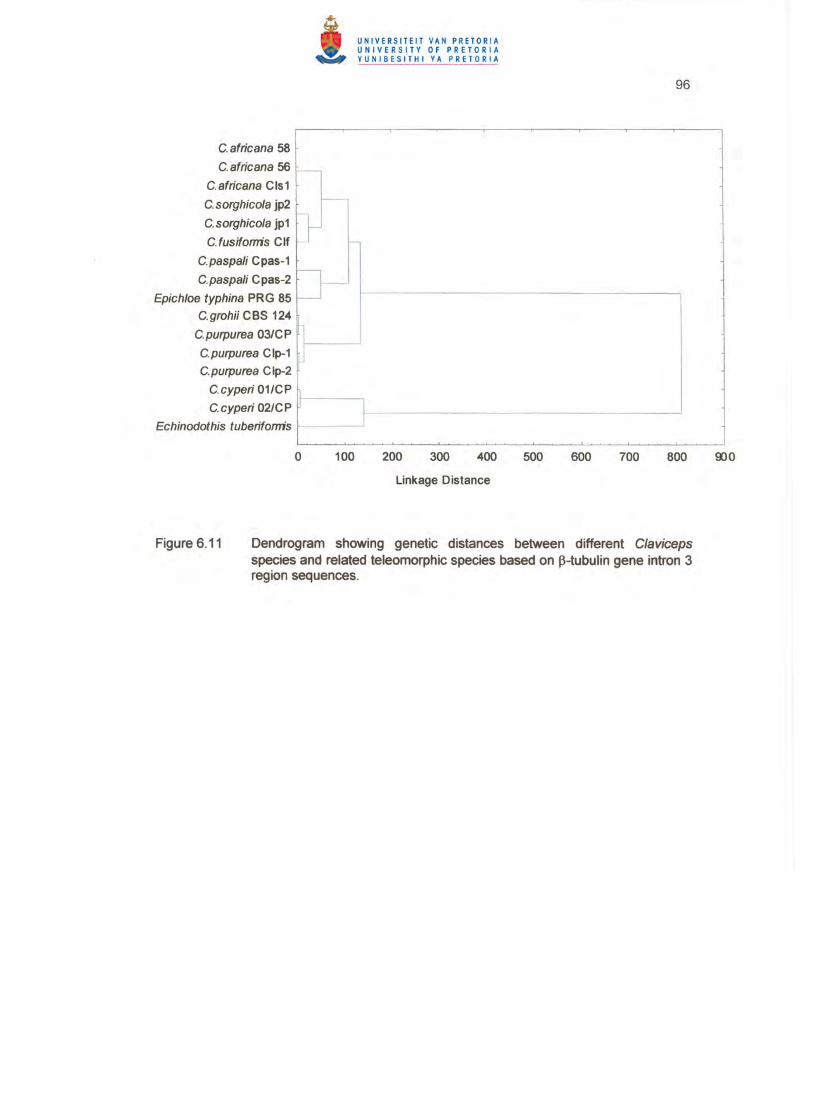
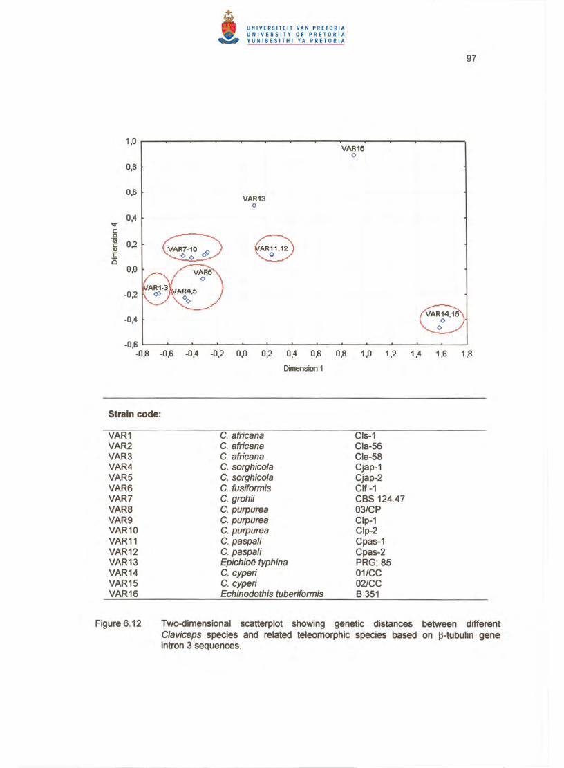

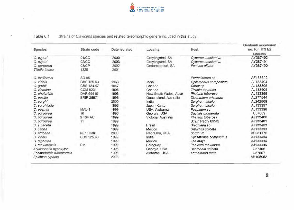
Table 6.1 Strains of Claviceps species and related teleomorphic genera included in this study.
Genbank accession Species Strain code Date isolated Locality Host no. for ITS1 /2
spacers C. cyperi 01 /CC 2000 Greylingstad, SA Cyperus esculentus AY387492 C. cyperi 02JCC 2003 Greylingstad, SA Cyperus esculentus AY387491 C. purpurea 03/CP 2002 Onderstepoort, SA Festuca eliator AY387490 Til/etia indica 1325 2001
C. fusiformis S085 Pennnisetum sp. AF133392 C. viridis CBS 125.63 1963 India Oplismenus compositus AJ1 33404 C. grohii CBS 124.47 1947 Canada Carex sp. AJ1 33395 C. zizaniae CCM 8231 1996 Canada Zizania aquatica AJ133405 C. phalaridis DAR 69619 1996 New South Wales, Austr Pha/aris tuberosa AJ133399 C. pusil/a BRIP 26571 1996 Queensland, Australia Dicanthium aristatum AJ277544 C. sorghi 2000 India Sorghum bic%r AJ242869 C. sorghicola 1996 Japan;Kanto Sorghum bic%r AJ133397 C.paspaJi MAL-1 1999 USA, Alabama Paspa/um sp. AJ133398 C. purpurea 10 1996 Georgia, USA DactyJis g/omerata U57669 C. purpurea 9134 AU 1999 Victoria, Australia Pha/aris tuberosa AJ133400 C. purpurea 11 1999 Strain Pepty 695/S AJ1 33401 C. su/acata 1999 Brazil Brachiaria sp. AJ 133403 C. citrina 1999 Mexico Distich/is spicata AJ 133393 C. africana NE1; Cafr 2000 Nebraska, USA Sorghum AF281176 C. viridis CBS 125.63 1999 Ind ia Oplismenus compositus AJ 133404 C. gigantea 1999 Mexico Zea mays AJ133394 C. maximensis PM 1999 Paraguay Panicum maximum AJ 133396 Atkinsonella hypoxylon 1996 Georgia, USA Danthonia spicata U57405 Echinodothis tuberiformis 1996 Alabama, USA Arundinaria tecta U57667 Epichloe typhina 2003 AB105952
CD CD

Genbank accession no. for ~-
Species Strain code Date isolated Locality Host tubulin intron 3 C. eyperi C. cyperi C. grohii C. purpurea C. purpurea C. purpurea C. fusiformis C.paspali C. paspali C. africana C. africana C. afrieana C. sorghieola C. sorghicola Epichloe typhina Echinodothis tuberiformis
01/CP 02/CP CBS 124.47 CIp-1 Clp-2 03/CP CIf-1 Cpas-1 Cpas-2 Cls-1 Cia-56 Cia-58 Cjap-1 Cjap-2 PRG; 85 B 351
2000 2003 1947
2002
1998 1997 1997 1998 1998 1989 1996 1990 1997
Greylingstad , SA Greylingstad, SA Canada Montana, USA East Germany South Africa Africa North Carolina Georgia India Potchefstroom, SA Potchefstroom, SA Tochigi, Japan Tochigi, Japan
Cyperus esculentus Cyperus esculentus Carex sp. Hordeum vulgare Secale eereale Festuca eliator Pennisetum typhoideum Paspalum sp. Paspalum sp. Sorghum bicolor Sorghum bic%r Sorghum bieo/or Sorghum bieolor Sorghum bieolor Lotium perenne
AY497005 AY497775 AY43867 1 AF263567 AF263568 AY438670 AF263569 AF263605 AF263606 AF263596 AF263591 AF263592 AF263600 AF263601
X52616 L78268
o o

101
Table 6.2 Dendrogram and K-means clustering of South African isolates of C/aviceps purpurea, C. grohii, C. eyperi and Til/etia indica based on multilocus fingerprinting.
Euclidean distances between clusters Distances below diagonal
No.1 No. 2 No.1 o No.2 46 .1 3407135 0
Distance from respective cluster centre '"
Variable Cluster Distance *
C. grohii CBS 2 15.5083758 124.47 C. purpurea 03/CP 2 2 15.5083758 T. indica 1325 3 C.cyperi 01/CC 4 1 1 .815852417 C. cyperi 02tCC 5 1 1.815852417
MANOVA discriminant analysis for BOX Ai R; ERIC2 and ARP-7 multilocus primers
DISCR_01 EIGV= 62.3% L= 0.0001 P<=0.001 % DISCR_02 EIGV= 37.7% L= 0.0154 P<=0.001 %
Similarity matrix
C. grohli CBS C. purpurea T. indica C. c011CC C. c02JCC C.grohii CBS 100 52.65 43.61 41 .56 46.18 124.47 C.purpurea 03/CP 52.65 100 52.2 34.09 32 .16 T.indica 43.61 52.2 100 20 .92 22 . 8~
C.cyperi 01/CC 41 .56 34 .09 20.92 100 95.69 C.cyperi 02/CC 46.1 8 32 .16 22.85 95.69 100
102
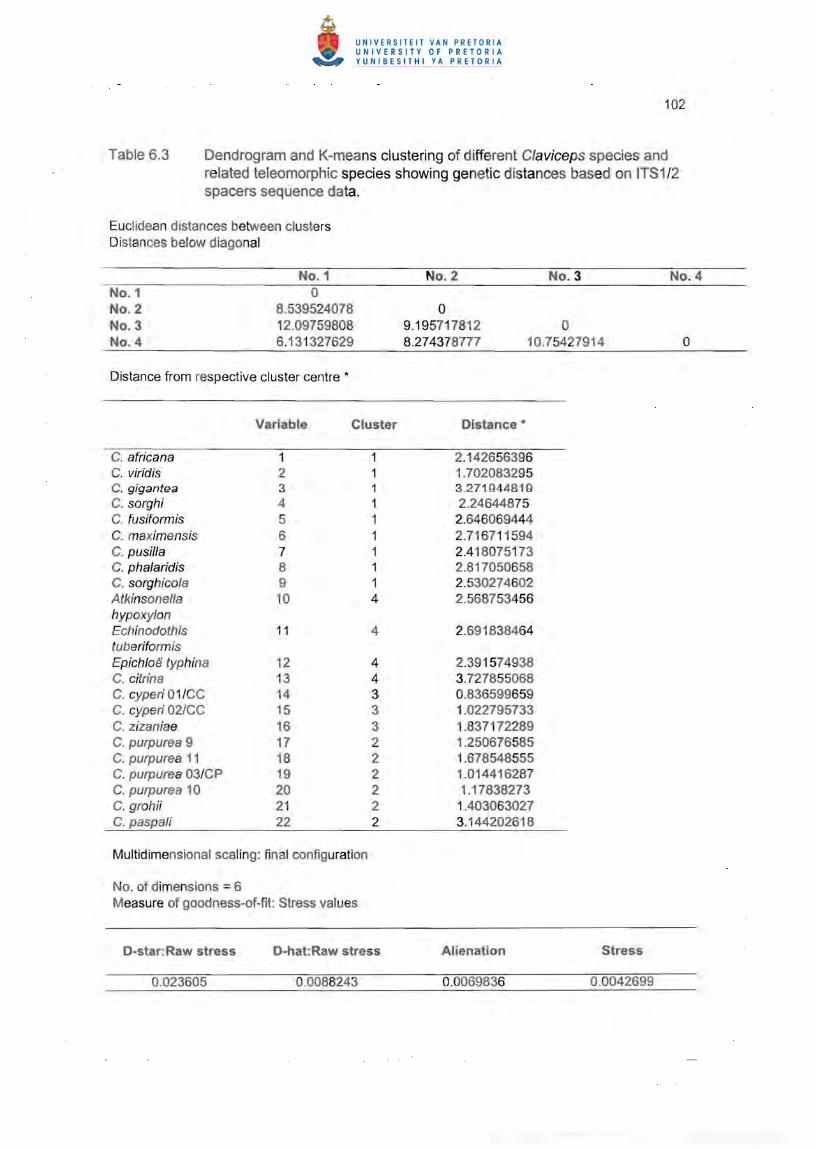
Table 6.3 Dendrogram and K-means clustering of different Claviceps species and related teleomorphic species showing genetic distances based on ITS1 /2 spacers sequence data.
Euclidean distances between clusters Distances below diagonal
No.1 No.2 No.3 No. 4 No.1 0 No.2 8.539524078 0 No.3 12.09759808 9.195717812 0 No.4 6.131327629 8.274378777 10.75427914 o
Distance from respective cluster centre *
Variable Cluster Distance *
C. africana 1 2.142656396 C. viridis 2 1 .702083295 C. gigantea 3 1 3 .271944819 C. sorghi 4 1 2.24644875 C. fusiformis 5 1 2.646069444 C. maximensis 6 1 2.716711594 C. pusil/a 7 1 2.418075173 C. phalaridis 8 1 2.817050658 C. sorghico/a 9 1 2.530274602 Atkinsonella 10 4 2.568753456 hypoxylon Echinodothis 11 4 2.691838464 tuberiformis Epichloe typhina 12 4 2.391574938 C. citrin a 13 4 3.727855068 C. cyperi 01/CC 14 3 0.836599659 C. cyperi 02/CC 15 3 1 .022795733 C. zizaniae 16 3 1.837172289 C. purpurea 9 17 2 1 .250676585 C. purpurea 11 18 2 1.678548555 C. purpurea 03/CP 19 2 1.014416287 C. purpurea 10 20 2 1.17838273 C. grahii 21 2 1 .403063027 C. paspa/i 22 2 3. 144202618
Multidimensional scaling: final configuration
No. of dimensions = 6 Measure of goodness-of-fit: Stress values
D-star:Raw stress D-hat:Raw stress Alienation Stress
0.023605 0.0088243 0.0069836 0.0042699

103
Table 6.4 Dendrogram and K-means clustering of different C/aviceps species and related teleomorphic species showing genetic distances based on ~-tub ulin gene intron 3 region sequence data.
Euclidean distances between clusters Distances below diagonal
No.1 No.2 No. 3 No. 4 No.5 No.6 No.7
No.1 0 No.2 9.537975311 0 NO. 3 10.74645329 11 .3850803 0 No.4 9.716282845 11.5703497 10.1820211 0 No.5 9.56938076 9.92785358 5.14885426 8.502935 0 No.6 46.33493423 47.7928696 51.6772575 51.1 3251 50.81779099 0 No.7 48.63568497 49.4319077 53.3657837 52.53014 52.49116898 26.51585 0
Distance from respective cluster centre *
Variable Cluster Distance*
C. africana Cls-1 1 3 0.06454972 C. africana Cia-56 2 3 0.06454972 C. africana Cia-58 3 3 0. 12909944 C. sorghico/a Cjap-1 4 5 1.53769759 C. sorghico/a Cjap-2 5 5 1.52661 572 C. Fusiformis Clf-1 6 5 3.06055777 C. grohii CBS124.47 7 4 0.98862673 C. purpurea 03/CP 8 4 1.37541187 C. purpurea Clp-1 9 4 1.16479947 C. purpurea Clp-2 10 4 1.01621617 C. paspali Cpas-1 11 2 0.073951 C. paspa/i Cpas-2 12 2 0.073951 Epichloe typhina 13 1 0 C. cyperi 01 ICC 14 7 0.77237459 C. cyperi 02/CC 15 7 0.77237459 Echinodothis 16 6 0 tuberiformis
Multid imensional scaling: final configuration
No. of dimensions = 6 Measure of goodness-of-fit: Stress values
D-star:Raw stress D-hat:Raw stress Alienation Stress
0.0000033 0.000001 0.00001139 0.00000624

104
7. GENERAL DISCUSSION
The previous chapters have provided descriptions of the symptoms of ergot on yellow nut
sedge (Cyperus esculentus L.) and of live specimens of the causal organism, Claviceps
cyperi Loveless. Evidence was also presented regarding the phylogeny and alkaloid
profile of the pathogen, the morphology, taxonomy, physiology and mode of infection of
its anamorph, and some aspects pertaining to the epidemiology and ecology of the
disease. Although many aspects have been elucidated in the study, some questions
remained unanswered or unclear, the most notable being the taxonomic position of the
pathogen and the origin, distribution and management of the disease.
Phylogenetically, C. cyperi proved to be a distinct species, but closely related to Claviceps
zizaniae (Fyles) Pantidou, with which it also shared a common alkaloid profile . However,
the presence of the extended ITS regions unique to these two species observed in the
present study may explain their outgroup placement insufficiently. Sequence data derived
from ITS regions should be interpreted with caution because not all copies of these
regions are identical in the same strain. Several strains of different species within a genus
need to be screened to ensure that only copy type is amplified and sequenced. Multiple
intron-rich protein coding genes should be sequenced as well , and compared with existing
sequence data in GenBank. These protein-coding genes evolve faster than the ITS
regions and are able to discriminate between closely related phylogenetic cryptic species
within a morphological species (Taylor et al. 2000). O'Donnell & Cigelnik (1997) have
demonstrated the potential of intron-rich protein-coding genes as species markers for
Fusarium, and probably other fungal genera as well. According to Geiser et al. (1998) and
Carbone & Kohn (2001), introns flanked by the more conserved exons evolve at a higher
rate than ITS regions, hence increaSing the resolving power of these genes to reveal
variation at intraspeCific level. Determination of the genealogies of these genes within the
genus C/aviceps may also prove rewarding with regard to the overall phylogeny of the
genus and determining a common ancestor or ancestors for different species. The
ongoing process of speciation within the genus Claviceps in association with different
hosts, as well as the geographic origin of different species may also be established using
appropriate species markers and the concept of Genealogical Concordance Phylogenetic
Species Recognition.
105
The apparent holoblastic conidiogenesis observed in the spacelial state of C. cyperi in this
study was a matter of concern as it implied a revision of the genus Sphacelia. Very
recently, however, Pazoutova et al. (2004) showed that conid iation in Claviceps is
pleomorphic, with conidiogenesis in C. zizaniae and Claviceps citrin a Pazoutova, Fucik.,
Leyva-Mir & Flieger being ephelidial (holoblastic and sympodial), typical of the genus
Ephelis. Although the existence of holoblastic conidiation in C/aviceps would have
resolved the taxonomy of C. cyperi, and placed its anamorph in Ephelis, this could not be
done as the conidia of Ephelis species are produced sympodially and often form whorls
consisting of 3 to 8 spores (Rykard et al. 1984; White 1997), whereas C. cyperi produces
conidia singly and certainly not in sympodial succession. The issue is further compounded
by the claim of Pazoutova et a/. (2004) that both macro- and microconidia have been
observed in all studied phialidic C/aviceps species except C. citrina and C. purpurea
(Fr.:Fr. ) Tul. , though microconidia could not be discerned in honeydew or cultures of C.
cyperi. The confusion surrounding the taxonomic status of the nut sedge ergot pathogen
therefore remains.
From an ecological perspective the most perplexing question obviously is the apparent
confinement of C. cyperi to South Africa and its disappearance for more than 50 years
since first recorded from in and around Pretoria during World War II until the incidents of
ergotism associated with the intake of ergotised nut sedge in 1996/1997 at Greylingstad,
Memel and Vrede. Despite concerted efforts the past eight years, the disease could not
be detected in the Pretoria/Kempton Park area. It should nevertheless be noted that
Greylingstad, Memel and Vrede do not feature in Doidge (1950) and the possibility
therefore exist that these sites have never been visited in previous disease surveys.
There is, however, a reference in Doidge (1950) to a Cerebella species on Cyperus from
Mount Edgecombe in KwaZulu-Natal. Examination of the specimen, which was collected
in 1945, indicated that the host was Cyperus rotundus L. and indeed colonised by
Cerebella. Although no sign of ergot could be found, the presence of Cerebella is a sure
indication of previous infection with a species of C/aviceps (Langdon 1942; Loveless
1964). It is thus possible that C. cyperi has been present in other areas of South Africa,
but remained unnoticed due to the inconspicuousness of the symptoms and the fact that it
was not involved in any reported incident of ergotism. The recent sporadic eruptions can
probably be ascribed to a combination of the following: (i) changes in tillage practices, (ii)
106
conducive climate, (iii) invasion by, or an increase in populations of spotted maize beetle
(Astylus atromaculatus Blanchard) and the unidentified thrips species associated with the
disease, and (iv) the conversion to forced crib-feeding by the farmers concerned.
Deep ploughing is an effective means of eliminating or confining ergot sclerotia
(Bandyopadhyay et al. 1998; Bhuiyan et al. 2002). This practice has, since about 1980,
increasingly been replaced by no-tillage or alternatively by shallow ripping and discing in
the affected areas, and could therefore have resulted in a gradual build-up of inoculum of
C. cyperi. Tillage is also an essential component in nut sedge control (Hauser 1962;
Glaze 1987), and the reduction in mechanical cultivation necessitated the use of
herbicides, which not only does not provide total control , but possibly could have
predisposed the weed to ergot infection (Altman & Campbell 1977). The first outbreak of
ergotism in 1996 was preceded by an exceptionally cold winter followed by high rainfall in
spring and early summer, a climatological pattern which incidentally also prevailed during
the latter part of World War II (SA Weather Services). Cold weather followed by rain and
high humidity is conducive to the germination of ergot sclerotia (Brentzel 1947;
Eleutherius &Meyers 1974). The ensuing inoculum propably was disseminated by insects
such as thrips and spotted maize beetle of which the numbers increased because
indiscriminate spraying of crops destined for fodder with insecticides obviously is
undesirable.
The main reason for the outbreaks of ergotism undoubtedly is the practice of forced crib
feeding. When allowed to graze ad-lib it is highly unlikely that cattle would consume nut
sedge plants, particularly when they are ergotised and the honeydew colonised by a
trichotecene-producing fungus such as Fusarium heterosporum Nees, as in the present
study. Crib-feeding is in any case not an effective farming practice and leads to various
metabolic and reproductive disorders, most notably acidosis. Unfortunately dairy farmers
in the new South Africa are compelled to resort to this practice for financial and security
reasons such as AgriBEE, accompanied by deregulation of the agricultural sector,
abolition of subsidies, land restitution, dwindling research funding and capacity, and the
ever-increasing incidence of stock theft, and maiming farm attacks.
Fortunately, it is not the end of the road for the farmers affected. World sales of Parlodel,
the dopaminergic agent of which the active ingredient 2-bromo-a.-ergocryptine, is derived
107
from a-ergocryptine, the main alkaloid produced by C. cyperi, amounts to approximately
$143 million (± R 860 million) per annum. This represents an annual usage of 247 kg of 2
bromo-a-ergocryptine, containing about 222 kg of a-ergocryptine. Yellow nut sedge
produces on average 600 000 inflorescences per hectare (Hill et al. 1963). When
ergotised, the mean number of sclerotia per inflorescence is 10 (Chapter 4), each
between 8- 15 x 1.5 mm in size (Chapter 2), with a mean volume of 6.5 1-11 and mean a
ergocryptine content of 3600 mg 1.1(Chapter 5). This implies that one hectare of ergotised
nut sedge yields about 140 g of the alkaloid to the value of R 436 842. Even when
accepted that the producer's price of sclerotia would probably be only 10 % of that of the
final product, it still means a profit of more than R 40 000 per hectare after harvesting. To
supply the world demand for Parlodel would require about 1 600 ha of ergotised nut
sedge. Synthetic production of a-ergocryptine with species such as C. zizaniae (and C.
cyperi if it can be induced to produce the alkaloid in culture) could add R 11 to R 70 g'1 to
the production cost. The cost of producing a-ergocryptine from wheat ergotised by C.
purpurea, the present means of commercial production, is not known but is likely to be
relatively high due to the low a-ergocryptine yield of C. purpurea and the added
expenditure of growing, infecting and maintaining the wheat.
As indicated in the first paragraph, facets of the nut sedge ergot complex that still need to
be clarified include the molecular systematics of C. cyperi and the taxonomic position of
the anamorph, as well as the distribution and dissemination of the disease. Negotiations
are presently underway to obtain material of the ergotised specimens of Cyperus latifolius
Pair. and Cyperus rigidifolius Steud. maintained at the International Mycological Institute
for inclusion in molecular analysis with new and existing isolates of C. cyperi. The above
two Cyperus species also occur in South Africa (Gordon-Gray 1995) and are
morphologically closely related to C. esculentus and C. rotundus. It should therefore be
worthwhile to conduct a survey of these and other related and common unrelated
Cyperus species in South Africa for ergot infection. Lastly, a collaborative study is
planned with a specialist entomologist to elucidate the involvement of the thrips species
and spotted maize beetle, the latter which incidentally also produces a toxin that can be
lethal when large numbers of the beetle are ensilaged in fodder fed to cattle (Drinkwater
1997), in the epidemiology of nut sedge ergot and pathology of ergotism.
108
REFERENCES
ALTMAN, J. & CAMPBELL, C. L. 1977. The influence of herbicides on plant diseases.
Annual Review of Phytopathology 15:361-386.
BANDYOPADHYAY, R. , FREDERICKSON, D.E. , MCLAREN, N.W., ODVODY, G.N. &
RYLEY, M.J. 1998. Ergot: a new disease threat to sorghum in the Americas and
Australia. Plant Disease 82:356- 367.
BHUIYAN, S.A. , GALEA, V .J., RYLEY, M.J., TAY, D. & LISLE, A.T. 2002. Survival of
conidia of sorghum ergot (caused by Claviceps africana) on panicles, seed and soil in
Australia. Australasian Plant Pathology 31 :137- 141.
BRENTZEL, W.E. 1947. Studies on ergot of grains and grasses. North Dakota
Experiment Station Bulletin 348:1 - 20.
CARBONE, L. & KOHN, L.M. 2001. A microbial population-species interface: Nested
cladistic and coalescent inference with multilocus data. Molecular Ecology 10: 947
964.
DOIDGE, E.M. 1950. The South African fungi and lichens to the end of 1945. Bothalia
5:1 - 1094.
DRINKWATER, T.W. 1997. Die bont mieliekewer. LNR-Instituut vir Graangewasse,
Gewasbeskermingsreeks 6: 1- 4.
ELEUTERIUS, L.N. & MEYERS, S.P. 1974. Claviceps purpurea on Spartina in coastal
marshes. Mycologia 66:978- 986.
GEISER, D.M., PITT, J.I. & TAYLOR, J.W. 1998. Cryptic speciation and recombination
in the aflatoxin-producing fungus Aspergillus flavus. Proceedings of the National
Academy of Sciences of the United States of America 95:388- 393.
GLAZE, N.C. 1987. Cultural and mechanical manipulation of Cyperus spp. Weed
109
Technology 1 :82- 83.
GORDON-GRAY, K.D. 1995. Cyperaceae in Natal. Strelitzia 2:1- 218.
HAUSER, E.W. 1962. Establishment of nutsedge from space-planted tubers. Weeds
10:209- 212.
HILL, E.R , LACHMAN, W.H. &MAYNARD, D.N. 1963. Reproductive potential of yellow
nutsedge by seed. Weeds 11 :160-161.
LANGDON, RF.N. 1942. The genus Cerebella Cesati - its biologic status and use.
Phytopathology 32:613- 617.
LOVELESS, A.R 1964. Use of the honeydew state in the identification of ergot species.
Transactions of the British Mycologic;CJI Suc;iely 41:205-213.
O'DONNELL, K. & CIGELNIK, E. 1997. Two divergent intragenomic rDNA ITS2 types
within a monophyletic lineage of the fungus Fusarium are nonorthologous. Molecular
and Phylogenetic Evolution 7: 103-116.
PAZOUTOVA, S. , KOLARiK, M. & KOLiNSKA, R. 2004. Pleomorphic conidiation in
Claviceps. Mycological Research 108:1 26- 135.
RYKARD, D.M., LUTTRELL, E.S. & BACON, C.W. 1984. Conidiogenesis and
conidiomata in the Clavicipitoideae. Mycologia 76:1095-1103.
TAYLOR, J.W., JACOBSON, D.J. , KROKEN, S., KASUGA, T. , GEISER, D.M.,
HIBBETT, D.S., & FISHER, M.C. 2000. Phylogenetic species recognition and species
concepts in fungi. Fungal Genetics and Biology 31 :21 - 32.
WHITE, J.F. 1997. Systematics of the graminicolous Clavicipitaceae: application of
morphological and molecular approaches. Pages 109- 124. In: C.W. Bacon & N.S. Hill
(eds. ). Neotyphodiumlgrass interactions. Plenum Press, New York.