epinephrine-induced ca2+ influx in vascular endothelial cells is mediated by cnga2 channels
TRANSCRIPT

Journal of Molecular and Cellular Cardiology 45 (2008) 437–445
Contents lists available at ScienceDirect
Journal of Molecular and Cellular Cardiology
j ourna l homepage: www.e lsev ie r.com/ locate /y jmcc
Original article
Epinephrine-induced Ca2+ influx in vascular endothelial cells is mediatedby CNGA2 channels
Bing Shen a,b,c, Kwong-Tai Cheng a,b,c, Yuk-Ki Leung a,b,c, Yuk-Chi Kwok a,b,c, Hiu-Yee Kwan a,b,c,Ching-On Wong a,b,c, Zhen-Yu Chen d, Yu Huang a,b,c, Xiaoqiang Yao a,b,c,⁎a Li Ka Shing Institute of Health Sciences, The Chinese University of Hong Kong, Hong Kong, Chinab Department of Physiology, The Chinese University of Hong Kong, Hong Kong, Chinac Institute of Vascular Medicine, The Chinese University of Hong Kong, Hong Kong, Chinad Department of Biochemistry, Faculty of Medicine, The Chinese University of Hong Kong, Hong Kong, China
⁎ Corresponding author. Department of Physiology, TKong, Hong Kong, China. Fax: +852 26035022.
E-mail address: [email protected] (X. Yao).
0022-2828/$ – see front matter © 2008 Elsevier Inc. Aldoi:10.1016/j.yjmcc.2008.06.005
a b s t r a c t
a r t i c l e i n f oArticle history:
Epinephrine, through its ac Received 12 February 2008Received in revised form 27 May 2008Accepted 13 June 2008Available online 24 June 2008Keywords:CNGA2 channelsEpinephrineIsoprenalineEndothelial cellsCa2+
tion on β-adrenoceptors, may induce endothelium-dependent vascular dilation,and this action is partly mediated by a cytosolic Ca2+ ([Ca2+]i) change in endothelial cells. In the present study,we explored the molecular identity of the channels that mediate epinephrine-induced endothelial Ca2+ influxand subsequent vascular relaxation. Patch clamp recorded an epinephrine- and cAMP-activated cationcurrent in the primary cultured bovine aortic endothelial cells (BAECs) and H5V endothelial cells. L-cis-diltiazem and LY-83583, two selective inhibitors for cyclic nucleotide-gated channels, diminished this cationcurrent. Furthermore, this cation current was greatly reduced by a CNGA2-specific siRNA in H5V cells. Withthe use of fluorescent Ca2+ dye, it was found that epinephrine and isoprenaline, a β-adrenoceptor agonist,induced endothelial Ca2+ influx in the presence of bradykinin. This Ca2+ influx was inhibited by L-cis-diltiazem and LY-83583, and by a β2-adrenoceptor antagonist ICI-118551. CNGA2-specific siRNA alsodiminished this Ca2+ influx in H5V cells. Furthermore, L-cis-diltiazem and LY-83583 inhibited the endothelialCa2+ influx in isolated mouse aortic strips. L-cis-diltiazem also markedly reduced the endothelium-dependent vascular dilation to isoprenaline in isolated mouse aortic segments. In summary, CNG channels,CNGA2 in particular, mediate β-adrenoceptor agonist-induced endothelial Ca2+ influx and subsequentvascular dilation.
© 2008 Elsevier Inc. All rights reserved.
1. Introduction
Epinephrine is the primary catecholamine released from theadrenal medulla in response to low blood glucose, exercise and stress.It exerts a profound effect on vascular system. Depending on vascularbeds and chemical concentration, epinephrine may either inducevascular dilation or contraction [1,2]. While the contractile action ofepinephrine is mainly mediated through α-adrenoceptors in vascularsmooth muscle cells, the relaxant effect of epinephrine is mainlymediated through β-adrenoceptors, which are located in bothvascular smooth muscle and endothelial cells [1]. In many vascularbeds, the dilation to β-adrenoceptor agonists is, at least partly,endothelium-dependent and can be attributed to an increasedproduction of nitric oxide (NO) in endothelial cells [3–7].
cAMP is an important second messenger that participates in theendothelium-dependent vasculardilation.Activationofβ-adrenoceptorsstimulates adenylyl cyclases, causing subsequent production of cAMP
he Chinese University of Hong
l rights reserved.
[1]. Elevated cAMP then activates endothelial nitric oxide synthase(eNOS) either via a Ca2+-independent pathway that involves proteinkinase A [8] or via a Ca2+-dependent pathway that involves Ca2+-calmodulin [9–11]. In the latter case, β-adrenoceptor agonists act onendothelial cells to elevate cAMP, which either increases cytosolic Ca2+
level ([Ca2+]i) by itself [9] or enhances agonist-induced [Ca2+]i rise[10,11], both of which result in an increased NO biosynthesis [7,10,11].
Little is known about the molecular identity of channels thatmediate epinephrine-induced Ca2+ influx in endothelial cells. Vascularendothelial cells express multiple Ca2+-permeable channels, whichinclude transient receptor potential (TRP) and cyclic nucleotide-gated(CNG) channels [12–14]. CNG channels are activated by cAMP andcGMP [15], the levels of which are elevated when endothelial cells areexposed to β-adrenoceptor agonists [4,10,16,17]. Therefore, CNGchannels could be a potential candidate that mediates β-adrenoceptoragonist-induced Ca2+ influx in endothelial cells.
In the present study, we used the methods of patch clamp, Ca2+-sensitive fluorescent dye and myograph to study the role of CNGchannels in vascular endothelial cells. Our data demonstrated that CNGchannels, especially CNGA2, mediate the endothelial Ca2+ influx inresponse to epinephrine and β-adrenoceptor agonists. Furthermore,

438 B. Shen et al. / Journal of Molecular and Cellular Cardiology 45 (2008) 437–445
inhibition of CNG channels greatly reduced isoprenaline-inducedvascular dilation in mouse aortic segments.
2. Materials and methods
2.1. Cell culture and aortic strip preparation
The animal study was conducted in conformity with the Guide foranimal Care and Use of Laboratory Animals published by the USNational Institute of Health. The primary cultured BAECs were isolatedfrom bovine aorta as described elsewhere [18]. Briefly, bovine aorticsegments were cut open longitudinally. The intima layer was peeledoff and then digested with 0.1% collagenase in PBS (in mmol/L: 140NaCl, 3 KCl, 25 Tris, pH 7.4) for 15 min at 37 °C under vigorous shaking.Dissociated cells were centrifuged, re-suspended and then grown in aculture medium that contained 90% RPMI-1640 and 10% FBS. Only thecells from the first four passages were used for experiments. H5V cells,which were derived frommurine embryonic heart endothelium, werea generous gift from Dr. Vecchi A, Italy [19]. Both H5V and a bronchialepithelial cell line 16HBE were grown in 90% DMEM and 10% FBS.
For mouse aortic strip preparation, thoracic aorta were dissectedfrom male C57 mice and cut into small strips (3 mm width×5 mmlong), and then mounted onto an experimental chamber withendothelial surface facing the objectives [18].
2.2. siRNA, CNGA2 clone and transfection
The vector-based siRNA strategy was used to allow H5V cells tostably express the CNGA2-specific siRNA [18]. A 19-nt siRNA sequenceagainst mouse CNGA2 gene was designed using Ambion siRNA TargetFinder. A pair of inverted repeat sequences containing the 19-nt siRNAwas then synthesized. The sequence for the strand 1 was 5′-TGGCAAAGATGACCACAGGTTCAAGAGACCTGTGGTCATCTTTGCCATTT-TTT-3′, and that for strand 2 was 5′-AATTAAAAAA TGGCAAAGATGAC-CACAGGTCTCTTGAACCTGTGGTCATCTTTGCCAGGCC-3′. The CNGA2-
Fig. 1. Effect of CNG channel inhibitors on epinephrine-activated whole-cell cation curcorresponding whole-cell current before (middle) and after (lower) epinephrine (1 μmol/L)current–voltage relationship for cells that were pretreated with 100 μmol/L L-cis-diltiazem (CSEM (n=5–9 independent experiments). ⁎Pb0.05 as compared to −epinephrine.
specific nucleotides are underlined. This sequence is specific to CNGA2only, and it does not cross-react with other CNG isoforms in Genbank.These two strands were annealed and then cloned into a self-constructed siRNA expression vector pcDU6C [18]. CNGA2 gene was agift from Dr. Ko WH, the Chinese University of Hong Kong. It wassubcloned in pcDNA6.
H5V cells were stably transfected with either the siRNA-containingconstruct or the control plasmid vector pcDU6C using Lipofectamine2000 [18]. 16HBE was stably transfected with the CNGA2-containingpcDNA6.
2.3. Immunoblots
Immunoblots were performed as described elsewhere [18].Briefly, whole-cell lysates were extracted with protein extractionbuffer, which contained 50 mmol/L Tris–HCl, 150 mmol/L NaCl,50 mmol/L NaF, 2 mmol/L EDTA, 1% Nonidet P-40, 0.1% SDS, 0.5%sodium deoxycholate, pH 7.5, with addition of the protease inhibitorcocktail tablets (Roche). 100 μg proteins was loaded onto each laneand separated on a SDS/PAGE gel. Proteins were transferred to aPVDF membrane, and blotted with the primary anti-CNGA2 (1:1000)or anti-CNGA4 (2 μg/ml) antibody (1:100). Immunodetection wasaccomplished with horseradish peroxidase-conjugated secondaryantibody, followed by ECLÔ Plus western blotting detection system.Immunoblots with anti-β-tubulin antibody were used to confirmthat an equal amount of proteins was loaded onto each lane. Theintensity of the bands was analyzed by FluorChem 8000 imagingsystem.
2.4. [Ca2+]i measurement
[Ca2+]i was measured as described elsewhere [18]. Briefly, culturedcells or isolated aortic strips were loaded with 10 μmol/L Fluo-4/AMand 0.02% pluronic F-127 for 1 h in dark at 37 °C in a normalphysiological saline solution (NPSS) that contained in mmol/L: 140
rent in BAECs. (A), representative traces showing voltage protocol (upper) and theapplication. (B), steady state current–voltage relationships as in A. (C, D), steady state) or 20 μmol/L LY-83583 (D). +Epi, with epinephrine; −Epi, without epinephrine. Mean±
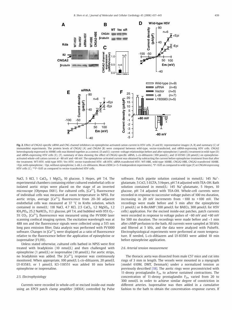
Fig. 2. Effect of CNGA2-specific siRNA and CNG channel inhibitors on epinephrine-activated cation current in H5V cells. (A and B): representative images (A, B) and summary (C) ofimmunoblot experiments. The protein levels of CNGA2 (A) and CNGA4 (B) were compared between wild-type, vector-transfected, and siRNA-expressing H5V cells. CNGA2heterologously expressed in 16HBE cells was blotted together as a control. (D and E): current–voltage relationships before and after epinephrine (1 μmol/L) treatment inwild-type (D)and siRNA-expressing H5V cells (E). (F), summary of data showing the effect of CNGA2-specific siRNA, L-cis-diltiazem (100 μmol/L), and LY-83583 (20 μmol/L) on epinephrine-activated whole-cell cation current at −80 mV and +80 mV. The epinephrine-activated current was obtained by subtracting the current before epinephrine treatment from that afterthe treatment. WT-H5V, wild-type H5V; Vec-H5V, vector-transfected H5V; siR-H5V, siRNA-transfected H5V; WT-HBE, wild-type 16HBE; CNGA2-HBE, CNGA2-transfected 16HBE.+Epi, with epinephrine; −Epi, without epinephrine; L-dil, L-cis-diltiazem. Mean±SEM (n=5–9 independent experiments). ⁎Pb0.05 as compared towild-type (F) or CNGA4 expressingH5V cells (C). ⁎⁎Pb0.05 as compared to vector-transfected H5V cells.
439B. Shen et al. / Journal of Molecular and Cellular Cardiology 45 (2008) 437–445
NaCl, 5 KCl, 1 CaCl2, 1 MgCl2, 10 glucose, 5 Hepes, pH 7.4. Theexperimental chambers containing either cultured endothelial cells orisolated aortic strips were placed on the stage of an invertedmicroscope (Olympus IX81). For cultured cells, [Ca2+]i fluorescenceof individual cells was measured at room temperature in NPSS. Foraortic strips, average [Ca2+]i fluorescence from 20–30 adjacentendothelial cells was measured at 37 °C in Krebs solution, whichcontained in mmol/L: 118 NaCl, 4.7 KCl, 2.5 CaCl2, 1.2 MgSO4, 1.2KH2PO4, 25.2 NaHCO3, 11.1 glucose, pH 7.4, and bubbled with 95% O2–
5% CO2. [Ca2+]i fluorescence was measured using the FV1000 laserscanning confocal imaging system. The excitation wavelength was at488 nm and the fluorescence signals were collected using a 515 nmlong pass emission filter. Data analysis was performed with FV1000software. Changes in [Ca2+]i were displayed as a ratio of fluorescencerelative to the fluorescence before the application of epinephrine orisoprenaline (F1/F0).
Unless stated otherwise, cultured cells bathed in NPSS were firsttreated with bradykinin (10 nmol/L) and then challenged withepinephrine (1 μmol/L) or isoprenaline (10 μmol/L). For aortic strips,no bradykinin was added. The [Ca2+]i response was continuouslymonitored. When appropriate, 100 μmol/L L-cis-diltiazem, 20 μmol/LLY-83583, or 1 μmol/L ICI-118551 was added 10 min beforeepinephrine or isoprenaline.
2.5. Electrophysiology
Currents were recorded in whole-cell or excised inside-out modeusing an EPC9 patch clamp amplifier (HEKA) controlled by Pulse
software. Patch pipette solution contained in mmol/L: 145 Na+-glutamate, 5 CsCl, 5 EGTA, 5 Hepes, pH 7.4 adjusted with TEA-OH. Bathsolution contained in mmol/L: 145 Na+-glutamate, 5 Hepes, 10glucose, pH 7.4 adjusted with TEA-OH. Whole-cell currents wererecorded in response to successive voltage pulses of 100 ms duration,increasing in 20 mV increments from −100 to +100 mV. Therecordings were made before and 5 min after the epinephrine(1 μmol/L) or 8-BrcAMP (100 μmol/L for BAECs, 300 μmol/L for H5Vcells) application. For the excised inside-out patches, patch currentswere recorded in response to voltage pulses of −60 mV and +60 mVfor 500 ms duration. The recordings were made before and ~1 minafter cAMP perfusion to the bath. All currents were sampled at 50 kHzand filtered at 5 kHz, and the data were analyzed with PulseFit.Electrophysiological experiments were performed at room tempera-ture. If needed, L-cis-diltiazem and LY-83583 were added 10 minbefore epinephrine application.
2.6. Arterial tension measurement
The thoracic aorta was dissected from male C57 mice and cut intorings of 3 mm in length. The vessels were mounted in a myograph(model 610M, DMT, Denmark) under a normalized tension aspreviously described [18]. The aortic rings were preconstricted with11-deoxy prostaglandin F2α to achieve sustained contractions. Theconcentration of 11-deoxy prostaglandin F2α varied from 20 to300 nmol/L in order to achieve similar degree of constriction indifferent arteries. Isoprenaline was then added in a cumulativefashion to the bath to obtain the concentration–response curves. If

440 B. Shen et al. / Journal of Molecular and Cellular Cardiology 45 (2008) 437–445
needed, L-cis-diltiazem (100 μmol/L) was added 30 min beforeisoprenaline application. L-cis-diltiazem did not cause a significantchange in contraction of aortic rings to 11-deoxy prostaglandin F2αThe relaxation response to isoprenaline was expressed as % of 11-deoxy prostaglandin F2α-induced tone. In some experiments, theendothelial layer was mechanically disrupted with a small piece ofplastic tubing. Endothelium integrity or functional removal wasverified by the presence or absence, respectively, of the relaxantresponse (over 80% relaxation) to 1 μmol/L acetylcholine at the start ofeach experiment. All tension experiments were performed at 37 °C inKrebs solution.
2.7. Materials
Fluo-4/AM and pluronic F-127 were obtained from MolecularProbes Inc. The primary antibodies against CNGA2 and CNGA4 werefrom Alpha-Diagnostic Int., USA. ECLÔ Plus western blotting detectionsystem was from Amersham Pharmacia. Protease inhibitor cocktailtablets were from Roche. L-cis-diltiazemwas from Biomol, USA. RPMI-1640, DMEM, FBS, blasticidin, and lipofectamine 2000 were fromInvitrogen. LY-83583 was from Calbiochem. Epinephrine, isoprena-line, ICI-118551, Nonidet P-40, sodium deoxycholate, TEA, SDS, EDTA,and Hepes were from Sigma.
3. Results
3.1. Role of CNG channels in epinephrine-induced cation current
Whole-cell voltage clamp was used to study the epinephrine-activated cation current in BAECs. The recorded current (Fig. 1A) and
Fig. 3. Effect of CNG channel inhibitors and CNGA2-specific siRNA on cAMP-activated wholetreatment in BAECs (A) and H5V cells (C). (B and D): summary of data showing the effect of , Lactivated whole-cell cation current at −80 mV and +80 mV in BAECs (B) and H5V cells (D).treatment from that after the treatment. +8-BrcAMP, with 8-BrcAMP (100 μmol/L for BAECs,SEM (n=4–8 independent experiments). ⁎Pb0.05 as compared to wild-type cells. ⁎⁎Pb0.05
the corresponding current–voltage (I–V) relationship (Fig. 1B) showedthat the current had slight outward rectification. Epinephrine treat-ment (1 μmol/L, 5 min) increased the magnitude of the whole-cellcurrent in both inward (negative) and outward (positive) directions(Figs. 1A and B). It is known that epinephrine, via its action on β-adrenergic receptors, causes an elevation in cytosolic cAMP. Thus, wetested the possible involvement of CNG channels. In cells pretreatedwith CNG channel blockers, either L-cis-diltiazem (100 μmol/L) or LY-83583 (20 μmol/L) [23,24], epinephrine was unable to cause asignificant increase in cation current (Figs. 1C and D). These datasuggest an involvement of CNG channels in the epinephrine-activatedcation current.
Because both L-cis-diltiazem and LY-83583 inhibit multiple CNGisoforms, they cannot be used to differentiate specific CNG isoforminvolved. Thus, we next used siRNA strategy. CNGA2 was chosen asthe possible target based on its high sensitivity to cAMP [15] and itsexpression in vascular endothelial cells [13,22,23]. Because bovineCNGA2 gene is still not cloned and its nucleotide sequence is notavailable in Genbank, we were unable to design siRNA againstbovine CNGA2. Instead, we designed siRNA against mouse CNGA2and studied its effect in the mouse endothelial cell line H5V. Inimmunoblot experiments, the CNGA2-specific antibody recognized aprotein band with molecular size of ~80 kDa, which correlates wellwith that of mouse CNGA2 proteins in Genbank (NM_007724) [24].This antibody was also able to detect the CNGA2 proteins over-expressed in a human bronchial epithelial cell line 16HBE, confirm-ing the specificity of this antibody (Fig. 2A). Stable expression ofCNGA2-specific siRNA reduced the CNGA2 protein level by 82±6%(n=5) (Figs. 2A and C). In contrast, transfection with control vectorhad no effect on the CNGA2 protein level (Fig. 2A). The effect of
-cell cation current. (A and C): current–voltage relationships before and after 8-BrcAMP-cis-diltiazem (100 μmol/L), LY-83583 (20 μmol/L), and CNGA2-specific siRNA on cAMP-The cAMP-activated current was obtained by subtracting the current before 8-BrcAMP300 μmol/L for H5V cells); −8-BrcAMP, without 8-BrcAMP; L-dil, L-cis-diltiazem. Mean±as compared to vector-transfected H5V cells.

Fig. 4. Dose–response relationship between cAMP concentrations and its inducedcation current in excised inside-out patches from BAECs. (A), representative tracesshowing voltage protocol (left) and the corresponding patch currents before (middleleft) and after cAMP (10 μmol/L, middle right; 30 μmol/L, right) application. (B),summary of data showing the relationship between cAMP concentrations and cAMP-induced peak inward currents at −60 mV. The cAMP-induced currents were obtained bysubtracting the peak currents in the absence of cAMP from the values in the presence ofcAMP. The currents were plotted as the percentage of maximal current in response to1 mmol/L cAMP. Mean±SEM (n=3–4 experiments).
Fig. 5. Effect of CNG channel inhibitors on epinephrine- and isoprenaline-induced [Ca2+]i rises in1 μmol/L) and isoprenaline (Iso, 10 μmol/L) in the presence of bradykinin (BK, 10 nmol/L). (C adisplayingepinephrine- and isoprenaline-induced [Ca2+]i rises (C) and on themagnitudeoffirst83583 (20 μmol/L); ICI, ICI-118551 (1 μmol/L). Mean±SEM (n=4–6 independent experiments, 1
441B. Shen et al. / Journal of Molecular and Cellular Cardiology 45 (2008) 437–445
siRNA was CNGA2-specific, because it had no influence on theexpression of another CNG isoform CNGA4, which has the molecularsize of ~66 kDa (NM_001033317) (Figs. 2B and C). Functionally, theexpression of siRNA diminished the epinephrine-activated cationcurrent (Figs. 2D–F), while the control vector had no effect (Fig. 2F).The effect of L-cis-diltiazem and LY-83583 was also tested. Asexpected, both inhibitors diminished the magnitude of epinephrine-activated cation current in both inward and outward directions (Fig.2F). Taken together, these data strongly suggest that CNGA2 are themain channels responsible for epinephrine-activated cation currentin endothelial cells.
3.2. Role of CNG channels in cAMP-activated cation current
CNGA2 channels are cAMP-activated channels. Therefore, we nextexamined cAMP-activated cation current in endothelial cells. 8-BrcAMP, which is a membrane-permeant analog of cAMP, increasedthe magnitude of whole-cell cation current in both BAECs (Figs. 3Aand B) and H5V cells (Figs. 3C and D). Furthermore, the cAMP-activated cation current was markedly reduced by L-cis-diltiazem(100 μmol/L) and LY-83583 (20 μmol/L) (Figs. 3B and D). Importantly,CNGA2-specific siRNA diminished the cAMP-activated cation currentin H5V cells (Fig. 3D). These data suggest that CNGA2 are the mainchannels responsible for cAMP-activated cation current in endothe-lial cells.
A cAMP-activated current was also recorded in the excised inside-out patches of endothelial cells (Figs. 4A and B). It was found that bathapplication of cAMP increased the cation currents in the inside-outpatches in a dose-dependentmanner (Fig. 4B)with EC50 at ~36 μmol/L.
BAECs. (A andB), representative traces of the [Ca2+]i rises in response to epinephrine (Epi,nd D), summary of data showing the effect of inhibitory agents on the percentage of cells[Ca2+]i transient among the responding cells (D). L-dil, L-cis-diltiazem (100 μmol/L); LY, LY-5 to 40 cells per experiment). ⁎Pb0.05 as compared to epinephrine or isoprenaline alone.

Fig. 6. Effect of CNG channel inhibitors and CNGA2-specific siRNA on epinephrine- and isoprenaline-induced [Ca2+]i rises in H5V cells. (A and B), representative traces of the [Ca2+]irises in response to epinephrine (Epi, 1 μmol/L) and isoprenaline (Iso, 10 μmol/L) in the presence of bradykinin (BK, 10 nmol/L). (C and D), summary of data showing the effect ofinhibitors and CNGA2-specific siRNA on the percentage of cells displaying epinephrine- and isoprenaline-induced [Ca2+]i rises (C) and on the magnitude of first [Ca2+]i transientamong the responding cells (D). L-dil, L-cis-diltiazem (100 μmol/L); LY, LY-83583 (20 μmol/L); ICI, ICI-118551 (1 μmol/L); siRNA, CNGA2-specific siRNA. Mean±SEM (n=3–5independent experiments, 15 to 40 cells per experiment). ⁎Pb0.05 as compared to epinephrine or isoprenaline alone. ⁎⁎Pb0.05 as compared to vector-transfected H5V cells.
442 B. Shen et al. / Journal of Molecular and Cellular Cardiology 45 (2008) 437–445
3.3. Role of CNG channels in epinephrine- and isoprenaline-induced Ca2+
influx in cultured endothelial cells
We then explored the functional role of CNG channels inendothelial cells. Previous reports showed that epinephrine andisoprenaline were able to elicit a [Ca2+]i rise in the endothelial cellsthat were pretreatedwith Ca2+-mobilizing agonists such as bradykininor thrombin [10,11]. It is speculated that, in these cases, Ca2+-mobilizing agonists serve to potentiate the epinephrine and isoprena-line-induced [Ca2+]i responses [10,11]. Here, we explored the possibi-lity of whether epinephrine- and isoprenaline-induced [Ca2+]i rises aremediated by CNG channels. BAECs were challenged with epinephrine(1 μmol/L) or isoprenaline (10 μmol/L). In the absence of bradykinin,only ~10–20% of cells responded with a small rise in [Ca2+]i (data notshown). However, in the presence of bradykinin (10 nmol/L), bothepinephrine (1 μmol/L) (Fig. 5A) and isoprenaline (10 μmol/L) (Fig. 5B)were able to elicit a [Ca2+]i rise in majority of BAECs (86±6%, n=6experiments). In some cells, this [Ca2+]i rise displayed an oscillatorypattern, whereas in other cells only a single [Ca2+]i transient could beobserved (Figs. 5A and B). This [Ca2+]i rise required the presence ofextracellular Ca2+, because epinephrine or isoprenaline failed to elicitthe [Ca2+]i rise in cells bathed in 0Ca2+-PSS. Importantly, L-cis-diltiazem (100 μmol/L) and LY-83583 (20 μmol/L) markedly reducedthe percentage of cells displaying epinephrine- and isoprenaline-induced [Ca2+]i rises (Fig. 5C), and they also suppressed the magnitudeof peak [Ca2+]i rises among the cells displaying epinephrine- andisoprenaline-induced [Ca2+]i rises (Fig. 5D). The effect of epinephrineand isoprenaline was likely to be mediated by β2-adrenoceptors,
because ICI-118551 (1 μmol/L), a selective antagonist for β2-adrenoceptors [1], effectively blocked the [Ca2+]i rises (Figs. 5C and D).
Experiments were also performed in H5V cells. Similar to BAECs,both epinephrine (1 μmol/L) (Fig. 6A) and isoprenaline (10 μmol/L)(Fig. 6B) induced a [Ca2+]i rise in H5V cells that were pretreated withbradykinin (10 nmol/L). L-cis-diltiazem (100 μmol/L) and LY-83583(20 μmol/L) markedly diminished epinephrine- and isoprenaline-induced [Ca2+]i rises (Figs. 6C and D). These [Ca2+]i rises were inhibitedby ICI-118551 (1 μmol/L). Importantly, CNGA2-specific siRNA alsosuppressed the [Ca2+]i rises, whereas the control vector had no effect(Figs. 6C and D). We also tested the effect of CNG channels inhibitorsand CNGA2-specific siRNA on bradykinin-induced [Ca2+]i rise, which isknown to be mainly resulted from Ca2+ release from inositol 1,4,5-trisphosphate-sensitive Ca2+ stores [25]. As expected, L-cis-diltiazem,LY-83583 and CNGA2-specific siRNA had no effect on bradykinin(10 nmol/L)-induced [Ca2+]i rise (Supplemental Fig. 1), Taken together,these data support a key role of CNGA2 channels in epinephrine- andisoprenaline-induced Ca2+ influx.
CNGA2 channels are highly permeable to Ca2+ [15]. Therefore wealso examined the role of CNGA2 in the maintenance of basal [Ca2+]ilevel in endothelial cells. As shown in Supplemental Fig. 2, L-cis-diltiazem (100 μmol/L) or LY-83583 (20 μmol/L) treatment for 1 hcaused a significant reduction in basal [Ca2+]i level for both BAECs andH5V cells (Supplemental Figs. 2A and B). Furthermore, CNGA2-specificsiRNA also decreased the basal [Ca2+]i level in H5V cells (SupplementalFig. 2B). These data suggest an important role of CNGA2 in themaintenance of basal [Ca2+]i level in endothelial cells under normalnon-stimulated condition.

Fig. 7. Effect of CNG channel inhibitors on epinephrine- and isoprenaline-induced endothelial [Ca2+]i rises within mouse aortic strips. (A and B), representative endotheliumfluorescence images before (A) and after (B) 1 μmol/L epinephrine. (C and D), summary of data showing the effect of L-cis-diltiazem (100 μmol/L) and LY-83583 (10 μmol/L) on themagnitude of first [Ca2+]i transient in response to 1 μmol/L epinephrine (Epi, C), 10 μmol/L isoprenaline (Iso, D), and 10 μmol/L acetylcholine (Ach, C and D). Mean±SEM (n=4–12experiments). ⁎Pb0.05 as compared to epinephrine or isoprenaline alone.
443B. Shen et al. / Journal of Molecular and Cellular Cardiology 45 (2008) 437–445
Note that, although Ca2+ influx was required for epinephrine- andisoprenaline-induced [Ca2+]i rises, itwasapparent that store Ca2+ releasealso contributed to this [Ca2+]i rise probably via the mechanism of Ca2+-induced store Ca2+ release. This was evident because in cells pretreatedwith thapsigargin (4 μM) for 30 min to deplete intracellular Ca2+ stores,the epinephrine- and isoprenaline-induced [Ca2+]i responses becamemuch smaller (Supplemental Fig. 3). Furthermore, in cells after Ca2+
store depletion, only a single [Ca2+]i transient (without [Ca2+]i oscilla-tion) could be observed in response to epinephrine and isoprenaline.
3.4. Role of CNG channels in epinephrine- and isoprenaline-inducedendothelial Ca2+ responses in mice aorta
There is a concern that endothelial phenotype may change duringcell culture and cell passage conditions. Thus, we studied the [Ca2+]ichanges in endothelial cells within isolated mouse aortic strips to
Fig. 8. Effect of L-cis-diltiazem on isoprenaline-induced vascular relaxation in mouseaortic segments. Mouse aortic segments were preconstricted with 11-deoxy prosta-glandin F2α in the absence or presence of 100 μmol/L L-cis-diltiazem. Isoprenaline wasthen added in a cumulative fashion to the bath to induce vascular relaxation. +Endo,endothelium-intact; −Endo, endothelium-denuded; L-dil, L-cis-diltiazem. Mean±SEM(n=5 independent experiments).
verify the above findings. Application of epinephrine (1 μmol/L) orisoprenaline (10 μmol/L) elicited a transient [Ca2+]i rise in more than90% of endothelial cells in the isolated mouse aortic strips (Figs. 7Aand B). Surprisingly, unlikely cultured endothelial cells, the endothe-lial cells within the isolated aortic strips were capable of responding toepinephrine or isoprenaline in the absence of exogenous bradykinin.We speculate that themicroenvironmentwithin isolated aortic tissuesmight already contain sufficient amount of Ca2+-mobilizing agonists,so that exogenous addition of agonists was no longer needed.Importantly, epinephrine- and isoprenaline-induced endothelial [Ca2+]irises were almost completely abolished in aortic strips that werepretreated with L-cis-diltiazem (100 μmol/L) or LY-83583 (10 μmol/L)for 10 min (Fig. 7C and D). In contrast, L-cis-diltiazem or LY-83583 hadno effect on acetylcholine-induced peak [Ca2+]i rise (Fig. 7C and D),which is known to be mainly resulted from store Ca2+ release [25].
3.5. Role of CNGchannels inβ-adrenoceptor-mediated vascular relaxation
We further examined the functional role of CNG channels in β-adrenoceptor-mediated vascular relaxation. Mouse aortic segmentswere preconstricted with 11-deoxy prostaglandin F2α, and in agree-ment with the results from other groups [26], isoprenaline inducedconcentration-dependent vascular relaxation (Fig. 8). The relaxationwasmostly endothelium-dependent, because removal of the endothe-lium almost completely abolished isoprenaline-induced relaxation(Fig. 8). Importantly, L-cis-diltiazem (100 μmol/L) markedly inhibitedthe relaxation to isoprenaline in endothelium-intact aortic rings (Fig.8), but it had no effect on the residual small relaxation to isoprenalinein endothelium-denuded aortic rings (Fig. 8). These data suggest thatCNGchannels playa key role in the endothelium-dependent relaxationinduced by β-adrenoceptor agonists.
4. Discussion
The major findings of this study are as follows: Firstly, with the useof whole-cell patch clamp, we recorded a current that was activated

444 B. Shen et al. / Journal of Molecular and Cellular Cardiology 45 (2008) 437–445
by cAMP and epinephrine in the primary cultured BAECs and H5Vendothelial cells. We demonstrated that this current was sensitive toselective CNG channel blockers L-cis-diltiazem and LY-83583. Further-more, a CNGA2-specific siRNA diminished this current in H5V cells.These results strongly suggest that epinephrine activates CNGchannels, CNGA2 in particular, in vascular endothelial cells. Secondly,we studied the functional role of CNGA2 in endothelial Ca2+ influx. Wefound that in the presence of bradykinin, both epinephrine andisoprenaline, via their action on β2-adrenoceptors, could stimulate Ca2+
influx in cultured endothelial cells. This Ca2+ influx was markedlyreduced by L-cis-diltiazem, LY-83583, and CNGA2-specific siRNA. Therole of CNGA2 in epinephrine- and isoprenaline-induced [Ca2+]iresponses in endothelial cells were also confirmed using isolatedmouse aortic strips. Thirdly, inhibition of CNG channels by L-cis-diltiazem reduced the endothelium-dependent vascular dilation toisoprenaline in isolated mouse aortic segments. Taken together, thesedata provide compelling evidence that CNG channels, CNGA2 inparticular, are the channels responsible for epinephrine- and β-adrenoceptor agonist-induced Ca2+ influx in endothelial cells andsubsequent vascular dilation.
With the use of RT-PCR and immunoblot, several groups havefound the expression of several CNG isoforms, including CNGA1,CNGA2, and CNGA4, in vascular endothelial cells [13,14,22,23].Regarding functional role of CNG channels, Zhang et al. [23] proposedthat CNGA2 contributes to the store-operated Ca2+ influx inpulmonary artery endothelial cells, whereas Wu et al. [22] suggestedthat CNGA2 mainly allows Na+ entry, resulting in membranedepolarization, which reduces the driving force for Ca2+ entry.However, in general there is a lack of convincing evidence for thefunctional role of CNG channels in vascular endothelial cells. In theprevious functional studies by Wu et al. and Zhang et al., the primaryevidence for the functional involvement of CNGwere mainly based onCNG-selective pharmacological blockers L-cis-diltiazem and LY-83583. While L-cis-diltilzem has good selectivity to CNG channels[20], LY-83853 is not very selective and it also acts on other targetssuch as guanylate cyclases [21]. Up to the present, there is still noreport employing the gene “knocking-down” technique, which is farmore selective to its target molecules, to study CNG function invascular tissue. Furthermore, no report has demonstrated “a cyclicnucleotide-activated current in excised inside-out patches” inendothelial cells, which is considered to be a hallmark of CNGchannels. In the present study, immunoblot experiments demon-strated the expression of CNGA2 protein in endothelial cells, andfurthermore excised inside-out patch clamp experiments showed acAMP-activated cation current. In addition, CNGA2-specific siRNAreduced the cAMP-activated cation current. These studies giveunequivocal support for an important functional role of CNGA2channels in vascular endothelial cells.
Previous studies have well demonstrated that stimulation ofendothelial β-adrenoceptors results in endothelium-dependent vas-cular dilation [1,3–7], and that the dilation is mediated by cAMP [1,5–7]. However, there is controversy on whether [Ca2+]i plays a role incAMP-induced endothelium-dependent vascular dilation. On the onehand, there is evidence that cAMP can activate eNOS in a Ca2+-independent manner via protein kinase A phosphorylation on Ser-1179 and Ser-635 [8]. On the other hand, Graier et al. provided theevidence that elevated cAMP following the stimulation of β-adrenoceptors elicits a [Ca2+]i rise in endothelial cells [10]. The risein endothelial [Ca2+]i would certainly activate eNOS, which is anenzyme sensitive to Ca2+-calmodulin activation [10]. In the presentstudy, we confirmed that both β-adrenoceptor agonists and cAMP caninduce a [Ca2+]i rise in endothelial cells. Importantly, we were able toidentify CNGA2 as the channel mediating β-adrenoceptor agonist-induced endothelial Ca2+ influx and vascular dilation. These findingsprovide a mechanistic support for the involvement of endothelial[Ca2+]i in β-adrenoceptor-mediated vascular dilation.
Our findings also fit well with a unique property of CNGA2, i.e. itshigher sensitivity to cAMP than other CNG isoforms [15]. Interestingly,vascular endothelial cells in vivo are also the targets of several otherphysiologically important cAMP-elevating agents, which includecalcitonin gene-related peptide [27], adrenomedullin [27] andadenosine [28]. Like β-adrenoceptor agonists, these cAMP-elevatingagents induce endothelium-dependent relaxation via two differentmechanisms [27–29]. The first mechanism is well characterized and itinvolves Ca2+-independent activation eNOS by cAMP and proteinkinase A [29]. The second mechanism is Ca2+-sensitive and theunderlying mechanism is largely unclear [30]. Recently, we havedemonstrated an important role of CNGA2 in adenosine-induced Ca2+
influx in endothelial cells [18]. Together with the present data, wehypothesize that the adenylyl cyclase-cAMP-mediated activation ofCNGA2 could be a pathway shared by many other cAMP-elevatingagents in vascular endothelial cells.
In conclusion, we demonstrate that CNG channels, especiallyCNGA2, are the main channels responsible for epinephrine- andisoprenaline-induced Ca2+ influx in vascular endothelial cells. Theeffect of epinephrine and isoprenaline is mediated through β2-adrenoceptors.
Acknowledgments
We thank the financial support of the Hong Kong Research GrantCouncil (CUHK4526/06M), Focused Investment Scheme of ChineseUniversity of Hong Kong, and Li Ka Shing Institute of Health Sciences.
Appendix A. Supplementary data
Supplementary data associated with this article can be found, inthe online version, at doi:10.1016/j.yjmcc.2008.06.005.
References
[1] Guimaraes S, Moura D. Vascular adrenoceptors: an update. Physiol Rev2001;53:319–56.
[2] Mark AL, Abboud FM, Schmid PG, Heistad DD, Mayer HE. Differences in directeffects of adrenergic stimuli on coronary, cutaneous, and muscular vessels. J ClinInvest 1972;51:279–87.
[3] Brawley L, Shaw AM, MacDonald A. Role of endothelium/nitric oxide in atypicalbeta-adrenoceptor-mediated relaxation in rat isolated aorta. Eur J Pharmacol2000;398:285–96.
[4] Priest RM, Hucks D, Ward JP. Noradrenaline, beta-adrenoceptor mediatedvasorelaxation and nitric oxide in large and small pulmonary arteries of the rat.Br J Pharmacol 1997;122:1375–84.
[5] Gray DW, Marshall I. Novel signal transduction pathway mediating endothelium-dependent beta-adrenoceptor vasorelaxation in rat thoracic aorta. Br J Pharmacol1992;107:637–84.
[6] Xu B, Li J, Gao L, Ferro A. Nitric oxide-dependent vasodilatation of rabbit femoralartery by beta(2)-adrenergic stimulation or cyclic AMP elevation in vivo. Br JPhramacol 2000;129:969–74.
[7] Ferro A, Queen LR, Priest RM, Xu B, Ritter JM, Poston L, Ward JP. Activation of nitricoxide synthase by beta 2-adrenoceptors in human umbilical vein endothelium invitro. Br J Pharmacol 1999;126:1872–80.
[8] Boo YC, Hwang J, Sykes M, Michell BJ, Kemp BE, Lum H, Jo H. Shear stressstimulates phosphorylation of eNOS at Ser(635) by a protein kinase A-dependentmechanism. Am J Physiol Heart Circ Physiol 2002;283:H1819–28.
[9] Brock TA, Dennis PA, Griendling KK, Diehl TS, Davies PF. GTP gamma S loading ofendothelial cells stimulates phospholipase C and uncouples ATP receptors. Am JPhysiol 1988;255:C667–73.
[10] Graier WF, Groschner K, Schmidt K, Kukovetz WR. Increases in endothelial cyclicAMP levels amplify agonist-induced formation of endothelium-derived relaxingfactor (EDRF). Biochem J 1992;288:345–9.
[11] Buchan KW, Martin W. Modulation of agonist-induced calcium mobilisation inbovine aortic endothelial cells by phorbol myristate acetate and cyclic AMP butnot cyclic GMP. Br J Pharmacol 1991;104:361–6.
[12] Nilius B, Groogman G. Ion channels and their functional role in vascularendothelium. Physiol Rev 2001;81:1415–59.
[13] Cheng KT, Chan FL, Huang Y, Chan WY, Yao X. Expression of olfactory-type cyclicnucleotide-gated channel (CNGA2) in vascular tissues. Histochem Cell Biol2003;120:475–81.
[14] Yao X, Leung PS, Kwan HY, Wong TP, Fong MW. Rod-type cyclic nucleotide-gatedcation channel is expressed in vascular endothelium and vascular smooth musclecells. Cardiovasc Res 1999;41:282–90.

445B. Shen et al. / Journal of Molecular and Cellular Cardiology 45 (2008) 437–445
[15] Kaupp UB, Seifert R. Cyclic nucleotide-gated ion channels. Physiol Rev2002;82:768–824.
[16] GraierWF, Kukovetz WR, Groschner K. Cyclic AMP enhances agonist-induced Ca2+
entry into endothelial cells by activation of potassium channels and membranehyperpolarization. Biochem J 1993;291:263–7.
[17] Trochy JN, Leblais V, Rautureau Y, Beverelli F, Le Marec H, Berdeaux A, Gauthier C.Beta3-adrenoceptor stimulation induces vasorelaxation mediated essentially byendothelium-derived nitric oxide in rat thoracic aorta. Br J Pharmacol1999;128:69–76.
[18] Cheng KT, Leung YK, Shen B, Kwok YC,Wong CO, Kwan HY, Man YB, Ma X, Huang Y,Yao X. CNGA2 channels mediate adenosine-induced Ca2+ influx in vascularendothelial cells. Arterioscler Thromb Vasc Biol 2008;28:913–8.
[19] Garlanda C, Parravicini C, Sironi M, De Rossi M, Wainstock de Calmanovici R,Carozzi F, Bussolino F, Colotta F, Mantovani A, Vecchi A. Progressive growth inimmunodeficient mice and host cell recruitment by mouse endothelial cellstransformed by polyoma middle-sized T antigen: implications for the pathogen-esis of opportunistic vascular tumors. Proc Natl Acad Sci U S A 1994;91:7291–5.
[20] Finn JT, Grunwald ME, Yau KW. Cyclic nucleotide-gated ion channels: an extendedfamily with diverse functions. Ann Rev Physiol 1996;58:395–426.
[21] Leinders-Zufall T, Zufall F. Block of cyclic nucleotide-gated channels insalamander olfactory receptor neurons by the guanylyl cyclase inhibitorLY83583. J Neurophysiol 1995;74:2759–62.
[22] Wu S, Moore TM, Brough GH, Whitt SR, Chinkers M, Li M, Stevens T. Cyclicnucleotide-gated channels mediate membrane depolarization following activa-
tion of store-operated calcium entry in endothelial cells. J Biol Chem2002;275:18887–96.
[23] Zhang J, Xia SL, Block ER, Patel JM. NO upregulation of a cyclic nucleotide-gatedchannel contributes to calcium elevation in endothelial cells. Am J Physiol CellPhysiol 2002;283:C1080–9.
[24] Bonigk W, Bradley J, Muller F, Sesti F, Boekhoff I, Ronnett GV, Kaupp UB, Frings S.The native rat olfactory cyclic nucleotide-gated channel is composed of threedistinct subunits. J Neurosci 1999;19:5332–47.
[25] Busse R, Pohl U, Lückhoff A. Mechanisms controlling the production of endothelialautacoids. Z Kardiol 1989;78 Suppl 6:64–9.
[26] Akimoto Y, Horinouchi T, Shibano M, Matsushita M, Yamashita Y, Okamoto T,Yamaki F, Tanaka Y, Koike K. Nitric oxide (NO) primarily accounts for endothelium-dependent component of beta-adrenoceptor-activated smooth muscle relaxationof mouse aorta in response to isoprenaline. J Smooth Muscle Res 2002;38:87–99.
[27] Brain SD, Grant AD. Vascular actions of calcitonin gene-related peptide andadrenomedullin. Physiol Rev 2003;84:903–34.
[28] Ray CJ, Marshall JM. The cellular mechanisms by which adenosine evokes releaseof nitric oxide from rat aortic endothelium. J Physiol 2006;570:85–96.
[29] Busse R, Edwards G, Feletou M, Fleming I, Vanhoutte PM, Weston AH. EDHF:bringing the concepts together. Trends Pharmacol Sci 2002;23:374–80.
[30] Shimekake Y, Nagata K, Ohta S, Kambayashi Y, Teraoka H, Kitamura K, Eto T,Kangawa K, Matsuo H. Adrenomedullin stimulates two signal transductionpathways, cAMP accumulation and Ca2+ mobilization, in bovine aortic endothelialcells. J Biol Chem 1995;270:4412–7.