epilepsy and brain tumor -...
TRANSCRIPT

Epilepsy and Brain Tumor
Apasri Lusawat M.D. Pediatric Neurology
Prasat Neurological Institute

Outline
• Overview
• Pathogenesis : Tumor-> epilepsy

Etiology of Epilepsy , age

EPILEPSY AND BRAIN TUMORS
• CNS neoplasms are the cause of (van Breeman MS, et al 2007) ≈ 10–15% of adult-onset epilepsy ≈ 0.2–6.0% of childhood-onset epilepsy
• Seizure is the most common presenting symptom of brain tumor (Lynam LM, et al 2007) – 38% of primary CNS tumors – 20% of secondary CNS tumors

Brain Tumor Classification
Glioma 50-67%
Pituitary T.
Pineal gland T.
Neuronal & mixed neuro-glial tumors
CN & PN T.
Sellar T
Germ cell T.
Melanocytic T.
Lymphomas
Histiocytic T.
Meningiomas
Mesenchymal T.
Embryonal T.
Astrocytic T.
Oligodendroglial T.
Glioblastomas
Choriod plexus T.
Metastatic Brain Tumor (2o BT)
Prim
ary
Brai
n Tu
mor
(1o B
T)
Brai
n Tu
mor
Dysembryoplastic Neuroepithelial T :DNET
Ganglioglioma
Others
Ependymal T.

Brain Tumor Grade
• “low grade” (WHO grade I or II)
• “high grade” (WHO grade III or IV).


Seizure frequency based on Tumor type Lancet Neurol 2007 421-430
Neuronal tumors have a higher incidence of seizures. Due to-> population of neurons could be epileptogenic within the tumor whereas in the glial tumors the seizure focus is generally in the peritumoral brain tissue

Seizures & Tumor Pathology
• Slow-growing, infiltrative tumors : DNET & LGG – Highest risk Sz 75%-100% in adults and children.
• Meningiomas and high-grade astrocytomas, – Less common in children compared with adults – 30%-60% incidence of Sz in adults and children.
• Seizures in 20-40% of pts brain metastases, – in 67% with melanoma, in 48% with lung cancer, – In 33% with CA breast , in 55% with unknown CA
Maschio M., 2012

Developmental brain & Tumor
• ≈ half of pts DNET and epilepsy have cortical dysplasia associated with the tumor (Chang et al., 2010).
Epilepsia, 52(1):158–174, 2011

Seizure and Tumor Volume
High-grade tumors presenting with seizures are likely to be smaller than those presenting with other symptoms
Lee et al., 2010

Seizure and Location of Tumor
• Higher seizure frequency associated with – Supratentorial tumors – Located in the cortex and superficially
• Higher epileptogenic supratentorial tumors in – Fronto-temporal region (80%) and fronto-parietal
region (71%).
• Epileptogenic infratentorially localized tumors in about 2.5% of cases

Evaluation of Sz in a Pt with a Known Tumor
Well EM., et al 2012
Possible causes of seizure in children with brain tumors

Late-Onset Seizures
• Late effects from brain tumors and their treatment defined as 5 years after diagnosis in cancer survivors
• Packer, 2003 reported seizure : – 25% long-term survivors of childhood BT – 6.5% had first seizure > 5 years after diagnosis
• Radiation therapy (RT) – at doses > 30 Gy to any cortical segment - 2-fold
increase in risk – Seizures more likely in younger children
Semin Pediatr Neurol 2012 ; 19:3-8

• Complete seizure control was achieved in only 12.6% patients with a brain tumor (Hildebrand et al., 2005)
• In 15–58% of cases of low-grade glioma, the epilepsy appears to be intractable (Duffau et al., 2002)
Brain Tumor-related Epilepsy Pharmacological Resistance.

Focal Generalized Unknown
Focal Generalized Combined
Generalized &Focal
Unknown
Genetic
Structural
Metabolic
Infection
Immune
Unknown
Scheffer IE, et al Epilepsia, 58(4):512–521, 2017

ILAE 2017 Classsification of seizure types (expanded version)
17
Focal Onset Generalized Onset Unknown Onset
Aware Impaired Awareness
Motor Onset Automatisms Atonic Clonic Epileptic spasms Hyperkinetic Myoclonic tonic
Non-motor Onset Autonomic Behavior arrest Cognitive Emotional Sensory
Focal to bilateral tonic-clonic
Motor Tonic-clonic Clonic Tonic Myoclonic Myoclonic-tonic-clonic Myoclonic-atonic Atonic Epileptic spasms Non-motor Absence •Typical •Atypical •Eyelid myoclonia
Motor Tonic-clonic Epileptic spasms Non-motor Behavior arrest
Unclassified

• Epilepsies attributed to organized by structural-metabolic causes o Malformations of cortical development
(hemimegalencephaly, heterotopias, etc.) o Neurocutaneous syndromes (tuberous
sclerosis complex, Sturge-Weber, etc) o Tumor, infection, trauma, angioma,
antenataland perinatal insults, stroke , etc
Epilepsies of unknown cause
Non-syndromic epilepsies

Symptomatology
• Focal onset seizure type
• Seizure semiology depends on –Tumor location or adjacent abnormal
brain (Primary epileptogenic)
–Secondary epileptogenic

Secondary epileptogenesis
• Occur in up to one-third of brain tumor patients where the epileptogenic focus does not correspond to the tumor location.
• Can be seen in low-grade brain tumors in the temporal lobe which have associated hippocampal sclerosis

Pathogenesis of tumor-related epilepsy • Cellular mechanisms - > epileptogenesis : NOT CLEAR • Two theories of pathogenesis
1. Tumor origin – Tumor itself may excrete molecules that could make the tumor tissue
epileptogenic, or it could change the peri-tumoral microenvironment and turn this into an epileptogenic zone
2. Peri-tumor – Tumor mechanically compresses the surrounding normal tissue, which
eventually becomes epileptogenic after suffering from ischemia and hypoxia.
• Both processes could potentially cause 2ochanges – In neurotransmitters and their receptors, metabolic changes, and
inflammatory responses, eventually leading to epileptic seizures.
Seizure 21 (2012) 153–159

Schematic of observed mechanisms underlying the pathogenesis and clinical strategies of tumor-related epilepsy
Extent of resection
Old AEDs
New AEDs
Tumor Histology Location
Gap junction Genetic factors BBB disruption
Hypoxia
Acidosis
Metabolic changes
Ionic Changes
K+ Na+
Mg2+ Fe 3+
Cl- Ca2+
Morphological changes
Immunological changes
Inflammatory changes
Amino acid and receptor
changes
GABA
Glutamate
Kynurenic acid
Seizure 21 (2012) 153–159 HISTOLOGY
• ECoG studies-> epileptiform activity is associated with a high neuronal density within the lesion (Ferrier et al., 2006).
• Glial tumor: abrupt tissue damage leading to necrosis and haemosiderin deposition and to oedema (Riva, 2005).
LOCATION Proximity to cortical gray matter ≈ associated with epilepsy HISTOLOGY preferred location - Most GLIONEURONAL tumors occur in the temporal,
- LGG tend to grow in2’functional areas near, but rarely within, primary eloquent parts of the brain - OLIGODENDROGLIAL tumors more likely located in frontal lobe
Tumor histology associated with the lobe during brain development Duffau and Capelle 2004

Brain 2012: 135; 1002–1016
Brain 2012: 135; 1002–1016
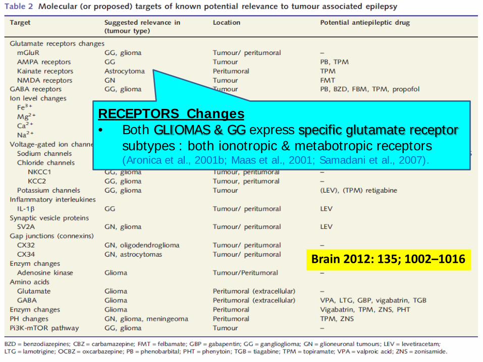
RECEPTORS Changes • Both GLIOMAS & GG express specific glutamate receptor
subtypes : both ionotropic & metabotropic receptors (Aronica et al., 2001b; Maas et al., 2001; Samadani et al., 2007).

Brain 2012: 135; 1002–1016
Receptor changes (Aronica et al., 2001b)
• PERITUMORAL ASTROCYTES express amount of kainate receptors -> downregulate GABAergic inhibition : may predispose to epilepsy.
• Multiple metabotropic glutamate receptors are overexpressed in the peritumoral cortex

Brain 2012: 135; 1002–1016
Voltage-gated Ion channels
• voltage-gated Na channels in tumor cells-> can generate AP. (Patt et al.,1996; Labrakakis et al., 1997)
• Na–K-Cl cotransporter (NKCC1) expression & K-Cl cotransporter (KCC2) reported in glioneuronal tumors (Aronica et al., 2007a, 2008). -> epileptogenicity in GG by modulation of GABA R (Yamada et al., 2004).
Extracelluar IONIC CHANGES
• Macro- or microhaemorrhage -> neuronal membrane injury -> Extracellular iron (Fe3+) ->change memb AP of neuron (Shamji, 2009)
• Mg2+ and Ca2+ probably released from oedema and haemorrhage. Extracellular Mg2+ -> spontaneous epileptiform discharges (Schaller and Ruegg, 2003).

Brain 2012: 135; 1002–1016
• Brain tumors often show a prominent immune response.
• Upregulation of inflammatory interleukins (such as IL-1B) in GG, have been suggested to play a role in epileptogenicity (Aronica et al.,2008; Vezzani et al., 2011).
• Inflammation activates a cascade of proinflammatory cytokines & induces alterations in the BBB may lead to epilepsy.

Brain 2012: 135; 1002–1016
•Dysplastic glial cells in GLIOMA express synaptic vesicle proteins SV2A ->dysfunction-> calcium accumulation during repeated AP generation -> may epileptogenesis. (De Groot et al., 2010)

Brain 2012: 135; 1002–1016
GAP JUNCTION Disturbed intercellular communication between glial cells->epileptogenic through gap junction channels: CONNEXINS (Aronica et al., 2001a)

Brain 2012: 135; 1002–1016
extracellular GLUTAMATE found in peritumoral brain parenchyma of patients with epilepsy compared to non-tumour patients with epilepsy (de Groot and Sontheimer, 2010).

Brain 2012: 135; 1002–1016
Genetic & Molecular pathways • In GG, several components of Pi3K-mTOR signalling pathways
are activated (Boer 2010). -> epileptogenesis , target of mTOR inhibitors
• In GLIOBLASTOMA and LGG, the pi3K-mTOR pathway plays a role in the development of the tumors (McBride 2010; Sunayama 2010)
• Pi3K-mTOR pathway Common genetic pathways for tumor-associated epilepsy &glioma (Berntsson 2009).
