epigenetic mechanisms in alzheimer's diseasetres/aging_seminar2011/mastroeni.2011.pdf ·...
TRANSCRIPT

s
y
8
Neurobiology of Aging 32 (2011) 1161–1180
Review Article
Epigenetic mechanisms in Alzheimer’s disease
Diego Mastroeni, Andrew Grover, Elaine Delvaux, Charisse Whiteside, Paul D. Coleman,Joseph Rogers*
Banner Sun Health Research Institute, PO Box 1278, Sun City, AZ 85372, USA.
Received 2 March 2010; received in revised form 20 July 2010; accepted 7 August 2010
Abstract
Epigenetic modifications help orchestrate sweeping developmental, aging, and disease-causing changes in phenotype by alteringtranscriptional activity in multiple genes spanning multiple biologic pathways. Although previous epigenetic research has focused primarilyon dividing cells, particularly in cancer, recent studies have shown rapid, dynamic, and persistent epigenetic modifications in neurons thathave significant neuroendocrine, neurophysiologic, and neurodegenerative consequences. Here, we provide a review of the major mecha-nisms for epigenetic modification and how they are reportedly altered in aging and Alzheimer’s disease (AD). Because of their reach acrossthe genome, epigenetic mechanisms may provide a unique integrative framework for the pathologic diversity and complexity of AD.© 2011 Elsevier Inc. All rights reserved.
Keywords: Epigenetics; DNA methylation; Histone acetylation; rDNA; miRNA; Genetics; Gene expression; Amyloid � peptide; Inflammation; Oxidativetress; Cell cycle
www.elsevier.com/locate/neuaging
ec
ot(nbodamptjgelmr
1. Introduction
Alzheimer’s disease (AD) is a progressive, irreversibleneurodegenerative disorder culminating in dementia. Its eti-ology and pathogenesis are complex, and encompass manygenetic and environmental risk factors, changes in the ex-pression of thousands of genes, and upregulation of multiplepathogenic pathways such as amyloid � peptide (A�) de-position, tau hyperphosphorylation, inflammation, oxidativestress, energy metabolism, and aberrant re-entry into the cellcycle/apoptosis. Moreover, with the exception of A�-induc-ing mutations, none of these molecular and genetic factorsappears to have absolute penetrance in causing the disorder:many individuals may possess the most salient risk factorsfor AD, as well as express profuse A� and tau pathology,et never develop the disorder (Lue et al., 1996). Indeed,
* Corresponding author at: Banner Sun Health Research Institute, P.O.Box 1278, Sun City, AZ 85372, USA. Tel.: �1 623 876 5467; fax: �1 62376 5461.
eE-mail address: [email protected] (J. Rogers).
0197-4580/$ – see front matter © 2011 Elsevier Inc. All rights reserved.doi:10.1016/j.neurobiolaging.2010.08.017
ven monozygotic twins can have dichotomous AD out-omes (Mastroeni et al., 2009; Räihä et al., 1997).
The emerging field of epigenetics has its roots in studiesf the structure of chromatin and modifications to the struc-ure of DNA, which extend back half a century or moreFelsenfeld, 2007). Although a unitary definition of epige-etics has yet to be reached, the many definitions that haveeen suggested all invoke heritability, lack of dependencen DNA sequence, and effects on transcription to produceiverse phenotypes. In particular, epigenetic modificationsre capable of altering transcriptional activity in a coherentanner across thousands of genes and dozens of biological
athways, yet can do so differentially in monozygotic twins,he same individual at different developmental stages, or ad-acent cells in the same organ, all of which share the sameenetic code. Epigenetics also provides a means by whichnvironmental factors such as diet, hazardous exposures, andife events can influence gene expression. As such, epigeneticechanisms may provide a point of intersection for the diverse
isk factors and pathophysiologic processes of AD.The purpose of this review is to briefly describe the major
pigenetic mechanisms, histone acetylation, DNA methylation,
1162 D. Mastroeni et al. / Neurobiology of Aging 32 (2011) 1161–1180
ribosomal DNA (rDNA), and microRNA (miRNA), and howthey are reportedly altered in aging and AD.
2. Epigenetic regulation of gene expression
Epigenetic mechanisms modify heritable and nonher-itable traits without necessarily altering the underlyingDNA sequence. Thus, through epigenetic modification thediverse cellular phenotypes and functions needed by thebody can be achieved using a single genetic code for allcells. These effects are typically accomplished by inhibitionof transcriptional access to various genes, leading to theirrepression or silencing. Conversely, release from normalepigenetic repression can enhance gene expression (Ban-dyopadhyay and Medrano, 2003; Fraga and Esteller, 2007;Liu et al., 2003; Poulsen et al., 2007; Wilson and Jones,1983). These modifications can occur at specific gene loci inspecific cells to yield specific cellular phenotypes, or canencompass many genes in many cells, an orchestratingmechanism that is widely assumed to help drive such broadbiological processes as development and aging (Bandyo-padhyay and Medrano, 2003; Fraga and Esteller, 2007; Liuet al., 2003; Poulsen et al., 2007; Wilson and Jones, 1983).
Because phenotype and function are affected and ef-fected by what genes are expressed and what genes arerepressed, epigenetically regulated and dysregulated tran-scription states can give rise not only to different cell typesand developmental stages, but also to favorable and unfa-vorable outcomes for specific cells within the same organsystem. Thus, changes in epigenetic regulation can causesome cells to develop structural, physiologic, and metabolicabnormalities, while other cells of the same type remainnormal. This is thought to occur, for example, in severalhematologic malignancies after aberrant epigenetic silenc-ing of genes that control proliferation (Mund et al., 2006).
In addition to direct regulation of gene expression, epige-netic modifications can mimic, exacerbate, or even cause ge-netic mutations. For example, epigenetic repression of tumorsuppressor genes can mimic loss of function mutations oftumor suppressor genes, and both are highly associated withcancer development (Mund et al., 2006). Epigenetic silencingof the alpha subunit of the stimulatory G protein, a signalingpeptide essential for the actions of parathyroid hormone, cancause pseudohypoparathyroidism, just as mutations to the al-pha subunit do (Bastepe, 2008). Epigenetic modifications canexacerbate the effects of gene mutations, as occurs with ade-nomatous polyposis coli (APC) gene mutations in colorectalcancers (Colnot et al., 2004), E-cadherin mutations in gastriccancers (Strathdee, 2002), and mitochondrial DNA mutationsin Leber’s disease (Johns and Neufeld, 1993). Beyond theseinteractions, epigenetic repression of DNA repair genes mayalso induce gene mutations (Jacinto and Esteller, 2007).
Finally, epigenetic mechanisms provide a means bywhich environmental events can be translated to the cellular
and molecular level. For example, ultraviolet ray exposuremay induce epigenetic modifications in skin cells that cul-minate in cutaneous malignancies (Millington, 2008). Like-wise, epigenetic changes induced by different environ-ments, or simply by stochastic events, are thought tounderlie the subtle phenotypic differences that emerge withtime in monozygotic twins (Flanagan et al., 2006; Fraga etal., 2005).
2.1. Histone modifications
Epigenetic mechanisms typically involve changes in themicro- and macrostructure of chromatin, a complex of DNA,chromosome proteins, and histone proteins in which the his-tone proteins are tethered together in structures around whichdouble-stranded DNA is wound. Conformational changes inhistone proteins or modifications of the way in which DNAwraps around the histones may then differentially alter accessof the transcriptional machinery to some genes while leavingaccess to other genes intact (Allfrey, 1966).
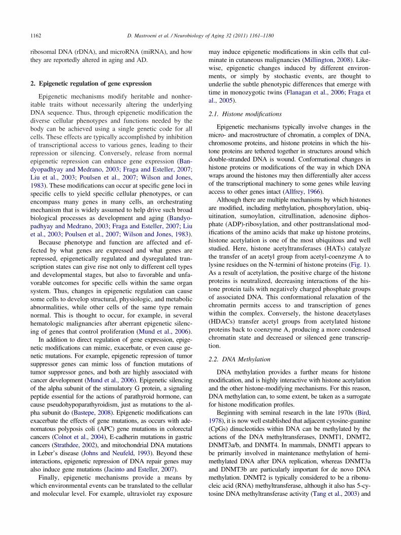
Although there are multiple mechanisms by which histonesare modified, including methylation, phosphorylation, ubiq-uitination, sumoylation, citrullination, adenosine diphos-phate (ADP)-ribosylation, and other posttranslational mod-ifications of the amino acids that make up histone proteins,histone acetylation is one of the most ubiquitous and wellstudied. Here, histone acetyltransferases (HATs) catalyzethe transfer of an acetyl group from acetyl-coenzyme A tolysine residues on the N-termini of histone proteins (Fig. 1).As a result of acetylation, the positive charge of the histoneproteins is neutralized, decreasing interactions of the his-tone protein tails with negatively charged phosphate groupsof associated DNA. This conformational relaxation of thechromatin permits access to and transcription of geneswithin the complex. Conversely, the histone deacetylases(HDACs) transfer acetyl groups from acetylated histoneproteins back to coenzyme A, producing a more condensedchromatin state and decreased or silenced gene transcrip-tion.
2.2. DNA Methylation
DNA methylation provides a further means for histonemodification, and is highly interactive with histone acetylationand the other histone-modifying mechanisms. For this reason,DNA methylation can, to some extent, be taken as a surrogatefor histone modification profiles.
Beginning with seminal research in the late 1970s (Bird,1978), it is now well established that adjacent cytosine-guanine(CpGs) dinucleotides within DNA can be methylated by theactions of the DNA methyltransferases, DNMT1, DNMT2,DNMT3a/b, and DNMT4. In mammals, DNMT1 appears tobe primarily involved in maintenance methylation of hemi-methylated DNA after DNA replication, whereas DNMT3aand DNMT3b are particularly important for de novo DNAmethylation. DNMT2 is typically considered to be a ribonu-cleic acid (RNA) methyltransferase, although it also has 5-cy-
tosine DNA methyltransferase activity (Tang et al., 2003) and
1163D. Mastroeni et al. / Neurobiology of Aging 32 (2011) 1161–1180
forms denaturant-resistant complexes with DNA (Dong et al.,2001). The methyl group that is transferred to cytosine by theDNMTs ultimately derives from methyltetrahydrofolatethrough its interactions with S-adenosylmethionine (SAM) in
Fig. 1. Simplified schematic of histone acetylation and DNA methylation.(blue cylinders) around which DNA is wrapped, is in a relaxed state, permin part, mediated by acetylation of histone tails (red rods) in which acetyl ghistone tails by histone acetyltransferases (HATs). (Bottom left) Withinmethylated. The methyl group ultimately derives from methyltetrahydrofolaS-adenosylmethionine (SAM) to the cytosine and incorporated into the geno(MBDs) and methylation complex proteins (MeCPs) (which may contain Maccess and repression of the gene. (Upper right) DNA methylation and histdeacetylases (HDACs) that transfer acetyl groups on the histone tail backstate characteristic of silenced or repressed genes.
the homocysteine-methionine cycle (Fig. 1).
Through these processes, approximately 70% of CpG di-nucleotides within the human genome are methylated. Al-though DNA methylation can take place at any CpG site,whether in coding or noncoding regions, previous studies have
left) In transcriptionally active genes the chromatin, made up of histonesanscriptional access to unwound DNA. This relaxed, euchromatin state is,green blocks) are transferred from acetyl-coenzyme A (acetyl-CoA) to theNA, the cytosines of adjacent C-G/G-C dinucleotides (CpGs) may benjunction with the methionine/homocysteine cycle, and is transferred fromDNA methyltransferases (DNMTs). CpG-methyl-binding-domain proteinsecome associated with methylated CpGs, further inhibiting transcriptional
difications are integrally linked, because MBDs and MeCPs attract histone. Histone deacetylation, in turn, promotes the condensed, heterochromatin
(Upperitting trroups (
the Dte in come byBDs) b
one moto CoA
often focused on CpG-rich stretches (CpG islands) within the
samicgmag
btayacnhssa(ntsp2bpgsDe
mimHcttmaM
stmRm1
padrMsthcCt
mt(H2roe(aeuamD5atctbf
2
Rc(rscdfap
1164 D. Mastroeni et al. / Neurobiology of Aging 32 (2011) 1161–1180
promoter region. Some 50,267 CpG islands exist in the humangenome, with 28,890 in simple repeat and low complexitysequences that are masked (Bandyopadhyay and Medrano,2003; Liu et al., 2003). Because CpG islands contain a highproportion of CpGs (and opportunities for CpG methylation),they are, perforce, among the most highly methylated regionsof the genome. However, it has recently been shown that thepercentage (rather than the absolute number) of methylatedCpGs is relatively low in conventional CpG islands, and isactually higher in promoters with intermediate CpG densities(Eckhardt et al., 2006; Weber et al., 2007). Moreover, tissue-pecific methylation patterns appear to be most pronounced nott CpG islands but at “CpG shores”, regions within approxi-ately 2 kb of CpG-enriched sequences. It has been suggested,
n fact, that methylation patterns of CpG shores are “suffi-iently conserved to completely discriminate tissue types re-ardless of species of origin” (Irizarry et al., 2009). Differentialethylation of CpG shores in brain compared with liver has
lso been found to be highly correlated with differences inene expression for these 2 organs (Irizarry et al., 2009).
Methylation of CpG sequences may alter gene expressiony inducing histone modifications that inhibit access of theranscriptional machinery (Bird and Wolffe, 1999; Zhang etl., 2002) (Fig. 1). One would expect, then, that highly meth-lated genes would be repressed, and that hypomethylation ofgene would lead to enhanced expression or overexpression
ompared with the normally repressed, methylated state. Someotable exceptions to these expected states have been found,owever. For example, the p16INK4a promoter is progres-ively hypermethylated with age (So et al., 2006), but expres-ion of the p16INK4a gene appears to increase with age (Kimnd Sharpless, 2006). More generally, Gius and colleaguesGius et al., 2004) found that chemical hypomethylation ofearly half the genes they surveyed resulted in silencing ratherhan upregulation. Indeed, altering the methylation status ofome CpG sites within a gene can be inconsequential com-ared with alterations at other sites (c.f. Murgatroyd et al.,009; Yakovlev et al., 2010). Such findings indicate that it wille important to follow up genome-wide DNA methylationrofiling, which can only survey a limited number of CpGs perene, with more detailed DNA methylation maps that includehores and flanking regions. In addition, even highly-detailedNA methylation profiles will require validation of functional
ffects at the gene expression and protein levels.A second, linked mechanism by which DNA methylation
ay modify gene expression is through methyl-CpG-bind-ng proteins (MeCPs) such as MeCP2. When bound toethylated DNA, MeCP2 has been shown to recruitDACs, which, as noted earlier, may then induce a more
ondensed chromatin state and decreased or silenced generanscription (Jones et al., 1998; Wade et al., 1998). Al-hough previous studies assumed that MeCP2 required aethylated chromatin substrate for direct binding (Lewis et
l., 1992), more recent research has demonstrated that
eCP2 can directly condense chromatin even in the ab- tence of DNA methylation, histone deacetylase activity, orhe cooperation of other transcriptional corepressors such asSin3A (Wade, 2001). Mutations of the MeCP2 gene causeett’s syndrome, with dysregulation of neural development,ental retardation, and motor dysfunction (Amir et al.,
999).MeCP1, a macromolecule made up of some 10 different
eptides, may also act as a mediator between DNA methyl-tion and histone acetylation, recognizing and binding to CpGinucleotides, recruiting HDACs, and inducing transcriptionalepression (Feng and Zhang, 2001). Unlike MeCP2, however,
eCP1 does not bind directly to methylated DNA, but to aingle methyl-CpG-binding domain protein, MBD-2. In addi-ion to inducing histone modifications, MBD-2-bound MeCP1elps maintain the DNA methylation status of CpGs by re-ruiting DNMT1. DNMT1 is then able to recognize and repairpGs that have lost methyl groups on one DNA strand but not
he other.Recent studies have also revealed a further modification to
ethylated CpGs that may make them even more inaccessibleo transcription. Here, the 5-methylcytosines are hydroxylatedoxidized) to form 5-hydroxymethylcytosine (Kriaucionis andeintz, 2009; Tahiliani et al., 2009; Valinluck and Sowers,007). Hydroxymethylated DNA has been observed in neu-ons (Kriaucionis and Heintz, 2009), and may occur as a resultf oxidative damage and/or the actions of specific oxidativenzymes, particularly Ten-Eleven-Translocation-1 (TET1)Tahiliani et al., 2009). Normally, 5-hydroxymethylcytosinesre stripped from genomic DNA by glycosylases of the basexcision repair (BER) system, permitting replacement withnmodified cytosines for subsequent de novo DNA methyl-tion by the DNMTs. When left intact, however, 5-hydroxy-ethylcytosines reportedly reduce the interaction of DNA withNA-binding proteins to an even greater extent than do-methylcytosines (Valinluck and Sowers, 2007; Valinluck etl., 2004). Because hydroxymethylated CpGs may go unde-ected by 5-methylcytosine antibodies, hydroxymethylationould lead to underreporting of DNA methylation, a possibilityhat the present authors are now investigating. Conversely,isulfite sequencing may not discriminate normally methylatedrom hydroxymethylated CpGs.
.3. RNA-related mechanisms
Epigenetic regulation also extends to mechanisms involvingNA such as microRNAs (miRNAs) and hereditable and cellycle-maintained silencing of a portion of the ribosomal RNArRNA) genes. Repeated rRNA transcripts from repeatedRNA genes (rDNA) provide a structure and primary catalyticite for the eukaryotic ribosome, wherein gene expressionulminates in protein synthesis. Different cell types exhibitifferent proportions of active rRNA genes, suggesting that theraction of rRNA gene copies may be altered in developmentnd differentiation. Epigenetic mechanisms are now known tolay important roles in this process by silencing rRNAs,
hereby providing a dynamic balance between active and in-
Sesc
nBcmm(taaint(tsds
1165D. Mastroeni et al. / Neurobiology of Aging 32 (2011) 1161–1180
active rRNAs. DNA methylation appears to be one of theseepigenetic mechanisms, and may be of particular importancegiven the unusually high frequency of CpG dinucleotideswithin the rRNA genes and their unusually high states of DNAmethylation. For example, two studies have shown a correla-tion between DNA methylation status and activity of rRNAgenes (Bird et al., 1981; Santoro and Grummt, 2001), andtreatment with 5-aza-2-deoxycytidine (azacytidine), a hypom-ethylating agent, enhances expression by rDNA genes (San-toro and Grummt, 2001). For an excellent and comprehensivesummary of rDNA methylation, as well as histone modifica-tions and chromatin remodeling, which also play key roles inepigenetic regulation of rDNA, the reader is referred to therecent review of McStay and Grummt (2008).
The study of miRNAs represents an additional, critical areafor epigenetics research. Often deriving from their own geneswith their own promoter and regulatory elements, approxi-mately 700–800 miRNAs have been identified in the humangenome. These small (�22 nucleotide) RNAs regulate geneexpression in a posttranscriptional manner by binding to theirtarget mRNAs, inhibiting translation or, less often, inducingcleavage of the mRNAs (reviewed in Yang et al., 2007).
3. Dynamic epigenetic regulation in adult neurons
3.1. Histone modifications
Histone modifications have been implicated in broad neu-robiological processes such as development of the central ner-vous system (CNS) (reviewed in MacDonald and Roskams,2009), posttraumatic stress disorders (Sokolova et al., 2006),childhood abuse/suicide (McGowan et al., 2009; Meaney et al.,2007), memory formation (Gupta et al., 2010), and addiction(Impey, 2007); specific physiologic processes such as neuronaldifferentiation (Kular et al., 2009), regulation of choline acetyl-transferase activity (Aizawa and Yamamuro, 2010), astrocyteglial-derived neurotrophic factor (GDNF) and brain-derivedneurotrophic factor (BDNF) transcription (Wu et al., 2008c),microglial apoptosis (Chen et al., 2007), and axon pathfinding(Zinovyeva et al., 2006); and various neurologic disorders,including Parkinson’s disease (Chen et al., 2007; Wu et al.,2008c), motor neuron disease (reviewed in Echaniz-Laguna etal., 2008), multiple sclerosis (reviewed in Gray and Dangond,2006), X-linked mental retardation (Tahiliani et al., 2007), andstroke/cerebral palsy (Meisel et al., 2006). All of these histone-related processes occur in the context of the CNS, and manyhave been reported to function as dynamic regulatory mecha-nisms in postmitotic neurons.
3.2. miRNA
Like histone modifications, miRNA has been implicated inboth broad and specific CNS processes and disorders. Forexample, miRNA-206 appears to promote neuromuscular syn-apse regeneration (Williams et al., 2009), and miR-329,miRNA-134, and miRNA-381, which are induced by neuronal
activity, have been suggested to be essential for activity-de- ppendent dendritic outgrowth of hippocampal neurons (Khu-dayberdiev et al., 2009). In turn, these and other miRNA-mediated mechanisms have been investigated in various CNSdisorders, including human immunodeficiency virus (HIV) de-mentia (Witwer et al., 2010), amyotrophic lateral sclerosis(Williams et al., 2009), Tourette’s syndrome (Abelson et al.,2005), AD (see section 5.2, below), and other neurologicconditions (reviewed in Maes et al., 2009).
Once again, these epigenetic processes have been demon-strated to occur in the context of postmitotic neurons. Indeed,it has been reported that the switch from a replicative state tothe postmitotic state of neurons may itself be controlled bymiRNA mechanisms (Yoo et al., 2009).
3.3. DNA methylation
Previously, DNA methylation reactions were consideredprimarily in the context of maintenance of the DNA methyl-ation pattern across cell divisions. Why neurons and otherpostmitotic cells should express DNA methylation markerssuch as DNMT3 was therefore unclear. Likewise, how longterm DNA methylation alterations might be accomplished andsustained in postmitotic cells was unknown. Recently, how-ever, new studies have shown that hypo- and hypermethylationare dynamic events that can occur within cells (Kangaspeska etal., 2008; Métivier et al., 2008), including postmitotic neurons(Levenson et al., 2006; Murgatroyd et al., 2009), on the scaleof tens of minutes. These findings open the door for longsuspected, dynamic epigenetic mechanisms that may help me-diate neuronal and synaptic plasticity (e.g., Arendt, 2005). Forexample, gene-specific hypomethylation of hippocampal neu-rons after DNMT inhibition blocks long term potentiation(Levenson et al., 2006) and fear conditioning (Miller and
weatt, 2007). Weaver et al. (2005) have suggested thatarly life events — specifically, maternal care — alter adulttress responses through sustained DNA methylationhanges in rat hippocampal neurons.
The role of DNA methylation in dynamic regulation ofeuronal activity and function began to emerge with studies byredy and colleagues (Bredy et al., 2003), and has been vividlyonfirmed by recent work linking early life stress to enduringolecular, physiologic, memory, and behavioral changes inice via epigenetic modifications to hypothalamic neurons
Murgatroyd et al., 2009). In particular, early exposure of miceo stress during the first 10 days of life has been found, as muchs a year later, to be associated with impaired step-downvoidance learning, sustained hyperactivity of the hypothalam-c-pituitary-adrenal axis, and corticosterone and pituitary adre-ocorticotropin prohormone hypersecretion. These effects, inurn, were elegantly traced to persistent arginine vasopressinAVP) overexpression by parvocellular neurons of the hypo-halamic paraventricular nucleus, and to hypomethylation ofpecific CpG sites within the AVP gene. Notably, age-depen-ent increases in AVP gene hypomethylation at multiple CpGites were observed in control mice in these studies, but hy-
omethylation of CpGs within a CpG island (CGI3) in the
1166 D. Mastroeni et al. / Neurobiology of Aging 32 (2011) 1161–1180
AVP enhancer region approximately 0.5 kb downstream fromthe AVP gene itself appeared to be the primary determinant ofAVP overexpression in early life stress mice. Further experi-ments went on to show that CGI3 hypomethylation was spe-cific to the paraventricular nucleus compared with the supraop-tic nucleus, and to provide a key mechanism for the epigeneticmodifications that were observed: CGI3 CpG sequences serveas preferential and selective DNA-binding sites for MeCP2.Phosphorylation of MeCP2 by calmodulin-dependent proteinkinase II as a result of neuronal membrane depolarizationdecreases MeCP2 occupancy of CGI3 CpGs, thereby enhanc-ing transcription (Murgatroyd et al., 2009). This landmarkstudy therefore shows that dynamic and long lasting DNAmethylation changes can and do occur in postmitotic neurons.
Similarly, Yakovlev et al. (2010) have traced age-depen-dent downregulation of caspase-3 production in rat brain tosignificantly lower levels of histone 3 acetylated Lys14 andhistone 4 acetylated Lys5, 8, 12, and 16, as well as to differ-ential methylation of specific CpG sites within the caspase-3promoter. These sites are in a region that is essential forcaspase-3 promoter activity, and correspond to predicted bind-ing sites for several transcription factors such as Ets-1 andEts-2 that are known to help control caspase-3 synthesis and toplay critical roles in neuronal differentiation, development, anddeath. Notably, Ets-1 and Ets-2 themselves did not show age-related decline in the study, highlighting the potential impor-tance of interactions between DNA methylation modificationsand transcription factor activity. That is, transcriptional controlof a gene by its transcription factors may be significantlyaltered by differential methylation of the binding sites for thosetranscription factors.
New research has also shown that, in addition to their rolesin DNA methylation, MBD-2 and DNMT3a/b may participatein dynamic demethylation processes. It has been reported, forexample, that transient coexpression of MBD-2 and methyl-ated promoters results in demethylation and activation of geneexpression, whereas knockdown of MBD-2 inhibits replica-tion-independent, active demethylation by valproate (reviewedin Szyf, 2009). Cyclical methylation/demethylation processesthat mediate bouts of active and inactive transcription overperiods of tens of minutes have also been demonstrated, andappear to help regulate the expression of multiple genes (Kan-gaspeska et al., 2008; Métivier et al., 2008). Here, DNMT3a/b,in concert with p68, bind and deaminate selective CpG sites,creating mismatches that are recognized by thymine-DNAglycosylase (TDG) and repaired by the BER. Because therepaired CpGs are no longer methylated, the local chromatinenvironment becomes poised for transcription. Following tran-scription, MBDs, MeCP2, and DNMTs are recruited and rem-ethylate the CpG sites. Transcription is therefore turned off orretarded. The cycle may then begin again with DNMT3-me-diated CpG deamination (Métivier et al., 2008). Althoughthese dynamic MBD and DNMT demethylating mechanismshave, as yet, been examined only in non-CNS cells, there is no
obvious reason why they should not occur in neurons. Simi-larly, a recent study has suggested that histone acetylation anddeacetylation, like DNA methylation and demethylation, aredynamic, rapid turnover processes that can poise genes fortranscription in peripheral cells (Clayton et al., 2006). As such,all these mechanisms may warrant significant attention in fu-ture neuroepigenetics research.
4. Epigenetic regulation of aging
Aging is universally considered to be one of the mostsalient risk factors for AD, with increasing risk for the disordercumulating until at least the ninth decade of life (Gao et al.,1998; Kukull et al., 2002). Why aging should be a risk factorfor AD (and other age-related disorders), however, is not wellunderstood, particularly at a mechanistic level. Potentially del-eterious changes in mitochondria/oxidative stress (Crouch etal., 2007), gonadotropins (Fuller et al., 2007), calcium (Thi-bault et al., 2007), glucocorticoids (Landfield et al., 2007),inflammation (Duenas-Gonzalez et al., 2008), trace metals(Brewer, 2007), insulin (Craft, 2005), cerebrovascular supply(Bailey et al., 2004), the cell cycle (Macaluso et al., 2005), A�(Selkoe, 2003), tau (Maeda et al., 2006), and hundreds tothousands of genes (Berchtold et al., 2008; Parachikova et al.,2007) occur both in aging and AD, but a coherent explanationfor why they occur and if their co-occurrence in aging and ADis coincidence or meaningful remains elusive.
DNA methylation and histone modifications have beenwidely implicated in the phenotypic alterations that occur dur-ing cellular senescence and the aging of various organisms(reviewed in Bandyopadhyay and Medrano, 2003; Fraga andEsteller, 2007; Liu et al., 2003; Poulsen et al., 2007; Wilsonand Jones, 1983), and may provide a link between aging andAD. Histone acetylation mechanisms, particularly those in-volving the Sir2 family of histone deacetylases, have beenlinked to aging and senescence in yeast and invertebrates(reviewed in Bandyopadhyay and Medrano, 2003), but haveyet to be investigated in mammals. By contrast, many studieshave reported a genome-wide tendency to DNA hypomethy-lation with age in multiple vertebrate organs, including brain,liver, small intestine mucosa, heart, and spleen; multiple celltypes, including fibroblasts and T lymphocytes; and multiplevertebrate species, including aging salmon, mice, rats, cows,and humans (Golbus et al., 1990; Richardson, 2002; Romanovand Vaniushin, 1980; Vanyushin et al., 1970, 1973; Wilson etal., 1987). Yu (2006) assayed human peripheral blood mono-nuclear cell DNA for the percentage of methylated to totalcytosines and observed a 3% per decade decrease from the firstto the tenth decade of life. In vitro, hypomethylation of humanand mouse fibroblasts cultured to senescence has also beenobserved (Wilson and Jones, 1983). Likewise, progressiveage-related decline in total genomic methylcytosine has beenreported in various organisms (Mays-Hoopes, 1989). BecauseDNMT1, which is responsible for maintaining DNA methyl-ation of CpG sites, is also progressively lost with age (Lopatina
et al., 2002), it has been speculated that progressive, age-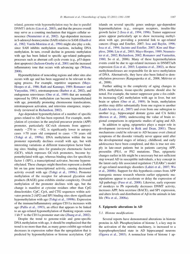
asmwpdi
c(pii(pfehampmcdvhoat1
Dtdp
ihggacIpt1eemoea
Dnibp(a(g
dmstappcstoetnA
1167D. Mastroeni et al. / Neurobiology of Aging 32 (2011) 1161–1180
related, genome-wide hypomethylation may be due to parallelDNMT1 deficits (Liu et al., 2003), and that the process overallmay serve as a counting mechanism that triggers cellular se-nescence (Neumeister et al., 2002). Age-dependent increasesin S-adenosyl-homocysteine (SAH) relative to SAM (Gharib etl., 1982; Varela-Moreiras et al., 1994) might also play a role,ince SAH inhibits methylation reactions, including DNAethylation. In turn, overall decline in genomic methylationith age has been linked to specific age-related pathogenicrocesses such as aberrant cell cycle events (e.g., p53-depen-ent apoptosis) (Jackson-Grusby et al., 2001) and the increasednflammatory tone that occurs with advancing age (Wilson,
2008).Hypomethylation of noncoding regions and other sites also
occurs with age and has been suggested to be relevant to theaging process. For example, repetitive sequences (Mays-Hoopes et al., 1986; Rath and Kanungo, 1989; Romanov andVanyushin, 1981), retrotransposons (Barbot et al., 2002), andendogenous retroviruses (Ono et al., 1989) that are normallyrepressed by DNA methylation can become hypomethylatedwith age, potentially promoting chromosome translocations,retrotransposon activation, and retrovirus emergence, respec-tively (reviewed in Richardson, 2002).
Age-dependent hypomethylation of a number of specificgenes related to AD has been reported. For example, meth-ylation of cytosines in the amyloid precursor protein (APP)promoter, particularly GC-rich elements from approxi-mately �270 to �182, is significantly lower in autopsyases �70 years old compared to cases �70 years oldTohgi et al., 1999a). DNA methylation within the tauromoter reportedly declines overall with age, but withnteresting variations at different transcription factor bind-ng sites: binding sites for granulocyte chemotactic factorGCF), which represses GC-rich promoters, become hy-omethylated with age, whereas binding sites for specificityactor 1 (SP1), a transcriptional activator, become hyperm-thylated. These changes might therefore represent a doubleit on tau gene transcriptional activity, causing decreasedctivity overall with age (Tohgi et al., 1999c). Promoterethylation of the receptor for advanced glycation end
roducts (RAGE) gene exhibits similar complexity. Overallethylation of the promoter declines with age, but the
hange is manifest at cytosine residues other than CpGinucleotides: CpC, CpA, and CTG sequences within acti-ator protein 2 (AP2) and SP1 binding sites show significantypomethylation with age (Tohgi et al., 1999b). Expressionf the immune/inflammatory antigen CD11a increases withge (Pallis et al., 1993), an effect that appears to be linkedo an age-related hypomethylation of flanking repeats somekb 5= to the CD11a promoter start site (Zhang et al., 2002).Despite the trend to genome-wide and gene-specific
NA methylation with age, it should be emphasized that therend is no more than that, as many genes exhibit age-relatedecreases in expression rather than the upregulation that is
redicted by hypomethylation or histone acetylation. CpGslands on several specific genes undergo age-dependentypermethylation (e.g., estrogen receptors, insulin-likerowth factor 2 (Issa et al., 1994, 1996). Tumor suppressorenes appear particularly apt to show increasing methyl-tion with age, providing a potential link to age-relatedancers (Fraga and Esteller, 2007; Issa and Baylin, 1996;ssa et al., 1994; Jacinto and Esteller, 2007; Kim and Shar-less, 2006; Liu et al., 2003; Mays-Hoopes, 1989; Neumeis-er et al., 2002; Richardson, 2002; Romanov and Vaniushin,980; So et al., 2006). Many of these hypermethylationvents could be due to age-related increases in DNMT3a/bxpression (Liu et al., 2003; Lopatina et al., 2002), as theseethyltransferases are responsible for de novo methylation
f DNA. Alternatively, they have also been linked to dem-thylation processes (Kangaspeska et al., 2008; Métivier etl., 2008).
Further adding to the complexity of aging changes inNA methylation, tissue-specific patterns should also beoted. For example, the tumor suppressor gene c-fos exhib-ts increasing CpG methylation with age in liver, but notrain or spleen (Ono et al., 1989). In brain, methylationrofiles may differ substantially from one region to anotherLadd-Acosta et al., 2007) and even from one subregion tonother (e.g., hippocampal dentate gyrus and CA fields)Brown et al., 2008), underscoring the value of brain re-ional comparisons in epigenetic studies of aging and AD.
In addition to aging, epigenetics plays a major role inevelopment (reviewed in Reik and Dean, 2001). Theseechanisms could be relevant to AD because overt clinical
ymptoms of the disorder virtually never appear until afterhe developmental stages of infancy, childhood, and earlydolescence have been completed, and this is true not sim-ly in late-onset patients but in patients carrying APP,resenilin (PS)1, or PS2 mutations. Thus, epigenetichanges earlier in life might be a necessary but not sufficienttep toward AD in susceptible individuals, a key concept inhe latent early-life associated regulation (“LEARn”) modelf age-related neurologic disorders (Lahiri et al., 2007; Wut al., 2008b). Support for this hypothesis comes from APPransgenic mouse research wherein earlier epigenetic ma-ipulations appear to accelerate or delay the expression of� pathology (Fuso et al., 2008). Likewise, early exposure
of monkeys to Pb reportedly decreases DNMT activity,increases APP, beta secretase (BACE), and SP1 expression,and alters levels and distribution of A� in the animals in latelife (Wu et al., 2008a).
5. Epigenetic alterations in AD
5.1. Histone modifications
Several reports have demonstrated alterations in histoneproteins in AD. Phosphorylation of histone 3, a key step inthe activation of the mitotic machinery, is increased to ahyperphosphorylated state in AD hippocampal neurons
(Ogawa et al., 2003). A nonnuclear form of histone 1 ap-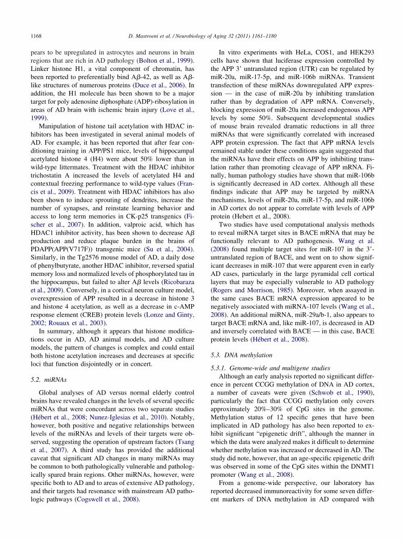
1168 D. Mastroeni et al. / Neurobiology of Aging 32 (2011) 1161–1180
pears to be upregulated in astrocytes and neurons in brainregions that are rich in AD pathology (Bolton et al., 1999).Linker histone H1, a vital component of chromatin, hasbeen reported to preferentially bind A�-42, as well as A�-like structures of numerous proteins (Duce et al., 2006). Inaddition, the H1 molecule has been shown to be a majortarget for poly adenosine diphosphate (ADP)-ribosylation inareas of AD brain with ischemic brain injury (Love et al.,1999).
Manipulation of histone tail acetylation with HDAC in-hibitors has been investigated in several animal models ofAD. For example, it has been reported that after fear con-ditioning training in APP/PS1 mice, levels of hippocampalacetylated histone 4 (H4) were about 50% lower than inwild-type littermates. Treatment with the HDAC inhibitortrichostatin A increased the levels of acetylated H4 andcontextual freezing performance to wild-type values (Fran-cis et al., 2009). Treatment with HDAC inhibitors has alsobeen shown to induce sprouting of dendrites, increase thenumber of synapses, and reinstate learning behavior andaccess to long term memories in CK-p25 transgenics (Fi-scher et al., 2007). In addition, valproic acid, which hasHDAC1 inhibitor activity, has been shown to decrease A�production and reduce plaque burden in the brains ofPDAPP(APP(V717F)) transgenic mice (Su et al., 2004).Similarly, in the Tg2576 mouse model of AD, a daily doseof phenylbutyrate, another HDAC inhibitor, reversed spatialmemory loss and normalized levels of phosphorylated tau inthe hippocampus, but failed to alter A� levels (Ricobarazaet al., 2009). Conversely, in a cortical neuron culture model,overexpression of APP resulted in a decrease in histone 3and histone 4 acetylation, as well as a decrease in c-AMPresponse element (CREB) protein levels (Lonze and Ginty,2002; Rouaux et al., 2003).
In summary, although it appears that histone modifica-tions occur in AD, AD animal models, and AD culturemodels, the pattern of changes is complex and could entailboth histone acetylation increases and decreases at specificloci that function disjointedly or in concert.
5.2. miRNAs
Global analyses of AD versus normal elderly controlbrains have revealed changes in the levels of several specificmiRNAs that were concordant across two separate studies(Hébert et al., 2008; Nunez-Iglesias et al., 2010). Notably,however, both positive and negative relationships betweenlevels of the miRNAs and levels of their targets were ob-served, suggesting the operation of upstream factors (Tsanget al., 2007). A third study has provided the additionalcaveat that significant AD changes in many miRNAs maybe common to both pathologically vulnerable and patholog-ically spared brain regions. Other miRNAs, however, werespecific both to AD and to areas of extensive AD pathology,and their targets had resonance with mainstream AD patho-
logic pathways (Cogswell et al., 2008).In vitro experiments with HeLa, COS1, and HEK293cells have shown that luciferase expression controlled bythe APP 3= untranslated region (UTR) can be regulated bymiR-20a, miR-17-5p, and miR-106b miRNAs. Transienttransfection of these miRNAs downregulated APP expres-sion — in the case of miR-20a by inhibiting translationrather than by degradation of APP mRNA. Conversely,blocking expression of miR-20a increased endogenous APPlevels by some 50%. Subsequent developmental studiesof mouse brain revealed dramatic reductions in all threemiRNAs that were significantly correlated with increasedAPP protein expression. The fact that APP mRNA levelsremained stable under these conditions again suggested thatthe miRNAs have their effects on APP by inhibiting trans-lation rather than promoting cleavage of APP mRNA. Fi-nally, human pathology studies have shown that miR-106bis significantly decreased in AD cortex. Although all thesefindings indicate that APP may be targeted by miRNAmechanisms, levels of miR-20a, miR-17-5p, and miR-106bin AD cortex do not appear to correlate with levels of APPprotein (Hebert et al., 2008).
Two studies have used computational analysis methodsto reveal miRNA target sites in BACE mRNA that may befunctionally relevant to AD pathogenesis. Wang et al.(2008) found multiple target sites for miR-107 in the 3=-untranslated region of BACE, and went on to show signif-icant decreases in miR-107 that were apparent even in earlyAD cases, particularly in the large pyramidal cell corticallayers that may be especially vulnerable to AD pathology(Rogers and Morrison, 1985). Moreover, when assayed inthe same cases BACE mRNA expression appeared to benegatively associated with miRNA-107 levels (Wang et al.,2008). An additional miRNA, miR-29a/b-1, also appears totarget BACE mRNA and, like miR-107, is decreased in ADand inversely correlated with BACE — in this case, BACEprotein levels (Hébert et al., 2008).
5.3. DNA methylation
5.3.1. Genome-wide and multigene studiesAlthough an early analysis reported no significant differ-
ence in percent CCGG methylation of DNA in AD cortex,a number of caveats were given (Schwob et al., 1990),particularly the fact that CCGG methylation only coversapproximately 20%–30% of CpG sites in the genome.Methylation status of 12 specific genes that have beenimplicated in AD pathology has also been reported to ex-hibit significant “epigenetic drift”, although the manner inwhich the data were analyzed makes it difficult to determinewhether methylation was increased or decreased in AD. Thestudy did note, however, that an age-specific epigenetic driftwas observed in some of the CpG sites within the DNMT1promoter (Wang et al., 2008).
From a genome-wide perspective, our laboratory hasreported decreased immunoreactivity for some seven differ-
ent markers of DNA methylation in AD compared with
oswda
maftt2apthtbiles(b(mmfSSeirds
5
m
Oe
e
hio
m2
pt(ly
1169D. Mastroeni et al. / Neurobiology of Aging 32 (2011) 1161–1180
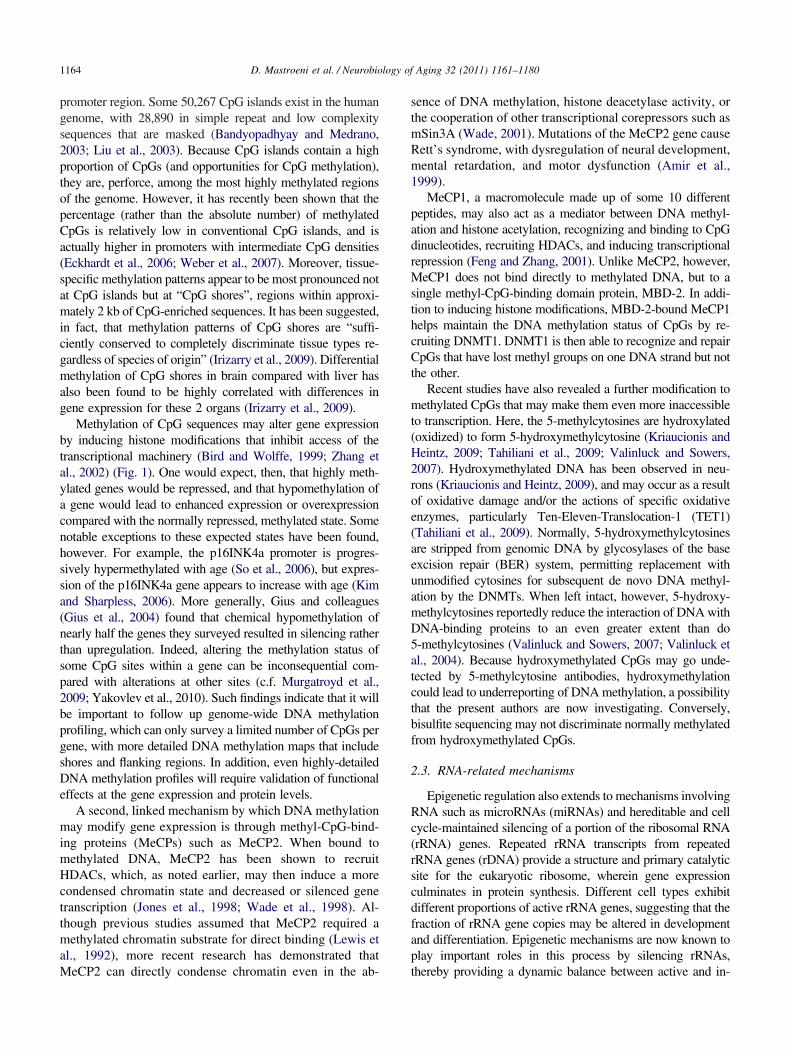
matched, nondemented elderly control (ND) cortical neu-rons and glia (Figs. 2 and 3), whereas no such changes werebserved in cerebellum, a brain region that is relativelypared in AD (Mastroeni et al., 2010). Highly similar resultsere subsequently obtained in a set of monozygotic twinsiscordant for AD (Mastroeni et al., 2009) (Fig. 4A), as wells in APP-overexpressing transgenic mice (Fig. 4B).
As previously noted, folate/methionine/homocysteineetabolism is critically linked to DNA methylation mech-
nisms. Folate deficiency in humans and in animal models,or example, typically results in global hypomethylationhat is at least partially reversible with folate supplementa-ion (reviewed in Choi and Friso, 2005; Choi and Mason,002; Fuso et al., 2005, 2008). Deficits in folate and alter-tions in the methionine/homocysteine cycle have been re-orted in aging and AD (reviewed in Smith, 2008), and mayherefore provide a basis for the tendency to genome-wideypomethylation summarized earlier in this review. Al-hough one prospective study failed to find an associationetween dietary folate, vitamin B12, or vitamin B6 withncident AD (Morris et al., 2006), CSF folate has nonethe-ess been reported to be significantly decreased in AD (Serott al., 2001), as has CSF and brain SAM and one of itsynthesizing enzymes, methionine S-adenosyltransferaseBottiglieri et al., 1990; Morrison et al., 1996). Increases inrain SAH (Kennedy et al., 2004) and plasma homocysteineClarke et al., 1998; Smith et al., 2008), which inhibit DNAethylation, have also been observed. Elevated plasma ho-ocysteine has been reported to be a significant risk factor
or AD in dementia-free cohorts of both the Framinghamtudy of Aging (Seshadri et al., 2002) and the Conselicetudy of Brain Aging (Ravaglia et al., 2005). In fact, dietaryffects of folate and homocysteine manipulation have beenmplicated in cognitive impairment generally, and in a wideange of neurologic conditions, including AD, Parkinson’sisease, depression, corticobasal degeneration, multipleclerosis, and frontotemporal dementia (Obeid et al., 2007).
.3.2. A�-related genesEpigenetic influences on A�-producing mutations have
long been suspected based on the heterogeneity of clinicalpresentation by patients who share mutations in the sameAPP, BACE, or PS1 genes — sometimes in identical pro-moter sites (Larner and Doran, 2006). Consistent with thisnotion, the APP (Davidson et al., 1992; Mani and Thakur,2006; West et al., 1995), BACE, and PS1 (Fuso et al., 2005)genes all contain manipulable, methylated CpG sites. A casestudy has reported complete demethylation of the APP genein an AD postmortem cortical sample, but not in similarsamples from a normal control or Pick’s disease patient(West et al., 1995). In vitro, expression of the BACE andPS1 genes is enhanced after folate deprivation-induced hy-pomethylation, and restored to normal when folate depriva-tion is accompanied by SAM supplementation. Expressionof TACE, ADAM10, and APP was unaffected by these
manipulations (Fuso et al., 2005). In vivo, exposure of cAPP-overexpressing transgenic mice to a folate/B12/B6-de-ficient diet is associated with enhanced SAH relative toSAM, as well as PS1 and BACE upregulation, enhanced A�deposition, and an accelerated appearance of intraneuronalA� and cognitive deficits (although the latter was quite
odest) (Fuso et al., 2008).Finally, it has been demonstrated that A� itself may
induce genome-wide hypomethylation in murine cerebralendothelial cell cultures while, at the same time, causingspecific hypermethylation and repression of the gene forneprilysin, an A� degrading enzyme (Chen et al., 2009).
ur laboratory has replicated the global hypomethylatingffects of A� in human SK-N-BE2 neuroblastoma cells, and
extended the results to show hypomethylation (as well asseveral instances of hypermethylation) of specific CpG is-lands in the BACE (Fig. 5) and caspase-3 genes (Grover etal., unpublished). Together with hypermethylation of nepri-lysin, these effects suggest the potential for a vicious cyclein which A�-induced methylation changes feed back tonhance A� production, further methylation changes, and
further A� production. Moreover, if the overall trend toypomethylation after A� exposure were to functionallympact other key AD genes, additional synergisms mightccur. For example, tumor necrosis factor (TNF)-� (Wil-
son, 2008) and caspase-3 are upregulated when hypomethy-lated (Müerköster et al., 2008), and increased levels ofTNF-� (Janelsins et al., 2008; McAlpine et al., 2009; Som-
er et al., 2009) and caspases (Xie et al., 2008; Xiong et al.,008) enhance A� expression, potentially generating new
vicious cycles.
5.3.3. TauMethylation mechanisms with respect to tau and neuro-
fibrillary tangle formation have also been explored. Aspreviously noted, in normal adults the AP2 binding site ofthe tau promoter is not methylated, but the SP1 and GCFbinding sites are. SP1, a transcriptional activator site, isincreasingly methylated and GCF, a promoter repressor site,is increasingly demethylated with age, suggesting an overalldownregulation of tau gene expression (Tohgi et al., 1999c).Although a corresponding age-related decrease in normaltau protein, particularly in frontal cortex and hippocampus,has been reported, there was no correlation with the modestneurofibrillary tangle pathology in the same subjects (Mu-kaetova-Ladinska et al., 1996).
Tau phosphorylation mechanisms are, however, subjectto cytoplasmic methylation reactions, and have been thesubject of several recent reports. Our studies, for example,revealed colocalized immunoreactivity for the methyl bind-ing complex component p66� (as well as HDAC1) withaired helical filament 1 (PHF1)-positive neurofibrillaryangles (Mastroeni et al., 2010). Protein phosphatase 2APP2A) is an enzyme that can dephosphorylate phosphory-ated tau, an action that may be potently activated by meth-lation of the PP2A catalytic subunit at its L309 site. In N2a
ultures carrying the APP Swedish mutation (APPswe) and
1170 D. Mastroeni et al. / Neurobiology of Aging 32 (2011) 1161–1180
Fig. 2. DNA methylation markers in Alzheimer’s disease (AD) and elderly control (ND) cortex. (A) Typical immunoreactivity for 5-methylcytosine, a globalmarker of DNA methylation, in AD and ND entorhinal cortex (from Mastroeni et al., 2008, with permission). Cases were well matched for age, gender, andpostmortem intervals, which were all less than 3 hours, 15 minutes. Shaded bars represent means for different cases. Although glia and virtually all typesof neurons exhibit 5-methylcytosine immunoreactivity, layer II “island” neurons, among the most vulnerable to AD pathology, exhibit particularly intensestaining in ND cases, as shown in the upper left micrograph at low power. Such staining is weak to absent in AD cases (upper right micrograph). High powermicrographs show the expected nuclear localization of immunoreactivity. Far right panel shows counts of immunoreactive neurons per total neurons per field.Normalizing to total neurons is important, as it helps to demonstrate loss of methylation within cell nuclei rather than loss of the methylated cell population
itself. The significant decrement in AD (p � 0.001) is typical of dozens of AD and ND cases examined, with little to no overlap in any case. (B)
et
gtH
5
repISCMNSsc�tte
hoi
s
a
5
yhryqntcmro2
1171D. Mastroeni et al. / Neurobiology of Aging 32 (2011) 1161–1180
in APPswe/PS1 transgenic mice, levels of demethylatedPP2A at L309 were significantly increased, correspondingwith increases in tau phosphorylation at the tau-1 and PHF1sites. Treatment with A�25-35 led to demethylation andnhanced tau phosphorylation (Zhou et al., 2008). Likereatment with A�25-35, exposure of rodent primary neuron
cultures to methotrexate, a folate antagonist, also has beenreported to result in demethylation of PP2A, with attendantenhancement of tau phosphorylation (as well as upregula-tion of APP and BACE) (Yoon et al., 2007). Consistent withall these findings, injections of homocysteine into rats for 2weeks yielded decreased PP2A L309 methylation and PP2Aactivity, effects that were reversed by simultaneous admin-istration of folate and vitamin B12. Hippocampal samplesfrom the rats and from AD patients exhibited immunohis-tochemical colocalization of demethylated, but not methyl-ated PP2A with hyperphosphorylated tau (Zhang et al.,2008).
5.3.4. Aberrant cell cycle/apoptosisA wide range of evidence suggests that attempted or
aberrant re-entry into the cell cycle and/or apoptosis ofneurons may be a common neurodegenerative mechanismin AD (reviewed in Neve and Mcphie, 2006). Many of thecritical components of the cell cycle and apoptosis pathwaysare upregulated in degenerating AD neurons, and are subjectto regulation by DNA methylation, including the P16, P21,P27, P53, RB1 (Moreira et al., 2009), cyclin B2 (Tschöpand Engeland, 2007), alternate open reading frame (ARF)protein product (Robertson and Jones, 1998), caspase 1 (Jeeet al., 2005), caspase 3, caspase 7, caspase 8, and caspase 9(Müerköster et al., 2008) genes. Hypomethylation of thesegenes would be expected to promote aberrant cell cycleevents, and global hypomethylation has been reported tooccur in cells as they move from the G(0) stage character-istic of postmitotic neurons to the G(1) stage characteristicof cell cycle re-entry (Brown et al., 2007). Indirect supportfor the role of hypomethylation in aberrant cell cycle eventsis provided by studies demonstrating apoptosis of culturedneurons when exposed to high homocysteine levels (Ho etal., 2002). Although many other biologic effects are possi-ble, such treatment is known to hypomethylate DNA (Reyn-olds, 2006), and concurrent treatment with SAM antago-nized the apoptotic effect (Ho et al., 2002). Interactions ofthe histone acetyltransferase Tip60 with the �-secretase-enerated APP C-terminal fragment APP-CT58, whichranslocates to the nucleus, also lead to apoptosis of human4 neuroglioma cells (Kinoshita et al., 2002).
Representative immunoreactivity and cell counts for various methyl-cytoSignificant AD decrements (p � 0.05) were observed with all the markersthese and other methylation markers exhibit immunoreactivity at approp
immunohistochemistry, suggesting that the latter are not due to cross-reactivity w.3.5. InflammationKey genes at almost every level of the inflammatory
esponse appear to be subject to DNA methylation influ-nces. A highly abbreviated list of examples includes com-lement C3, factor B, interleukin (IL)-1�, IL-1�, IL-2, IL-4,L-5, IL-6, IL-13, TNF-�, TNFRI, interferon (INF)-�,OCS, S100A2, the chemokine receptors CXCR4 andCR7, clusterin (ApoJ), and iNOS (Buslei et al., 2006;ejía et al., 1995; Mi and Zeng, 2008; Mori et al., 2005;ile et al., 2008; Parker et al., 2008; Pieper et al., 2008;uuronen et al., 2007; Van Panhuys et al., 2008; Van Riet-choten et al., 2008). The TNF-� promoter, for example,ontains 12 CpG-rich sites. Two of those sites (�304 and205) are hypomethylated on exposure of macrophages to
he classic inflammatory stimulus lipopolysaccharide, andheir extent of hypomethylation is correlated with increasingxpression of TNF-� (Wilson, 2008). Likewise, a single
CpG site in the IL-6 promoter (�1181) is significantlyhypomethylated in rheumatoid arthritis and this hypomethy-lation is correlated with IL-6 mRNA levels (Nile et al.,2008). Although virtually none of the many factors in in-flammation has been investigated with respect to its DNAmethylation status in AD, this could be a fertile area forinvestigation given the heightened expression of these mol-ecules in the disorder. A recent Parkinson’s disease study,for example, has demonstrated that the TNF-� promoter isypomethylated in the substantia nigra relative to severalther brain regions. Because DNA methylation of CpG sitesn the TNF-� promoter inhibits SP1 and AP2 transcription
factor binding and decreases TNF-� expression, it has beenpeculated that nigral TNF-� hypomethylation might ex-
plain the heightened vulnerability of nigral dopamine neu-rons to TNF-�-mediated inflammatory reactions (Pieper etl., 2008).
.3.6. ApolipoproteinsAgain, no specific studies have been done on the meth-
lation status of apolipoprotein E (ApoE) in AD. Notably,owever, Wang and colleagues (Wang et al., 2008) haveeported that although the ApoE promoter is poorly meth-lated, the ApoE �4 allele contains methylated CpG se-uences that are not extant in the �2 or �3 alleles. Becauseot all �4 carriers develop AD, it would be of great interesto know if methylation status at �4 CpG sites is altered in �4arriers who develop (or do not develop) AD. Similarly, itight be useful to determine whether the significant but
elatively low penetrance of ApoJ (clusterin) single nucle-tide polymorphisms (SNPs) to AD risk (Harold et al.,009; Lambert et al., 2009) might be explained by methyl-
anine-binding protein (MeCP)1 components in AD and ND neocortex.in with little to no overlap. (C) Western blots (normalized to �-actin) forolecular weights, with AD/ND differences similar to those observed by
sine-gu— aga
riate m
ith other antigens.
W
1172 D. Mastroeni et al. / Neurobiology of Aging 32 (2011) 1161–1180
ation changes, because the ApoJ promoter is rich in CpGsites and expression is increased on hypomethylation (Suu-ronen et al., 2007).
6. Conclusions
Global epigenetic changes, acting on a wide range ofgenes and biological pathways, appear to help orchestratethe cellular alterations that drive development, aging, and,in some cases, disease. Likewise, global epigenetic changeshave been observed in pathologically vulnerable regions of
Fig. 3. Immunoreactivity for DNA methyltransferase (DNMT)1, the mostactivity and cell counts (p � 0.001) in Alzheimer’s disease (AD) and elde
estern blots (normalized to �-actin standards) show immunoreactive badecrement in AD cases.
the AD brain, and key genes in virtually every mainstream
pathologic pathway in AD are known to be labile to suchchanges. The ability of epigenetic mechanisms to initiate anextremely wide range of pathogenic responses — an orches-trating capacity perhaps equaled only by transcription fac-tors (which themselves often work together with epigeneticmechanisms to direct expression in specific sets of genes)(e.g., Agrawal et al., 2007; Ivascu et al., 2007; Yakovlev etal., 2010) — provides a relatively unique integrative frame-work for the diverse genetic factors and multifactorial pa-thology of AD, including A�, tau, inflammation, mitochon-
nt methyltransferase in adult mammals. (A) Typical DNMT1 immunore-trol (ND) neocortex. Shaded bars represent means for different cases. (B)appropriate molecular weights for DNMT1 and a significant (p � 0.01)
prevalerly connds at
drial metabolism, oxidative stress, and aberrant cell cycle/

1173D. Mastroeni et al. / Neurobiology of Aging 32 (2011) 1161–1180
apoptosis events. Moreover, the epigenetic modificationsthat have been reported in AD, particularly with respect toDNA methylation, typically resonate with similar trends inaging, and may therefore help explain not only the patho-logic complexity of AD, but also the particular salience ofaging as an AD risk factor.
Finally, whereas the field of epigenetics has previouslyemphasized mechanisms for preserving epigenetic profilesacross generations of dividing cells, there is now ampleprecedent for active, dynamic epigenetic alterations in post-mitotic cells, including neurons, that play important roles in
Fig. 4. Decreased overall DNA methylation in monozygotic twins discordaimmunoreactivity) in an AD monozygotic twin compared with his norm(Mastroeni et al., 2009) (courtesy of PLoS One). (B) Similar findings have rmice compared with their wild type littermates.
neuroendocrine, learning and memory, and apoptotic pro-
cesses. These latter, landmark studies provide the tools forsubsequent explorations of how the epigenetic modifica-tions that have already been reported in AD occur, as wellas a mechanistic underpinning for the AD genome-widemethylation profiling that is now in progress.
7. Future directions
At the level of basic research, DNA methylation profiling ofaging and AD subjects is eagerly awaited in order to developa better portrait of the normal methylation status of all genes
lzheimer’s disease (AD). (A) Global hypomethylation (5-methylcytosineling at low (upper micrographs) and high (bottom micrographs) powerbeen observed by our group in amyloid precursor protein (APP) transgenic
nt for Aal sib
ecently
across the AD genome, how that status may change in AD, and

pG site
1174 D. Mastroeni et al. / Neurobiology of Aging 32 (2011) 1161–1180
whether or not such changes implicate AD-related proteins andpathogenetic processes. These studies would be especially sig-nificant if they were conducted in tandem with genome-widegene expression arrays because the experiments would thenprovide validation of the functional effects of DNA methyl-ation changes on gene expression. In addition, knowing themethylation states of genes containing putative AD SNPscould be useful. Many such SNPs, for example, remain con-troversial because their odds ratios for disease risk consistentlyhover at the statistical edges of significance or they fail toreplicate in some studies but not others. Epigenetic regulationof the SNP genes could account for this variability. For exam-ple, a gain or decrease of function SNP could be compensatedfor by epigenetic downregulation or upregulation, respectively,of the gene’s expression, so that some carriers might in effectpossess the SNP with relative impunity. Finally, as valuable aslarge-scale epigenetic profiling of the AD genome will be, itwill still not tell us why or how the profiles were altered, norwill it give us a detailed profile of each gene. Genome-widemethylation profiling is presently only able to sample a few ofthe CpG-rich sites within each gene, although the technology israpidly expanding. Because both hyper- and hypomethylationcan occur at different CpG sites in the same gene, with one butnot the other causing functional changes in gene expression(e.g., Murgatroyd et al., 2009), follow-up studies giving de-tailed methylation maps of AD-relevant genes and concomi-tant changes in their expression will be essential. These sameconsiderations may also apply to other neurologic conditionssuch as schizophrenia and bipolar disorders, where epigeneticmechanisms are being pursued (reviewed in Pidsley and Mill,2011).
Of course, to hypothesize that epigenetic changes play arole in brain aging, AD, and other neurologic disorders stillbegs the question of what causes the epigenetic changes? Theenvironment that cells and organisms are exposed to can havea profound influence on epigenetic mechanisms (Smith et al.,2008; Waterland and Michels, 2007), but simple stochastic
Fig. 5. MethylMiner methylation profile of selected flanking and initial BACto 10 �M A�42. MethylMiner™ Methylated DNA Enrichment Kits (Invstranded DNA based on cytosine-phosphate-guanine (CpG) methylation dpoorly methylated regions exhibited hypermethylation (Grover et al., unputhe functional relevance of methylation modifications at these and other C
processes may do so as well (Jaenisch and Bird, 2003).
Whether as environmentally driven or randomly occurringevents, however, the probability of epigenetic modificationsmust logically increase with time, and increased time is pre-cisely what the advanced ages reached by many human beingsmay afford. Some of these modifications may be inconsequen-tial, depending on the CpG site, the gene, or the organ. Forexample, inadvertent upregulation of an A�-synthesizing genemight have little to no impact on a muscle cell, whereas itcould be highly significant in a pyramidal neuron. Similarly,the brain lacks several major defenses against inflammatoryattack (Gasque et al., 2002; Zanjani et al., 2005), so that theinadvertent upregulation of a proinflammatory gene might beuniquely problematic there. Thus, the origin and organ-specificconsequences of epigenetic modifications are important con-siderations for AD epigenetic studies, and will continue to becritical targets for AD basic research into epigenetic mecha-nisms.
At the clinical level, the initiation of AD trials with folate/Bvitamins/SAM may constitute one means of testing an epige-netic orchestration hypothesis of AD, although it is becomingincreasingly evident that reversing the course of human ADwith any treatment may be an overambitious goal. For exam-ple, in a recent trial B vitamin treatment significantly slowedcognitive decline in mild AD, but was without effect in moreadvanced cases (Aisen et al., 2008). A trial in mild cognitiveimpairment patients might therefore be of great interest. Asecond impediment to successful treatment of epigenetic de-fects may be achieving sufficiently high levels of epigenetictherapeutics not only within cells, but within neuronal nuclei.Moreover, the many different biochemical pathways impactedby epigenetic mechanisms may make targeting specific diseaseprocesses difficult. Beyond folate and other such approaches,cancer chemotherapeutics that are directed at epigenetic mech-anisms are available, but would need to be considered care-fully. DNA demethylating agents such as 5-azacytidine anddecitabine, for example, might actually prove harmful in ADgiven the profound global hypomethylation of AD neurons
oter sites after exposure of differentiated SK-N-BE(2) neuron-like cultures, Carlsbad, CA, USA) were employed to enrich and fractionate double-The highly methylated region showed significant hypomethylation while). Correlations with amyloid � peptide (A�) production can help establishs within the BACE gene.
E promitrogenensity.blished
(Mastroeni et al., 2009, 2010). HDAC inhibitors such as val-

1175D. Mastroeni et al. / Neurobiology of Aging 32 (2011) 1161–1180
proate, by contrast, might counter many of the epigeneticchanges that have been reported in AD, and have, in fact, beenused successfully to improve cognitive outcome measures inAD transgenic mouse models (Fischer et al., 2007; Francis etal., 2009; Ricobaraza et al., 2009; Su et al., 2004). Treatmenttrials in human AD patients, however, have not been particu-larly encouraging, perhaps due to somnolence, agitation, andother side effects of the drug (Herrman et al., 2007; Profennoet al., 2005) or to lack of specificity of the drug to epigeneticmechanisms alone.
With respect to the development of new treatments for AD,a direct role for epigenetics would be the design and applica-tion of epigenetic therapeutics that have appropriate effects onspecific epigenetic mechanisms in specific genes or sets ofgenes. As we have emphasized throughout, however, one ofthe defining hallmarks of epigenetic mechanisms is their abilityto exert effects over many genes, and, accordingly, virtually allpresent epigenetic therapeutics also exert their effects overmany genes. Unless a broad modifier such as an HDAC in-hibitor can be found that just happens to impact the right genes,while sparing significant effects in others, the specificity re-quirements of an AD epigenetic therapeutic will be challeng-ing. Nonetheless, elucidating specific genes that undergo sig-nificant epigenetic alterations in AD — as is now in progressin our laboratory and elsewhere — could, at the very least, helpdirect our attention to the most salient pathogenic elements ofthe disorder and to more conventional (e.g., agonist/antagonist)approaches to the protein products of the epigenetically-mod-ified genes.
Disclosure statement
The authors state that they have no actual or potentialconflict of interest that could inappropriately influence thiswork.
Acknowledgements
Preparation of this review was supported by NIA AGO-7367-19 (JR), NIA AG 036400 (PC), and the Arizona Alz-heimer’s Disease Consortium (JR).
References
Abelson, J.F., Kwan, K.Y., O’Roak, B.J., Baek, D.Y., Stillman, A.A.,Morgan, T.M., Mathews, C.A., Pauls, D.L., Rasin, M.R., Gunel, M.,Davis, N.R., Ercan-Sencicek, A.G., Guez, D.H., Spertus, J.A., Leck-man, J.F., Dure, L.S., Kurlan, R., Singer, H.S., Gilbert, D.L., Farhi, A.,State, M.W., Lifton, R.P., Sestan, N., State, M.W., 2005. Sequencevariants in SLITRK1 are associated with Tourette’s syndrome. Science310, 317–320.
Agrawal, A., Murphy, R.F., Agrawal, D.K., 2007. DNA methylation inbreast and colorectal cancers. Mod. Pathol. 20, 711–721.
Aisen, P.S., Schneider, L.S., Sano, M., Diaz-Arrastia, R., Van Dyck, C.H.,Weiner, M.F., Bottiglieri, T., Jin, S., Stokes, K.T., Thomas, R.G., Thal,L.J., Alzheimer Disease Cooperative Study, 2008. High-dose B vitaminsupplementation and cognitive decline in Alzheimer disease: a random-
ized controlled trial. JAMA 300, 1774–1783.Aizawa, S., Yamamuro, Y., 2010. Involvement of histone acetylation in theregulation of choline acetyltransferase gene in NG108-15 neuronalcells. Neurochem. Int. 56, 627.
Allfrey, V.G., 1966. Structural modifications of histones and their possiblerole in the regulation of ribonucleic acid synthesis. Proc. Can. CancerConf. 6, 313–335.
Amir, R.E., Van Den Veyver, I.B., Wan, M., Tran, C.Q., Francke, U.,Zoghbi, H.Y., 1999. Rett syndrome is caused by mutations in X-linkedMECP2, encoding methyl-CpG-binding protein. Nat. Genet. 2, 23,185–188.
Arendt, T., 2005. Alzheimer’s disease as a disorder of dynamic brainself-organization. Prog. Brain Res. 147, 355–378.
Bailey, T.L., Rivara, C.B., Rocher, A.B., Hof, P.R., 2004. The nature andeffects of cortical microvascular pathology in aging and Alzheimer’sdisease. Neurol. Res. 26, 573–578.
Bandyopadhyay, D., Medrano, E.E., 2003. The emerging role of epigenet-ics in cellular and organismal aging. Exp. Gerontol. 38, 1299–1307.
Barbot, W., Dupressoir, A., Lazar, V., Heidmann, T., 2002. Epigeneticregulation of an IAP retrotransposon in the aging mouse: progressivedemethylation and de-silencing of the element by its repetitive induc-tion. Nucleic Acids Res. 30, 2365–2373.
Bastepe, M., 2008. The GNAS locus and pseudohypoparathyroidism. Adv.Exp. Med. Biol. 626, 27–40.
Berchtold, N.C., Cribbs, D.H., Coleman, P.D., Rogers, J., Head, E., Kim,R., Beach, T., Miller, C., Troncoso, J., Trojanowski, J.Q., Zielke, H.R.,Cotman, C.W., 2008. Gene expression changes in the course of normalbrain aging are sexually dimorphic. Proc. Natl. Acad. Sci. U. S. A. 105,15605–15610.
Bird, A.P., 1978. The occurrence and transmission of a pattern of DNAmethylation in Xenopus laevis ribosomal DNA. Philos. Trans. R. Soc.Lond. B Biol. Sci. 283, 325–327.
Bird, A.P., Taggart, M., Macleod, D., 1981. Loss of rDNA methylationaccompanies the onset of ribosomal gene activity in early developmentof X. laevis. Cell. 26, 381–90.
Bird, A.P., Wolffe, A.P., 1999. Methylation-induced repression--belts,braces, and chromatin. Cell 99, 451–454.
Bolton, S.J., Russelakis-Carneiro, M., Betmouni, S., Perry, V.H., 1999.Non-nuclear histone H1 is upregulated in neurones and astrocytes inprion and Alzheimer’s diseases but not in acute neurodegeneration.Neuropathol. Appl. Neurobiol. 25, 425–432.
Bottiglieri, T., Godfrey, P., Flynn, T., Carney, M.W., Toone, B.K., Reyn-olds, E.H., 1990. Cerebrospinal fluid S-adenosylmethionine in depres-sion and dementia: effects of treatment with parenteral and oral S-adenosylmethionine. J. Neurol. Neurosurg. Psychiatry 53, 1096–1098.
Bredy, T., Grant, R., Champagne, D., Meaney, M., 2003. Maternal careInfluences neuronal survival in the hippocampus of the rat. Eur. J. Neu-rosci. 10, 2903–2909.
Brewer, G.J., 2007. Iron and copper toxicity in diseases of aging, partic-ularly atherosclerosis and Alzheimer’s disease. Exp. Biol. Med. 232,323–335.
Brown, S.E., Fraga, M.F., Weaver, I.C., Berdasco, M., Szyf, M., 2007.Variations in DNA methylation patterns during the cell cycle of HeLacells. Epigenetics 2, 54–65.
Brown, S.E., Weaver, I.C., Meaney, M.J., Szyf, M., 2008. Regional-specific global cytosine methylation and DNA methyltransferase ex-pression in the adult rat hippocampus. Neurosci. Lett. 440, 49–53.
Buslei, R., Kreutzer, J., Hofmann, B., Schmidt, V., Siebzehnrübl, F.,Hahnen, E., Eyupoglu, I.Y., Fahlbusch, R., Blümcke, I., 2006. Abun-dant hypermethylation of SOCS-1 in clinically silent pituitary adeno-mas. Acta Neuropathol. 111, 264–271.
Chen, K.L., Wang, S.S., Yang, Y.Y., Yuan, R.Y., Chen, R.M., Hu, C.J.,2009. The epigenetic effects of amyloid-beta(1–40) on global DNAand neprilysin genes in murine cerebral endothelial cells. Biochem.Biophys. Res. Commun. 378, 57–61.
Chen, P.S., Wang, C.C., Bortner, C.D., Peng, G.S., Wu, X., Pang, H., Lu,
R.B., Gean, P.W., Chuang, D.M., Hong, J.S., 2007. Valproic acid and
1176 D. Mastroeni et al. / Neurobiology of Aging 32 (2011) 1161–1180
other histone deacetylase inhibitors induce microglial apoptosis andattenuate lipopolysaccharide-induced dopaminergic neurotoxicity.Neuroscience 149, 203–212.
Choi, S.W., Friso, S., 2005. Interactions between folate and aging forcarcinogenesis. Clin. Chem. Lab. Med. 43, 1151–1157.
Choi, S.W., Mason, J.B., 2002. Folate status: effects on pathways ofcolorectal carcinogenesis. J. Nutr. 132, 2413S–2418S.
Clarke, R., Smith, A.D., Jobst, K.A., Refsum, H., Sutton, L., Ueland, P.M.,1998. Folate, vitamin B12, and serum total homocysteine levels inconfirmed Alzheimer disease. Arch. Neurol. 55, 1449–1455.
Clayton, A.L., Hazzalin, C.A., Mahadevan, L.C., 2006. Enhanced histoneacetylation and transcription: a dynamic perspective. Mol. Cell 23,289–296.
Cogswell, J.P., Ward, J., Taylor, I.A., Waters, M., Shi, Y., Cannon, B.,Kelnar, K., Kemppainen, J., Brown, D., Chen, C., Prinjha, R.K., Rich-ardson, J.C., Saunders, A.M., Roses, A.D., Richards, C.A., 2008. Iden-tification of miRNA changes in Alzheimer’s disease brain and CSFyields putative biomarkers and insights into disease pathways. J. Alz-heimers Dis. 14, 27–41.
Colnot, S., Niwa-Kawakita, M., Hamard, G., Godard, C., Le Plenier, S.,Houbron, C., Romagnolo, B., Berrebi, D., Giovannini, M., Perret, C.,2004. Colorectal cancers in a new mouse model of familial adenoma-tous polyposis: influence of genetic and environmental modifiers. Lab.Invest. 84, 1619–1630.
Craft, S., 2005. Insulin resistance syndrome and Alzheimer’s disease: age-and obesity-related effects on memory, amyloid, and inflammation.Neurobiol. Aging 26 Suppl 1, 65–69.
Crouch, P.J., Cimdins, K., Duce, J.A., Bush, A.I., Trounce, I.A., 2007.Mitochondria in aging and Alzheimer’s disease. Rejuvenation Res. 10,349–357.
Davidson, J.S., West, R.L., Kotikalapudi, P., Maroun, L.E., 1992. Se-quence and methylation in the beta/A4 region of the rabbit amyloidprecursor protein gene. Biochem. Biophys. Res. Commun. 188, 905–911.
Dong, A., Yoder, J.A., Zhang, X., Zhou, L., Bestor, T.H., Cheng, X., 2001.Structure of human DNMT2, an enigmatic DNA methyltransferasehomolog that displays denaturant-resistant binding to DNA. NucleicAcids Res. 29, 439–448.
Duce, J.A., Smith, D.P., Blake, R.E., Crouch, P.J., Li, Q.X., Masters, C.L.,Trounce, I.A., 2006. Linker histone H1 binds to disease associatedamyloid-like fibrils. J. Mol. Biol. 361, 493–505.
Duenas-Gonzalez, A., Candelaria, M., Perez-Plascencia, C., Perez-Carde-nas, E., De La Cruz-Hernandez, E., Herrera, L.A., 2008. Valproic acidas epigenetic cancer drug: preclinical, clinical and transcriptional ef-fects on solid tumors. Cancer Treat. Rev. 34, 206–222.
Echaniz-Laguna, A., Bousiges, O., Loeffler, J.P., Boutillier, A.L., 2008.Histone deacetylase inhibitors: therapeutic agents and research tools fordeciphering motor neuron diseases. Curr. Med. Chem. 15, 1263–1273.
Eckhardt, F., Lewin, J., Cortese, R., Rakyan, V.K., Attwood, J., Burger,M., Burton, J., Cox, T.V., Davies, R., Down, T.A., Haefliger, C.,Horton, R., Howe, K., Jackson, D.K., Kunde, J., Koenig, C., Liddle, J.,Niblett, D., Otto, T., Pettett, R., Seemann, S., Thompson, C., West, T.,Rogers, J., Olek, A., Berlin, K., Beck, S., 2006. DNA methylationprofiling of human chromosomes 6, 20 and 22. Nat. Genet. 38, 1378–1385.
Felsenfeld, G., 2007. A brief history of epigenetics, in: Allis, C.D., Jenu-wein, T., Reinberg, D. (Eds.), Epigenetics. Cold Spring Harbor Labo-ratory Press (Cold Spring Harbor, Maine), pp. 15–22.
Feng, Q., Zhang, Y., 2001. The MeCP1 complex represses transcriptionthrough preferential binding, remodeling, and deacetylating methylatednucleosomes. Genes Dev. 15, 827–832.
Fischer, A., Sananbenesi, F., Wang, X., Dobbin, M., Tsai, L.H., 2007.Recovery of learning and memory is associated with chromatin remod-elling. Nature 447, 178–182.
Flanagan, J.M., Popendikyte, V., Pozdniakovaite, N., Sobolev, M., As-
sadzadeh, A., Schumacher, A., Zangeneh, M., Lau, L., Virtanen, C.,Wang, S.C., Petronis, A., 2006. Intra- and interindividual epigeneticvariation in human germ cells. Am. J. Hum. Genet. 79, 67–84.
Fraga, M.F., Ballestar, E., Paz, M.F., Ropero, S., Setien, F., Ballestar,M.L., Heine-Suñer, D., Cigudosa, J.C., Urioste, M., Benitez, J., Boix-Chornet, M., Sanchez-Aguilera, A., Ling, C., Carlsson, E., Poulsen, P.,Vaag, A., Stephan, Z., Spector, T.D., Wu, Y.Z., Plass, C., Esteller, M.,2005. Epigenetic differences arise during the lifetime of monozygotictwins. Proc. Natl. Acad. Sci. U. S. A. 102, 10604–10609.
Fraga, M.F., Esteller, M., 2007. Epigenetics and aging: the targets and themarks. Trends Genet. 23, 413–418.
Francis, Y.I., Fà, M., Ashraf, H., Zhang, H., Staniszewski, A., Latchman,D.S., Arancio, O., 2009. Dysregulation of Histone Acetylation in theAPP/PS1 Mouse Model of Alzheimer’s Disease. J. Alzheimers Dis. 18,131–139.
Fuller, S.J., Tan, R.S., Martins, R.N., 2007. Androgens in the etiology ofAlzheimer’s disease in aging men and possible therapeutic interven-tions. J. Alzheimers Dis. 12, 129–142.
Fuso, A., Nicolia, V., Cavallaro, R.A., Ricceri, L., D’anselmi, F., Coluccia,P., Calamandrei, G., Scarpa, S., 2008. B-vitamin deprivation induceshyperhomocysteinemia and brain S-adenosylhomocysteine, depletesbrain S-adenosylmethionine, and enhances PS1 and BACE expressionand amyloid-beta deposition in mice. Mol. Cell. Neurosci. 37, 731–746.
Fuso, A., Seminara, L., Cavallaro, R.A., D’anselmi, F., Scarpa, S., 2005.S-adenosylmethionine/homocysteine cycle alterations modify DNAmethylation status with consequent deregulation of PS1 and BACE andbeta-amyloid production. Mol. Cell. Neurosci. 28, 195–204.
Gao, S., Hendrie, H.C., Hall, K.S., Hui, S., 1998. The relationships be-tween age, sex, and the incidence of dementia and Alzheimer disease:a meta-analysis. Arch. Gen. Psychiatry 55, 809–815.
Gasque, P., Neal, J.W., Singhrao, S.K., Mcgreal, E.P., Dean, Y.D., Van,B.J., Morgan, B.P., 2002. Roles of the complement system in humanneurodegenerative disorders: pro-inflammatory and tissue remodelingactivities. Mol. Neurobiol. 25, 1–17.
Gharib, A., Sarda, N., Chambannes, B., Cronenberger, L., Pacheco, H.,1982. The regional concentrations of S-andenosyl-L-methionine, S-adenosyl-L-homocysteine, and adenosine in rat brain. J. Neurochem.38, 810–815.
Gius, D., Cui, H., Bradbury, C.M., Cook, J., Smart, D.K., Zhao, S., Young,L., Brandenburg, S.A., Hu, Y., Bisht, K.S., Ho, A.S., Mattson, D., Sun,L., Munson, P.J., Chuang, E.Y., Mitchell, J.B., Feinberg, A.P., 2004.Distinct effects on gene expression of chemical and genetic manipula-tion of the cancer epigenome revealed by a multimodality approach.Cancer Cell 6, 361–371.
Golbus, J., Palella, T.D., Richardson, B.C., 1990. Quantitative changes inT cell DNA methylation occur during differentiation and ageing. Eur.J. Immunol. 20, 1869–1872.
Gray, S.G., Dangond, F., 2006. Rationale for the use of histone deacetylaseinhibitors as a dual therapeutic modality in multiple sclerosis. Epige-netics 1, 67–75.
Gupta, S., Kim, S.Y., Artis, S., Molfese, D.L., Schumacher, A., Sweatt,J.D., Paylor, R.E., Lubin, F.D., 2010. Histone methylation regulatesmemory formation. J. Neurosci. 30, 3589–3599.
Harold, D., Abraham, R., Hollingworth, P., Sims, R., Gerrish, A., Ham-shere, M.L., Pahwa, J.S., Moskvina, V., Dowzell, K., Williams, A.,Jones, N., Thomas, C., Stretton, A., Morgan, A.R., Lovestone, S.,Powell, J., Proitsi, P., Lupton, M.K., Brayne, C., Rubinsztein, D.C.,Gill, M., Lawlor, B., Lynch, A., Morgan, K., Brown, K.S., Passmore,P.A., Craig, D., Mcguinness, B., Todd, S., Holmes, C., Mann, D.,Smith, A.D., Love, S., Kehoe, P.G., Hardy, J., Mead, S., Fox, N.,Rossor, M., Collinge, J., Maier, W., Jessen, F., Schurmann, B., VanDen Bussche, H., Heuser, I., Kornhuber, J., Wiltfang, J., Dichgans, M.,Frolich, L., Hampel, H., Hull, M., Rujescu, D., Goate, A.M., Kauwe,J.S., Cruchaga, C., Nowotny, P., Morris, J.C., Mayo, K., Sleegers, K.,Bettens, K., Engelborghs, S., De Deyn, P.P., Van Broeckhoven, C.,
Livingston, G., Bass, N.J., Gurling, H., Mcquillin, A., Gwilliam, R.,
1177D. Mastroeni et al. / Neurobiology of Aging 32 (2011) 1161–1180
Deloukas, P., Al-Chalabi, A., Shaw, C.E., Tsolaki, M., Singleton, A.B.,Guerreiro, R., Muhleisen, T.W., Nothen, M.M., Moebus, S., Jockel,K.H., Klopp, N., Wichmann, H.E., Carrasquillo, M.M., Pankratz, V.S.,Younkin, S.G., Holmans, P.A., O’Donovan, M., Owen, M.J., Williams,J., 2009. Genome-wide association study identifies variants at CLU andPICALM associated with Alzheimer’s disease. Nat. Genet. 41,1088–1093.
Hébert, S.S., Horré, K., Nicolaï, L., Papadopoulou, A.S., Mandemakers,W., Silahtaroglu, A.N., Kauppinen, S., Delacourte, A., De Strooper, B.,2008. Loss of microRNA cluster miR-29a/b-1 in sporadic Alzheimer’sdisease correlates with increased BACE1/beta-secretase expression.Proc. Natl. Acad. Sci. U. S. A. 105, 6415–6420.
Herrman, N., Lanctot, K.L., Rothenburg, L.S., Eryavec, G., 2007. A pla-cebo-controlled trial of valproate for agitation and aggression in Alz-heimer’s disease. Dement. Geriatr. Cogn. Disord. 23, 116–119.
Ho, P.I., Ortiz, D., Rogers, E., Shea, T.B., 2002. Multiple aspects ofhomocysteine neurotoxicity: glutamate excitotoxicity, kinase hyperac-tivation and DNA damage. J. Neurosci. Res. 70, 694–702.
Impey, S., 2007. A histone deacetylase regulates addiction. Neuron 56,415–417.
Irizarry, R.A., Ladd-Acosta, C., Wen, B., Wu, Z., Montano, C., Onyango,P., Cui, H., Gabo, K., Rongione, M., Webster, M., Ji, H., Potash, J.B.,Sabunciyan, S., Feinberg, A.P., 2009. The human colon cancer methy-lome shows similar hypo- and hypermethylation at conserved tissue-specific CpG island shores. Nat. Genet. 41, 178–186.
Issa, J.P., Baylin, S.B., 1996. Epigenetics and human disease. Nat. Med. 2,281–282.
Issa, J.P., Ottaviano, Y.L., Celano, P., Hamilton, S.R., Davidson, N.E.,Baylin, S.B., 1994. Methylation of the oestrogen receptor CpG islandlinks ageing and neoplasia in human colon. Nat. Genet. 7, 536–540.
Issa, J.P., Vertino, P.M., Boehm, C.D., Newsham, I.F., Baylin, S.B., 1996.Switch from monoallelic to biallelic human IGF2 promoter methylationduring aging and carcinogenesis. Proc. Natl. Acad. Sci. U. S. A. 93,11757–11762.
Ivascu, C., Wasserkort, R., Lesche, R., Dong, J., Stein, H., Thiel, A.,Eckhardt, F., 2007. DNA methylation profiling of transcription factorgenes in normal lymphocyte development and lymphomas. Int.J. Biochem. Cell Biol. 39, 1523–1538.
Jacinto, F.V., Esteller, M., 2007. Mutator pathways unleashed by epige-netic silencing in human cancer. Mutagenesis 22, 247–253.
Jackson-Grusby, L., Beard, C., Possemato, R., Tudor, M., Fambrough, D.,Csankovszki, G., Dausman, J., Lee, P., Wilson, C., Lander, E., Jae-nisch, R., 2001. Loss of genomic methylation causes p53-dependentapoptosis and epigenetic deregulation. Nat. Genet. 27, 31–39.
Jaenisch, R., Bird, A., 2003. Epigenetic regulation of gene expression: howthe genome integrates intrinsic and environmental signals. Nat. Genet.33 Suppl, 245–254.
Janelsins, M.C., Mastrangelo, M.A., Park, K.M., Sudol, K.L., Narrow,W.C., Oddo, S., Laferla, F.M., Callahan, L.M., Federoff, H.J., Bowers,W.J., 2008. Chronic neuron-specific tumor necrosis factor-alpha ex-pression enhances the local inflammatory environment ultimately lead-ing to neuronal death in 3xTg-AD mice. Am. J. Pathol. 173, 1768–1782.
Jee, C.D., Lee, H.S., Bae, S.I., Yang, H.K., Lee, Y.M., Rho, M.S., Kim,W.H., 2005. Loss of caspase-1 gene expression in human gastriccarcinomas and cell lines. Int. J. Oncol. 26, 1265–1271.
Johns, D.R., Neufeld, M.J., 1993. Pitfalls in the molecular genetic diag-nosis of Leber hereditary optic neuropathy (LHON). Am. J. Hum.Genet. 53, 916–920.
Jones, P.L., Veenstra, G.J., Wade, P.A., Vermaak, D., Kass, S.U., Lands-berger, N., Strouboulis, J., Wolffe, A.P., 1998. Methylated DNA andMeCP2 recruit histone deacetylase to repress transcription. Nat. Genet.19, 187–191.
Kangaspeska, S., Stride, B., Métivier, R., Polycarpou-Schwarz, M., Ibber-
son, D., Carmouche, R.P., Benes, V., Gannon, F., Reid, G., 2008.Transient cyclical methylation of promoter DNA. Nature 452,112–115.
Kennedy, B.P., Bottiglieri, T., Arning, E., Ziegler, M.G., Hansen, L.A.,Masliah, E., 2004. Elevated S-adenosylhomocysteine in AlzheimerBrain: influence on methyltransferases and cognitive funtion. J. NeuralTransm. 111, 547–567.
Khudayberdiev, S., Fiore, R., Schratt, G., 2009. MicroRNA as modulatorsof neuronal responses. Commun. Integr. Biol. 2, 411–413.
Kim, W.Y., Sharpless, N.E., 2006. The regulation of INK4/ARF in cancerand aging. Cell 127, 265–275.
Kinoshita, A., Whelan, C.M., Berezovska, O., Hyman, B.T., 2002. Thegamma secretase-generated carboxyl-terminal domain of the amyloidprecursor protein induces apoptosis via Tip60 in H4 cells. J. Biol.Chem. 277, 28530–28536.
Kriaucionis, S., Heintz, N., 2009. The nuclear DNA base 5-hydroxymeth-ylcytosine is present in Purkinje neurons and the brain. Science 324,929–930.
Kukull, W.A., Higdon, R., Bowen, J.D., Mccormick, W.C., Teri, L.,Schellenberg, G.D., Van Belle, G., Jolley, L., Larson, E.B., 2002.Dementia and Alzheimer disease incidence: a prospective cohort study.Arch. Neurol. 59, 1737–1746.
Kular, R.K., Cvetanovic, M., Siferd, S., Kini, A.R., Opal, P., 2009. Neu-ronal differentiation is regulated by leucine-rich acidic nuclear protein(LANP), a member of the inhibitor of histone acetyltransferase com-plex. J. Biol. Chem. 284, 7783–7792.
Ladd-Acosta, C., Pevsner, J., Sabunciyan, S., Yolken, R.H., Webster, M.J.,Dinkins, T., Callinan, P.A., Fan, J.B., Potash, J.B., Feinberg, A.P.,2007. DNA methylation signatures within the human brain. Am. J.Hum. Genet. 81, 1304–1315.
Lahiri, D.K., Maloney, B., Basha, M.R., Ge, Y.W., Zawia, N.H., 2007.How and when environmental agents and dietary factors affect thecourse of Alzheimer’s disease: the “LEARn” model (latent early-lifeassociated regulation) may explain the triggering of AD. Curr. Alzhei-mer Res. 4, 219–228.
Lambert, J.C., Heath, S., Even, G., Campion, D., Sleegers, K., Hiltunen,M., Combarros, O., Zelenika, D., Bullido, M.J., Tavernier, B., Leten-neur, L., Bettens, K., Berr, C., Pasquier, F., Fiévet, N., Barberger-Gateau, P., Engelborghs, S., De Deyn, P., Mateo, I., Franck, A.,Helisalmi, S., Porcellini, E., Hanon, O., De Pancorbo, M.M., Lendon,C., Dufouil, C., Jaillard, C., Leveillard, T., Alvarez, V., Bosco, P.,Mancuso, M., Panza, F., Nacmias, B., Bossu, P., Piccardi, P., Annoni,G., Seripa, D., Galimberti, D., Hannequin, D., Licastro, F., Soininen,H., Ritchie, K., Blanche, H., Dartigues, J.F., Tzourio, C., Gut, I., VanBroeckhoven, C., Alperovitch, A., Lathrop, M., Amouyel, P.,Amouyel, P., 2009. Genome-wide association study identifies variantsat CLU and CR1 associated with Alzheimer’s disease. Nat. Genet. 41,1094–1099.
Landfield, P.W., Blalock, E.M., Chen, K.C., Porter, N.M., 2007. A newglucocorticoid hypothesis of brain aging: implications for Alzheimer’sdisease. Curr. Alzheimer Res. 4, 205–212.
Larner, A.J., Doran, M., 2006. Clinical phenotypic heterogeneity of Alz-heimer’s disease associated with mutations of the presenilin-1 gene.J. Neurol. 253, 139–158.
Levenson, J.M., Roth, T.L., Lubin, F.D., Miller, C.A., Huang, I.C., Desai,P., Malone, L.M., Sweatt, J.D., 2006. Evidence that DNA (cytosine-5)methyltransferase regulates synaptic plasticity in the hippocampus.J. Biol. Chem. 281, 15763–15773.
Lewis, J.D., Meehan, R.R., Henzel, W.J., Maurer-Fogy, I., Jeppesen, P.,Klein, F., Bird, A., 1992. Purification, sequence, and cellular localiza-tion of a novel chromosomal protein that binds to methylated DNA.Cell 69, 905–914.
Liu, L., Wylie, R.C., Andrews, L.G., Tollefsbol, T.O., 2003. Aging, cancerand nutrition: the DNA methylation connection. Mech. Ageing Dev.124, 989–998.
Lonze, B.E., Ginty, D.D., 2002. Function and regulation of CREB family
transcription factors in the nervous system. Neuron 35, 605–623.
1178 D. Mastroeni et al. / Neurobiology of Aging 32 (2011) 1161–1180
Lopatina, N., Haskell, J.F., Andrews, L.G., Poole, J.C., Saldanha, S.,Tollefsbol, T., 2002. Differential maintenance and de novo methylatingactivity by three DNA methyltransferases in aging and immortalizedfibroblasts. J. Cell. Biochem. 84, 324–334.
Love, S., Barber, R., Wilcock, G.K., 1999. Increased poly(ADP-ribosyl)a-tion of nuclear proteins in Alzheimer’s disease. Brain 122, 247–253.
Lue, L.F., Brachova, L., Civin, W.H., Rogers, J., 1996. Inflammation, Abeta deposition, and neurofibrillary tangle formation as correlates ofAlzheimer’s disease neurodegeneration. J. Neuropathol. Exp. Neurol.55, 1083–1088.
Macaluso, M., Montanari, M., Cinti, C., Giordano, A., 2005. Modulation ofcell cycle components by epigenetic and genetic events. Semin. Oncol.32, 452–457.
MacDonald, J.L., Roskams, A.J., 2009. Epigenetic regulation of nervoussystem development by DNA methylation and histone deacetylation.Prog. Neurobiol. 88, 170–183.
Maeda, S., Sahara, N., Saito, Y., Murayama, S., Ikai, A., Takashima, A.,2006. Increased levels of granular tau oligomers: an early sign of brainaging and Alzheimer’s disease. Neurosci. Res. 54, 197–201.
Maes, O.C., Chertkow, H.M., Wang, E., Schipper, H.M., 2009. Mi-croRNA: Implications for Alzheimer Disease and other Human CNSDisorders. Curr. Genomics 10, 154–168.
Mani, S.T., Thakur, M.K., 2006. In the cerebral cortex of female and malemice, amyloid precursor protein (APP) promoter methylation is higherin females and differentially regulated by sex steroids. Brain Res. 1067,43–47.
Mastroeni, D., Grover, A., Delvaux, E., Whiteside, C., Coleman, P.D.,Rogers, J., 2010. Epigenetic changes in Alzheimer’s disease: Decre-ments in DNA methylation. Neurobiol. Aging 31, 2025–2037.
Mastroeni, D., Mckee, A., Grover, A., Rogers, J., Coleman, P.D., 2009.Epigenetic differences in cortical neurons from a pair of monozygotictwins discordant for Alzheimer’s disease. PLoS One 4, e6617.
Mays-Hoopes, L., Chao, W., Butcher, H.C., Huang, R.C., 1986. Decreasedmethylation of the major mouse long interspersed repeated DNA dur-ing aging and in myeloma cells. Dev. Genet. 7, 65–73.
Mays-Hoopes, L.L., 1989. DNA methylation in aging and cancer. J. Geron-tol. 44, 35–36.
Mcalpine, F.E., Lee, J.K., Harms, A.S., Ruhn, K.A., Blurton-Jones, M.,Hong, J., Das, P., Golde, T.E., Laferla, F.M., Oddo, S., Blesch, A.,Tansey, M.G., 2009. Inhibition of soluble TNF signaling in a mousemodel of Alzheimer’s disease prevents pre-plaque amyloid-associatedneuropathology. Neurobiol. Dis. 34, 163–177.
McGowan, P., Sasaki, A., D’Alessio, A.C., Dymov, S., Labonte, B., Szyf,M., Turecki, G., Meaney, M.J., 2009. Epigenetic regulation of theglucocorticoid receptor in human brain associates with childhoodabuse. Nat. Neurosci. 12, 241–243.
McStay, B., Grummt, I., 2008. The epigenetics of rRNA genes: frommolecular to chromosome biology. Annu. Rev. Cell Dev. Biol. 24,131–157.
Meaney, M.J., Szyf, M., Seckl, J.R., 2007. Epigenetic mechanisms ofperinatal programming of hypothalamic-pituitary-adrenal function andhealth. Trends Mol. Med. 13, 269–277.
Meisel, A., Harms, C., Yildirim, F., Bösel, J., Kronenberg, G., Harms, U.,Fink, K.B., Endres, M., 2006. Inhibition of histone deacetylation pro-tects wild-type but not gelsolin-deficient neurons from oxygen/glucosedeprivation. J. Neurochem. 98, 1019–1031.
Mejía, J.E., Jahn, I., Hauptmann, G., 1995. DNA methylation and theorigin of complement factor B polymorphism. Hum. Immunol. 42,241–244.
Métivier, R., Gallais, R., Tiffoche, C., Le Péron, C., Jurkowska, R.Z.,Carmouche, R.P., Ibberson, D., Barath, P., Demay, F., Reid, G., Benes,V., Jeltsch, A., Gannon, F., Salbert, G., 2008. Cyclical DNA methyl-ation of a transcriptionally active promoter. Nature 452, 45–50.
Mi, X.B., Zeng, F.Q., 2008. Hypomethylation of interleukin-4 and -6promoters in T cells from systemic lupus erythematosus patients. Acta
Pharmacol. Sin. 29, 105–112.Miller, C.A., Sweatt, J.D., 2007. Covalent modification of DNA regulatesmemory formation. Neuron 53, 857–869.
Millington, G.W., 2008. Epigenetics and dermatological disease. Pharma-cogenomics 9, 1835–1850.
Moreira, P.R., Guimarães, M.M., Guimarães, A.L., Diniz, M.G., Gomes,C.C., Brito, J.A., Gomez, R.S., 2009. Methylation of P16, P21, P27,RB1 and P53 genes in odontogenic keratocysts. J. Oral Pathol. Med.38, 99–103.
Mori, T., Kim, J., Yamano, T., Takeuchi, H., Huang, S., Umetani, N.,Koyanagi, K., Hoon, D.S., 2005. Epigenetic up-regulation of C–Cchemokine receptor 7 and C-X-C chemokine receptor 4 expression inmelanoma cells. Cancer Res. 65, 1800–1807.
Morris, M.C., Evans, D.A., Schneider, J.A., Tangney, C.C., Bienias, J.L.,Aggarwal, N.T., 2006. Dietary folate and vitamins B-12 and B-6 notassociated with incident Alzheimer’s disease. J. Alzheimers Dis. 9,435–443.
Morrison, L.D., Smith, D.D., Kish, S.J., 1996. Brain S-adenosylmethioninelevels are severely decreased in Alzheimer’s disease. J. Neurochem. 67,1328–1331.
Müerköster, S.S., Werbing, V., Koch, D., Sipos, B., Ammerpohl, O.,Kalthoff, H., Tsao, M.S., Fölsch, U.R., Schäfer, H., 2008. Role ofmyofibroblasts in innate chemoresistance of pancreatic carcinoma--epigenetic downregulation of caspases. Int. J. Cancer 123, 1751–1760.
Mund, C., Brueckner, B., Lyko, F., 2006. Reactivation of epigeneticallysilenced genes by DNA methyltransferase inhibitors: basic conceptsand clinical applications. Epigenetics 1, 7–13.
Murgatroyd, C., Patchev, A.V., Wu, Y., Micale, V., Bockmühl, Y., Fischer,D., Holsboer, F., Wotjak, C.T., Almeida, O.F., Spengler, D., 2009.Dynamic DNA methylation programs persistent adverse effects ofearly-life stress. Nat. Neurosci. 12, 1559–1566.
Mukaetova-Ladinska, E.B., Harrington, C.R., Roth, M., Wischik, C.M.,1996. Alterations in tau protein metabolism during normal aging.Dementia 7, 95–103.
Neumeister, P., Albanese, C., Balent, B., Greally, J., Pestell, R.G., 2002.Senescence and epigenetic dysregulation in cancer. Int. J. Biochem.Cell Biol. 34, 1475–1490.
Neve, R.L., Mcphie, D.L., 2006. The cell cycle as a therapeutic target forAlzheimer’s disease. Pharmacol. Ther. 111, 99–113.
Nile, C.J., Read, R.C., Akil, M., Duff, G.W., Wilson, A.G., 2008. Meth-ylation status of a single CpG site in the IL6 promoter is related to IL6messenger RNA levels and rheumatoid arthritis. Arthritis Rheum. 58,2686–2693.
Nunez-Iglesias, J., Liu, C.C., Morgan, T.E., Finch, C.E., Zhou, X.J., 2010.Joint genome-wide profiling of miRNA and mRNA expression inAlzheimer’s disease cortex reveals altered miRNA regulation. PLoSOne 5, e8898.
Obeid, R., Mccaddon, A., Herrmann, W., 2007. The role of hyperhomo-cysteinemia and B-vitamin deficiency in neurological and psychiatricdiseases. Clin. Chem. Lab. Med. 45, 1590–1606.
Ogawa, O., Zhu, X., Lee, H.G., Raina, A., Obrenovich, M.E., Bowser, R.,Ghanbari, H.A., Castellani, R.J., Perry, G., Smith, M.A., 2003. Ectopiclocalization of phosphorylated histone H3 in Alzheimer’s disease: amitotic catastrophe? Acta Neuropathol. 105, 524–528.
Ono, T., Shinya, K., Uehara, Y., Okada, S., 1989. Endogenous virusgenomes become hypomethylated tissue--specifically during aging pro-cess of C57BL mice. Mech. Ageing Dev. 50, 27–36.
Pallis, M., Robins, A., Powell, R., 1993. Quantitative analysis of lympho-cyte CD11a using standardized flow cytometry. Scand. J. Immunol. 38,559–564.
Parachikova, A., Agadjanyan, M.G., Cribbs, D.H., Blurton-Jones, M.,Perreau, V., Rogers, J., Beach, T.G., Cotman, C.W., 2007. Inflamma-tory changes parallel the early stages of Alzheimer disease. Neurobiol.Aging 28, 1821–1833.
Parker, M.D., Chambers, P.A., Lodge, J.P., Pratt, J.R., 2008. Ischemia-reperfusion injury and its influence on the epigenetic modification of
the donor kidney genome. Transplantation 86, 1818–1823.
1179D. Mastroeni et al. / Neurobiology of Aging 32 (2011) 1161–1180
Pidsley, R., Mill, J., 2011. Epigenetic Studies of Psychosis: Current Find-ings, Methodological Approaches, and Implications for PostmortemResearch. Biol. Psychiatry 69, 146–156.
Pieper, H.C., Evert, B.O., Kaut, O., Riederer, P.F., Waha, A., Wüllner, U.,2008. Different methylation of the TNF-alpha promoter in cortex andsubstantia nigra: Implications for selective neuronal vulnerability. Neu-robiol. Dis. 32, 521–527.
Poulsen, P., Esteller, M., Vaag, A., Fraga, M.F., 2007. The epigenetic basisof twin discordance in age-related diseases. Pediatr. Res. 61, 38R–42R.
Profenno, L.A., Jakimovich, L., Holt, C.J., Porsteinsson, A., Tariot, P.N.,2005. A randomized, double-blind, placebo-controlled pilot trial ofsafety and tolerability of two doses of divalproex sodium in outpatientswith probable Alzheimer’s disease. Curr. Alzheimer Res. 2, 553–558.
Räihä, I., Kaprio, J., Koskenvuo, M., Rajala, T., Sourander, L., 1997.Alzheimer’s disease in twins. Biomed. Pharmacother. 51, 101–104.
Rath, P.C., Kanungo, M.S., 1989. Methylation of repetitive DNA se-quences in the brain during aging of the rat. FEBS Lett. 244, 193–198.
Ravaglia, G., Forti, P., Maioli, F., Martelli, M., Servadei, L., Brunetti, N.,Porcellini, E., Licastro, F., 2005. Homocysteine and folate as riskfactors for dementia and Alzheimer disease. Am. J. Clin. Nutr. 82,636–643.
Reik, W., Dean, W., 2001. DNA methylation and mammalian epigenetics.Electrophoresis 22, 2838–2843.
Reynolds, E., 2006. Vitamin B12, folic acid, and the nervous system.Lancet Neurol. 5, 949–960.
Richardson, B.C., 2002. Role of DNA methylation in the regulation of cellfunction: autoimmunity, aging and cancer. J. Nutr. 132, 2401S–2405S.
Ricobaraza, A., Cuadrado-Tejedor, M., Pérez-Mediavilla, A., Frechilla, D.,Del Río, J., García-Osta, A., 2009. Phenylbutyrate ameliorates cogni-tive deficit and reduces tau pathology in an Alzheimer’s disease mousemodel. Neuropsychopharmacology 34, 1721–1732.
Robertson, K.D., Jones, P.A., 1998. The human ARF cell cycle regulatorygene promoter is a CpG island which can be silenced by DNA meth-ylation and down-regulated by wild-type p53. Mol. Cell. Biol. 18,6457–6473.
Rogers, J., Morrison, J.H., 1985. Quantitative morphology and regionaland laminar distributions of senile plaques in Alzheimer’s disease. J.Neurosci. 5, 2801–2808.
Romanov, G.A., Vaniushin, B.F., 1980. Intragenomic specificity of DNAmethylation in animals. Qualitative differences in tissues and changesin methylation of repeating sequences during aging, carcinogenesis andhormonal induction [in Russian]. Mol. Biol. (Mosk.) 14, 357–368.
Romanov, G.A., Vanyushin, B.F., 1981. Methylation of reiterated se-quences in mammalian DNAs. Effects of the tissue type, age, malig-nancy and hormonal induction. Biochim. Biophys. Acta 653, 204–218.
Rouaux, C., Jokic, N., Mbebi, C., Boutillier, S., Loeffler, J.P., Boutillier,A.L., 2003. Critical loss of CBP/p300 histone acetylase activity bycaspase-6 during neurodegeneration. EMBO J. 22, 6537–6549.
Santoro, R., Grummt, I., 2001. Molecular mechanisms mediating methy-lation-dependent silencing of ribosomal gene transcription. Mol. Cell.8, 719-25.
Schwob, N.G., Nalbantoglu, J., Hastings, K.E., Mikkelsen, T., Cashman,N.R., 1990. DNA cytosine methylation in brain of patients with Alz-heimer’s disease. Ann. Neurol. 28, 91–94.
Selkoe, D.J., 2003. Aging, amyloid, and Alzheimer’s disease: a perspectivein honor of Carl Cotman. Neurochem. Res. 28, 1705–1713.
Serot, J.M., Christmann, D., Dubost, T., Béne, M.C., Faure, G.C., 2001.CSF-folate levels are decreased in late-onset AD patients. J. NeuralTransm. 108, 93–99.
Seshadri, S., Beiser, A., Selhub, J., Jacques, P.F., Rosenberg, I.H.,D’Agostino, R.B., Wilson, P.W., Wolf, P.A., 2002. Plasma homocys-teine as a risk factor for dementia and Alzheimer’s disease. N. Engl.J. Med. 346, 476–483.
Smith, A.D., 2008. The worldwide challenge of the dementias: a role for B
vitamins and homocysteine? Food Nutr. Bull. 29, s143–s172.Smith, A.D., Kim, Y.I., Refsum, H., 2008. Is folic acid good for everyone?Am. J. Clin. Nutr. 87, 517–533.
So, K., Tamura, G., Honda, T., Homma, N., Waki, T., Togawa, N.,Nishizuka, S., Motoyama, T., 2006. Multiple tumor suppressor genesare increasingly methylated with age in non-neoplastic gastric epithelia.Cancer Sci. 97, 1155–1158.
Sokolova, N.E., Shiryaeva, N.V., Dyuzhikova, N.A., Savenko, Y.N.,Vaido, A.I., 2006. Effect of long-term mental and pain stress on thedynamics of H4 histone acetylation in hippocampal neurons of ratswith different levels of nervous system excitability. Bull. Exp. Biol.Med. 142, 341–343.
Sommer, G., Kralisch, S., Lipfert, J., Weise, S., Krause, K., Jessnitzer, B.,Lössner, U., Blüher, M., Stumvoll, M., Fasshauer, M., 2009. Amyloidprecursor protein expression is induced by tumor necrosis factor alphain 3T3-L1 adipocytes. J. Cell. Biochem. 108, 1418–1422.
Strathdee, G., 2002. Epigenetic versus genetic alterations in the inactiva-tion of E-cadherin. Semin. Cancer Biol. 12, 373–379.
Su, Y., Ryder, J., Li, B., Wu, X., Fox, N., Solenberg, P., Brune, K., Paul,S., Zhou, Y., Liu, F., Ni, B., 2004. Lithium, a common drug for bipolardisorder treatment, regulates amyloid-beta precursor protein process-ing. Biochemistry 43, 6899–6908.
Suuronen, T., Nuutinen, T., Ryhänen, T., Kaarniranta, K., Salminen, A.,2007. Epigenetic regulation of clusterin/apolipoprotein J expression inretinal pigment epithelial cells. Biochem. Biophys. Res. Commun. 357,397–401.
Szyf, M., 2009. Epigenetics, DNA methylation, and chromatin modifyingdrugs. Annu. Rev. Pharmacol. Toxicol. 49, 243–263.
Tahiliani, M., Koh, K.P., Shen, Y., Pastor, W.A., Bandukwala, H., Brudno,Y., Agarwal, S., Iyer, L.M., Liu, D.R., Aravind, L., Rao, A., 2009.Conversion of 5-methylcytosine to 5-hydroxymethylcytosine in mam-malian DNA by MLL partner TET1. Science 324, 930–935.
Tahiliani, M., Mei, P., Fang, R., Leonor, T., Rutenberg, M., Shimizu, F.,Li, J., Rao, A., Shi, Y., 2007. The histone H3K4 demethylase SMCXlinks REST target genes to X-linked mental retardation. Nature 447,601–605.
Tang, L.Y., Reddy, M.N., Rasheva, V., Lee, T.L., Lin, M.J., Hung, M.S., Shen,C.K., 2003. The eukaryotic DNMT2 genes encode a new class of cyto-sine-5 DNA methyltransferases. J. Biol. Chem. 278, 33613–33616.
Thibault, O., Gant, J.C., Landfield, P.W., 2007. Expansion of the calciumhypothesis of brain aging and Alzheimer’s disease: minding the store.Aging Cell 6, 307–317.
Tohgi, H., Utsugisawa, K., Nagane, Y., Yoshimura, M., Genda, Y., Ukitsu,M., 1999a. Reduction with age in methylcytosine in the promoterregion �224 approximately �101 of the amyloid precursor proteingene in autopsy human cortex. Brain Res. Mol. Brain Res. 70, 288–292.
Tohgi, H., Utsugisawa, K., Nagane, Y., Yoshimura, M., Ukitsu, M., Genda,Y., 1999b. Decrease with age in methylcytosines in the promoter regionof receptor for advanced glycated end products (RAGE) gene in au-topsy human cortex. Brain Res. Mol. Brain Res. 65, 124–128.
Tohgi, H., Utsugisawa, K., Nagane, Y., Yoshimura, M., Ukitsu, M., Genda,Y., 1999c. The methylation status of cytosines in a tau gene promoterregion alters with age to downregulate transcriptional activity in humancerebral cortex. Neurosci. Lett. 275, 89–92.
Tsang, J., Zhu, J., van Oudenaarden, A., 2007. MicroRNA-mediated feed-back and feedforward loops are recurrent network motifs in mammals.Mol. Cell 26, 753–767.
Tschöp, K., Engeland, K., 2007. Cell cycle-dependent transcription ofcyclin B2 is influenced by DNA methylation but is independent ofmethylation in the CDE and CHR elements. FEBS J. 274, 5235–5249.
Valinluck, V., Sowers, L.C., 2007. Endogenous cytosine damage productsalter the site selectivity of human DNA maintenance methyltransferaseDNMT1. Cancer Res. 67, 946–950.
Valinluck, V., Tsai, H.H., Rogstad, D.K., Burdzy, A., Bird, A., Sowers,
L.C., 2004. Oxidative damage to methyl-CpG sequences inhibits the
1180 D. Mastroeni et al. / Neurobiology of Aging 32 (2011) 1161–1180
binding of the methyl-CpG binding domain (MBD) of methyl-CpGbinding protein 2 (MeCP2). Nucleic Acids Res. 32, 4100–4108.
Van Panhuys, N., Le Gros, G., Mcconnell, M.J., 2008. Epigenetic regula-tion of Th2 cytokine expression in atopic diseases. Tissue Antigens 72,91–97.
Van Rietschoten, J.G., Gal-Yam, E.N., Jeong, S., Cortez, C.C., Verweij,C.L., Jones, P.A., 2008. Epigenetic regulation and nucleosome posi-tioning in the human TATA-less IL-1 alpha promoter. Genes Immun.9, 582–590.
Vanyushin, B.F., Nemirovsky, L.E., Klimenko, V.V., Vasiliev, V.K., Be-lozersky, A.N., 1973. The 5-methylcytosine in DNA of rats. Tissue andage specificity and the changes induced by hydrocortisone and otheragents. Gerontologia 19, 138–152.
Vanyushin, B.F., Tkacheva, S.G., Belozersky, A.N., 1970. Rare bases inanimal DNA. Nature 225, 948–949.
Varela-Moreiras, G., Pérez-Olleros, L., García-Cuevas, M., Ruiz-Roso, B.,1994. Effects of ageing on folate metabolism in rats fed a long-termfolate deficient diet. Int. J. Vitam. Nutr. Res. 64, 294–299.
Wade, P.A., 2001. Methyl CpG-binding proteins and transcriptional re-pression. Bioessays 23, 1131–1137.
Wade, P.A., Jones, P.L., Vermaak, D., Veenstra, G.J., Imhof, A., Sera, T.,Tse, C., Ge, H., Shi, Y.B., Hansen, J.C., Wolffe, A.P., 1998. Histonedeacetylase directs the dominant silencing of transcription in chroma-tin: association with MeCP2 and the Mi-2 chromodomain SWI/SNFATPase. Cold Spring Harb. Symp. Quant. Biol. 63, 435–445.
Wang, S.C., Oelze, B., Schumacher, A., 2008. Age-specific epigenetic driftin late-onset Alzheimer’s disease. PLoS One 3, e2698.
Wang, W.X., Rajeev, B.W., Stromberg, A.J., Ren, N., Tang, G., Huang, Q.,Rigoutsos, I., Nelson, P.T., 2008. The expression of microRNA miR-107 decreases early in Alzheimer’s disease and may accelerate diseaseprogression through regulation of beta-site amyloid precursor protein-cleaving enzyme. J. Neurosci. 1, 28, 1213–1223.
Waterland, R.A., Michels, K.B., 2007. Epigenetic epidemiology of thedevelopmental origins hypothesis. Annu. Rev. Nutr. 87, 517–533.
Weaver, I.C., Champagne, F.A., Brown, S.E., Dymov, S., Sharma, S.,Meaney, M.J., Szyf, M., 2005. Reversal of maternal programming of stressresponses in adult offspring through methyl supplementation: alteringepigenetic marking later in life. J. Neurosci. 25, 11045–11054.
Weber, M., Hellmann, I., Stadler, M.B., Ramos, L., Pääbo, S., Rebhan, M.,Schübeler, D., 2007. Distribution, silencing potential and evolutionaryimpact of promoter DNA methylation in the human genome. Nat.Genet. 39, 457–466.
West, R.L., Lee, J.M., Maroun, L.E., 1995. Hypomethylation of the amy-loid precursor protein gene in the brain of an Alzheimer’s diseasepatient. J. Mol. Neurosci. 6, 141–146.
Williams, A.H., Valdez, G., Moresi, V., Qi, X., McAnally, J., Elliott, J.L.,Bassel-Duby, R., Sanes, J.R., Olson, E.N., 2009. MicroRNA-206 de-lays ALS progression and promotes regeneration of neuromuscularsynapses in mice. Science 326, 1549–1554.
Wilson, A.G., 2008. Epigenetic regulation of gene expression in the in-flammatory response and relevance to common diseases. J. Periodontol.79, 1514–1519.
Wilson, V.L., Jones, P.A., 1983. DNA methylation decreases in aging butnot in immortal cells. Science 220, 1055–1057.
Wilson, V.L., Smith, R.A., Ma, S., Cutler, R.G., 1987. Genomic 5-methylde-
oxycytidine decreases with age. J. Biol. Chem. 262, 9948–9951.Witwer, K.W., Sisk, J.M., Gama, L., Clements, J.E., 2010. MicroRNA regu-lation of IFN-{beta} protein expression: rapid and sensitive modulation ofthe innate immune response. J Immunol. 184, 2369–2376.
Wu, J., Basha, M.R., Brock, B., Cox, D.P., Cardozo-Pelaez, F., Mcpherson,C.A., Harry, J., Rice, D.C., Maloney, B., Chen, D., Lahiri, D.K., Zawia,N.H., 2008a. Alzheimer’s disease (AD)-like pathology in aged mon-keys after infantile exposure to environmental metal lead (Pb): evi-dence for a developmental origin and environmental link for AD. J.Neurosci. 28, 3–9.
Wu, J., Basha, M.R., Zawia, N.H., 2008b. The environment, epigeneticsand amyloidogenesis. J. Mol. Neurosci. 34, 1–7.
Wu, X., Chen, P.S., Dallas, S., Wilson, B., Block, M.L., Wang, C.C.,Kinyamu, H., Lu, N., Gao, X., Leng, Y., Chuang, D.M., Zhang, W., Lu,R.B., Hong, J.S., 2008c. Histone deacetylase inhibitors up-regulateastrocyte GDNF and BDNF gene transcription and protect dopaminer-gic neurons. Int. J. Neuropsychopharmacol. 11, 1123–1134.
Xie, Z., Culley, D.J., Dong, Y., Zhang, G., Zhang, B., Moir, R.D., Frosch,M.P., Crosby, G., Tanzi, R.E., 2008. The common inhalation anestheticisoflurane induces caspase activation and increases amyloid beta-pro-tein level in vivo. Ann. Neurol. 64, 618–627.
Xiong, M., Zhang, T., Zhang, L.M., Lu, S.D., Huang, Y.L., Sun, F.Y.,2008. Caspase inhibition attenuates accumulation of beta-amyloid byreducing beta-secretase production and activity in rat brains afterstroke. Neurobiol. Dis. 32, 433–441.
Yakovlev, A., Khafizova, M., Abdullaev, Z., Loukinov, D., Kondratyev,A., 2010. Epigenetic regulation of caspase-3 gene expression in ratbrain development. Gene 450, 103–108.
Yang, Z., Vilkaitis, G., Yu, B., Klimasauskas, S., Chen, X., 2007. Ap-proaches for studying microRNA and small interfering RNA methyl-ation in vitro and in vivo. Meth. Enzymol. 427, 139–154.
Yoo, A.S., Staahl, B.T., Chen, L., Crabtree, G.R., 2009. MicroRNA-mediated switching of chromatin-remodelling complexes in neural de-velopment. Nature 460, 642–646.
Yoon, S.Y., Choi, H.I., Choi, J.E., Sul, C.A., Choi, J.M., Kim, D.H., 2007.Methotrexate decreases PP2A methylation and increases tau phosphor-ylation in neuron. Biochem. Biophys. Res. Commun. 363, 811–816.
Yu, M.K., 2006. Epigenetics and chronic lymphocytic leukemia. Am.J. Hematol. 81, 864–869.
Zanjani, H., Finch, C.E., Kemper, C., Atkinson, J., Mckeel, D., Morris,J.C., Price, J.L., 2005. Complement activation in very early Alzheimerdisease. Alzheimer Dis. Assoc. Disord. 19, 55–66.
Zhang, C.E., Tian, Q., Wei, W., Peng, J.H., Liu, G.P., Zhou, X.W., Wang,Q., Wang, D.W., Wang, J.Z., 2008. Homocysteine induces tau phos-phorylation by inactivating protein phosphatase 2A in rat hippocampus.Neurobiol. Aging 29, 1654–1665.
Zhang, Z., Deng, C., Lu, Q., Richardson, B., 2002. Age-dependent DNAmethylation changes in the ITGAL (CD11a) promoter. Mech. AgeingDev. 123, 1257–1268.
Zhou, X.W., Gustafsson, J.A., Tanila, H., Bjorkdahl, C., Liu, R., Winblad,B., Pei, J.J., 2008. Tau hyperphosphorylation correlates with reducedmethylation of protein phosphatase 2A. Neurobiol. Dis. 31, 386.
Zinovyeva, A.Y., Graham, S.M., Cloud, V.J., Forrester, W.C., 2006. TheC. elegans histone deacetylase HDA-1 is required for cell migration
and axon pathfinding. Dev. Biol. 289, 229–242.