epidemiology of enterococcus infection a …jcm.asm.org/content/30/8/1953.full.pdf · have...
TRANSCRIPT
JOURNAL OF CLINICAL MICROBIOLOGY, Aug. 1992, p. 1953-1957 Vol. 30, No. 80095-1137/92/081953-05$02.00/0Copyright X 1992, American Society for Microbiology
Epidemiology of Enterococcus faecalis Urinary TractInfection in a Teaching Hospital in London, United Kingdom
LUCINDA M. C. HALL,"* BRIGID DUKE,1 GILLIAN URWIN,1 AND MARGARET GUINEY2
Department ofMedical Microbiology, London Hospital Medical College, Turner Street, London El 2AD,1and Department ofMedical Microbiology, The Royal London Hospital,
Whitechapel, London El JBB,2 United Kingdom
Received 26 December 1991/Accepted 29 April 1992
Enterococcusfaecalis is a frequent cause of urinary tract infection in hospitalized patients. Recent reportshave suggested that the organism may frequently be acquired by cross-infection from other patients. In thisstudy, we used total DNA restriction patterns to type 135 urine isolates ofE.faecalis from four sets of patients.Isolates were placed into types (all bands identical) and into groups (most bands identical). Most isolates werediscriminated by the typing method, and the results suggested that direct cross-infection occurred rarely if atall. However, two groups of clonally related isolates occurred frequently in the urine specimens and also in fecesfrom hospital-associated patients and were often associated with antibiotic resistance. Isolates from these twogroups were found less frequently in feces from people not associated with the hospital.
Enterococci are among the most frequent causes of hos-pital-acquired infection, yet our understanding of theirspread is very incomplete. Resistance to many antibiotics isalready common among enterococci. High-level gentamicinresistance (henceforth referred to as gentamicin resistance)in Enterococcus faecalis has now become widespread. Themore recent findings of ,-lactamase production and espe-cially of vancomycin resistance highlight the need to under-stand routes of infection, in the hope of preventing strainswith these properties from becoming endemic in our hospi-tals.
It was long assumed that enterococcal infections wereautogenous, arising from organisms already present in thepatient's intestinal flora. This view was supported by a studyof enterococcal urinary tract infections that used antibio-grams for typing and that revealed no clustering of antibio-gram types by location in the hospital (6). However, a morerecent study of colonization by gentamicin-resistant E.faecalis, typed by plasmid profiles, showed that patients inadjacent beds acquired indistinguishable isolates (26). Thisfinding and other reports of outbreaks (9, 12) have led tospeculation that enterococcal infection may frequently bespread between patients, with consequent implications forcontrol methods. The epidemiology of enterococci has beenreviewed in detail by Chenoweth and Schaberg (3).The potential for detailed epidemiological studies has been
improved recently by the development of DNA fingerprint-ing methods for typing enterococci. Pulsed-field electropho-resis of restriction fragments has shown the relatedness of anumber of P-lactamase-producing E. faecalis isolates acrossthe United States (15), while ribotyping has been used todemonstrate the unrelatedness of vancomycin-resistant E.faecium isolates in one hospital in France (2). In our labo-ratory, a simpler method that uses conventional electropho-resis of restriction fragments was shown to be highly dis-criminatory for enterococci (7). In the present study, weused this method to type E. faecalis urine isolates obtainedfrom four sets of patients at the Royal London Hospital overa 9-month period. The findings are correlated with other
* Corresponding author.
factors, such as antibiotic resistance. A number of fecalisolates were also investigated.
MATERIALS AND METHODS
Description of isolates. All isolates of E. faecalis from urinespecimens from four sets of patients were selected retro-spectively from a survey of enterococci (6a) at the RoyalLondon Hospital, a large teaching hospital in the East End ofLondon. The survey was conducted from January to Octo-ber 1989, during which all enterococcal isolates were col-lected. Identification of isolates has been described else-where (7). Patients were from an ethnically diverse localpopulation or referred from elsewhere in the country. Thesets of patient represented (i) a general surgical ward (43isolates), (ii) a urology-nephrology ward (30 isolates), (iii) amaternity unit (27 isolates), and (iv) urology outpatients (35isolates). All replicate isolates from patients were excluded.Seventy six of the 135 cases were considered to have aclinically significant infection, defined as the detection of> 105 CFU/ml of urine, with growth of E. faecalis alone orwith one other species. For the remainder of the cases,results were reported as mixed growth (probable fecal con-tamination) or no significant growth (colony count below 105CFU/ml). All urine isolates were tested for resistance togentamicin, streptomycin, erythromycin, tetracycline, andchloramphenicol. MICs of erythromycin, tetracycline, andchloramphenicol were determined by conventional agar di-lution methods (24). Isolates for which MICs were .16,ug/ml for erythromycin, .32 ,ug/ml for tetracycline, and .16,ug/ml for chloramphenicol were considered resistant tothose antibiotics. In addition, the isolates were screened forhigh-level aminoglycoside resistance on agar containing1,000 ,ug of gentamicin or streptomycin per ml. All isolateswere also characterized by their ability to lyse horse eryth-rocytes.
Fecal isolates were obtained from specimens submitted tothe diagnostic laboratory for other investigations during thesummer of 1991. These specimens were either from hospital-associated patients (including outpatients) or from patientsattending a general practice in the local area (consideredcommunity specimens). A sample of feces was plated di-
1953
on May 5, 2018 by guest
http://jcm.asm
.org/D
ownloaded from
1954 HALL ET AL.
rectly on kanamycin-esculin-azide agar (Oxoid, Basing-stoke, United Kingdom). Colonies with a dark halo wereGram stained and tested for group D agglutination with latexgrouping reagent D (Oxoid). E. faecalis was distinguishedfrom other enterococci by hydrolysis of pyruvate. Thirtyisolates were obtained from hospital-associated patients, and46 were obtained from community specimens. Fecal isolateswere screened for gentamicin resistance by the disk diffusionmethod with 100-,ug disks.
Statistical analysis. Chi-square tests were performed withSPSS software (SPSS Inc.) or Epilnfo software (Centers forDisease Control-World Health Organization). Results wereconsidered significant when P was <0.05.DNA fingerprinting. DNA was extracted by the method of
Pitcher et al. (18) exactly as described elsewhere (7). DNAwas digested with SstI and separated by electrophoresis in0.9% agarose with TBE buffer at 120 V by standard proce-dures (20). Ethidium bromide-stained gels were photo-graphed with Polaroid 665 (positive-negative) film. The pho-tographed patterns of all urine isolates were compared witheach other by eye by superimposing negatives on a light box.Fragments between the 1.6- and 8-kb molecular size markerswere considered in the comparison. When patterns of two ormore isolates were possibly identical, the DNAs were run onadjacent lanes of a gel. Patterns were scored as identical,similar (the majority of fragments in each of two patternsmatched in size), or different.
Gentamicin probe. An oligonucleotide representing 30bases from the 6'-aminoglycoside acetyltransferase [AAC(6')]coding region (amino acid codons 158 to 167 from Ferretti et al.[5] or nucleotides 1171 to 1200 from Rouch et al. [19]) ofthe AAC(6')-2"-aminoglycoside phosphotransferase [APH(2")]gentamicin resistance gene found in staphylococci and entero-cocci, which had been used previously for studies of methicil-lin-resistant Staphylococcus aureus (11), was used in thisstudy. It was labelled with digoxigenin by use of terminaltransferase and a digoxigenin oligonucleotide 3'-end-labellingkit from Boehringer Mannheim. Total DNA from selectedisolates was digested, electrophoresed, and transferred bycapillary transfer to a Hybond N membrane (AmershamInternational, Amersham, United Kingdom) by standard pro-cedures (20). The probe was hybridized to blots at 55°C andwashed at a maximum stringency of 0.2x SSC (lx SSC is 0.15M NaCl plus 0.015 M sodium citrate)-0.1% sodium dodecylsulfate at 55°C. Hybridization was detected by use of a digox-igenin nonradioactive detection kit from Boehringer Mann-heim.
RESULTS
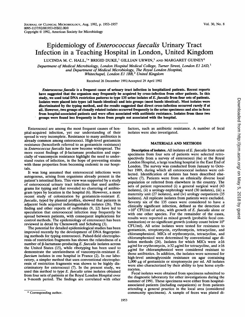
Typing of isolates and relationship to place and time. E.faecalis was isolated from single urine specimens from 135patients: 30 from a urology-nephrology ward, 43 from asurgical ward, 27 from a maternity unit, and 35 who wereurology outpatients. When DNA restriction fragments of allthe isolates were compared, 94 different patterns wereobserved. A single band difference in the size range of 1.6 to8 kb was used as the criterion for a pattern difference;identity was always checked by running potentially identicalisolates in adjacent lanes of a gel (Fig. 1). The largest numberof isolates in a type was six. Two types were represented bysix isolates, 3 were represented by five isolates, 5 wererepresented by three isolates, 9 were represented by twoisolates, and 75 were represented by single isolates (Table 1).Isolates of the same type frequently differed in their profileof resistance to antibiotics (data not shown).
M Ah At Ae Ag
-- =_
M BI Bf Ba Be
- M - m -
23 5 M 11
= - -
FIG. 1. Examples of SstI restriction fragment patterns. Fourpattern types from group A, four pattern types from group B, andexamples from three other groups (groups 23, 5, and 11) are shown.Patterns were traced from Polaroid negative images of gels; bands ofless than 1.6 kb and of more than 8 kb in length were omitted. LanesM contain molecular size markers of 1.6, 2, 3, 4, 5, 6, 7, and 8 kb inlength (1-kb ladder; Life Technologies, Paisley, Scotland).
The types were related to the four patient sets from whichthe isolates were derived (Table 1) and to the date ofisolation to identify any clustering of cases. Four isolates ofone type (Bf; Table 1) were obtained from urology outpa-tients, one on 9 January, one on 18 April, and two on 4 and9 May. Only the second and third isolates had the sameantibiotic resistance pattern. In 15 instances, two isolates ofa single type were obtained from the same patient set (Table1), and in 6 of these instances, the antibiogram was also thesame. One pair was isolated on the same day from theurology-nephrology ward (same antibiogram), a second pairwas isolated 2 days apart from the maternity unit (differentantibiograms), and the others were all isolated at least 2weeks apart.
Grouping of isolates and relationship to other factors. WhenDNA patterns were compared, it was clear that manypatterns, while not identical, were very similar to each other.Isolates were therefore placed in pattern groups, such thatfor any two isolates in a group the majority of bands (>50%)were identical (see examples in Fig. 1). (Within each groupmost pairs of isolates had at least 75% of bands in common.)With this criterion, two groups of 24 isolates each, twogroups of 11 isolates each, and eight groups with between 2and 6 isolates each were found similar, and the remaining 32isolates were found unique.The two most common groups, designated A and B (Fig.
1), contained over one-third of all urine isolates. The asso-ciation of these two groups with other factors was examined(Table 2). Both groups had a higher frequency of antibioticresistance than other isolates. Group B had the highestfrequency of isolates resistant to all five antibiotics exam-ined. The association of gentamicin resistance with thisgroup was particularly marked. Group A had a high fre-quency of tetracycline resistance, and resistance to strepto-mycin, erythromycin, and chloramphenicol was also morefrequent in this group than in the other groups. No group Aisolates were resistant to gentamicin. The distribution ofbeta-hemolytic isolates was also examined, and 19 of 31beta-hemolytic isolates were from group B. Chi-square anal-ysis confirmed that the number of resistant isolates in groupsA and B, and other groups differed significantly (P < 0.01)from a random distribution for all antibiotics except strepto-mycin and for beta-hemolysis.
J. CLIN. MICROBIOL.
on May 5, 2018 by guest
http://jcm.asm
.org/D
ownloaded from
EPIDEMIOLOGY OF E. FAECALIS 1955
TABLE 1. Distribution of pattern types among patient sets, in order of prevalence of the types
Patient No. of isolates of the following type:set' Aa la Ad Bf Bl Af Be 11 17 23 Ba Bh lc 3a 5c 5i 16a 16b 19c UT*
URO 1 1 1 0 2 1 0 1 1 0 2c 0 0 2 0 0 1 0 0 17SURG 2 2 1 1 2 1 0 0 1 0 0 2 1 0 2 1 1 1 2 23UO 1 2 1 4d 1 1 2 0 0 1 0 0 1 0 0 1 0 0 0 20MAT 2e 1 2 0 0 0 1 2 1 2 0 0 0 0 0 0 0 1 0 15
a URO, urology-nephrology ward; SURG, surgical ward; UO, urology outpatients; MAT, maternity unit.b UT, unique type not found elsewhere.c Isolates obtained on the same day.d Two isolates obtained within 5 days of each other.e Isolates obtained within 2 days of each other.
The frequency of isolation of the two most commongroups from the four sets of patients was compared (Table3). Group B was isolated less frequently from maternitypatients than from the other patients, but this result was notfound statistically significant by chi-square analysis.
All isolates of E. faecalis from urine specimens were
analyzed, but not all specimens were considered to show aclinically significant infection (see Materials and Methods).Fourteen of 24 (58%) group A isolates, 16 of 24 (67%) groupB isolates, and 42 of 87 (48%) other isolates came frompositive specimens. This distribution was not significantlydifferent from a random distribution.Frequency of group A and B isolates in fecal specimens. To
determine the underlying frequency of groups A and B, weexamined E. faecalis isolates obtained from fecal specimens.Group A and especially group B patterns were found lessfrequently in community specimens (Table 4). For hospital-associated patients, the frequency of isolation of thesegroups was closer to that obtained with urine specimens(Table 4). Chi-square analysis indicated that the probabilityof the distribution shown in Table 4 occurring at random was0.0695.Two of 45 community fecal isolates tested and 2 of 30
hospital fecal isolates tested were gentamicin resistant; ineach set of isolates, one of the resistant isolates belonged togroup B and the other belonged to a different group.
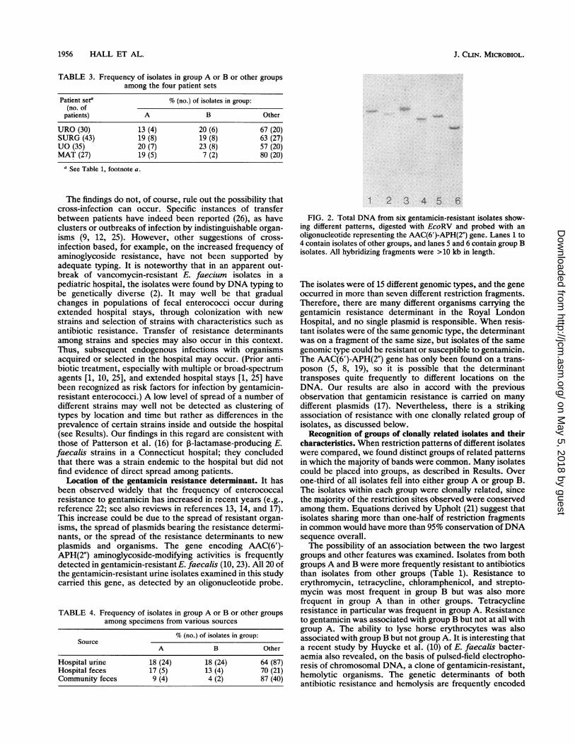
Further analysis of gentamicin-resistant isolates. E. faecalisurine isolates showing resistance to gentamicin (20 in total)were found to have eight different patterns in group B andseven other patterns. Thus, although there was an associa-tion of resistance with group B patterns, the isolates did notrepresent a single identical clone. An alternative hypothesiswas that a single plasmid was spreading among organisms.The enzyme most frequently responsible for gentamicinresistance in enterococci is AAC(6')-APH(2"), which isidentical to the staphylococcal enzyme with the same activ-ity. A 30-base oligonucleotide was used to probe for the geneencoding gentamicin resistance in restriction digests of totalDNA. All resistant isolates hybridized with the probe, andfour susceptible isolates did not hybridize. The probe hy-
bridized to EcoRI fragments of at least seven different sizes(five isolates had fragments of >20 kb in length that were notwell resolved by conventional electrophoresis). Tests ofsome isolates after digestion of DNA with EcoRV (Fig. 2)showed that more differences could be distinguished. EcoRIand EcoRV do not digest within the resistance gene or withinthe transposon that carries it (8, 19), so the fragment sizedifferences demonstrated that the DNA sequences flankingthe gene were different.
DISCUSSION
Lack of evidence for clustering of types by ward or time. Wehave demonstrated elsewhere (7) that digestion of total DNAfrom E. faecalis with SstI is a simple and discriminatorytyping method for distinguishing isolates. Furthermore, thepatterns produced appear to be very stable, with no differ-ences being detected after repeated subculturing (7) (25times) or upon later isolation from the same patient (7).
In the present study, 135 urine isolates from differentpatients were typed and compared. A total of 94 types wereobtained, with no type containing more than six isolates. Thepatients were from three different wards or were urologyoutpatients. Isolates of the same type were not generallyclustered in any ward or patient set (Table 1), nor were theyobviously clustered in time. In three instances, a pair ofisolates of the same type was obtained from patients in thesame set within less than 1 week. Of these, only one pair alsohad the same antibiotic resistance profile. All other isolatesof the same type and from the same patient set were obtainedat least 2 weeks apart, and most were obtained more than 1month apart. Furthermore, in more than half of these cases,pairs of isolates with identical DNA patterns had differentantibiograms, suggesting differences in plasmid carriage.The large number of different types obtained and the lack
of clustering of isolates of the same type suggest that directpatient-to-patient spread is not a frequent occurrence. Thesame conclusions are reached whether isolates that mayhave come from fecal contamination or low-level coloniza-tion were excluded.
TABLE 2. Frequencies of resistance to antibiotics and of beta-hemolysis among isolates from groups A and Band all other groups combined
Group (no. % (no.) of isolates resistant to: % (no.) ofof isolates) beta-hemolyticOtIsolatesGentamicin Streptomycin Erythromycin Tetracycline Chloramphenicol isolates
A (24) 0 (0) 17 (4) 21 (5) 75 (18) 29 (7) 4 (1)B (24) 54 (13) 29 (7) 54 (13) 79 (19) 50 (12) 79 (19)Other (87) 8 (7) 10 (9) 18 (16) 33 (29) 13 (11) 13 (11)
VOL. 30, 1992
on May 5, 2018 by guest
http://jcm.asm
.org/D
ownloaded from
1956 HALL ET AL.
TABLE 3. Frequency of isolates in group A or B or other groupsamong the four patient sets
Patient set' % (no.) of isolates in group:(no. of
patients) A B Other
URO (30) 13 (4) 20 (6) 67 (20)SURG (43) 19 (8) 19 (8) 63 (27)UO (35) 20 (7) 23 (8) 57 (20)MAT (27) 19 (5) 7 (2) 80 (20)
a See Table 1, footnote a.
The findings do not, of course, rule out the possibility thatcross-infection can occur. Specific instances of transferbetween patients have indeed been reported (26), as haveclusters or outbreaks of infection by indistinguishable organ-isms (9, 12, 25). However, other suggestions of cross-infection based, for example, on the increased frequency ofaminoglycoside resistance, have not been supported byadequate typing. It is noteworthy that in an apparent out-break of vancomycin-resistant E. faecium isolates in apediatric hospital, the isolates were found by DNA typing tobe genetically diverse (2). It may well be that gradualchanges in populations of fecal enterococci occur duringextended hospital stays, through colonization with newstrains and selection of strains with characteristics such asantibiotic resistance. Transfer of resistance determinantsamong strains and species may also occur in this context.Thus, subsequent endogenous infections with organismsacquired or selected in the hospital may occur. (Prior anti-biotic treatment, especially with multiple or broad-spectrumagents [1, 10, 25], and extended hospital stays [1, 25] havebeen recognized as risk factors for infection by gentamicin-resistant enterococci.) A low level of spread of a number ofdifferent strains may well not be detected as clustering oftypes by location and time but rather as differences in theprevalence of certain strains inside and outside the hospital(see Results). Our findings in this regard are consistent withthose of Patterson et al. (16) for P-lactamase-producing E.faecalis strains in a Connecticut hospital; they concludedthat there was a strain endemic to the hospital but did notfind evidence of direct spread among patients.
Location of the gentamicin resistance determinant. It hasbeen observed widely that the frequency of enterococcalresistance to gentamicin has increased in recent years (e.g.,reference 22; see also reviews in references 13, 14, and 17).This increase could be due to the spread of resistant organ-isms, the spread of plasmids bearing the resistance determi-nants, or the spread of the resistance determinants to newplasmids and organisms. The gene encoding AAC(6')-APH(2") aminoglycoside-modifying activities is frequentlydetected in gentamicin-resistant E. faecalis (10, 23). All 20 ofthe gentamicin-resistant urine isolates examined in this studycarried this gene, as detected by an oligonucleotide probe.
TABLE 4. Frequency of isolates in group A or B or other groupsamong specimens from various sources
% (no.) of isolates in group:Source
A B Other
Hospital urine 18 (24) 18 (24) 64 (87)Hospital feces 17 (5) 13 (4) 70 (21)Community feces 9 (4) 4 (2) 87 (40)
1 2 3 4 5 6
FIG. 2. Total DNA from six gentamicin-resistant isolates show-ing different patterns, digested with EcoRV and probed with anoligonucleotide representing the AAC(6')-APH(2") gene. Lanes 1 to4 contain isolates of other groups, and lanes 5 and 6 contain group Bisolates. All hybridizing fragments were >10 kb in length.!
The isolates were of 15 different genomic types, and the geneoccurred in more than seven different restriction fragments.Therefore, there are many different organisms carrying thegentamicin resistance determinant in the Royal LondonHospital, and no single plasmid is responsible. When resis-tant isolates were of the same genomic type, the determinantwas on a fragment of the same size, but isolates of the samegenomic type could be resistant or susceptible to gentamicin.The AAC(6')-APH(2") gene has only been found on a trans-poson (5, 8, 19), so it is possible that the determinanttransposes quite frequently to different locations on theDNA. Our results are also in accord with the previousobservation that gentamicin resistance is carried on manydifferent plasmids (17). Nevertheless, there is a strikingassociation of resistance with one clonally related group ofisolates, as discussed below.
Recognition of groups of clonally related isolates and theircharacteristics. When restriction patterns of different isolateswere compared, we found distinct groups of related patternsin which the majority of bands were common. Many isolatescould be placed into groups, as described in Results. Overone-third of all isolates fell into either group A or group B.The isolates within each group were clonally related, sincethe majority of the restriction sites observed were conservedamong them. Equations derived by Upholt (21) suggest thatisolates sharing more than one-half of restriction fragmentsin common would have more than 95% conservation ofDNAsequence overall.The possibility of an association between the two largest
groups and other features was examined. Isolates from bothgroups A and B were more frequently resistant to antibioticsthan isolates from other groups (Table 1). Resistance toerythromycin, tetracycline, chloramphenicol, and strepto-mycin was most frequent in group B but was also morefrequent in group A than in other groups. Tetracyclineresistance in particular was frequent in group A. Resistanceto gentamicin was associated with group B but not at all withgroup A. The ability to lyse horse erythrocytes was alsoassociated with group B but not group A. It is interesting thata recent study by Huycke et al. (10) of E. faecalis bacter-aemia also revealed, on the basis of pulsed-field electropho-resis of chromosomal DNA, a clone of gentamicin-resistant,hemolytic organisms. The genetic determinants of bothantibiotic resistance and hemolysis are frequently encoded
J. CLIN. MICROBIOL.
on May 5, 2018 by guest
http://jcm.asm
.org/D
ownloaded from

EPIDEMIOLOGY OF E. FAECALIS 1957
on plasmids. One possible explanation for the association ofthese characteristics with isolates of some groups of organ-isms more than others is the influence of chromosomal geneson plasmid uptake and propagation. For example, in E.faecalis the transfer of certain plasmids is promoted by sexpheromones secreted by the recipient (4).
Prevalence of group A and B isolates in fecal specimens. Thecommon occurrence of group A and B isolates in urinespecimens led us to question the frequency of such organ-isms in the general population and whether they were foundonly in the hospital or were widespread in the community.Only two group B isolates were obtained from maternitypatients (Table 3), most of whom have only a short-termconnection with the hospital and no prior antibiotic treat-ment. However, the total number of isolates in this patientset was low. E. faecalis was therefore isolated from fecalspecimens submitted for other investigations and obtainedfrom both hospital-associated patients and general-practice(community) patients (Table 4). Group A and B isolates werealmost as frequent in feces from hospital-associated patientsas in urine specimens. Both groups were rare in fecalspecimens from the community, although this result was notstatistically significant. (Since hospital isolates includedthose from outpatients and day-care patients, the associationof groups A and B with the hospital may have been under-estimated.)The findings of this study suggest that direct cross-infec-
tion was not the source of most urinary tract infections at thetime of this investigation. On the other hand, they areconsistent with the proposition that the fecal flora of hospi-tal-associated patients changes through the acquisition orselection of specific strains. A subsequent autogenous infec-tion could then involve hospital-associated enterococci andan increased likelihood of antibiotic resistance problems.
ACKNOWLEDGMENTS
We are grateful to Rosamund Williams and J. D. Williams for helpand support on this project. We thank David Beighton and NickBanatvala for help with statistical analysis and Esther Simpson forproviding some of the fecal isolates.
This study was funded by a grant from the Special Trustees of TheRoyal London Hospital Trust.
REFERENCES1. Axelrod, P., and G. H. Talbot. 1989. Risk factors for acquisition
of gentamicin-resistant enterococci. Arch. Intern. Med. 149:1397-1401.
2. Bingen, E. H., E. Denamur, N. Y. Lambert-Zechovsky, and J.Elion. 1991. Evidence for the genetic unrelatedness of noso-comial vancomycin-resistant Enterococcus faecium strains in apediatric hospital. J. Clin. Microbiol. 29:1888-1892.
3. Chenoweth, C., and D. Schaberg. 1990. The epidemiology ofenterococci. Eur. J. Clin. Microbiol. Infect. Dis. 9:80-89.
4. Clewell, D. B. 1990. Movable genetic elements and antibioticresistance in enterococci. Eur. J. Clin. Microbiol. Infect. Dis.9:90-102.
5. Ferretti, J. J., K. S. Gilmore, and P. Courvalin. 1986. Nucleo-tide sequence analysis of the gene specifying the bifunctional6'-aminoglycoside acetyltransferase 2"-aminoglycoside phos-photransferase enzyme in Streptococcus faecalis and identifica-tion and cloning of gene regions specifying the two activities. J.Bacteriol. 167:631-638.
6. Gross, P. A., L. M. Harkavy, G. E. Barden, and M. F. Flower.1976. The epidemiology of nosocomial enterococcal urinarytract infection. Am. J. Med. Sci. 272:75-81.
6a.Guiney, M., et al. Unpublished data.7. Hall, L. M. C., B. Duke, M. Guiney, and R. Williams. 1992.
Typing of Enterococcus species by DNA restriction fragmentanalysis. J. Clin. Microbiol. 30:915-919.
8. Hodel-Christian, S. L., and B. E. Murray. 1991. Characteriza-tion of the gentamicin resistance transposon Tn5281 from En-terococcus faecalis and comparison to staphylococcal trans-posons Tn4001 and Tn4031. Antimicrob. Agents Chemother.35:1147-1152.
9. Hussain, Z., M. Kuhn, R. Lannigan, and T. W. Austin. 1988.Microbiological investigation of an outbreak of bacteraemia dueto Streptococcus faecalis in an intensive care unit. J. Hosp.Infect. 12:263-271.
10. Huycke, M. M., C. A. Spiegel, and M. S. Gilmore. 1991.Bacteremia caused by hemolytic, high-level gentamicin-resis-tant Enterococcus faecalis. Antimicrob. Agents Chemother.35:1626-1634.
11. Jordens, J. Z., and L. M. C. Hall. 1989. Chromosomally-encoded gentamicin resistance in 'epidemic' methicillin-resis-tant Staphylococcus aureus: detection with a synthetic oligonu-cleotide probe. J. Antimicrob. Chemother. 23:327-334.
12. Luginbuhl, L. M., H. A. Rotbart, R. R. Facklam, M. H. Roe, andJ. A. Elliot. 1987. Neonatal enterococcal sepsis: case-controlstudy and description of an outbreak. Pediatr. Infect. Dis. J.6:1022-1026.
13. Moellering, R. C., Jr. 1991. The enterococcus: a classic exampleof the impact of antimicrobial resistance on therapeutic options.J. Antimicrob. Chemother. 28:1-12.
14. Murray, B. E. 1990. The life and times of the enterococcus.Clin. Microbiol. Rev. 3:46-65.
15. Murray, B. E., K. V. Singh, S. M. Markowitz, H. A. Lopardo,J. E. Patterson, M. J. Zervos, E. Rubeglio, G. M. Eliopoulos,L. B. Rice, F. W. Goldstein, S. G. Jenkins, G. M. Caputo, R.Nasnas, L. S. Moore, E. S. Wong, and G. Weinstock. 1991.Evidence for clonal spread of a single strain of P-lactamase-producing Enterococcusfaecalis to six hospitals in five states. J.Infect. Dis. 163:780-785.
16. Patterson, J. E., K. V. Singh, and B. E. Murray. 1991. Epidemi-ology of an endemic strain of 3-lactamase-producing Enterococ-cus faecalis. J. Clin. Microbiol. 29:2513-2516.
17. Patterson, J. E., and M. J. Zervos. 1990. High-level gentamicinresistance in Enterococcus: microbiology, genetic basis, andepidemiology. Rev. Infect. Dis. 12:644-652.
18. Pitcher, D. G., N. A. Saunders, and R. J. Owen. 1989. Rapidextraction of bacterial genomic DNA with guanidium thiocya-nate. Lett. Appl. Microbiol. 8:151-156.
19. Rouch, D. A., M. E. Byrne, Y. C. Kong, and R. A. Skurray.1987. The aacA-aphD gentamicin and kanamycin resistancedeterminant of Tn4001 from Staphylococcus aureus: expressionand nucleotide sequence analysis. J. Gen. Microbiol. 133:3039-3052.
20. Sambrook, J., E. F. Fritsch, and T. Maniatis. 1989. Molecularcloning: a laboratory manual. Cold Spring Harbor Laboratory,Cold Spring Harbor, N.Y.
21. Upholt, W. B. 1977. Estimation of DNA sequence divergencefrom comparison of restriction endonuclease digests. NucleicAcids Res. 4:1257-1265.
22. Watanakunakorn, C. 1989. The prevalence of high-level amino-glycoside resistance among enterococci isolated from bloodcultures during 1980-1988. J. Antimicrob. Chemother. 24:63-68.
23. Weems, J. J., Jr., J. H. Lowrance, L. M. Baddour, and W. A.Simpson. 1989. Molecular epidemiology of nosocomial, multiplyaminoglycoside resistant Enterococcus faecalis. J. Antimicrob.Chemother. 24:121-130.
24. Working Party on Antibiotic Sensitivity Testing of the BritishSociety for Antimicrobial Chemotherapy. 1991. A guide to sen-sitivity testing. Academic Press, London.
25. Zervos, M. J., S. Dembinski, T. Mikesell, and D. R. Schaberg.1986. High-level resistance to gentamicin in Streptococcusfaecalis: risk factors and evidence for exogenous acquisition ofinfection. J. Infect. Dis. 153:1075-1083.
26. Zervos, M. J., C. A. Kauffman, P. M. Therasse, A. G. Bergman,T. S. Mikesell, and D. R. Schaberg. 1987. Nosocomial infectionby gentamicin-resistant Streptococcusfaecalis: an epidemiolog-ical study. Ann. Intern. Med. 106:687-691.
VOL. 30, 1992
on May 5, 2018 by guest
http://jcm.asm
.org/D
ownloaded from