eparation of carbonated apatite nano-whiskpr ers in sbf...
TRANSCRIPT

Journal of Ceramic Processing Research. Vol. 10, No. 5, pp. 606~608 (2009)
606
J O U R N A L O F
CeramicProcessing Research
Preparation of carbonated apatite nano-whiskers in SBF with ultrasonic assistance
Yanli Geng, Yanbao Li and Dongxu Li*
State Key Laboratory of Materials-Oriented Chemical Engineering, College of Materials Science and Engineering, Nanjing
University of Technology, Nanjing 210009, China
Carbonated apatite nano-whiskers were prepared in simulated body fluids (SBF) with ultrasonic assistance at 37 oC. Theeffects of the initial pH value in the SBF solution and reaction times were investigated in this study. The calcium phosphateprecipitates were examined by XRD, FTIR and TEM techniques. The results showed that the initial pH value in the SBF hadan important effect on the morphology and composition of the calcium phosphate precipitates, and carbonated apatite nano-whiskers with length/diameter ratio of ~20 were obtained at an initial pH = 7.5 after ultrasonic treatment for 24 h.
Key words: Carbonated apatite, Nano-whisker, Simulated body fluids, Ultrasonic assistance.
Introduction
Carbonated apatite (denoted as CAP) is the mineral phaseof bone and teeth [1-3]. CAP have attracted substantialattentions due to its biocompatibility and bioactivity, theability to form a chemical bond in the interface between animplant and living tissue. Furthermore, CAP can be used inindustrial fields such as for separation and adsorptionbecause of its specific surface structure [3]. CAP is limited toapplication in the non-loaded or low-loaded fields due to itsbrittle nature. Recently, CAP has been introduced to fillpolymer matrix to improve its mechanical properties [4, 5].Generally speaking, crystal whiskers are free from theinternal defects such as dislocations due to their smalldiameter and its strength tends to the maximum theoreticalvalue [6]. Hence, various methods have been employedto prepare CAP whiskers, including co-precipitation,homogeneous precipitation, sol-gel synthesis, pyrolysis ofaerosols, using a micro-emulsion and hydrothermal reaction.[7-11]
In this study, simulated body fluid (SBF) was used asa medium to prepare CAP nano-whiskers in order tosimulate the biological environment in human bodies.Ultrasonic treatment was also used to reduce the aging time.
Experimental
Materials and methods
The starting materials were listed as follows: K2HPO4·3H2O,CaCl2, sorbitol (C6H14O6), NaCl, NaHCO3, KCl,K2HPO4·3H2O, MgCl2·6H2O, 1 mol/l HCl, CaCl2, Na2SO4
and (CH2OH)3CNH2. A simulated body fluid (SBF) solution
was prepared according to the method developed byKokubo et al.[12]
Firstly, K2HPO4·3H2O (0.012 mol) and sorbitol (0.04 mol)were dissolved into 100 ml SBF to form a phosphatesolution. Secondly, CaCl2 (0.02 mol) was dissolved into100 ml SBF to obtain a calcium solution. Then, a milkysuspension appeared after adding phosphate solutiondropwise into the calcium solution with an initial Ca/Pratio of 1.67 (stoichiometric ratio of HA) at differentinitial pH values of 7.15 to 7.80. The milky suspensionwas aged for 1 h under continuous and gentle magneticstirring, and put into an ultrasonic bath at 37 oC with anultrasonic power of 40 W. The suspensions were treatedultrasonically for different times of 6-24 h. After thetreatment, the calcium phosphate precipitates were filteredand washed twice with deionized water and then withanhydrous ethanol twice, to remove the residual sorbitoland ions. The filtered cakes were dried at 70 oC for 24 h toobtain calcium phosphate powders.
Characterization
X-ray diffraction (XRD, ARL X'TRA. Cu Kα radiation)was employed to characterize the phase of the calciumphosphate precipitates. Fourier transformed infraredspectroscopy (FTIR, Nexus 670. Resolution: 2 cm−1)was conducted in the wave number range of 4000-400 cm−1.High resolution transmission electron microscopy (TEM,JEM-2010 UHR, accelerating voltage: 200 kV; crystallattice resolution = 0.143 nm) was used to observe thecalcium phosphate precipitates.
Results and Discussion
Fig.1 shows the XRD patterns of the Samples 1#, 2#,3# and 4# prepared at the initial pHs in the SBF solution of7.15, 7.30, 7.50 and 7.60, respectively, after ultrasonictreatment for 24 h. It can be seen that when the initial pH was
*Corresponding author: Tel : +86-25-83587256Fax: +86-25-83588967E-mail: [email protected]

Preparation of carbonated apatite nano-whiskers in SBF with ultrasonic assistance 607
7.15, The main phase is CaHPO4 and only a few CAP peakswere observed. When the initial pH was higher, the yield ofCAP increased and only the CAP phase could be obtainedif pH ≥ 7.45. The reaction may be described by Eq.1:
CaHPO4 = Ca2+ + HPO4
2− (1)
HPO4
2− = PO4
3− + H+ (2)
10Ca2+ + 6PO4
3− + 2H2O = Ca10(PO4)6(OH)2 (3)
6CaHPO4 + 4Ca2+ + 2H2O = Ca10(PO4)6(OH)2 + 8H+ (4)
When the initial pH in the SBF solution was higher,the reaction would be more easily progressed to the rightdirection for preparing Ca10(PO4)6(OH)2 as described in Eq.4.
Fig.2 gives the XRD patterns of samples 5#, 6#, 7#and 8# prepared at an initial pH = 7.45 in SBF solutionafter ultrasonic treatment for 6, 12, 18 and 24 h, respectively.The intensity of the (002) peak is much higher than that ofstandard apatite (JCPDS card No. 9-432). This may bedue to the predominant crystal growth along the c-axis, whichcould make the precipitates to be whiskers. Furthermore,there is no obvious difference among these four patternsand all the peaks agree with the standard apatite patternwhich shows that our precipitates are a pure apatite phase.
Fig. 3 shows the FTIR spectra of samples 1#, 2#, 3#
and 4#. It can be seen that in sample 1#, only one PO4
3−
band were detected at 1038 cm−1 compared with sample 4#in which almost all of the PO4
3− bands are shown in thespectrum. So it can be deduced that the main phase in
Fig. 1. XRD patterns of samples 1#, 2#, 3# and 4# prepared atinitial pHs in the SBF solution of 7.15, 7.30, 7.50 and 7.60,respectively, after ultrasonic treatment for 24 h. Fig. 2. XRD patterns of samples 5#, 6#, 7# and 8# prepared at an
initial pH = 7.45 in the SBF solution after treated by ultrasonic for6, 12, 18 and 24 h, respectively.
Fig. 3. FTIR spectra of samples 1#, 2#, 3# and 4# prepared atinitial pHs in the SBF solution of 7.15, 7.30, 7.50 and 7.60,respectively, after ultrasonic treatment for 24 h.
608 Yanli Geng, Yanbao Li and Dongxu Li
sample 1# is CaHPO4 while only the CAP phase existedin sample 4#. The difference between sample 1# andsample 4# is in accord with their XRD patterns asshown in Fig. 1. In addition, the band at 1450 cm−1
corresponded to the carbonate group (CO3
2−) and indicatedthat the CAP precipitates contained CO3
2−, which issimilar to the minerals in bone [13].
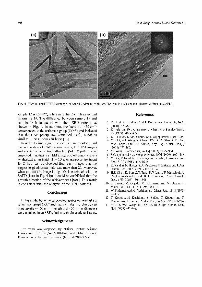
In order to investigate the detailed morphology andcharacterization of CAP nano-whiskers, HRTEM imagesand selected area electron diffraction (SAED) pattern wereemployed. Fig. 4(a) is a TEM image of CAP nano-whiskerssynthesized at an initial pH = 7.5 after ultrasonic treatmentfor 24 h. It can be observed from such images that thebiggest length/diameter ratio was more than 20. Moreover,when an HRTEM image in Fig. 4(b) is combined with theSAED (inset in Fig. 4(b)), it could be established that thegrowth direction of the whiskers was [001]. This resultis consistent with the analysis of the XRD patterns.
Conclusions
In this study, bonelike carbonated apatite nano-whiskerswhich contained CO3
2− and had a similar morphology tobone apatite (~100 nm in length and ~20 nm in diameter)were obtained in an SBF solution with ultrasonic assistance.
Acknowledgements
This work was supported by National Nature ScienceFoundation of China (No. 50802042), and Nature ScienceFoundation of Jiangsu province (No. BK2008379).
References
1. T. Hirai, M. Hodono And I. Komasawa, Langmuir, 16[3](2000) 955-960.
2. E. Dalas and P.G. Koutsoukos, J. Chem. Soc.-Faraday Trans.,85 (1989) 2465-2472.
3. L.L. Hench, J. Am. Ceram. Soc., 81[7] (1998) 1705-1728.4. Y.B. Li, W.J. Weng, K. Cheng, P.Y. Du, G. Shen, G.R. Han,
M.A. Lopes and J.D. Santos, Key Eng. Mater., 254[2](2004) 477-480.
5. M. Wang, Biomaterials, 24[13] (2003) 2133-2151.6. S.C. Tjong and Y.Z. Meng, Polymer, 40[5] (1999) 1109-1117.7. Y. Ota, T. Iwashita, T. Kasuga and Y. Abe, J. Am. Ceram.
Soc., 81[6] (1998) 1665-1668.8. K. Kandori, N. Horigami, A. Yasukawa, T. Ishikawa and J. Am.
Ceram. Soc., 80[5] (1997) 1157-1164.9. H.F. Chen, K. Sun, Z.Y. Tang, R.V. Law, J.F. Mansfield, A.
Czajka-Jakubowska and B.H. Clarkson, Cryst. GrowthDes., 6[6] (2006) 1504-1508.
10. S. Suzuki, M. Ohgaki, M. Ichiyanagi and M. Ozawa, J.Mater. Sci. Lett., 17[5] (1998) 381-383.
11. W. Suchanek and M. Yoshimura, J. Mater. Res., 13[1] (1998)94-117.
12. T. Kokubo, H. Kushitani, S. Sakka, T. Kitsugi and T.Yamamuro, J. Biomed. Mater. Res., 24[6] (1990) 721-734.
13. Y.B. Li, W.J. Weng and D.X. Li, Int J Appl Ceram Tech,5[5] (2008) 442-448.
Fig. 4. TEM (a) and HRTEM (a) images of typical CAP nano-whiskers. The inset is a selected area electron diffraction (SAED).