enzyme‐mediated, site‐specific protein coupling strategies
TRANSCRIPT

German Edition: DOI: 10.1002/ange.201805034Protein Immobilization Hot PaperInternational Edition: DOI: 10.1002/anie.201805034
Enzyme-Mediated, Site-Specific Protein Coupling Strategies forSurface-Based Binding AssaysWolfgang Ott+, Ellis Durner+, and Hermann E. Gaub*
Abstract: Covalent surface immobilization of proteins forbinding assays is typically performed non-specifically via lysineresidues. However, receptors that either have lysines near theirbinding pockets, or whose presence at the sensor surface iselectrostatically disfavoured, can be hard to probe. To over-come these limitations and to improve the homogeneity ofsurface functionalization, we adapted and optimized threedifferent enzymatic coupling strategies (4’-phosphopante-theinyl transferase, sortase A, and asparaginyl endopeptidase)for biolayer interferometry surface modification. All of theseenzymes can be used to site-specifically and covalently ligateproteins of interest via short recognition sequences. Theenzymes function under mild conditions and thus immobiliza-tion does not affect the receptors� functionality. We successfullyemployed this enzymatic surface functionalization approach tostudy the binding kinetics of two different receptor–ligand pairs.
The binding properties of receptor–ligand complexes havebeen studied in vitro with numerous assays developed duringthe last decades.[1–3] Mainly, covalent approaches for receptorimmobilization have been established to precisely determineon-rate (kon), off-rate (koff), and equilibrium constant (Kd).[4]
For these methods, the receptor is immobilized onto a surfaceand a change in signal upon ligand application is evaluated.While sometimes the terminology “ligand-analyte” is used,throughout this article the molecule immobilized to thesensor surface is called the receptor and its binding partnerthe ligand. In general, accessible side chains of correspondingamino acids (amine-, carboxyl-, or thiol-groups)[5] can beemployed to covalently link the receptor to a surface.However, non-specific attachment requires an electrostati-cally driven surface pre-concentration step, where the pH andsalt conditions of the buffer must be chosen such that thesensor surface and the receptors are predominantly oppo-sitely charged. This pre-concentration step requires a bufferof low ionic strength in order to prevent screening of surface
charges, which in turn may cause unfolding and aggregationissues.[6] Additionally, proteins with a low isoelectric pointmight not be sufficiently protonated, and thus remainnegatively charged.
Another challenge with non-specific surface chemistry isthat proteins often contain more than one reactive residue,which leads to inhomogeneous surface anchoring. Conse-quently, sensorgrams of a binding experiment represent thesuperimposed response of multiple populations of differentlyattached receptors. Varying attachment sites may stronglyinfluence binding kinetics solely due to the molecules�orientation. The binding behaviour can be altered or bindingmay even be prevented, especially with receptors immobi-lized via reactive residues close to their binding interface(Figure 1A).[7]
In this study, we expand the toolbox for surface function-alization by adapting advances in enzyme-based proteinmodification strategies to overcome the limitations of non-specific pull-down strategies in binding assays. The employedenzymes are a 4’-phosphopantetheinyl transferase fromBacillus subtilis (Sfp),[8] an evolved sortase A (SrtA) fromStaphylococcus aureus (d59SrtA, P94R/D160N/D165A/K190E/K196T),[9] and an engineered asparaginyl endopepti-dase from the plant Oldenlandia affinis (OaAEP1)(C247A).[10] All of these enzymes recognize specific aminoacid sequences (tags) and covalently attach these tags to other
Figure 1. Schematic of BLI Kinetics. A) Non-specific immobilization ofthe receptor on the sensor in different geometries as a result of severalaccessible amine-groups. B) Specific and site-directed immobilizationof a receptor to a sensor. All receptors are homogeneously orientated.The red arrows in (A) represent different binding geometries withpossibly different kinetics, whereas specific attachment (B) providesa uniform population of binders. C) The principle of a BLI kineticexperiment. A receptor-functionalized sensor is immersed into a ligandsolution. The increasing signal shows binding of the ligand. When thesensor signal has reached a steady state, the rates of ligand associa-tion and dissociation are equal—the system has reached equilibrium.The sensor is then moved to a buffer solution, the receptor starts todissociate and the detected signal decreases again.
[*] Dr. W. Ott,[+] E. Durner,[+] Prof. Dr. H. E. GaubLehrstuhl f�r Angewandte Physik and Center for NanoScienceLudwig-Maximilians-Universit�t M�nchenAmalienstrasse 54, 80799 Munich (Germany)andCenter for Integrated Protein Science Munich(CIPSM), Ludwig-Maximilians-Universit�t M�nchenButenandtstrasse 5–13, 81377 Munich (Germany)E-mail: [email protected]
[+] These authors contributed equally to this work.
Supporting information (including experimental details) and theORCID identification number(s) for the author(s) of this article canbe found under:https://doi.org/10.1002/anie.201805034.
AngewandteChemieCommunications
1Angew. Chem. Int. Ed. 2018, 57, 1 – 5 � 2018 Wiley-VCH Verlag GmbH & Co. KGaA, Weinheim
These are not the final page numbers! � �

amino acid sequences (SrtA and OaAEP1) or to Coenzyme A(CoA; Sfp). In case of SrtA and OaAEP1, the tags have to beat the termini of the protein, whereas the ybbR-tag (11 aminoacids) recognized by Sfp can also be internal (if accessible)since its ligation mechanism does not rely on peptidaseactivity. These tags can be fused to proteins and employed insurface pull-down strategies, hence allowing homogeneousloading of a surface (Figure 1B). In single-moleculeapproaches, such as single-molecule force spectroscopy, site-specific reactions[11–13] are already well established and ensurereliable mechano-probing of receptor–ligand systems withoutremoving the proteins from the surface or cantilever. Weadapted these enzyme-based techniques, which enabled us tolink a receptor of interest to a sensor surface in very mildreaction conditions while using only low micromolar quanti-ties of receptor.
We chose a biolayer interferometer (BLI) as a develop-ment platform because of its fast and flexible assay format.However, it should be noted that the approach presented hereis applicable to other surface sensitive techniques, such assurface plasmon resonance (SPR) or quartz crystal micro-balance (QCM), since the receptor immobilization relies onthe same chemistry. The underlying principle of a BLI makesuse of light reflection at interfaces between media of differentoptical densities to analyse the spectral shift of interferencesignals upon binding—which effectively modifies the opticalpath length—to the sensor.[14, 15] The interference signalchanges whenever binding/unbinding to the sensor fibreoccurs (Figure 1 C).
In order to establish our enzyme-based BLI bindingassays, we selected two different systems (Table S1, Figure S1in the Supporting Information). Firstly, we chose GFP-binding nanobodies (LaG9).[16] Nanobodies are small func-tional single-chain antibodies[17] and are popular tools indiagnostic as well as in therapeutic applications. As a secondsystem, we chose the mechanically highly robust cohesin–dockerin type III complex (CohE–XDocIII) from Rumino-coccus flavefaciens. As previous single-molecule force spec-troscopy studies have shown, its unbinding behaviour underforce depends on the anchoring geometry of the cohesin.When immobilized via its C-terminus, a most probablerupture force of around 700 pN (at 100 nNs�1)[18] is observed,in contrast to only 100 pN (at 0.7 nNs�1)[19] when anchored viaits N-terminus. With the site-specific immobilization strat-egies presented here (Figure 2), we were able to probe thegeometry dependence in the absence of force.
Experimental details, traces for Sfp-, SrtA- and OaAEP1-based sensor modifications (Figure S4–S19), and an overviewof all possible immobilization geometries (Figure S20) can befound in the Supporting Information. Once the sensors weresite-specifically loaded with the protein of interest, they wereequilibrated in the same measuring buffer throughout allexperiments.
In order to compare the different immobilization strat-egies, a kinetic binding series with each coupling approachwas recorded. Figure 3A shows an example sensorgram of anSrtA-based experiment. Despite using another GFP variantwhich differs in the binding epitope (Figure S21), we obtainedsimilar binding kinetics to the reported ones (Kd = 3.5 nm,
kon = 2.3 � 106m�1 s�1, koff = 8.0 � 10�3 s�1) determined with
SPR[16] (compare Figure 3B).The obtained kinetic rates were independent of the
functionalization method (specific and non-specific). Thesite-specific approach anchors proteins at their termini anddecreases the chance of binding site obstruction (spatialseparation of surface coupling and ligand binding), which thusallows us to determine the unaltered (un)binding rates. Thisincreased reliance is an intrinsic advantage of our site-specificsurface functionalization. Based on this, we can compare thedata with the non-specifically anchored proteins and concludethat the multiple lysine anchoring possibilities do not obstructthe binding behaviour in the case of TagGFP2–LaG9interaction. TagGFP2 contains 17 lysines that may take partin the non-specific immobilization procedure. Hence, it is notsurprising that enough primary amines non-adjacent to thebinding epitope that do not disturb binding are available asanchoring sites. Other receptor–ligand systems might be morestrongly affected by the non-specific anchoring (see cohesin–dockerin interaction below). Especially if the surface area atthe ligand binding site is charged such that it is electrostati-cally favoured to make surface contact during the pre-concentration step, the binding site could be obstructed.
Figure 2. Overview of the different covalent, site-specific immobiliza-tion techniques. Left: Sfp catalyses the reaction between ybbR-tag ofthe TagGFP2 and coenzyme a (CoA). First, the amine-group of PDEAreacts with the EDC/NHS-activated carboxyl groups of the sensor.PDEA can then undergo a thiol exchange reaction with CoA, whichpresents a free thiol. Middle: SrtA links C-terminal LPETGG with N-terminal GGG. In the case shown here, a C-GGGGG peptide wasreacted with the EDC/NHS-PDEA-activated sensor. Right: OaAEP1recognizes the C-terminal amino acids NGL and fuses it to TagGFP2containing the N-terminal amino acids GLP. EDC/NHS-activatedsensors were reacted with the amine-groups of a KK-GSGS-NGLpeptide. All three immobilization methods yield a homogeneousTagGFP2-modified sensor ready for binding kinetic measurements.
AngewandteChemieCommunications
2 www.angewandte.org � 2018 Wiley-VCH Verlag GmbH & Co. KGaA, Weinheim Angew. Chem. Int. Ed. 2018, 57, 1 – 5� �
These are not the final page numbers!

TagGFP2-NGL and TagGFP2-ybbr could not be fused toa BLI sensor. However, both tags were functional to fuseprotein domains in an in vitro bulk reaction. Thus, it appearsthat both tags are sterically hindered by the GFP domainwhen used for surface functionalization. A longer linkerbetween GFP domain and the recognition sequence couldpossibly provide both enzymes (Sfp, OaAEP1) more space toligate the protein to the sensor.
This enzyme-based and site-specific surface reaction alsoallowed us to probe the inverse geometry with the nanobodynow immobilized to the sensor. Two kinetic titration series,one using the SrtA-based and the other using the non-specificimmobilization approach, were recorded (Figure S10). Fitsdeviated notably from a 1:1 binding model, which might beexplained by either ligand–surface interactions or by potentialavidity effects should TagGFP2 present more than onebinding interface. The ability to site-specifically anchor bothbinding partners allows us to exclude that the deviations froma simple 1:1 binding model stem from heterogeneous surfacepreparation due to non-specific protein anchoring. Based onthe conducted experiments, we were able to show that allthree enzymes can be used for sensor functionalization.
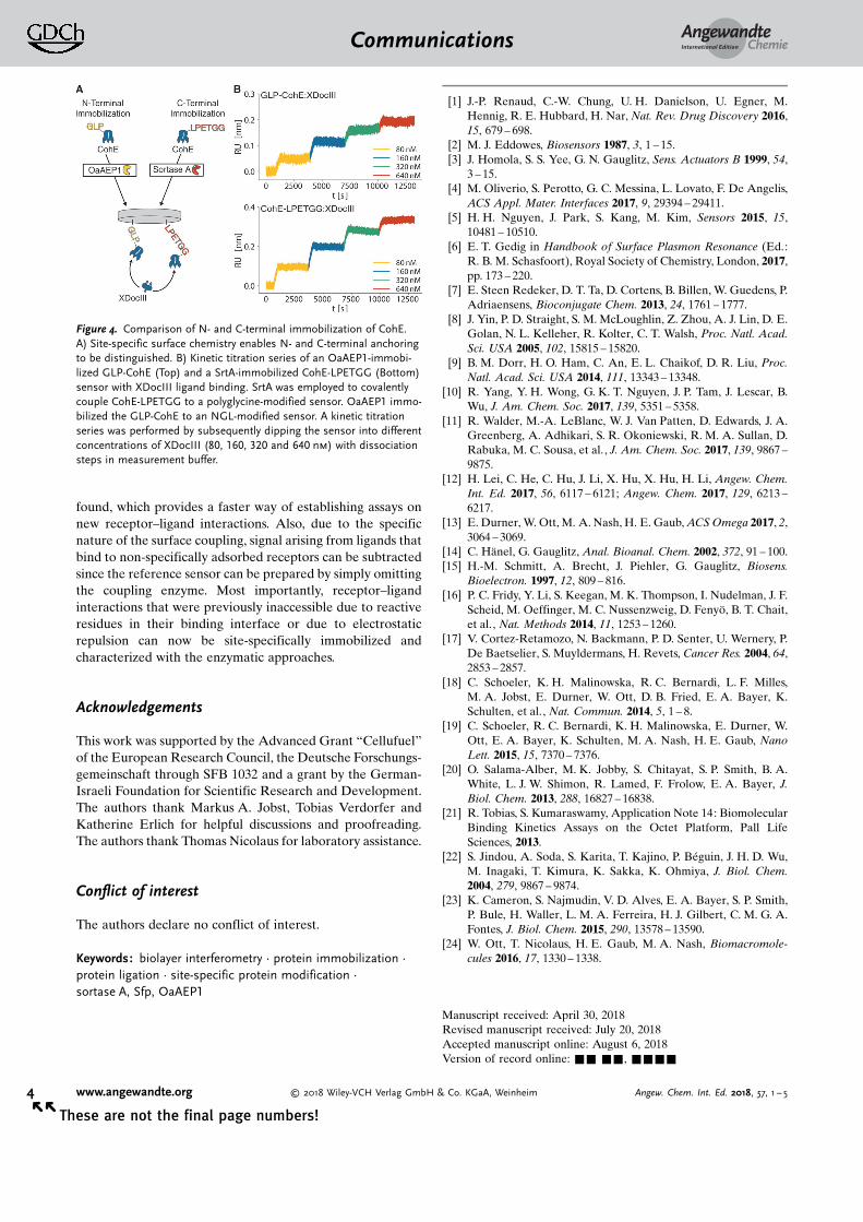
The advantages of defined surface immobilization emergemore clearly when investigating the CohE–XDocIII inter-action of R. flavefaciens. This cohesin–dockerin pair hasalready been characterized in bulk studies[20] as well as single-molecule studies.[18, 19] However, we were not previously ableto non-specifically immobilize the cohesin in a functionalstate (Figure S3), possible due to a lysine in its bindingpocket.[18] However, not only were we able to attach thecohesin site-specifically and in biologically active form withthe enzymatic approach, we were also able to do so from
either terminus. This was of particular interest since theunbinding behaviour of this complex under external force wasshown to strongly depend on the anchoring geometry of thecohesin. The complete sensorgram of the sensor modificationcan be found in Figure S11. Because of the evidently very lowoff-rate of the cohesin–dockerin complex, and because noregeneration conditions could be found to force liganddissociation, we chose to perform a kinetic titration experi-ment. Full dissociation of the complex would take too longand by far exceed the four hours of total experiment timesuggested for BLI. Longer measurements would suffer fromevaporation of liquids in the microwell plate, thus falsifyingconcentrations. Initial experiments showed that sensor-drifteffects seemed to exceed the actual ligand dissociation due tothe low off-rate (Figure S12). Thus, as recommended,[21] wemodified our protocol such that both sensors are loaded witha receptor; in our case they were functionalized site-specifically with cohesin. For referencing, one sensor wasonly dipped into measurement buffer while the other waspresented with ligand. Despite having minimized drift, itbecame clear that the off-rate of the complex is too low to beassessed through BLI. Too little dissociation occurred so thatadequate fitting of the data was not possible. For an exactdetermination of the off-rate, alternative techniques such asSPR or QCM might be more promising, since they are notlimited by evaporation effects and can thus measure forextended periods of time. The apparent low off-rate iscommon in cohesin–dockerin systems.[22, 23] However, a qual-itative statement about the (un)binding behaviour of thecohesin–dockerin complex is possible, namely that on- andoff-rates appear to be independent of the anchoring geometry(Figure 4).
This stands in contrast to force spectroscopy experi-ments,[18, 19] where the anchoring geometry strongly changesthe force necessary to dissociate cohesin and dockerin. Bycomparing the findings obtained by force spectroscopy withthose from site-directed BLI, we can conclude that thedifferent rupture forces are indeed a consequence of forcepropagation within the receptor–ligand complex, rather thanan artefact caused by surface effects or the employedanchoring chemistry.
In summary, the presented strategy provides an efficientmeans to covalently and site-specifically couple receptorsunder mild reaction conditions. The employed tags are allsmall and should not influence the overall functionality ofa protein. This makes it viable to use the same constructs forcharacterization in a surface-based assay as well as for otherbulk and single-molecule studies. Moreover, these small tagscan be further used for post-translational protein modifica-tions, that is, attachment of a fluorescent dye[24] or anadditional protein domain,[13] or as a pull-down technique.[13]
Hence, label-free and label-dependent techniques can be usedwith the same batch of proteins. While the enzymaticapproaches presented here are shown for sensor modificationin BLI, they can be easily adopted for other assays, such asSPR or QCM. Overall, the site-directed and covalentimmobilization techniques present a viable, easily implement-able alternative to the non-specific approach. Additionally, nobuffer conditions suitable for pre-concentration need to be
Figure 3. Binding kinetics of the TagGFP2-LPETGG receptor with theGLP-LaG9-HIS ligand. A) An example sensorgram of LaG9 ligandbinding to the TagGFP2-LPETGG receptor at different concentrations(25, 50, 100 and 200 nm). B) The kinetic rates obtained from perform-ing global fits to sensorgrams for each immobilization method. Valuesand the respective standard errors are obtained from triplicates.
AngewandteChemieCommunications
3Angew. Chem. Int. Ed. 2018, 57, 1 – 5 � 2018 Wiley-VCH Verlag GmbH & Co. KGaA, Weinheim www.angewandte.org
These are not the final page numbers! � �
found, which provides a faster way of establishing assays onnew receptor–ligand interactions. Also, due to the specificnature of the surface coupling, signal arising from ligands thatbind to non-specifically adsorbed receptors can be subtractedsince the reference sensor can be prepared by simply omittingthe coupling enzyme. Most importantly, receptor–ligandinteractions that were previously inaccessible due to reactiveresidues in their binding interface or due to electrostaticrepulsion can now be site-specifically immobilized andcharacterized with the enzymatic approaches.
Acknowledgements
This work was supported by the Advanced Grant “Cellufuel”of the European Research Council, the Deutsche Forschungs-gemeinschaft through SFB 1032 and a grant by the German-Israeli Foundation for Scientific Research and Development.The authors thank Markus A. Jobst, Tobias Verdorfer andKatherine Erlich for helpful discussions and proofreading.The authors thank Thomas Nicolaus for laboratory assistance.
Conflict of interest
The authors declare no conflict of interest.
Keywords: biolayer interferometry · protein immobilization ·protein ligation · site-specific protein modification ·sortase A, Sfp, OaAEP1
[1] J.-P. Renaud, C.-W. Chung, U. H. Danielson, U. Egner, M.Hennig, R. E. Hubbard, H. Nar, Nat. Rev. Drug Discovery 2016,15, 679 – 698.
[2] M. J. Eddowes, Biosensors 1987, 3, 1 – 15.[3] J. Homola, S. S. Yee, G. N. Gauglitz, Sens. Actuators B 1999, 54,
3 – 15.[4] M. Oliverio, S. Perotto, G. C. Messina, L. Lovato, F. De Angelis,
ACS Appl. Mater. Interfaces 2017, 9, 29394 – 29411.[5] H. H. Nguyen, J. Park, S. Kang, M. Kim, Sensors 2015, 15,
10481 – 10510.[6] E. T. Gedig in Handbook of Surface Plasmon Resonance (Ed.:
R. B. M. Schasfoort), Royal Society of Chemistry, London, 2017,pp. 173 – 220.
[7] E. Steen Redeker, D. T. Ta, D. Cortens, B. Billen, W. Guedens, P.Adriaensens, Bioconjugate Chem. 2013, 24, 1761 – 1777.
[8] J. Yin, P. D. Straight, S. M. McLoughlin, Z. Zhou, A. J. Lin, D. E.Golan, N. L. Kelleher, R. Kolter, C. T. Walsh, Proc. Natl. Acad.Sci. USA 2005, 102, 15815 – 15820.
[9] B. M. Dorr, H. O. Ham, C. An, E. L. Chaikof, D. R. Liu, Proc.Natl. Acad. Sci. USA 2014, 111, 13343 – 13348.
[10] R. Yang, Y. H. Wong, G. K. T. Nguyen, J. P. Tam, J. Lescar, B.Wu, J. Am. Chem. Soc. 2017, 139, 5351 – 5358.
[11] R. Walder, M.-A. LeBlanc, W. J. Van Patten, D. Edwards, J. A.Greenberg, A. Adhikari, S. R. Okoniewski, R. M. A. Sullan, D.Rabuka, M. C. Sousa, et al., J. Am. Chem. Soc. 2017, 139, 9867 –9875.
[12] H. Lei, C. He, C. Hu, J. Li, X. Hu, X. Hu, H. Li, Angew. Chem.Int. Ed. 2017, 56, 6117 – 6121; Angew. Chem. 2017, 129, 6213 –6217.
[13] E. Durner, W. Ott, M. A. Nash, H. E. Gaub, ACS Omega 2017, 2,3064 – 3069.
[14] C. H�nel, G. Gauglitz, Anal. Bioanal. Chem. 2002, 372, 91 – 100.[15] H.-M. Schmitt, A. Brecht, J. Piehler, G. Gauglitz, Biosens.
Bioelectron. 1997, 12, 809 – 816.[16] P. C. Fridy, Y. Li, S. Keegan, M. K. Thompson, I. Nudelman, J. F.
Scheid, M. Oeffinger, M. C. Nussenzweig, D. Fenyç, B. T. Chait,et al., Nat. Methods 2014, 11, 1253 – 1260.
[17] V. Cortez-Retamozo, N. Backmann, P. D. Senter, U. Wernery, P.De Baetselier, S. Muyldermans, H. Revets, Cancer Res. 2004, 64,2853 – 2857.
[18] C. Schoeler, K. H. Malinowska, R. C. Bernardi, L. F. Milles,M. A. Jobst, E. Durner, W. Ott, D. B. Fried, E. A. Bayer, K.Schulten, et al., Nat. Commun. 2014, 5, 1 – 8.
[19] C. Schoeler, R. C. Bernardi, K. H. Malinowska, E. Durner, W.Ott, E. A. Bayer, K. Schulten, M. A. Nash, H. E. Gaub, NanoLett. 2015, 15, 7370 – 7376.
[20] O. Salama-Alber, M. K. Jobby, S. Chitayat, S. P. Smith, B. A.White, L. J. W. Shimon, R. Lamed, F. Frolow, E. A. Bayer, J.Biol. Chem. 2013, 288, 16827 – 16838.
[21] R. Tobias, S. Kumaraswamy, Application Note 14: BiomolecularBinding Kinetics Assays on the Octet Platform, Pall LifeSciences, 2013.
[22] S. Jindou, A. Soda, S. Karita, T. Kajino, P. B�guin, J. H. D. Wu,M. Inagaki, T. Kimura, K. Sakka, K. Ohmiya, J. Biol. Chem.2004, 279, 9867 – 9874.
[23] K. Cameron, S. Najmudin, V. D. Alves, E. A. Bayer, S. P. Smith,P. Bule, H. Waller, L. M. A. Ferreira, H. J. Gilbert, C. M. G. A.Fontes, J. Biol. Chem. 2015, 290, 13578 – 13590.
[24] W. Ott, T. Nicolaus, H. E. Gaub, M. A. Nash, Biomacromole-cules 2016, 17, 1330 – 1338.
Manuscript received: April 30, 2018Revised manuscript received: July 20, 2018Accepted manuscript online: August 6, 2018Version of record online: && &&, &&&&
Figure 4. Comparison of N- and C-terminal immobilization of CohE.A) Site-specific surface chemistry enables N- and C-terminal anchoringto be distinguished. B) Kinetic titration series of an OaAEP1-immobi-lized GLP-CohE (Top) and a SrtA-immobilized CohE-LPETGG (Bottom)sensor with XDocIII ligand binding. SrtA was employed to covalentlycouple CohE-LPETGG to a polyglycine-modified sensor. OaAEP1 immo-bilized the GLP-CohE to an NGL-modified sensor. A kinetic titrationseries was performed by subsequently dipping the sensor into differentconcentrations of XDocIII (80, 160, 320 and 640 nm) with dissociationsteps in measurement buffer.
AngewandteChemieCommunications
4 www.angewandte.org � 2018 Wiley-VCH Verlag GmbH & Co. KGaA, Weinheim Angew. Chem. Int. Ed. 2018, 57, 1 – 5� �
These are not the final page numbers!

Communications
Protein Immobilization
W. Ott, E. Durner,H. E. Gaub* &&&&—&&&&
Enzyme-Mediated, Site-Specific ProteinCoupling Strategies for Surface-BasedBinding Assays
Coupling strategies for surface-basedkinetic assays based on the enzymes Sfp,SrtA, and OaAEP were developed to site-specifically and covalently ligate proteinsof interest via short recognition sequen-ces. The enzymes function under mildconditions without affecting the receptorfold, thereby enabling study of the bind-ing kinetics of receptor–ligand pairs thatare inaccessible to non-specific surfacechemistry.
AngewandteChemieCommunications
5Angew. Chem. Int. Ed. 2018, 57, 1 – 5 � 2018 Wiley-VCH Verlag GmbH & Co. KGaA, Weinheim www.angewandte.org
These are not the final page numbers! � �