enzymatic debridement: is ha-collagenase the right synergy ... · 4operative unit of angiology,...
TRANSCRIPT

1421
Abstract. – OBJECTIVE: The aim of this study is to evaluate the efficacy and safety of a new ointment containing Hyaluronic Acid and collagenase from non-pathogenic Vibrio algi-nolyticus.
PATIENTS AND METHODS: Double blind, multicenter, controlled clinical trial (no. IS-RCTN71239043) conducted to demonstrate the superiority of Hyaluronic Acid-Collagenase ap-plied once a day over placebo in mean reduc-tion of devitalized/fibrinous/slough tissue after 15 days of treatment. 113 patients with venous ulcers were enrolled and randomized to receive active treatment therapy or vehicle preparation. Both arms also received compression therapy. Subjects were assessed at baseline and at 4 dif-ferent clinical study visits up to a maximum of 30 days. Outcome measures included mean per-centage debridement evaluated by digital plani-metry, pain during change of dressing meas-ured on a visual analogue scale and adverse event assessment for tolerance.
RESULTS: After 15 days the debridement rate in the active group was 67.5% compared to 59% in the placebo group (p = 0.0436). A significant-ly higher number of patients in the treatment group achieved 100% debridement by day 15 (p = 0.0025) than in the control group, and a high-er percentage also demonstrated complete de-bridement at every other time point. Pain per-ception was similar in both groups with low lev-els during medication. No differences in toler-ance were observed between groups.
CONCLUSIONS: Chronic venous ulcers treat-ed with this novel compound of Hyaluronic Ac-
id and collagenase resulted in a significantly higher debridement rate at Day 15 vs. the con-trol group. Hyaluronic Acid-Collagenase was well tolerated and a low degree of pain was perceived during dressing change. The prepa-ration of 0.2% of Hyaluronic acid-collagenase shows significant benefits in the management of chronic ulcers.
Key Words:Collagenase, Enzymatic debridement, Hayluronic
acid, Leg ulcer.
Abbreviations
VAS = visual analogue scale; VLU = venous leg ulcers; DVT = deep vein thrombosis; LMW = low molecular weight; HA = Hyaluronic Acid; ITT = Intention-to-Treat; PP: Per-Protocol; SP: Safety Population; SDS-PAGE = sodium dodecyl-sulfate electrophoresis; WBP = wound bed preparation.
Introduction
Venous leg ulcers (VLU) are a medical con-dition of important clinical relevance and high social impact. They affect 0.3-2% of the general population and 2-4% of patients with chronic venous insufficiency1,2. Values may reach 5%
European Review for Medical and Pharmacological Sciences 2017; 21: 1421-1431
A. SCALISE1, F. CAMPITIELLO2, A. DELLA CORTE2, P. LONGOBARDI3, M. DI SALVO4, C. TARTAGLIONE1, C. SANTIN5, N. GIORDAN5, G. GUARNERA6
1Department of Clinical and Experimental Medicine, Clinic of Plastic and Reconstructive Surgery, Ancona United Hospitals, Marche Polytechnic University, Ancona, Italy 2Department of Medical, Surgical, Neurologic, Metabolic and Ageing Sciences, Second University of Naples, Naples, Italy3Wound Care Centre, Hyperbaric Center of Ravenna, Ravenna, Italy 4Operative Unit of Angiology, P.O. Vittorio Emanuele, Catania, Italy5Fidia Farmaceutici S.p.A., Abano Terme, Italy6Department of Vascular Surgery and Pathology, Vascular Ulcer Unit, Istituto Dermopatico dell’Immacolata, Rome, Italy
Corresponding Author: Alessandro Scalise, MD; e-mail: [email protected]
Enzymatic debridement: is HA-collagenase the right synergy? Randomized double-blind controlled clinical trial in venous leg ulcers
A. Scalise, F. Campitiello, A. Della Corte, P. Longobardi, M. Di Salvo, et al.
1422
when considering patients over 65 years of age3. Up to one-third of ulcerations become chronic and need continuous treatment. Poor healing is patient-related (advanced age, comorbidities, in-creased BMI, diabetes mellitus, history of deep venous thrombosis, non-compliance with com-pression therapy) and local factor-related (ulcer dimensions, oxygenation status, duration of the ulcer)4. These considerations lead to the need for a full understanding of the pathophysiological mechanisms that are at the basis of the lesion to reach a fast and effective diagnosis and identify appropriate therapies. The two main causes of venous ulceration are primary degenerative dis-ease and/or post-thrombotic disease. One-half to two-thirds of venous ulcers are due to slowly progressive primary reflux disease that begins as varicose veins5 and can lead to ulceration. The risk of this kind of progression and development of VLU increases as patients reach 60-70 years. The other one-third to one-half of venous ulcers develop after deep vein thrombosis (DVT) and are prone to advance more rapidly to the ulcer stage in periods from 6 months to several years after the DVT event5. The clinical appearance of ulcers, due to primary venous reflux disease and post-thrombotic deep vein changes, are so sim-ilar that diagnosis of the ulcer’s cause requires an imaging examination by duplex scanning in every case for confirmation. Due to their chronicity and relatively high prevalence, the im-pact of chronic venous ulcers on healthcare costs and patient quality of life is quite significant. Venous hypertension influences the microcircu-lation triggering an inflammatory process with a reduction of the shear stress (tangential force imposed by the flow of blood on the endothelial surface) leading to an interaction between en-dothelial cells and leukocytes. The latter releases proteolytic enzymes, inflammatory cytokines, interleukins and tumor necrosis factor-alpha in the interstitial space6. Tumor necrosis factor-al-pha contributes to fibrin deposition because it inhibits fibrinolysis in patients with venous leg ulcers7. Although the gold standard in the treat-ment of venous leg ulcers is compression thera-py, debridement is recognized as a fundamental aspect for removing devitalized/fibrinous tissue. Debridement may be surgical, mechanical, bio-logical, autolytic, enzymatic or chemical. Sur-gical debridement using scalpels is the fastest method to remove necrotic tissue. Mechanical debridement involves the use of special dressings or ultrasounds to remove the eschar of a sta-
ble chronic wound. Larval biologic debridement therapy is based on the use of larvae for medical use that feed on the necrotic tissue and clean the wound by removing bacteria. Autolytic debride-ment promotes autolysis through enzymes pro-duced by the body and the white blood cells. It is much more effective in patients with compro-mised immune systems. Enzymatic debridement is a highly selective method consisting in the use of particular enzymes, which promote the re-moval of necrotic tissue. Topical administration of collagenase has been shown to increase the effects of macrophage collagenase. This allows wound debridement by breaking down proteins in the wound eschar. In the case of enzymat-ic debridement, the standard treatment uses a collagenase derived from Clostridium histolyt-icum8. A new enzymatic debrider has recently been marketed. It is a semi-solid preparation consisting of a hydrophobic ointment for topical application containing 0.2% of low molecular weight (LMW) Hyaluronic Acid (HA) as the principal component and a novel collagenase as the enzymatic component (bacterial collagenase from non-pathogenic Vibrio V. alginolyticus >2.0 nkat/g ointment). Collagenases are proteolytic enzymes with a high specificity for native and denatured collagen. In particular V. alginolyticus collagenase performs its action by cleaving the Y-Gly bond of the sequence -Pro-Y-Gly-Pro-, while the more commonly used C. histolyticum collagenase cleaves the Y-Gly bond in the se-quence Y-Gly-X (where Y and X are, respective-ly, a neutral or any aminoacid). V. alginolyticus collagenase is characterized by high purity (> 99.0%) and does not contain non-specific pro-teases, migrating as a single 82 kDa band on SDS-PAGE. This collagenase preparation shows the highest activity at pH between 7 and 9, de-creasing significantly at pH<7 and completely loses activity at pH<5.5, where a transition in the protein folding pattern occurs. This property enables V. alginolyticus collagenase to act in a selective manner purely on non-healing wounds, since chronic wounds are characterized by al-kaline pH values between 7.15 and 8.9, while healing wounds present pH in mildly acidic range9. HA is an endogenous glycosaminogly-can consisting of repeating disaccharide units of N-acetyl-glucosamine and glucuronic acid distributed in the extracellular matrix of most tissues, and particularly concentrated whenev-er rapid tissue proliferation, regeneration and repair occur10. HA has important mechanical
HA-collagenase synergy for venous leg ulcers treatment
1423
and structural functions and plays a key role in wound healing processes11. Its addition maintains an optimal moist environment reducing crusting, discomfort and swelling. Moreover, the LMW-HA fraction specifically enhances the healing process as it acts at the cellular level by increas-ing the migration and proliferation of fibroblasts and the formation of new blood vessels (neo angiogenesis) and favors re-epithelialization12,13. Collagenase was added to the HA-based device as a debriding agent to make the treatment more adequate for the management of ulcers in the first phase of wound bed preparation. The pres-ence of collagenase offers a series of advantages as it combines wound cleaning action with the beneficial effect of hyaluronic acid in the prepa-ration of the wound bed and makes this formu-lation suitable for the management of chronic ulcers, particularly those with areas of necrosis and slough. Very few placebo-controlled studies comparing enzymatic debridement vs. placebo are available14 and the quality of many studies in the field remains poor15. Considering that chronic wounds are a marked healthcare expense, there is an urgent need for efficient and evidence-based treatment guidelines16. For this reason, this dou-ble-blind placebo-controlled study was designed to evaluate the safety and efficacy of the recently marketed LMW-HA-Collagenase product.
Patients and Methods
PatientsThis was a perspective, randomized, dou-
ble-blind, controlled study vs. placebo. Patients affected by chronic venous ulcers,
who fulfilled the study inclusion/exclusion cri-teria as described in Table I, were recruited at ten Italian renowned specialized referral Wound Healing Centers for the management of VLU.
The purpose of this comparative investiga-tion was to demonstrate the superiority of the LMW-HA-Collagenase device (Bionect Start®, Fidia Farmaceutici S.p.A., Abano Terme, Italy) applied once a day to the comparator in terms of mean reduction in devitalized/fibrinous/slough tissue after 15 days of treatment in patients af-fected by chronic venous ulcers. The primary study outcome was the mean debridement rate (regarding percentage change in the devitalized/fibrinous/slough tissue area) measured by digital planimetry at Day 15. Secondary objectives in-cluded the assessment of the device’s efficacy in terms of reduction of pain during change of medi-cation, reduction in wound size and improvement in periwound skin status.
This project was developed in cooperation with the “Associazione Italiana Ulcere Cutanee” (AIUC) and the study protocol was approved by
Table I. Inclusion and exclusion criteria.
Main Inclusion Criteria
1. Both sexes, all ethnic backgrounds, both ambulatory and hospitalized subjects, between 18 and 85 years of age. 2. Subjects with a diagnosis of chronic venous ulcers (CEAP classification: C6) with devitalized/fibrinous/slough tissue comprising more than 40% of the lesion. 3. Subjects who have had a venous leg ulcer for at least 6 months.4. Subjects who have a target wound which is between 5 cm squared to 30 cm squared in area at the baseline assessment.5. Subjects who have given their written informed consent in accordance with the relevant provisions of the Declaration of Helsinki (revised October 2008) and GCP for medical devices.
Main Exclusion Criteria
1. Subjects who have exposed bone, tendon or fascia visible around the target wound.2. Subjects who have an Ankle Brachial Pressure Index lower than 0.8 (ABPI < 0.8) measured by Doppler sonography, absent pulses and peripheral arterial disease.3. Concomitant use of local antibiotics, hydrogels, hydrocolloids (the administration of oral antibiotics is allowed in the presence of infection).4. Concomitant use of detergents, hexachlorophene, acid solutions, antiseptics containing heavy metal ions or soaks containing metal ions or acidic solutions. Concomitant use of disinfectants containing quaternary ammonium.5. Subjects with a known hypersensitivity to collagenase or Hyaluronic acid.6. Immunocompromised Subjects; known seropositivity to HIV virus.7. Subjects affected by severe renal, dismetabolic or hepatic failure which represents a risk to the subjects; presence of underlying medical conditions that might interfere with study completion. 8. Females who are pregnant, lactating or who have not reached menopause and are not abstinent or practising an acceptable means of birth control as determined by the Investigator for the duration of the study. 9. Subjects unlikely to be compliant/cooperative during the study, in the judgment of the Investigator.

A. Scalise, F. Campitiello, A. Della Corte, P. Longobardi, M. Di Salvo, et al.
1424
the Italian Competent Authority and by the Eth-ics Committees of all recruitment centers. The clinical trial is registered in the ISRCTN registry with the identification number ISRCTN71239043. One hundred and thirteen patients were recruited from November 2011 to October 2013. Written in-formed consent was obtained from each enrolled patient.
MethodsPatients who were eligible to participate were
randomized to the intervention group or the con-trol group following a 4 balanced-block random-ization list kept in sequentially numbered sealed opaque envelopes. Randomization was generated with a prevalidated computer program. The dou-ble-blind design was made possible thanks to a placebo ointment that was identical to the test product in terms of appearance, color, consist-ency, packaging and mode of administration. Furthermore, both products had the same com-position with the difference that the test ointment also contained the active ingredients: LMW-HA (0.2% w/w) and collagenase (≥ 2.0 nkat/g of ointment). Both ointments were provided free of charge by the manufacturer. Treatment group pa-tients received LMW-HA-Collagenase, the con-trol group received the comparator, both products were topically self-administered on the wound once a day (except the day of the visits when the product was applied by the investigator). Before application, the wound was cleansed of debris by gently rubbing with a gauze pad saturated with normal saline solution. Dry wounds had to be moistened with physiological saline (0.9% NaCl) or glucose solution before treatment. The wounds were covered with a layer of about 2 mm of product, as per clinical practice and as indicat-ed in the manufacturer user’s instructions. The same dose of treatment has previously been used in other clinical trials17,18. Test preparations were covered using a non-occlusive dressing to ensure contact with the wound surface. The investigator instructed subjects, or their care-givers, on the proper treatment administration procedure that had to be followed once a day at home. Treatment continued for up to 1 month or until complete wound debridement.
Whatever arm of the patient was assigned to compression therapy, the standard of care for ve-nous ulcers, was guaranteed to all subjects by a double layer compression stocking (UlcerKit®, PRO, 40 mmHg, Gloria Med S.p.A., Menaggio, Como, Italy). Both groups were assessed at base-
line and at 4 different clinical study visits up to a maximum of 30 days. Clinical data including de-mographics, medical/surgical history, medication, wound characteristics (i.e., total lesion area, area of devitalized tissue, presence of infection, ulcer du-ration) and status of the wound bed were collected by trained study staff at days 1 (baseline), 7 (visit 1), 15 (visit 2), 21 (visit 3), and 30 (final visit) using case report forms. Wounds were measured and the devitalized area was assessed using a digital plani-metry system: the margins of the reference ulcer were traced on an acetate paper grid that was then transferred to a portable digital tablet to calculate both total lesion area and devitalized area. Pain during target wound medication was assessed by means of a visual analogue scale (0-100 mm) at each visit; no medication influencing the evalua-tion of pain was allowed within 24 hours before each visit (48 hours if systemic corticosteroids were used in addition to analgesics to treat pain). Local and systemic tolerability was also assessed: participants were monitored throughout the dura-tion of the trial for serious and non-serious adverse events, which were recorded as they occurred by the assigned investigator.
Statistical AnalysisA power estimate of 80% suggested that a
minimum sample size of 110 participants was required (significance level of 0.05). Enrollment was closed with 113 patients.
All data summaries and listings were per-formed using the SAS System version 9.2 under Windows 7 Ultimate. Continuous variables were summarized by descriptive statistics [number of cases, mean, standard deviation, median, min-imum, maximum, first (Q1) and third quartile (Q3)]. Continuous variables were summarized using counts of patients and percentages. For con-tinuous endpoints, differences between treatment arms were tested using the X2-test or Fisher’s Ex-act Test, where applicable, at the 5% significance level. Two-sided 95% confidence intervals for proportions were also calculated. The analysis of the primary efficacy endpoint was performed us-ing the analysis of covariance on ranks due to the non-normality of the primary endpoint. Normali-ty assumption was verified with the Shapiro-Wilk test. The model included the treatment arm as an independent factor and the area of devitalized/fibrinous/slough tissue at baseline as a covariate. Treatment difference was estimated using least squares means and a corresponding 95% confi-dence interval was derived from the same mod-

HA-collagenase synergy for venous leg ulcers treatment
1425
el. The percentage of patients that reached full debridement at each time point was compared between groups using the X2-test.
The analysis populations defined for this study were:• Intention-to-Treat (ITT): all randomized pa-
tients who received at least one application of HA-Collagenase/comparator and had at least one post-baseline measurement for debridement.
• Per-Protocol (PP): extracted from the ITT pop-ulation after the exclusion of subjects with major protocol violations and subjects who withdrew from the study for reasons not relat-ed to treatment performance and tolerability.
• Safety Population (SP): all randomized pa-tients who received at least one application of HA-Collagenase/comparator.
The analysis of primary and secondary effi-cacy endpoints was performed using the Inten-tion-To-Treat population. Analysis of the prima-ry efficacy endpoint was also repeated on the Per-Protocol population. The results obtained on the PP population were seen as supportive.
Analysis of safety was performed on the SP set: data were summarized with descriptive sta-tistics. No statistics tests were applied.
Results
One hundred and thirteen patients were re-cruited between November 2011 and October 2013; then they were randomly allocated, 58 to the LMW-HA-Collagenase group and 55 to the comparator group. Ninety-nine patients overall completed the study, while 14 patients in total, 8 in the treatment group and 6 in the control group, prematurely discontinued the study. Figure 1 shows the reasons for study discontinuation in the randomized population. The treatment groups were comparable in all baseline characteristics (Table II).
Observations on the Primary Outcome Measure
The main endpoint examined across the two groups was the mean percentage debridement after 15 days of daily ointment application. At Day 15 LMW-HA Collagenase treated ulcers (Figure 2A and 2B) had a significantly higher de-bridement rate compared with the control group (Figure 3A and 3B).
The wound characteristics after 15 days of treatment with placebo (Figure 3B) presented more fibrinous tissue than lesions in patients af-
Figure 1. Study flow diagram.

A. Scalise, F. Campitiello, A. Della Corte, P. Longobardi, M. Di Salvo, et al.
1426
Table II. Baseline characteristics of participants.
HA-Collagenase Inactive comparator (N = 58) (N = 55) p-value
Age (years) Mean (SD) 66.48 (13.64) 65.13 (15.81) 0.6260 Median (range) 70 (28-86) 70 (28-93) Gender 0.6622Male N (%) 24 (41.4) 25 (45.6) Female N (%) 34 (58.6) 30 (54.5) BMI (kg/m2) Mean (SD) 29.42 (4.331) 31.07 (7.01) 0.1320 Median (range) 29.9 (21-38.6) 30 (20-48.5) Time suffering from the target ulcer (months) Mean (SD) 15.47 (23.75) 29.73 (55.58) 0.0762 Median (range) 8 (2-166) 11 (6-310) ABPI/ABI Mean (SD) 0.982 (0.077) 0.972 (0.067) 0.4593 Median (range) 1 (0.8-1.2) 1 (0.8-1.2) Total lesion area (cm2) Mean (SD) 11.83 (9.93) 12.48 (8.429) 0.7095 Median (range) 7.55 (2.1-48.2) 8.2 (2.0-42.6) Devitalized/fibrinous/slough tissue (cm2) Mean (SD) 8.188 (7.696) 7.929 (5.535) 0.8385 Median (range) 5.35 (0.8-34.4) 6.1 (1.9-25) Devitalized area (%) Mean (SD) 68.99 (22.9) 66.24 (21.33) 0.5108 Median (range) 64.35 (26.7-100) 60.4 (24.8-100) Presence of ulcer infection 0.4169No infection N (%) 45 (77.6%) 46 (83.6%) Infection N (%) 13 (22.4%) 9 (16.4%) Administration of oral antibiotic therapy 0.4169No N (%) 45 (77.6%) 46 (83.6%) Yes N (%) 13 (22.4%) 9 (16.4%)
Figure 2. Treatment group. A, Pre-treatment aspect of the wound; B, Aspect after 15 days of HA-Collagenase treatment; C, Aspect after 30 days of treatment.
Figure 3. Placebo Group: A, Pre-treatment lesion; B, Lesion after 15 days of treatment with placebo ointment; C, Lesion after 30 days of treatment.
HA-collagenase synergy for venous leg ulcers treatment
1427
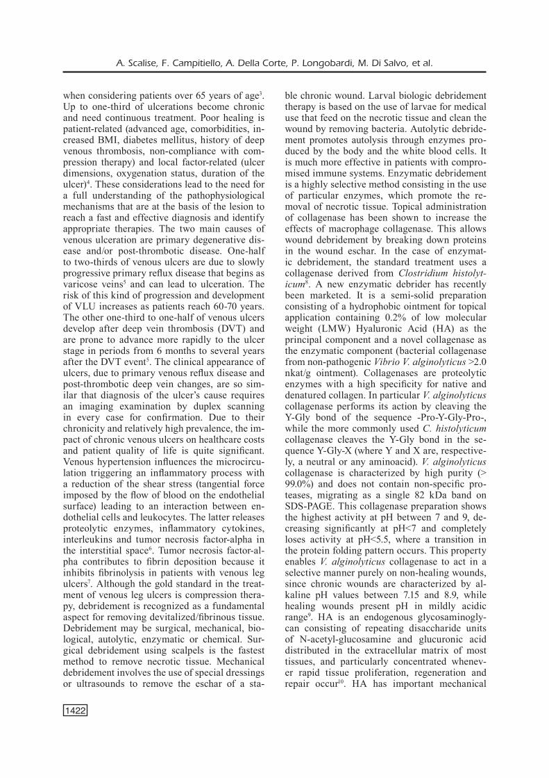
ter 15 days of LMW-HA-Collagenase treatment (Figures 2B and 2C). Consequently, after 30 days wounds treated with placebo ointment (Figure 3C) showed delayed healing. The debridement rate was 67.5 ± 6.7% (median 86.9%, range -148 to 100%) in the device group and 59.0±7.6% (me-dian 74.4%, range -253 to 100%) in the placebo group (Figure 4). The comparison between groups showed that the difference between the adjusted means of the treatment and the control group was 11.816% (95% CI: 0.348 to 23.283%), thus show-ing the difference was statistically significant (p = 0.0436) in favour of the LMW-HA-Collagenase group (Figure 4). The same analysis was also repeated in the PP population and it was found to be confirmatory with a debridement rate of 79.5% in the treatment group and 66.9% in the control group (p = 0.0031).
Observations on Secondary Outcome Measure
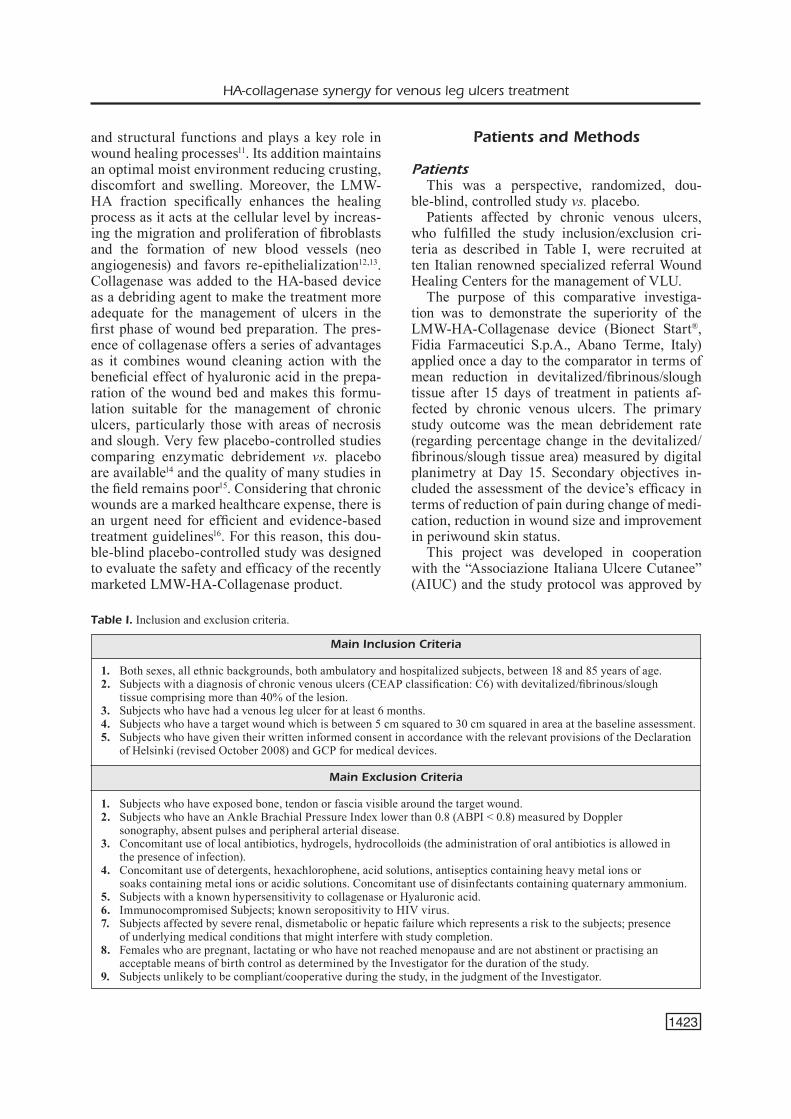
The mean debridement rate was higher in the LMW-HA-Collagenase group than in the control group at all other time points (Day 7, 21 and fi-nal visit). The LMW-HA-Collagenase group had more than three times the number of ulcers that achieved complete wound debridement by 15 days post-baseline visit compared to those in the
control arm, with 20 (39.2%) and 6 (12.5%) ulcers completely debrided respectively (p = 0.0025). Even at Day 7 and at the final visit the number of completely debrided wounds was higher in the LMW-HA-Collagenase group than in the com-parator group. This same analysis repeated in the PP population confirmed the LMW-HA-Colla-genase superiority at 15 days (p = 0.0002) and at the other time points, with 18.9% of ulcers com-pletely debrided at Day 7 in the treatment arm vs. 4.8% in the control arm (p = 0.0481) and 63.2% at the final visit in the treatment arm vs. 39.5% in the control arm (p = 0.0338) (Figure 5). The mean total lesion area decreased from baseline at all post-baseline time points in both groups. The number and percentage of patients with at least a 50% reduction in wound size at the different time points were analyzed. Although the reduction in total lesion area ≥ 50%, ≥ 75% and equal to 100% was generally observed in higher percentages of patients in the treatment group than in the control group at any time point in the overall sample, the differences between the groups were not sta-tistically significant in either the ITT or the PP population. Pain intensity during dressing change decreased from baseline at all post-baseline time points in both groups. The mean decrease from baseline was comparable in the two groups. The
Figure 4. Primary efficacy endpoint: Mean percentage debridement at day 15, ITT population.
A. Scalise, F. Campitiello, A. Della Corte, P. Longobardi, M. Di Salvo, et al.
1428
mean decrease from baseline to the last visit was -4.4 in the LMW-HA-Collagenase group and -2.5 in the placebo group. The frequency of adverse events and serious adverse events was very sim-ilar in the treatment and control groups and we recorded no significant differences between the two. As an additional measure of tolerability, the status of periwound skin was observed at each visit: the number and rate of patients with the presence of erythema/redness in the periwound skin decreased from baseline to the last visit in both groups, with no statistically significant
difference between the two arms. Furthermore, there were no cases of treatment-related serious adverse events in either group (Table III).
Discussion
Managing skin ulcers is based primarily on a holistic approach to the patient, with the identifi-cation of the disease responsible for the injury and the implementation of fast and effective diag-nostic-therapeutic treatments. A successful out-
Figure 5. Proportion of patients with complete debridement at different time points (PP).
Table III. Summary of treatment related adverse events, by SOC and PT (Safety Population).
Bionect Placebo Primary System Organ Class (SOC) Preferred Term (PT) (N = 58) (N = 55)
Number of patients with at least one related AE n (%) 7 (12.1%) 8 (14.5%)Skin and subcutaneous tissue disorders n (%) 5 (8.6%) 3 (5.5%)Erythema n (%) 3 (5.2%) 1 (1.8%)Venous ulcer pain n (%) 2 (3.4%) 2 (3.6%)Skin disorder n (%) 1 (1.7%) Injury, poisoning and procedural complications n (%) 3 (5.2%) 2 (3.6%)Thermal burn n (%) 2 (3.4%) 1 (1.8%)Wound secretion n (%) 1 (1.7%) Wound complication n (%) 1 (1.8%) General disorders and administration site conditions n (%) 2 (3.4%) 1 (1.8%)Pain n (%) 2 (3.4%) 1 (1.8%)Infections and infestations n (%) 1 (1.7%) 2 (3.6%)Infected skin ulcer n (%) 1 (1.7%) 1 (1.8%)Erysipelas n (%) 1 (1.8%) Musculoskeletal and connective tissue disorders n (%) 1 (1.8%) Pain in extremity n (%) 1 (1.8%)
HA-collagenase synergy for venous leg ulcers treatment
1429
come is dependent on the presence of a multidis-ciplinary team working closely together. Wound healing is a complex series of events that are in-terlinked and dependent on each other19. The first step in wound bed preparation (WBP) entails the global and coordinated management of the wound performed under an appropriate diagnostic proto-col to identify the cause of the wound, treat it and meet the patient’s needs. Subsequently, the prob-lems related to the wound are addressed and ef-fective actions undertaken to facilitate endoge-nous wound healing processes. In this context, WBP is essential for accelerating endogenous healing and facilitating the effectiveness of other therapeutic measures when the wound does not heal spontaneously. The recommendations con-tained in the European Wound Management As-sociation position document have become a touchstone in WBP literature on continuous wound debridement, moisture balance and reso-lution of bacterial overload. In brief, debridement is a fundamental act performing all the actions described above: it allows the removal of necrotic tissue and devitalized and senescent cells, reduc-es the bacterial overload and excess exudate and exerts a stimulating action on keratinocytes of the lesion edges. Enzymatic, autolytic, mechanical and surgical methods can be used and the choice of the debridement technique should be related to clinical and management criteria. Enzymatic de-bridement can be considered as a first choice due to its selectivity of action, the lack of pain follow-ing application, the low cost, and the relative speed of action. The clinical examination of the ulcer is the guide: the presence of fibrin and devi-talized tissue, without excessive exuding and signs of infection, is the indication for the use of enzymes. Proteolytic enzymes have been used for wound debridement for many years; the main advantage of their use in the debridement of pa-tients with chronic wounds is their easy, safe handling. Therapies are bloodless and generally considered quite painless. Because of the highly selective mode of action, this type of debride-ment can be appropriate to use in long-term care facilities and in homecare settings20. This study evaluated the activity of a novel collagenase product vs. placebo and demonstrated superiority of this new debriding agent. Based on the results of this trial, the LMW-HA-Collagenase device appears to have a rapid onset of action and achieves complete debridement in a shorter time compared to its comparator. Patients treated with LMW-HA-Collagenase achieved a faster wound
cleansing: after 15 days of treatment not only was the percentage debridement significantly higher in the treatment group (p = 0.0436) but complete wound debridement was also obtained by a sig-nificantly higher number of subjects (p = 0.0025). At baseline, the two arms of the study were sta-tistically comparable overall, although a small, albeit non-significant, difference between the ul-cer duration of the two groups was observed (15.5 months in the treatment group 29.7 months in the placebo group [p=0.07]). This difference may have influenced the clinical evidence. The physi-cian’s choice of a debriding agent is influenced by multiple factors including patient pain and the tolerability profile of the device; HA-Collagenase was well tolerated by patients: the prevalence and severity of adverse events were comparable to the comparator. Furthermore, it is well known that products containing proteolytic enzymes can lead to irritation of the periwound skin, with clinical signs of inflammation or discomfort. The present study demonstrated that LMW-HA-Collagenase is also safe from this point of view, which can probably be attributed in part to the HA protec-tive action on skin surrounding the ulcer. The purity and substrate selectivity of V. alginolyticus collagenase could partly be the reason why V. al-ginolyticus collagenase /LMW-HA ointment seems to be milder on healthy tissue and able to ensure the protection of the periwound skin. This is consistent with the findings obtained in a recent study21 that compared two different colla-genase-based ointments with mechanical de-bridement and that furthermore suggested a more rapid action of V. alginolyticus collagenase com-pared to C. histolyticum collagenase. Also, it must be mentioned that LMW-HA, apart from maintaining an optimal moist environment, pro-motes the healing process with proangiogenic effects, facilitating the migration and prolifera-tion of endothelial cells10 and fibroblasts11. Con-cerning keratinocytes, the main cell type in-volved in wound re-epithelialization, the role of LMW HA is even more interesting: an averaged 200kDa HA fraction has been demonstrated to induce a significant migration and proliferation of primary human keratinocyte cultures in an in vitro wound closure model, while higher or lower MW HA fractions were not effective22. Similarly, a topical application of a 0.2% 200kDa HA cream caused a significant skin thickening in patients with age- or corticosteroid-related skin atrophy23. Both responses were completely eliminated when hyaluronan receptor binding was blocked using

A. Scalise, F. Campitiello, A. Della Corte, P. Longobardi, M. Di Salvo, et al.
1430
specific antibodies and/or inhibitors. In our study, a tendency to enhanced re-epithelialization was found in the LMW-HA–Collagenase group, but the difference compared to the placebo group was not statistically significant. In any case, it must be underlined that the present study did not have a follow-up period to investigate long term ulcer healing and once complete debride-ment was achieved, patients left the study and no additional observation was scheduled. In all likelihood, LMW-HA achieves a better tolera-bility profile compared to products containing C. histolyticum collagenase alone, where the biological behavior of the collagenase is more likely to be associated with periwound skin ir-ritation and increased aggressiveness on the wound edges. As to pain experienced by the patient, the study results show that levels of perceived pain during change of medication at the target wound, evaluated by means of a visual analogue scale, were quite low (mean value: 8.5) at each visit and similar in both arms. This is in contrast with general clinical experience where change of medication proves to be the most painful moment for patients (Pain at Wound Dressing Changes, EWMA, 2002). Nevertheless, Gravante et al17 had al-ready reported that 87% of patients treated in his study with 0.2% HA-Collagenase referred no pain during dressing removal. These find-ings suggest a relationship with the ointment composition of both the active product and its vehicle, which has a softer texture than other collagenases resulting in less pain for the pa-tient and more frequent and easier applicabili-ty19. A weakness of this study may be the ab-sence of follow up. Moreover, it is worth re-membering that, for ethical reasons, compres-sion therapy, which is the mainstay of treatment of venous leg ulcers24, was guaranteed to all subjects. Compression is known to be a treat-ment that plays an essential role in itself in promoting healing and prolonging the recur-rence-free period after complete healing25 and this should be taken into consideration when evaluating the study healing outcomes. For all these reasons it is likely that a follow-up period would add validity to the healing frequency and this aspect needs to be considered for future trials. For this study, the quality of the wound fluid was assessed and the evaluation was made by a clinician. Wound assessment was per-formed by the use of a digital planimetry sys-tem. Arguably a more effective and accurate
monitoring of skin lesions should be performed measuring the complete status and evolution of the wound in an objective, precise, and repro-ducible manner. This level of monitoring could be achieved by using 3-dimensional scanners and systems based on active optical approaches26-28. However, these kinds of devices are generally not yet present in most outpatient clinics.
Conclusions
In this study, the percentage reduction in devitalized tissue was statistically significantly greater in patients randomized to LMW-HA-Col-lagenase therapy compared to vehicle after 15 days of treatment. The improvement was ob-served regardless of the age of the wound, the size of the wound, or compounding comorbid-ities reported in this cohort. Participants treat-ed with LMW-HA-Collagenase also reported a statistically significant higher complete debride-ment rate within 15 days of treatment and an improved healing rate, though not significant, over those treated with the comparator. Perform-ing fast and effective debridement, as well as avoiding pain and periwound skin irritation, can be considered the first step towards successful healing. Thus, with better effectiveness, good tolerability and less pain for patients, the prepa-ration of 0.2% of LMW-Hyaluronic Acid and V. alginolyticus collagenolytic enzyme used in the study holds great promise for the management of chronic ulcers.
AcknowledgementsThis project was developed in cooperation with the “Associ-azione Italiana Ulcere Cutanee” AIUC and 10 affiliated cen-ters. We thank all the participants who took part in this trial; the Principal Investigators, research nurses, and health care professionals who screened and recruited patients into the study, gathered trial data, and ensured the success of the tri-al. Principal investigators of the study were: Piero Bonadeo – Asl AI P.O. Tortona; Paolo Bortolotti – Department of Gen-eral Surgery ULSS 2 Lucca; Ferdinando Campitiello – De-partment of Medical, Surgical, Neurologic, Metabolic and Ageing Sciences, Second University of Naples; Aldo Cres-pi – Department of cure and prevention of vascular lesions, NH Novara; Michelangelo Di Salvo – Operative Unit of An-giology, “P.O. Vittorio Emanuele”, Catania; Ornella Forma – Department of Medical and infection control, San Raffa-ele Hospital, Milan; Giorgio Guarnera – Vascular Ulcer Unit, Department of Vascular Surgery and Pathology, Istituto Der-mopatico dell’Immacolata, Rome; Pasquale Longobardi – Wound care Centre, Hyperbaric center of Ravenna, Marco Romanelli – Dermatology department of Santa Chiara Hos-pital, Pisa; Alessandro Scalise – Clinic of Plastic and Recon-
HA-collagenase synergy for venous leg ulcers treatment
1431
structive Surgery, Department of Clinical and Experimental Medicine, Ancona United Hospitals.
FundingFidia Farmaceutici S.p.A. funded, supplied study materials and carried out the trial.
Conflict of InterestThe Authors declare that they have no conflict of interests.
References
1) Valencia ic, Falabella a, Kirsner rs, eaglstein WH. Chronic venous insufficiency and venous leg ul-ceration. J Am Acad Dermatol 2001; 44: 401-421.
2) reicHenberg J, DaVis M. Venous ulcers. Semin Cu-tan Med Surg 2005; 24: 216-226.
3) etuFugH cn, PHiliPs tJ. Venous ulcers. Clin Derma-tol 2007; 25: 121-130.
4) labroPoulos n, Wang eD, lanier st, KHan su. Fac-tors associated with poor healing and recurrence of venous ulceration. Plast Reconstr Surg 2012; 129: 179-186.
5 abbaDe lPF, lastória s. Venous ulcer: epidemiolo-gy, physiopathology, diagnosis and treatment. Int J Dermatol 2005; 44: 449-456.
6) raFFetto JD. Inflammation in chronic venous ul-cers. Phlebology 2013; 28: 61-67.
7) clauDy al, MirsHaHi M, soria c, soria J. Detection of undegraded fibrin and tumor necrosis factor-al-pha in venous leg ulcers. J Am Acad Dermatol 1991; 25: 623-627.
8) Falconi M, teti g, Zago M, galanZi a, brescHi l, Pelot-ti s, ruggeri a, MaZZotti g. Influence of a commer-cial tattoo ink on protein production in human fibro-blasts. Arch Dermatol Res 2009; 301: 539-547.
9) PerciVal sl, Mccarty s, Hunt Ja, WooDs eJ. The ef-fects of pH on wound healing, biofilms, and an-timicrobial efficacy. Wound Repair Regen 2014; 22: 174-186.
10) West D, HaMPson i, arnolD F, KuMar s. Angiogene-sis induced by degradation products of hyaluron-ic acid. Science 1985; 228: 1324-1326.
11) cHen WJ, abatangelo g. Functions of hyaluronan in wound repair. Wound Repair Regen 1999; 7: 79-89.
12) ProsDociMi M, beVilacqua c. Exogenous hyaluronic acid and wound healing: an updated vision. Pan-minerva Med 2012; 54: 129-135.
13) graVante g, sorge r, Merone a, taMisani aM, Di lonarDo a, scalise a, DoneDDu g, MelanDri D, stracuZZi g, onesti Mg, cerulli P, Pinn r, esPosito g. Hyalomatrix PA in burn care practice: results from a national retrospective survey, 2005 to 2006. Ann Plast Surg 2010; 64: 69-79.
14) MeKKes Jr, Zeegelaar Je, WesterHoF W. Quantita-tive and objective evaluation of wound debriding properties of collagenase and fibrinolysin/desox-
yribonuclease in a necrotic ulcer animal model. Arch Dermatol Res 1998; 290: 152-157.
15) Price P, gottruP F, abel M. EWMA study recom-mendations for clinical investigations in leg ul-cers and wound care. J Wound Care 2014; 23: S1-S36.
16) Doerler M, reicH-scHuPKe s, altMeyer P, stücKer M. Impact on wound healing and efficacy of various leg ulcer debridement techniques. J Dtsch Der-matol Ges 2012; 10: 624-632.
17) graVante g, sorge r, giorDan n, georgescu sr, Morariu sH, stoicescu i, clatici V. Multicenter clin-ical trial on the performance and tolerability of the Hyaluronic acid-collagenase ointment for the treatment of chronic venous ulcers: a preliminary pilot study. Eur Rev Med Pharmacol Sci 2013; 17: 2721-2727.
18) onesti Mg, FioraMonti P, carella s, Fino P, sorVillo V, scuDeri n. A new association between hyaluronic acid and collagenase in wound repair: an open study. Eur Rev Med Pharmacol Sci 2013; 17: 210-216.
19) Position DocuMent: Wound Bed Preparation in Practice. European Wound Management Associ-ation (EWMA), 2004.
20) Strohal R, Apelqvist J, Dissemond J, Jordan O’Brien J, Piaggesi A, Rimdeika R, Young T. EW-MA document: debridement. J Wound Care 2013; 22: S1-S52.
21) onesti Mg, FioraMonti P, Fino P, sorVillo V, carel-la s, scuDeri n. Effect of enzymatic debridement with two different collagenases versus mechani-cal debridement on chronic hard-to-heal wounds. Int Wound J 2016; 13: 1111-1115.
22) gHaZi K, Deng-PicHon u, Warnet JM, rat P. Hyalu-ronan fragments improve wound healing on in vit-ro cutaneous model through P2X7 purinoreceptor basal activation: role of molecular weight. PLoS One 2012; 7: e48351.
23) Kaya g, tran ct, sorg o, HotZ r, granD D, carraux P, DiDierJean l, staMenKoVic i, saurat JH. Hyaluronate fragments reverse skin atrophy by a CD44-de-pendent mechanism. PLoS Med 2006; 3: e493.
24) nair b. Compression therapy for venous leg ul-cers. Indian Dermatol Online J 2014; 5: 378-382.
25) scotton MF, Miot Ha, abbaDe lPF. Factors that in-fluence healing of chronic venous leg ulcers: a retrospective cohort. An Bras Dermatol 2014; 89: 414-422.
26) roManelli M, Dini V, biancHi t, roManelli P. Wound assessment by 3-dimensional laser scanning Arch Dermatol 2007; 143: 1333-1344.
27) scalise a, bottoni M, scalise l, Pierangeli M, Di bene-Detto g. Evaluation of the efficacy of an innova-tive system for measuring chronic wounds: mo-bile wound analyzer. Acta Vulnologica 2015; 13: 15-23.
28) neuenDorF aD, Pierangeli M, Forlini W, taleVi D, as-tolFi M, carboni a, grassetti l, Petrucci e, PonZio i, Di beneDetto g, bertani a, scalise a. L’importanza della misurazione nelle ferite difficili. Acta Vulno-logica 2007; 3: 95.