environmental enrichment causes a global … · environmental enrichment causes a global ......
TRANSCRIPT

ORIGINAL RESEARCH ARTICLEpublished: 08 August 2013
doi: 10.3389/fncel.2013.00124
Environmental enrichment causes a global potentiation ofneuronal responses across stimulus complexity and laminaof sensory cortexDasuni S. Alwis and Ramesh Rajan*
Department of Physiology, Monash University, Clayton, VIC, Australia
Edited by:
Andreas Frick, Institut National de laSanté et de la Recherche Médicale,France
Reviewed by:
Dirk Feldmeyer, RWTH AachenUniversity, GermanyFrancesca Prestori, University ofPavia, Italy
*Correspondence:
Ramesh Rajan, Department ofPhysiology, Monash University,Wellington Road, Clayton, VIC 3800,Australiae-mail: [email protected]
Enriched social and physical housing produces many molecular, anatomical,electrophysiological and behavior benefits even in adult animals. Much less is known ofits effects on cortical electrophysiology, especially in how sensory cortex encodes thealtered environment, and extant studies have generally been restricted to neurons in inputlaminae in sensory cortex. To extend the understanding of how an enriched environmentalters the way in which cortex views the world, we investigated enrichment-inducedchanges in neuronal encoding of sensory stimuli across all laminae of the rat barrel cortexreceiving input from the face whisker tactile system. Animals were housed in Enriched(n = 13) or Isolated housing (n = 13) conditions for 8 weeks before extracellular recordingswere obtained from barrel cortex in response to simple whisker deflections and whiskermotions modeling movements seen in awake animals undertaking a variety of differenttasks. Enrichment resulted in increases in neuronal responses to all stimuli, ranging fromthose modeling exploratory behavior through to discrimination behaviors. These increaseswere seen throughout the cortex from supragranular layers through to input Layer 4 andfor some stimuli, in infragranular Layer 5. The observed enrichment-induced effect isconsistent with the postulate that enrichment causes shift in cortical excitatory/inhibitorybalance, and we demonstrate this is greatest in supragranular layers. However, we alsoreport that the effects are non-selective for stimulus parameters across a range of stimuliexcept for one modeling the likely use of whiskers by the rats in the enriched housing.
Keywords: EE, barrel cortex, electrophysiology, hyperexcitability
INTRODUCTIONEnvironmental enrichment (EE) for laboratory animals, in hous-ing that allows more social interaction and cognitive and motorchallenges compared with standard housing (Hebb, 1947; VanPraag et al., 2000; Nithianantharajah and Hannan, 2006), pro-vides a remarkable array of cognitive and behavioral benefits innormal development and in adulthood (Rosenzweig and Bennett,1996; Buonomano and Merzenich, 1998; Nilsson et al., 1999; VanPraag et al., 2000; Zimmermann et al., 2001; Lewis, 2004; Bruel-Jungerman et al., 2005; Li and Tang, 2005; Meshi et al., 2006;Nithianantharajah and Hannan, 2006; Sale et al., 2009; Veyracet al., 2009).
Corresponding to these cognitive and behavioral changes, EEinduces anatomical and molecular changes in the brain. The for-mer include neuro-, glio-, synapto- and angio-genesis, decreasedcell death, and increases in receptor numbers, transmitter synthe-sis, dendritic length, and branching, and thickness of the cerebralcortex (Holloway, 1966; Diamond et al., 1972; Globus et al., 1973;Greenough and Volkmar, 1973; Uylings et al., 1978; Sirevaag andGreenough, 1987; Rosenzweig and Bennett, 1996; Buonomanoand Merzenich, 1998; Van Praag et al., 2000; Li and Tang, 2005;Nithianantharajah and Hannan, 2006; Sale et al., 2009), andincreased levels of neurotransmitters, such as acetylcholine and
noradrenaline, which promote neurogenesis and plasticity (Poret al., 1982; Rosenzweig and Bennett, 1996; Soares et al., 1999).Molecular changes include increases in levels of neurotrophicand growth factors, including brain-derived neurotrophic factor,nerve growth factor, and vascular endothelial growth factor whichcontribute to neuronal proliferation, development, signaling, sur-vival, and ultimately, plasticity (Falkenberg et al., 1992; Phamet al., 1999; Ickes et al., 2000; During and Cao, 2006). Enhancedsynaptogenesis after EE is coupled to an increase in synaptic pro-teins such as postsynaptic density-95 protein and synaptophysin(Frick and Fernandez, 2003; Nithianantharajah et al., 2004). EE-induced changes can occur rapidly—e.g., even structural changes(viz., number of excitatory and inhibitory synapses in Layer 4 ofbarrel cortex) can occur with just 24 h of EE (Landers et al., 2011).
Much less is known about EE effects on neuronal func-tionality, especially to behaviorally relevant inputs or outputs.Exposure to EE results in increased field potentials and greaterexcitatory post-synaptic potential slopes in in vitro hippocam-pal slice recordings (Sharp et al., 1985; Green and Greenough,1986) and auditory cortex slices show increases in excitatory post-synaptic currents in layers 2/3, likely from enhanced glutamater-gic transmission (Nichols et al., 2007). In extracellular recordingsfrom auditory, visual and somatosensory cortices, EE effects
Frontiers in Cellular Neuroscience www.frontiersin.org August 2013 | Volume 7 | Article 124 | 1
CELLULAR NEUROSCIENCE

Alwis and Rajan Sensory cortical changes after EE
include increased sensitivity and responsiveness in granular orsupragranular layers (Engineer et al., 2004; Polley et al., 2004;Mainardi et al., 2010; Jakkamsetti et al., 2012; Tognini et al.,2012). Thus, in auditory cortex, there are significant increasesin spontaneous and stimulus-evoked responses to sounds, longerresponse latency, and narrower receptive field (RF) bandwidths;additionally, improved temporal information processing is sug-gested by increased responsiveness to higher temporal modula-tion rates and increased paired-pulse depression (Engineer et al.,2004; Percaccio et al., 2005, 2007; Jakkamsetti et al., 2012).Somatosensory cortex maps are made more precise after EEthrough decreases in neuronal RF sizes (Polley et al., 2004; Frostig,2006) and improved response selectivity (Coq and Xerri, 1998;Polley et al., 2004). In visual cortex, EE causes a decrease in cor-tical inhibition (Scali et al., 2012) promoting ocular dominanceplasticity in aged rats, and accelerating development of visual cor-tex in animals exposed to EE from birth (Cancedda et al., 2004).The general view is that the core change induced by EE to producethese varied outcomes in cortical neurophysiology and functionalencoding is a shift in cortical excitation/inhibition (E/I) ratios(Beaulieu and Colonnier, 1987; Coq and Xerri, 1998; Polley et al.,2004; Sale et al., 2009; Baroncelli et al., 2011 see Discussion). Thegreat majority of studies of such cortical neuronal properties havebeen restricted to thalamo-recipient layers and little is known ofchanges in other layers, especially the supra-granular layers 2 and3 (L2 and 3) that are considered to be a “privileged substrate”for consolidating EE-induced cortical plasticity (Nichols et al.,2007) with their great capacity for amplification of changes occur-ring in the thalamo-recipient layers (Komai et al., 2006; Brecht,2007; Feldmeyer et al., 2013; Petersen and Crochet, 2013). Theeffects observed in thalamo-recipient layers may not simply pre-dict effects in upper layers as indicated by the observation in themature barrel cortex that deprivation-induced plasticity decreasesLayer 4 feed-forward excitation to L2/3 inhibitory neurons butimproves inhibition to L2/3 pyramidal cells (House et al., 2011),resulting in E/I balance being maintained in L2/3. Further thescattered body of data on the neurophysiological consequencesof EE on cortex are generally limited to descriptions of RF sizesand response strength, generally to simple stimuli. We have nowattempted to redress these deficits in knowledge by investigatingEE related changes in circuit dynamics of neurons from supra-granular layer 2 through to infra-granular layer 5 of rat barrelcortex to simple stimuli and to complex stimuli that model theways in which awake trained rats use their whiskers in manynatural behaviors.
MATERIALS AND METHODSANIMALSMale Sprague–Dawley rats (aged ∼7 weeks, weight 250 g) wereobtained from Monash Animal Services (MAS) and littermatesrandomly assigned to either Isolated (Isol.; n = 13) or Enriched(EE; n = 13) housing conditions, always in a 12 h light/dark cyclewith ad libitum food and water, for 8–10 weeks; from week 8onwards, animals were removed for terminal electrophysiologi-cal experiments conducted over a 2 week period to ensure thatthere was no confound from ageing. All experiments were con-ducted in accordance with guidelines from the National Health
and Medical Research Council and received approval from theMonash University Standing Committee on Ethics in AnimalExperimentation.
HOUSING CONDITIONSRats in EE housing were housed in groups of 3 in a large (69 ×60 × 270 cm) run which featured a front-facing Plexiglas walland 3 steel walls with a steel base and a wire mesh cage lid. Thecage floor was covered with wood shavings and contained numer-ous plastic and metal toys and objects of various colors, sizes,and textures. The objects used for enrichment were kept constantbut their locations were re-arranged every 2.5 days. The Isolatedhousing rats were housed individually in standard sized cages(31 × 24 × 45 cm) with wood shavings and shredded paper, butno toys.
EXTRACELLULAR RECORDINGS FROM BARREL CORTEXCortical responses from animals in the two housing conditionswere recorded 8–10 weeks after commencement of housing inthe test condition. Extracellular recordings were taken from theposteromedial barrel subfield region of somatosensory cortex(PMBSF; the so-called barrel cortex), as detailed elsewhere (Rajanet al., 2006, 2007; Alwis et al., 2012). Briefly, animals were anes-thetized with 5% halothane (Sigma Aldrich, USA) mixed in O2
(O2 flow rate = 1 ml/min) and tracheotomised for mechanicalventilation (2.5–3.5 mL tidal volume, 72–80 breaths/min; bothdependent on animal size) to maintain anesthesia thereafter with0.5–2% halothane mixed in O2 (O2 flow rate = 0.3 ml/min). Athermostatically-controlled heat pad with feedback from a rectalprobe (Fine Science Tools Inc., U.S.A) was used to maintain bodytemperature at 37–38◦C.
Once anesthesia was established, as determined from absentpalpebral reflexes or responses to strong forepaw pinching, theskull was exposed and anchored to a head bar with a screwand dental cement. Then a section of skull ∼5 mm in diame-ter, located above barrel cortex (∼2 mm caudal of bregma and6 mm lateral of the midline), was removed and the exposed cortex(dura intact) covered with silicone oil. Recordings were obtainedusing a parylene-coated tungsten microelectrode (2–4 M� resis-tance; FHC, ME, U.S.A) which was moved using a fast-steppingmicro-drive (Kopf Instruments, California, U.S.A). Initially, theelectrode was advanced to a depth between 600 and 800 μm fromthe surface to allow determination of the Principal Whisker (PW)by manual deflection of the whiskers using a hand held probe.Here the PW was classified as the whisker which produced thegreatest neuronal firing response to manual whisker deflection(with electrode output monitored aurally through speakers aswell as on an oscilloscope screen) and, in barrel cortex, was alwaysunequivocally identifiable at this depth. Where drive was weak, ora result of multi-whisker activity, the electrode was retracted anda new penetration was made until a single PW could be identified;the electrode output was monitored (see below) after amplifi-cation and filtering, on an oscilloscope and through speakers. Ifstrong PW drive was obtained (see Alwis et al., 2012 for details)the electrode was then retracted to the cortical surface undervisual control and zeroed here. Then it was advanced systemat-ically to record from neurons at a number of different depths,
Frontiers in Cellular Neuroscience www.frontiersin.org August 2013 | Volume 7 | Article 124 | 2

Alwis and Rajan Sensory cortical changes after EE
using stimuli delivered under computer control to the PW. Thefirst recording was made at a depth of about 150 μm from thesurface; thereafter recordings were made at regular intervals toensure data was collected from all animals from the following lay-ers: Layer 2 (150–300 μm); Upper Layer 3 (350–500 μm); DeepLayer 3 (550–700 μm); Layer 4 (750–1000 μm); and Layer 5(1100–1400 μm). To ensure that data representation from deeperlayers was not compromised by the later recordings perforce ofthis strategy, in some cases we advanced the electrode first toLayer 4 and collected data from that layer and then from Layer5 before retracting back to the surface to advance systematicallyas above to obtain data from the other layers. We have previouslydemonstrated that recorded responses in halothane anesthetizedanimals, using the same experimental techniques outlined in thepresent study and in our previous work (see Rajan et al., 2006;Alwis et al., 2012; Johnstone et al., 2013), were comparable tothose seen in awake animals, where the pattern of temporal andspatial responses to whisker deflection were similar in both theanesthetized and awake states (Rajan et al., 2006; Maravall et al.,2007).
Neural signals from the microelectrode were treated asdescribed previously (see Rajan et al., 2006, 2007; Alwis et al.,2012 for details), being amplified, band-pass filtered (0.3–10 kHz)and displayed on an oscilloscope, and through speakers for auralmonitoring of neural activity. A Schmitt trigger box was used toset a voltage trigger level, while monitoring on the oscilloscopeat a level 2× noise level, for neuronal cluster activity record-ings. Trigger crossings generated digital pulses that were fed toa PC; this computer used Spike 2 software (CED, UK) for stim-ulus generation and/or delivery (Alwis et al., 2012) and stimulustrigger events were stored along with the Schmitt trigger pulses.The Spike2 software was also used to generate on-line displaysof rasters of spike occurrences and peristimulus time histograms(PSTHs). A minimally-filtered copy of the signals recorded by theelectrode was also stored for any offline analysis if needed later(Rajan et al., 2006).
CONTROLLED WHISKER DEFLECTIONS FOR QUANTITATIVE BARRELCORTEX DATA COLLECTIONFor quantitative data recording, the PW was threaded through ahole at the end of a motor-controlled lever arm system positioned5 mm from the mystacial pad to deflect the PW in computer-controlled patterns. At each recording site, voltage triggers wereset at a level 2× noise level to obtain responses from neuronalclusters (of 4–6 neurons, as determined by online spike sortingusing Spike 2 software). Responses were first characterized forlaminar location using a suite of 3 trapezoidal stimuli, where onlythe onset ramp velocity was varied (60, 150, 400 mm/s; Alwiset al., 2012). This suite of trapezoids was presented for 150–300repetitions in a pseudo-random manner.
Then a series of four complex “naturalistic” whisker deflec-tions, obtained from studies in which whisker motion wasrecorded in awake behaving rats, were played out from text fileswhich stored stimulus characteristics. These stimuli have beendescribed in detail in our previous studies (Alwis et al., 2012;Johnstone et al., 2013); the four waveforms were those seen inwhisker motion across a smooth and rough surface (Ritt et al.,
2008); when rats made contact with a rod placed in the path ofthe whiskers (Hartmann et al., 2003); and in head-fixed rats thatwere engaging in “free” whisking (Gao et al., 2001). Details of theacquisition and conversion of these waveforms to whisker deflec-tion patterns have been previously described by our group (Alwiset al., 2012; Johnstone et al., 2013). Each suite consisted of 10stimulus amplitudes, from the lowest amplitude of 0.2 mm, andthen continuing from 0.4 to 3.6 mm in 0.4 mm steps. Each suite of10 stimulus amplitude was presented 50 times, in pseudo-randomorder across successive presentations.
DATA ANALYSISCluster responses were segregated into lamina by depth (from thecortical surface) as noted above: Layer 2 (150–300 μm); UpperLayer 3 (350–500 μm); Deep Layer 3 (550–700 μm); Layer 4(750–1000 μm); and Layer 5 (1100–1400 μm). Data were repre-sented as firing rate (in spikes/s) in 1 ms bins over the periodfrom 200 ms prior to stimulus onset until 100 ms post stim-ulus offset. The data collected in the 200 ms pre-stimulus binwas used to calculate spontaneous firing rates, which were sub-tracted from responses during the rest of the data collectionperiod to correct for the spontaneous firing rate. The sponta-neous activity-corrected firing rates were used for all subsequentanalyses.
Offline analysis was conducted to generate population peri-stimulus time histograms (PSTHs) showing the pattern of pop-ulation responses within a lamina, in animals housed in eitherEnrichment or Isolation. PSTHs were obtained by averagingcluster responses within a lamina across each presentation of astimulus. A 5-point weighted moving average was then applied tothe data to smooth out noise and a grand PSTH was produced byaveraging the data across all multi-units.
For quantitative analysis for each stimulus, we used only datafrom multi-neuronal clusters considered to be responsive to thatstimulus. Clusters were classified as responsive if their responserates were significantly greater (more than 1.4 SD >) than sponta-neous firing rates at more than two consecutive stimulus velocities(for the trapezoidal stimuli) or at more than two consecutivestimulus amplitudes (for the four naturalistic stimuli). We thenextracted the following metrics for each stimulus: peak firingrate, area under the curve, latency to peak and half-peak width.These calculations were done using specific counting windowsfor each stimulus: for the trapezoidal and object contact stim-uli, a counting window from 5 to 50 ms after stimulus onset wasused, for the surface texture discrimination stimuli, a 5–30 mscounting window, while the free whisking stimulus was analysedusing a 5–200 ms counting window. Almost identical effects wereseen for the firing rate measures (peak firing rate and area underthe curve), and almost identical effects for the timing measures(latency to peak and half-peak width) and hence, we present dataonly from the peak response (PFR) and the latency to the PFR(LPFR) for each stimulus in its designated analysis window.
Statistical analysis was carried out using two-way repeatedmeasures ANOVAs to determine laminar-specific differences,with peak firing rates or latencies being the dependent vari-able and the independent between-animal factors being housingcondition and stimulus amplitude/velocity. When the ANOVA
Frontiers in Cellular Neuroscience www.frontiersin.org August 2013 | Volume 7 | Article 124 | 3

Alwis and Rajan Sensory cortical changes after EE
revealed a significant main factor effect of Group (housingcondition) or Stimulus parameter (Amplitude/Velocity) or a sig-nificant interaction term between these two factors, post-hocBonferroni tests were used to where the differences lay.
RESULTSDATABASE AND MEASURES OF NEURONAL RESPONSESElectrophysiological recordings were obtained from 13 ratshoused in Isolation and 13 rats in EE housing, from multi-unit clusters in layers 2 (L2), Upper layer 3 (U3), Deep layer 3(D3), Layer 4 (L4), and Layer 5 (L5), in response to simple andcomplex, “naturalistic” whisker motion patterns. Only data fromresponsive multi-neuronal clusters (see Materials and Methodsfor definition) were extracted for analysis. We present below, foreach stimulus, laminar-specific data on the response patterns ofclusters in the two housing conditions, and then data for twomajor metrics of the responses.
To visualize the laminar-specific response pattern of neu-ronal clusters to any specific stimulus, Grand Peri-Stimulus TimeHistograms (GPSTHs), which are defined as histograms of firingrate against time from stimulus onset, were generated. For this,responses of each cluster during and after the stimulus periodwere corrected for the spontaneous activity measured in that clus-ter in the 200 ms period prior to stimulus onset. Then, for rats ineach housing condition, responses from all responsive clusters ina specific lamina were averaged to generate the GPSTH of firingrate against stimulus period for that lamina. Finally, to quantifythe effects, analysis windows were defined for each stimulus inrelation to aspects of that stimulus (e.g., the onset period, or thepost-stimulus offset period; defined in Materials and Methods foreach stimulus), and for these windows we present two represen-tative quantitative metrics from all responsive clusters: (1) peakexcitatory firing rate (PFR) and (2) latency to the peak (LPFR). ThePFR was the across-clusters average peak firing rate during thestimulus period as seen in the population GPSTH and expressedas spikes/s, and the LPFR was the time from stimulus onset to thispeak in the GPSTH. Note that other firing rate and timing valueswere also calculated (see Materials and Methods) but the effectswere the same across all firing rate measures or across all timingmeasures and hence only PFR and LPFR are presented here.
CHANGES IN NEURONAL RESPONSES TO COMPLEX WHISKERSTIMULI AFTER EXPOSURE TO EEPopulation responses to a simple whisker motion stimulusTrapezoidal whisker displacements. The first stimulus applied tothe PW of each cluster was a suite of 3 trapezoid stimuli, withone of three onset ramp (whisker protraction) velocities (60, 150,and 400 mm/s) which we have shown (Rajan et al., 2007) toelicit population responses that cover the range from low neu-ral response rates (velocity of 60 mm/s) to saturation of neuralresponses (velocity of 400 mm/s). Data were obtained to thesestimuli from 86 responsive clusters in Isolation-housed animalsand 84 responsive clusters in EE animals; there was no differencebetween the two housing conditions in the number of responsiveclusters per layer (χ2 = 3.6, df = 4, p > 0.05).
The pattern of population responses to the trapezoid stimuliare exemplified in Figure 1A by the GPSTHs from the highest
onset ramp velocity of 400 mm/s for all cortical laminae. InIsolated housing animals, responses to the trapezoid consisted of awell-defined response during the onset ramp, evident in all layers,and a second peak, poorly defined in all layers, corresponding tostimulus offset. The GPSTHs from EE animals showed the sametwo peaks, but with the offset response now being well-defined,and with a marked increase in firing rates in all cortical layers.Increases in peak firing rate were observed for both onset andoffset responses, while tonic excitation in the period between theonset and offset responses was also increased in the supragranularL2–D3 in EE animals.
An analysis window of 5–50 ms from stimulus onset, coveringthe entire onset peak response at all three onset ramp veloci-ties, was used to obtain quantitative metrics on response strength(Peak firing rate, PFR, in the onset response; Figure 1B) and tim-ing (Latency to Peak firing, LPFR; Figure 1C). Each dataset wasanalysed using Two-Way repeated measures ANOVAs, with hous-ing condition and lamina being between-subjects variables andonset ramp velocity being the within-subjects variable. In all cor-tical layers, the PFRs in EE animals were significantly higher thanPFRs in Isolation-housed animals (p < 0.05; see SupplementaryTable S1 for statistical details). In L2, a significant increase infiring rate was seen at the highest two velocities; in U3 only atthe highest velocity; at all velocities in Deep Layer 3 and 4; andat the two lowest velocities in L5 (see Supplementary Table S1for details: Two-Way ANOVA, Bonferroni post-hoc, p < 0.05).However, this EE-related marked increase in firing strength inall layers occurred with no changes in the latency to the peakonset response, LPFR (Figure 1C) at any onset ramp velocity inany cortical layer (p > 0.05; see Supplementary Table S1).
Responses to complex whisker motion stimuliObject contact stimulus. Hartmann et al. (2003; Figure 8A fromHartmann paper) imaged whisker motion in an unrestrained ratthat was using its whiskers to brush past a metal post to obtain aliquid reward. We extracted a significant segment of this “objectcontact” stimulus waveform (see Materials and Methods; Alwiset al., 2012) and applied this segment to the PW in record-ings from 79 responsive clusters in Isolation-housed animalsand 82 responsive clusters in EE animals. There was no differ-ence between the two housing groups in numbers of responsiveclusters in each layer (χ2 = 3.5, df = 4, p > 0.05).
We illustrate the pattern of neuronal responses to this complexstimulus using Grand PSTHs (GPSTHs) obtained at the high-est stimulus amplitude (3.6 mm) from all responsive clusters ina layer for rats in a particular housing (Figure 2A). In Isolation-housed animals, the GPSTH generally consisted of two responsepeaks corresponding to stimulus onset and offset, with low lev-els of tonic excitation in between. Specifically, GPSTHs from L2and U3 showed poorly defined onset responses but larger, moredefined peaks at stimulus offset (Figure 2A), whereas in D3, L4,and L5, clearly-defined onset and offset responses were present,with the onset response consisting of two peaks for all eightstimulus amplitudes >0.4 mm whisker deflection. Following thestimulus offset response in L4 and L5, responses decreased tobelow spontaneous activity rates (recorded in the 200 ms pre-stimulus period) and were classified as inhibitory responses. This
Frontiers in Cellular Neuroscience www.frontiersin.org August 2013 | Volume 7 | Article 124 | 4

Alwis and Rajan Sensory cortical changes after EE
FIGURE 1 | EE effects on pattern, strength and timing of responses
evoked by simple trapezoidal stimuli. For this simple stimulus, responsestrength significantly increased in all laminae after EE, with no changes inresponse timing. (A) Population Grand PSTHs in response to the trapezoidwith the fastest onset ramp velocity (400 mm/s) in a specific lamina(indicated to left of panel) in EE and Isolated animals. Laminardesignations: L2, Layer 2; U3, Upper Layer 3; D3, Deep Layer 3; L4,Layer 4; L5, Layer 5. Each Grand PSTH was generated by averagingresponses across all responsive clusters in that lamina. The analysis
window used to extract response metrics is represented by the grayshaded box (5–50 ms). Stimulus waveform is presented above both panels.(B) Peak firing rate (PFR) and (C) Latency to peak (LPFR) extracted fromthe onset response to simple trapezoidal stimuli from clusters in EEanimals (gray circles) and in Isolated animals (black squares). Datarepresents averages from all responsive clusters (±SEM) at all tested rampvelocities, separated by cortical lamina. Each row of data comes from thesame lamina as designated by the labels on the left; cluster numbers foreach layer are listed in the key in the last column. ∗p < 0.05.
same general pattern of activity was seen in EE animals but with amarked increase in onset and offset excitation in all layers exceptL5. Post-offset inhibition, as evidenced by a decrease in firingrate below spontaneous firing rate was now clearly evident bothduring and after the stimulus in all layers except L2. In general,compared to neuronal responses in Isolation-housed animals, EEanimals showed increased excitation in L2–4 for onset and offsetresponses, and minor changes in L5.
Quantitative metrics of population responses to the objectcontact stimulus were extracted using an analysis window of5–50 ms from stimulus onset, encompassing the entire onsetresponse. For the peak excitatory firing rate (PFR) in thisonset window (Figure 2B), Two-Way repeated measures ANOVAs(between-subjects variables: housing condition and lamina;within-subjects variable: stimulus amplitude) revealed significantdifferences (p < 0.05; see Supplementary Table S2 for statistical
details) between Isolated housing and EE animals in L2–4 butno differences in L5. The PFR was higher in EE animals than inIsolated housing animals in all layers, with significant increasesat the four highest stimulus amplitudes in L2 (2.4–3.6 mm),at all but one intermediate amplitude (0.8 mm) in U3, and atall amplitudes in D3 and L4 (Two-Way ANOVA, Bonferronipost-hoc, p < 0.05). For the onset response timing, measuredas the latency to the onset peak (LPFR), Two-Way ANOVAsfound no significant differences between Isolation-housed andEE animals in L2–4 (p < 0.05; Figure 2C; SupplementaryTable S2): in both housing conditions LPFR decreased sys-tematically with increasing stimulus amplitude. There was asignificant housing condition effect but no housing × ampli-tude interaction in L5, reflecting a shorter LPFR in EE animalswhen compared with Isolation-housed animals, at all stimulusamplitudes.
Frontiers in Cellular Neuroscience www.frontiersin.org August 2013 | Volume 7 | Article 124 | 5

Alwis and Rajan Sensory cortical changes after EE
FIGURE 2 | EE effects on pattern, strength and timing of responses
evoked by the object contact stimulus. For this complex stimulus,response strength significantly increased in all layers from Layer 2 toLayer 4 after EE, with no changes in response timing. (A) PopulationGrand PSTHs in response to the trapezoid with the fastest onset rampvelocity (400 mm/s) in a specific lamina (indicated to left of panel) in EEand Isolated animals. Laminar designations: L2, Layer 2; U3, UpperLayer 3; D3, Deep Layer 3; L4, Layer 4; L5, Layer 5. Each Grand PSTHwas generated by averaging responses across all responsive clusters inthat lamina. The analysis window used to extract response metrics is
represented by the gray shaded box (5–50 ms). Stimulus waveform ispresented above both panels. Inset shows a magnified view ofpost-stimulus inhibition (B) Peak firing rate (PFR) and (C) Latency topeak (LPFR) extracted from the onset response to simple trapezoidalstimuli from clusters in EE animals (gray circles) and in Isolated animals(black squares). Data represents averages from all responsive clusters(±SEM) at all tested stimulus amplitudes, separated by cortical lamina.Each row of data comes from the same lamina as designated by thelabels on the left; cluster numbers for each layer are listed in the keyin the last column. ∗p < 0.05.
Thus, as in the case of the simpler trapezoid stimuli, for this“object contact” stimulus waveform, response strength in L2–4was greater at all or most stimulus amplitudes in EE animals thanin Isolated housing controls. Generally, there were no differencesin response timing between Isolation-housed and EE animals inall layers except L5, where LPFR was found shorter in EE animals.
Surface texture discrimination stimuli.
Smooth surface discrimination whisker motion. As in our previ-ous studies (Alwis et al., 2012; Johnstone et al., 2013) our suite of
complex whisker stimuli included two motion patterns (recordedby Ritt et al., 2008) which mimic whisker motion in awake, unre-strained rats trained to discriminate between smooth and roughsurface textures. First, we consider the responses to the whiskerstimulus which mimics a significant portion of whisker motionacross a smooth surface (Ritt et al., 2008). This stimulus was usedwhile recording from 78 responsive clusters in Isolation-housedanimals and 85 responsive clusters in EE animals (no differencebetween groups in number of responsive clusters in every layer:χ2 = 3.5, df = 4, p > 0.05).
Frontiers in Cellular Neuroscience www.frontiersin.org August 2013 | Volume 7 | Article 124 | 6
Alwis and Rajan Sensory cortical changes after EE
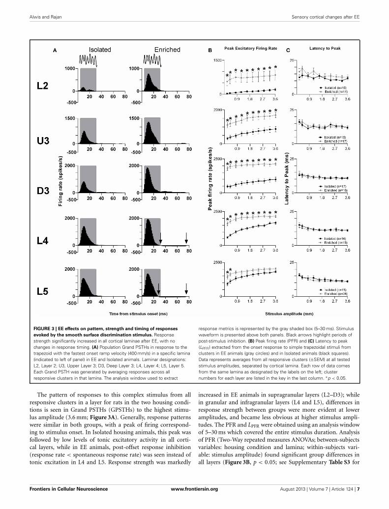
FIGURE 3 | EE effects on pattern, strength and timing of responses
evoked by the smooth surface discrimination stimulus. Responsestrength significantly increased in all cortical laminae after EE, with nochanges in response timing. (A) Population Grand PSTHs in response to thetrapezoid with the fastest onset ramp velocity (400 mm/s) in a specific lamina(indicated to left of panel) in EE and Isolated animals. Laminar designations:L2, Layer 2; U3, Upper Layer 3; D3, Deep Layer 3; L4, Layer 4; L5, Layer 5.Each Grand PSTH was generated by averaging responses across allresponsive clusters in that lamina. The analysis window used to extract
response metrics is represented by the gray shaded box (5–30 ms). Stimuluswaveform is presented above both panels. Black arrows highlight periods ofpost-stimulus inhibition. (B) Peak firing rate (PFR) and (C) Latency to peak(LPFR) extracted from the onset response to simple trapezoidal stimuli fromclusters in EE animals (gray circles) and in Isolated animals (black squares).Data represents averages from all responsive clusters (±SEM) at all testedstimulus amplitudes, separated by cortical lamina. Each row of data comesfrom the same lamina as designated by the labels on the left; clusternumbers for each layer are listed in the key in the last column. ∗p < 0.05.
The pattern of responses to this complex stimulus from allresponsive clusters in a layer for rats in the two housing condi-tions is seen in Grand PSTHs (GPSTHs) to the highest stimu-lus amplitude (3.6 mm; Figure 3A). Generally, response patternswere similar in both groups, with a peak of firing correspond-ing to stimulus onset. In Isolated housing animals, this peak wasfollowed by low levels of tonic excitatory activity in all corti-cal layers, while in EE animals, post-offset response inhibition(response rate < spontaneous response rate) was seen instead oftonic excitation in L4 and L5. Response strength was markedly
increased in EE animals in supragranular layers (L2–D3); whilein granular and infragranular layers (L4 and L5), differences inresponse strength between groups were more evident at loweramplitudes, and became less obvious at higher stimulus ampli-tudes. The PFR and LPFR were obtained using an analysis windowof 5–30 ms which covered the entire stimulus duration. Analysisof PFR (Two-Way repeated measures ANOVAs; between-subjectsvariables: housing condition and lamina; within-subjects vari-able: stimulus amplitude) found significant group differences inall layers (Figure 3B, p < 0.05; see Supplementary Table S3 for
Frontiers in Cellular Neuroscience www.frontiersin.org August 2013 | Volume 7 | Article 124 | 7

Alwis and Rajan Sensory cortical changes after EE
statistical detail), with PFRs in EE animals being higher thanthose of Isolation-housed animals. Post-hoc analysis showed thatthe significant increases in PFRs in EE animal occurred acrossall amplitudes in L2–4 (p < 0.05); in L5, PFRs in EE animalswere only significantly higher than those in Isolation-housed ani-mals at the two lowest amplitudes (0.2 and 0.4 mm). As with theother stimuli detailed above, there were no significant changesin onset peak timing (LPFR; Figure 3C) between the two housingconditions in all layers (p > 0.05; see Supplementary Table S3 forfurther details); in both groups there was a significant systematicdecrease in LPFR with increasing stimulus amplitude.
Rough surface discrimination whisker motion. The second of thetwo discrimination stimulus waveforms we used was a whiskermotion pattern which mimicked the movement of whiskersacross a rough surface (Ritt et al., 2008), characterized by a num-ber of “stick-slip” events (Wolfe et al., 2008). For this stimulus,we recorded from 74 clusters in Isolation-housed animals and85 clusters in EE animals (cluster numbers not significantly dif-ferent between groups and layers: χ2 = 3.7, df = 4, p > 0.05).Response patterns were similar to those elicited to the smoothsurface discrimination stimulus (Figure 4A), as were measuresof firing rate (Figure 4B; see Supplementary Table S4 for further
FIGURE 4 | EE effects on pattern, strength and timing of responses
evoked by the rough surface discrimination stimulus. Significantincreases in response strength were found in L2–4 after EE (but not L5), withno changes in response timing. (A) Population Grand PSTHs in response tothe trapezoid with the fastest onset ramp velocity (400 mm/s) in a specificlamina (indicated to left of panel) in EE and Isolated animals. Laminardesignations: L2, Layer 2; U3, Upper Layer 3; D3, Deep Layer 3; L4, Layer 4;L5, Layer 5. Each Grand PSTH was generated by averaging responses acrossall responsive clusters in that lamina. The analysis window used to extract
response metrics is represented by the gray shaded box (5–30 ms). Stimuluswaveform is presented above both panels. Black arrows highlight periods ofpost-stimulus inhibition. (B) Peak firing rate (PFR) and (C) Latency to peak(LPFR) extracted from the onset response to simple trapezoidal stimuli fromclusters in EE animals (gray circles) and in Isolated animals (black squares).Data represents averages from all responsive clusters (±SEM) at all testedstimulus amplitudes, separated by cortical lamina. Each row of data comesfrom the same lamina as designated by the labels on the left; clusternumbers for each layer are listed in the key in the last column. ∗p < 0.05.
Frontiers in Cellular Neuroscience www.frontiersin.org August 2013 | Volume 7 | Article 124 | 8

Alwis and Rajan Sensory cortical changes after EE
details) and response timing (Figure 4C; Supplementary Table S4for further details).
In summary, for both discrimination stimuli, in L2–4EE animals showed an increase in PFR compared withIsolation-housed animals, with no changes in the timing ofresponses.
Exploratory “free whisking” stimulus. The last complex stimu-lus waveform we consider is one that models the movement ofrat whiskers when undergoing normal exploratory whisking (Gaoet al., 2001), characterized by two cycles of sinusoid-like backand forth motion. Data for this stimulus waveform were obtainedfrom 72 responsive multi-unit clusters in Isolation-housed ani-mals and 84 multi-unit clusters in EE animals (cluster numbersnot significantly different between groups and layers: χ2 = 3.6,df = 4, p > 0.05).
The laminar-related pattern of responses to this stimulus isshown in Grand PSTHs (GPSTHs) from the highest stimu-lus amplitude (3.6 mm) from all responsive clusters in a layer(Figure 5). In L2 and U3, responses in Isolation-housed ani-mals consisted of low level excitation that was poorly definedthroughout the stimulus. In deeper layers (D3–L5), responses inIsolation-housed animals were better defined as consisting of sev-eral peaks which appeared to coincide with changes in velocityin the stimulus waveform. In contrast, responses in the superfi-cial layers (L2 and U3) in EE animals showed an overall increasein excitatory activity throughout the stimulus duration, result-ing in responses being much better defined and consisting of anumber of clear peaks appearing to align with velocity changes.Deeper layers (D3–L5) exhibited well-defined responses and sim-ilar response patterns to those in Isolated housing animals, albeitwith much higher firing rates.
Given the complex nature of the response pattern, we firstused an analysis window of 5–200 ms post-stimulus onset, tostudy changes in PFR throughout the stimulus (Figure 6A).Laminar-specific Two-Way repeated measures ANOVAs (detailsin Supplementary Table S5) found significant differences in PFRas a function of housing condition, and significant housing ×amplitude interactions in L2–5, where again, PFRs in EE animalswere higher than those in Isolated housing animals. Post-hoc anal-ysis revealed that in L2, increased responses in EE animals wereconfined to higher stimulus amplitudes (2.4–3.6 mm), while inU3, D3, and L4, responses in EE animals were significantly higherat mostly higher stimulus amplitudes (Bonferroni post-hoc, p <
0.05). L5 responses were significantly higher in EE animals atonly two of the highest stimulus amplitudes (2.8 and 3.6 mm;Bonferroni post-hoc, p < 0.05).
To allow comparison to metrics obtained to the other stimuliusing shorter analysis windows, we then used a shorter analysiswindow (5–50 ms) to characterize changes in PFR and LPFR dur-ing the onset ramp component of the stimulus (Figures 6B,C).Laminar-specific Two-Way repeated measures ANOVAs (seeSupplementary Table S5 for further details) found significantdifferences in PFR as a function of housing condition and signif-icant housing × amplitude interactions in L2–4, where responsesin EE animals were higher than those in Isolation-housed ani-mals but only at the higher amplitudes (Bonferroni post-hoc,
FIGURE 5 | EE effects on pattern of responses evoked by the free
whisking stimulus. Response strength increased and was more preciselydefined across all laminae after EE. Population Grand PSTHs in response tothe trapezoid with the fastest onset ramp velocity (400 mm/s) in a specificlamina (indicated to left of panel) in EE and Isolated animals. Laminardesignations: L2, Layer 2; U3, Upper Layer 3; D3, Deep Layer 3; L4, Layer4; L5, Layer 5. Each Grand PSTH was generated by averaging responsesacross all responsive clusters in that lamina. The two analysis windowsused to extract response metrics are represented by the gray shaded box(5–200 ms) and the black dashed box (5–50 ms). Stimulus waveform ispresented above both panels.
p < 0.05; Figure 6B). In L5 there was no effect of housing con-dition but there was a significant Group × Amplitude inter-action (Two-Way ANOVA, p < 0.05). Finally, analysis of LPFR
data from each lamina (Figure 6C) found a significant Group ×Amplitude interaction only in D3, with no significant groupeffects in any layer (p > 0.05; statistical details in SupplementaryTable S5).
In summary, in L2–4 the effects of housing on neuronal clus-ter responses for this stimulus was similar to the effects with thesurface discrimination stimuli: EE animals showed an increase inPFR compared with Isolation-housed animals, with no changesin timing of responses.
Frontiers in Cellular Neuroscience www.frontiersin.org August 2013 | Volume 7 | Article 124 | 9

Alwis and Rajan Sensory cortical changes after EE
FIGURE 6 | EE effects on strength and timing of responses evoked by
the free whisking stimulus. (A) Peak firing rate (PFR) extracted from a5–200 ms window, (B) PFR and (C) Latency to peak (LPFR) extracted froma 5–50 ms window in response to the free whisking stimulus from clustersin EE animals (grey circles) and in Isolated animals (black squares). PFRresponses were significantly increased across L2–4 for both analysiswindows, with a significant PFR increase in L5 only for the 5–200 ms
window. No changes in response timing were found. Data representsaverages from all responsive clusters (±SEM) at all tested stimulusamplitudes, separated by cortical lamina. Laminar designations: L2, Layer2; U3, Upper Layer 3; D3, Deep Layer 3; L4, Layer 4; L5, Layer 5. Eachrow of data comes from the same lamina as designated by the labels onthe left; cluster numbers for each layer are listed in the key in the lastcolumn. ∗p < 0.05.
Comparison of the effect sizes to the different stimuli in the differentlaminaeAs shown above, EE induced an increase in firing rate across allstimuli for most laminae. To make a comparison of the size of theEE-induced effects, for each stimulus type we calculated the ratioof PFR in EE animals for a specific stimulus condition (stimulus
type × velocity/amplitude) vs. PFR for the same condition forclusters from Isolation-housed animals. This PFREE/PFRIsol ratiois shown in Figure 7 for four stimuli (trapezoid whisker deflec-tions, the two surface discrimination stimuli, and the objectcontact stimulus) and in Figure 8 for the exploratory whiskingstimulus.
Frontiers in Cellular Neuroscience www.frontiersin.org August 2013 | Volume 7 | Article 124 | 10

Alwis and Rajan Sensory cortical changes after EE
FIGURE 7 | Peak firing rate ratios in EE vs. Isol animals for
trapezoid, object contact and surface discrimination stimuli.
PFREE/PFRIsol across all onset ramp velocities in (A) a simpletrapezoidal stimulus; across all stimulus amplitudes in (B) the objectcontact stimulus, (C) the smooth surface discrimination stimulus and(D) the rough surface discrimination stimulus. PFREE/PFRIsol was
greatest at lower velocities/amplitudes and decreased with increasingvelocity/amplitude in all cortical layers as responses in EE animalscame closer to those in Isolation-housed animals. Laminardesignations: L2, Layer 2; U3, Upper Layer 3; D3, Deep Layer 3;L4, Layer 4; L5, Layer 5. Each row of data comes from the samelamina as designated by the labels on the left.
A striking feature for the first four stimuli is that, asidefrom L2 responses to one stimulus (Figure 7B), a similarpattern of effects is seen in all laminae. In all cases, EEanimals showed greater responses than Isolation-housed ani-mals but the increase was inversely related to the appro-priate stimulus parameter (trapezoids: onset ramp velocity;
all other stimuli: stimulus amplitude)—i.e., the PFREE/PFRIsol
was greater at lower velocities/amplitudes and decreased withincreasing velocity/amplitude in all cortical layers. At thehighest velocity/amplitude increased, responses in EE animalscame closer to those in Isolation-housed animals. This patternwas consistent across the four very different stimuli, ranging
Frontiers in Cellular Neuroscience www.frontiersin.org August 2013 | Volume 7 | Article 124 | 11

Alwis and Rajan Sensory cortical changes after EE
FIGURE 8 | Peak firing rate ratios in EE vs. Isol animals for free
whisking stimulus. PFREE/PFRIsol across all stimulus amplitudes in (A) the5–200 ms analysis window, and (B) the 5–50 ms analysis window of thefree whisking stimulus. For the 5–200 ms analysis window, there was anon-monotonic increase in PFREE/PFRIsol, especially in layers U3–L5. Forthe 5–50 ms window, the PFREE/PFRIsol also increased non-monotonicallyand was followed by a plateau/decrease in magnitude of increasedresponsiveness in EE vs. Isolated animals. Laminar designations: L2, Layer2; U3, Upper Layer 3; D3, Deep Layer 3; L4, Layer 4; L5, Layer 5. Each rowof data comes from the same lamina as designated by the labels on the left.
from simple trapezoids to three of the complex, naturalisticstimuli.
However, a very different pattern of effects was seen forthe PFREE/PFRIsol ratio to the exploratory whisking stimulus(Figure 8). Using a 5–200 ms analysis window to cover the wholestimulus, the PFREE/PFRIsol increased with stimulus amplitudebut non-monotonically (Figure 8A), especially in layers U3–L5,where the greatest PFREE/PFRIsol is at intermediate amplitudesand decreased toward 1 at higher amplitudes. When the anal-ysis window was restricted to only the “onset ramp” of thesinusoid-like stimulus (5–50 ms), a window similar to that usedfor the other stimuli (see above), the PFREE/PFRIsol still showed
non-monotonicity, with maximum difference between EE andIsolated groups at intermediate stimulus amplitudes, followed bya plateau or even a decrease in magnitude of increased responsive-ness in EE relative to Isolated animals (Figure 8B). Again, at highamplitudes responses in EE animals approached the peak firingrates of Isolated animals.
One other feature evident in the previous two figures is thatthe largest PFREE/PFRIsol ratio (i.e., the greatest increase in fir-ing rate in EE animals relative to firing rate in Isolation-housedanimals) appeared to occur in supra-granular layers. To demon-strate this, Figure 9A plots the maximum ratio of PFREE/PFRIsol
for all the stimuli; to demonstrate that the effects were not dis-torted by a singular maximum value, Figure 9B plots the averagePFREE/PFRIsol ratio calculated across the three highest stimulusamplitudes (for the four complex naturalistic stimuli) or acrossall velocities (for the three trapezoid stimuli). Both plots showthe same effect: both the maximum PFREE/PFRIsol ratio and theaverage PFREE/PFRIsol ratio occurred in Layers 2 and Upper 3,with a systematic decrease with depth to L5 (Figures 8, 9). Bothplots also indicate that while the EE-induced increase in responsemagnitude decreased with cortical depth, responses in EE animalsalways remain higher than those in animals housed in isolation.
Spontaneous firing rates in multi-neuronal clustersFinally, in addition to the stimulus-driven activity, we examinedif EE conditions altered spontaneous activity. For this calcula-tion, average spontaneous cluster firing rates were obtained fromeach cluster in the 200 ms period prior to onset of the basictrapezoidal stimulus, for all cortical layers in the two housingconditions (Figure 10). As with the changes in stimulus-drivenactivity reported above, average spontaneous firing rates in EEanimals were significantly higher (Mann–Whitney U t-test, p <
0.05; see Figure 10 for exact p-values) than those in Isolation-housed animals in L2–4 (Isolated vs. EE (spikes/s): L2 = 9.78 ±1.75 vs. 31.13 ± 5.92; U3 = 10.81 ± 3.70 vs. 40.33 ± 7.09; D3= 21.29 ± 3.70 vs. 77.91 ± 11.08; L4 = 16.49 ± 2.82 vs. 45.15± 9.55). In L5, there were no differences in spontaneous firingrates between the two housing conditions (Isolated vs. EE: 32.77± 5.16 vs. 71.28 ± 10.48 spikes/s, p > 0.05). The highest averagespontaneous firing rate after EE was observed in D3.
DISCUSSIONIn response to exposure to EE, barrel cortex neurons demon-strated a very large increase in responsiveness to sensory inputwithout any changes in response timing, throughout all cor-tical laminae of barrel cortex, in response to simple and var-ied complex, naturalistic stimuli, across a variety of responsepatterns, and in both short-term responses (up to 50 ms post-stimulus onset for short-duration stimuli) and longer-lastingresponses (up to 200 ms post-stimulus onset for a longer dura-tion stimulus). Thus, the plasticity underlying the effects of EEappears to be exercised across lamina, stimulus type, responsepattern, and response duration. Additionally, EE also causedan increase in spontaneous activity in all laminae except inLayer 5. EE provides animals with cognitive, sensory and motorstimulation, through interactions with a complex environment,
Frontiers in Cellular Neuroscience www.frontiersin.org August 2013 | Volume 7 | Article 124 | 12

Alwis and Rajan Sensory cortical changes after EE
FIGURE 9 | Depth-dependent changes in maximum and average PFREE/
PFRIsol ratios. (A) Maximum PFREE/PFRIsol ratio in all stimuli plotted acrossall cortical layers. The maximum PFREE/PFRIsol ratio is in supragranular layersfor all stimuli, and steadily declines as cortical depth increases. (B) AveragePFREE/PFRIsol ratio in all stimuli from all onset ramp velocities/3 highest
stimulus amplitudes, plotted across all cortical layers. The average PFREE/PFRIsol ratio is highest in supragranular layers for all stimuli, and decreases ascortical depth increases. Laminar designations: L2, Layer 2; U3, Upper Layer3; D3, Deep Layer 3; L4, Layer 4; L5, Layer 5. Symbols corresponding to thefive stimuli are presented in the keys to the right of the figure.
FIGURE 10 | Elevated spontaneous firing rate following EE.
Spontaneous firing rate was measured in the 200 ms window prior tostimulus onset, and was extracted from multi-neuronal clusters for EE(n = 84 clusters) and Isolated (n = 84 clusters) animals. The figure plots themean spontaneous firing rate in clusters grouped according to cortical layer.Significant increases in spontaneous firing rate were observed after EE inlayers L2–L4, with no significant differences in spontaneous firing ratesbetween the two housing conditions in L5. Data represents averages fromall responsive clusters (±SEM) at all tested ramp velocities, separated bycortical lamina. ∗p < 0.05.
increased physical activity, and social interactions. The combi-nation of these factors is thought to be responsible for pro-moting cortical plasticity in normal animals (as used in ourstudy) and in models of disease and deprivation (Diamondet al., 1993; Van Praag et al., 2000; Nithianantharajah andHannan, 2006; Rema et al., 2006; Baroncelli et al., 2010). Inthe following discussion we restrict ourselves to studies of adultplasticity and its mechanisms as developmental plasticity mayinclude mechanisms not available in adulthood or much atten-uated in adulthood (viz. Lendvai et al., 2000; Rajan and Irvine,2010).
EXCITATION/INHIBITION BALANCE AFTER EE AND SENSORY CORTICALRESPONSIVENESSEE is generally reported to increase sensory cortical responsive-ness in sensory cortex studies restricted to recordings from somelayers. Mainardi et al. (2010) found that evoked potentials wereincreased in layers 3/4 of visual cortex of rats exposed to EE and,at the single neuron level, enrichment resulted in no change in
RF size or spontaneous activity (Beaulieu and Cynader, 1990a)but increased responsiveness, orientation tuning, and temporalcontrast tuning (Beaulieu and Cynader, 1990b, layers unknown).In primary auditory cortex (A1), 8 weeks of auditory enrichmentresulted in increased stimulus-evoked neuronal response rates,spontaneous rate, and response latency, in layer IV (Engineeret al., 2004), increased auditory evoked potential amplitudes(Engineer et al., 2004; Percaccio et al., 2005, 2007) and decreasedability to follow high stimulus repetition rates (Percaccio et al.,2005, 2007). We too found increases in driven and spontaneousactivity but no change in response latency. In other studies inadult auditory cortex, different types of enhanced auditory expe-rience distorts the tonotopic map (Pienkowski and Eggermont,2009; Zhou et al., 2011) and the ability to follow high stimulusrates (Zhou et al., 2011). However, there is dispute as to whether itdecreases neuronal frequency selectivity at all frequencies (Zhouet al., 2011) or increases it within the frequency band of theenrichment stimulus (Pienkowski and Eggermont, 2009). In non-primary sensory cortex (posterior auditory field) EE increasedresponse strength and decreased RF size, leading to an increasein spectral and temporal selectivity (Jakkamsetti et al., 2012). Incontrast, Polley et al. (2004) reported that naturalistic sensoryexperience in their EE conditions decreased neuronal responsesand RF size in barrel cortex, but only in the upper layers and notin layer 4. However, (Guic et al., 2008) suggested that the effectsin the Polley et al. (2004) study reflected the effects of habitua-tion even despite EE, and a lack of active exploration; Guic et al.(2008) found that EE caused an increase in RF size in contrastto Polley et al. (2004). Megevand et al. (2009) report increasedgreater whisker-driven evoked potentials in barrel cortex after EE.
A variety of mechanisms may contribute to cortical plastic-ity after EE (reviews by Van Praag et al., 2000; Nithianantharajahand Hannan, 2006), including structural and molecular changeslike changes in dendritic structure and function and enhancedsynaptic plasticity (Yang et al., 2009; Fu and Zuo, 2011; Jung andHerms, 2012), increases in certain neurotransmitters (Giovanniniet al., 2001; Naka et al., 2002) or in synaptic release proba-bility (Percaccio et al., 2005), and shifts in cortical E/I ratios(Coq and Xerri, 1998; Engineer et al., 2004; Polley et al., 2004;Baroncelli et al., 2011), given that a decrease in inhibition can
Frontiers in Cellular Neuroscience www.frontiersin.org August 2013 | Volume 7 | Article 124 | 13
Alwis and Rajan Sensory cortical changes after EE
trigger plasticity, even in non-critical periods (Maya Vetencourtet al., 2008; Southwell et al., 2010; Maya-Vetencourt et al., 2012),through changes in neuronal connectivity (Baroncelli et al., 2010,2012). Our results can most parsimoniously be explained by thislast factor (not excluding that the other mechanisms may alsocontribute to this factor)—that EE-induced plasticity in our casewas due to a shift in the E/I balance to favor E. The stabilityof neuronal connections in the adult brain is held to be dueto maturation of cortical inhibitory interneurons which resultsin decreased plasticity (Fagiolini and Hensch, 2000; Hensch andFagiolini, 2005). Induction of adult cortical plasticity appearsthen to be brought about primarily through a decrease in cor-tical inhibitory activity (Hensch and Fagiolini, 2005; Sale et al.,2007; Benali et al., 2008; Baroncelli et al., 2010, 2011; Luz andShamir, 2012). Consistent with this idea, EE causes a reductionin the number of visual cortical neurons expressing GAD67 (Scaliet al., 2012; Tognini et al., 2012), and a decrease in extracellu-lar basal GABA levels, coupled with a restoration of white matterlong-term potentiation (WM-LTP), suggesting decreased corticalinhibition (Sale et al., 2007). Decreases in GABAergic inhibitionafter EE, through a reduction in expression of GABAA receptorsubunits, have been demonstrated in adult rat auditory cortex(Zhou et al., 2011), and in cat visual cortex enrichment decreasesthe number of inhibitory synapses (Beaulieu and Colonnier,1987). Treatment with IGF-1, a peptide thought to play a rolein EE-induced plasticity by enhancing synaptic plasticity (Torres-Aleman, 1999; Aberg et al., 2000), causes a decrease in basalinhibitory neurotransmitter levels with no overall changes inbasal glutamate levels (Maya-Vetencourt et al., 2012) and a shift inocular dominance as measured by visual evoked potentials, indi-cating visual cortical plasticity in adult rats that had undergonemonocular deprivation (Maya-Vetencourt et al., 2012).
The hypothesis that EE simply decreases cortical inhibi-tion is not universally accepted: Nichols et al. (2007) foundthat GABAA receptor-mediated inhibitory postsynaptic cur-rents do not change in supragranular auditory cortex after EEbut there was an increase in AMPA-mediated current ampli-tudes. Further, the group had previously shown that EE resultsin a decreased ability of AI neurons to follow rapid stimuli(Percaccio et al., 2005), inconsistent with effects predicted by adecrease in inhibition. Similarly, Zhou et al. (2011) reported thatenhanced auditory experience decreased expression of GABAA
and NMDA receptors, but had no effect on the AMPA glutamatereceptor.
Our extracellular recordings do not allow direct measurementsof inhibition but the large increase in driven and spontaneousresponse strength strongly supports the inference that EE in ourstudy also caused a shift in the cortical E/I ratio to favor exci-tation over inhibition. Sensory deprivation also induces corticalplasticity through a decrease in inhibition (Welker et al., 1989;Buonomano and Merzenich, 1998). Kelly et al. (1999) reportedthat whisker trimming leads to a loss of inhibitory input fromsurrounding whiskers, as well as a net decrease in tonic corti-cal inhibition, resulting in increased barrel cortex responses tothe PW. In the mature barrel cortex, deprivation-induced plas-ticity decreases L4 feed-forward excitation to L2/3 inhibitoryneurons but with improved inhibition to L2/3 pyramidal cells
(House et al., 2011), resulting in E/I balance being maintainedin L2/3. We found increased activity in all layers, indicating thatan E/I balance was not maintained in L2/3. Thus, EE, in ourcase, appears to exert global cross-lamina effects primarily, ifnot solely, directed to favoring excitatory over inhibitory inputs.This does not mean that large changes in excitation need haveoccurred. It has been elegantly demonstrated that during nor-mal processing in somatosensory cortex, excitation and inhibitionare highly synchronized and well-correlated in time and strength,in a continuous manner (Okun and Lampl, 2008); thus, evensmall changes in timing and strength of the two processes couldresult in a very large increase in excitation as we see (Isaacson andScanziani, 2011).
Only certain forms of inhibition may be affected after exposureto EE since, even in our extracellular recordings, we do see someindications of inhibition after EE, with decreases in post-stimulusresponses to levels below pre-stimulus spontaneous activity (seeFigure 4A). Jakkamsetti et al. (2012) suggest that EE affects basallevels of GABA and evoked GABA differently, such that stimulus-evoked inhibition remains intact, while basal inhibitory activity issuppressed after EE, which could explain our evidence of intactpost-stimulus inhibition but increased spontaneous activity.
Cortical inhibition is heterogeneous, either acting to affectneuronal responses (Wehr and Zador, 2003; Haider et al., 2013)by increasing response selectivity and hence, RF sizes, or alteringresponse gain (Isaacson and Scanziani, 2011; Wilson et al., 2012),including acting through “silent” shunting inhibition (Isaacsonand Scanziani, 2011). Selective loss of some forms of corticalinhibition may be all that is required to obtain the EE-inducedincreases in cortical spontaneous and driven responses. In audi-tory cortex, loss of surround inhibition can occur without lossof stimulus-driven within-field inhibition (Rajan, 1998, 2001)andthen results in no change cortical in map topography (Rajan,1998) but an increase in bandwidth of cortical neurons (Rajan,2001), and thereby likely an increase in the overall representationof any specific frequency and increase response rates to the “best”frequency (Rajan, 1998). This parallels the observations that thatpost-stimulus inhibition is preserved in EE animals (our presentstudy), and that EE does not change the exclusive representationarea of a whisker in barrel cortex though there is an expansion ofthe total representation area of that whisker (Guic et al., 2008).
LOCUS OF EE-INDUCED PLASTICITYOur finding of a global increase in neuronal responsiveness inbarrel cortex after EE without response timing changes suggestsan intra-cortical locus for plasticity (cf. Fox et al., 2002; Polleyet al., 2004), especially given this pattern of effects even in thethalamo-recipient Layer 4 responses. Long-term whisker depriva-tion increases the functionality of specific GABAA receptors onLayer 4 cells such that there is faster decay of inhibition in thesecells (Li et al., 2009). In this case, a normal thalamic input couldproduce larger responses in Layer 4 as we find with EE. Our find-ings of an inverse relationship between cortical depth and theamount of EE induced increase in responses can be explainedby postulating that a Layer 4 change is amplified by local mech-anisms when information flows from Layer 4 to upper layers(Armstrong-James et al., 1992; Jellema et al., 2004; Lubke and
Frontiers in Cellular Neuroscience www.frontiersin.org August 2013 | Volume 7 | Article 124 | 14

Alwis and Rajan Sensory cortical changes after EE
Feldmeyer, 2007; Megevand et al., 2009), as has been suggestedfor experience-dependent plasticity in barrel cortex (Fox et al.,2002). This is consistent with the fact that supra-granular cor-tical layers show longer-lasting and greater amounts of plasticitythan layers 4 or 5 (Glazewski and Fox, 1996; Wallace and Fox,1999; Fox et al., 2002; Nichols et al., 2007). It is also con-sistent with the postulate (Nichols et al., 2007) that layer 2/3is a “privileged substrate” for consolidating EE-induced cor-tical plasticity, possibly through spike-timing-dependent plas-ticity, or even structural plasticity, with studies reporting anEE-induced increase in dendritic branching in supragranu-lar layers of the occipital cortex (Volkmar and Greenough,1972; Greenough et al., 1973). A similar depth-dependenteffect has been seen for plasticity evoked by rhythmic whiskerstimulation in anesthetized animals, a paradigm suggested toengage the same intra-cortical plasticity mechanisms as EE(Megevand et al., 2009).
The absence of changes in response timing, as would beexpected for plasticity evoked sub-cortically, is consistent withstudies showing that thalamic plasticity is limited in the adultbrain (Fox et al., 1996, 2002), except in the case of peripheralnerve injury (Li et al., 1995; Jones and Pons, 1998). However,it must be recognized that the debate on sub-cortical contribu-tions to cortical changes in EE is not yet resolved. EE is capable ofenhancing thalamocortical transmission in adult rat visual cor-tex (Mainardi et al., 2010), while EE in an experimental modelof adult monocular amblyopia causes an increase in presynapticthalamo-cortical activity, hence increasing postsynaptic stimula-tion of the visual cortex (Tognini et al., 2012). In the somatosen-sory cortex, whisker deprivation increases neuronal response ratesin cortical Layer 4, with no changes in response rates in thalamicVPM (Wallace and Fox, 1999). In the auditory system it has beenargued that cortical changes induced by enriched auditory expe-rience are likely to occur in thalamus but be modified by localmechanisms in cortex (Pienkowski and Eggermont, 2009).
We are currently conducting further studies to investigatethe exact mechanisms underlying the EE-induced changes inneuronal plasticity reported in the present study, using slicepreparations and immunohistochemical techniques.
PATTERN OF EE INDUCED CHANGES IN RESPONSES TO DIFFERENTBEHAVIORALLY RELEVANT STIMULIOur results show a stimulus amplitude-dependent increase inpeak firing rates, where for all stimuli (except one) EE inducedgreater increases in excitatory responses at lower stimulus ampli-tudes than at higher stimulus amplitudes. The effect may simplyreflect response saturation at higher levels; a multiplier effectwill produce a larger change at lower stimulus amplitudes whereresponses are not saturated than it would at higher levels whereresponses are closer to saturation. This is consistent with theobservation of Engineer et al. (2004) that stimuli that elicitedsmaller responses which did not reach saturation were moresuitable to reveal whether EE has an effect on auditory corticalactivity.
The odd one out of the stimuli in this inverse relationshipbetween EE-induced increases and stimulus parameter(amplitude or velocity) was the stimulus waveform mimicking
whisker motions seen in exploratory, free-whisking behavior. Forthis stimulus, the EE effect was non-monotonic with greatesteffects at intermediate amplitudes than at higher amplitudeswhere EE effects either plateaued or even decreased in relativepotency compared to the EE effects at intermediate amplitudes.EE influences exploratory behavior (Dell’omo et al., 2000;Frostig, 2006), thus making a stimulus that mimics exploratorywhisker motion most relevant for investigation of EE effects. Itis interesting to speculate that the maximization of EE effectsto particular amplitudes of free, exploratory whisking may belinked to the effects of EE on exploratory behaviors—does EEpromote rat behavior such that the animals are more likely toexplore objects when the object is at a particular distance fromthe animal, i.e., does the animal tend to position itself so that itcan best explore an object at a certain distance away? Szwed et al.(2006) have shown that spike rate is particularly important inencoding (in trigeminal ganglion neurons) radial distance of anobject and whisker exploration of environmental objects and theEE-induced optimization of firing rate at particular amplitudesmay be seen to support this hypothesis. However, against this isthat an object a certain fixed distance from the animal will beat different radial distances along the length of whiskers arrayedacross the rostro-caudal axis of the mystacial pad. Thus, therelevance of the EE-induced increase in firing rate at particulardeflection amplitudes for this specific stimulus alone remainsunknown.
NOVELTY OF OBJECTS AND HABITUATIONOne final point that merits consideration is that it is gener-ally held that novel objects in EE housing should be changedregularly to prevent habituation. We did not do so but stillfound highly elevated response rates, especially in supragran-ular layers, whereas Polley et al. (2004) reported that EE ledto decreased responses in upper layers of barrel cortex (butnot in layer 4), and a decrease in RF sizes. As noted above,Guic et al. (2008) suggested that the Polley et al. (2004) resultsreflected the effects of habituation and a lack of active explo-ration even despite EE conditions. If this criticism is valid, itwould indicate that, in our case, the constancy of the objectsin the EE housing was not a deterrent to active explorationof those objects, insofar as such active exploration led to theobserved increase in response rates to all stimuli. In part thismay reflect the fact that, in our study, when cages were cleanedand food and water replenished, the experimenters made a pointof bringing back to the surface all objects that had becomesubmerged under the bedding material. It is possible that thisre-appearance of the objects contributed to maintaining activeexploration of the objects and thereby contributed to the effectsobserved in this study. Under such conditions the effects ofEE appear to be remarkably well-expressed in barrel cortex,across all layers, stimulus types, response patterns, and responsedurations.
SUPPLEMENTARY MATERIALThe Supplementary Material for this article can be foundonline at: http://www.frontiersin.org/Cellular_Neuroscience10.3389/fncel.2013.00124/abstract
Frontiers in Cellular Neuroscience www.frontiersin.org August 2013 | Volume 7 | Article 124 | 15

Alwis and Rajan Sensory cortical changes after EE
Table S1 | Results of Two-Way repeated measures ANOVA statistical
analysis of firing rate (PFR) and Latency to Peak (LPFR) in clusters
responsive to the trapezoidal stimulus from 5 to 50 ms from stimulus
onset (related to Figures 1B,C). The table lists F statistics and degrees of
freedom for both significant and non-significant factors for main and
interaction terms.
Table S2 | Results of Two-Way repeated measures ANOVA statistical
analysis of firing rate (PFR) and Latency to Peak (LPFR) in clusters
responsive to the onset response of the object contact stimulus from 5 to
50 ms from stimulus onset (related to Figures 2B,C). The table lists F
statistics and degrees of freedom for both significant and non-significant
factors for main and interaction terms.
Table S3 | Results of Two-Way repeated measures ANOVA statistical
analysis of firing rate (PFR) and Latency to Peak (LPFR) in clusters
responsive to the smooth surface discrimination whisker motion stimulus
from 5 to 30 ms from stimulus onset (related to Figures 3B,C). The table
lists F statistics and degrees of freedom for both significant and
non-significant factors for main and interaction terms.
Table S4 | Results of Two-Way repeated measures ANOVA statistical
analysis of firing rate (PFR) and Latency to Peak (LPFR) in clusters
responsive to the rough surface discrimination whisker motion stimulus
from 5 to 30 ms from stimulus onset (related to Figures 4B,C). The table
lists F statistics and degrees of freedom for both significant and
non-significant factors for main and interaction terms.
Table S5 | Results of Two-Way repeated measures ANOVA statistical
analysis of firing rate (PFR) in clusters responsive to the exploratory “free
whisking” stimulus from 5 to 200 ms from stimulus onset (related to
Figure 6A); and firing rate (PFR) and Latency to Peak (LPFR) from 5 to 50 ms
from stimulus onset (related to Figures 6B,C). The table lists F statistics
and degrees of freedom for both significant and non-significant factors for
main and interaction terms.
REFERENCESAberg, M. A., Aberg, N. D., Hedbacker,
H., Oscarsson, J., and Eriksson, P.S. (2000). Peripheral infusion ofIGF-I selectively induces neurogen-esis in the adult rat hippocampus.J. Neurosci. 20, 2896–2903.
Alwis, D., Yan, E. B., Morganti-Kossmann, M. C., and Rajan, R.(2012). Sensory cortex underpin-nings of traumatic brain injurydeficits. PLoS ONE 7:e52169. doi:10.1371/journal.pone.0052169
Armstrong-James, M., Fox, K., andDas-Gupta, A. (1992). Flow ofexcitation within rat barrel cor-tex on striking a single vibrissa.J. Neurophysiol. 68, 1345–1358.
Baroncelli, L., Bonaccorsi, J., Milanese,M., Bonifacino, T., Giribaldi, F.,Manno, I., et al. (2012). Enrichedexperience and recovery fromamblyopia in adult rats: impactof motor, social and sensorycomponents. Neuropharmacology62, 2388–2397. doi: 10.1016/j.neuropharm.2012.02.010
Baroncelli, L., Braschi, C., Spolidoro,M., Begenisic, T., Maffei, L., andSale, A. (2011). Brain plasticityand disease: a matter of inhibi-tion. Neural Plast. 2011:286073. doi:10.1155/2011/286073
Baroncelli, L., Sale, A., Viegi, A., MayaVetencourt, J. F., De Pasquale,R., Baldini, S., et al. (2010).Experience-dependent reactivationof ocular dominance plastic-ity in the adult visual cortex.Exp. Neurol. 226, 100–109. doi:10.1016/j.expneurol.2010.08.009
Beaulieu, C., and Colonnier, M. (1987).Effect of the richness of the envi-ronment on the cat visual cortex.J. Comp. Neurol. 266, 478–494. doi:10.1002/cne.902660404
Beaulieu, C., and Cynader, M. (1990a).Effect of the richness of theenvironment on neurons in cat
visual cortex. I. Receptive fieldproperties. Brain Res. Dev. BrainRes. 53, 71–81.
Beaulieu, C., and Cynader, M. (1990b).Effect of the richness of the envi-ronment on neurons in cat visualcortex. II. Spatial and temporal fre-quency characteristics. Brain Res.Dev. Brain Res. 53, 82–88.
Benali, A., Weiler, E., Benali, Y.,Dinse, H. R., and Eysel, U. T.(2008). Excitation and inhi-bition jointly regulate corticalreorganization in adult rats.J. Neurosci. 28, 12284–12293. doi:10.1523/JNEUROSCI.1952-08.2008
Brecht, M. (2007). Barrel cortex andwhisker-mediated behaviors. Curr.Opin. Neurobiol. 17, 408–416. doi:10.1016/j.conb.2007.07.008
Bruel-Jungerman, E., Laroche, S., andRampon, C. (2005). New neuronsin the dentate gyrus are involvedin the expression of enhanced long-term memory following environ-mental enrichment. Eur. J. Neurosci.21, 513–521. doi: 10.1111/j.1460-9568.2005.03875.x
Buonomano, D. V., and Merzenich,M. M. (1998). Cortical plastic-ity: from synapses to maps. Annu.Rev. Neurosci. 21, 149–186. doi:10.1146/annurev.neuro.21.1.149
Cancedda, L., Putignano, E., Sale,A., Viegi, A., Berardi, N., andMaffei, L. (2004). Accelerationof visual system developmentby environmental enrichment.J. Neurosci. 24, 4840–4848. doi:10.1523/JNEUROSCI.0845-04.2004
Coq, J. O., and Xerri, C. (1998).Environmental enrichment altersorganizational features of theforepaw representation in the pri-mary somatosensory cortex of adultrats. Exp. Brain Res. 121, 191–204.doi: 10.1007/s002210050452
Dell’omo, G., Ricceri, L., Wolfer, D. P.,Poletaeva, II., and Lipp, H. (2000).
Temporal and spatial adaptation tofood restriction in mice under natu-ralistic conditions. Behav. Brain Res.115, 1–8.
Diamond, M. C., Rosenzweig, M.R., Bennett, E. L., Lindner, B.,and Lyon, L. (1972). Effects ofenvironmental enrichment andimpoverishment on rat cerebralcortex. J. Neurobiol. 3, 47–64. doi:10.1002/neu.480030105
Diamond, M. E., Armstrong-James, M.,and Ebner, F. F. (1993). Experience-dependent plasticity in adult ratbarrel cortex. Proc. Natl. Acad.Sci. U.S.A. 90, 2082–2086. doi:10.1073/pnas.90.5.2082
During, M. J., and Cao, L. (2006).VEGF, a mediator of theeffect of experience on hip-pocampal neurogenesis. Curr.Alzheimer Res. 3, 29–33. doi:10.2174/156720506775697133
Engineer, N. D., Percaccio, C. R.,Pandya, P. K., Moucha, R.,Rathbun, D. L., and Kilgard,M. P. (2004). Environmentalenrichment improves responsestrength, threshold, selectivity,and latency of auditory cortexneurons. J. Neurophysiol. 92,73–82. doi: 10.1152/jn.00059.2004
Fagiolini, M., and Hensch, T. K. (2000).Inhibitory threshold for critical-period activation in primary visualcortex. Nature 404, 183–186. doi:10.1038/35004582
Falkenberg, T., Mohammed, A. K.,Henriksson, B., Persson, H.,Winblad, B., and Lindefors, N.(1992). Increased expressionof brain-derived neurotrophicfactor mRNA in rat hippocam-pus is associated with improvedspatial memory and enrichedenvironment. Neurosci. Lett. 138,153–156. doi: 10.1016/0304-3940(92)90494-R
Feldmeyer, D., Brecht, M., Helmchen,F., Petersen, C. C., Poulet, J.F., Staiger, J. F., et al. (2013).Barrel cortex function. Prog.Neurobiol. 103, 3–27. doi:10.1016/j.pneurobio.2012.11.002
Fox, K., Glazewski, S., Chen,C. M., Silva, A., and Li, X.(1996). Mechanisms underlyingexperience-dependent potentia-tion and depression of vibrissaeresponses in barrel cortex. J. Physiol.Paris 90, 263–269. doi: 10.1016/S0928-4257(97)81436-2
Fox, K., Wallace, H., and Glazewski, S.(2002). Is there a thalamic compo-nent to experience-dependent cor-tical plasticity. Philos. Trans. R. Soc.Lond. B Biol. Sci. 357, 1709–1715.doi: 10.1098/rstb.2002.1169
Frick, K. M., and Fernandez, S. M.(2003). Enrichment enhances spa-tial memory and increases synapto-physin levels in aged female mice.Neurobiol. Aging 24, 615–626. doi:10.1016/S0197-4580(02)00138-0
Frostig, R. D. (2006). Functionalorganization and plasticity inthe adult rat barrel cortex: mov-ing out-of-the-box. Curr. Opin.Neurobiol. 16, 445–450. doi:10.1016/j.conb.2006.06.001
Fu, M., and Zuo, Y. (2011).Experience-dependent struc-tural plasticity in the cortex.Trends Neurosci. 34, 177–187. doi:10.1016/j.tins.2011.02.001
Gao, P., Bermejo, R., and Zeigler,H. P. (2001). Whisker deafferenta-tion and rodent whisking patterns:behavioral evidence for a centralpattern generator. J. Neurosci. 21,5374–5380.
Giovannini, M. G., Rakovska, A.,Benton, R. S., Pazzagli, M., Bianchi,L., and Pepeu, G. (2001). Effectsof novelty and habituation onacetylcholine, GABA, and gluta-mate release from the frontal cortex
Frontiers in Cellular Neuroscience www.frontiersin.org August 2013 | Volume 7 | Article 124 | 16

Alwis and Rajan Sensory cortical changes after EE
and hippocampus of freely movingrats. Neuroscience 106, 43–53. doi:10.1016/S0306-4522(01)00266-4
Glazewski, S., and Fox, K. (1996). Timecourse of experience-dependentsynaptic potentiation and depres-sion in barrel cortex of adolescentrats. J. Neurophysiol. 75, 1714–1729.
Globus, A., Rosenzweig, M. R.,Bennett, E. L., and Diamond, M. C.(1973). Effects of differential expe-rience on dendritic spine countsin rat cerebral cortex. J. Comp.Physiol. Psychol. 82, 175–181. doi:10.1037/h0033910
Green, E. J., and Greenough, W.T. (1986). Altered synaptic trans-mission in dentate gyrus of ratsreared in complex environments:evidence from hippocampal slicesmaintained in vitro. J. Neurophysiol.55, 739–750.
Greenough, W. T., and Volkmar, F.R. (1973). Pattern of dendriticbranching in occipital cortex ofrats reared in complex environ-ments. Exp. Neurol. 40, 491–504.doi: 10.1016/0014-4886(73)90090-3
Greenough, W. T., Volkmar, F. R.,and Juraska, J. M. (1973). Effectsof rearing complexity on dendriticbranching in frontolateral and tem-poral cortex of the rat. Exp. Neurol.41, 371–378. doi: 10.1016/0014-4886(73)90278-1
Guic, E., Carrasco, X., Rodriguez,E., Robles, I., and Merzenich, M.M. (2008). Plasticity in primarysomatosensory cortex resultingfrom environmentally enrichedstimulation and sensory discrim-ination training. Biol. Res. 41,425–437. doi: 10.4067/S0716-97602008000400008
Haider, B., Hausser, M., and Carandini,M. (2013). Inhibition dominatessensory responses in the awakecortex. Nature 493, 97–100. doi:10.1038/nature11665
Hartmann, M. J., Johnson, N. J.,Towal, R. B., and Assad, C. (2003).Mechanical characteristics of ratvibrissae: resonant frequencies anddamping in isolated whiskers andin the awake behaving animal.J. Neurosci. 23, 6510–6519.
Hebb, D. O. (1947). The efects of earlyexperience on problem-solving atmaturity. Am. Psychol. 2, 306–307.
Hensch, T. K., and Fagiolini, M. (2005).Excitatory-inhibitory balance andcritical period plasticity in develop-ing visual cortex. Prog. Brain Res.147, 115–124. doi: 10.1016/S0079-6123(04)47009-5
Holloway, R. L. Jr. (1966). Dendriticbranching: some preliminary resultsof training and complexity in ratvisual cortex. Brain Res. 2, 393–396.doi: 10.1016/0006-8993(66)90009-6
House, D. R., Elstrott, J., Koh, E.,Chung, J., and Feldman, D. E.(2011). Parallel regulation of feed-forward inhibition and excitationduring whisker map plastic-ity. Neuron 72, 819–831. doi:10.1016/j.neuron.2011.09.008
Ickes, B. R., Pham, T. M., Sanders, L.A., Albeck, D. S., Mohammed, A.H., and Granholm, A. C. (2000).Long-term environmental enrich-ment leads to regional increases inneurotrophin levels in rat brain.Exp. Neurol. 164, 45–52. doi:10.1006/exnr.2000.7415
Isaacson, J. S., and Scanziani, M.(2011). How inhibition shapes cor-tical activity. Neuron 72, 231–243.doi: 10.1016/j.neuron.2011.09.027
Jakkamsetti, V., Chang, K. Q.,and Kilgard, M. P. (2012).Reorganization in processing ofspectral and temporal input in therat posterior auditory field inducedby environmental enrichment.J. Neurophysiol. 107, 1457–1475.doi: 10.1152/jn.01057.2010
Jellema, T., Brunia, C. H., andWadman, W. J. (2004). Sequentialactivation of microcircuits under-lying somatosensory-evokedpotentials in rat neocortex.Neuroscience 129, 283–295. doi:10.1016/j.neuroscience.2004.07.046
Johnstone, V. P., Yan, E. B., Alwis,D. S., and Rajan, R. (2013).Cortical hypoexcitation definesneuronal responses in the imme-diate aftermath of traumatic braininjury. PLoS ONE 8:e63454. doi:10.1371/journal.pone.0063454
Jones, E. G., and Pons, T. P. (1998).Thalamic and brainstem contribu-tions to large-scale plasticity of pri-mate somatosensory cortex. Science282, 1121–1125. doi: 10.1126/sci-ence.282.5391.1121
Jung, C. K., and Herms, J. (2012).Structural dynamics of den-dritic spines are influencedby an environmental enrich-ment: an in vivo imaging study.Cereb. Cortex. doi: 10.1093/cercor/bhs317. [Epub ahead of print].
Kelly, M. K., Carvell, G. E., Kodger,J. M., and Simons, D. J. (1999).Sensory loss by selected whiskerremoval produces immediate disin-hibition in the somatosensory cor-tex of behaving rats. J. Neurosci. 19,9117–9125.
Komai, S., Licznerski, P., Cetin, A.,Waters, J., Denk, W., Brecht, M.,et al. (2006). Postsynaptic excitabil-ity is necessary for strengtheningof cortical sensory responsesduring experience-dependentdevelopment. Nat. Neurosci. 9,1125–1133. doi: 10.1038/nn1752
Landers, M. S., Knott, G. W., Lipp,H. P., Poletaeva, I., and Welker,E. (2011). Synapse formation inadult barrel cortex following natu-ralistic environmental enrichment.Neuroscience 199, 143–152. doi:10.1016/j.neuroscience.2011.10.040
Lendvai, B., Stern, E. A., Chen, B., andSvoboda, K. (2000). Experience-dependent plasticity of dendriticspines in the developing rat barrelcortex in vivo. Nature 404, 876–881.doi: 10.1038/35009107
Lewis, M. H. (2004). Environmentalcomplexity and central nervous sys-tem development and function.Ment. Retard. Dev. Disabil. Res. Rev.10, 91–95. doi: 10.1002/mrdd.20017
Li, L., and Tang, B. L. (2005).Environmental enrichmentand neurodegenerative dis-eases. Biochem. Biophys. Res.Commun. 334, 293–297. doi:10.1016/j.bbrc.2005.05.162
Li, P., Rudolph, U., and Huntsman,M. M. (2009). Long-term sen-sory deprivation selectively rear-ranges functional inhibitory circuitsin mouse barrel cortex. Proc. Natl.Acad. Sci. U.S.A. 106, 12156–12161.doi: 10.1073/pnas.0900922106
Li, X., Glazewski, S., Lin, X., Elde,R., and Fox, K. (1995). Effectof vibrissae deprivation on folli-cle innervation, neuropeptide syn-thesis in the trigeminal ganglion,and S1 barrel cortex plasticity.J. Comp. Neurol. 357, 465–481. doi:10.1002/cne.903570310
Lubke, J., and Feldmeyer, D. (2007).Excitatory signal flow and connec-tivity in a cortical column: focus onbarrel cortex. Brain Struct. Funct.212, 3–17. doi: 10.1007/s00429-007-0144-2
Luz, Y., and Shamir, M. (2012).Balancing feed-forward excita-tion and inhibition via Hebbianinhibitory synaptic plasticity. PLoSComput. Biol. 8:e1002334. doi:10.1371/journal.pcbi.1002334
Mainardi, M., Landi, S.,Gianfranceschi, L., Baldini, S.,De Pasquale, R., Berardi, N., et al.(2010). Environmental enrichmentpotentiates thalamocortical trans-mission and plasticity in the adultrat visual cortex. J. Neurosci. Res.88, 3048–3059. doi: 10.1002/jnr.22461
Maravall, M., Petersen, R. S., Fairhall,A. L., Arabzadeh, E., and Diamond,M. E. (2007). Shifts in coding prop-erties and maintenance of informa-tion transmission during adaptationin barrel cortex. PLoS Biol. 5:e19.10.1371/journal.pbio.0050019
Maya-Vetencourt, J. F., Baroncelli, L.,Viegi, A., Tiraboschi, E., Castren, E.,Cattaneo, A., et al. (2012). IGF-1
restores visual cortex plasticity inadult life by reducing local GABAlevels. Neural Plast. 2012:250421.doi: 10.1155/2012/250421
Maya Vetencourt, J. F., Sale, A., Viegi,A., Baroncelli, L., De Pasquale, R.,O’leary, O. F., et al. (2008). Theantidepressant fluoxetine restoresplasticity in the adult visual cortex.Science 320, 385–388.
Megevand, P., Troncoso, E., Quairiaux,C., Muller, D., Michel, C. M., andKiss, J. Z. (2009). Long-term plas-ticity in mouse sensorimotor cir-cuits after rhythmic whisker stim-ulation. J. Neurosci. 29, 5326–5335.doi: 10.1523/JNEUROSCI.5965-08.2009
Meshi, D., Drew, M. R., Saxe, M.,Ansorge, M. S., David, D., Santarelli,L., et al. (2006). Hippocampalneurogenesis is not required forbehavioral effects of environmen-tal enrichment. Nat. Neurosci. 9,729–731. doi: 10.1038/nn1696
Naka, F., Shiga, T., Yaguchi, M., andOkado, N. (2002). An enrichedenvironment increases nora-drenaline concentration in themouse brain. Brain Res. 924,124–126. doi: 10.1016/S0006-8993(01)03257-7
Nichols, J. A., Jakkamsetti, V. P.,Salgado, H., Dinh, L., Kilgard,M. P., and Atzori, M. (2007).Environmental enrichment selec-tively increases glutamatergicresponses in layer II/III of the audi-tory cortex of the rat. Neuroscience145, 832–840. doi: 10.1016/j.neuroscience.2006.12.061
Nilsson, M., Perfilieva, E., Johansson,U., Orwar, O., and Eriksson, P.S. (1999). Enriched environmentincreases neurogenesis in the adultrat dentate gyrus and improvesspatial memory. J. Neurobiol. 39,569–578.
Nithianantharajah, J., and Hannan, A.J. (2006). Enriched environments,experience-dependent plasticity anddisorders of the nervous system.Nat. Rev. Neurosci. 7, 697–709. doi:10.1038/nrn1970
Nithianantharajah, J., Levis, H., andMurphy, M. (2004). Environmentalenrichment results in cortical andsubcortical changes in levels ofsynaptophysin and PSD-95 pro-teins. Neurobiol. Learn. Mem. 81,200–210. doi: 10.1016/j.nlm.2004.02.002
Okun, M., and Lampl, I. (2008).Instantaneous correlation of excita-tion and inhibition during ongo-ing and sensory-evoked activities.Nat. Neurosci. 11, 535–537. doi:10.1038/nn.2105
Percaccio, C. R., Engineer, N. D.,Pruette, A. L., Pandya, P. K.,
Frontiers in Cellular Neuroscience www.frontiersin.org August 2013 | Volume 7 | Article 124 | 17

Alwis and Rajan Sensory cortical changes after EE
Moucha, R., Rathbun, D. L., et al.(2005). Environmental enrichmentincreases paired-pulse depression inrat auditory cortex. J. Neurophysiol.94, 3590–3600. doi: 10.1152/jn.00433.2005
Percaccio, C. R., Pruette, A. L., Mistry,S. T., Chen, Y. H., and Kilgard,M. P. (2007). Sensory experiencedetermines enrichment-inducedplasticity in rat auditory cor-tex. Brain Res. 1174, 76–91. doi:10.1016/j.brainres.2007.07.062
Petersen, C. C., and Crochet, S. (2013).Synaptic computation and sen-sory processing in neocorticallayer 2/3. Neuron 78, 28–48. doi:10.1016/j.neuron.2013.03.020
Pham, T. M., Ickes, B., Albeck, D.,Soderstrom, S., Granholm, A. C.,and Mohammed, A. H. (1999).Changes in brain nerve growth fac-tor levels and nerve growth fac-tor receptors in rats exposed toenvironmental enrichment for oneyear. Neuroscience 94, 279–286. doi:10.1016/S0306-4522(99)00316-4
Pienkowski, M., and Eggermont, J.J. (2009). Long-term, partially-reversible reorganization offrequency tuning in mature catprimary auditory cortex canbe induced by passive expo-sure to moderate-level sounds.Hear. Res. 257, 24–40. doi:10.1016/j.heares.2009.07.011
Polley, D. B., Kvasnak, E., and Frostig,R. D. (2004). Naturalistic expe-rience transforms sensory mapsin the adult cortex of caged ani-mals. Nature 429, 67–71. doi:10.1038/nature02469
Por, S. B., Bennett, E. L., and Bondy,S. C. (1982). Environmentalenrichment and neurotrans-mitter receptors. Behav.Neural Biol. 34, 132–140. doi:10.1016/S0163-1047(82)91514-X
Rajan, R. (1998). Receptor organ dam-age causes loss of cortical surroundinhibition without topographicmap plasticity. Nat. Neurosci. 1,138–143. doi: 10.1038/388
Rajan, R. (2001). Plasticity of excita-tion and inhibition in the receptivefield of primary auditory corticalneurons after limited receptor organdamage. Cereb. Cortex 11, 171–182.doi: 10.1093/cercor/11.2.171
Rajan, R., Bourke, J., and Cassell,J. (2006). A novel stimulus sys-tem for applying tactile stimuli tothe macrovibrissae in electrophys-iological experiments. J. Neurosci.Methods 157, 103–117. doi: 10.1016/j.jneumeth.2006.04.008
Rajan, R., Browning, A. S., and Bourke,J. L. (2007). Heterogeneity in thecoding in rat barrel cortex ofthe velocity of protraction of the
macrovibrissae. Eur. J. Neurosci.25, 2383–2403. doi: 10.1111/j.1460-9568.2007.05481.x
Rajan, R., and Irvine, D. R. (2010).Severe and extensive neonatal hear-ing loss in cats results in auditorycortex plasticity that differentiatesinto two regions. Eur. J. Neurosci.31, 1999–2013. doi: 10.1111/j.1460-9568.2010.07214.x
Rema, V., Armstrong-James, M.,Jenkinson, N., and Ebner,F. F. (2006). Short exposureto an enriched environmentaccelerates plasticity in thebarrel cortex of adult rats.Neuroscience 140, 659–672. doi:10.1016/j.neuroscience.2006.02.043
Ritt, J. T., Andermann, M. L., andMoore, C. I. (2008). Embodiedinformation processing: vibrissamechanics and texture featuresshape micromotions in activelysensing rats. Neuron 57, 599–613.doi: 10.1016/j.neuron.2007.12.024
Rosenzweig, M. R., and Bennett, E. L.(1996). Psychobiology of plasticity:effects of training and experienceon brain and behavior. Behav. BrainRes. 78, 57–65. doi: 10.1016/0166-4328(95)00216-2
Sale, A., Berardi, N., and Maffei, L.(2009). Enrich the environment toempower the brain. Trends Neurosci.32, 233–239. doi: 10.1016/j.tins.2008.12.004
Sale, A., Maya Vetencourt, J. F., Medini,P., Cenni, M. C., Baroncelli, L.,De Pasquale, R., et al. (2007).Environmental enrichment inadulthood promotes amblyopiarecovery through a reduc-tion of intracortical inhibition.Nat. Neurosci. 10, 679–681. doi:10.1038/nn1899
Scali, M., Baroncelli, L., Cenni,M. C., Sale, A., and Maffei, L.(2012). A rich environmentalexperience reactivates visualcortex plasticity in aged rats.Exp. Gerontol. 47, 337–341. doi:10.1016/j.exger.2012.01.007
Sharp, P. E., McNaughton, B. L., andBarnes, C. A. (1985). Enhancementof hippocampal field potentials inrats exposed to a novel, com-plex environment. Brain Res. 339,361–365. doi: 10.1016/0006-8993(85)90105-2
Sirevaag, A. M., and Greenough, W. T.(1987). Differential rearing effectson rat visual cortex synapses. III.Neuronal and glial nuclei, boutons,dendrites, and capillaries. Brain Res.424, 320–332. doi: 10.1016/0006-8993(87)91477-6
Soares, J., Holmes, P. V., Renner, K. J.,Edwards, G. L., Bunnell, B. N., andDishman, R. K. (1999). Brain nora-drenergic responses to footshock
after chronic activity-wheel run-ning. Behav. Neurosci. 113, 558–566.doi: 10.1037/0735-7044.113.3.558
Southwell, D. G., Froemke, R. C.,Alvarez-Buylla, A., Stryker, M.P., and Gandhi, S. P. (2010).Cortical plasticity induced byinhibitory neuron transplanta-tion. Science 327, 1145–1148. doi:10.1126/science.1183962
Szwed, M., Bagdasarian, K.,Blumenfeld, B., Barak, O.,Derdikman, D., and Ahissar, E.(2006). Responses of trigem-inal ganglion neurons to theradial distance of contactduring active vibrissal touch.J. Neurophysiol. 95, 791–802. doi:10.1152/jn.00571.2005
Tognini, P., Manno, I., Bonaccorsi, J.,Cenni, M. C., Sale, A., and Maffei,L. (2012). Environmental enrich-ment promotes plasticity and visualacuity recovery in adult monoc-ular amblyopic rats. PLoS ONE7:e34815. doi: 10.1371/journal.pone.0034815
Torres-Aleman, I. (1999). Insulin-likegrowth factors as mediators of func-tional plasticity in the adult brain.Horm. Metab. Res. 31, 114–119. doi:10.1055/s-2007-978707
Uylings, H. B., Kuypers, K., Diamond,M. C., and Veltman, W. A. (1978).Effects of differential environmentson plasticity of dendrites of cor-tical pyramidal neurons in adultrats. Exp. Neurol. 62, 658–677. doi:10.1016/0014-4886(78)90276-5
Van Praag, H., Kempermann, G.,and Gage, F. H. (2000). Neuralconsequences of environmentalenrichment. Nat. Rev. Neurosci. 1,191–198. doi: 10.1038/35044558
Veyrac, A., Sacquet, J., Nguyen, V.,Marien, M., Jourdan, F., and Didier,A. (2009). Novelty determines theeffects of olfactory enrichmenton memory and neurogenesisthrough noradrenergic mecha-nisms. Neuropsychopharmacology34, 786–795. doi: 10.1038/npp.2008.191
Volkmar, F. R., and Greenough, W.T. (1972). Rearing complexityaffects branching of dendritesin the visual cortex of the rat.Science 176, 1445–1447. doi:10.1126/science.176.4042.1445
Wallace, H., and Fox, K. (1999). Theeffect of vibrissa deprivation patternon the form of plasticity inducedin rat barrel cortex. Somatosens.Mot. Res. 16, 122–138. doi: 10.1080/08990229970564
Wehr, M., and Zador, A. M. (2003).Balanced inhibition underlies tun-ing and sharpens spike timingin auditory cortex. Nature 426,442–446. doi: 10.1038/nature02116
Welker, E., Soriano, E., and Van derLoos, H. (1989). Plasticity in thebarrel cortex of the adult mouse:effects of peripheral depriva-tion on GAD-immunoreactivity.Exp. Brain Res. 74, 441–452. doi:10.1007/BF00247346
Wilson, N. R., Runyan, C. A., Wang,F. L., and Sur, M. (2012). Divisionand subtraction by distinctcortical inhibitory networksin vivo. Nature 488, 343–348. doi:10.1038/nature11347
Wolfe, J., Hill, D. N., Pahlavan, S.,Drew, P. J., Kleinfeld, D., andFeldman, D. E. (2008). Texture cod-ing in the rat whisker system: slip-stick versus differential resonance.PLoS Biol. 6:e215. doi: 10.1371/jour-nal.pbio.0060215
Yang, G., Pan, F., and Gan, W. B. (2009).Stably maintained dendritic spinesare associated with lifelong mem-ories. Nature 462, 920–924. doi:10.1038/nature08577
Zhou, X., Panizzutti, R., de Villers-Sidani, E., Madeira, C., andMerzenich, M. M. (2011). Naturalrestoration of critical periodplasticity in the juvenile andadult primary auditory cortex.J. Neurosci. 31, 5625–5634. doi:10.1523/JNEUROSCI.6470-10.2011
Zimmermann, A., Stauffacher, M.,Langhans, W., and Wurbel, H.(2001). Enrichment-dependentdifferences in novelty explorationin rats can be explained by habit-uation. Behav. Brain Res. 121,11–20. doi: 10.1016/S0166-4328(00)00377-6
Conflict of Interest Statement: Theauthors declare that the researchwas conducted in the absence of anycommercial or financial relationshipsthat could be construed as a potentialconflict of interest.
Received: 20 May 2013; accepted: 20 July2013; published online: 08 August 2013.Citation: Alwis DS and Rajan R (2013)Environmental enrichment causes aglobal potentiation of neuronal responsesacross stimulus complexity and laminaof sensory cortex. Front. Cell. Neurosci.7:124. doi: 10.3389/fncel.2013.00124Copyright © 2013 Alwis and Rajan.This is an open-access article dis-tributed under the terms of the CreativeCommons Attribution License (CC BY).The use, distribution or reproduction inother forums is permitted, provided theoriginal author(s) or licensor are cred-ited and that the original publication inthis journal is cited, in accordance withaccepted academic practice. No use, dis-tribution or reproduction is permittedwhich does not comply with these terms.
Frontiers in Cellular Neuroscience www.frontiersin.org August 2013 | Volume 7 | Article 124 | 18