environmental and neuroendocrine control of smoltification
TRANSCRIPT

HAL Id: tel-02091418https://tel.archives-ouvertes.fr/tel-02091418
Submitted on 5 Apr 2019
HAL is a multi-disciplinary open accessarchive for the deposit and dissemination of sci-entific research documents, whether they are pub-lished or not. The documents may come fromteaching and research institutions in France orabroad, or from public or private research centers.
L’archive ouverte pluridisciplinaire HAL, estdestinée au dépôt et à la diffusion de documentsscientifiques de niveau recherche, publiés ou non,émanant des établissements d’enseignement et derecherche français ou étrangers, des laboratoirespublics ou privés.
Environmental and neuroendocrine control ofsmoltification in long-river (Loire - Allier) Atlantic
salmonMitchell Fleming
To cite this version:Mitchell Fleming. Environmental and neuroendocrine control of smoltification in long-river (Loire -Allier) Atlantic salmon. Animal biology. Museum national d’histoire naturelle - MNHN PARIS, 2018.English. �NNT : 2018MNHN0015�. �tel-02091418�

0
MUSEUM NATIONAL D’HISTOIRE NATURELLE
Ecole Doctorale Sciences de la Nature et de l’Homme – ED 227
Année 2018
N°attribué par la bibliothèque
|_|_|_|_|_|_|_|_|_|_|_|_|
THESE
Pour obtenir le grade de
DOCTEUR DU MUSEUM NATIONAL D’HISTOIRE NATURELLE
Spécialité : Biologie
Présentée et soutenue publiquement par
Mitchell S. Fleming Le 20 Décembre 2018
Environmental and neuroendocrine control of smoltification
in long-river (Loire-Allier) Atlantic salmon
Sous la direction de: Dr Dufour Sylvie, DR CNRS, Paris, France
Dr Rousseau Karine, MC MNHN, Paris, France
JURY:
Pasqualini, Catherine CR CNRS, Gif-sur-Yvette, France Rapporteur
Mayer, Ian PR NMBU, Oslo, Norway Rapporteur
Boeuf, Gilles PR UPMC, Paris, France Examinateur
Sachs, Laurent DR CNRS, Paris, France Examinateur
Martin, Patrick Dir CNSS, Chanteuges, France Examinateur
Rousseau, Karine MC MNHN, Paris, France Examinateur
Dufour, Sylvie DR CNRS, Paris, France Examinateur

1
Acknowledgements
First and foremost I would like to extend my gratitude to the jury members for being a part
of my PhD defense. It is a pleasure of mine to present my thesis to you and I appreciate you
taking the time out of your busy schedules to come to Paris and evaluate the work I have
done over the past three years. A special thanks to both Madame Pasqualini and Monsieur
Mayer for agreeing to review my manuscript.
This work would have not been possible without the amazing support and supervision from
my PhD supervisors, Dr.Sylvie Dufour, Dr. Karine Rousseau and Patrick Martin. Their
unbelievable guidance through this PhD has allowed me to produce a thesis I am truly proud
of. The quality of science and leadership displayed by Sylvie, Karine and Patrick was essential
to this PhD and a continuous source of motivation. I look forward to many years of good
science and friendship together.
In addition to my supervisors I have must thank both Gersende Maugars and Anne-Gaëlle
Lafont for their perpetual help and guidance. Dr. Maugars and Dr. Lafont were an enormous
help during my laboratory experiments and the success of the experiments were a result of
their excellent teaching. Thank you very much to the both of you and I apologize for the
million questions.
Thank you very much to the entire team at the Conservatoire National du Saumon Sauvage.
Their excellent work at the hatchery allowed me to carry out an intense experiment with
zero problems. Their knowledge of Atlantic salmon rearing is admirable and I give them huge
respect to their dedication to sustaining the Loire-Allier Atlantic salmon.
In particular both Céline Berard and Adeline Martin for making my trips and stay in
Chanteuges a true pleasure. Céline has helped me so much over the years with the ever
confusing administration in France and I can’t thank you enough. Maybe the next train down
to Chanteuges will arrive on time.
Thank you to Aurora Campo and the entire group of amazing PhD students in the IMPRESS
project. Wonderful friendships have sprouted from this project and I can’t thank all of you
enough for the support, both emotionally and academically over the past three years.
Aurora, our time together has been special and I appreciate all of our talks, support and
therapy sessions. We walked this road together and achieved our goals, well done.
Last but not least, to my amazing family and partner. This past year has had its ups and
downs and your continuous love and support has allowed me to overcome the many
challenges. To my mother in particular, your strength, courage and positive attitude not only
impressed me every day but kept me going when things were tough. I love all of you from
the bottom of my heart and I would have not been able to do this without you.
2
Abbreviations
1R First round of whole genome duplication 2R Second round of whole genome duplication 3R Third round of whole genome duplication 4R Fourth round of whole genome duplication ap4b1 Adaptor Related Protein Complex 4 Subunit Beta 1 ACTH Adrenocorticotropic hormone Bp Base pairs cDNA Complementary deoxyribonucleic acid CDS Coding sequence CFTR Cystic fibrosis transmembrane regulator Cq Quantitation cycle CNSS Conservatoire national du saumon sauvage CRH Corticotropin-releasing hormone cRNA Complementary ribonucleic acid DAPI 4’,6-diamidino-2-phenylindole Dio2 Deiodinase type-2 enzyme EtOH Ethanol FITC Fluorescein isothiocyanate FISH Fluorescent in situ hybridization FSH Follicle stimulating hormone FW Freshwater GH Growth hormone GpHβ Glycoprotein hormone β-subunit HPT Hypothalamus-pituitary-thyroid axis HW Hybridization wash IGF-1 Insulin like growth factor-1 K Condition factor kcna10 Potassium voltage-gated channel subfamily A member 10 LBP Light-Brain-Pituitary pathway LD Long-day photoperiod LH Luteinising hormone MR Mitochondrion-rich cells mRNA Messenger ribonucleic acid N5 Natural photoperiod with warm water (+5°C) NKA Na+-K+-ATPase (Sodium-potassium adenosine triphosphatase) NKA α1a Na+-K+-ATPase α-subunit 1a paralog NKA α1b Na+-K+-ATPase α-subunit 1b paralog NKCC Na+-K+-2Cl- cotransporter NN Natural photopeiod with natural water temperature PBS Phosphate buffered saline PBST Phosphate buffered saline with Tween PCR Polymerase chain reaction PD Pars distalis PFA Paraformaldehyde

3
PN Pars nervosa PPD Proximal pars distalis PRL Prolactin PT Pars tuberalis PT-TSH Pars tuberalis thyrotropin PTU Propylthiouracil PVN Periventricular nucleus qPCR Real-time quantitative polymerase chain reaction RNA Ribonucleic acid RPD Rostral pars distalis rplp2 Ribosomal Protein Lateral Stalk Subunit P2 SD Short-day photoperiod s.e.m Standard mean error slc16a1 Solute Carrier Family 16 Member SSWGD Salmonid specific whole genome duplication SW Seawater T3 Triiodothyronine T4 Thyroxine TH Thyroid hormones TRH Thyrotropin-releasing hormone TSH Thyrotropin (Thyroid stimulating hormone) Tshßa Thyroid stimulating hormone β-subunit a paralog Tshßb Thyroid stimulating hormone β-subunit b paralog TSH-R TSH receptor tspan Tetraspanin TSWGD Teleost specific whole genome duplication WGD Whole genome duplication W5 Winter photoperiod with warm water (+5°C) WN Winter photoperiod with natural water temperature

4
Table of content
Introduction 6
1. Model of research: Loire-Allier Atlantic salmon 6
1.1 The Loire-Allier River 8
1.2 Restoration projects 9
1.3 Conservatoire national du Saumon Sauvage 9
1.4 Atlantic salmon life cycle 10
1.5 The Loire-Allier Atlantic salmon 13
1.6 Physiological and Environmental “windows” of opportunity 14
1.7 Genome duplication events 15
2. Smoltification 16
2.1 Morphology 16
2.1.1 Early growth 16
2.1.2 Colouration 17
2.1.3 Condition factor 18
2.1.4 Other organs and tissues 18
2.2. Metabolism 18
2.3. Behaviour 19
2.4. Osmoregulation 19
2.5. Environmental regulation of Smoltification 21
2.5.1. Photoperiod 21
2.5.2. Temperature 22
2.5.3. Water flow and turbidity 23
2.5.4. Lunar cycle 24
3. Endocrinology of smoltification 24
3.1. Hypothalamus-pituitary-thyroid (HPT) or Thyrotropic axis 24
3.1.1. Major actors of the HPT axis 24
3.1.1.1. Mammals 24
3.1.1.2. Other vertebrates 25
3.1.2. HPT and the control of major developmental process 26
3.1.2.1. HPT axis and circannual rhythms 26
3.1.2.2. HPT axis and metamorphosis 27
3.1.2.2.1. HPT axis and amphibian metamorphosis 28
3.1.2.2.2. HPT axis and teleost metamorphosis 29
3.1.2.2.2.1. HPT axis and flatfish metamorphosis 29
3.1.2.2.2.2. HPT axis and salmonid metamorphosis 30
3.2. Other endocrine factors involved in the control of smoltification 31
3.2.1. GH and IGF-1 31
3.2.2. Prolactin 33
3.2.3. ACTH and Cortisol 33
5
4. Study objectives 35
Methodology 36
Results 44
Chapter 1: Functional divergence of thyrotropin beta-subunit paralogs gives new insights
into salmon smoltification metamorphosis 45
Chapter 2: Environmental modulation of thyrotropin beta subunit paralogs and downstream
migratory behaviour during Long-River Atlantic salmon smoltification metamorphosis. 84
Discussion and Perspectives 104
A novel TSH in Atlantic salmon 105
Local Action of TSHb in the hypothalamus 106
Modulation of pituitary tshßb expression 108
Inversion of rheotaxis is predetermined 111
Implications for conservation in the frame of global climate change 113
References 116
6
Introduction
This PhD has been carried out in the frame of the European ITN project IMPRESS “Improved
Production Strategies for Endangered Freshwater Species”. This European wide Horizon
2020 project seeks to understand and provide solutions to the decline of wild populations of
Europe’s most economically and socially important diadromous fish species: European eel
(Anguilla anguilla), European sturgeon (Acipenser sturio) and Atlantic salmon (Salmo salar).
Together, this project aims to provide novel knowledge on life histories, broodstock
management and hatchery management practices in order to help conserve these unique
species throughout European waterways.
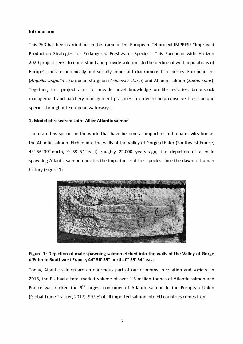
1. Model of research: Loire-Allier Atlantic salmon
There are few species in the world that have become as important to human civilization as
the Atlantic salmon. Etched into the walls of the Valley of Gorge d'Enfer (Southwest France,
44° 56′ 39″ north, 0° 59′ 54″ east) roughly 22,000 years ago, the depiction of a male
spawning Atlantic salmon narrates the importance of this species since the dawn of human
history (Figure 1).
Figure 1: Depiction of male spawning salmon etched into the walls of the Valley of Gorge d'Enfer in Southwest France, 44° 56′ 39″ north, 0° 59′ 54″ east
Today, Atlantic salmon are an enormous part of our economy, recreation and society. In
2016, the EU had a total market volume of over 1.5 million tonnes of Atlantic salmon and
France was ranked the 5th largest consumer of Atlantic salmon in the European Union
(Global Trade Tracker, 2017). 99.9% of all imported salmon into EU countries comes from

7
Figure 2: Rivers colonized in France by Atlantic salmon in the mid-eighteenth century, end of the nineteenth century and end of the twentieth century (Thibault, 1994)
Norwegian aquaculture production of Atlantic salmon. Fish produced in aquaculture are very
different to that of their wild counterparts and in fact, as we witness a global increase of
Atlantic salmon production, we are simultaneously experiencing a massive decline in wild
populations. Rivers throughout Europe and North America are being subjected to human
globalization as well as anthropogenic and climate related threats, pushing many Atlantic
salmon populations towards extinction (Parrish et al., 1998).In France, the historical runs of
Atlantic salmon were enormous, reaching estimated levels of over 800,000 returning adults
in the mid-1800s. However, due to the disconnection of river continuity by hydroelectric
dams and the degradation of juvenile habitat, French populations of Atlantic salmon have
greatly diminished (Figure 2) (Thibault, 1994). In addition, river modification for navigation
and flood control left many of the natural river ways no longer suitable for Atlantic salmon
(Prouzet, 1990).
The Loire River is the largest river in France, spanning over 1,000km long, and is home to a
diverse range of flora and fauna. The river winds through 1/5 of the metropolitan regions of
France and once had a carry capacity of over 100,000 Atlantic salmon. Catch records indicate
a severe decline of Atlantic salmon from 1928-1975, dropping from 15,000 fish per month to
less than 1,000 fish per season (Cohendet, 1993). In 1984, as Atlantic salmon were on the
brink of extinction, the French government banned all Atlantic salmon fishing in the Loire
River and its tributaries. Despite this, wild returning populations of the Loire went extinct by
8
the mid-1990s and its main tributary, the Allier, became the last remaining tributary of the
Loire with wild returning Atlantic salmon.
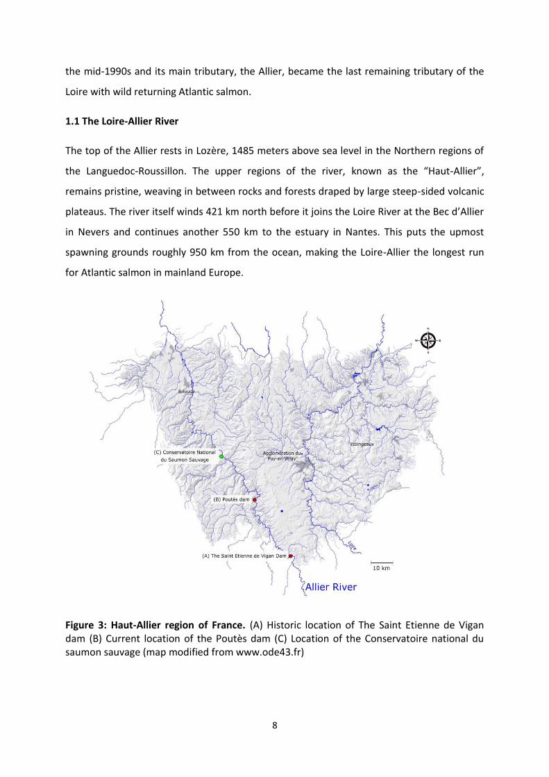
1.1 The Loire-Allier River
The top of the Allier rests in Lozère, 1485 meters above sea level in the Northern regions of
the Languedoc-Roussillon. The upper regions of the river, known as the “Haut-Allier”,
remains pristine, weaving in between rocks and forests draped by large steep-sided volcanic
plateaus. The river itself winds 421 km north before it joins the Loire River at the Bec d’Allier
in Nevers and continues another 550 km to the estuary in Nantes. This puts the upmost
spawning grounds roughly 950 km from the ocean, making the Loire-Allier the longest run
for Atlantic salmon in mainland Europe.
Figure 3: Haut-Allier region of France. (A) Historic location of The Saint Etienne de Vigan dam (B) Current location of the Poutès dam (C) Location of the Conservatoire national du saumon sauvage (map modified from www.ode43.fr)

9
Unfortunately the Allier has had hydroelectric dams, weirs and other obstructions which
have restricted access to spawning grounds since the late 1800s. One dam in particular, The
Saint Etienne de Vigan (Figure 3 A), was located near the source of the Allier and completely
blocked Atlantic salmon access to 70 acres of the best spawning grounds the region had to
offer. Another severe obstruction, the Poutès hydroelectric dam (Figure 3 B), was built in
1941 cutting off all salmon migration to the Haut-Allier until a salmon elevator was installed
in 1986. This region, prior to the dam, was estimated to produced nearly 10 tonnes of
salmon per year, which contributed heavily to the local economy and exemplifies the
population health prior to anthropogenic interference. Despite recent efforts to facilitate
upstream migration, only few salmon make it above Poutès dam each year (24 fish in 2017;
CNSS data). After years of conservational efforts, The Poutès dam is undergoing a
reconstruction project in hopes to better facilitate the down and upstream passage of
Atlantic salmon.
1.2 Restoration projects
In the late 1980s, the SOS Loire Vivante (“Living Loire”) was created to oppose the creation
of the Seree de la Fare dam project, which would have been detrimental to the pristine
environment of the upper Loire basin. Backed by the WWF and international media
coverage, the SOS Loire Vivante fought diligently against the project and finally won the
battle with the cancellation of all 4 dams, confirmed by the French government in January of
1994. The SOS Loire Vivante brought forward, through large demonstrations, strong
scientific research, alternatives to damming and novel approaches to river management
which sparked the creation of “Plan Loire Grandeur Nature” in 1994. The plan coordinated a
series of projects aimed at preserving the Loire River ecosystem. The plan had a particular
focus on saving the remaining French Atlantic salmon population by the removal of poorly
designed or inefficient dams, suspension of all Atlantic salmon fishing and the construction
of the Conservatoire national du saumon sauvage (CNSS). The SOS Loire Vivante network
won the Nobel Prize for environment protectors in 1992 for their relentless efforts of
preserving the Loire River.
1.3 Conservatoire National du Saumon Sauvage
10
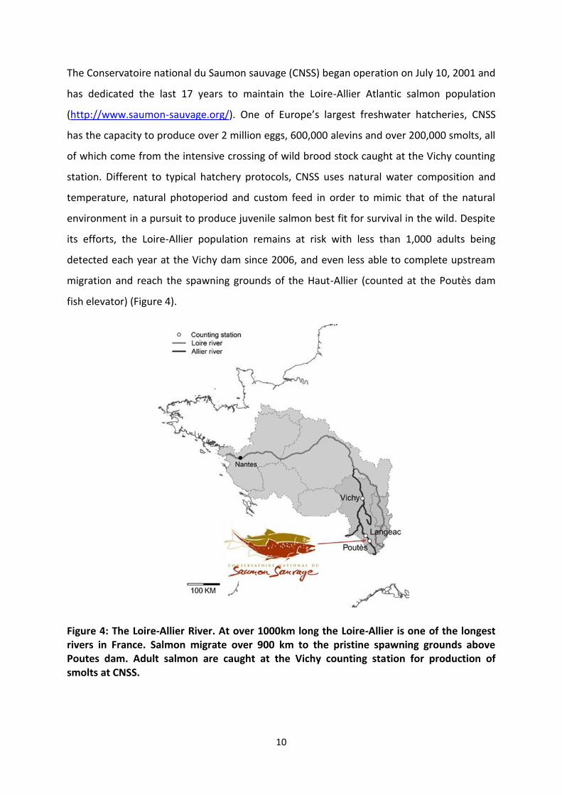
The Conservatoire national du Saumon sauvage (CNSS) began operation on July 10, 2001 and
has dedicated the last 17 years to maintain the Loire-Allier Atlantic salmon population
(http://www.saumon-sauvage.org/). One of Europe’s largest freshwater hatcheries, CNSS
has the capacity to produce over 2 million eggs, 600,000 alevins and over 200,000 smolts, all
of which come from the intensive crossing of wild brood stock caught at the Vichy counting
station. Different to typical hatchery protocols, CNSS uses natural water composition and
temperature, natural photoperiod and custom feed in order to mimic that of the natural
environment in a pursuit to produce juvenile salmon best fit for survival in the wild. Despite
its efforts, the Loire-Allier population remains at risk with less than 1,000 adults being
detected each year at the Vichy dam since 2006, and even less able to complete upstream
migration and reach the spawning grounds of the Haut-Allier (counted at the Poutès dam
fish elevator) (Figure 4).
Figure 4: The Loire-Allier River. At over 1000km long the Loire-Allier is one of the longest rivers in France. Salmon migrate over 900 km to the pristine spawning grounds above Poutes dam. Adult salmon are caught at the Vichy counting station for production of smolts at CNSS.

11
1.4 Atlantic salmon life cycle
Atlantic salmon is an anadromous fish species able to live in both freshwater and saltwater
environments (Figure 5) (For review on life history: Klemetsen et al., 2003). Spawning and
most of the early developmental stages of the Atlantic salmon life cycle takes place in
freshwater (FW). Eggs, or ova, are released into small nests built by adult spawning females.
The ova are fertilized by one or more males and will develop throughout the winter,
hatching in the spring. Just-hatched fish are called alevins and have a nutrient sack attached
to them, which they will absorb throughout the coming months staying hidden between
pebbles and small substrate. Once the yolk sack is absorbed and the fish have become strong
enough to swim to the surface, they will do so to fill their air bladders and gain better
control in the moving streams. Now referred to as fry, the young fish will develop
throughout the summer and into the fall feeding on microscopic invertebrates and seeking
refuge in slower moving water. Over the autumn, fry will develop into parr which have
distinct colouration and behaviours associated with them. Dark green and brown body
colour and dark vertical bands along the side of the body provide camouflage from predators
as the young parr begin to scavenge for aquatic insects. They are much more territorial
during this stage than their other early life stages and will aggressively compete for food.
Once a certain growth stage is reached, juvenile salmon will undergo a dramatic life history
transition referred to as smoltification. This is when the salmon develop from their parr
stage into the smolt stage and encompasses many behavioural, morphological and
physiological changes which pre-adapt the salmon for life in seawater (SW). The
environmental and neuroendocrine control of this metamorphic event will be covered
extensively in the following sections. During this period, downstream migration is initiated
and the juvenile fish, now referred to as smolts, will begin their journey to the sea. During
this stage, smolts give up their territorial behaviour in favour of schooling and through a
series of environmental and physiological cues, will begin to swim downstream to the ocean.
Smolts will remain in the estuary for a short period for acclimatization to changes in
temperature and salinity before making the journey to the North Sea feeding grounds.

12
Figure 5: Atlantic salmon life cycle. Early stages of life take place in freshwater. Eggs or ova are layed in nests called reds which they incubate through the winter and hatch in the spring. Once juvenile salmon develop into parr, smotlfication and downstream migration occurs in which salmon transition to life in seawater. Feeding and growth phase takes place in the Oceans where they develop into Adults. During upstream migration adult salmon return to their native rearing grounds and spawn (image modified from: Life Cycle of Atlantic Salmon, Barbara Harmon, 2011)
Salmon will feed and mature in the North Sea ranging from 1-5 years depending on the
specific population. Salmon that mature after one sea winter are referred to as griles and
will return to their native stream or river for spawning after just one year of feeding. These
fish tend to come from shorter rivers or rivers that have an overall relatively short migration
length. Salmon populations whose migration to the southernmost limit, like those in France,
will spend upwards of 3-4 winters feeding in the open ocean and tend to be much larger in
size when beginning their spawning migration (Martin and Verspoor, 2011).

13
1.5 The Loire-Allier Atlantic salmon
The Loire-Allier Atlantic salmon have many unique characteristics which separate them from
most short river Atlantic salmon stocks. The long 900 km+ migration results in a unique
upstream migration consisting of multiple phases, separated by stopping periods according
to the salmon cohort and date of arrival to the estuary (Martin and Verspoor, 2011).
Spawning migration begins from mid-September to mid-November and involves large
salmon which have spent at least 3 winters in the oceans feeding. These salmon will migrate
upriver until they are halted by low winter temperatures, recommencing once again when
water temperatures rise around mid-March. The salmon will continue migrating upriver until
mid-June when temperatures reach too high for migration forcing salmon to take refuge
until the final leg in the autumn where salmon will reach the upper catchment and spawning
areas of the Loire-Allier (Martin and Verspoor, 2011)..
The Loire-Allier Atlantic salmon are notoriously large fish, reaching approximately 5-7kg and
over a meter in length at spawning. Females will typically lay between 1200-2000 eggs per kg
of weight. Spawning in the Loire-Allier takes place in late autumn and the majority of adults
will die after spawning, due to the enormous energetic demands of the long migration.
However, if the salmon is of good health, multiple migration and spawning events can take
place. Adult salmon used as broodstock at CNSS will be nursed back to good health after
spawning and multiple spawning events are common within the hatchery. Eggs develop
throughout the winter and will hatch the following spring (Martin and Verspoor, 2011).
The majority of Loire-Allier parr will undergo smoltification after 1 year of growth in the river
and begin their downstream migration around March-April. In comparison, salmon from
Scotland or Norway will smoltify around May-June and as late as August for some of the
most northern latitude rivers (Davidsen et al., 2005; Antonsson and Gudjonsson, 2002) The
earlier smoltification and downstream migration of the Loire-Allier population is most likely
combination of genetic component of long river migrating fish as well as the response to
southern environmental conditions. The period in which salmon reach the estuary must be
coordinated with their body’s physiology and thus the synchronization of downstream
migration with physiological development is imperative. Estuary arrival data for the Loire-
Allier salmon is scarce as conservation programs prevent scientific research downstream of
14
the Vichy dam. However the SALSEA-Merge project estimates the Loire-Allier population
should arrive at the Nantes estuary around the end of April in order to complete successful
seaward migration within tolerable environmental conditions (SALSEA-MERGE, 2012).
1.6 Physiological and environmental “windows” of opportunity
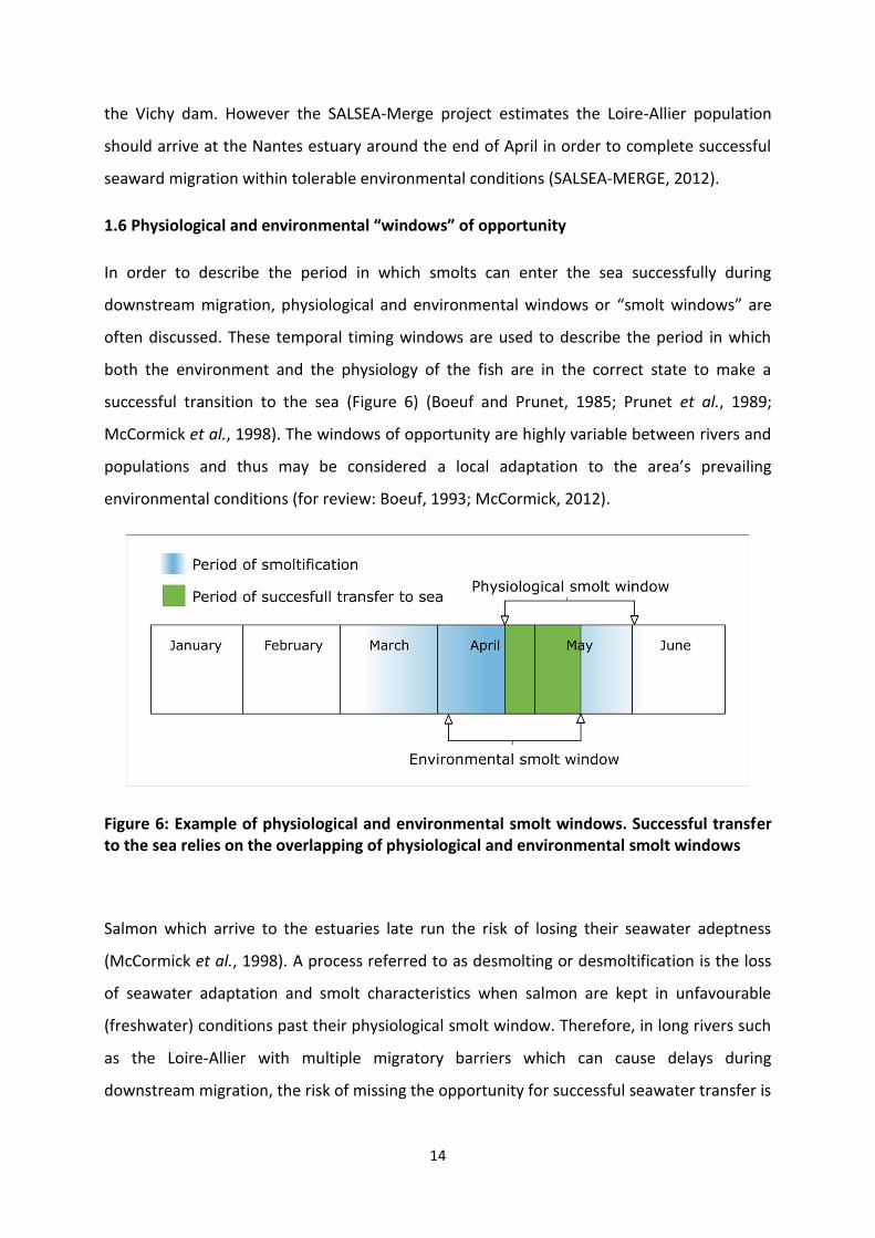
In order to describe the period in which smolts can enter the sea successfully during
downstream migration, physiological and environmental windows or “smolt windows” are
often discussed. These temporal timing windows are used to describe the period in which
both the environment and the physiology of the fish are in the correct state to make a
successful transition to the sea (Figure 6) (Boeuf and Prunet, 1985; Prunet et al., 1989;
McCormick et al., 1998). The windows of opportunity are highly variable between rivers and
populations and thus may be considered a local adaptation to the area’s prevailing
environmental conditions (for review: Boeuf, 1993; McCormick, 2012).
Figure 6: Example of physiological and environmental smolt windows. Successful transfer to the sea relies on the overlapping of physiological and environmental smolt windows
Salmon which arrive to the estuaries late run the risk of losing their seawater adeptness
(McCormick et al., 1998). A process referred to as desmolting or desmoltification is the loss
of seawater adaptation and smolt characteristics when salmon are kept in unfavourable
(freshwater) conditions past their physiological smolt window. Therefore, in long rivers such
as the Loire-Allier with multiple migratory barriers which can cause delays during
downstream migration, the risk of missing the opportunity for successful seawater transfer is

15
much higher than in short rivers (Martin et al., 2012). Therefore, the time in which juvenile
salmon smoltify and initiate downstream migration in long rivers has a direct effect on the
long term survivability. Understanding the environmental and endocrine mechanisms which
regulate this metamorphosis will elucidate a better understanding of the smoltification
process and lead to better conservational efforts for high risk long river Atlantic salmon
populations such as the Loire-Allier.
1.7 Genome duplication events
Nearly 50 years ago, Ohno (1970) presented evidence that whole genome duplication events
(WGD) supply genetic raw material for the emergence of new functions through natural
selection forces and allow for large-scale adaptation to new environments. Generally
speaking, there are three fates for a gene after it has been duplicated: one copy will become
silenced due to degenerative mutations (nonfunctionalization), one copy will acquire a
completely new and beneficial function which then will be passed on through natural
selection (neofunctionalization) or copies of the duplicated gene will adopted sub functions
of the original role with the combined capacity equal to that of the original functions
(subfunctionalization) (Ohno, 1970; Lynch and Conery, 2000). The majority of genes after
duplication events will acquire deleterious mutations which render them non-functional and
if still present in the genome can be characterized as pseudogene.
Teleosts belong to the Actinopterygian infraclass which is amazingly diverse with over
26,000 discovered species. Three rounds of whole genome duplication events have taken
place in the teleost lineage; 1R and 2R before the divergence of lamprey from jawed
vertebrates and a third teleost-specific (3R; TSWGD) WGD at the base of teleosts roughly
320 million years ago (Meyer and Van De Peer, 2005; Smith et al., 2013). The diversification
of the teleost infraclass has been hypothesized to be a consequence of the 3R supported by
the fact that classes which diverged from Aactinopterygians before the 3R event are
extremely species-poor in comparison (Meyer and Van De Peer, 2005).
Recent advances in sequencing technology have concluded that salmonids have undergone a
4th WGD event. The salmonid-specific whole genome duplication (4R; SSWGD) is an
additional genome duplication event that took place roughly 80 million years ago in the
common ancestor of the salmonid species. Interestingly, Lein and collaborators, using the

16
gene expression patterns of Atlantic salmon compared to a pre-4R outgroup, concluded that
the instances of neofunctionalization were much higher than that of subfunctionalization
(Lien et al., 2016). Thus there seems to be the potential for a number of genes duplicated via
the 4R which have acquired novel functions not yet discovered.
2. Smoltification
The parr-smolt transformation of salmonid species has captivated scientists for decades.
Fuelled by the economic importance of this family of teleosts, the research into the
metamorphic event of smoltification has been robust (for reviews: (Folmar and Dickhoff,
1980; Wedemeyer, Saunders and Clarke, 1980; Hoar, 1988; Boeuf, 1993; Björnssone et al.,
2011; Rousseau et al., 2012; McCormick, 2012). Despite this, the discussion of to what
extent smoltification is regulated by exogenous or endogenous factors remains unclear.
Smoltification is a complex series of the morphological, behavioural, and physiological
changes managed by the endocrine system and suggested to be synchronized with
environmental factors such as photoperiod, temperature, lunar phase, salinity, turbidity and
river flow (Hoar, 1988).
2.1 Morphology
2.1.1 Early growth
Within a freshlyl hatched cohort of juvenile Atlantic salmon, referred to as 0+, differential
growth rates between individuals will result in a bimodal size-frequency distribution that is
related to smolt development (Kristinsson et al., 1985). The upper mode represents the
potential pre-smolts which will continue to eat and grow throughout the autumn and winter
in preparation for smoltification and migration in the spring. The lower mode will enter a
state of anorexia during the winter ceasing growth and development until the following
spring (Thorpe et al., 1982; Metcalfe and Thorpe, 1992). Juvenile salmon that develop into
smolts after 1 full winter are called +1 smolts, 2 winters are called 2+ smolts and so forth
(Thorpe, 1989). The majority of the Loire-Allier population smoltify and migrate at +1 or 2+,
however 3+ is seen on occasion.

17
2.1.2 Colouration
There are distinct morphological differences between parr and smolt of Atlantic salmon,
which can lend to the visual observation of the smoltification process. Parr have dark
pigmented bands along the side of the body referred to as “parr marks”, which slowly fade
during the smolting process, as deposition of crystalline purines (guanine and hypoxanthine)
in the skin and scales causes extensive silvering of the body (Johnston and Eales, 1967).
Synchronous to silvering, caudal, dorsal and pectoral fins become significantly darker
throughout the smolting process due to the diffusion of melanin granules in melanophore
cells (Figure 7) (Mizuno et al., 2004).
Figure 7: Morphological changes of the Loire-Allier Atlantic salmon during the smoltification metamorphosis. (A) Parr marks regress, (B) Fin margins darken and (C) extensive silvering of the body occurs during this metamorphic event which helps preadapt the salmon for life at sea.
18
2.1.4 Condition factor
The body shape of a developing smolt is different to that of a parr. Smolts become elongated
as their linear growth rate increases relative to total growth, leading to a reduction in
condition factor [k=(L/w3)*100]. Condition factor is often used for fish to help indicate the
overall health of a fish; indicating such things as feeding condition, parasitic infections and
physiological factors (Le Cren, 1951; for review: Froese, 2006). Elongation during the parr-
smolt transformation is expected and thus condition factor can be used to characterize the
smolting process. Parr tend to increase condition factor during the spring as feeding
increases, meanwhile maintaining low lipid demands. During smoltification, increased linear
growth rate, metabolic rate and reduced lipid intake result in a decreasing condition factor
(Mccormick et al., 1987).
2.1.5 Other organs and tissues
As smoltification is a metamorphosis, many organs and tissues are affected during the
process. Although not the focus of this thesis, these changes are worth mentioning: the eye,
brain, gills, intestine and kidney all undergo some sort of adjustment to coincide with the
change of habitats during smoltification (for review: Rousseau et al., 2012)
2.2 Metabolism
Overall standard metabolic rates are 30-50% greater in smolts than in parr when body
weight is accounted for (Maxime et al., 1989; Baraduc and Fontaine; 1955). The increased
metabolism is characteristic of smoltification and due in part to the increased growth,
feeding, sodium-potassium adenosine triphosphatase enzyme (Na+-K+-ATPase : NKA) activity
and concentration of mitochondrial rich (MR) cells (Blake et al., 1984; Hoar, 1988; Maxime et
al., 1989). Increased haemoglobin concentrations and complexity has been reported during
smoltification due to the increased oxygen demands during migration (Zaugg and McLain,
1976; Sullivan et al., 1985). Overall, lipid concentrations decrease during smoltification,
mediated by lipase enzymes, which in part may be regulated by endocrine factors such as
thyroxine (T4), cortisol, growth hormone (GH) and prolactin (PRL) (Sheridan, 1988, 1989; Bell
et al., 1997).

19
2.3 Behaviour
Parr have a distinct territorial behaviour, which they exhibit during their residence in rivers
and streams. They remain near the bottom of the water column hidden between the
substrate, and use fast bursts of swimming to feed on passing food particles. As the winter
ends and the parr begin the smolt transformation, they reduce their territorial behaviour in
favour of forming shoals. Increased social behaviour coincides with a reduction in fast
swimming activity and an increase in migratory readiness behaviour (Hoar, 1988; Boeuf,
1993). As spring approaches, smoltification will commence and migratory behaviour will
change from predominantly facing the stream (positive rheotaxis) to predominantly facing
downstream (negative rheotaxis), and begin their journey downward to the sea (Boeuf,
1993; McCormick et al., 1998)
The factors contributing to the change in rheotaxis may be the most critical in determining
the overall timing of smolt migration, yet these factors remain poorly understood (for
review: McCormick, 2012). Laboratory studies have demonstrated environmental factors
contributing to a change in rheotaxis such as advanced photoperiod (Muir et al., 1994),
accumulated thermal units (Zydlewski et al., 2005; Sykes and Shrimpton, 2009), water
temperature (Jonsson and Ruud-Hansen, 1985), water discharge (Thorpe et al., 1981;
Thorpe, 1989) and endocrine factors such as thyroid hormones (TH) (Ojima and Iwata, 2007)
and growth hormone (Prunet et al., 1989); however no single entity takes full responsibility
of migration onset (McCormick et al., 1998). Due to the variability in river geographic
location, topography and length of the initiation of migration, a genetic component that is
unique to river populations is likely involved. Furthermore, the onset of migration may also
depend on things such as social cues or increased exothermic predation (Hansen and
Jonsson, 1985). Nevertheless, the change in rheotaxis during the smoltification period is a
marked characteristic of the smolting process (Boeuf, 1993; McCormick et al., 1998). Arriving
to the estuary at the correct period depends on the correct time of departure from rearing
grounds and therefore indicative to smolt survival.
2.4 Osmoregulation and increased salinity preference
One of the most important changes that occur during smoltification is the change in
osmoregulatory function as salmon go from a freshwater to salt water environment (for

20
reviews: (Sakamoto et al., 1993; McCormick, 2012). In freshwater, fish normally do not drink
and will excrete excess water through diluted urine while absorbing salt through the gills. In
salt water, fish drink seawater to compensate for passive water loss and actively secrete
excess salt via gills and kidney. For salmon migrating down river, it is important that they
maintain the ability to absorb ions while they are still in freshwater meanwhile preparing for
the increasing salinity as they approach the estuary.
Smolts develop increased salinity tolerance over several weeks prior to SW entry. Many
tissues including the gut, intestine, kidney and bladder manage the internal salt and water
balance using the gills for major source of ion uptake in freshwater and salt secretion in
seawater (For review: Evans et al., 2005). Mitochondrion-rich cells, also known as chloride
cells, are specialized ionocytes responsible for the active transport of ions across the gills.
Three main transport proteins are localized in gill ionocytes cells: NKA, Na+-K+-2Cl-
cotransporter (NKCC) and the cystic fibrosis trans membrane regulator (CFTR) (Evans et al.,
2005). The NKA enzyme is a solute pump that actively pumps sodium out of the cell while
pumping potassium into the activity of which increase during the smoltification process
(Rainbow trout (Oncorhynchus mykiss): Richards, 2003; Arctic char (Salvelinus alpinus):
Bystriansky, LeBlanc and Ballantyne, 2006; Atlantic salmon: (Nilsen et al., 2007; McCormick
et al., 2009)). In Atlantic salmon, two isoforms of NKA α are present; NKA α1a and NKA α1b,
which have shown to be expressed in variable abundance depending on the aquatic
environment the fish are in (McCormick et al., 2009). In freshwater, NKA α1a expression is
highly abundant in distinct chloride cells while NKA α1b becomes the dominant isoform after
seawater acclimation. Albeit low numbers, NKA α1b mRNA is detectable in chloride cells
while in freshwater suggesting the mechanism for seawater tolerance is available before fish
experience an increase in salinity (Nilsen et al., 2007).
Two NKCC isoforms (NKCC1 and NKCC2) are present in teleosts; one for secretory function
(NKCC1) and the other for absorptive function (NKCC2) (for review: McCormick, 2012). In
Atlantic salmon, mRNA expression of the NKCC1 isoform increases during smolt
development and especially in gill ionocyte cells (Nilsen et al., 2007). Juvenile Atlantic
salmon kept under continuous light disrupts smolt development and inhibits NKCC
transcription and development of seawater tolerance (Stefansson et al., 2007). In brown
21
trout (Salmo trutta), NKCC and NKA abundance increases gradually with smolt development
and dramatically after seawater exposure (Tipsmark et al., 2002)
There have been two confirmed CFTR isoform in Atlantic salmon which has differential
expression during smolt development. CFTR I mRNAs increase during smolt development
and after seawater transfer, whereas CFTR II expression shows no increase and only a slight
increase, respectively (Singer et al., 2003; Nilsen et al., 2007).
2.5 Environmental regulation of smoltification
Environmental conditions act through the endocrine system to relay vital information for the
timing of physiological and behavioural changes during smoltification (Hoar, 1976, 1988;
Björnsson, Stefansson and McCormick, 2011).
2.5.1 Photoperiod
Similar circannual rhythms of smoltification characteristics such as increased growth rate,
silvering and reduced condition factor can be achieved under vastly different photoperiods;
however, correct light and dark conditions are needed for normal and healthy smolt
development able to complete successful migrations and transfer to the sea (Eriksson and
Lundqvist, 1982). Parr exposed to continuous light experienced less retinal innervation to
the preoptic nucleus than controls (Stefansson et al., 2007). These fibres contribute to the
light-brain-pituitary (LBP) pathway and demonstrate a direct or indirect regulation of
pituitary hormones such as GH, thyroid stimulating hormone (TSH) or adrenocorticotropic
hormone (ACTH) (Holmqvist et al., 1994; Ebbesson et al., 2003).
In salmonids, as like other teleosts, melatonin profiles accurately reflect the prevailing
light/dark cycles and are known to be involved in the synchronization of a number of
rhythmic physiological processes (for review: Korf, 1994). Melatonin synthesis increases
during hours of darkness and is produced from photoreceptor cells in the pineal gland
(Falcon et al., 1992). Pinealectomized Atlantic salmon still undergo smoltification, but with a
delay of nearly 3 weeks as compared to controls, while pinealectomized salmon given
intramuscular injection of melatonin have advanced smoltification (Porter et al., 1998).
Additionally, melatonin injections caused increased growth rate of smolting salmon and thus

22
suggest that melatonin is an important component of somatic growth and smoltification,
while not necessary for the metamorphosis to occur (Porter et al., 1998; Mayer, 2000).
For Atlantic salmon, the vast knowledge concerning the effect of photoperiod on
physiological development during smoltification comes from salmon aquaculture research
and the pursuit to consistently produce healthy 0+ smolts. Using artificial photoperiod,
hastened and abrupt simulations of summer and winter, 0+ smolts able to advance elevated
plasma GH (Björnsson et al., 2000), cortisol (Sundell et al., 2003) and increased NKA activity
(Berge et al., 1995) to levels similar to a natural +1 smolt. Juvenile salmon raised under
continuous light have increased growth rate and NKA activity while in freshwater as
compared to controls (Mccormick et al., 1987; Stefansson et al., 1991). These studies
emphasis the entrainment of smoltification with photoperiod and concluded that
photoperiod is one of the, if not the most, important environmental factor for the initiation
of smoltification.
Photoperiod however is not the only primary factor when determining the timing of
smoltification and does not contribute to smoltifying characteristics when body growth or
water temperature are too low (Thorpe et al., 1982; McCormick et al., 2000). Thus it has
been suggested that photoperiod determines the temporal range in which the fish become
sensitive to factors such as water temperature, flow, turbidity and lunar cycle in order to
synchronize the endocrine system with environmental factors (for review: McCormick, 2012)
2.5.2 Temperature
Increased water temperatures can hasten the rate of smolt development (Duston et al.,
1991; Sigholt et al., 1998). Thus, European stocks of wild Atlantic salmon from southern
populations typically smoltify at 1-2+ whereas cooler northern populations will smoltify at 3-
5+ (Martin et al., 2012; Erkinaro, 1997). It has been suggested that a determining factor for
smoltification is related to the degree days the fish are exposed to (Zydlewski et al., 2005);
whereas others suggest a critical temperature threshold must be met in order for smolting
and migration to take place (Hansen and Jonsson, 1985). Reported temperature thresholds
vary between populations and species, likely being genetically specific to populations of
certain river geographic location (Handeland et al., 2004; Handeland et al., 2008). For
example, river Orkla in Northern Norway has shown migration to commence when water

23
temperatures range between 1.7 and 4.4°C (Hvidsten et al., 1995), while migration onset
was observed at temperatures between 8.5-13°C for the Varzuga River in Russia (Davidsen et
al., 2005).
For the Loire-Allier, downstream movements has been suggested to commence when water
temperatures ranged from 7.5-13°C with maximum swimming speed at 10.5°C; however,
downstream migration quickly diminishes when water temperatures exceed 17°C and
completely halt above 20°C (Martin et al., 2012). Generally speaking, salmonids are
negatively impacted by temperatures outside their thermal range, which can inhibit smolt
development, halt migration and hasten the loss of some smoltification characteristics (de-
smoltifying) (Duston, Saunders and Knox, 1991; Handeland et al., 2004). Physiological and
morphological parameters such as NKA activity and silvering can be advanced with increased
rearing temperatures; however, loss of seawater adeptness and smolt characterises are also
advanced at temperatures above thermal range of the species (Duston et al., 1991).
Increased temperature can hasten the NKA activity during smoltification, but the date of
which peak activity occurs is the same between experimental groups of Atlantic salmon
raised at two different temperature regimes (McCormick et al., 2002). In coho salmon
(Oncorhynchus kisutch), Larsen et al. (2001) found no difference in the timing of NKA activity
increase at salmon reared throughout winter at 2.5 and 10˚C. Interestingly, advanced
silvering and NKA activity induced via increased water temperature are nullified without
photoperiod stimulus (Sigholt et al., 1998; McCormick et al., 2002). Thus a common
conclusion is that temperature is an important modulator of smoltification and downstream
migratory behaviour, but not sufficient as directive factor alone (Clarke et al., 1978;
Wedemeyer et al., 1980; Boeuf, 1993; McCormick et al., 1998; Sigholt et al., 1998; Martin et
al., 2012).
2.5.3 Water flow and turbidity
Early investigations into the downstream movements of smolts related migratory behaviour
to increased water flow and concluded that smolt movement is the passive displacement
primarily controlled by flow rate (Thorpe et al., 1981; Jonsson and Ruud-Hansen, 1985;
Thorpe, 1989; Thorpe et al., 1994). However, further studies argue that smolt migration is a
mix of passive and active swimming, using water flow as a facilitating mechanism to

24
downstream movements (Hansen and Jonsson, 1985; Aarestrup et al., 2002; Davidsen et al.,
2005; Martin et al., 2012). The migratory strategies used by different populations of
migrating smolts may depend on the length and geographic region of the river and could be
a heritable trait. Nevertheless, water flow seems to play an important role in the movement
of smolts once smoltification has commenced but does not regulate downstream movement
alone (McCormick et al., 1998).
2.5.4 Lunar cycle
Compelling evidence for the influence of lunar cycle and migratory behaviour was first
reported in coho salmon, by Grau et al (1981) who observed a tight correlation to new moon
events with T4 surges during smoltification. Further studies observed similar correlations
between new moon events and T4 surges in Atlantic salmon (Boeuf and Prunet, 1985),
Chinook salmon (Nishioka et al., 1985) and masu salmon (Yamauchi et al., 1984) and suggest
that T4 surges during smoltification may be regulated by exogenous factors (for review:
(Grau et al., 1982)
3. Endocrinology of smoltification
3.1. Hypothalamus-pituitary-thyroid (HPT) or Thyrotropic axis
Thyroid hormones (TH) are responsible for several critical biological phenomena and are
important throughout growth and development of most organisms. In adult vertebrates, TH
control basal metabolic rates and energy metabolism whereas in developing vertebrates,
nearly all processes concerning neurogenesis and central nervous system maturation are
depending on the availability of thyroid hormones (Tata, 2006). The thyrotropic axis is the
main neuroendocrine axis involved in the regulation of salmonid smoltification, as in the
control of amphibian and flatfish metamorphosis.
3.1.1. Major actors of HPT axis
3.1.1.1. Mammals
In mammals, hypothalamic thyrotropin-releasing hormone (TRH) located in the
periventricular nucleus (PVN) of the brain have been shown to be responsible for the
regulation of TSH from the pars distalis (PD) of the anterior pituitary, which subsequently

25
stimulates the synthesis and release of T4 from the thyroid gland (for reviews: Fekete and
Lechan, 2007; Zoeller, Tan and Tyl, 2007). T4 gets then converted to bioactive
triiodothyronine (T3) by the deiodinase type-2 enzyme (dio2) in various tissues. Interestingly,
TRH has also been shown to have multiple stimulatory effects on other pituitary hormones
such as PRL and GH in vertebrates (Galas et al., 2009). TRH neurons in the PVN are regulated
by complex web of neuronal axons and humoral signalling, which allow the HPT axis to
respond to environmental stimuli (Lechan and Fekete, 2006; Ortiga-Carvalho et al., 2016).
3.1.1.2. Other vertebrates
The involvement of TRH as the hypothalamic factor controlling the thyrotropic axis has been
subject to debate among vertebrates other than mammals. Studies on non-mammalian
vertebrates showed that TRH does not always act as a TSH releasing factor and led to the
discovery that corticotropin-releasing hormone (CRH) was acting as a potent TSH stimulator
in amphibians, reptiles, birds and teleosts (Denver, 1988; Denver and Licht, 1989; De Groef
et al., 2006). CRH is generally known as the central regulator of the corticotropic axis (or
stress axis), which produces corticosteroids. With its dual action, CRH may coordinate both
and thyrotropic axes during vertebrate life stage transitions (Watanabe et al, 2016).
In amphibians, Denver and Licht (1989) showed that American bullfrog (Rana pipiens)
tadpole pituitaries increase TSH secretion after ovine CRH treatment, but did not respond to
TRH. In addition, Denver (1988) showed pituitaries from adult Rana pipiens, Hyla regilla, and
Xenopus laevis increased TSH secretion in a dose dependent manner with exposure to TRH.
These studies suggest that TRH has a TSH-releasing action only in metamorphosed
amphibians but not larval amphibians (for review: De Groef et al. 2006).
In reptiles, in vivo and in vitro studies on adult turtles (Chrysemys picta) and lizards (Anolis
carolinesis) observed an increase of TSH after TRH stimulation (Preece and Licht, 1987; Licht
and Denver, 1988), but also after CRH treatment in turtle (Pseudemys scripta: Denver and
Licht, 1989).
Similar results have been reported in birds in which CRH acts as a potent TSH stimulator. In
chicken embryos, ovine CRH caused both an increase in T4 as well as corticosterone levels
(Meeuwis et al., 1989). Furthermore, Geris and collaborators confirmed the direct action of

26
CRH as a TSH-releasing factor on domestic fowl (Gallus domesticus) pituitaries both in vitro
and in vivo (Geris et al., 1996, 1999).
In basal teleosts, lungfish and hagfish, TRH does not stimulate the release of TSH (African
lungfish Protopterus ethiopicus: Gorbman and Mohamed, 1971); Pacific hagfish Eptatretus
stouti: Dickhoff, Crim and Gorbman, 1978). However some more recent studies have shown
an increase of tsh mRNA expression in Japanese eel (Anguilla japonica; Han et al., 2004) and
bighead carp (Aristichthys nobilis; Chatterjee, Hsieh and Yu, 2001) after TRH treatment. In
coho salmon, Larsen et al (1998) discovered a potent stimulation of TSH release by CRH in
pituitary cell cultures, whereas TRH did not induce any changes. In supporting evidence for
the interaction between CRH and TSH in salmonids, Matz and Hofeldt (1999) showed that
CRH-immunoreactive fibres terminated in pituitary regions containing TSH immunopositive
cells in Chinook salmon. In flatfish (Pleuronectiformes), few studies have investigated the
hypothalamic regulation of TSH. Exposure to the goitrogen, thiourea, or to T4 did not induce
any changes in TRH mRNA levels, indicating that TRH do not participate in the regulation of
HPT in the sole (Iziga et al., 2010). In addition, TRH expression was shown to decrease
progressively from 2-3 days after hatching till metamorphosis in Senegalese sole, suggesting
that TRH was not involved in the induction of flatfish metamorphosis (Iziga et al., 2010).
More recently, Campinho and collaborators hypothesize that flatfish metamorphosis may be
an hypothalamic independent process as neither trh nor crh expression changes during
metamorphosis, and hypothalamus does not respond to an anti-thyroid drug blockade
whereas pituitary and thyroid do (Senegalese sole (Solea senegalensis): Campinho et al.
2015).
3.1.2. HPT and the control of major developmental processes
3.1.2.1. HPT and circannual rhythms
Birds and mammals have developed a novel role for HPT in the regulation of seasonal
changes in physiological functions such as reproduction (for reviews: Ikegami and
Yoshimura, 2017). Recent studies in the quail (Nakao et al., 2008), sheep (Hanon et al., 2008)
and mouse (Ono et al., 2008) revealed that TSH could also be produced by the pars tuberalis
(PT) of the anterior pituitary under long day stimulus. This PT-TSH production is independent

27
of the HPT axis, but is instead regulated by melatonin from the pineal gland in mammals
(Malpaux et al., 2001) and direct photoperiodic information through deep brain
photoreceptors in birds (Nakane and Yoshimura, 2010). PT-TSH locally activates thyroid
hormones in the hypothalamus, which in turn induces the activation of the gonadotropic
axis involved in the control of reproduction (for reviews: Yoshimura, 2013; Ikegami and
Yoshimura, 2017). For now, in fish, it is the saccus vasculosus, an organ located on the
ventral side of the diencephalon posterior to the pituitary, which has been described as the
seasonal sensor (Nakane et al., 2013).
In birds, thyroid hormones are related to many circannual processes including reproduction,
molting and migratory readiness (for reviews: Dawson et al., 2001; Kuenzel, 2003).
Exogenous treatment of T4 can mimic the reproductive effects of long photoperiod in a
variety of bird species measured by increased levels of LH and FSH secretion and testicular
growth (tree sparrow: Wilson and Reinert, 1996; Japanese quail: Follett and Nicholls, 1988;
European starling: Goldsmith and Nicholls, 1992). Removal of the thyroid gland in redheaded
bunting (Emberiza bruniceps) was found to inhibit the fat deposition and nocturnal
restlessness, which are related to pre-migratory disposition; both were restored with
exogenous treatment of T4 or T3 (Pant and Chandola-Saklani, 1993).
In seasonal mammals, thyroid hormones are also involved in the control of seasonal
processes such as reproduction. In sheep, a short day (SD) breeder, thyroidectomy blocks
the suppression of gonadotropin induced by long photoperiod, and hypothalamic T4
microimplants reverse this blocking effect (Anderson et al., 2003). In Siberian hamster, a
long day breeder (LD), daily T3 injections to short-day animals induce testicular growth,
mimicking thus long day length (Freeman et al., 2007). Similarly, T3 intrahypothalamic
microimplants prevent the onset of testicular regression normally observed in short
photoperiod (Barrett et al., 2007).
3.1.2.2. HPT and metamorphosis
Metamorphosis is a term used to define a remarkable body change during development that
is normally accompanied by a change in habitat or behaviour. Metamorphic events ranging
from mild to extreme have been described in a variety of groups including cnidarian, insects,
crustacean, molluscs, tunicates and some vertebrates (amphibians and some fish; for review:

28
Laudet, 2011; Rousseau et al., 2012). Although the molecular mechanism involved in
metamorphosis throughout various chordate lineages are largely unknown, THs and the HPT
axis seem to predominantly regulated metamorphosis in amphibians and teleost fishes (Paris
and Laudet, 2008). In fish, two types of metamorphoses have been described; First or larval
metamorphosis is a true metamorphosis (as amphibian metamorphosis) which occurs for
example in pleuronectiforms (flatfish). Secondary metamorphosis is observed in some
diadromic migratory teleosts and compared to first/larval metamorphosis involves less
drastic morphological changes in post-embryonic juveniles; the smoltification
metamorphosis has been debated as a secondary metamorphosis (Björnsson et al. 2012;
Rousseau et al., 2012).
3.1.2.2.1. HPT and amphibian metamorphosis
Amphibian metamorphosis is the best example of dramatic/extensive morphological,
biochemical and cellular changes occurring during development and its regulation has been
extensively studied and documented. This post-embryonic metamorphosis is obligatorily
initiated and sustained by TH (Figure 8). The involvement of thyroid gland in the induction of
metamorphosis has been suggested as early as 1912. At that time, Gudernatsch observed
that common frog (Rana temporaria) tadpoles were turning into frogs when given powdered
sheep thyroid gland (Gudernatsch, 1912). In addition, when surgical or chemical ablation of
the thyroid gland was performed by Allen (1917), the larvae, instead of metamorphosing,
became giant tadpoles (Allen, 1917). Further studies of early exposure to TH confirmed
these first observations (for review: Tata, 2006). External environmental factors and
stressors, from higher brain centers, influence hypothalamic CRH as the main stimulator of
TSH and ultimately TH (Denver et al., 2002). TH then target via their receptors, a wide variety
of tissues causing a series of anatomical and functional changes including the simultaneous
emergence of limbs and loss of larval tails, gills and digestive system. CRH and TSH regulation
of TH during metamorphosis has been clearly demonstrated in a variety of studies (for
reviews: (Denver et al., 2002; Manzon and Denver, 2004; Tata, 2006; Grimaldi et al., 2013).

29
Figure 8: Proposed hormonal regulation of amphibian metamorphosis (Tata, 1993)
3.1.2.2.2. HPT and teleost metamorphoses
3.1.2.2.2.1. HPT and flatfish larval metamorphosis
Flatfish (Pleuronectiformes) have an extreme asymmetry with both eyes being on the same
side of the head after metamorphosis. Similar to amphibians, larval metamorphosis in
flatfish is primarily regulated by the HPT axis (for reviews: (Inui and Miwa, 1985; Schreiber
and Specker, 1998; Power et al., 2008). During this metamorphic event, many changes
happen to the body and include the dramatic migration of one eye to the contralateral side
of the head resulting in both eyes located on the same side of the head. Radioimmunoassay
studies have shown a peak of T4 and histology studies have shown activation of TSH cells
during the climax of the metamorphosis (Miwa et al., 1988). Injection of bovine TSH in
Japanese flounder can increase T4 levels and accelerate metamorphosis concluding that TSH
is the main stimulatory factor of T4 and thus the major regulatory of metamorphosis (for
review: Inui and Miwa, 2012).

30
3.1.2.2.2.2. HPT and salmonid smoltification (secondary metamorphosis)
Thyroid hormones
As early as 1939, thyroid hormones have been hypothesized to be involved in the control of
smoltification. Hoar discovered via histological studies that the thyroid follicles of Atlantic
salmon became highly active during the period of smoltification (Hoar, 1939). Since then,
surges of T4 during the period of smolitfication has been shown in a variety of salmonid
species (coho salmon: Dickhoff et al., 1978; Larsen et al., 2011; masu salmon, (Oncorhynchus
masou): Nishikawa et al., 1979; Atlantic salmon: Boeuf and Prunet, 1985; Prunet et al., 1989)
These peaks of T4 led scientists to suggest that T4 surge during smoltification is the critical
component to the timing of the smoltification metamorphosis. Further studies concluded
through treatments with thyroid hormones or anti-thyroid drugs such as propylthiouracil
(PTU) (immersion, oral administration, intraperitoneal implants etc) that thyroid hormones
could stimulate silvering and fin margination, metabolism and downstream migration (for
review: Rousseau et al., 2012). However they could not induce other changes of
smoltification such as increased osmoregulatory ability (Refstie, 1982; Brown et al., 1987).
Thyroid hormones seem crucial for smoltification; however the mechanisms that regulate TH
availability are not fully understood.
CRH
Administration of human CRH increased plasma T4 levels in coho salmon and stimulated
downstream movements in coho, Chinook (Oncorhynchus tshawytscha) and chum
(Oncorhynchus keta) salmon when administered during the smoltification period (Clements
and Schreck, 2004; Ojima and Iwata, 2009, 2010). At the time of smoltification, neurogenesis
of CRH neurons is activated in Atlantic salmon (Ebbesson et al., 2011).
TSH
Among teleosts, pituitary content of TSH is characteristically low in relation to thyroid
hormone activity (for review: MacKenzie, Jones and Miller, 2009). Within salmonids, the
data obtained for TSH has been contradictory. Fridberg et al. (1981) reported in the Atlantic
salmon that pituitary TSH-cells were more numerous and had increased activity in presmolts
and smolts than in parr; yet Nishioka et al. (1982) showed no ultra-structural changes of TSH
cells in coho salmon. No change or a slight decrease in pituitary tsh mRNA levels were

31
measured in smolts as compared to parr in Atlantic salmon (Martin et al., 1999; Robertson
and McCormick, 2012) and coho salmon (Larsen et al. 2011). No variations in pituitary and
plasma TSH protein levels were reported in coho salmon throughout smoltification (Larsen
et al., 2011). More work is needed to understand the role of TSH in teleosts.
TSH is comprised of two subunits, a common alpha subunit shared with the gonadotropins,
luteinizing hormone and follicle-stimulating hormone, and a beta subunit (TSHβ) conferring
hormone specificity (Pierce and Parsons 1981). Recent work by Maugars and collaborators
has outlined the detailed discovery and characterization of thyrotropin beta subunit paralogs
in the teleost lineage (Maugars et al., 2014). Through detailed phylogeny and synteny
analysis, the authors presented TSH paralogs in various teleost species and used the
European eel to display differential expression of the two paralogs throughout peripheral
tissues (Maugars et al., 2014). In addition, the authors used the deduced amino acid
sequence to confirm the conservation of 12 cysteine residues essential for proper folding
and functionality of thyrotropin suggesting a functional role of these paralogs in multiple
species. Functional paralogs of TSH have already been observed in stickleback. Kitano et al.
(2010) discovered paralogs of the tshß subunit (named tshß1 and tshß2 by the authors) in
stickleback and reported higher pituitary transcripts of tshß2 in populations migrating to the
sea compared to stream-resident populations, meanwhile expressing similar transcript levels
of tshß1.
3.2 Other factors involved in the control of smoltification
3.2.1 GH and IGF-1
Growth hormone and insulin like growth factor -1 (IGF-1) are important regulators of growth
and metabolism in vertebrates including teleost fish (for reviews: Moriyama et al., 2000;
Wood et al., 2005). Both have been shown to be crucial components during the smolting
process of salmonid species (Sakamoto et al., 1993; Larsen et al., 1995; Agustsson et al.,
2001). Pituitary GH is the main stimulator of IGF-1, which is primarily secreted from the liver.
In Atlantic salmon parr, plasma GH and IGF-1 remain low and increase with smolt
development peaking slightly before the peak of NKA activity (Prunet et al., 1989;
McCormick et al., 2007). The mRNA expression of the two genes for GH (GH1 and GH2)

32
present in Atlantic salmon increases during the parr-smolt transformation and peaks
concomitantly to that of TSH (for review: Rousseau et al., 2012). In coho salmon, plasma GH
concentrations increase at the end of the smoltification period, and exposure to seawater
causes increase in GH circulating levels (Sweeting et al., 1985).
Exogenous treatment of GH in Atlantic salmon pre-smolts increases NKA activity and salinity
tolerance, while causing no affect in smolts (Prunet et al., 1994). In coho salmon, GH also
induces gill corticosteroid receptor abundance, leading to increased responsiveness of gills
to cortisol and partially accounting for the increase in NKA activity (Shrimpton et al., 1995).
Interestingly, the natural increase of GH during smoltification is absent in landlocked
populations of salmon, which is associated with their poor salinity tolerance (Nilsen et al.,
2008). The strong induction of NKA activity by GH and IGF-1 during smoltification has been
suggested to play a role in behaviour change during smolting as they cause an increased
salinity preference in smolts (for review: Iwata 1995). Injections of GH in Atlantic salmon
parr cause increased Na+–K+–2Cl- cotransporter abundance in gill chloride cells (Pelis and
McCormick, 2001). Low levels of GH receptors in the liver and subsequent low levels of
plasma IGF-1 have been related to extreme reduced growth (“stunting” phenomenon) after
seawater exposure suggesting the importance of increased GH and IGF-1 in proper SW
tolerance (Duan et al., 1995).
In addition to its role in the control of salinity tolerance, GH possesses also its “classical”
strong growth promoting action in salmonids (Björnsson, 1997), which can be associated
with increased food intake (Johnsson and Björnsson, 1994). GH usually stimulates length
growth over weight growth resulting in leaner elongated fish (Johnsson and Björnsson,
1994). Increased growth could be due to the stimulation of lipid mobilisation and protein
synthesis (Björnsson, 1997). In Atlantic salmon, the increased metabolic rate in smolts when
compared to parr could come from the increased NKA activity and mitochondrial cell
proliferation as a consequence of increased GH (Maxime et al., 1989; Handeland, Imsland
and Stefansson, 2008).
33
3.2.2 Prolactin
In 1928, Stricker and Grueter discovered that pituitary prolactin (PRL) could stimulate milk in
rabbits (Stricker and Grueter, 1928). Over the following decades, PRL was accredited with
over 300 biological activates among various vertebrae species (for reviews: Bole-Feysot et
al., 1998; Manzon, 2002). In fish, PRL was first demonstrated by Pickford and Phillips to be
part of the ion regulation mechanism in freshwater Killifish (Fundulus geteroclitus) in 1959
(Pickford and Phillips, 1956). Further studies reported high levels of PRL in winter and spring,
which decreased with the progression of smoltification in Atlantic and coho salmon (Prunet
et al., 1989; Young et al., 1989). In Atlantic salmon, Morin et al., (1994) observed no changes
in PRL over the period of smoltification. However if smolts are kept in freshwater for
extended periods of time, PRL levels will begin to increase (Boeuf and Prunet, 1985; Morin
and Haug, 1994). PRL in parr may be a smolt inhibitory mechanism supported by the fact
that PRL inhibits the development of seawater chloride cells meanwhile promoting ion
uptake cells in sea-water adapted tilapia (Oreochromis niloticus) (Herndon, McCormick and
Bern, 1991; Pisam et al., 1993). Atlantic salmon PRL promotes acclimation to freshwater
while GH promotes acclimation to seawater mediated by the dual osmoregulatory function
of cortisol (Pelis and McCormick, 2001).
3.2.3 ACTH and Cortisol
Commonly called the stress axis, the corticotropic axis involves hypothalamic CRH, which
stimulates the pituitary to release ACTH, which subsequently acts on the interrenal to
stimulate the production and release of cortisol. The activation of this axis during
smoltification was first reported in 1957 by Fontaine and Olivereau who observed
hypertrophy of the interrenal in smolting Atlantic salmon (Fontaine and Olivereau, 1957)
Cortisol is suggested to be involved in the control of smoltification since 1954 when Fontaine
and Hatey first observed increased plasma cortisol levels in Atlantic salmon smolts (Fontaine
and Hatey, 1954). Further studies confirmed the dramatic increase of circulating cortisol
during smoltification of multiple Atlantic salmon populations, which experience an increase
in salinity, while is absent in non-anadromous populations (McCormick et al., 2007; Nilsen et
al., 2008). In coho salmon, plasma cortisol increases at the same time as plasma GH during
smoltification (Young et al., 1989). Treatment with cortisol stimulates salinity tolerance in

34
Atlantic salmon smolts and has been shown to increase the major ion transport proteins
(Na+-K+-2Cl--cotransporter) in the gill and gut (Pelis and McCormick, 2001).
There seems to be a tight relationship between GH, IGF-1 and cortisol and the increased
salinity tolerance during smoltification. Cortisol treatment stimulates the production of both
NKA isoforms, while in the presence of GH it only increases the NKA α1b isoform responsible
for ion excretion in seawater (for review: McCormick, 2012). In addition, Pelis and
McCormick observed an increase of Na+-K+-2Cl—cotransporter abundance after injections of
GH and cortisol separately, and greater increase when GH and cortisol were given in
combination (Pelis and McCormick, 2001). Therefore, the peak of GH early in smoltification
may lead to an increased salinity preference driven by the switch in NKA isoforms by cortisol
regulation. In addition, cortisol then in turn increases the transcription of GH and IGF-1
receptors in the gill, which may further increases salinity preference of smolts (Tipsmark et
al., 2008).

35
4. Study Objectives
The primary objective of this thesis is to investigate the environmental and neuroendocrine
regulation of smoltification and downstream migratory behaviour of the endangered
Atlantic salmon population from the Loire-Allier River in France. Such knowledge will help
better understand the complex life cycle of the Atlantic salmon and bring new insights to the
smoltification metamorphosis including the crucial period of downstream migration
initiation. The results of this study hope to better understand the dynamics of long river
migration in order to help preserve the last remaining long river population of Atlantic
salmon in western Europe as well as provide new knowledge to the restocking programs of
other long rivers throughout Europe.
1. To characterize and investigate the presence and functional divergence of
thyrotropin beta subunits and their relationship to smoltification and change of
rheotaxis behaviour (Chapter 1)
2. To investigate the modulation of pituitary expression of tsh beta subunits and change
in rheotaxis behaviour by increased water temperature and short day photoperiod
conditions (Chapter 2)
Together, these objectives will seek to give new insights to the neuroendocrine regulation of
smoltification and in particular the functional divergence of the thyrotropic pathway in
Atlantic salmon. In addition, modulation of environmental conditions and how they affect
tsh and downstream migratory behaviour may foreshadow the consequences of global
climate change and bring attention to precautionary measures that must be taken in order
to preserve the unique Loire-Allier Atlantic salmon population.

36
Methodology
Experimental design
The experimental design included 4 different conditions which juvenile salmon were raised
in throughout the period of smoltification. The 4 conditions were as follows; Natural
photoperiod with natural water temperature (NN), Natural photoperiod with warm water
(goal of +5°C; N5), winter photoperiod with natural water temperature (WN) and winter
photoperiod with warm water (W5) (Figure 9).
Figure 9: Experimental design. Four conditions were investigated: Natural photoperiod with natural water temperature (NN), Natural photoperiod with warm water (goal of +5°C; N5), winter photoperiod with natural water temperature (WN) and winter photoperiod with warm water (W5). Two tanks for each condition, one dedicated to video monitoring and the other to sampling. Fish were raised from parr to smolt stage from December to June of 2016.
Natural photoperiod was obtained using sensors outside which synchronized the turning
on/off of the tank lights with that of sunrise and sunset. This produced a “natural” light
regime for the fish that mimiced the natural photoperiod. Winter photoperiod was obtained
by maintaining the Light/Dark (LD) hours to that of winter solstice of the current year which
fell on December 21, 2015. From this point on, lights above the tank remained at the specific
light/dark regime of December 21, 2015 which was equivalent to a LD regime of 8:14 .

37
Natural river water is extracted from the Desges river, small tributary of the Allier river
(Figure 10), and pumped directly to the tanks passing only through a small UV filter to
removed potential bacteria. To obtain “warm water”, natural water from the Desges River
passes through a heat exchange system with that of the local well water of Chanteuges.
River water passing through his system draws heat from the local well water bring the
temperature up to a goal of +5°C to that of the natural water temperature. Due to the
nature of the design, larger differences in water temperature were more easily achievable
during periods of cooler temperatures when the difference between the river water and well
water are at its greatest. As the river temperature rises throughout the spring the efficiency
of the heat exchange system decreases and the difference between the well and river water
is reduced. Water temperatures were measure on an hourly basis using temperature probes
in each experimental tank providing a detailed profile of water temperature throughout the
experiment.
Figure 10: Simplified schematic of the recirculating and heat exchange system at the Conservatoire national du Saumon Sauvage. Natural water is drawn from the Desges river, a tributary of the Allier. For increased water temperature, natural water is passed through a heat exchange system. The system runs on a 80/20% recirculating system.
Smolt production
All Atlantic salmon smolts were raised at the Conservatoire National du Saumon Savage in
Chanteuges France (south central France: N 45°04′48, E03°31′55). The conservatory uses

38
wild returning adult salmon caught at the Vichy dam to produce F1 progeny used for
restocking of the Allier and tributaries of the Allier and Loire in part of the Plan Loire
Grandeur Nature restoration project. For the current experiment, 390 under-yearling fish at
approximately ~8 months old and exceeding 145mm (“upper mode”: (Thorpe et al., 1982))
were transferred to experimental tanks on December 1st, 2015 and were let to acclimatize
to the tanks for 3 weeks. The number of fish used was decided by accounting for sampling
frequency and growth in order to maintain an acceptable density of fish not to disturb
normal behaviour. In addition, the low rearing density (3-4kg/m3) compared to fish farms
(25kg/m3) is a strategy used by CNSS to increase fish health and post-release survival rates.
Each tank was equipped with tangentially oriented water inlet which produced an anti-
clockwise flow. Water flow was maintained at 1.5L/s until the start of April where the water
flow increased to 2L/s in order to maintain a dissolved oxygen concentration of 7mg/L. Fish
were fed a custom blend (Turbot label Rouge, Le Gouessant, France) by automatic feeders 5
times a day. Feeding times were distributed evenly throughout daylight hours.
Tissue collection
Fish were anesthetized with an overdose of ms222 (0.4 ml/l; Sigma-Aldrich, St Louis, MI,
USA) and total body length, fork length and total body weight were recorded. Fish were then
killed by decapitation. For qPCR analyses of the tissue distribution of tshβa and tshβb
transcripts, 10 fish (5 males and 5 females) were sampled in March 2015; the following
organs were individually collected and stored in RNALater (Ambion Inc, Austin, USA) at -20°C
until RNA extraction: retina, brain (dissected into olfactory bulbs, telencephalon, epiphysis,
optic lobes, hypothalamus, saccus vasculosus, cerebellum, medulla oblongata), pituitary, as
well as samples of gill filaments, kidney, liver, spleen, muscle, skin, abdominal fat, testis or
ovary. For qPCR analyses of pituitary tshβa and tshβb expression profiles throughout
smoltification, 20 fish (mixed sex) were sampled once a month from January to June in 2013
and 2014, and 8 fish were sampled at more frequent intervals from December 2015 to June
2016; individual pituitaries were collected in RNALater and stored at -20°C until RNA
extraction. For in situ hybridization of tshβa- and tshβb-expressing pituitary cells, 8 fish were
sampled in April and 8 in June 2017; individual pituitaries were collected, fixed in

39
paraformaldehyde (PFA) overnight at 4°C, dehydrated in increasing series of ethanol (EtOH)
concentration and stored in 98% methanol at -20°C before further processing.
Identification of tshβ paralogs in the Atlantic salmon
Atlantic salmon tshβ loci were identified in the recent Atlantic salmon genome assembly
(ICSASG_v2, GCA_000233375.4) (Lien et al., 2016) after interrogation of the Atlantic salmon
annotated gene database in GenBank. The presence of additional tshβ genes were
investigated by blasting salmon tshβa and tshβb against the Atlantic salmon genome. Gene
sequences were examined with CLC Main Workbench 8 (Qiagen Bioinformatics, Hilden,
Germany) for prediction of exons, introns, coding sequence (CDS) and signal peptide.
Cloning and sequencing of partial cDNA of Atlantic salmon tshβa and tshβb paralogs
Cloning primers for Atlantic salmon tshβa and tshβb were designed on predicted mRNA
sequences of corresponding genes (LOC100136355 and LOC106572976) using Primer3
(http://bioinfo.ut.ee/primer3-0.4.0/, Whitehead Institute/Massachusetts Institute of
Technology, Boston, MA, US)(Untergasser et al., 2012) (Table 1). PCR was performed using
cDNA of smolt pituitaries collected in April using Taq DNA polymerase (Invitrogen, Carlsbad,
CA, USA) for tshβa and Platinum Taq DNA polymerase (Invitrogen) for tshβb according to the
manufacturer’s instructions. Purified PCR fragments were subcloned into PCRII vectors
(Thermo-Fisher, Wahtham, MA, USA) before sequencing by GATC Biotech (Brussels,
Belgium). Plasmids were used for preparing cRNA probes (section fluorescence in situ
hybridization).
Gene
Name
Primer
name
Purpos
e Primer Sequence
Annealing
Temperature
Primer
Reference
tshßa tshßa-CL-F PCR GTCTCCTTTGCCTGCTCTTC 55°C This paper
tshßa-CL-R PCR GTCTCCTTTGCCTGCTCTTC 55°C This paper
tshßb tshßb-CL-F PCR CTGGTCGCTGTGTGGATAGG 55°C This paper
tshßb-CL-R PCR CGCTGTGTGGATAGGCAGTT 55°C This paper
tshßa tshßa-F qPCR CTCCTTTGCCTGCTCTTCAG 60°C This paper
tshßa-R qPCR GGCCAGCTCCTTCATGTTAC 60°C This paper

40
tshßb tshßb-F qPCR TTGCCGTCAACACCACCAT 62°C This paper
tshßb-R qPCR GGGATGATAGACCAGGGAGTG 62°C This paper
ß-actin actin-F qPCR CCAAAGCCAACAGGGAGAAG 60°C 56
actin-R qPCR AGGGACAACACTGCCTGGAT 60°C 56
Table 1. Primer sequences. The table provides the sequences of Forward and Reverse primers used for PCR of Atlantic salmon tshβa and tshβb (cloning and FISH) and for qPCR of Atlantic salmon tshβa, tshβb and β-actin.
Phylogeny analysis.
Phylogeny analysis of 39 vertebrate TSHβ amino-acid sequences was performed using a part
of dataset from (Maugars et al., 2014), enriched with additional teleost TSHβ sequences,
including TSHβ paralogs of salmonids identified in this study. New tshβ genes were either
retrieved from GenBank or were identified by blasting (TBLASTN algorithm) genome
assembly databases when genes were not annotated in GenBank. The amino-acid sequences
of TSHβ were deduced and signal peptides were predicted using CLC Main Workbench 8.
The sequence alignment was performed on CLC Main workbench 8 and manually adjusted.
Phylogenetic tree was constructed using Maximum Likelihood algorithm with
PhyML:3.0(Guindon et al., 2010) combined to the SMS model selection(Lefort, Longueville
and Gascuel, 2017) and SPR as tree improvement on ATGC browser (http://www.atgc-
montpellier.fr/phyml/). Tree topology was assessed by bootstrapping on 1000 replicates.
Synteny analysis.
Synteny analysis was performed on tshβ genomic region in actinopterygians, using a
holostean, the spotted gar as a reference (LepOcu1 (GCA_000242695.1)). Comparisons were
made with tshβ parlogons in the pike (Eluc_V3 (GCA_000721915.3)), and in two salmonid
representatives, Atlantic salmon (ICSASG_v2 (GCA_000233375.4)) and rainbow trout
(Omyk_1.0 (GCA_002163495.1)). Neighbouring genes of tshβ loci were identified and
compared manually using chromosome annotation. Blast analyses on the genomes were
41
performed to search for un-annotated genes and additional paralogs. Genes fractionated,
showing a frameshift mutation or missing exon were considered pseudogenes.
RNA extraction and cDNA synthesis.
Total RNA was extracted by homogenizing tissues in TRIzol (Thermo-Fisher) according to the
manufacturer’s protocol, using TissueLyser II (Qiagen). After a chloroform separation step,
RNA was precipitated in ice cold isopropanol with 1 µl of glycoblue (Ambion). Total RNA was
treated with DNase I (Roche Diagnostics, Basel, Switzerland) according to the manufacturer’s
instructions. RNA concentration was measured using Nanopore 2000c/2000 (Thermo-
Fisher).
Reverse transcription was performed using 75 ng random hexamer primers (Invitrogen) and
SuperScriptIII First Strand cDNA Synthesis Kit (Invitrogen) following the manufacturer’s
protocol. For pituitaries, 250 ng of total RNA were used and 750 ng for brain and peripheral
tissues.
Quantitative RT-PCR.
Specific quantitative real-time PCR (qPCR) primers for tshβa and tshβb were designed using
Primer3 (http://bioinfo.ut.ee/primer3-0.4.0/)(Untergasser et al., 2012) with forward and
reverse primers on two different exons to prevent amplification of genomic DNA
(Supplementary Table S3); primers were purchased from Eurofins scientific (Luxembourg).
Specificity of the primers was controlled by sequencing PCR product. β-actin was used as
reference gene using previously published primers(Olsvik et al., 2005).
Quantitative PCR assays were performed using LightCycler 1.2 (Roche Diagnostics) and
LightCycler FastStart Master plus SYBR Green I kit (Roche Diagnostics). Each reaction
contained: 4 µl of diluted cDNA template, 2 µl of SYBR green master mix and 1µl of specific
primers (500 nM final concentration). The following thermal cycling steps were used for each
qPCR run: initial denaturation 94°C for 10 min followed by 41 cycles of 10 s of denaturation
at 95°C, 5 s of annealing temperature (60°C or 62°C; Supplementary Table S3) and 6 s of
elongation at 72°C. The program ended by slowly increasing temperatures (0.1°C/s) from 68-
95°C for amplification specificity controlled by melting curve analysis. Relative quantification
42
was performed using standard curves created for each gene with serial dilutions of pooled
pituitary cDNA. One dilution of the cDNA pool was added in each run as a calibrator. All
samples were analysed in duplicates and each qPCR run contained a negative control using
water in substitute for template cDNA. Calculations of sample concentrations were made
using the Roche LightCycler 1.2 manufacturer’s software.
Fluorescence in situ hybridization (FISH).
Antisense and sense cRNA probes for FISH were synthesized by in vitro transcription from
tshβa or tshβa plasmids using T7 and SP6 RNA polymerase (Promega, Madison, Wisconsin
USA) and labelled with digoxigenin-11 UTP (Roche Diagnostics) at 37°C for 2 h. Probes were
purified using Nucleospin RNA clean-UP kit (Machere-Nagal, Hoerdt, France) and controlled
by gel electrophoresis.
Whole fixed pituitaries were rehydrated (96, 70, 50, 25% EtOH), included in 3% RNase free
agarose gel and sliced into 70 µm parasagittal sections using VT1000S Leica vibratome (Leica,
Wetzlar, Germany). Sections were permeabilized using proteinase K (Sigma-Aldrich, 1 µg/ml
in PBS with 0.1% Tween 20, PBST) for 45 min at 37°C, then proteinase K was inactivated
using glycine (Sigma-Aldrich, 2 mg/ml in PBST) for 30 min at room temperature (RT),
followed by a post fixation step in 4% PFA for 15 min and washing in PBST.
Prior to FISH, sections were incubated with hybridization buffer (HB: 50% formamide, SSC
5X, 0.1% Tween 20, 15 ng/ml Heparin, Sigma-Aldrich; 80 µg/ml Torula yeast tRNA, Sigma-
Aldrich; pH 6.5) at 55°C for 4 h. FISH was performed in fresh HB containing 300 ng/ml tshβa
probe or 600 ng/ml tshβb probe at 55°C. After 18 h, sections were washed with a series of 4
different hybridization washes (HW): HW1 (50% formamide, SSC 5X, 0.1% Tween 20) 2 x 30
min, HW2 (50% HW1, 50% SSC 2X) 2 x 30 min, HW3 (SSC 2X, 1% Tween 20) 2 x 30 min, HW4
(SSC 0.2X, 0.1% Tween 20) 2 x 2 min. Sections were soaked in TNE buffer (10 mM Tris HCL pH
7.6, 500 mM NaCl, 1 mM EDTA) at 37°C for 30 min then treated with RNaseA (Sigma-Aldrich,
20 µg/ml in TNE buffer) for 30 min at 37°C. Sections were washed in TNE buffer, 2 x 10 min
at 37°C followed by SSC 0.2X 0.1% Tween 20, 2 x 30 min at 55°C. Sections were washed in
PBST for 10 min with agitation at RT and incubated in PBST with 2% H2O2 for 30 min in order
to inactivate endogenous peroxidases, followed by washes in PBST, 3 x 10 min with
agitation. Blocking was performed using 1% Blocking Reagent (Roche Diagnostics) in Maleic

43
acid buffer (MAB) for 2 h with agitation. Sections were incubated in blocking/MAB buffer
with anti-digoxigenin peroxidase-conjugated antibody (1/250, Roche Diagnostics) overnight
at 4°C, and then washed in PBST for 2 h with agitation. Tyramide revelation was carried out
using green FITC conjugated tyramide (Sigma-Aldrich, 1/200 in PBST with 0.01% H2O2) for 30
min in darkness at RT. After washing in PBST for 5 x 20 min, sections were let overnight in
PBST at 4°C in darkness. Cell nuclei were stained using DAPI staining (Sigma-Aldrich, 1/1000
in PBST) for 20 min at RT. Sections were mounted in Vectashield H-1000 Mounting Medium
(Vector, Eurobio/Abcys, CA, USA). Confocal images were obtained using a confocal
microscope (Zeiss LSM710, Oberkochen, Germany). Channels were acquired sequentially to
avoid signal crossover between the different filters. Images were processed using the ZEN
software (version 2009, Zeiss). Z-plan and Z-projection images were obtained using Image J
software (Fiji software; http://rsbweb.nih.gov/ij/).

44
Chapter 1

45
ABSTRACT
Smoltification is a metamorphic event in salmon life history, which initiates downstream
migration and pre-adapts juvenile salmon for seawater entry. While a number of reports
concern thyroid hormones and smoltification, few and inconclusive studies have addressed
the potential role of thyrotropin (TSH). TSH is composed of a α-subunit common to
gonadotropins, and a β-subunit conferring hormone specificity. We report the presence and
functional divergence of duplicated TSH β-subunit paralogs (tshβa and tshβb) in Atlantic
salmon. Phylogeny and synteny analyses allowed us to infer that they originated from
teleost-specific whole genome duplication. Expression profiles of both paralogs in the
pituitary were measured by qPCR throughout smoltification in Atlantic salmon from the
endangered Loire-Allier population raised at the Conservatoire National du Saumon Sauvage.
This revealed a striking peak of tshβb expression in April, concomitant with downstream
migration initiation, while tshβa expression remained relatively constant. In situ
hybridization showed two distinct pituitary cell populations, tshβa cells in the anterior
adenohypophysis, and tshβb cells near to the pituitary stalk, a location comparable to the
pars tuberalis TSH cells involved in seasonal physiology and behaviour in birds and
mammals. Functional divergence of tshβ paralogs in Atlantic salmon supports a specific role
of tshβb in smoltification.
Introduction
The Atlantic salmon (Salmo salar), like other salmonids, has a complex life cycle with the
reproduction occurring in the upper part of the rivers, while the growth phase taking place in
the ocean. A crucial life history transition, called smoltification (or parr-smolt
transformation) initiates downstream migration and pre-adapts the juvenile salmon to

46
seawater entry1-3. With multiple morphological, physiological, and behavioural changes,
smoltification can be viewed as a metamorphosis4,5. In amphibians, extensive research has
demonstrated that larval metamorphosis is triggered mainly by thyroid hormones (TH,
thyroxine T4 and triiodothyronine T3), the production of which is stimulated by a pituitary
hormone, thyrotropin (or thyroid-stimulating hormone, TSH). TSH is comprised of two
subunits, a common alpha subunit shared with the gonadotropins, luteinizing hormone and
follicle-stimulating hormone, and a beta subunit (TSHβ) conferring hormone specificity6.
A histological study by Hoar first reported an activation of thyroid follicles in Atlantic salmon
during smoltification7. An increase in T4 plasma levels was then shown during smoltification
in various salmonids (coho salmon, Oncorhynchus kisutch8,9; masu salmon, Oncorhynchus
masou10; Atlantic salmon11), even though changes in T4 plasma levels were relatively limited
as compared to other hormones such as cortisol, insulin-like growth factor-1 and growth
hormone3,5. Experimental hormonal treatments suggested that TH may be responsible for
smoltification-related change in rheotaxis and swimming behaviour12 and olfactory
imprinting13, but would be insufficient to induce preadaptation to osmoregulation in
seawater14.
While many studies addressed TH, little is known on TSH and smoltification, and
contradictory data have been obtained. In the Atlantic salmon, pituitary TSH-cells were more
numerous and had increased activity in presmolts and smolts than in parr15, but no ultra-
structural changes of TSH cells were observed in coho salmon during smoltification16. No
change or a slight decrease in pituitary tshβ mRNA levels were measured in smolts as
compared to parr in Atlantic salmon17,18 and coho salmon9. No variations in pituitary and
plasma TSH protein levels were reported in coho salmon throughout smoltification9.

47
While extant amphibians, birds and mammals possess only a single TSH (a single tshβ gene)
recent studies revealed the presence of duplicated tshβ paralogs in some other
vertebrates19. Duplicated tshβ genes originated from whole genome duplication events that
occurred in early vertebrates (“1R/2R”, for “1st and 2nd rounds of whole genome
duplication”) and in early teleosts (“3R”, for “3rd round of whole genome duplication”)19.
Thus, chondrichthyans, such as the elephant shark, Callorhinchus milii, and basal
sarcopterygians, such as the coelacanth, Latimeria chalumnae, have two tshβ paralogs
issued from 2R (tshβ and tshβ2), while tetrapods have lost the tshβ2 paralog and conserved
only a single tshβ gene19. Tshβ2 has also been lost in the actinopterygian lineage, but various
teleost species possess two tshβ paralogs as a result of 3R-duplication of tshβ19,20, named in
the present study tshβa and tshβb, according to the most common nomenclature “a and b”
for teleost 3R-paralogs. In the present study, we searched for tshβ paralogs in the Atlantic
salmon, considering also the additional genome duplication that occurred in the salmonid
lineage (“4R”) 21.
We revealed the presence and expression of two tshβ paralogs in Atlantic salmon and
brought new knowledge on the evolutionary history of tshβ genes in salmonids. We
investigated the potential involvement of the tshβ paralogs in smoltification, using the
Atlantic salmon from the Loire-Allier basin as a model. This population is the last extant
salmon population able to migrate long rivers in Western Europe and is currently
endangered. Samplings were performed at the “Conservatoire National du Saumon
Sauvage” (CNSS), Chanteuges, France, whom breeds wild brood stock and produces juvenile
salmon which are released at different developmental stages, as part of a conservation
programme. Quality and timing of smoltification are key issues for this population, as smolts
need to achieve a 900 km-downstream migration before reaching the Loire estuary in a
48
narrow window of suitable physiological and environmental conditions. This requires new
research advances on environmental and neuroendocrine regulatory mechanisms of
smoltification and initiation of downstream migration. With the demonstration of a striking
expression peak of one of the tshβ paralog, this study provides the first evidence of the
involvement of TSH in smoltification.
Results
Two thyrotropin beta subunit (tshβ) paralogs in the Atlantic salmon.
Using the recent Atlantic salmon genome assembly (GCA_000233375.4)22, we retrieved two
genes with two exons each, encoding complete TSH β-subunit sequences: one gene (named
in the present study tshβa) located on the chromosome ssa22 and corresponding to the tshβ
sequence previously isolated17, and the second gene (named in the present study tshβb)
located on ssa15 and coding for another tshβ; this tshβb sequence encompassed two exons,
as all vertebrate tshβ, and included a previously identified exon 119. In order to assess that
the two tshβ paralogs are transcribed, cDNA sequences were successfully cloned using
pituitary RNA from Atlantic salmon sampled during the parr-smolt transformation. A partial
tshβa mRNA sequence (401 bp) was cloned and its sequence shared 100% identity with
already characterized tshβ (AF060566)17. A tshβb mRNA sequence (517 bp) including the full
length CDS was cloned (MG948546, this study); it presented 100% identity with the
corresponding predicted tshβb sequence in the genome. A third putative tshβ locus was
identified on ssa12, corresponding to a partial exon 2 with a frameshift mutation resulting in
an early stop codon. Therefore this locus was identified as a tshβ-pseudogene.
Comparison of TSHβ deduced amino-acid sequences (Supplementary Fig. S1) showed that
Atlantic salmon paralogs TSHβa and TSHβb shared 31.5% identity and 47.5% similarity and

49
that both have conserved the twelve cysteine residues that have been shown to be required
for proper folding and functional activity of TSH in mammals23. Both paralogs also shared the
typical N-glycosylation site conserved among vertebrate glycoprotein hormone beta
subunits; in addition, Atlantic salmon TSHβb presented a second N-glycosylation site located
between the two first cysteine residues, as with the other teleost TSHβb19.
Phylogeny analysis of TSHβ.
Molecular phylogeny analysis was performed on 38 gnathostome TSH β-subunit sequences
and using lamprey, Petromyzon marinus, glycoprotein hormone β-subunit (GpHβ) as
outgroup (Figure 1; Supplementary Fig. S1). As previously shown19, most sequences grouped
into a “classical TSHβ” clade, whereas a few chondrichthyan and basal sarcopterygian
sequences formed a small TSHβ2 sister clade. Among the TSHβ clade, the analysis also
supported the two sister clades for teleost sequences resulting from teleost 3R, named here
TSHβa and TSHβb following the teleost 3R paralogs nomenclature (previously named TSHβ
and TSHβ319). The two Atlantic salmon TSHβ branched into the two teleost TSHβa and TSHβb
clades, respectively (Figure 1) allowing us to classify and name the Atlantic salmon TSHβ
paralogs, TSHβa and TSHβb. The pike, Esox lucius, representative species of a sister group of
Salmoniforms, the Esociforms which have not undergone the salmonid 4R, also possessed
the two teleost tshβ 3R paralogs, encoding for TSHβa and TSHβb (Figure 1). In contrast, we
identified up to three tshβ genes in the genomes of rainbow trout, Oncorhynchus mykiss
(GCA_002163495.1) and coho salmon (GCF_002021735.1): a single tshβb and two tshβa
genes (tshβaα and tshβaβ according to the current nomenclature “α and β” for salmonid 4R
paralogs21). They encode for three putative TSHβ (TSHβaα, TSHβaβ and TSHβb, Figure 1,
Supplementary Fig. S1).

50
Figure 1. Consensus phylogenetic tree of TSHβ amino-acid sequences. Analysis was performed on 38 gnathostome TSHβ amino-acid sequences using the Maximum likelihood method, with 1000 bootstrap replicates. The tree was rooted using lamprey (Petromyzon marinus) GpHβ as outgroup. Bootstrap values are indicated at each node. The gnathostome TSHβ and TSHβ2 sister clades are highlighted in purple and yellow, and the teleost TSHβa and TSHβb sister clades, in blue and green, respectively. For sequence alignment, see Supplementary Fig. S1 and for sequence references, see Supplementary Table S1.

51
Synteny analysis of tshβ genomic region.
Figure 2. Synteny analysis of tshβ genomic region in actinopterygians. Tshβ genomic region
of a non-teleost actinopterygian, a holostean, the spotted gar (Lepisosteus oculatus) was
used as a reference. Tshβ genomic region was duplicated by teleost-specific whole genome
duplication (3R) as seen in the pike (Esox lucius), resulting in two paralogons (a and b), and
further duplicated by salmonid-specific whole genome duplication (4R) as seen in the
Atlantic salmon (Salmo salar) and rainbow trout (Oncorhynchus mykiss), resulting in four
paralogons (aα, aβ, bα, bβ). For each species, chromosome or scaffold number is indicated.
Gene positions are given (in Mega base) below the genes. Full names and references of tshβ
and neighbouring genes are given in Supplementary Table S2. Red cross indicates loss of tshβ
paralog; dotted red cross indicates tshβ pseudogene.
To further assess the origin and nomenclature of salmonid tshβ paralogs, we performed a
synteny analysis (Figure 2) of the tshβ genomic region of the pike and of two salmonids, the
Atlantic salmon and the rainbow trout. We used as a reference the tshβ genomic region of

52
the spotted gar, Lepisosteus oculatus, holostean basal actinopterygian, which has not
undergone the teleost 3R. Synteny analysis confirmed that the tshβ genomic region has been
duplicated into two paralogons in the pike, in agreement with the teleost 3R, and further
duplicated into four paralogons in the Atlantic salmon and rainbow trout, in agreement with
the salmonid 4R. As an example, kcna10, one of tshβ neighbouring genes, was present as a
single gene in the spotted gar, as two paralogs in the pike, and as four paralogs in the
Atlantic salmon and rainbow trout, reflecting full conservation of 3R- and 4R-duplicated
paralogs (Figure 2). Other tshβ neighbouring genes, such as rplp2, tspan, or slc16a1, showed
full conservation of 3R duplicated paralogs, but incomplete conservation of 4R duplicated
paralogs leading to only three paralogs in salmonids (Figure 2). Further paralog gene losses
were observed for other neighbouring genes, and in particular for ap4b1, with a single gene
present in the pike and salmonids, as in the spotted gar, indicating losses of 3R- and 4R-
duplicated paralogs (Figure 2). For tshβ, synteny analysis confirmed that teleost 3R gave rise
to tshβa and tshβb paralogs, as shown in the pike (Figure 2). Concerning tshβa, it assessed
that salmonid 4R further gave rise to tshβaα and tshβaβ paralogs, both conserved in the
rainbow trout, while only tshβaα was conserved in the Atlantic salmon. For simplicity, in this
study we named the tshβaα paralog conserved in the Atlantic salmon, tshβa. Concerning the
4R duplicated tshβb paralogs, synteny supported that only one paralog was conserved in the
rainbow trout and Atlantic salmon, named here tshβb.
Differential tissue distribution of tshβa and tshβb transcripts in the Atlantic salmon.
We developed specific qPCRs for each Atlantic salmon tshβa and tshβb paralogs and
compared the tissue distribution of their expression in smolts (Figure 3). Both tshβa and
tshβb paralogs were mainly expressed in the pituitary. While salmon tshβa transcript was

53
exclusively found in the pituitary, tshβb was expressed also at low levels in various brain
regions, and at lower but detectable levels in some peripheral tissues such as gills, kidney,
liver, muscle, fat and gonads; tshβb transcripts were not detectable in heart, spleen and skin
(Figure 3).
Figure 3. Tissue distribution of tshβa and tshβb transcripts in the Atlantic salmon. Messenger RNA levels of tshβa and tshβb paralogs were measured by qPCR in various tissues from smolts sampled in March 2015. Owing to the different nature of the tissues, transcripts levels were normalized to the amount of total RNA, and expressed as arbitrary units. Results are means ± s.e.m (n= 5 females for ovary; n= 5 males for testis; n= 10 mixed sex for the other tissues as there was no differences between sex).
Peak expression of pituitary tshβb paralog during smoltification.

54
In 2013 and 2014, under-yearling juveniles were sampled every month from January to June,
which covers the smoltification period that occurs in early spring (end of March-April) for the
Loire-Allier population (Figure 4a, b). Smoltification was marked by a drastic change in
swimming behaviour, as visually recorded during day time, from positive rheotaxis (facing
the current) in parr to negative rheotaxis (facing downstream) in smolt, which triggers the
onset of downstream migration (Figure 4a, b). Pituitary tshβa and tshβb mRNA levels were
measured by qPCR. While the expression profile of tshβa remained relatively constant
throughout the sampling period, a dramatic peak in the expression of the other paralog,
tshβb, was measured in March-April, during the smoltification period (Figure 4a, b).
Figure 4. Expression profiles of pituitary tshβa and tshβb transcripts in the Atlantic salmon throughout the smoltification period. Three independent experiments (a, b, c) were performed. Under-yearling Atlantic salmon produced at CNSS were transferred in December, for each experimental year, to the experimental tanks under natural river water, temperature and photoperiod, and with circular water flow. For experiments a and b, monthly fish samplings were made from January to June in 2013 and in 2014. The third experiment (c) was performed with more frequent fish samplings, from December 2015 to June 2016. Messenger RNA levels of tshβa and tshβb paralogs were measured by qPCR, normalized to beta-actin as reference gene, and expressed as arbitrary units. Results are
55
means ± s.e.m (experiment a, n= 20 individual pituitaries; experiment b, n= 10 pools of 2 pituitaries; experiment c, n= 8 individual pituitaries). Photoperiod and mean daily water temperature are indicated for experiment c. Fish swimming behaviour and rheotaxis was observed during daytime. For experiment c, water current is indicated by an arrow and fish orientation symbolised. * indicates date of inversion of fish rheotaxis from positive to negative, in experiments a, b and c.
In order to confirm the differential regulation of the pituitary expression of tshβa and b
paralogs, we performed a third experiment from December 2015 to June 2016, with high
frequency sampling during the smoltification period (Figure 4c). The results were in full
agreement with the previous experiments. Pituitary expression of tshβa remained stable
throughout the experiment, while a large expression peak of tshβb was recorded during the
smoltification period. Pituitary tshβb transcript levels started to rise during February,
reached a dramatic peak in early April and then dropped at the end of April until June, to
reach lower levels than in December (Figure 4c). It is noteworthy that qPCR results (Figure 4)
were expressed as arbitrary units for each paralog; however comparison of quantification
cycle values (Cq) between paralogs suggested lower pituitary levels of tshβb than tshβa in
December and June (8 Cq mean difference), and even in April at the time of tshβb peak (4 Cq
mean difference). We observed again that the change in rheotaxis from positive to negative,
a characteristic of smoltification, was concomitant with the expression peak of tshβb paralog
(Figure 4c).
Distinct populations of tshβa- and tshβb-expressing cells in the Atlantic salmon pituitary.
In order to get more insight into the pituitary expression of the two paralogs, we compared
the localization of tshβa and tshβb transcripts by fluorescent in situ hybridization (FISH),
using pituitaries from smolts sampled in April and June. Tshβa-expressing cells could be well

56
detected by FISH in April as well as in June, while tshβb-expressing cells could be observed
only in April. The lack of detection of tshβb-expressing cells by FISH in June is in agreement
with the very low expression of this paralog outside of the smoltification period, as shown by
qPCR. Tshβa cells were located in the anterior adenohypophysis, in the rostral pars distalis
(RPD) close to the follicles formed by the prolactin cells, at the border with the proximal pars
distalis (PPD) (Figure 5). In contrast, tshβb cells were observed in the dorsal region of the
PPD, boarding the dorsal region of the pars nervosa (PN) near the pituitary stalk (Figure 5).
No labelling was observed when using control sense probes (Supplementary Fig. S2). FISH
results demonstrated that salmon tshβa and tshβb paralogs are expressed by two distinct
cell populations within the pituitary.
Figure 5. Localization by FISH of tshβa and tshβb transcripts in the Atlantic salmon pituitary. Fluorescent in situ hybridization (FISH) of tshβa and tshβb was performed on 70 µm parasagittal sections of pituitaries of smolts sampled in April, 2017. FISH photos: tshβa and tshβb cells are labelled in green (FITC); cell nuclei are labelled in blue (DAPI); upper: tshβb labelling: Confocal Z-projection from 3µm Z-stack; lower: tshβa labelling: Confocal Z-plan image. Diagram: representation of the localization of tshβa- and tshβb-expressing cell
57
populations; tshβa-expressing cells are located in the rostral pars distalis (RPD) close to the prolactin follicles (white circles), at the border with the proximal pars distalis (PPD); tshβb-expressing cells are less numerous and located in the dorsal PPD close to the pars nervosa (PN) of the pituitary stalk. No FITC labelling was observed in the pars intermedia (PI). Controls were performed using FITC sense probes and showed no labelling (see Supplementary Fig. S2).
Discussion
The present study revealed that two paralogous genes, named tshβa and tshβb based on
phylogeny and synteny analyses, are expressed in the Atlantic salmon. The deduced protein
sequences TSHβa corresponded to the subunit previously characterized in Atlantic salmon17,
while TSHβb corresponded to a novel not yet investigated subunit. A third homologous gene
sequence was identified as a pseudogene. Atlantic salmon TSHβa and b sequences shared
the 12 cysteine residues and the N-glycosylation site conserved among vertebrate TSHβ. Yet,
they were largely different with only 47.5% amino-acid similarity reflecting their divergence
since the teleost 3R genome duplication event. Differently to TSHβa, Atlantic salmon TSHβb
possessed a second glycosylation site, as previously observed for other teleost TSHβb19,
which may confer differential biological properties. Recent work in mice has shown that
tissue specific glycosylation of TSH produced in pars distalis versus in pars tuberalis, induces
differential bioactivity in the blood24,25.
Phylogeny and synteny analyses brought new advances on the evolutionary scenario of tshβ
genes, as illustrated in Figure 6. As previously indicated19 the duplicated paralogs tshβ and
tshβ2 originated from vertebrate 2R, but only tshβ was conserved in the actinopterygian
lineage as observed in an extant holostean, the spotted gar. Teleost 3R duplicated tshβ into
tshβa and tshβb, both conserved in various extant teleosts. Salmonid 4R further duplicated
tshβa into tshβaα and tshβaβ paralogs, both conserved in Oncorhynchus species; however,

58
tshβaβ is undergoing a loss in Atlantic salmon, where it was detected as a pseudogene.
Salmonid 4R would have also duplicated tshβb into two paralogs, but a single tshβb gene has
been conserved in the Oncorhynchus and Salmo species investigated in this study; this
suggests that one of the tshβ 4R paralogs would have been lost in the salmonid lineage,
shortly after the 4R, before the divergence between Oncorhynchus and Salmo species
(Figure 6).
Figure 6. Proposed evolutionary scenario of vertebrate tshβ with a special focus on salmonids. The evolutionary scenario is based on phylogeny and synteny analyses (19 and the present study). Examples of extant representative species are listed on the right. Tshβ and tshβ2 arose from the 2nd round of whole genome duplication in early vertebrates (2R). Tshβ2 paralog was lost early in the actinopterygian lineage. Tshβa and tshβb arose from the teleost-specific 3rd round of whole genome duplication (3R) and were further duplicated by the salmonid-specific 4th round of whole genome duplication (4R). One of the 4R parlogs, tshβbβ, was lost early in the salmonid lineage, before the divergence between the Onchorynchus and Salmo lineages. Another 4R paralog, tshβaβ, still present in the rainbow trout (Onchorynchus mykiss) and coho salmon (Onchorynchus kisutch), is undergoing a loss in Atlantic salmon (Salmo salar) where it is detected as a pseudogene.

59
Development of specific qPCR for Atlantic salmon tshβa and tshβb allowed us to show that
both genes are mainly expressed in the pituitary, and indicated that tshβa transcript levels
were much higher than tshβb transcript levels. The expression of both tshβa and tshβb
paralogs in the pituitary has been previously reported in two other teleost species, the
stickleback, Gasterosteus aculeatus (named tshβ1 and tshβ2 by the authors20) and the
European eel, Anguilla anguilla (named tshβ and tshβ3 by the authors19).
As measured by qPCR in the Atlantic salmon, tshβa was exclusively expressed in the
pituitary, while tshβb was also expressed at lower levels in different brain regions, and in
various peripheral tissues. A similar result was previously obtained in the European eel, with
a pituitary-only expression of tshβa and a more ubiquitous tissue distribution of tshβb (tshβ
and tshβ3 respectively19). In amphibians, reptiles and birds, which possess a single tshβ
gene, most studies have reported the expression of tshβ mRNA only in the pituitary
(chicken26; bullfrog27; quail28; turtle29; duck30,31) while investigating a wide range of other
tissues (brain, gonads, liver, thyroid, muscle, lung, heart, intestine, kidney and spleen).
However in mammals, which also possess a single tshβ mainly expressed in the pituitary,
tshβ transcript or TSH protein were reported at low levels in the brain32 and in some
peripheral tissues, such as myometrium and skin33.
We further investigated by FISH the respective localization of tshβa and tshβb in the Atlantic
salmon pituitary and revealed that the paralogs were expressed by distinct pituitary cell
populations. Numerous tshβa-expressing cells were located at the border between the RPD
and antero-ventral PPD, while fewer tshβb-expressing cells were observed in the dorsal PPD
close to the pituitary stalk. Early histological works in salmonids using radiothyroidectomy
already reported the localization of TSH cells mainly in the RPD at the junction with PPD
(Atlantic salmon34; chinook salmon, Oncorhynchus tshawytscha35). Immunocytochemical

60
studies, using an antibody against human TSHβ, revealed TSH cells in the ventral PPD
adjacent to the RPD (chum salmon, Oncorhynchus keta and rainbow trout36,37). A similar
localization was observed using an antibody raised against purified coho salmon TSH
(rainbow trout38; chinook salmon39). In light of our present study, these previous
investigations likely observed the localization of the abundant TSHβa.
Two distinct populations of TSH cells have also been described in the pituitary of birds and
mammals, but both expressing the same single gene present in tetrapods (tshβ). The
“classical” TSH cell population is located in the pars distalis (PD), while a less numerous TSH
cell population is located in the pars tuberalis (PT) which surrounds the pituitary stalk
(quail40; Soy sheep41; mice42; European hamster43). We may relate the localization of salmon
tshβa- and tshβb-expressing cells to that of amniote PD- and PT-tshβ cells, respectively, and
infer that the specific expression of tshβa and tshβb in these distinct pituitary cell
populations would represent a typical case of subfunctionalization of duplicated paralogs.
The potential involvement of tshβa and tshβb paralogs in smoltification was investigated by
measuring their pituitary expression profiles in juvenile Atlantic salmon from Loire-Allier
basin. Three independent sampling experiments were performed in 2013, 2014 and 2016 at
CNSS. Remarkably, a striking peak in the expression of tshβb, with no change in tshβa, was
recorded in April, at the period of smoltification, in each yearly experiment. This is the first
demonstration of a surge in pituitary tshβ in relation to smoltification metamorphosis in
salmonids. In contrast, previous studies reported slightly lower9,17 or no change18 in pituitary
tshβ transcript levels during smoltification; these investigations were in fact targeting the
tshβa paralog, the expression of which remains relatively stable as shown in the present
study. The demonstration of a differential regulation of the pituitary expression of salmon

61
tshβa and tshβb paralogs, with a specific peak of tshβb, revealed a marked functional
divergence of the two paralogs, conferring a specific role in smoltification to tshβb paralog.
In birds and mammals, TSH produced by PT cells plays a key role in the seasonal regulation
of major steps of life cycles, including reproduction, migration, hibernation41,44,45.
Photoperiodic regulation of PT-TSH production is mediated via melatonin in mammals, or via
deep brain photoreceptors in birds, but PT-TSH exerts a similar action on neighbouring
hypothalamus in both birds and mammals; PT-TSH stimulates the expression of type 2
deiodinase (DIO2), which catalyses the conversion of T4 into the more biologically active T3,
thus leading to the activation of TH-regulated brain functions (Figure 7). In a recent study46,
a salmonid specific 4R-issued DIO2 paralog (dio2b) has been identified in the Atlantic
salmon, the expression of which increases in circumventricular brain area of cell
proliferation, during experimental photoperiod-induced smoltification. The authors
proposed a specific role of DIO2b in promotion of TH-dependent brain development during
smoltification46.
In the line of PT-TSH role and action mechanism in amniotes, we propose that TSHb
produced by dorsal PPD cells in Atlantic salmon may stimulate brain DIO2b expression and
promote TH-activated brain functions related to smoltification (Figure 7). As a support to this
hypothesis, in our three yearly experiments, the expression peak of tshβb paralog occurred
simultaneously with smoltification-related changes in rheotaxis, which triggers the onset of
downstream migration.
In the Atlantic salmon, tshβb was expressed not only in the pituitary but also at lower levels
in various brain regions, so that additional regulatory pathways may also occur locally in
some brain regions. Such a local signalling between light sensors, TSH and DIO2, has been
proposed in the saccus vasculosus for the photoperiodic regulation of reproduction in masu

62
salmon47,48. In our study, tshβb transcripts were detectable in the saccus vasculosus of the
Atlantic salmon smolt but were at least 100 times less expressed than in the pituitary;
tshβ1a transcripts were not detectable outside the pituitary.
Figure 7. Proposed subfunctionalization of TSHa and TSHb, with potential role of TSHb in Atlantic salmon smoltification. In birds and mammals, the same gene (tshβ) is expressed by the “classical” TSH cells of the adenohypophysis and by the fewer TSH cells from the pars tuberalis (PT). PT-TSH is involved in the photoperiodic regulation of seasonal life traits40,41,44,45 by increasing brain deiodinase 2 (DIO2) and stimulating the conversion of thyroxin (T4) into triiodothyronine (T3). Salmon tshβa and tshβb paralogs are expressed by distinct cell populations as shown by FISH. We propose that tshβb cells located in the dorsal region of the pars proximal distalis (PPD) may be related to the PT-TSH cells in birds and mammals, and infer a subfunctionalization of salmon tshβa and tshβb paralogs. We revealed a peak expression of tshβ at smoltification, concomitant to the inversion of rheotaxis initiating downstream migration. An increase in brain DIO2b paralog was reported at smoltification in Atlantic salmon46, and we hypothesize that the peak expression of tshβb may be responsible for this activation. Salmon TSHb may play a key-role in the environmental and internal regulation of smoltification and downstream migration, via similar signalling pathway as bird and mammal PT-TSH.

63
In mammals, PT-TSH has been suggested to act not only on the basal hypothalamus but also
to exert some paracrine effects in the pituitary49, so that we may also hypothesize pituitary
actions of tshβb paralog during salmon smoltification. Using a basal teleost, the European
eel, as a model, we previously identified 3R duplicated TSH receptor (TSHR) paralogs and
revealed the specific expression of tshrb paralog in various brain regions as well as in the
pituitary, supporting brain and pituitary actions of TSH in teleosts19. Further studies should
aim at investigating TSH receptor paralog number and tissue distribution in the Atlantic
salmon.
In the present study, we propose a specific role of Atlantic salmon tshβb paralog at
smoltification, related to the change in rheotaxis and triggering of downstream migration.
The remarkable expression peak of pituitary tshβb demonstrated in our study may be of high
relevance for deciphering the internal and environmental regulation of salmonid
smoltification and initiation of downstream migration; especially concerning the endangered
long-river Loire-Allier salmon population, understanding these mechanisms may provide
new basis for its conservation. A specific role in migration of tshβb paralog (named tshβ2 by
the authors20) was also suggested in the stickleback20, based on higher pituitary transcript
levels of tshβb in populations migrating to the sea, as compared to stream-resident
populations, with no difference in tshβa paralog (named tshβ1 by the authors20) between
the two ecotypes. The authors suggested that genetic differences in cis-regulatory regions of
tshβb gene may convey this adaptive divergence in migratory behaviour during stickleback
radiation20,50. Similarly, there is large diversity of migration strategies among salmonid
species and populations, from long-river anadromous to landlocked. Our present findings
open new research avenues for comparing tshβb expression during smoltification, as well
tshβb regulatory genomic sequences, between salmonid species and ecotypes.

64
In conclusion, two tshβ paralogs are expressed in the Atlantic salmon by distinct pituitary cell
populations, and exhibit a striking functional divergence, with a large expression peak of
tshβb, but not of tshβa, during smoltification. This is the first demonstration of an
involvement of TSH in salmonid metamorphic event of smoltification. A specific role of tshβb
paralog is suggested in the onset of smoltification-related downstream migratory behaviour,
possibly mediated by the stimulation of brain DIO2 and T3 production, as shown for PT-TSH
involved in the seasonal regulation of life cycle traits in birds and mammals. The remarkable
functional divergence of tshβa and tshβb in salmon may have represented selective forces
for the conservation of these duplicated paralogs.
Methods
Fish. The study was carried on juvenile Atlantic salmon (Salmo salar) of the Loire-Allier
population raised indoor under natural water, temperature, and photoperiod conditions, at
the Conservatoire National du Saumon Sauvage (CNSS), Chanteuges, France (Agreement N°
B43 056 005; according to the ARRETE N° DDCSPP/CS/2016/40), which breeds wild returning
adult Atlantic salmon genitors caught at the Vichy dam, 620km from the Loire estuary. The
research project was performed in accordance with guidelines and regulations according
to the protocol approved by Cuvier Ethic Committee France.
For each experimental year, 390 under-yearling fish were transferred in December into two
circular tanks (3 m diameter; depth range 0.5 m) supplied with UV filtered natural running
water from the Desges River (tributary of the Allier). Water temperature was measured
using probes (Johnson control, Colombes, France; TS 9101: accuracy ± 0.2°C). An anti-
clockwise flow was achieved by a tangentially oriented water inlet at the periphery of the
65
tank and a central drain as previously described51. Photoperiod regime mimicked the natural
photoperiod by using an outside light sensor that controlled the light above each tank. Each
tank had a LedBulb (D 14-75W E27 827 A67; Philips, Amsterdam, Netherlands) 3 m above
the water surface. Fish were fed automatically with a custom fish diet (Turbot label Rouge,
Le Gouessant, Lamballe France) in excess five times a day at equal intervals during daylight
hours. Fish swimming behaviour was visually observed during daytime: positive rheotaxis for
fish facing the water current versus negative rheotaxis for fish swimming with the current.
Tissue collection. Fish were anesthetized with an overdose of ms222 (0.4 ml/l; Sigma-
Aldrich, St Louis, MI, USA) and killed by decapitation. For qPCR analyses of the tissue
distribution of tshβa and tshβb transcripts, 10 fish (5 males and 5 females) were sampled in
March 2015; the following organs were individually collected and stored in RNALater
(Ambion Inc, Austin, USA) at -20°C until RNA extraction: retina, brain (dissected into
olfactory bulbs, telencephalon, epiphysis, optic lobes, hypothalamus, saccus vasculosus,
cerebellum, medulla oblongata), pituitary, as well as samples of gill filaments, kidney, liver,
spleen, muscle, skin, abdominal fat, testis or ovary. For qPCR analyses of pituitary tshβa and
tshβb expression profiles throughout smoltification, 20 fish (mixed sex) were sampled once a
month from January to June in 2013 and 2014, and 8 fish were sampled at more frequent
intervals from December 2015 to June 2016; individual pituitaries were collected in
RNALater and stored at -20°C until RNA extraction. For in situ hybridization of tshβa- and
tshβb-expressing pituitary cells, 8 fish were sampled in April and 8 in June 2017; individual
pituitaries were collected, fixed in paraformaldehyde (PFA) overnight at 4°C, dehydrated in
increasing series of ethanol (EtOH) concentration and stored in 98% methanol at -20°C
before further processing.

66
Identification of tshβ paralogs in the Atlantic salmon. Gene and transcript names are in
lower case and italics (e.g. tshβ) and protein names are in upper case (e.g. TSHβ).
Atlantic salmon tshβ loci were identified in the recent Atlantic salmon genome assembly
(ICSASG_v2, GCA_000233375.4)22 after interrogation of the Atlantic salmon annotated gene
database in GenBank. The presence of additional tshβ genes were investigated by blasting
salmon tshβa and tshβb against the Atlantic salmon genome. Gene sequences were
examined with CLC Main Workbench 8 (Qiagen Bioinformatics, Hilden, Germany) for
prediction of exons, introns, coding sequence (CDS) and signal peptide.
Cloning and sequencing of partial cDNA of Atlantic salmon tshβa and tshβb paralogs.
Cloning primers for Atlantic salmon tshβa and tshβb were designed on predicted mRNA
sequences of corresponding genes (LOC100136355 and LOC106572976) using Primer3
(http://bioinfo.ut.ee/primer3-0.4.0/, Whitehead Institute/Massachusetts Institute of
Technology, Boston, MA, US)52 (Supplementary Table S3). PCR was performed using cDNA of
smolt pituitaries collected in April using Taq DNA polymerase (Invitrogen, Carlsbad, CA, USA)
for tshβa and Platinum Taq DNA polymerase (Invitrogen) for tshβb according to the
manufacturer’s instructions. Purified PCR fragments were subcloned into PCRII vectors
(Thermo-Fisher, Wahtham, MA, USA) before sequencing by GATC Biotech (Brussels,
Belgium). Plasmids were used for preparing cRNA probes (section fluorescence in situ
hybridization).

67
Phylogeny analysis. Phylogeny analysis of 39 vertebrate TSHβ amino-acid sequences was
performed using a part of dataset from Maugars et al., 201419, enriched with additional
teleost TSHβ sequences, including TSHβ paralogs of salmonids identified in this study. New
tshβ genes were either retrieved from GenBank or were identified by blasting (TBLASTN
algorithm) genome assembly databases when genes were not annotated in GenBank. The
amino-acid sequences of TSHβ were deduced and signal peptides were predicted using CLC
Main Workbench 8.
The sequence alignment was performed on CLC Main workbench 8 and manually adjusted.
Phylogenetic tree was constructed using Maximum Likelihood algorithm with PhyML:3.053
combined to the SMS model selection54 and SPR as tree improvement on ATGC browser
(http://www.atgc-montpellier.fr/phyml/). Tree topology was assessed by bootstrapping on
1000 replicates.
Synteny analysis. Synteny analysis was performed on tshβ genomic region in
actinopterygians, using a holostean, the spotted gar as a reference (LepOcu1
(GCA_000242695.1)). Comparisons were made with tshβ parlogons in the pike (Eluc_V3
(GCA_000721915.3)), and in two salmonid representatives, Atlantic salmon (ICSASG_v2
(GCA_000233375.4)) and rainbow trout (Omyk_1.0 (GCA_002163495.1)). Neighbouring
genes of tshβ loci were identified and compared manually using chromosome annotation.
Blast analyses on the genomes were performed to search for un-annotated genes and
additional paralogs. Genes fractionated, showing a frameshift mutation or missing exon
were considered pseudogenes.

68
RNA extraction and cDNA synthesis. Total RNA was extracted by homogenizing tissues in
TRIzol (Thermo-Fisher) according to the manufacturer’s protocol, using TissueLyser II
(Qiagen). After a chloroform separation step, RNA was precipitated in ice cold isopropanol
with 1 µl of glycoblue (Ambion). Total RNA was treated with DNase I (Roche Diagnostics,
Basel, Switzerland) according to the manufacturer’s instructions. RNA concentration was
measured using Nanopore 2000c/2000 (Thermo-Fisher).
Reverse transcription was performed using 75 ng random hexamer primers (Invitrogen) and
SuperScriptIII First Strand cDNA Synthesis Kit (Invitrogen) following the manufacturer’s
protocol. For pituitaries, 250 ng of total RNA were used and 750 ng for brain and peripheral
tissues.
Quantitative RT-PCR. Specific quantitative real-time PCR (qPCR) primers for tshβa and tshβb
were designed using Primer3 (http://bioinfo.ut.ee/primer3-0.4.0/)52 with forward and
reverse primers on two different exons to prevent amplification of genomic DNA
(Supplementary Table S3); primers were purchased from Eurofins scientific (Luxembourg).
Specificity of the primers was controlled by sequencing PCR product. β-actin was used as
reference gene using previously published primers55.
Quantitative PCR assays were performed using LightCycler 1.2 (Roche Diagnostics) and
LightCycler FastStart Master plus SYBR Green I kit (Roche Diagnostics). Each reaction
contained: 4 µl of diluted cDNA template, 2 µl of SYBR green master mix and 1µl of specific
primers (500 nM final concentration). The following thermal cycling steps were used for each
qPCR run: initial denaturation 94°C for 10 min followed by 41 cycles of 10 s of denaturation
at 95°C, 5 s of annealing temperature (60°C or 62°C; Supplementary Table S3) and 6 s of
elongation at 72°C. The program ended by slowly increasing temperatures (0.1°C/s) from 68-
69
95°C for amplification specificity controlled by melting curve analysis. Relative quantification
was performed using standard curves created for each gene with serial dilutions of pooled
pituitary cDNA. One dilution of the cDNA pool was added in each run as a calibrator. All
samples were analysed in duplicates and each qPCR run contained a negative control using
water in substitute for template cDNA. Calculations of sample concentrations were made
using the Roche LightCycler 1.2 manufacturer’s software.
Fluorescence in situ hybridization (FISH). Antisense and sense cRNA probes for FISH were
synthesized by in vitro transcription from tshβa or tshβa plasmids using T7 and SP6 RNA
polymerase (Promega, Madison, Wisconsin USA) and labelled with digoxigenin-11 UTP
(Roche Diagnostics) at 37°C for 2 h. Probes were purified using Nucleospin RNA clean-UP kit
(Machere-Nagal, Hoerdt, France) and controlled by gel electrophoresis.
Whole fixed pituitaries were rehydrated (96, 70, 50, 25% EtOH), included in 3% RNase free
agarose gel and sliced into 70 µm parasagittal sections using VT1000S Leica vibratome (Leica,
Wetzlar, Germany). Sections were permeabilized using proteinase K (Sigma-Aldrich, 1 µg/ml
in PBS with 0.1% Tween 20, PBST) for 45 min at 37°C, then proteinase K was inactivated
using glycine (Sigma-Aldrich, 2 mg/ml in PBST) for 30 min at room temperature (RT),
followed by a post fixation step in 4% PFA for 15 min and washing in PBST.
Prior to FISH, sections were incubated with hybridization buffer (HB: 50% formamide, SSC
5X, 0.1% Tween 20, 15 ng/ml Heparin, Sigma-Aldrich; 80 µg/ml Torula yeast tRNA, Sigma-
Aldrich; pH 6.5) at 55°C for 4 h. FISH was performed in fresh HB containing 300 ng/ml tshβa
probe or 600 ng/ml tshβb probe at 55°C. After 18 h, sections were washed with a series of 4
different hybridization washes (HW): HW1 (50% formamide, SSC 5X, 0.1% Tween 20) 2 x 30

70
min, HW2 (50% HW1, 50% SSC 2X) 2 x 30 min, HW3 (SSC 2X, 1% Tween 20) 2 x 30 min, HW4
(SSC 0.2X, 0.1% Tween 20) 2 x 2 min. Sections were soaked in TNE buffer (10 mM Tris HCL pH
7.6, 500 mM NaCl, 1 mM EDTA) at 37°C for 30 min then treated with RNaseA (Sigma-Aldrich,
20 µg/ml in TNE buffer) for 30 min at 37°C. Sections were washed in TNE buffer, 2 x 10 min
at 37°C followed by SSC 0.2X 0.1% Tween 20, 2 x 30 min at 55°C. Sections were washed in
PBST for 10 min with agitation at RT and incubated in PBST with 2% H2O2 for 30 min in order
to inactivate endogenous peroxidases, followed by washes in PBST, 3 x 10 min with
agitation. Blocking was performed using 1% Blocking Reagent (Roche Diagnostics) in Maleic
acid buffer (MAB) for 2 h with agitation. Sections were incubated in blocking/MAB buffer
with anti-digoxigenin peroxidase-conjugated antibody (1/250, Roche Diagnostics) overnight
at 4°C, and then washed in PBST for 2 h with agitation. Tyramide revelation was carried out
using green FITC conjugated tyramide (Sigma-Aldrich, 1/200 in PBST with 0.01% H2O2) for 30
min in darkness at RT. After washing in PBST for 5 x 20 min, sections were let overnight in
PBST at 4°C in darkness. Cell nuclei were stained using DAPI staining (Sigma-Aldrich, 1/1000
in PBST) for 20 min at RT. Sections were mounted in Vectashield H-1000 Mounting Medium
(Vector, Eurobio/Abcys, CA, USA). Confocal images were obtained using a confocal
microscope (Zeiss LSM710, Oberkochen, Germany). Channels were acquired sequentially to
avoid signal crossover between the different filters. Images were processed using the ZEN
software (version 2009, Zeiss). Z-plan and Z-projection images were obtained using Image J
software (Fiji software; http://rsbweb.nih.gov/ij/).
Data availability: Data generated or analysed during this study are included in this article
(and its Supplementary Information file).

71
References
1. Hoar, W. S. Smolt Transformation: Evolution, behavior, and physiology. J. Fish. Res.
Board Canada 33, 1233–1252 (1976).
2. Boeuf, G. Salmonid smolting: a pre-adaptation to the oceanic environment in Fish
Ecophysiology (eds Rankin J.C., Jensen F.B.) Fish and Fisheries Series, 9, 105–135
(Springer, 1993).
3. McCormick, S. D. Smolt physiology and endocrinology. Fish Physiology 32, 199-251
(2012).
4. Rousseau, K., Martin, P., Boeuf, G. & Dufour, S. Salmonid smoltification in Fish
Metamorphorsis. (eds Dufour S., Rousseau K., Kapoor B.G.) 167–215 (CRC Press,
2012).
5. Björnsson, B. T., Einarsdottir, I. E. & Power, D. Is salmon smoltification an example of
vertebrate metamorphosis? Lessons learnt from work on flatfish larval development.
Aquaculture 362–363, 264–272 (2012).
6. Pierce, J. G. & Parsons, T. F. Glycoprotein hormones: Structure and function. Annu.
Rev. Biochem. 50, 465–495 (1981).
7. Hoar, W. S. The thyroid gland of Atlantic salmon. J. Morphol. 62, 257–293 (1937).
8. Dickhoff, W.W, Folmar, L.C. & Gorbman, A. Changes in plasma thyroxine during
smoltification of coho salmon, Oncorhynchus kisutch. Gen. Comp. Endocrinol. 36, 229–
232 (1978)
9. Larsen, D. A., Swanson, P. & Dickhoff, W. W. The pituitary-thyroid axis during the parr-

72
smolt transformation of Coho salmon, Oncorhynchus kisutch: Quantification of TSH β
mRNA, TSH, and thyroid hormones. Gen. Comp. Endocrinol. 171, 367–372 (2011).
10. Nishikawa, K., Hirashima, T., Suzuki, S., Suzuki, M. Changes in circulating L-Thyroxine
and L-Triiodothyronine of the masu salmon, Oncorhynchus masou, accompaning the
smoltification, measured by radioimmunoassay. Endocrinol. Japon. 26, 731–735
(1979).
11. Boeuf, G. & Prunet, P. Measurements of gill (Na+K+)-ATPase activity and plasma
thyroid hormones during smoltification in Atlantic salmon (Salmo salar L.).
Aquaculture 45, 111–119 (1985).
12. Iwata, M. Downstream migratory behavior of salmonids and its relationship with
cortisol and thyroid hormones: A review. Aquaculture 135, 131–139 (1995).
13. Ueda, H. et al. Involvement of hormones in olfactory imprinting and homing in chum
salmon. Sci. Rep. 6, 1–13 (2016).
14. Miwa, S., Inui, Y. Effects of l-thyroxine and ovine growth hormone on smoltification of
amago salmon (Oncorhynchus rhodurus). Gen. Comp. Endocrinol. 58, 436–442 (1985).
15. Fridberg, G., Llndahl, K. & Ekengren, B. The Thyrotropic Cell in the Atlantic Salmon,
Salmo salar. 62, 43–51 (1981).
16. Nishioka, R. S., Bern, H. A., Lai, K. V., Nagahama, Y. & Grau, E. G. Changes in the
endocrine organs of coho salmon during normal and abnormal smoltification - An
electron-microscope study. Aquaculture 28, 21–38 (1982).
17. Martin, S. A. M., Wallner, W., Youngson, A. F. & Smith, T. Differential expression of
73
Atlantic salmon thyrotropin β subunit mRNA and its cDNA sequence. J. Fish Biol. 54,
757–766 (1999).
18. Robertson, L. S. & McCormick, S. D. Transcriptional profiling of the parr-smolt
transformation in Atlantic salmon. Comp. Biochem. Physiol. - Part D Genomics
Proteomics 7, 351–360 (2012).
19. Maugars, G., Dufour, S., Cohen-Tannoudji, J. L. & Quérat, B. Multiple thyrotropin β-
subunit and thyrotropin receptor-related genes arose during vertebrate evolution.
PLoS One 9(11) e111361 10.1371/journal.pone.0111361 (2014).
20. Kitano, J. et al. Adaptive divergence in the thyroid hormone signaling pathway in the
stickleback radiation. Curr. Biol. 20, 2124–2130 (2010).
21. Robertson, F. M. et al. Lineage-specific rediploidization is a mechanism to explain
time-lags between genome duplication and evolutionary diversification. Genome Biol.
18, 1–14 (2017).
22. Lien, S. et al. The Atlantic salmon genome provides insights into rediploidization.
Nature 533, 200–205 (2016).
23. Cahoreau, C., Klett, D. & Combarnous, Y. Structure-function relationships of
glycoprotein hormones and their subunits’ ancestors. Front. Endocrinol. 6, 1–14
(2015).
24. Ikegami, K. et al. Tissue-specific post-translational modification allows functional
targeting of thyrotropin. Cell Rep. 9, 801–809 (2014).
25. Ikegami, K. & Yoshimura, T. The hypothalamic–pituitary–thyroid axis and biological

74
rhythms: The discovery of TSH’s unexpected role using animal models. Best Pract. Res.
Clin. Endocrinol. Metab. 31, 475–485 (2017).
26. Gregory, C. C. & Porter, T. E. Cloning and sequence analysis of a cDNA for the beta
subunit of chicken thyroid-stimulating hormone. Gen. Comp. Endocrinol. 107, 182–90
(1997).
27. Okada, R. et al. Cloning of bullfrog thyroid-stimulating hormone (TSH) β subunit cDNA:
Expression of TSHβ mRNA during metamorphosis. Gen. Comp. Endocrinol. 119, 224–
231 (2000).
28. Catena, M. L., Porter, T. E., McNabb, F. M. A. & Ottinger, M. A. Cloning of a partial
cDNA for Japanese quail thyroid-stimulating hormone β and effects of methimazole
on the thyroid and reproductive axes. Poult. Sci. 82, 381–387 (2003).
29. Chien, J. T. et al. Molecular cloning and sequence analysis of a cDNA encoding
pituitary thyroid stimulating hormone β-subunit of the Chinese soft-shell turtle
Pelodiscus sinensis and regulation of its gene expression. Gen. Comp. Endocrinol. 146,
74–82 (2006).
30. Hsieh, Y. L., Chatterjee, A., Lee, G. & Yu, J. Y. L. Molecular cloning and sequence
analysis of the cDNA for thyroid-stimulating hormone β subunit of Muscovy duck.
Gen. Comp. Endocrinol. 120, 336–344 (2000).
31. Hsieh, Y. L., Chowdhury, I., Chien, J. T., Chatterjee, A. & Yu, J. Y. L. Molecular cloning
and sequence analysis of the cDNA encoding thyroid-stimulating hormone β-subunit
of common duck and mule duck pituitaries: In vitro regulation of steady-state TSHβ
mRNA level. Comp. Biochem. Physiol. - B Biochem. Mol. Biol. 146, 307–317 (2007).

75
32. Hojvat, S., Baker, G., Kirsteins, L. & Lawrence, A. M. TSH in the rat and monkey brain.
Neuroendocrinology 34, 327–332 (1982).
33. Bodó, E. et al. Thyroid-stimulating hormone, a novel, locally produced modulator of
human epidermal functions, is regulated by thyrotropin-releasing hormone and
thyroid hormones. Endocrinology 151, 1633–1642 (2010).
34. Baker, B. I. The response of teleost pituitary thyrotrophs to thyroxin in vitro-a
histological study. Gen. Comp. Endocrinol. 12, 427–437 (1969).
35. Olivereau, M. Identification des cellules thyréotropes dans l’hypophyse du Saumon du
Pacifique (Oncorhynchus tshawytscha Walbaum) après radiothyroidectomie.
Z.Zellforsch 187, 175–187 (1972).
36. Ueda, H., Young, G. & Nagahama, Y. Immunocytochemical identification of
thyrotropin (TSH)-producing cells in pituitary glands of several species of teleosts with
antiserum to human TSH β subunit. Cell Tissue Res. 231, 199–204 (1983).
37. Saga, T., Oota, Y., Nozaki, M., Swanson, P. Salmonid pituitary gonadotrophs III.
Chronological apperance of GTH I and other adenohypophysial hormones in the
pituitary of the developing rainbow trout (Oncorhynchus mykiss irideus). Gen. Comp.
Endocrinol. 92, 233–241 (1993).
38. Moriyama, S. et al. Salmon thyroid-stimulating hormone : Isolation, characterization
and development of a radioimmunoassay. Gen. Comp. Endocrinol. 108, 457–471
(1997).
39. Matz, S. P. & Takahashi, T. T. Immunohistochemical localization of thyrotropin-
releasing hormone in the brain of chinook salmon (Oncorhynchus tshawytscha). J.

76
Comp. Neurol. 345, 214–223 (1994).
40. Nakao, N. et al. Thyrotrophin in the pars tuberalis triggers photoperiodic response.
Nature 452, 317–322 (2008).
41. Hanon, E. A. et al. Ancestral TSH mechanism signals summer in a photoperiodic
mammal. Curr. Biol. 18, 1147–1152 (2008).
42. Ono, H. et al. Involvement of thyrotropin in photoperiodic signal transduction in mice.
Proc. Natl. Acad. Sci. 105, 18238–18242 (2008).
43. Sáenz De Miera, C. et al. A circannual clock drives expression of genes central for
seasonal reproduction. Curr. Biol. 24, 1500–1506 (2014).
44. Hazlerigg, D. & Loudon, A. New insights into ancient seasonal life timers. Curr. Biol. 18,
795–804 (2008).
45. Dardente, H., Hazlerigg, D. G. & Ebling, F. J. P. Thyroid hormone and seasonal
rhythmicity. Front. Endocrinol. 5, 1–11 (2014).
46. Lorgen, M. et al. Functional divergence of type 2 deiodinase paralogs in the Atlantic
salmon. Curr. Biol. 25, 936–941 (2015).
47. Nakane, Y. et al. The saccus vasculosus of fish is a sensor of seasonal changes in day
length. Nat. Commun. 4, 10.1038/ncomms3108 (2013).
48. Nakane, Y. & Yoshimura, T. Universality and diversity in the signal transduction
pathway that regulates seasonal reproduction in vertebrates. Front. Neurosci. 8, 1–7
(2014).

77
49. Unfried, C., Ansari, N., Yasuo, S., Korf, H. W. & Von Gall, C. Impact of melatonin and
molecular clockwork components on the expression of thyrotropin β-chain (Tshb) and
the Tsh receptor in the mouse pars tuberalis. Endocrinology 150, 4653–4662 (2009).
50. Kitano, J., Ishikawa, A., Kume, M. & Mori, S. Physiological and genetic basis for
variation in migratory behavior in the three-spined stickleback, Gasterosteus
aculeatus. Ichthyol. Res. 59, 293–303 (2012).
51. Martin, P. et al. Experimental study of the influence of photoperiod and temperature
on the swimming behaviour of hatchery-reared Atlantic salmon (Salmo salar L.)
smolts. Aquaculture 362–363, 200–208 (2012).
52. Untergasser, A. et al. Primer3-new capabilities and interfaces. Nucleic Acids Res. 40,
1–12 (2012).
53. Guindon, S. et al. New algorithms and methods to estimate maximum-likelihood
phylogenies: Assessing the performance of PhyML 3.0. Syst. Biol. 59, 307–321 (2010).
54. Lefort, V., Longueville, J. E. & Gascuel, O. SMS: Smart Model Selection in PhyML. Mol.
Biol. Evol. 34, 2422–2424 (2017).
55. Olsvik, P. A., Lie, K. K., Jordal, A. E. O., Nilsen, T. O. & Hordvik, I. Evaluation of potential
reference genes in real-time RT-PCR studies of Atlantic salmon. BMC Mol. Biol. 6, 1–9
(2005).

78
Acknowledgements
This work was supported by grants from the European Community H2020 Marie
Sklodowska-Curie ITN IMPRESS no 642893, the Agence Nationale de la Recherche ANR
SALTEMP no ANR-12-ADAP-0021 and the Research Council of Norway DigiBrain no 248828.
We thank the team from CNSS for raising the fish. We also thank the team at BIOM, Banyuls
for the help with tissue sampling and Xavier Mauvois (MNHN) for the contribution to
laboratory experiments.
Author Contributions
M.S.F and J.R. performed fish monitoring. M.S.F., G.M., A.G.L., S.D., P.M. performed tissue
sampling. M.S.F, G.M and A.G.L performed qPCR analyses. G.M. did phylogeny and synteny
analyses. M.S.F, R.F and F.A.W performed FISH. M.S.F., G.M., K.R. and S.D. wrote the main
manuscript. M.S.F. and G.M created the figures. S.D and P.M. supervised the work. All
authors reviewed the manuscript.

79
Supplementary Information for
Functional divergence of thyrotropin beta-subunit paralogs in the Atlantic salmon gives new insight into smoltification
Mitchell S. Fleming 1,2,4, Gersende Maugars1,3,4, Anne-Gaëlle Lafont1, Jocelyn Rançon2,
Romain Fontaine3, Finn-Arne Weltzien3, Karine Rousseau1, Patrick Martin2, Sylvie Dufour1,*
1Biology of Aquatic Organisms and Ecosystems (BOREA), Muséum National d’Histoire
Naturelle, CNRS, IRD, Sorbonne Université, Université de Caen Normandie, Université des
Antilles, 75231 Paris Cedex 05, France
2Conservatoire National du Saumon Sauvage, 43300 Chanteuges, France
3Faculty of Veterinary Medicine, Norwegian University of Life Sciences, 0102 Oslo, Norway
4co-first authors
*Correspondence: [email protected]

80
Supplementary Figures
A. anguilla TSHβb M—-ALDSLACVLLCLLLGQALAKCVPQNYTLYVEREGCEHCVAVNTTVCRGFCFSRDTNMKKCGL-KGFPVQRACMYQSLVYHAVSLPGC
S. formosus TSHβb M--TVSPFASGILCLMMIWAVFACAPKNYTLYVQRLHCDRCVAINTTVCSGFCYSQDTNLRGQMG-RWQPYQRGCTYQMLAYQTAVLPGC
D. rerio TSHβb M----RVLLCSFLLLLGEDALLACSLKNYTLYVEKHECGHCMAINTTVCSGMCFTRDTNVQGFVG-KRFLLQQSCMHRSLVYRSARMPGC
P. nattereri TSHβb M--GFVALPHILLFFSAAGVLLGCSLRNFTLHVEKPECGRCMVINTTICSGMCFSQDSNLRGLMG-RAFLIQAVCVYQSVEYRSVKLPGC
E. lucius TSHβb* M----CVLMWLLLCVLMGGGMCGCMTENYTLHIEKSGCNQCVTINTTICSGFCHTQDTNVKGRVG-RSYLIQRGCMPHTLVHRAARVPGC
S. salar TSHβb M----YVLAWVLLFVWLGGGVCVCMMENYTLLIEKRGCSQCIAVNTTICSGFCHTQDTNVKGRVG-KSYLIQRGCMPHSLVYHPARVPGC
O. kisutch TSHβb M----YALTWVLLFVWLGGGVSVCMMENYTLLIEKRACSQCIAVNTTICSGFCHTQDTNVKGRVG-KSYLIQRGCMPHSLVYHPARVPGC
O. mykiss TSHβb M----YALTWVLLFVWLGGGVSVCMMENYTLLIEKRGCSQCIAVNTTICSGFCHTQDTNVKGRVG-KSYLIQRGCMPHSLVYHPARVPGC
G. morhua TSHβb M--SL--FVCVLLCVLIADTACGCTLKNFTLMIEKYECEQCVLINTTICSGYCYTQDTNFRGRVG-KNFLIQRGCTPGSLVYRTARLVGC
O. niloticus TSHβb M--PLSALKSLLLCALMVGAVYTCMLKNYTLWIEKQDCTQCVAINTTICSGYCYTQDTNLKGRFG-RTFLIQRSCVPLSLVYQAAHIPGC
O. latipes TSHβb M--SLFMLKSALVLAVMAGTVCACVLKNHTIWVEKQNCTQCIAINTTICSGYCYSRDTNFRGRFG-RTFLIQRSCMPLSLVYRVAHIPGC
D. labrax TSHβb M--PSLALKCMLLCTLTGWTVCACMLKNHTLWIERHDCAQCVAVNTTICSGYCYTQDTNLRGRFG-RTFLIQRSCVPLSLVYRPARLPGC
G. aculeatus TSHβb M--PLLVLKCMLLCALMHRAACACMLSNHTLWIESRDCAQCVAINTTICKGYCYTKDTNLKGRFG-RDFMIQRSCVPLSLVYRAVHLPGC
A. anguilla TSHβa M--RVVLLASAVLCLLAGQVLSICSPVDYTLYVEKPECDFCVAINTTICMGFCYSLDPNVVGPAVKR-LVVQRGCTYQAVEYRTAELPGC
S. formosus TSHβa MGSSTLALTCGLLYMLGGRALSLCSLTDYTLYVEKPGCDYCVAINTTICMGFCYSWDTNMVGLVGKR-LLLQRGCTYRSIEYQTITLPGC
D. rerio TSHβa M--SL-LYVIGMLGLLMKVAVPMCAPTDYTIYIERQECNYCVAVNTTICMGFCFSRDSNIKELVGPR-FIVQRGCTYQEVEYRTAVLPGC
P. nattereri TSHβa M--SAAVLVAGILGLLLGSAMPMCTPTEYTLYIEKQECDYCVAINTTICMGFCFSRDSNMKELVGPR-FLIQRSCTYQEVEYRTAALPGC
E. lucius TSHβa M--ESSVVMCGL-CLLFSQAVTICVPNEYTLYVEKQECDFCVAINTTICMGFCYSRDSNMREMAGPR-FLIQRGCTYNKVEYRTVTLPGC
S. salar TSHβa M--ELSVAMCGLLCLLFSQAVPMCVPTDYTLYEERRECDFCVAINTTICMGFCYSRDSNMKELAGPR-FLIQRGCTYDQVEYRTVILPGC
O. kisutch TSHβaα M--ELSVAMCGLLCLLFSQAVPMCVPTDYTLYEERRECDFCVAINTTICMGFCYSRDSNMKELAGPR-FLIQRGCTYDQVEYRTVILPGC
O. mykiss TSHβaα M--ELSVAMYGLLCLLFSQAVPMCVPTDYTLYEERRECDFCVAINTTICMGFCYSRDSNMKELAGPR-FLIQRGCTYDQVEYRTVILPGC
O. kisutch TSHβaβ M--ESSVAMCGLLCLLFSQAVPMCVPTDYTLYEERHECDFCVAINTTICMGFCYSRDSNMKELAGPR-FLVQRGCTYDQVEYRTVILPGC
O. mykiss TSHβaβ M--ESSVAMCGLLCLLFSQAVPMCVPTDYTLYEERHECDFCVAINTTICMGFCYSRDSNMKELAGPR-FLVQRGCTYDQVEYRTVILPGC
G. morhua TSHβa M--DYFVFVGSVLLLMFSPAAPMCVPTDYTLYVEKPECNFCVAINTTICMGFCYSRDSNIGDLVGLR-FLLQRGCTYNQVEYRTAILPGC
O. niloticus TSHβa M--EATVFNCWLFFLMFSPAVPMCLPTDFTLYVEKPECEFCVAINTTICMGFCYSRDSNMRDILGPR-FLVQRGCTYDKVEYHTAILPGC
O. latipes TSHβa M--NTVLFPFWMLFLLLSPVVPMCLPTDFTLYVEKPECDYCVAVNTTICTGFCYSRDSNMRDIFGPR-FLIQRGCTYDKVEYRSAILPGC
D. labrax TSHβa M--ETAVFSCWLLFLLFSPAVPMCLPTDFTLYVERPECDFCVAINTTICMGFCYSRDSNMRDILGPR-FLIQRGCTYDKVEYRTAILPGC
G. aculeatus TSHβa M--ETAVFPCWLLFLLLSPAVPTCFPTDFTMYVERPECDYCVAINTTICMGFCYSRDSNVRAIVGPR-FLIQTGCNYDKVEYRAALLPGC
L. oculatus TSHβa M--GAALLVCGLLCLVASQTLSKCAPTDYMLYVEKYGCAYCVAINTTICSGFCYSRDTNVKGVVG-KSYFLQRSCTYQVLEYRTVLLPGC
H. sapiens TSHβ M--TALFLMSMLFGLTCGQAMSFCIPTEYTMHIERRECAYCLTINTTICAGYCMTRDINGK-LFLPKYALSQDVCTYRDFIYRTVEIPGC
S. vulgaris TSHβ M--SPFFVMSLLLGLIFGQTASLCAPSEYTIHVEKRECAYCLAINTTICAGFCMTRDSNGKKLLL-KSALSQNVCTYKEMLYRTALIPGC
C. mydas TSHβ M--SPIFLMSLLFGLTFGQAMSFCAPIEYIIHVEKRECAYCLAINTTICAGFCMTRDSNGKKLLL-KSALSQNVCTYKDMVYRTVVLPGC
N. parkeri TSHβ M--TSVFMVSFLLCFAYGHAALLCMLTEYTMYVEKEECAYCIAVNTTICSGYCQTKDPNMKR-SLPEINLNQNICTYSDYIHKTVSVPGC
L. chalumnae TSHβ M--NHICLVSILLYLVVRQALSICSLTQHTIYVEKRECTSCLVVNTTICSGYCRTRDVIMKNRLLPKTALSQHVCTYKDIEYNSVTVPGC
C. milii TSHβ M-SSRLLLLILFLCGGRAH--PYCSPSPYLQYLEQDQCEFCLVINTTICSGSCLTRDANVK-RLLPKSALSQNICTFDELEYRTVRIPGC
L. chalumnae TSHβ2 M-NFTWLVPVVICMSC-TSVNSLCTVTRYMMYVEKEQCSHCIAINTTICSGYCITRDPNLKA-LLPRTALSQSVCTYNKVKYLTIRIPGC
C. milii TSHβ2 M-NAMWLLPLVLCLSG-SQIGFTCSLTRHVVYVEKEECSYCMAINTTVCAGYCMSRDVNIKT-LLPKNALVQNVCTFHNIRYMMIRLPGC
R. typus TSHβ2 M-NTLQLLALILSLSC-HRVVSQCSLTRFVSYVEKEECSHCLAINTTGCAGFCISRDVNTKS-LLPKIALIQRVCTYQDVKYISIKLPGC
P. marinus GpHβ M-GPLQLFQLALWLEV-AYSNSLCKLHNTTIAVEKSGCAECRYINTTVCSGYCYTW----QLIGHNMRKIAQEVCTYTDVGYETVTLHGC
A. anguilla TSHβb PPDVDPLFSFPVALRCHCSRCNTSNTECLHRGKRLPNPCDSTLCYAKGPPKAATATSLTGTYQENQKMGEAAVIYQEV*-------
S. formosus TSHβb QLNVDSLYSYPAALSCHCARCDTASSDCIHKVKDITR-ANTSSCLANHTYDLQLHSH------QPQTHLHTHLGSN*---------
D. rerio TSHβb PVHIDPLFFYPVARRCNCTKCNTSRNECVFRHK-----HKHNRC-SK-----------------QLRTV*----------------
P. nattereri TSHβb PAHADPLFVYPIARGCHCSKCNTVRNECVH-TL-----RRSHTCRLK-----------------QLQTTDQ*--------------
E. lucius TSHβb* PLHISPLLYFPEVHRCHCTRCDGHAHRCVHKAQD-----TPAPCPRTSPSP-----------------------------------
S. salar TSHβb PLHANNVLYYPESRRCHCTRCDGHTHRCVHVTQV-----TPTPCTRKNPATHRTSSSTRRTWKRPPVKKNSDQET*----------
O. kisutch TSHβb PLHVNNVLYYPESRRCHCTRCDGHAHRCVHMTQA-----TPTPCTRKSPAT-------RRTWTRPPVKKHSDQET*----------
O. mykiss TSHβb PLHVNNVLYYPESRRCHCTRCDGHAHRCVHMTQA-----TPTPCTRKSPAT-------RRTWTRPPVKKHSDQET*----------
G. morhua TSHβb PRNVNPVIYYPEFHRCKCRSCDRRTHHCVQKSRY-----PLNQC-RKTR------------------HKRKGKDSN*---------
O. niloticus TSHβb PKDVNPQLYYPAAHCCSCRRCDTRTHRCVRTSRI-----PYDQCFTTLDSVKK----------QNQSALEISQYSNTSGSALARK*
O. latipes TSHβb PPDVNAELYYPVAHCCSCRCCDTRTYHCVQPRRF-----SYDQCSVKLGSGGH----------QNENCFGNITNC*----------
D. labrax TSHβb PHGVDPQLYYPAARRCSCRRCDTRTHHCVRTSRI----SSYYRCSTGLKGSVGS---------QKQPHALGN*-------------
G. aculeatus TSHβb PPGVNPQVYYPAAHRCLCKRCDTRTHHCVRTSRV-----STEGCSATLDGVK-SQT-------QPSVATQQYVNTGDAA*------
A. anguilla TSHβa PPHVDPRFSYPVALHCTCRACDPARDECTHRASA-----DGDRCSKPLLLH---------MHAYPGQSNYIQTL*-----------
S. formosus TSHβa QRHANPLFSYPVAQDCYCSTCDTGSHECTHKGAG----DSSVQCAKPLLHI----------YPYPGQSNHV*--------------
D. rerio TSHβa PSHADPHFTYPVALSCHCSTCKTHSDECALRTRS-----AGMRCSKPVHHL----------YPE--ENNYAQAYWDQ-YE*-----
P. nattereri TSHβa PPHADPNFTYPVALSCHCSMCNTRSDECSHKSSR-----SAVKCSRPVRHL----------YPYPIESDLIQPGWLQMLE*-----
E. lucius TSHβa PLRADPLFTYPVALSCHCGTCNTNSDECAHKAGS----GDGARCSKPLRHV----------FPYAGLNNYIHPN*-----------
S. salar TSHβa PLHANPLFTYPVALSCHCGTCNTDSDECAHKASS----GDGARCSKPLRHI----------YHTLA*-------------------
O. kisutch TSHβaα PLHANPLFTYPVALSCHCGTCNTDSDECAHKASS----GDGARCSKPLRHI----------YP*----------------------
O. mykiss TSHβaα PLHANPLFTYPVALSCHCGTCNTDSDECAHKASS----GDGARCSKPLRHI----------YPYPGLNSYIHPN*-----------
O. kisutch TSHβaβ PLHANPLFTYPVALSCHCGTCNTDSDECAHKASS----GDGPRCTKPLRNI----------YPYPGLSNYIHPN*-----------
O. mykiss TSHβaβ PLHANPLFTYPVALSCHCGTCNTDSDECAHKASS----GDGPRCTKPLRHI----------YPYPGLNNYIHPN*-----------
G. morhua TSHβa PSEGSSLFSYPVALSCHCGACNTAVDECAHRASS----NR-PTCTKPVRHI----------Y----QSNFLLPF*-----------
O. niloticus TSHβa PIEANPVFTYPVALSCHCSACRTDTDECAHRA-S----MDGTKCTKPVRRI----------YPYPGHSNYVIPF*-----------
O. latipes TSHβa PLESNPVFTYPVALSCHCSACRTDSDECTHRA-S----TGGGRCTKPVRLV----------HPYPGQSTYMILF*-----------
D. labrax TSHβa PLDANPMFTYPVALSCHCGACRTDSDECAHRA-S----VDGTRCTKPVRRL----------YPYPGQTNYMIPF*-----------
G. aculeatus TSHβa PIDSDPVFSYPVALSCRCGTCRTDSDECVHRAPG----VGGARCTKPVRRI----------YPYPGQSTYMTPF*-----------
L. oculatus TSHβ PLHINPLLSYPLALRCHCSRCNTDNNDCTHKA------SETNECTKPIQPA----------DSYPGQSNYIQLD*-----------
H. sapiens TSHβ PLHVAPYFSYPVALSCKCGKCNTDYSDCIH------EAIKTNYCTKPQK-------------------SYLVGFSV*---------
S. vulgaris TSHβ PHHTVPYYSYPVAVSCKCGKCNTDYSDCVR------ERLRTNYCTKPQK-------------------LCNL*-------------
C. mydas TSHβ PRHTVSYYSYPVATNCKCGKCNTDYSDCIH------ETVRTDYCTKPQK-------------------PYNV*-------------
N. parkeri TSHβ PMHVNSLYTYPVALSCRCDKCNTDYIDCVQ------DRIESNYCTKPRM-------------PKDFLYNYAKNIIRQRFK*-----
L. chalumnae TSHβ PPHVSPHYSYPVARSCSCDKCNTDYTDCTH------ETVRTNHCIKPTV------------------ASPLKLF*-----------
C. milii TSHβ PTGVSSQHSYPTALSCKCKNCDTDYTDCTVQ-----ENLEANVCRKPQ----------------SETNSQD*--------------
L. chalumnae TSHβ2 APHIDPYYRFAVAINCKCSLCNTDSTDCTNEGE------NPNECNQPQWR-------------IPAMKSRLLLI*-----------
C. milii TSHβ2 PPDIDPFYRLPVVLSCQCSQCATETTDCTNDIAN----QNPYHCTKPQWR-------------IPATNSRIFIL*-----------
R. typus TSHβ2 PPNVDPFYRLPVILSCVCSQCTTDTTDCINGV-N-----ASFDCTKPQWS-------------IPISRSRILPLSGNV*-------
P. marinus GpHβ DPGVDPTLHYPVALSCQCSQCQTDTTDCTVRS------LRPDYCSHPSQIKGPPLGVDLTNETVPAAGSYRV*-------------
Figure S1. Alignment of TSHβ sequences. The alignment displays amino-acid sequences of
gnathostome TSHβ and of lamprey glycoprotein hormone β-subunit, used for phylogeny
analysis (Fig. 1). The amino-acid sequences were deduced from genomic sequences,
alignment was performed and signal peptides predicted, using CLC Main Workbench 8.

81
Sequence references are provided in Supplementary Table S1. Signal peptide sequences are
highlighted in green, cysteine residues are in red, and conserved potential N-glycosylation
sites in blue. *: indicates partial sequence.
Figure S2. Control fluorescent in situ hybridization. Fluorescent in situ hybridization (FISH)
with sense probes of Atlantic salmon tshβa and tshβb was performed on 70 µm parasagittal
sections of pituitaries of smolts sampled in April 2017. Photos: Confocal z-plan images. No
tshβa and tshβb labelling was observed (green, FITC); cell nuclei are labelled in blue (DAPI).

82
Supplementary Tables
Table S1. References of sequences used for alignment (Figure S1) and phylogeny (Figure 1).
The table provides species names, gene names and gene sequence references.
Scientific name Common name Gene Sequence reference
tshβa X73493
tshβb LM655248
tshβ LOC103183879
tshβ2 LOC103179889
Chelonia mydas green sea turtle tshβ LOC102944542
tshβa LOC353223
tshβb LOC100001596
tshβa HG916837
tshβb HG916841
tshβa LOC105007569
tshβb GATF01036761.1 (tsa-eluc-ngs-13528670)
tshβa CAEA01180393 (WGS)
tshβb CAEA01110666 (WGS)
thsβb AANH01002636
tshβa AANH01007474
Homo sapiens Human tshβ LOC7252
tshβ LOC102363810
tshβ2 LOC102362870
Lepisosteus oculatus spotted gar tshβ LOC102690058
Nanorana parkeri Tibetan frog tshβ LOC108799084
tshβaα LOC109890149
tshβaβ LOC109899110
tshβb LOC109882835
tshβaα LOC100136289
tshβaβ LOC110494700
tshβb LOC110531299
tshβa LOC100534562
tshβb LOC102082579
tshβa LOC101171658
tshβb LOC105354380
Petromyzon marinus sea lamprey gphβ AY730276.1
tshβa LOC108427544
tshβb LOC108424941
Rhincodon typus whale shark tshbβ2 LOC109924889
tshβaα LOC100136355
tshβaβ LOC106566072
tshβb LOC106572976
tshβa LOC108918388
tshβb LOC108939147
Sturnus vulgaris common starling tshβ LOC106851066
Atlantic cod
three-spined stickleback
Nile tilapia
rainbow trout
European eel
Danio rerio
Gadus morhua
European sea bass
Oncorhynchus mykiss
Anguilla anguilla
Dicentrarchus labrax
Esox lucius
zebrafish
elephant shark
Gasterosteus aculeatus
northern pike
Callorhinchus milii
Oreochromis niloticus
coho salmonOncorhynchus kisutch
coelacanthLatimeria chalumnae
Japanese medakaOryzias latipes
Asian arowanaScleropages formosus
Atlantic salmonSalmo salar
red piranhaPygocentrus nattereri

83
Table S2. References of genes used for synteny. The table provides gene name description
and GenBank identification number for tshβ and its neighbouring genes used for the synteny
analysis (Figure 2). Chromosome or scaffold numbers are highlighted in grey. Each gene is
single in the spotted gar (used as a reference) while they are represented by up to 2 paralogs
in the pike and up to 4 paralogs in the Atlantic salmon and rainbow trout.
Gene Name
Primer name
Purpose
Primer Sequence Annealing
Temperature Primer
Reference
tshßa tshßa-CL-F PCR GTCTCCTTTGCCTGCTCTTC 55°C This paper
tshßa-CL-R PCR GTCTCCTTTGCCTGCTCTTC 55°C This paper
tshßb tshßb-CL-F PCR CTGGTCGCTGTGTGGATAGG 55°C This paper
tshßb-CL-R PCR CGCTGTGTGGATAGGCAGTT 55°C This paper
tshßa tshßa-F qPCR CTCCTTTGCCTGCTCTTCAG 60°C This paper
tshßa-R qPCR GGCCAGCTCCTTCATGTTAC 60°C This paper
tshßb tshßb-F qPCR TTGCCGTCAACACCACCAT 62°C This paper
tshßb-R qPCR GGGATGATAGACCAGGGAGTG 62°C This paper
ß-actin actin-F qPCR CCAAAGCCAACAGGGAGAAG 60°C 56
actin-R qPCR AGGGACAACACTGCCTGGAT 60°C 56
Table S3. Primer sequences. The table provides the sequences of Forward and Reverse
primers used for PCR of Atlantic salmon tshβa and tshβb (cloning and FISH) and for qPCR of
Atlantic salmon tshβa, tshβb and β-actin.
Spotted gar
(Lepisosteus
oculatus )
Gene Gene description LG3 LG17 NW_017858932 LG12 ssa22 ssa12 ssa15 ssa13 17 7 9 16
ap4b1 adaptor related protein complex 4 beta 1 subunit 102684786 105030751 106572995 110532865
rplp2 ribosomal protein lateral stalk subunit P2 102692126 105008159 105009991 100194651 106566032 106572979 110494710 110527753 110532870
kcna10 potassium voltage-gated channel subfamily A member 10 102689659 105008161 106582604 106566035 106572980 106566322 110494706 110527755 110531301 110492706
ngf nerve growth factor 102692738 105007566 105028952 106582728 106566077 106566305 110527108
tspan2 tetraspanin 2 102689859 105007567 105028940 106582727 106566039 106572998 110494702 110527760 110531298
slc25a22 mitochondrial glutamate carrier 1-like 102692936 105007568 105028941 106582726 106566040 110494701 110527759
tshβ1 thyroid stimulating hormone beta 102690058 105007569 join(484522.486383) 100136355 106566072 106572976 110494700 100136289 110531299
slc16a1 solute carrier family 16 member 1 102684984 105023676 105028950 106582732 106566043 106566331 110494698 110527763 110492722
Pike (Esox lucius ) Atlantic Salmon (Salmo salar ) Rainbow trout (Oncorhynchus mykiss )

84
Chapter 2

85
Manuscript in Preparation
Environmental modulation of thyrotropin beta subunit paralogs and downstream
migratory behaviour during Long-River Atlantic salmon smoltification metamorphosis.
Mitchell S. Fleming1, Gersende Maugars1,3, Anne-Gaëlle Lafont1, Gilles Segura2, Karine
Rousseau1, Patrick Martin2, and Sylvie Dufour1,*
1Biology of Aquatic Organisms and Ecosystems (BOREA), Muséum National d’Histoire
Naturelle, CNRS, IRD, Sorbonne Université, Université de Caen Normandie, Université des
Antilles, 75231 Paris Cedex 05, France
2Conservatoire National du Saumon Sauvage, 43300 Chanteuges, France
3Faculty of Veterinary Medicine, Norwegian University of Life Sciences, 0102 Oslo, Norway
*Correspondence: [email protected]

86
Abstract
In preperation
Introduction
The Atlantic salmon (Salmo salar), like other salmonids, has a complex life cycle with the
reproduction occurring in the upper part of the rivers, while the majority of feeding and
growth phase taking place in the ocean. A crucial life history transition, called smoltification
(or parr-smolt transformation) initiates downstream migration and pre-adapts the juvenile
salmon to seawater entry (Boeuf, 1993). The parr-smolt transformation of salmonid species
has captivated scientists for decades. Fuelled by the drastic decline of wild populations and
economic importance of farmed salmon, the research into the metamorphic of
smoltification has been robust (reviews: (Wedemeyer, Saunders and Clarke, 1980; Hoar,
1988; Boeuf, 1993; Björnsson, Stefansson and McCormick, 2011; Rousseau et al., 2011;
McCormick, 2012). Despite this, the discussion of to what extent smoltification is regulated
by exogenous or endogenous factors remains unclear.
Smoltification can be considered a metamorphosis due to multiple morphological,
physiological, and behavioural changes that occur. These changes are considered to be
managed by the endocrine system and synchronized with environmental factors (for
reviews: (Rousseau et al., 2011; Björnsson, Einarsdottir and Power, 2012). In amphibians,
extensive research has demonstrated that larval metamorphosis is triggered mainly by
thyroid hormones (TH, thyroxine T4 and triiodothyronine T3). In Atlantic salmon, a
histological study by Hoar first reported an activation of thyroid follicles during smoltification
(Hoar, 1939). Since then, surges of thyroxine (T4) during the period of smolitfication has
been shown in a variety of salmonid species (coho salmon, Oncorhynchus kisutch: Dickhoff,
et al., 1978; Larsen et al., 2011; masu salmon, Oncorhynchus masou: Nishikawa, Kazuyoshi,
1979; Atlantic salmon: Boeuf and Prunet, 1985; Prunet et al., 1989); even though changes in
T4 plasma levels were relatively limited as compared to other hormones such as cortisol,
insulin-like growth factor-1, and growth hormone (for reviews: (Björnsson, Einarsdottir and
Power, 2012; McCormick, 2012). Experimental hormonal treatments suggested that TH may

87
be responsible for smoltification-related changes such increased skin silvering, inversion of
rheotaxis and swimming behaviour yet concrete evidence is still lacking (Iwata, 1995).
In amphibians, TH production is stimulated by a pituitary hormone, thyrotropin (or thyroid-
stimulating hormone, TSH). While many studies have addressed TH in salmonids, little is
known about salmonid thyrotropin. TSH is comprised of two subunits, a common alpha
subunit shared with the gonadotropins, luteinizing hormone and follicle-stimulating
hormone, and a beta subunit (TSHβ) conferring hormone specificity. Our recent work
discovered the presence and functional divergence of the duplicated thyrotropin beta
subunit paralog in Atlantic salmon during the period of smoltification (tshßa/tshßb; Fleming
et al., in review). The beta subunit paralog, tshßb, displayed a dramatic peak of pituitary
expression during the period of smoltification concomitant to the inversion of rheotaxis
(Fleming et al., in review). This was the first time pituitary expression of tsh was related with
smoltification. Localization of tshßb expression cells in the pituitary revealed a distinct
location on the dorsal region of the pituitary near the point of attachment to the
hypothalamus. This region could be related to the pars tuberalis in mammals and birds
which produces a particular TSH that is responsible for seasonal life history traits such as
reproduction and migration which can be stimulated via long day photoperiod exposure
(Nakao et al., 2008; for review: Nakane and Yoshimura, 2014).
Prior to and during smoltification, environmental conditions act through the endocrine
system to relay vital information for the timing of physiological and behavioural changes
(Hoar, 1976, 1988; Björnsson, Stefansson and McCormick, 2011). The synchronization of
physiological changes with environmental conditions is crucial for successfully smolitfication
and is often discussed through the idea of physiological and environmental “windows of
opportunity” or “smolt windows” (Berglund, 1991; McCormick et al., 1998). These temporal
timing windows are used to describe the period in which both the environment and the
physiology of the fish are in the correct state to make a successful transition to the sea
(Boeuf and Prunet, 1985; McCormick et al., 1998). Therefore departure from the rearing
grounds, especially for long river populations may be a crucial factor for the success of
downstream migration and transition to the sea (for review: (McCormick, 2012).

88
Photoperiod and temperature are two of the main environmental factors proposed to
regulate the smoltification metamorphosis (for reviews: Boeuf, 1993; Rousseau et al., 2011;
McCormick, 2012). For Atlantic salmon, the vast knowledge of the effect of photoperiod and
temperature on the physiological development during smoltification comes from salmon
aquaculture research. Using artificial photoperiod and temperature schemes, early
smoltification can be trigged out of season in juvenile salmon as measured by increase of
plasma GH (Björnsson et al., 2000), plasma cortisol (Sundell et al., 2003) and increased NKA
activity (Berge et al., 1995) similar to that of naturally occurring smolts. Interestingly, some
endocrine factors could not be manipulated by photoperiod and temperature such as
plasma IGF-1 and plasma T3 (McCormick et al., 2002).
With regards to the initiation of migration, mixed and non-conclusive evidence has
accumulated for the exogenous regulation of downstream migration. Photoperiod,
temperature, lunar cycle and social interactions have all been reported to contribute to the
timing of downstream migration; it has been suggested that migration timing is an
adaptation to the area’s specific prevailing environmental conditions and regulated by
endogenous factors (for reviews: Boeuf, 1993; Rousseau et al., 2011; McCormick, 2012). For
the Loire-Allier, inversion of rheotaxis and increased swimming behaviour has been reported
to commence when water temperatures ranged from 7.5-13°C (Martin et al., 2012a) yet
more studies are needed to understand the endocrine regulation of this behaviour.
This study will further explore the tshßb paralog during smoltification by investigating the
modulation of pituitary expression by increased water temperature and constant short day
photoperiod. In addition, we will investigate the effect these environmental conditions on
body weight, length and condition factor and the timing of rheotaxis change indicating
downstream migration. Investigating these factors will enhance our knowledge on the
smoltification metamorphosis and bring new insights to the timing of downstream
migration.
89
Materials and Methods
Fish
The study was carried on juvenile Atlantic salmon (Salmo salar) of the Loire-Allier population
raised indoor in six tanks under three experimental conditions at the Conservatoire National
du Saumon Sauvage (CNSS), Chanteuges, France (Agreement N° B43 056 005; according to
the ARRETE N° DDCSPP/CS/2016/40). CNSS breeds wild returning adult Atlantic salmon
genitors caught at the Vichy dam, 620km from the Loire estuary in part of an on-going
conservation project to augment the endangered Loire-Allier population. The research
project was performed in accordance with guidelines and regulations according to the
protocol approved by Cuvier Ethic Committee France.
The three experimental conditions were as follows: Natural photoperiod with natural water
temperature (NN), natural photoperiod with increased water temperature (N+5) and
constant short day photoperiod with natural water temperature (SN). Each condition was
replicated in two tanks; one tank was dedicated to sampling and the other tank was
dedicated to video monitoring. No fish were sampled from the video tanks in order to avoid
perturbation of behaviour from repeated sampling. Tanks (3 m diameter; depth range 0.5 m)
were supplied with UV filtered natural running water from the Desges River (tributary of the
Allier). Increased water temperature was achieved through a heat exchange system with
that of the local well water and sought to have a difference of +5°C to that of natural water
temperature. Water temperature was measured hourly using probes (Johnson control,
Colombes, France; TS 9101: accuracy ± 0.2°C). An anti-clockwise flow was achieved by a
tangentially oriented water inlet at the periphery of the tank and a central drain as
previously described (Martin et al., 2012).
Photoperiod regime mimicked the natural photoperiod by using an outside light sensor that
controlled the light above each tank. For the winter photoperiod conditions, the light regime
was kept constant at 8L:16D throughout the experiment representing the LD regime of
winter solstice. Each tank had a LedBulb (D 14-75W E27 827 A67; Philips, Amsterdam,
Netherlands) 3 m above the water surface. Fish were fed automatically with a custom fish
90
diet (Turbot label Rouge, Le Gouessant, Lamballe France) in excess five times a day at equal
intervals during daylight hours.
Behaviour
Fish swimming behaviour was observed using a video recording system mounted above each
tank. Video was captured continuously throughout the experiment and was used to analyse
the time in which fish changed from positive (facing the streaming) to negative rheotaxis
(facing downstream). Fish rheotaxis was measured 3 times a day at 9:00, 13:00 and 17:00
during the months of March and April, the period of time in which rheotaxis inversion occurs
for the Loire-Allier population (Martin et al., 2012). During each measurement, a line was
drawn in each tank and the number of fish and their orientation when crossing the line was
recorded over a 10 second period. The total numbers of fish movements were summed
number of fish exhibiting negative rheotaxis for each day and gave a representation of total
daily movements within the tanks.
Biometric parameters
Fish were anaesthetized with an overdose of ms222 (0.4 ml/l; Sigma-Aldrich, St Louis, MI,
USA) and biometric measurements were taken. Total body length, fork length and total body
weight were recorded and condition factor (K) was calculated [k=(W/L3)*100] using Fork
length for L.
Tissue collection
After body measurements were taken the fish were killed by decapitation. For qPCR analyses
of pituitary tshβa and tshβb expression profiles throughout smoltification, 8 fish (mixed sex)
of each condition were sampled at frequent intervals from December 2015 to June 2016;
individual pituitaries were collected in RNALater and stored at -20°C until RNA extraction.
RNA extraction and cDNA synthesis.
Total RNA was extracted by homogenizing tissues in TRIzol (Thermo-Fisher) according to the
manufacturer’s protocol, using TissueLyser II (Qiagen). After a chloroform separation step,
RNA was precipitated in ice cold isopropanol with 1 µl of glycoblue (Ambion). Total RNA was
treated with DNase I (Roche Diagnostics, Basel, Switzerland) according to the manufacturer’s

91
instructions. RNA concentration was measured using Nanopore 2000c/2000 (Thermo-
Fisher).
Reverse transcription was performed using 75 ng random hexamer primers (Invitrogen) and
SuperScriptIII First Strand cDNA Synthesis Kit (Invitrogen) following the manufacturer’s
protocol. For pituitaries, 250 ng of total RNA were used.
Quantitative RT-PCR.
Specific quantitative real-time PCR (qPCR) primers for tshßa and tshßb were designed and
confirmed by Fleming et al. (in review)
Quantitative PCR assays were performed using LightCycler 480 II (Roche Diagnostics) and
LightCycler FastStart Master plus SYBR Green I kit (Roche Diagnostics). For each sample date,
pools of cDNA from 8 pituitaries were used. Each reaction contained: 4 µl of diluted cDNA
template, 2 µl of SYBR green master mix and 1µl of specific primers (500 nM final
concentrations). The following thermal cycling steps were used for each qPCR run: initial
denaturation 94°C for 10 min followed by 41 cycles of 10 s of denaturation at 95°C, 5 s of
annealing temperature (60°C or 62°C) and 6 s of elongation at 72°C (Table S1). The program
ended by slowly increasing temperatures (0.1°C/s) from 68-95°C for amplification specificity
controlled by melting curve analysis. Relative quantification was performed using standard
curves created for each gene with serial dilutions of pooled pituitary cDNA. One dilution of
the cDNA pool was added in each run as a calibrator. All samples were analysed in duplicates
and each qPCR run contained a negative control using water in substitute for template
cDNA. Calculations of sample concentrations were made using the Roche LightCycler 1.2
manufacturer’s software.
Statistics
In preparation

92
Results
Water temperature
Due to the nature of the heat exchange system, the difference in water temperature
between natural and warm water conditions varied throughout the experiment. From
December 21 to May 1, the temperature difference between the tanks varied from 1.61°-
4.20°C with an average difference of 2.68°C. Daily average waters began to significantly
increase during the second week of March and increased steadily until the end of the
Experiment. Temperatures between the two conditions had less difference as the natural
temperature increased. By the first week April only a slight difference between the tanks
was achieved and by mid-April the water temperature in each condition was nearly identical
(Figure 1).
0.00
2.00
4.00
6.00
8.00
10.00
12.00
14.00
16.00
Tem
pe
ratu
re (
C°)
Average +5°
Average natural
Figure 1: Water temperature in Natural temperature (blue) and increased water temperature (red) tanks through the experimental period

93
Growth rate,
Growth, in both length and weight was significantly greater in fish raised in warm water and
began to diverge near the end of January. Warm water fish remained significantly larger and
heavier throughout the experiment and on average were roughly 20-30% (both length and
weight) larger than controls from February to May. At the end of the experiment, increased
water temperature fish were on average 10% larger than controls (Figure 2).
Figure 2: Loire-Allier Atlantic salmon fork length, weight and condition factor (K). Salmon were raised at the Conservatoire national du salmon sauvage (CNSS: 45.07°90’ North, 3.53°20’ East) in natural photoperiod and temperature (NN: Green), natural photoperiod with increased water temperature (N+5: red) and constant short day photoperiod with natural water temperature (SN: blue)

94
Fish raised in natural and winter photoperiod conditions shared similar body length and
weight from first sampling until May. A significant divergence between the two conditions
happened after May where the constant short day photoperiod fish were significantly
shorter and lighter than the natural condition fish. In May and June, constant short day
photoperiod fish were approximately 25-30% smaller (both length and weight) than the
natural condition fish (Figure 2).
Condition factor for all three conditions increased significantly from the December to
January. Condition factor in all conditions remained generally constant with no significant
differences between conditions from February to the end of April. In May, both natural and
increased water condition fish had a significant drop in condition factor whereas constant
short day photoperiod remained high at similar levels to that of previous months (Figure 2).
Swimming behaviour
Pattern of activity
During the winter and up to the first change in rheotaxis, all fish faced the current (positive
rheotaxis) and did not exhibit any net movement. Some downstream drifting occurred
during periods of high flow but during these events all fish remained facing the current (data
not shown). In early spring, around the end of March, fish began to exhibit a change in
rheotaxis behaviour during daylight hours facing downstream (negative rheotaxis) for
periods of time before returning again to positive rheotaxis. By the first week of April, in all
conditions, the majority of fish exhibited negative rheotaxis behaviour during daylight hours
with the majority of movements being observed during 17:00 recording. Daily accumulated
negative rheotaxis movements were recorded for each condition and are displayed in Figure
3. These results reveals a distinct change to negative rheotaxis during the first week of April
disregarding the prevailing photoperiod and temperature conditions. Periods of high flow
brought murky water into the tanks and block vision in the tanks. During these times,
behaviour analyses were not possible (light grey regions; Fig 3)

95
Effect of increased water temperature
Increased water temperature had no effect on the time at which the fish changed from
positive to negative rheotaxis (Figure 3A). During the first week of April fish from both
Figure 3: Total daily negative rheotaxis movments of fish raised in Natural (green), increased water temperature (red) and constant short day photoerpiod (blue). Dash line in in figures represents water temperature. Dotted line in bottom figure represents photoperiod (day length)

96
control and warm water conditions began to change rheotaxis and move downstream. The
numbers of fish movements were initially higher in the warm water conditions however
after mid-April; both conditions had similar negative rheotaxis movements which fluctuated
with that of water temperature. Drops in daily mean water temperature decreased the
number of negative rheotaxis movements while an increased daily mean water temperature
increased negative rheotaxis movements.
Effect of short day photoperiod
Constant short day photoperiod had no effect on the time at which the fish changed from
positive to negative rheotaxis (Figure 3B). During the first week of April from both control
and constant short-day photoperiod conditions began to change rheotaxis and move
downstream. The number of fish movements were initially the same. Slightly less negative
rheotaxis movements were recorded in the constant short day photoperiod condition during
mid-April however increased again by the end of April. Like that of the other conditions,
negative rheotaxis movements in the constant short-day photoperiod condition also
fluctuated with that of daily mean water temperature.
Tsh beta paralog profiles
Pattern of expression
An increase of tshßb pituitary expression was observed in all three conditions (Figure 4). In
controls, tshßb expression began to increase in the middle of March reaching peak
expression during the mid of April. Expression began to drop during the start of May and
returned to basal levels by mid-June. Tshßa expression levels remained constant throughout
the whole experiment in all conditions.
Effect of increased water temperature
Tshßb expression in warm water conditions began to increase during the mid of March
reaching peak expression during the mid of April. Peak expression lasted till the starts of May
were it then dropped faster than controls, reaching basal levels by the first week of May.
Expression continued to drop reaching levels lower than initial levels by the mid of June.
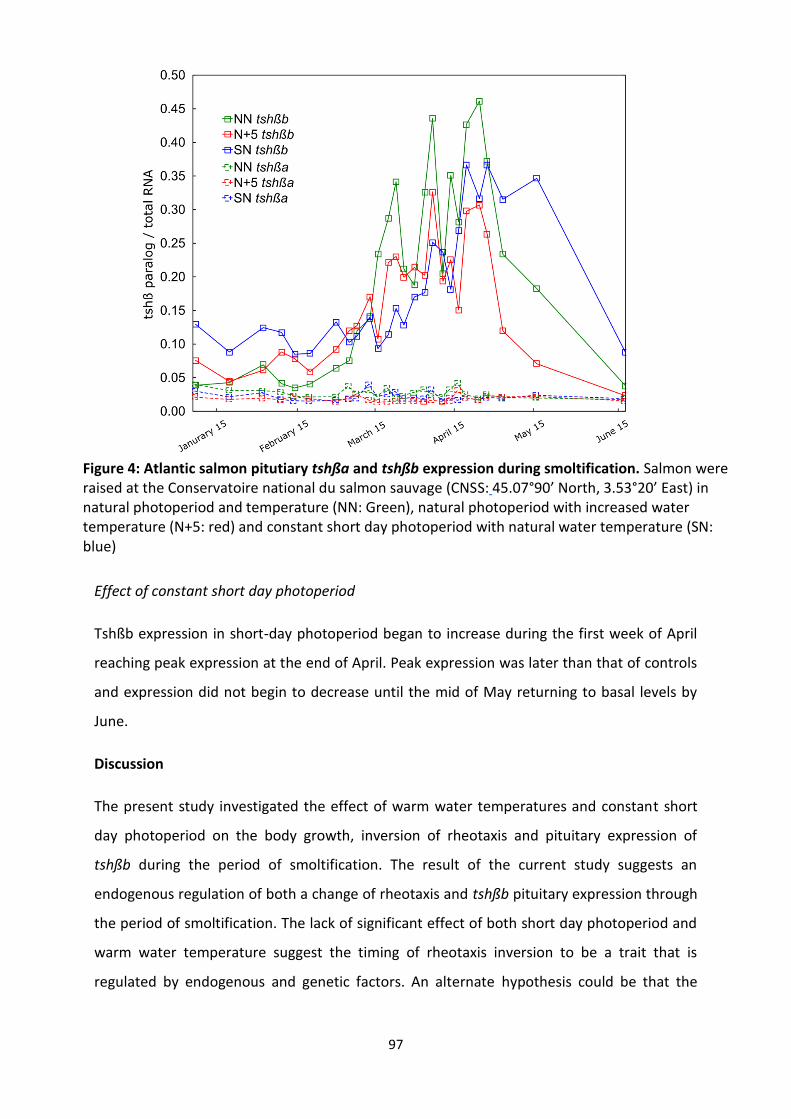
97
Effect of constant short day photoperiod
Tshßb expression in short-day photoperiod began to increase during the first week of April
reaching peak expression at the end of April. Peak expression was later than that of controls
and expression did not begin to decrease until the mid of May returning to basal levels by
June.
Discussion
The present study investigated the effect of warm water temperatures and constant short
day photoperiod on the body growth, inversion of rheotaxis and pituitary expression of
tshßb during the period of smoltification. The result of the current study suggests an
endogenous regulation of both a change of rheotaxis and tshßb pituitary expression through
the period of smoltification. The lack of significant effect of both short day photoperiod and
warm water temperature suggest the timing of rheotaxis inversion to be a trait that is
regulated by endogenous and genetic factors. An alternate hypothesis could be that the
Figure 4: Atlantic salmon pitutiary tshßa and tshßb expression during smoltification. Salmon were raised at the Conservatoire national du salmon sauvage (CNSS: 45.07°90’ North, 3.53°20’ East) in natural photoperiod and temperature (NN: Green), natural photoperiod with increased water temperature (N+5: red) and constant short day photoperiod with natural water temperature (SN: blue)
98
timing of rheotaxis inversion depends on conditions before juvenile fish begin to smoltify,
during the autumn and winter months prior to smoltification.
Nevertheless, the timing of migration onset is extremely important to the success of
downstream migration and transition to the sea the results of this study emphasizes the
importance of using local strains for the augmentation of declining fish populations during
conservation restocking programs.
Length of photoperiod does not affect daytime behaviour
Photoperiod had no effect on the body growth of the fish during the smoltification period.
Up to May, controls and short day photoperiod fish had nearly identical body growth
suggests that photoperiod does not play a role in the growth of fish during this period. This is
in agreement with McCormick et al. (2002) who observed no difference in growth rates
between control and constant short day photoperiod and displayed similar body growth in
tanks raised in the same water temperature in Atlantic salmon reared at the White River
hatchery in Vermont USA.
Interestingly, the authors conclude that constant short day photoperiod reared fish
remained at a stage of incomplete smoltification measured by lower NKA activity and no
decrease in condition factor when compared to controls. In the current study, NKA activity
was not measured but we report a similar tendency in condition factor with constant short
day photoperiod not declining like that of increased water temperature condition and
controls. The parameters used in the current study as indication of smoltification were
rheotaxis inversion and pituitary expression peak of tshßb. In this case, smolitfication was
similar between controls and constant short-day photoperiod showing both a change in
rheotaxis and tshßb peak expression, albeit slightly delayed. The completion and success of
smolitfication would only be known after successful transfer to sea hence the varying results
of smoltification parameters by environmental factors exaggerates the complexity of this
metamorphic event and the multiple components involved.
The investigation of tshßb is new and the relationship to smoltification has not been clearly
established. The dramatic peak of activity indicates a significant role and previous research
indicates a relationship to downstream migration. The current study supports this idea as we
99
see report similar timings of increase tshßb expression and the same timings of rheotaxis
change. More studies will be needed to confirm the relationship between the two but the
current study supports the integral role of tshßb in the smoltification process. No
modulation of both the timing of tshßb expression increase and change in rheotaxis by
environmental factors suggesting a strong endogenous regulation of these smoltification
characteristics. Our results suggest that tshßb is similar to that of plasma IGF-1 and plasma
T3 which have shown to be unaffected by varying environmental conditions (McCormick et
al., 2002). On the other hand, other mechanisms of smoltification such as NKA and plasma
GH have been shown to be modulated by photoperiod and water temperature again
exemplifying the multiple regulatory mechanisms involved in the smoltification
metamorphosis.
Warmer water does not cause earlier rheotaxis inversion
The current experimental design used a heat exchange system to increase the water
temperature in order to mimic possible future warmer river conditions and foreshadow the
consequences of so on the endangered Loire-Allier Atlantic salmon population. The Loire-
Allier River has risen 2°C in the past two decades (Moatar and Gailhard, 2006) and thus a
+5°C difference between controls and increased water temperature conditions would have
represented a realistic case of future conditions. Albeit, the temperature difference achieved
in this experiment is still an accurate representation of the coming decades and displays an
effect on growth rate however shows no little to any effect on the change of rheotaxis and
migration onset behaviour.
Martin et al., (2012) observed a significant increase of swimming behaviour related to
rheotaxis inversion at 7.5C for Loire-Allier population (Martin et al., 2012a). In the current
study, the water temperature in the increased water temperature tanks remained above
7.5C for the majority of the experiment without a change in swimming behaviour or
rheotaxis inversion until the first weeks of April. Therefore, the timing of rheotaxis inversion
appears unrelated to water temperature.
The significant increase in body size due to warm water is in agreement with past studies
and clearly shows that growth is direct response to prevailing water temperatures (REFS). No
change to rheotaxis change or tshßb expression was observed in increased water

100
temperature condition suggesting that tshßb expression is not regulated by body growth
signalling pathways.
Implication for conservation
For the Loire-Allier population, smolts must reach the oceans before water temperatures
reach 20°C as swimming speed for this population severely decreases above this
temperature (Martin et al., 2012a). In addition, warm water has lower oxygen carrying
capacity further decreasing migratory ability of Atlantic salmon smolts (for review: Jonsson
and Jonsson, 2009). Thus, if river water temperature continues to rise in the coming
decades, the period of time before the water temperature meets this critical threshold
decreases, lowering the period of time in which salmon have for successful migration to the
sea. Interestingly, the increased growth rate could be advantageous for the future as larger
fish migrate faster allowing for long river fish, such as the Loire-Allier population to complete
longer migrations in a shorter time (Brett, 1965; Brett and Glass, 1973). This may partially
compensate for the fact that increased water temperature did not affect the time in which
fish changed rheotaxis in the water which may otherwise cause a mismatch between the
physiological and environmental smolt window.
In conclusion, in the future when river temperatures are warmer, salmon may have
increased risk of incomplete migration due to the fact that departure from the rearing
grounds does not seem to be related to water temperature. The increased body size may
compensate for this and allow for migration to be complete successfully. More importantly,
these results suggest a strong endogenous regulation to the timing of migration onset and
exaggerates the importance of using local strains for the augmentation of declining Atlantic
salmon populations, especially true for the endangered Loire-Allier population.
101
References
1. Berge, A. I. et al. (1995) ‘Development of Salinity Tolerance in Underyearling Smolts
of Atlantic Salmon (Salmo-Salar) Reared under Different Photoperiods’, Canadian
Journal of Fisheries and Aquatic Sciences, 52(2), pp. 243–251. doi: 10.1139/f95-024.
2. Berglund, I. (1992). Growth and early sexual maturation in Baltic salmon (Salmo salar)
parr. Canadian Journal of Zoology, 70(2), 205-211.
3. Björnsson, B. T. et al. (2000) ‘Photoperiod regulation of plasma growth hormone
levels during induced smoltification of underyearling atlantic salmon’, General and
Comparative Endocrinology, 119(1), pp. 17–25. doi: 10.1006/gcen.2000.7439.
4. Björnsson, B. T., Einarsdottir, I. E. and Power, D. (2012) ‘Is salmon smoltification an
example of vertebrate metamorphosis? Lessons learnt from work on flatfish larval
development’, Aquaculture. Elsevier B.V., 362–363, pp. 264–272. doi:
10.1016/j.aquaculture.2011.03.002.
5. Björnsson, B. T., Stefansson, S. O. and McCormick, S. D. (2011) ‘Environmental
endocrinology of salmon smoltification’, General and Comparative Endocrinology,
170(2), pp. 290–298. doi: 10.1016/j.ygcen.2010.07.003.
6. Boeuf, G. (1993). Salmonid smolting: a pre-adaptation to the oceanic environment.
In Fish ecophysiology (pp. 105-135). Springer, Dordrecht.
7. Boeuf, G. and Prunet, P. (1985) ‘Measurements of gill (Na+K+)-ATPase activity and
plasma thyroid hormones during smoltification in Atlantic salmon (Salmo salar L.)’,
Aquaculture, 45(1–4), pp. 111–119. doi: 10.1016/0044-8486(85)90262-5.
8. Brett, J. R. (1965) ‘The Relation of Size to Rate of Oxygen Consumption and Sustained
Swimming Speed of Sockeye Salmon ( Oncorhynchus nerka )’, Journal of the Fisheries
Research Board of Canada, 22(6), pp. 1491–1501. doi: 10.1139/f65-128.
9. Brett, J. R. and Glass, N. R. (1973) ‘Metabolic Rates and Critical Swimming Speeds of
Sockeye Salmon ( Oncorhynchus nerka ) in Relation to Size and Temperature’, Journal
of Fisheries Research Board of Canada, 30(Brett 1962), pp. 379–387. doi:
10.1139/f73-068.
10. Dickhoff, W. W., Folmar, L. C. and Gorbman, A. (1978) ‘Changes in plasma thyroxine
during smoltification of coho salmon, Oncorhynchus kisutch’, General and
Comparative Endocrinology, 36(2), pp. 229–232. doi: 10.1016/0016-6480(78)90027-
102
8.
11. Hoar, W. S. (1976) ‘Smolt Transformation: Evolution, Behavior, and Physiology’,
Journal of the Fisheries Research Board of Canada, 33(5), pp. 1233–1252. doi:
10.1139/f76-158.
12. Hoar, W. S. (1988) ‘4 The physiology of smolting salmonids’, Fish Physiology. doi:
10.1016/S1546-5098(08)60216-2.
13. Iwata, M. (1995) ‘Downstream migratory behavior of salmonids and its relationship
with cortisol and thyroid hormones: A review’, Aquaculture, 135(1–3), pp. 131–139.
doi: 10.1016/0044-8486(95)01000-9.
14. Jonsson, B. and Jonsson, N. (2009) ‘A review of the likely effects of climate change on
anadromous Atlantic salmon Salmo salar and brown trout Salmo trutta, with
particular reference to water temperature and flow’, Journal of Fish Biology, 75(10),
pp. 2381–2447. doi: 10.1111/j.1095-8649.2009.02380.x.
15. Larsen, D. A., Swanson, P. and Dickhoff, W. W. (2011) ‘The pituitary-thyroid axis
during the parr-smolt transformation of Coho salmon, Oncorhynchus kisutch:
Quantification of TSH β mRNA, TSH, and thyroid hormones’, General and
Comparative Endocrinology, 171(3), pp. 367–372. doi: 10.1016/j.ygcen.2011.03.003.
16. Martin, P. et al. (2012) ‘Experimental study of the influence of photoperiod and
temperature on the swimming behaviour of hatchery-reared Atlantic salmon (Salmo
salar L.) smolts’, Aquaculture, 362–363, pp. 200–208. doi:
10.1016/j.aquaculture.2011.11.047.
17. McCormick, S. D. et al. (1998) ‘Movement, migration, and smolting of Atlantic salmon
(Salmo salar )’, Canadian Journal of Fisheries and Aquatic Sciences, 55(S1), pp. 77–92.
doi: 10.1139/d98-011.
18. McCormick, S. D. et al. (2002) ‘Effects of an advanced temperature cycle on smolt
development and endocrinology indicate that temperature is not a zeitgeber for
smolting in Atlantic salmon.’, The Journal of experimental biology, 205, pp. 3553–
3560.
19. McCormick, S. D. (2012) Smolt Physiology and Endocrinology, Fish Physiology. Elsevier
Inc. doi: 10.1016/B978-0-12-396951-4.00005-0.
20. Moatar, F. and Gailhard, J. (2006) ‘Water temperature behaviour in the River Loire
since 1976 and 1881’, Comptes Rendus - Geoscience, 338(5), pp. 319–328. doi:

103
10.1016/j.crte.2006.02.011.
21. Nakane, Y. and Yoshimura, T. (2014) ‘Universality and diversity in the signal
transduction pathway that regulates seasonal reproduction in vertebrates’, Frontiers
in Neuroscience, 8(8 MAY), pp. 1–7. doi: 10.3389/fnins.2014.00115.
22. Nakao, N. et al. (2008) ‘Thyrotrophin in the pars tuberalis triggers photoperiodic
response’, Nature, 452(7185), pp. 317–322. doi: 10.1038/nature06738.
23. Nishikawa, Kazuyoshi, et al. (1979) ‘Changes in Circulating L-Thyroxine and L-
Triiodothyronine of the masu salmon, Oncorhynchus masou accompaning the
smoltification, Measured by Radioimmunoassay.’, Endocrinologia japonica, 26(6), pp.
731–735. doi: 10.1507/endocrj1954.26.731.
24. Prunet, P. et al. (1989) ‘Smoltification and seawater adaptation in Atlantic salmon
(Salmo salar): Plasma prolactin, growth hormone, and thyroid hormones’, General
and Comparative Endocrinology, 74(3), pp. 355–364. doi: 10.1016/S0016-
6480(89)80031-0.
25. Rousseau, K., Martin, P., Boeuf, G., & Dufour, S. (2012). Salmonid
smoltification. Metamorphosis in fish. CRC Press, Boca Raton, 167-215.
26. Sundell, K. et al. (2003) ‘Intestinal transport mechanisms and plasma cortisol levels
during normal and out-of-season parr-smolt transformation of Atlantic salmon,
Salmo salar’, Aquaculture, 222(1–4), pp. 265–285. doi: 10.1016/S0044-
8486(03)00127-3.
27. Wedemeyer, G. A., Saunders, R. L. and Clarke, W. C. (1980) ‘Environmental Factors
Affecting Smoltification and Early Marine Survival of Anadromous Salmonids’, Marine
Fisheries Review, 42(6), pp. 1–14.

104
Discussion & Perspectives
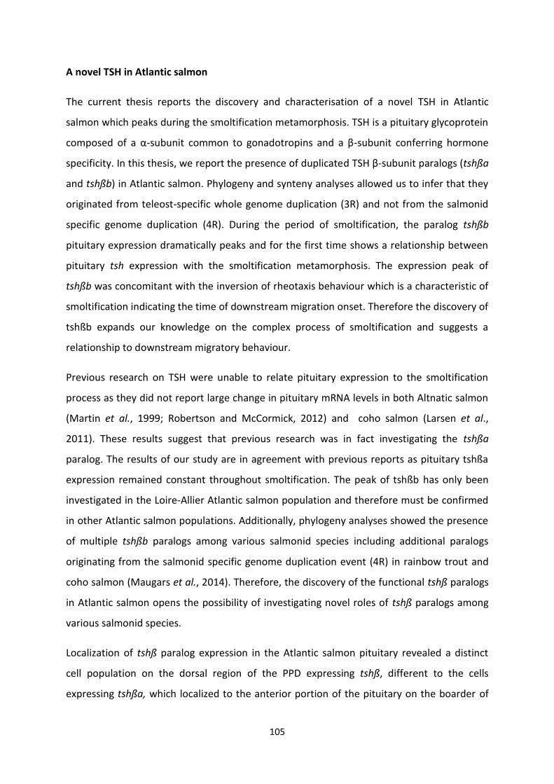
105
A novel TSH in Atlantic salmon
The current thesis reports the discovery and characterisation of a novel TSH in Atlantic
salmon which peaks during the smoltification metamorphosis. TSH is a pituitary glycoprotein
composed of a α-subunit common to gonadotropins and a β-subunit conferring hormone
specificity. In this thesis, we report the presence of duplicated TSH β-subunit paralogs (tshßa
and tshßb) in Atlantic salmon. Phylogeny and synteny analyses allowed us to infer that they
originated from teleost-specific whole genome duplication (3R) and not from the salmonid
specific genome duplication (4R). During the period of smoltification, the paralog tshßb
pituitary expression dramatically peaks and for the first time shows a relationship between
pituitary tsh expression with the smoltification metamorphosis. The expression peak of
tshßb was concomitant with the inversion of rheotaxis behaviour which is a characteristic of
smoltification indicating the time of downstream migration onset. Therefore the discovery of
tshßb expands our knowledge on the complex process of smoltification and suggests a
relationship to downstream migratory behaviour.
Previous research on TSH were unable to relate pituitary expression to the smoltification
process as they did not report large change in pituitary mRNA levels in both Altnatic salmon
(Martin et al., 1999; Robertson and McCormick, 2012) and coho salmon (Larsen et al.,
2011). These results suggest that previous research was in fact investigating the tshßa
paralog. The results of our study are in agreement with previous reports as pituitary tshßa
expression remained constant throughout smoltification. The peak of tshßb has only been
investigated in the Loire-Allier Atlantic salmon population and therefore must be confirmed
in other Atlantic salmon populations. Additionally, phylogeny analyses showed the presence
of multiple tshßb paralogs among various salmonid species including additional paralogs
originating from the salmonid specific genome duplication event (4R) in rainbow trout and
coho salmon (Maugars et al., 2014). Therefore, the discovery of the functional tshß paralogs
in Atlantic salmon opens the possibility of investigating novel roles of tshß paralogs among
various salmonid species.
Localization of tshß paralog expression in the Atlantic salmon pituitary revealed a distinct
cell population on the dorsal region of the PPD expressing tshß, different to the cells
expressing tshßa, which localized to the anterior portion of the pituitary on the boarder of

106
rostral pars distalis (RPD) and pars proximal distalis (PPD). The location of tshßb expression is
comparable to the location of pars tuberalis TSH discovered in mammals and birds. TSH
produced by PT cells plays a key role in the seasonal regulation of major steps of life cycles,
including reproduction, migration and hibernation (for reviews: Hanon et al., 2008; Hazlerigg
and Loudon, 2008; Dardente, Hazlerigg and Ebling, 2014; Nakane and Yoshimura, 2014). In
our study during Atlantic salmon smoltification, the peak of tshßb was concomitant to the
inversion of rheotaxis in spring and may suggest similarities of the TSH pathway in seasonal
life history events between vertebrate lineages.
Local action of TSHb in the hypothalamus
In mammals, photoperiodic regulation of PT-TSH production is mediated via melatonin
production from the pineal gland. In contrast, birds PT-TSH production is regulated through
deep brain photoreceptors and is not affected by melatonin levels. Despite the differences in
the integration of photoperiodic information between mammals and birds, PT-TSH exerts a
similar action on neighbouring hypothalamus. PT-TSH stimulates the expression of DIO2 in
the hypothalamus which catalyses the conversion of T4 into the more biologically active T3,
thus leading to the activation of TH-regulated brain functions (for reviews: Hanon et al.,
2008; Hazlerigg and Loudon, 2008; Dardente, Hazlerigg and Ebling, 2014; Nakane and
Yoshimura, 2014). In a recent study, a salmonid specific 4R-issued DIO2 paralog (dio2b) has
been identified in the Atlantic salmon, the expression of which increases in circumventricular
brain areas of cell proliferation, during experimental photoperiod-induced smoltification.
The authors proposed a specific role of DIO2b in promotion of TH-dependent brain
development during smoltification (Lorgen et al., 2015). Thus, in-line of PT-TSH role and
action mechanism in amniotes, we propose that TSHb produced by dorsal PPD cells in
Atlantic salmon may stimulate brain DIO2b expression and promote TH-activated brain
functions related to smoltification.
Studies are needed to confirm the tshßb/dio2b pathway and could be done by further
investigating the brain distribution of tsh receptor (tshr) paralog expression. Three tsh
receptors have been characterized in Atlantic salmon and more could be present due to the
4R in salmonid species (Maugars et al., 2014). In European eel (Anguilla anguilla), tsh
receptor paralog (called tshrb by the authors) showed expression in various parts of the
brain (Maugars et al., 2014). In the current study, brain distribution investigated by qPCR

107
showed expression of salmon tshßb in multiple regions of the brain while tshßa was only
expressed in the pituitary. Additional experiments are needed to confirm these expressions
but suggest potential roles in other parts of the brain aside from the hypothalamus.
Investigating, the co-localization of tshr and dio2 expressing cells in the hypothalamus and
other brain regions could help better understand the tshßb/dio2b pathway and potentially
discover other pathways between paralogs.. The investigation into the tshr paralogs and
their co-localization with dio2 paralogs would help characterize multiple pathways of
tsh/dio2 and lead to potential roles in the regulation of seasonal life history events.
Furthermore, tissue distribution of tshßb paralog showed expression throughout peripheral
tissues such as the gills, whereas tshßa was only expressed in the pituitary. It could be
hypothesized that a similar mechanism of tshßb activation of dio2 may take place
throughout the body to promote local TH-activated peripheral tissue functions during
smoltification or other developmental processes. This is supported by the fact that dio2 and
dio3 paralogs are observed throughout the tissues of flatfish during larval metamorphosis
(Itoh et al., 2010; Alves et al., 2017). These results lend support to the peripheral regulation
model of thyroid function proposed by Eales and Brown (1993) who propose thyroid
hormones to be regulated peripherally in teleosts (for review: Eales and Brown, 1993).
Further experiments are needed to confirm these peripheral expressions observed in this
study but suggest potential roles of tshßb in various peripheral tissues. Investigating the co-
localization of tshr and dio2 expressing cells among various tissues would help decipher
activation of local TH depend processes (Figure 11). Additionally, tshßb expression profiles in
the brain and peripheral tissues through the period of smoltification would be interesting as
to date only the expression profile for pituitary have been investigated.

108
Figure 11: Proposed mechanims of tshß/dio2 paralogs in both the hypothalamus and perifpheral tissues such as the gills during the smoltification metamorphosis
Modulation of pituitary tshßb peak by environmental factors
Constant short day photoperiod throughout smoltification had a minor effect on the
expression of tshßb in our study. A slight delay in the peak expression of tshßb was observed
and the drop was also delayed. In a previous experiment, McCormick et al. (2002) fount that
constant short day photoperiod were able to delay the increase of gill NKA activity and
plasma levels of GH without affecting the levels of plasma IGF-1 (McCormick et al., 2002).
The slight modulation of the peak of pituitary tshßb expression by constant short day
photoperiod will need to be confirmed in future studies. The minor effect of drastically
different photoperiod regimes suggests that the peak of tshßb expression is not particularly
regulated via photoperiod cues; rather a genetic/developmental aspect may be slightly
modulated by photoperiodic conditions.

109
The teleost pineal gland is known to be involved in the synchronization of a number of
physiological events and is responsible for the synthesis and release of melatonin (for
review: Korf, 1994). The duration of nocturnal melatonin surge in Atlantic salmon is dictated
by photoperiod while the amplitude of the surge depends on water temperature (for review:
Falcón et al., 2010). Pinealectomised Atlantic salmon undergo delayed smoltification as
compared to controls and authors suggest melatonin to be an important factor in the
regulation of certain smoltification processes (Porter et al., 1998). In order to determine if
tshßb production is regulated via melatonin I assayed samples from a previous experiment
that took place at CNSS which investigated the effect of melatonin implants in juvenile Loire-
Allier Atlantic salmon. I tested via qPCR the tshßa and tshßb paralog expression levels in
melatonin treated fish and saw a dramatic peak of tshßb expression similar to that of control
fish (Figure 12). This suggests that tshßb production does not rely on melatonin signalling
and may be modulated by other photoperiodic integration mechanism, possibly through the
eyes and deep brain receptors similar to that of birds (for review: Dawson et al., 2001).
110
Figure 12: tshßa and tshßb pituitary expression in control (blue) placebo (green) and melatonin implanted Atlantic salmon during the period of smoltification
Interestingly, in the same melatonin experiment, no inversion of rheotaxis was observed in
the melatonin treated fish during daytime. The melatonin implants caused the fish to remain
with positive rheotaxis in the water corresponding to the night-time behaviour observed in
control fish (data not shown). Similarly, putting naturally smolting Atlantic salmon in
constant darkness stopped diurnal downstream migratory behaviour which was restored
after returning salmon back to natural photoperiod (Martin et al., 2012). Therefore, it seems
that melatonin has the ability to block day time behaviour (negative rheotaxis) by simulating
constant darkness via high melatonin levels; however the peak of tshßb expression remains
similar to that of controls. It may be that the expression of tshßb only affects day time
behaviour is smoltifying salmon and that the behaviour mechanism of melatonin is able to
override the photoperiodic information relayed through the light-brain-pituitary axis. The

111
relationship between endocrine and behavioural mechanisms of smoltification is something I
will continue to investigate after my PhD.
Similarly to photoperiod, increased water temperature did not cause a significant effect on
the tshßb expression. Although the fish were subjected to increased water temperature for
months before the peak of tshßb expression both conditions peaked during the first weeks
of April. The expression decreased slightly sooner than that of controls. The similar peaks
between controls and increased water temperature suggest that tshßb expression is not
predominantly regulated via thermal cues. In previous studies, increased water temperature
has been shown to advance many aspects of smolting including silvering, body growth, swim
speed and behaviour (Johnston and Saunders, 1981; Muir et al., 1994; McCormick et al.,
2002; Zydlewski, Haro and Mccormick, 2005) . The peak of tshßb however is unaffected by
prevailing water temperatures further suggesting the endogenous regulation of tshßb.
Interestingly, other endogenous regulated process such as body weight and length were
significantly affected by increased water temperature. Despite a 20-30% increase in body
size in increased water temperature reared fish only minor differences of the time of
rheotaxis inversion and peak tshßb expression were observed as compared to controls.
Therefore, this suggests that the induction of tshßb expression and rheotaxis inversion does
not rely on endogenous body growth signalling pathways.
Inversion of rheotaxis is predetermined
Concomitant to the increase of tshßb expression was the inversion from positive to negative
rheotaxis. This behaviour change indicates the timing of migration onset and is a crucial
component to the smoltification process (Hoar, 1976; Boeuf, 1993). The time in which
salmon deport from the rearing grounds is a determining factor in the survival of smolts
(Boeuf, 1993; McCormick et al., 1998) and therefore is an important consideration for
conservation programs. Completing migration and arriving at the estuary within the
physiological and environmental smolt windows depends on the time in which salmon
depart from the rearing grounds. Particularly true for long river populations such as the
Loire-Allier were barriers, weirs and other migratory obstacles can cause significant delays to
downstream migration.
112
The change in rheotaxis in the current experiment happened during the first weeks of April
in all experimental conditions. Neither increased water temperature nor constant short day
photoperiod disrupted the time in which salmon began to change from positive to negative
rheotaxis during day light hours as compared to controls. During this period, fish would keep
positive rheotaxis during morning hours and begin to change to negative rheotaxis towards
the end of the day. Downstream movements increased significantly during the dusk hours.
This is in agreement with Martin et al., (2012) who saw a rapid change switch from positive
to negative rheotaxis during the end of March or start of April depending on the year
(Martin et al., 2012). Nocturnal behaviour remained the same throughout the experiment
represented by persistent positive rheotaxis with little to no movements. Similar behaviour
for the Loire-Allier population has been reported with only slight movements at night
through the period of smoltification across many years of study (Martin et al., 2012).
Downstream migratory strategies of smolts have been investigated for many years and
reports include nocturnal (Thorpe et al., 1981; Aarestrup, Nielsen and Koed, 2002), diurnal
(Davidsen et al., 2005) or a mix of the two movements (McCormick et al., 1998; Ibbotson et
al., 2006). The current study reports little nocturnal movements throughout the experiment
with a rapid change to diurnal migration during the first weeks of April for the Loire-Allier
population. Interestingly, the time in which this inversion of rheotaxis takes places seems to
be only slightly modified by environmental factors. The timing of rheotaxis and the migratory
strategy used may be a heritable trait, genetically specific for the specific river. The specific
timing of such crucial events may be genetically coded in a way which disregards the
variations in environmental factors which would protect crucial life history events from
yearly fluctuations in river temperature. Long term increases in water temperature however
may pose a serious threat as timing of migration onset appears to be predetermined.
In order to compare Loire-Allier Atlantic salmon population with that of others, common
garden experiments could be done to investigate if whether inversion of rheotaxis and peak
expression of tshßb are endogenously and genetically regulated. The Loire-Allier is highly
protected through the “Plan Loire Grandeur Nature” and thus transferring salmon to CNSS is
forbidden; however, transferring Loire-Allier salmon eggs to more northern latitudes and
raising them to the smolt stage under foreign photoperiodic and thermal regimes would
reveal if indeed rheotaxis inversion and tshßb expression are regulated internally. It could be

113
hypothesized that these smoltification characterises are determined during the autumn and
winter months prior to the period of smoltification and common garden experiments would
help answer these questions.
To date, the most recent published genome is that of an Atlantic salmon from the AquaGen
strain, a commercially farmed strain of Atlantic salmon that has been selectively bread for
multiple generations (Lien et al., 2016). As an approach to investigate the uniqueness of the
Loire-Allier population, the genomes of a wild smolt from the Loire-Allier caught in 2017 and
one caught in 1894 (provided by the collections at MNHN) are being sequenced and
assembled by Future genomics in Leiden, Netherlands in collaboration with CNSS and
MNHN. This research will compare both the current and ancient Loire-Allier genome to that
of the published genome and could reveal a level of genomic divergence allowing to
characterize the uniqueness of the Loire-Allier population. As an example, this research
could be used to investigate the promoter regions of Atlantic salmon tsh paralogs which may
explain the differences in pituitary expression during smoltification. A similar mechanism
was reported in stickleback (Gasterosteus aculeatus) by Kitano et al (2010) in which cis-
regulatory changes in tshß paralogous genes were suggested to account for the difference in
mRNA expression and behaviour between marine and freshwater populations (Kitano et al.,
2010). Of course, multiple different candidate genes may account for the differences in gene
expression and behaviour between various Atlantic salmon populations.
Implications for conservation in the frame of global climate change
Persistent increasing of river water temperature has occurred in the Loire-Allier for the last
25 years. Since the late 1970s the Loire-Allier has raised approximately 2˚C during the late
winter and spring months and the trends indicate further warming in the coming decades
(Moatar and Gailhard, 2006). The current study mimics possible future conditions by
subjecting fish to increased water temperature throughout the entire smoltification process.
Reports have hypothesized that warming rivers in the frame of global climate change will
cause earlier migration onset for various salmonid species (for review: Jonsson and Jonsson,
2009) however the results of this study provides arguments against this hypothesis.
The current study saw no advancement of rheotaxis change and thus suggests an alarming
consequence of global climate and anthropogenic changes. In addition to global climate

114
change, there are many situations along the Loire-Allier which cause the occurrence of local
warming which will further hinder the downstream migratory ability of smolts. In the Haut-
Allier region, water is often drawn from streams and tributaries of the Loire-Allier for
agriculture purposes causing a reduction in water flow further increasing river water
temperatures. During downstream migration, smolts are subjected to many occurrences of
local pockets of warm water from nuclear power plant cooling station run offs and other city
activities and influence which can threaten the migratory ability of smolts. Thus, no change
in departure time from rearing grounds in a warmer future will significantly reduce the
period of time smolts have to migrate down river before water temperatures reach 20
degrees or above, the temperature at which downstream swimming halts for the Loire-Allier
population (Martin et al., 2012b). Furthermore, along with warm water come decreased
oxygen availability and increase pathogens compounding the negative consequences of
increased water temperature for the Loire-Allier Atlantic salmon (for review: Jonsson and
Jonsson, 2009).
A possible compensatory effect of increased water temperature for the wild population
could be that the increased water temperature is shown to have a significant effect on the
rate of body growth. The current study saw a 20-30% increase in body size in the increased
water temperature fish which would result in faster migrating smolts as swim speed has
been shown to increase with body size (Brett, 1965; Brett and Glass, 1973). Therefore the
high plasticity in body growth regulated by prevailing water temperatures could partially
compensate for the consistent timing of rheotaxis inversion and migration onset.
Although increased growth rate in warmer waters may contribute to faster migration, the
situation for the Loire-Allier looks alarming. As the last long river migrating population of
Atlantic salmon in Europe, the Loire-Allier salmon should be given special attention to
ensure this population prevails. Multiple global and anthropogenic related threats are
putting enormous pressure on this salmon population to adapt faster than they are
physiologically capable of. In order to help, conservation strategies could be set in place
which would facilitate successful downstream migration and transition to the sea. First,
attention must be put into restoring river continuity and increase the ease of access to the
upper portions of the Loire-Allier. The pristine spawning grounds of the region should be
used as much as possible before environmental and anthropogenic factors force salmon out

115
of the area. Large progress was recently made in the restoration of river continuity as the
Poutès dam began reconstruction in the spring of 2018 and the new dam designed for the
location will drastically improve the bypass methods allowing for better access to the Haut-
Allier region.
Secondly, conservational efforts should allocate resources to the restoration of rearing
grounds lower down river than the Haut-Allier region. Lower spawning grounds have been
modified or destroyed by human presence and restoration to these locations would reduce
the overall migratory distance for Loire-Allier Atlantic salmon. As global climate change
continues, focus must be made to provide sufficient spawning locations lower than the Haut-
Loire region to ensure downstream migration will be successful before physiological
thresholds of water temperature are reached halting migration; especially in the light of the
results of this thesis which demonstrates a strong endogenous regulation of migration onset
for the Loire-Allier population. Providing suitable rearing grounds for migrating salmon
downriver of the Haut-Allier may allow for the prevalence of this special population in a
warmer future.
The current thesis provides novel knowledge to the smoltification metamorphosis of Atlantic
salmon. Functional divergence of TSH in Atlantic salmon provides for the first time, tshß
expression with that of the smoltification metamorphosis. Similarities to PT-TSH in mammals
and birds helps conserve the idea that this is an ancestral mechanism regulating seasonal life
history events, in this case the inversion of rheotaxis. Dominantly regulated by endogenous
factors and only slightly modified by prevailing environmental conditions suggests genetic
regulation of tshßb expression and migration onset for the Loire-Allier population. In the
frame of global climate change these results suggest focus should be put on restoring
downriver rearing grounds to facilitate shorter migration distances and help the special
Loire-Allier population prevail in a warming future.
116
References
Aarestrup, K., Nielsen, C., & Koed, A. (2002). Net ground speed of downstream migrating radio-tagged Atlantic salmon (Salmo salar L.) and brown trout (Salmo trutta L.) smolts in relation to environmental factors. In Aquatic telemetry (pp. 95-102). Springer, Dordrecht.
Ágústsson, T., Sundell, K., Sakamoto, T., Johansson, V., Ando, M., & Bjornsson, B. T. (2001). Growth hormone endocrinology of Atlantic salmon (Salmo salar): pituitary gene expression, hormone storage, secretion and plasma levels during parr-smolt transformation. Journal of endocrinology, 170(1), 227-234.
Allen, B. M. (1917). Effects of the extirpation of the anterior lobe of the hypophysis of Rana pipiens. The Biological Bulletin, 32(3), 117-130.
Alves, R. N. et al. (2017) ‘Duplication of Dio3 genes in teleost fish and their divergent expression in skin during flatfish metamorphosis’, General and Comparative Endocrinology. Elsevier Inc., 246, pp. 279–293. doi: 10.1016/j.ygcen.2017.01.002.
Anderson, G. M. et al. (2003) ‘Evidence that thyroid hormones act in the ventromedial preoptic area and the premammillary region of the brain to allow the termination of the breeding season in the ewe’, Endocrinology, 144(7), pp. 2892–2901. doi: 10.1210/en.2003-0322.
Baker, B. I. (1969). The response of teleost pituitary thyrotrophs to thyroxin in vitro—a histological study. General and comparative endocrinology, 12(3), 427-437.
Barrett, P. et al. (2007) ‘Hypothalamic thyroid hormone catabolism acts as a gatekeeper for the seasonal control of body weight and reproduction’, Endocrinology, 148(8), pp. 3608–3617. doi: 10.1210/en.2007-0316.
Bell, J. G. et al. (1997) ‘The effect of dietary lipid on polyunsaturated fatty acid metabolism in Atlantic salmon (Salmo salar) undergoing parr-smolt transformation’, Lipids, 32(5), pp. 515–525. doi: 10.1007/s11745-997-0066-4.
Berge, A. I. et al. (1995) ‘Development of Salinity Tolerance in Underyearling Smolts of Atlantic Salmon (Salmo-Salar) Reared under Different Photoperiods’, Canadian Journal of Fisheries and Aquatic Sciences, 52(2), pp. 243–251. doi: 10.1139/f95-024.
Berglund, I. (1992). Growth and early sexual maturation in Baltic salmon (Salmo salar) parr. Canadian Journal of Zoology, 70(2), 205-211.
Björnsson, B. T. (1997) ‘The biology of salmon growth hormone: from daylight to dominance’, Fish Physiology and Biochemistry, 17(1–6), pp. 9–24. doi: 10.1023/A:1007712413908.
Björnsson, B. T. et al. (2000) ‘Photoperiod regulation of plasma growth hormone levels during induced smoltification of underyearling atlantic salmon’, General and Comparative Endocrinology, 119(1), pp. 17–25. doi: 10.1006/gcen.2000.7439.
Björnsson, B. T., Einarsdottir, I. E. and Power, D. (2012) ‘Is salmon smoltification an example

117
of vertebrate metamorphosis? Lessons learnt from work on flatfish larval development’, Aquaculture. Elsevier B.V., 362–363, pp. 264–272. doi: 10.1016/j.aquaculture.2011.03.002.
Björnsson, B. T., Stefansson, S. O. and McCormick, S. D. (2011) ‘Environmental endocrinology of salmon smoltification’, General and Comparative Endocrinology, 170(2), pp. 290–298. doi: 10.1016/j.ygcen.2010.07.003.
Blake, M. S., Johnston, K. H., Russell-Jones, G. J., & Gotschlich, E. C. (1984). A rapid, sensitive method for detection of alkaline phosphatase-conjugated anti-antibody on Western blots. Analytical biochemistry, 136(1), 175-179.
Bodó, E. et al. (2010) ‘Thyroid-stimulating hormone, a novel, locally produced modulator of human epidermal functions, is regulated by thyrotropin-releasing hormone and thyroid hormones’, Endocrinology, 151(4), pp. 1633–1642. doi: 10.1210/en.2009-0306.
Boeuf, G. (1993). Salmonid smolting: a pre-adaptation to the oceanic environment. In Fish ecophysiology (pp. 105-135). Springer, Dordrecht.
Boeuf, G. and Prunet, P. (1985) ‘Measurements of gill (Na+K+)-ATPase activity and plasma thyroid hormones during smoltification in Atlantic salmon (Salmo salar L.)’, Aquaculture, 45(1–4), pp. 111–119. doi: 10.1016/0044-8486(85)90262-5.
Bole-Feysot, C. et al. (1998) ‘Prolactin (PRL) and its receptor:Actions, signal transduction pathways and phenotypes observed in PRL receptor knockout mice.’, Endocrine reviews, 19(3), pp. 225–268. doi: 10.1210/edrv.19.3.0334.
Brett, J. R. (1965) ‘The Relation of Size to Rate of Oxygen Consumption and Sustained Swimming Speed of Sockeye Salmon ( Oncorhynchus nerka )’, Journal of the Fisheries Research Board of Canada, 22(6), pp. 1491–1501. doi: 10.1139/f65-128.
Brett, J. R. and Glass, N. R. (1973) ‘Metabolic Rates and Critical Swimming Speeds of Sockeye Salmon ( Oncorhynchus nerka ) in Relation to Size and Temperature’, Journal of Fisheries Research Board of Canada, 30(Brett 1962), pp. 379–387. doi: 10.1139/f73-068.
Brown, C. L. et al. (1987) ‘Occurrence of thyroid hormones in early developmental stages of teleost fish’, American Fisheries Society Symposium Series, 2(January), pp. 144–150.
Bystriansky, J. S., LeBlanc, P. J. and Ballantyne, J. S. (2006) ‘Anaesthetization of Arctic charr Salvelinus alpinus (L.) with tricaine methanesulphonate or 2-phenoxyethanol for immediate blood sampling’, Journal of Fish Biology, 69(2), pp. 613–621. doi: 10.1111/j.1095-8649.2006.01109.x.
Cahoreau, C., Klett, D. and Combarnous, Y. (2015) ‘Structure-function relationships of glycoprotein hormones and their subunits’ ancestors’, Frontiers in Endocrinology, 6(FEB), pp. 1–14. doi: 10.3389/fendo.2015.00026.
Campinho, M. A. et al. (2015) ‘Flatfish metamorphosis: A hypothalamic independent process?’, Molecular and Cellular Endocrinology. Elsevier Ireland Ltd, 404, pp. 16–25. doi: 10.1016/j.mce.2014.12.025.
Catena, M. L. et al. (2003) ‘Cloning of a partial cDNA for Japanese quail thyroid-stimulating
118
hormone β and effects of methimazole on the thyroid and reproductive axes’, Poultry Science, 82(3), pp. 381–387. doi: 10.1093/ps/82.3.381.
Chatterjee, a, Hsieh, Y. L. and Yu, J. Y. (2001) ‘Molecular cloning of cDNA encoding thyroid stimulating hormone beta subunit of bighead carp Aristichthys nobilis and regulation of its gene expression.’, Molecular and cellular endocrinology, 174(1–2), pp. 1–9. doi: S0303-7207(01)00392-6 [pii].
Chien, J. T. et al. (2006) ‘Molecular cloning and sequence analysis of a cDNA encoding pituitary thyroid stimulating hormone β-subunit of the Chinese soft-shell turtle Pelodiscus sinensis and regulation of its gene expression’, General and Comparative Endocrinology, 146(2), pp. 74–82. doi: 10.1016/j.ygcen.2005.09.021.
Clarke, W. C., Shelbourn, J. E. and Brett, J. R. (1978) ‘Growth and adaptation to sea water in “underyearling” sockeye (Oncorhynchus nerka) and coho (O. kisutch) salmon subjected to regimes of constant or changing temperature and day length’, Can. J. Zoo., 56(11), pp. 1421–2413.
Clements, S. and Schreck, C. B. (2004) ‘Central administration of corticotropin-releasing hormone alters downstream movement in an artificial stream in juvenile chinook salmon (Oncorhynchus tshawytscha)’, General and Comparative Endocrinology, 137(1), pp. 1–8. doi: 10.1016/j.ygcen.2004.02.004.
Dardente, H., Hazlerigg, D. G. and Ebling, F. J. P. (2014) ‘Thyroid hormone and seasonal rhythmicity’, Frontiers in Endocrinology, 5(FEB), pp. 1–11. doi: 10.3389/fendo.2014.00019.
Davidsen, J. et al. (2005) ‘Spatial and temporal migration of wild Atlantic salmon smolts determined from a video camera array in the sub-Arctic River Tana’, Fisheries Research, 74(1–3), pp. 210–222. doi: 10.1016/j.fishres.2005.02.005.
Dawson, A. et al. (2001) ‘Photoperiodic control of seasonality in birds’, Journal of Biological Rhythms, 16(4), pp. 365–380. doi: 10.1177/074873001129002079.
Denver, R. J. (1988) ‘Several hypothalamic peptides stimulate in vitro thyrotropin secretion by pituitaries of anuran amphibians’, General and Comparative Endocrinology, 72(3), pp. 383–393. doi: 10.1016/0016-6480(88)90160-8.
Denver, R. J., Glennemeier, K. A., & Boorse, G. C. (2002). Endocrinology of complex life cycles: amphibians. In Hormones, brain and behavior (pp. 469-XI).
Denver, R. J. and Licht, P. (1989) ‘Neuropeptide stimulation of thyrotropin secretion in the larval bullfrog: Evidence for a common neuroregulator of thyroid and interrenal activity in metamorphosis’, Journal of Experimental Zoology, 252(1), pp. 101–104. doi: 10.1002/jez.1402520114.
Dickhoff, W. W., Crim, J. W. and Gorbman, A. (1978) ‘Lack of effect of synthetic thyrotropin releasing hormone on Pacific hagfish (Eptatretus stouti) pituitary-thyroid tissues in vitro’, General and Comparative Endocrinology, 35(1), pp. 96–98. doi: 10.1016/0016-6480(78)90173-9.
Dickhoff, W. W., Folmar, L. C. and Gorbman, A. (1978) ‘Changes in plasma thyroxine during

119
smoltification of coho salmon, Oncorhynchus kisutch’, General and Comparative Endocrinology, 36(2), pp. 229–232. doi: 10.1016/0016-6480(78)90027-8.
Duan, C., Plesetskaya, E. M. and Dickhoff, W. W. (1995) Expression of insulin-like growth factor I in normally and abnormally developing coho salmon (Oncorhynchus kisutch). Endocrinology, 136(2), 446-452.
Duston, J., Saunders, L. and Knox, D. E. (1991) ‘Effects of increases in Freshwater Temperature on Loss of Smolt Characteristics in Atlantic Salmon (Salmo salar)’, Canadian Journal of Fisheries and Aquatic Sciences, 48, pp. 164–169. doi: 10.1139/f91-022.
Le Cren, E. D. (1951). The length-weight relationship and seasonal cycle in gonad weight and condition in the perch (Perca fluviatilis). The Journal of Animal Ecology, 201-219.
Eales, J. G. and Brown, S. B. (1993) ‘Measurement and regulation of thyroidal status in teleost fish’, Reviews in Fish Biology and Fisheries, 3(4), pp. 299–347. doi: 10.1007/BF00043383.
Ebbesson, L. O. E. et al. (2003) ‘Neural circuits and their structural and chemical reorganization in the light-brain-pituitary axis during parr-smolt transformation in salmon’, Aquaculture, 222(1–4), pp. 59–70. doi: 10.1016/S0044-8486(03)00102-9.
Ebbesson, L. O. E. et al. (2011) ‘Corticotropin-releasing factor neurogenesis during midlife development in salmon: Genetic, environmental and thyroid hormone regulation’, Journal of Neuroendocrinology, 23(8), pp. 733–741. doi: 10.1111/j.1365-2826.2011.02164.x.
Eriksson, L. O. and Lundqvist, H. (1982) ‘Circannual rhythms and photoperiod regulation of growth and smolting in Baltic salmon (Salmo salar L.)’, Aquaculture, 28(1–2), pp. 113–121. doi: 10.1016/0044-8486(82)90014-X.
Evans, D. H., Piermarini, P. M. and Keith, C. P. (2005) ‘The Multifunctional Fish Gill: Dominant Site of Gas Exchange, Osmoregulation, Acid-Base Regulation, and Excretion of Nitrogenous Waste’, Physiological Reviews, 85(1), pp. 97–177. doi: 10.1152/physrev.00050.2003.
Falcón, J. et al. (2010) ‘Current knowledge on the melatonin system in teleost fish’, General and Comparative Endocrinology. Elsevier Inc., 165(3), pp. 469–482. doi: 10.1016/j.ygcen.2009.04.026.
Fekete, C. and Lechan, R. M. (2007) ‘Negative feedback regulation of hypophysiotropic thyrotropin-releasing hormone (TRH) synthesizing neurons: Role of neuronal afferents and type 2 deiodinase’, Frontiers in Neuroendocrinology, 28(2–3), pp. 97–114. doi: 10.1016/j.yfrne.2007.04.002.
Follett, B. K. and Nicholls, T. J. (1988) ‘Acute effect of thyroid hormones in mimicking photoperiodically induced release of gonadotropins in Japanese quail’, Journal of Comparative Physiology B: Biochemical, Systemic and Environmental Physiology, 157(6), pp. 837–843. doi: 10.1007/BF00691016.
Folmar, L. C. and Dickhoff, W. W. (1980) ‘The parr—Smolt transformation (smoltification) and seawater adaptation in salmonids: A review of selected literature’, Aquaculture, pp. 1–37. doi: 10.1016/0044-8486(80)90123-4.

120
Freeman, D. A. et al. (2007) ‘Exogenous T3 mimics long day lengths in Siberian hamsters.’, American journal of physiology. Regulatory, integrative and comparative physiology, 292(6), pp. R2368–R2372. doi: 10.1152/ajpregu.00713.2006.
Fridberg, G., Lindahl, K., & Ekengren, B. (1981). The thyrotropic cell in the Atlantic salmon, Salmo salar. Acta Zoologica, 62(1), 43-51.
Froese, R. (2006) ‘Cube law, condition factor and weight-length relationships: History, meta-analysis and recommendations’, Journal of Applied Ichthyology, 22(4), pp. 241–253. doi: 10.1111/j.1439-0426.2006.00805.x.
Galas, L. et al. (2009) ‘TRH acts as a multifunctional hypophysiotropic factor in vertebrates’, General and Comparative Endocrinology. Elsevier Inc., 164(1), pp. 40–50. doi: 10.1016/j.ygcen.2009.05.003.
Geris, K. L. et al. (1996) ‘Evidence of a thyrotropin-releasing activity of ovine corticotropin-releasing factor in the domestic fowl (Gallus domesticus).’, General and comparative endocrinology, 104(2), pp. 139–146. doi: 10.1006/gcen.1996.0156.
Geris, K. L. et al. (1999) ‘The drop in plasma thyrotropin concentrations in fasted chickens is caused by an action at the level of the hypothalamus: Role of corticosterone’, Domestic Animal Endocrinology, 16(4), pp. 231–237. doi: 10.1016/S0739-7240(99)00016-8.
Goldsmith, A. R., & Nicholls, T. J. (1992). Thyroxine effects upon reproduction, prolactin secretion and plumage moult in intact and in thyroidectomised European Starlings Sturnus vulgaris. Ornis Scandinavica, 398-404.
Gorbman, A., & Hyder, M. (1973). Failure of mammalian TRH to stimulate thyroid function in the lungfish. General and comparative endocrinology, 20(3), 588-589.
Grau, E. G. et al. (1982) ‘Factors determining the occurrence of the surge in thyroid activity in salmon during smoltification’, Aquaculture, 28(1–2), pp. 49–57. doi: 10.1016/0044-8486(82)90007-2.
Gregory, C. C. and Porter, T. E. (1997) ‘Cloning and sequence analysis of a cDNA for the beta subunit of chicken thyroid-stimulating hormone.’, General and comparative endocrinology, 107(2), pp. 182–90. doi: 10.1006/gcen.1997.6911.
Grimaldi, A. et al. (2013) ‘Mechanisms of thyroid hormone receptor action during development: Lessons from amphibian studies’, Biochimica et Biophysica Acta - General Subjects. Elsevier B.V., 1830(7), pp. 3882–3892. doi: 10.1016/j.bbagen.2012.04.020.
De Groef, B. et al. (2006) ‘Role of corticotropin-releasing hormone as a thyrotropin-releasing factor in non-mammalian vertebrates’, General and Comparative Endocrinology, 146(1), pp. 62–68. doi: 10.1016/j.ygcen.2005.10.014.
Gudernatsch, J. F. (1912) ‘Feeding Experiments on tadpoles’, Archiv für Entwicklungsmechanik der Organismen, 35, pp. 457–483. doi: 10.1007/BF02277051.
Guindon, S. et al. (2010) ‘New algorithms and methods to estimate maximum-likelihood phylogenies: Assessing the performance of PhyML 3.0’, Systematic Biology, 59(3), pp. 307–

121
321. doi: 10.1093/sysbio/syq010.
Han, Y.-S. et al. (2004) ‘Cloning of the cDNA for thyroid stimulating hormone beta subunit and changes in activity of the pituitary-thyroid axis during silvering of the Japanese eel, Anguilla japonica.’, Journal of molecular endocrinology, 32(1), pp. 179–194. doi: 10.1677/jme.0.0320179.
Handeland, S. O. et al. (2004) ‘Temperature influence on the development and loss of seawater tolerance in two fast-growing strains of Atlantic salmon’, Aquaculture, 233(1–4), pp. 513–529. doi: 10.1016/j.aquaculture.2003.08.028.
Handeland, S. O., Imsland, A. K. and Stefansson, S. O. (2008) ‘The effect of temperature and fish size on growth, feed intake, food conversion efficiency and stomach evacuation rate of Atlantic salmon post-smolts’, Aquaculture, 283(1–4), pp. 36–42. doi: 10.1016/j.aquaculture.2008.06.042.
Hanon, E. A. et al. (2008) ‘Ancestral TSH Mechanism Signals Summer in a Photoperiodic Mammal’, Current Biology, 18(15), pp. 1147–1152. doi: 10.1016/j.cub.2008.06.076.
Hansen, L. P., & Jonsson, B. (1985). Downstream migration of hatchery-reared smolts of Atlantic salmon (Salmo salar L.) in the River Imsa, Norway. Aquaculture, 45(1-4), 237-248..
Hazlerigg, D. and Loudon, A. (2008) ‘New Insights into Ancient Seasonal Life Timers’, Current Biology, 18(17), pp. 795–804. doi: 10.1016/j.cub.2008.07.040.
Herndon, T. M., McCormick, S. D. and Bern, H. A. (1991) ‘Effects of prolactin on chloride cells in opercular membrane of seawater-adapted tilapia’, General and Comparative Endocrinology, 83(2), pp. 283–289. doi: 10.1016/0016-6480(91)90032-2.
Hoar, W. S. (1939). The thyroid gland of the Atlantic salmon. Journal of morphology, 65(2), 257-295.
Hoar, W. S. (1976) ‘Smolt Transformation: Evolution, Behavior, and Physiology’, Journal of the Fisheries Research Board of Canada, 33(5), pp. 1233–1252. doi: 10.1139/f76-158.
Hoar, W. S. (1988). 4 The Physiology of Smolting Salmonids. In Fish physiology (Vol. 11, pp. 275-343). Academic Press.
Hojvat, S., Baker, G., Kirsteins, L., & Lawrence, A. M. (1982). TSH in the rat and monkey brain. Neuroendocrinology, 34(5), 327-332.
Holmqvist, B. I., Östholm, T. and Ekström, P. (1994) ‘Neuroanatomical analysis of the visual and hypophysiotropic systems in Atlantic salmon (Salmo salar) with emphasis on possible mediators of photoperiodic cues during parr-smolt transformation’, Aquaculture, 121(1–3), pp. 1–12. doi: 10.1016/0044-8486(94)90003-5.
Hsieh, Y. L. et al. (2000) ‘Molecular cloning and sequence analysis of the cDNA for thyroid-stimulating hormone β subunit of Muscovy duck’, General and Comparative Endocrinology, 120(3), pp. 336–344. doi: 10.1006/gcen.2000.7572.
Hsieh, Y. L. et al. (2007) ‘Molecular cloning and sequence analysis of the cDNA encoding thyroid-stimulating hormone β-subunit of common duck and mule duck pituitaries: In vitro

122
regulation of steady-state TSHβ mRNA level’, Comparative Biochemistry and Physiology - B Biochemistry and Molecular Biology, 146(3), pp. 307–317. doi: 10.1016/j.cbpb.2006.11.018.
Ibbotson, A. T. et al. (2006) ‘Diel migration patterns of Atlantic salmon smolts with particular reference to the absence of crepuscular migration’, Ecology of Freshwater Fish, 15(4), pp. 544–551. doi: 10.1111/j.1600-0633.2006.00194.x.
I Ikegami, K., Liao, X. H., Hoshino, Y., Ono, H., Ota, W., Ito, Y., ... & Shigeyoshi, Y. (2014 ) ‘Tissue-specific post-translational modification allows functional targeting of thyrotropin’, 9(3), pp. 801–809. doi: 10.1016/j.celrep.2014.10.006.Tissue-specific.
Ikegami, K. and Yoshimura, T. (2017) ‘The hypothalamic–pituitary–thyroid axis and biological rhythms: The discovery of TSH’s unexpected role using animal models’, Best Practice and Research: Clinical Endocrinology and Metabolism. Elsevier Ltd, 31(5), pp. 475–485. doi: 10.1016/j.beem.2017.09.002.
Inui, Y. and Miwa, S. (1985) ‘Thyroid hormone induces metamorphosis of flounder larvae’, General and Comparative Endocrinology, 60(3), pp. 450–454. doi: 10.1016/0016-6480(85)90080-2.
Inui, Y. and Miwa, S. (2012) ‘Metamorphosis of Flatfish (Pleuronectiformes)’, Metamorphosis in Fish, pp. 107–153.
Itoh, K. et al. (2010) ‘Three Members of the Iodothyronine Deiodinase Family, dio1 , dio2 and dio3 , are Expressed in Spatially and Temporally Specific Patterns During Metamorphosis of the Flounder, Paralichthys olivaceus’, Zoological Science, 27(7), pp. 574–580. doi: 10.2108/zsj.27.574.
Iwata, M. (1995) ‘Downstream migratory behavior of salmonids and its relationship with cortisol and thyroid hormones: A review’, Aquaculture, 135(1–3), pp. 131–139. doi: 10.1016/0044-8486(95)01000-9.
Iziga, R. et al. (2010) ‘Molecular characterization and gene expression of thyrotropin-releasing hormone in Senegalese sole (Solea senegalensis)’, Comparative Biochemistry and Physiology - B Biochemistry and Molecular Biology. Elsevier Inc., 157(2), pp. 167–174. doi: 10.1016/j.cbpb.2010.05.013.
Johnsson, J. I. and Björnsson, B. T. (1994) ‘Growth hormone increases growth rate, appetite and dominance in juvenile rainbow trout, Oncorhynchus mykiss’, Animal Behaviour, pp. 177–186. doi: 10.1006/anbe.1994.1224.
Johnston, C. E. and Eales, J. G. (1967) ‘Purines in the integument of the Atlantic salmon (_Salmo salar_) during parr-smolt transformation’, Journal of the Fisheries Research Board of Canada, 24(5), pp. 955–964. doi: 10.1139/f67-085.
Johnston, C. E. and Saunders, R. L. (1981) ‘Parr–Smolt Transformation of Yearling Atlantic Salmon ( Salmo salar ) at Several Rearing Temperatures’, Canadian Journal of Fisheries and Aquatic Sciences, 38(10), pp. 1189–1198. doi: 10.1139/f81-160.
Jonsson, B. and Jonsson, N. (2009) ‘A review of the likely effects of climate change on anadromous Atlantic salmon Salmo salar and brown trout Salmo trutta, with particular

123
reference to water temperature and flow’, Journal of Fish Biology, 75(10), pp. 2381–2447. doi: 10.1111/j.1095-8649.2009.02380.x.
Jonsson, B. and Ruud-Hansen, J. (1985) ‘Water Temperature as the Primary Influence on Timing of Seaward Migrations of Atlantic Salmon ( Salmo salar ) Smolts’, Canadian Journal of Fisheries and Aquatic Sciences, 42(3), pp. 593–595. doi: 10.1139/f85-076.
Kitano, J. et al. (2010) ‘Adaptive divergence in the thyroid hormone signaling pathway in the stickleback radiation’, Current Biology, 20(23), pp. 2124–2130. doi: 10.1016/j.cub.2010.10.050.
Kitano, J. et al. (2012) ‘Physiological and genetic basis for variation in migratory behavior in the three-spined stickleback, Gasterosteus aculeatus’, Ichthyological Research, 59(4), pp. 293–303. doi: 10.1007/s10228-012-0289-8.
Klemetsen, A. et al. (2003) ‘Atlantic salmon Salmo salar L., brown trout Salmo trutta L. and Arctic charr Salvelinus alpinus (L.): a review of aspects of their life histories’, Ecology of Freshwater Fish, 12(1), pp. 1–59. doi: 10.1034/j.1600-0633.2003.00010.x.
KORF, H. ‐W (1994) ‘The Pineal Organ as a Component of the Biological Clock: Phylogenetic and Ontogenetic Considerations’, Annals of the New York Academy of Sciences, 719(1), pp. 13–42. doi: 10.1111/j.1749-6632.1994.tb56818.x.
Kristinsson, J. B., Saunders, R. L. and Wiggs, A. J. (1985) ‘Growth dynamics during the development of bimodal length-frequency distribution in juvenile Atlantic salmon (Salmo salar L.)’, Aquaculture, 45(1–4), pp. 1–20. doi: 10.1016/0044-8486(85)90254-6.
Kuenzel, W. J. (2003) ‘Neurobiology of molt in avian species’, Poultry Science, 82(6), pp. 981–991. doi: 10.1093/ps/82.6.981.
Larsen, D. A., Moriyama, S., Dickhoff, W. W., & Beckman, B. R. (1995). Endocrine and growth control of salmon smoltification. Aquaculture, 135(1-3), 147-147.
Larsen, D. A., Swanson, P., Dickey, J. T., Rivier, J., & Dickhoff, W. W. (1998). In VitroThyrotropin-Releasing Activity of Corticotropin-Releasing Hormone-Family Peptides in Coho Salmon, Oncorhynchus kisutch. General and Comparative Endocrinology, 109(2), 276-285.
Larsen, D. A., Swanson, P. and Dickhoff, W. W. (2011) ‘The pituitary-thyroid axis during the parr-smolt transformation of Coho salmon, Oncorhynchus kisutch: Quantification of TSH β mRNA, TSH, and thyroid hormones’, General and Comparative Endocrinology, 171(3), pp. 367–372. doi: 10.1016/j.ygcen.2011.03.003.
Laudet, V. (2011) ‘The origins and evolution of vertebrate metamorphosis’, Current Biology. Elsevier Ltd, 21(18), pp. R726–R737. doi: 10.1016/j.cub.2011.07.030.
Lechan, R. M. and Fekete, C. (2006) ‘Chapter 12: The TRH neuron: a hypothalamic integrator of energy metabolism’, Progress in Brain Research, 153(06), pp. 209–235. doi: 10.1016/S0079-6123(06)53012-2.
Lefort, V., Longueville, J. E. and Gascuel, O. (2017) ‘SMS: Smart Model Selection in PhyML’,

124
Molecular biology and evolution, 34(9), pp. 2422–2424. doi: 10.1093/molbev/msx149.
Licht, P. and Denver, J. (1988) ‘Effects of TRH on Hormone Release from Pituitaries Anolis carolinensis of the Lizard , Anolis carolinensis’, General and Comparative Endocrinology, 362(70), pp. 355–362.
Lien, S., Koop, B. F., Sandve, S. R., Miller, J. R., Kent, M. P., Nome, T., ... & Grammes, F. (2016) ‘The Atlantic salmon genome provides insights into rediploidization’, Nature, 533, pp. 200–205. doi: 10.1038/nature17164.
Lorgen, M. et al. (2015) ‘Functional divergence of type 2 deiodinase paralogs in the Atlantic salmon’, Current Biology, 25(7), pp. 936–941. doi: 10.1016/j.cub.2015.01.074.
Lynch, M. and Conery, J. S. (2000) ‘The evolutionary fate and consequences of duplicate genes’, Science, 290(5494), pp. 1151–1155. doi: 10.1126/science.290.5494.1151.
MacKenzie, D. S., Jones, R. A. and Miller, T. C. (2009) ‘Thyrotropin in teleost fish’, General and Comparative Endocrinology, 161(1), pp. 83–89. doi: 10.1016/j.ygcen.2008.12.010.
Malpaux, B. et al. (2001) ‘Biology of mammalian photoperiodism and the critical role of the pineal gland and melatonin’, Journal of Biological Rhythms, 16(4), pp. 336–347. doi: 10.1177/074873001129002051.
Manzon, L. A. (2002) ‘The role of prolactin in fish osmoregulation: A review’, General and Comparative Endocrinology, 125(2), pp. 291–310. doi: 10.1006/gcen.2001.7746.
Manzon, R. G. and Denver, R. J. (2004) ‘Regulation of pituitary thyrotropin gene expression during Xenopus metamorphosis: Negative feedback is functional throughout metamorphosis’, Journal of Endocrinology, 182(2), pp. 273–285. doi: 10.1677/joe.0.1820273.
Martin, P. et al. (2012a) ‘Experimental study of the influence of photoperiod and temperature on the swimming behaviour of hatchery-reared Atlantic salmon (Salmo salar L.) smolts’, Aquaculture, 362–363, pp. 200–208. doi: 10.1016/j.aquaculture.2011.11.047.
Martin, P. et al. (2012b) ‘Experimental study of the influence of photoperiod and temperature on the swimming behaviour of hatchery-reared Atlantic salmon (Salmo salar L.) smolts’, Aquaculture. Elsevier B.V., 362–363, pp. 200–208. doi: 10.1016/j.aquaculture.2011.11.047.
Martin, S. A. M. et al. (1999) ‘Differential expression of Atlantic salmon thyrotropin β subunit mRNA and its cDNA sequence’, Journal of Fish Biology, 54(4), pp. 757–766. doi: 10.1006/jfbi.1998.0912.
Matz, S. P. and Hofeldt, G. t. (1999) ‘Immunohistochemical Localization of Corticotropin-Releasing Factor in the Brain and Corticotropin-Releasing Factor and Thyrotropin-Stimulating Hormone in the Pituitary of Chinook Salmon (Oncorhynchus tshawytscha) Stuart’, The Journal of Comparative Neurology, 345(2), pp. 214–223. doi: 10.1002/cne.903450205.
Matz, S. P. and Takahashi, T. T. (1994) ‘Immunohistochemical localization of thyrotropin-releasing hormone in the brain of chinook salmon (oncorhynchus tshawytscha)’, The Journal of Comparative Neurology, 345(2), pp. 214–223. doi: 10.1002/cne.903450205.
125
Maugars, G. et al. (2014) ‘Multiple thyrotropin b-subunit and thyrotropin receptor-related genes arose during vertebrate evolution’, PLoS ONE, 9(11). doi: 10.1371/journal.pone.0111361.
Maxime, V. et al. (1989) ‘Comparative study of the energetic metabolism of Atlantic salmon (Salmo salar) parr and smolts’, Aquaculture, 82(1–4), pp. 163–171. doi: 10.1016/0044-8486(89)90405-5.
Mayer, I. (2000) ‘Effect of long-term pinealectomy on growth and precocious maturationin Atlantic salmon, Salmo salar parr’, Aquatic Living Resources, 13(3), pp. 139–144. doi: 10.1016/S0990-7440(00)01053-6.
McCormick, S. D., Saunders, R. L., Henderson, E. B., & Harmon, P. R. (1987). Photoperiod Control of Parr-Smo t Transformation in Atlantic salmon (Salmo salar): Changes in Salinty Tolerance, Gil Na+,K+-ATPase Activity, and Plasma Thyroid Hormones. Canadian Journal of Fisheries and Aquatic Sciences, 44, pp. 1462-1468
McCormick, S. D. et al. (1998) ‘Movement, migration, and smolting of Atlantic salmon (Salmo salar )’, Canadian Journal of Fisheries and Aquatic Sciences, 55(S1), pp. 77–92. doi: 10.1139/d98-011.
McCormick, S. D. et al. (2002) ‘Effects of an advanced temperature cycle on smolt development and endocrinology indicate that temperature is not a zeitgeber for smolting in Atlantic salmon.’, The Journal of experimental biology, 205, pp. 3553–3560.
McCormick, S. D. et al. (2007) ‘Differential hormonal responses of Atlantic salmon parr and smolt to increased daylength: A possible developmental basis for smolting’, Aquaculture, 273(2–3), pp. 337–344. doi: 10.1016/j.aquaculture.2007.10.015.
McCormick, S. D. (2012) Smolt Physiology and Endocrinology, Fish Physiology. Elsevier Inc. doi: 10.1016/B978-0-12-396951-4.00005-0.
McCormick, S. D., Moriyama, S. and Björnsson, B. T. (2000) ‘Low temperature limits photoperiod control of smolting in Atlantic salmon through endocrine mechanisms’, pp. 1352–1361.
McCormick, S. D., Regish, A. M. and Christensen, A. K. (2009) ‘Distinct freshwater and seawater isoforms of Na+/K+-ATPase in gill chloride cells of Atlantic salmon’, Journal of Experimental Biology, 212(24), pp. 3994–4001. doi: 10.1242/jeb.037275.
Metcalfe, N. B. and Thorpe, J. E. (1992) ‘Anorexia and Defended Energy Levels in Over-Wintering Juvenile Salmon Author ( s ): Neil B . Metcalfe and John E . Thorpe Published by : British Ecological Society Stable URL : http://www.jstor.org/stable/5520 REFERENCES Linked references are available on’, 61(1), pp. 175–181.
Meyer, A. and Van De Peer, Y. (2005) ‘From 2R to 3R: Evidence for a fish-specific genome duplication (FSGD)’, BioEssays, pp. 937–945. doi: 10.1002/bies.20293.
Miwa, S. et al. (1988) ‘Thyroxine surge in metamorphosing flounder larvae’, General and Comparative Endocrinology, 70(1), pp. 158–163. doi: 10.1016/0016-6480(88)90105-0.

126
Mizuno, S. et al. (2004) ‘Quantitative changes of black pigmentation in the dorsal fin margin during smoltification in masu salmon, Oncorhynchus masou’, Aquaculture, 229(1–4), pp. 433–450. doi: 10.1016/S0044-8486(03)00368-5.
Moatar, F. and Gailhard, J. (2006) ‘Water temperature behaviour in the River Loire since 1976 and 1881’, Comptes Rendus - Geoscience, 338(5), pp. 319–328. doi: 10.1016/j.crte.2006.02.011.
Morin, P. and Haug, E. (1994) ‘Changes in Serum Free Thyroxine, Prolactin, and Olfactory Activity during Induced Smoltification in Atlantic Salmon (Salmo’.
Moriyama, S. et al. (1997) ‘Salmon thyroid-stimulating hormone : Isolation , characterization and development of a radioimmunoassay’, 108d, pp. 457–471.
Moriyama, S., Ayson, F. G., & Kawauchi, H. (2000). Growth regulation by insulin-like growth factor-I in fish. Bioscience, biotechnology, and biochemistry, 64(8), 1553-1562.
Muir, W. D. et al. (1994) ‘Accelerating smolt development and downstream movement in yearling chinook salmon with advanced photoperiod and increased temperature’, Aquaculture, 123(3–4), pp. 387–399. doi: 10.1016/0044-8486(94)90073-6.
Nakane, Y., Ikegami, K., Iigo, M., Ono, H., Takeda, K., Takahashi, D., ... & Suga, T. (2013). The saccus vasculosus of fish is a sensor of seasonal changes in day length. Nature communications, 4, 2108. doi: 10.1038/ncomms3108.
Nakane, Y. and Yoshimura, T. (2010) ‘Deep brain photoreceptors and a seasonal signal transduction cascade in birds’, Cell and Tissue Research, 342(3), pp. 341–344. doi: 10.1007/s00441-010-1073-6.
Nakane, Y. and Yoshimura, T. (2014) ‘Universality and diversity in the signal transduction pathway that regulates seasonal reproduction in vertebrates’, Frontiers in Neuroscience, 8(8 MAY), pp. 1–7. doi: 10.3389/fnins.2014.00115.
Nakao, N. et al. (2008) ‘Thyrotrophin in the pars tuberalis triggers photoperiodic response’, Nature, 452(7185), pp. 317–322. doi: 10.1038/nature06738.
Nilsen, T. O. et al. (2007) ‘Differential expression of gill Na+,K+-ATPase - and -subunits, Na+,K+,2Cl- cotransporter and CFTR anion channel in juvenile anadromous and landlocked Atlantic salmon Salmo salar’, Journal of Experimental Biology, 210(16), pp. 2885–2896. doi: 10.1242/jeb.002873.
Nilsen, T. O. et al. (2008) ‘Endocrine systems in juvenile anadromous and landlocked Atlantic salmon (Salmo salar): Seasonal development and seawater acclimation’, General and Comparative Endocrinology, 155(3), pp. 762–772. doi: 10.1016/j.ygcen.2007.08.006.
Nishikawa, K. et al. (1979) ‘Changes in Circulating L-Thyroxine and L-Triiodothyronine of the masu salmon, Oncorhynchus masou accompaning the smoltification, Measured by Radioimmunoassay.’, Endocrinologia japonica, 26(6), pp. 731–735. doi: 10.1507/endocrj1954.26.731.
Nishioka, R. S. et al. (1982) ‘Changes in the endocrine organs of coho salmon during normal

127
and abnormal smoltification - An electron-microscope study’, Aquaculture, 28(1–2), pp. 21–38. doi: 10.1016/0044-8486(82)90005-9.
Nishioka, R. S. et al. (1985) ‘Attempts to intensify the thyroxin surge in coho and king salmon by chemical stimulation’, Aquaculture, 45(1–4), pp. 215–225. doi: 10.1016/0044-8486(85)90271-6.
Ohno, S. (1970) ‘The Enormous Diversity in Genome Sizes of Fish as a Reflection of Nature’s Extensive Experiments with Gene Duplication’, Transactions of the American Fisheries Society, 99(1), pp. 120–130. doi: 10.1577/1548-8659(1970)99<120:TEDIGS>2.0.CO;2.
Ojima, D. and Iwata, M. (2007) ‘The relationship between thyroxine surge and onset of downstream migration in chum salmon Oncorhynchus keta fry’, Aquaculture, 273(2–3), pp. 185–193. doi: 10.1016/j.aquaculture.2007.10.024.
Ojima, D. and Iwata, M. (2009) ‘Central administration of growth hormone-releasing hormone triggers downstream movement and schooling behavior of chum salmon (Oncorhynchus keta) fry in an artificial stream’, Comparative Biochemistry and Physiology - A Molecular and Integrative Physiology. Elsevier Inc., 152(3), pp. 293–298. doi: 10.1016/j.cbpa.2008.06.037.
Ojima, D. and Iwata, M. (2010) ‘Central administration of growth hormone-releasing hormone and corticotropin-releasing hormone stimulate downstream movement and thyroxine secretion in fall-smolting coho salmon (Oncorhynchus kisutch)’, General and Comparative Endocrinology. Elsevier Inc., 168(1), pp. 82–87. doi: 10.1016/j.ygcen.2010.04.007.
Okada, R. et al. (2000) ‘Cloning of bullfrog thyroid-stimulating hormone (TSH) β subunit cDNA: Expression of TSHβ mRNA during metamorphosis’, General and Comparative Endocrinology, 119(2), pp. 224–231. doi: 10.1006/gcen.2000.7515.
Olivereau, M. (1972) ‘Identification des cellules thyreotropes dans l’hypophyse du Saumon du Pacifique (Oncorhynchus tshawytscha Walbaum) apres radiothyroidectomie*’, Z.Zellforsch, 187, pp. 175–187.
Olsvik, P. A. et al. (2005) ‘Evaluation of potential reference genes in real-time RT-PCR studies of Atlantic salmon’, BMC Molecular Biology, 6, pp. 1–9. doi: 10.1186/1471-2199-6-21.
Ono, H. et al. (2008) ‘Involvement of thyrotropin in photoperiodic signal transduction in mice’, Proceedings of the National Academy of Sciences, 105(47), pp. 18238–18242. doi: 10.1073/pnas.0808952105.
Ortiga-Carvalho, T. M. et al. (2016) ‘Hypothalamus-pituitary-thyroid axis’, Comprehensive Physiology, 6(3), pp. 1387–1428. doi: 10.1002/cphy.c150027.
Pant, K. and Chandola-Saklani, A. (1993) ‘A role for thyroid hormones in the development of premigratory disposition in redheaded bunting, Emberiza bruniceps’, Journal of Comparative Physiology B, 163(5), pp. 389–394. doi: 10.1007/BF00265643.
Paris, M. and Laudet, V. (2008) ‘The history of a developmental stage: Metamorphosis in chordates’, Genesis, 46(11), pp. 657–672. doi: 10.1002/dvg.20443.

128
Parrish, D. L. et al. (1998) ‘Why aren’t there more Atlantic salmon (Salmo salar)’, Canadian Journal of Fisheries and Aquatic Science, 55(May), pp. 281–287. doi: 10.1139/cjfas-55-S1-281.
Pelis, R. M. and McCormick, S. D. (2001) ‘Effects of growth hormone and cortisol on Na+-K+2Clcotransporter localization and abundance in the gills of Atlantic salmon’, General and Comparative Endocrinology, 124(2), pp. 134–143. doi: 10.1006/gcen.2001.7703.
Pickford, G. E. and Phillips, J. G. (1956) ‘Prolactin , a Factor in Promoting Survival of Hypophysectomized Killifish in Fresh Water’, Science, 130(3373), pp. 454–455.
Pierce, J. G. and Parsons, T. F. (1981) ‘Glycoprotein Hormones: Structure and Function’, Annual Review of Biochemistry, 50(1), pp. 465–495. doi: 10.1146/annurev.bi.50.070181.002341.
Pisam, M. et al. (1993) ‘Effects of prolactin on α and β chloride cells in the gill epithelium of the saltwater adapted tilapia “Oreochromis niloticus”’, The Anatomical Record, 235(2), pp. 275–284. doi: 10.1002/ar.1092350211.
Porter, M. J. R. et al. (1998) ‘The role of melatonin and the pineal gland on development and smoltification of Atlantic salmon (Salmo salar) parr’, Aquaculture, 168(1–4), pp. 139–155. doi: 10.1016/S0044-8486(98)00345-7.
Power, D. M. et al. (2008) ‘The molecular and endocrine basis of flatfish metamorphosis’, Reviews in Fisheries Science, 16(SUPPL.1), pp. 93–109. doi: 10.1080/10641260802325377.
Preece, H. and Licht, P. (1987) ‘Effects of thyrotropin-releasing hormone in vitro on thyrotropin and prolactin release from the turtle pituitary’, General and Comparative Endocrinology, 67(2), pp. 247–255. doi: 10.1016/0016-6480(87)90154-7.
Prouzet, P. (1990) ‘Stock characteristics of Atlantic salmon (Salmo salar) in France: a review’, Aquatic Living Resources, 3(2), pp. 85–97. doi: 10.1051/alr:1990008.
Prunet, P. et al. (1989) ‘Smoltification and seawater adaptation in Atlantic salmon (Salmo salar): Plasma prolactin, growth hormone, and thyroid hormones’, General and Comparative Endocrinology, 74(3), pp. 355–364. doi: 10.1016/S0016-6480(89)80031-0.
Prunet, P. et al. (1994) ‘Effects of growth hormone on gill chloride cells in juvenile Atlantic salmon (Salmo salar).’, The American journal of physiology, 266(3 Pt 2), pp. R850-7. Available at: http://www.ncbi.nlm.nih.gov/pubmed/8160880.
Refstie, T. (1982) ‘The effect of feeding thyroid hormones on saltwater tolerance and growth rates of Atlantic salmon’, Canadian Journal of Zoology, 60(experiment I), pp. 2706–2712.
Richards, J. G. (2003) ‘Na+/K+-ATPase -isoform switching in gills of rainbow trout (Oncorhynchus mykiss) during salinity transfer’, Journal of Experimental Biology, 206(24), pp. 4475–4486. doi: 10.1242/jeb.00701.
Robertson, F. M. et al. (2017) ‘Lineage-specific rediploidization is a mechanism to explain time-lags between genome duplication and evolutionary diversification’, Genome Biology. Genome Biology, 18(1), pp. 1–14. doi: 10.1186/s13059-017-1241-z.

129
Robertson, L. S. and McCormick, S. D. (2012) ‘Transcriptional profiling of the parr-smolt transformation in Atlantic salmon’, Comparative Biochemistry and Physiology - Part D: Genomics and Proteomics. Elsevier B.V., 7(4), pp. 351–360. doi: 10.1016/j.cbd.2012.07.003.
Rousseau, K., Martin, P., Boeuf, G., & Dufour, S. (2012). Salmonid smoltification. Metamorphosis in fish. CRC Press, Boca Raton, 167-215.
Sáenz De Miera, C. et al. (2014) ‘A circannual clock drives expression of genes central for seasonal reproduction’, Current Biology, 24(13), pp. 1500–1506. doi: 10.1016/j.cub.2014.05.024.
Saga, T., Oota, Y., Nozaki, M., Swanson, P. (1993) ‘Saga-1993-Chronology apparition GtH1 hypophyse truite-GCE.pdf’, General and Comparatiave Endocrinology, pp. 233–241.
Sakamoto, T., McCormick, S. D. and Hirano, T. (1993) ‘Osmoregulatory actions of growth hormone and its mode of action in salmonids: A review’, Fish Physiology and Biochemistry, 11(1–6), pp. 155–164. doi: 10.1007/BF00004562.
Schreiber, a M. and Specker, J. L. (1998) ‘Metamorphosis in the summer flounder (Paralichthys dentatus): stage-specific developmental response to altered thyroid status.’, General and comparative endocrinology, 111(2), pp. 156–166. doi: 10.1006/gcen.1998.7095.
Sheridan, M. A. (1988) ‘Exposure to seawater stimulates lipid mobilization from depot tissues of juvenile coho (Oncorhynchus kisutch) and chinook (O. tshawytscha) salmon’, Fish Physiology and Biochemistry, 5(4), pp. 173–180. doi: 10.1007/BF01874793.
Sheridan, M. A. (1989) ‘Alterations in lipid metabolism accompanying smoltification and seawater adaptation of salmonid fish’, Aquaculture, 82(1–4), pp. 191–203. doi: 10.1016/0044-8486(89)90408-0.
Shrimpton, J. M. et al. (1995) ‘Increases in Gill Cytosolic Corticosteroid Receptor Abundance and Saltwater Tolerance in Juvenile Coho Salmon (Oncorhynchus kisutch) Treated with Growth Hormone and Placental Lactogen’, General and Comparative Endocrinology, pp. 1–15. doi: 10.1006/gcen.1995.1039.
Sigholt, T., Åsgård, T. and Staurnes, M. (1998) ‘Timing of parr-smolt transformation in Atlantic salmon (Salmo salar): Effects of changes in temperature and photoperiod’, Aquaculture, 160(1–2), pp. 129–144. doi: 10.1016/S0044-8486(97)00220-2.
Singer, T. D. et al. (2003) ‘Interactive effects of cortisol treatment and ambient seawater challenge on gill Na+,K+-ATPase and CFTR expression in two strains of Atlantic salmon smolts’, Aquaculture, 222(1–4), pp. 15–28. doi: 10.1016/S0044-8486(03)00099-1.
Smith, J. J. et al. (2013) ‘Sequencing of the sea lamprey (Petromyzon marinus) genome provides insights into vertebrate evolution’, Nature Genetics. Nature Publishing Group, 45(4), pp. 415–421. doi: 10.1038/ng.2568.
Stefansson, S. O. et al. (1991) ‘Growth, Parr-Sm t Transformation, and Changes in Growth Hormone of At Salmo salar) Reared under Different Photoperiods’, 48.
Stefansson, S. O. et al. (2007) ‘Molecular mechanisms of continuous light inhibition of

130
Atlantic salmon parr-smolt transformation’, Aquaculture, 273(2–3), pp. 235–245. doi: 10.1016/j.aquaculture.2007.10.005.
Sullivan, C. V. et al. (1985) ‘Changes in the hemoglobin system of the coho salmon Oncorhynchus kisutch during smoltification and triiodothyronine and propylthiouracil treatment’, Comparative Biochemistry and Physiology -- Part A: Physiology, 81(4), pp. 807–813. doi: 10.1016/0300-9629(85)90911-9.
Sundell, K. et al. (2003) ‘Intestinal transport mechanisms and plasma cortisol levels during normal and out-of-season parr-smolt transformation of Atlantic salmon, Salmo salar’, Aquaculture, 222(1–4), pp. 265–285. doi: 10.1016/S0044-8486(03)00127-3.
Sweeting, R. M., Wagner, G. F. and McKeown, B. A. (1985) ‘Changes in plasma glucose, amino acid nitrogen and growth hormone during smoltification and seawater adaptation in coho salmon, Oncorhynchus kisutch’, Aquaculture, 45(1–4), pp. 185–197. doi: 10.1016/0044-8486(85)90269-8.
Sykes, G. E. and Shrimpton, J. M. (2009) ‘Temperature and Flow Effects on Migration Timing of Chinook Salmon Smolts’, Transactions of the American Fisheries Society, 138(6), pp. 1252–1265. doi: 10.1577/T08-180.1.
Tata, J. R. (1993) ‘Metamorphosis : An Ideal System for Studying’, BioEssays, Vol. 15(No. 4), pp. 239–248.
Tata, J. R. (2006) ‘Amphibian metamorphosis as a model for the developmental actions of thyroid hormone’, Molecular and Cellular Endocrinology, 246(1–2), pp. 10–20. doi: 10.1016/j.mce.2005.11.024.
Thorpe, J. E. et al. (1981) ‘Tracking Atlantic salmon smolts, Salmo salar L., through Loch Voil, Scotland’, Journal of Fish Biology, 19(5), pp. 519–537. doi: 10.1111/j.1095-8649.1981.tb03820.x.
Thorpe, J. E. (1989) ‘Developmental variation in salmonid populations’, Journal of Fish Biology, 35, pp. 295–303. doi: 10.1111/j.1095-8649.1989.tb03073.x.
Thorpe, J. E., Metcalfe, N. B. and Fraser, N. H. C. (1994) ‘Temperature dependence of switch between nocturnal and diurnal smolt migration in Atlantic salmon’, High performance fish: proceedings of an international fish physiology symposium held at the University of British Columbia in Vancouver, Canada, July 16-21, 1994., pp. 83–86.
Thorpe, J. E., Talbot, C. and Villarreal, C. (1982) ‘Bimodality of growth and smolting in Atlantic salmon, Salmo salar L.’, Aquaculture, 28(1–2), pp. 123–132. doi: 10.1016/0044-8486(82)90015-1.
Tipsmark, C. K. et al. (2002) ‘Dynamics of Na+,K+,2Cl- cotransporter and Na+,K+-ATPase expression in the branchial epithelium of brown trout (Salmo trutta) and Atlantic salmon (Salmo salar)’, Journal of Experimental Zoology, 293(2), pp. 106–118. doi: 10.1002/jez.10118.
Tipsmark, C. K. et al. (2008) ‘Branchial expression patterns of claudin isoforms in Atlantic salmon during seawater acclimation and smoltification.’, American journal of physiology. Regulatory, integrative and comparative physiology, 294(5), pp. R1563–R1574. doi:
131
10.1152/ajpregu.00915.2007.
Ueda, H. et al. (2016) ‘Involvement of hormones in olfactory imprinting and homing in chum salmon’, Scientific Reports. Nature Publishing Group, 6(February), pp. 1–13. doi: 10.1038/srep21102.
Ueda, H., Young, G. and Nagahama, Y. (1983) ‘Immunocytochemical identification of thyrotropin (TSH)-producing cells in pituitary glands of several species of teleosts with antiserum to human TSH β subunit’, Cell and Tissue Research, 231(1), pp. 199–204. doi: 10.1007/BF00215786.
Unfried, C. et al. (2009) ‘Impact of melatonin and molecular clockwork components on the expression of thyrotropin ??-chain (Tshb) and the Tsh receptor in the mouse pars tuberalis’, Endocrinology, 150(10), pp. 4653–4662. doi: 10.1210/en.2009-0609.
Untergasser, A. et al. (2012) ‘Primer3-new capabilities and interfaces’, Nucleic Acids Research, 40(15), pp. 1–12. doi: 10.1093/nar/gks596.
Watanabe, Y., Grommen, S. V. H. and De Groef, B. (2016) ‘Corticotropin-releasing hormone: Mediator of vertebrate life stage transitions?’, General and Comparative Endocrinology. Elsevier Inc., 228, pp. 60–68. doi: 10.1016/j.ygcen.2016.02.012.
Wedemeyer, G. A., Saunders, R. L. and Clarke, W. C. (1980) ‘Environmental Factors Affecting Smoltification and Early Marine Survival of Anadromous Salmonids’, Marine Fisheries Review, 42(6), pp. 1–14.
Wilson, F. E. and Reinert, B. D. (1996) ‘The timing of thyroid-dependent programming in seasonally breeding male American tree sparrows (Spizella arborea)’, General and Comparative Endocrinology, 103(1), pp. 82–92. doi: 10.1006/gcen.1996.0096.
Wood, A. W., Duan, C. and Bern, H. A. (2005) ‘Insulin-Like Growth Factor Signaling in Fish Insulin-Like Growth Factor Signaling in Fish’, International Review of Cytology, 243, pp. 215-285. doi: 10.1016/S0074-7696(05)43004-1.
Yamauchi, K. et al. (1984) ‘Changes in seawater adaptability and blood thyroxine concentrations during smoltification of the masu salmon, Oncorhynchus masou, and the amago salmon, Oncorhynchus rhodurus’, Aquaculture, 42(3–4), pp. 247–256. doi: 10.1016/0044-8486(84)90105-4.
Yoshimura, T. (2013) ‘Thyroid hormone and seasonal regulation of reproduction’, Frontiers in Neuroendocrinology. Elsevier Inc., 34(3), pp. 157–166. doi: 10.1016/j.yfrne.2013.04.002.
Young, G. et al. (1989) ‘Smoltification and Seawater Adaptation in Coho Salmon (Oncorhynchus-Kisutch) - Plasma Prolactin, Growth-Hormone, Thyroid-Hormones, and Cortisol’, General and Comparative Endocrinology, 74(3), pp. 335–345. Available at: isi:A1989U587600002.
Zaugg, W. S. and McLain, L. R. (1976) ‘Influence of water temperature on gill sodium, potassium-stimulated ATpase activity in juvenile Coho salmon (Oncorhynchus-kisutch)’, Comparative Biochemistry and Physiology a-Physiology, 54(4), pp. 419–421. doi: 10.1016/0300-9629(76)90043-8.

132
Zoeller, R. T., Tan, S. W. and Tyl, R. W. (2007) ‘General background on the hypothalamic-pituitary-thyroid (HPT) axis’, Critical Reviews in Toxicology, 37(1–2), pp. 11–53. doi: 10.1080/10408440601123446.
Zydlewski, G. B., Haro, A. and Mccormick, S. D. (2005) ‘Evidence for cumulative temperature as an initiating and terminating factor in downstream migratory behavior of Atlantic salmon (Salmo salar) smolts’, Can. J. Fish. Aquat. Sci., 62, pp. 68–78. doi: 10.1139/F04-179.

Résumé: Contrôle environnemental et neuroendocrine de la smoltification chez le saumon Atlantique, Salmo salar, de longue rivière
La smoltification est un évènement métamorphique chez le saumon qui initie la migration de dévalaison et pré-adapte le juvénile à l’entrée en mer. Cette thèse a pour objectif d’étudier les régulations endocrines et environnementales de la smoltification chez le saumon Atlantique de la longue rivière Loire-Allier, population qui est en danger. Nous montrons la présence et la divergence fonctionnelle de deux paralogues de la sous-unité ß (tshßa & tshßb) de la thyrotropine (TSH) chez le saumon Atlantique et observons un pic d’expression de tshßb dans l’hypophyse à la smoltification, pic concomitant à l’initiation de la migration de dévalaison. Ce résultat est le premier à mettre en relation l’expression hypophysaire de TSH avec la smoltification et le comportement migratoire de dévalaison. L’exposition expérimentale à une photopériode constante de jours courts ou à une température augmentée n’affecte pas nettement le pic de tshßb ni l’initiation de la migration de dévalaison, ce qui met en évidence l’importance de contrôles endogènes. Cette étude apporte de nouvelles connaissances fondamentales sur le cycle de vie du saumon Atlantique avec la découverte de nouveaux acteurs dans le processus de smoltification et avec des implications dans le domaine de la conservation.
Mots-clés: Salmo salar, smoltification, migration de dévalaison, hormone thyréotrope, photopériode, température
Abstract: Environmental and Neuroendocrine control of smoltification in long-river (Loire-Allier) Atlantic salmon, Salmo salar
Smoltification is a metamorphic event in salmon, which initiates downstream migration and pre-adapts juvenile for seawater entry. The PhD aimed at investigating endocrine and environmental regulation of smoltification in the endangered long-river Loire-Allier Atlantic salmon. We report the presence and functional divergence of thyrotropin ß-subunit paralogs (tshßa & tshßb) in Atlantic salmon and showed a peak pituitary expression of tshßb at smoltification which was concomitant with the initiation of downstream migration. This is the first time pituitary TSH expression is related to smoltification and downstream migratory behavior. Experimental exposure to constant short-day photoperiod or to increased temperature did not markedly affect the peak of tshßb nor the initiation of downstream migration, highlighting the importance of endogenous controls. This study brings new insights to the life cycle of Atlantic salmon with the discovery of novel components of the smoltification process, and with implications for conservation.
Key-words: Salmo salar, smoltification, downstream migration, thyrotropin, photoperiod, temperature