environmental and biological samples analyzed by inductively coupled plasma emission spectrometry
TRANSCRIPT

E N V I R O N M E N T A L AND B I O L O G I C A L S A M P L E S A N A L Y Z E D
BY I N D U C T I V E L Y C O U P L E D P L A S M A E M I S S I O N
S P E C T R O M E T R Y
A N N A B R Z E Z I N S K A , A G N E S B A L I C K I , a n d J O N VAN L O O N
Institute for Environmental Studies and Departments of Geology and Chemistry, University of Toronto, Toronto M5S 1A1, Canada
(Received December 1, 1982; revised February 8, 1983)
Abstract. A number of closely related acid digestion procedures are proposed for the accurate analysis of animal tissue, plant tissue, sediment and particulate samples. A closed teflon vessel is employed to reduce contamination and to speed up digestion. Procedures which minimize evaporations should be employed where applicable. Hydrofluoric acid is essential when plant tissue, sediment and particulate material are to be dissolved. Accuracy and precision is assessed using standard reference samples.
1. Introduction
There have been many procedures proposed for the dissolution of biological and environmental samples to be analyzed for major and trace elements by inductively coupled plasma emission spectrometry (ICPES). Generally these fall into two cate- gories: dry ashing followed by acid attack or oxidizing acid attack. Controversy exists over use of a dry ash procedure because of the possible loss of volatile metals at the elevated temperatures required for this purpose. Thus oxidizing acid attack is more widely accepted. Fusions have also been used for sample decompositions. However, impurities in the flux are often high enough to severely limit the trace element detection limits.
Contamination can be a problem when decompositions are done in open vessels. It is our experience that contamination due to use of an open vessel often invalidates the determination of elements present at ppm or lower levels.
Table I is a representative summary of the procedures commonly used for sample dissolution for ICPES.
Work in our laboratory indicates that closed tube acid digestions [2, 7 ] are preferable to open vessel digestions, dry ashing or fusions. This approach has three advantages: (a) contamination is minimized; (b) decomposition is speeded up; and (c)volume of acids needed is minimized. For plant tissue samples HF was found to be necessary. Closely related, rapid, closed teflon tube digestion methods are proposed below which can be used to analyze a wide range of samples. The proposed procedures have been proven using standard reference samples. The elements covered are Ca, Mg, P, A1, Fe, Mn, Zn, Cu, Mo, Ni, Pb, Co, Cr, Cd, As, Se, Sb, and Ag. Samples analyzed include animal and plant tissue, algae, sediments and particulate matter.
Water, Air, and Soil Pollution 21 (1984) 323-333. 0049-6979/84/0213-0323501.65, © 1984 by D. Reidel Publishing Company.

324 A. BRZEZINSKA ET AL.
TABLE I
Comparison of sample preparation methods for ICPES
Reference Method Sample type Comments
[1] (a) HF/Hc104 geological (b) fusion LiBO 2
[2] HNO3/HCIO4/HF marine sediments
[3] (a) HNO3/HCIO 4 tissue serum
[4]
(b) Dry ash (485 °C) organic samples dissolve in HNO 3
(a) HNO3/HCIO 4 biological argricultural
(b) Dry ash (550 °C) biological dissolve in agricultural HC1/HNO3/H2S04
(c) HNO3/H202 biological agricultural
[5] HNO3/HCIO4/H2SO 4 plant and animal tissue
(a) open vessel (b) high dissolved solids can be a pro-
blem
sealed teflon vessel then evaporated in teflon vessels to dryness, centrifuge resi- due
dichromate and vanadate added as catalysts and to indicate complete oxi- dation of organics (b) report no problems due to loss of
valite metals
(a) in teflon beaker evap. to dryness
no loss of volatile metal reported
add H202 drop wise until clarified solu- tion obtained
in 100 ml Kjeldahl flask other acid mix- tures found to be inferior
[6] HF/HNO3/HC10 4 soils, air samples, in open beakers biological tissues
[7] HNO3/HCIO 4 serum, blood teflon bomb
2. Experimental
2.1. EQUIPMENT
An Appl ied Research Labora tor ies Mode l 34 000 was employed. Wavelengths and
operat ing parameters used are given in Tables II and I I I respectively. A commercial ly
available pressure cooker suitable for home cooking was employed. Teflon bombs were
made at the Universi ty of Toron to from teflon available from Canplas Industr ies Ltd.
(Ont. Canada) .
Figure 1 is a photograph of the teflon digestion vessel used. These are cleaned using
a concent ra ted HNO3 leach, followed by K M n O 4 t reatment which in turn is folowed
by HC1 cleaning to remove residual MnO2.
2.2. REAGENTS
Reagent G r a d e (Baker Analysed) chemicals were found to be pure enough for the t race
elements covered in the p roposed procedures , Stock 1000 ppm s tandard solutions were

ENVIRONMENTAL AND BIOLOGICAL SAMPLES 325
TABLE II
Spectral lines used
Elements Lines (nm) Order
A1 308.22 2 Ag 328.07 2 As 189.04 3 Ca 317.93 2 Cd 226.50 3 Co 228.62 3 Cr 267.72 2 Cu 324.75 2 Fe 259.94 2 Mg 279.08 2 Mn 257.61 3 Mo 202.03 3 Ni 231.60 3 P 178.28 4 Pb 220.35 3 Sb 217.59 3 Se 196.03 3 Zn 2t3.86 3
Fig. 1. Teflon closed tube digestion vessel.

326 A. BRZEZINSKA ET AL.
TABLE III
ICPES operating parameters
Plasma and Nebulizer/Premix chamber - Argon
flow rates (l min i) coltant gas 121 min- 1 plasma gas 0.81 min- carrier gas 1.01 min - 1
- Nebulizer concentric pneumatic with automatic tip wash sample up-take rate, 2.5 ml rain- 1 viewing height of plasma above coil is 15 mm
RF Generator - Frequency 27.12 MHz - Operating power 1.2 kW - Reflected power < 3 W
Instrument - An ARL Model ICPQ 34000 - The grating spectrometer has 32 fixed, channels and can be evacuated - A wavelength scanning accessory consisting of a moving primary source was used - A variable channel comprising an 0.25 Spex Monochromator was also available
Optics - Laser ruled tripartite concave A1 coated on SiO 2 blank - 1080 lines mm-1 - Metal-dialectric-metal narrow band pass filters for order-sorting - Blaze angle 600 nm (lst order) - Resolving power 43 200 (lst order) - Reciprocal linear dispersion 0.926 nm mm- 1 (lst order) - 4 orders are used - Primary slit 20 gm - Secondary slits 35 to 50 p.m
p repa red f r o m meta l s or meta l salts. A 1 • 1 mix ture o f H C 1 0 4 / H N O 3 was added to
s tock and work ing solut ions to give a 6~o final acid con t en t [6].
2.3. PROCEDURES AND RESULTS
2.3.1. Animal Tissue
(a) We igh 0.2 to 0.5 g o f we t t issue into a teflon vessel . A d d 10 ml o f c o n c e n t r a t e d
H N O 3 , c lose the vesse l and keep at r o o m t empera tu r e for about 1 h r for initial d e c o m -
pos i t ion . H e a t the vesse l in the oven at 100 ° C for about 2 hr or in the p ressure c o o k e r
( temp. abou t 120 ° C ) for 45 min. Af ter cool ing, r e m o v e the top o f the vesse l and
e v a p o r a t e the sample on a ho t plate to about 0.1 ml. Di lu te the digest wi th 0.5 ml o f
c o n c e n t r a t e d H C 1 0 4 and e v a p o r a t e the sample to nea r dryness . D i lu te the res idue in
10 ml 6~0 v / v mix ture o f 1 : 1 H N O 3 and H C 1 0 4 and hea t on a ho t plate for about
10 min. Af te r coo l ing t ransfer the d iges ted sample to a 25 ml vo lumet r i c f lask and ad jus t

ENVIRONMENTAL AND BIOLOGICAL SAMPLES 327
the v o l u m e wi th the same 6 ~o acid solut ion. S to re the samples in po lye thy lene bot t les .
R u n 3 b lanks wi th each series o f samples .
(b) We igh 0.25 g o f d r ied or 0.3 to 0.4 g o f we t t issue into a tef lon vessel . A d d 1 ml
o f dist i l led water , 4 ml o f c o n c e n t r a t e d H N O 3 and 2 ml o f c o n c e n t r a t e d H C 1 0 4. C lose
the vesse l and a l low to digest at r o o m t e m p e r a t u r e for about 0.5 hr. H e a t the vesse l at
140 ° C in the o v e n for 1 hr. (This t ime m a y vary slightly d e p e n d i n g on sample type.)
O p e n the vesse l and t ransfer the samples to 100 ml vo lumet r i c flasks and adjus t to
v o l u m e with dist i l led water . S to re the samples in po lye thy lene bot t les . R u n 3 b lanks wi th
each series o f samples . P r e p a r e ca l ib ra t ion s t anda rds to con ta in the same acid concen -
t ra t ion.
T h e accu racy and prec i s ion have been as sessed us ing the samples l is ted in Tab l e IV.
TABLE IV
Results of the analysis of animal tissue (gg g i of dry matter)
Element Oyster tissue NBS 1566 Bovine liver NBS 1577
NBS value Method Method NBS value Method Method (a) (b) (a) (b)
Ca 1500 + 200 1508 + 180 1460 +_ 50 124 + 6 122 + 8 122 + 5 Mg 1280 + 90 1410 + 100 1320 + 80 604 _+ 9 632 + 13 610 + 11 P (8100) 7760 _+ 200 7800 + 150 11000 10 300 + 400 11000 + 350 A1 - 88 + 25 114 + 6 (0.2) ND ND Fe 195 + 34 198 + 32 188 _+ 18 268 + 8 298 + 15 272 + 10 Mn 17.5 + 1.2 17.1 _+ 1.8 15.9 + 0.5 10 _+ 3 9.5 + 5 9.8 _+ 4 Zn 852 _+ 14 868 + 28 850 + 16 130 + 3 128.5 + 6 129 + 4 Cu 63 _+ 3.5 65.7 + 3 64.0 + 2 193 + 10 184 + 12 190 + 6 Mo (<0.2) 0.4 + 0.1 ND 3.5 _+ 0.5 3.5 _+ 0.8 3.3 + 0.5 Ni 2.03 _+ 0.19 2.19 + 0.2 1.8 + 0.3 - ND ND Pb 0.48 + 0.04 0.5 _+ 0.3 ND 0.34 + 0.08 0.5 + 0.3 ND Co (0.4) 0.35 + 0.1 ND (0.18) 0.20 + 0.06 ND Cr 0.69 _+ 0.07 0.54 + 0.15 0.6 + 0.4 0.088 + 0.012 0.25 + 0.2 ND Cd 3.65 + 0.4 3.4 _+ 0.6 3.2 + 0.7 0.27 + 0.04 0.30 + 0.07 ND As 13.4 + 1.9 16.5 + 5 15.2 + 4 0.055 + 0.005 ND ND Se 2.1 + 0.5 4.0 + 1.6 3.0 _+ 1.1 1.1 _+ 0.1 2.0 + 0.9 1.2 + 0.5 Sb - - - (0.005) ND ND Ag 0.89 _+ 0.09 2 + 0.5 1.8 + 0.6 0.04 + 0.01 - -
ND: Not detected. ( ): Not certified, given for information.
M e t h o d (a) can be used for all the e lements in the samples l is ted in Tab l e IV.
H o w e v e r , it is m o r e t ime c o n s u m i n g than m e t h o d (b) and c o n t a m i n a t i o n dur ing the
e v a p o r a t i o n m a y be a p rob lem. In our l abo ra to ry var iab le b lanks were ob t a ined for Zn ,
Pb, Cd, Ni , As , Se, and Sb. M e t h o d (b) is r ap id and s impler and can be r e c o m m e n d e d
for Ca, Mg, P, A1, Fe , M n , Zn , Cu, and Mo.
2.3.2. Plant Tissue
(a) We igh abou t 0.5 g o f dr ied or 0.8 to 1 g o f we t p l a n t "tissue and p lace in a tef lon
vessel . A d d 4 ml o f a q u a regia (1 ml o f c o n c e n t r a t e d H N O 3 and 3 ml o f c o n c e n t r a t e d

328 A. B R Z E Z I N S K A ET AL,
HC1). Keep at room temperature in the closed vessel for about 0.5 hr. Heat in the oven at 100 °C for 2 to 3 hr or in a pressure cooker for 1 hr. Evaporate the digest on a hot
plate to about 1 ml. Cool and add 3 ml of concentrated H N O 3 and 1 ml of concentrated HC10 4. Heat again in the oven or in the pressure cooker for 0.5 hr and evaporate the
digest to white fumes. After cooling, add 1 ml of concentrated H F and evaporate the sample to near dryness. (In case of high slica content the volume of H F must be
increased.) Dilute using 10 ml of 6~o v/v mixture of 1 : 1 HNO3 and HC104 and heat on the hot plate for about 5 to 10 min. Transfer the solutions to 25 ml volumetric flasks
and adjust the volume with the 6~o acid solution. Store samples in polyethylene bottles. Run three blanks with each series of samples. Prepare calibration standards to have the same acid composition as the samples.
(b) Weigh 0.3 to 0.4 g of wet sample into a teflon vessel. Add 4 ml of concentrated
H N O 3, 1 ml of concentrated HC10 4 and 0.5 ml of concentrated HF. Allow to react in
the closed vessels at room temperature for about 0.5 hr. Heat in the oven at 140 °C for
about 2 to 2.5 hr or in the pressure cooker for 1.5 hr. After cooling, transfer the sample to 100 ml volumetric flask and adjust to volume with distilled water. Store the samples in polyethylene bottles. Run 3 blanks with each sample set. Prepare calibration standards
to have the same acid composition as samples. Note that H F is necessary for complete dissolution of plant material due to the
presence of SiO 2. The accuracy and precision was assessed using the samples listed in Table V.
Method (a) is much more time consuming than method (b) and is subject to contami-
nation problems. The latter should be used when applicable. It is only when very low
levels are encountered (e.g. Ni in these samples) that the greater dilution which is required in method (b) invalidates its use.
2.3.3. Sediments
(a) Weigh 0.4 to 0.6 g of wet, well powdered sediment into a teflon vessel. Add 3 ml
of concentrated HNO3 and 1 ml of concentrated HC10 4. Close the vessel and keep at room temperature for about 2 hr. Heat in the oven at 100 °C for 1 hr or in a pressure cooker about 0.5 hr. Evaporate the sample to fumes on hot plate. Cool and add 10 ml
of HF. Close the tube, heat in the oven or in the pressure cooker as above. After cooling, evaporate the solution to near dryness. Dilute the residue with 10 ml of6~o 1 : 1 mixture
of H N O 3 and HC10 4 and heat on hot plate for about 10 min. Filter the sample through a Whatman 540 filter into a 25 ml volumetric flask and adjust the volume with the same acid mixture. When the silica content is higher (e.g. sands), the volume of H F should be increased. Run 3 blanks with the samples by the same procedure.
(b) Weigh 0.1 to 0.2 g of wet sediments into a teflon vessel. Add 2 ml of concentrated HNO3, 0.5 ml of concentrated HC10 4 and 4 ml of HF. Keep the samples in closed vessels at room temperature for about 1 hr. Heat the sample in the oven at 140 °C for 2 hr or in the pressure cooker for 1 hr. Evaporate the sample to 2 ml on a hot plate at 250 °C. After cooling add 3 ml of concentrated HNO3. Place 1 g of H3BO 3 into a 100 ml volumetric flask and then add about 50 ml of distilled water. Transfer the sample

E N V I R O N M E N T A L A N D B I O L O G I C A L S A M P L E S 329
>.
<
6 Z
e,
©
,-g
Z
~ +1 + l ~ +1 +1 +1 +1
+1 +1 +1 +1 ~ ~ +1 ~1 +1 +1 +1
+ 1 ~ +1 ~
-t-I
-t-i
I I
d d d d d d d d ~ + 1 ~ ~1 +1 +l +1 +1 +1 +1 +1 +1 +1
I I ~ I I
~ ~ ~ ~ ~ .
4-1
Z ~
+ r
o o
I
¢:~ ~ ~ ~ ~ +1 +1 ¢~ +1 +1 4-1 +1 4-1 4-1
+1 +[ +1 +1 ~ +1
E E ~ +l+i+l+l+l+J +l + lo +l
©
©
&
~ z Z ~ . ~ Z ~

TABLE Via
Results of the analysis of sediments (~tg g - 1 of dry matter)
Element River sediment, NBS 1645 Pond sediment, NIES Japan
NBS value Method Method NIES value Method (a) (b) (a)
Method (b)
Ca - 28500 _+ 400 26200 -+ 400 8100 -+ 600 8000 _+ 700 Mg (4000) 7000 -+ 800 5100 -+ 700 - 9000 -+ 700 P 910 _+ 140 906 -+ 156 908 -+ 120 (1400) 1200 _+ 100 A1 22000 20600 -+ 550 23000 -+ 500 106000 -+ 5000 102780 -+ 6000 Fe 11 300 -+ 1200 13 000 -+ 3500 10 800 -+ 1500 65 300 -+ 3500 80000 _+ 5000 Mn 785 _+ 97 742 -+ 100 750 -+ 86 (770) 717 -+ 85 Zn 1720 _+ 169 1636 -+ 88 1680 -+ 100 343 -+ 20 330 -+ 25 Cu 109 _+ 19 109 _+ 25 108 _+ 20 210 -+ 12 147 -+ 30 Mo - 20 -+ 4 18 -+ 35 - 25 -+ 3.5 Ni 45.8 _+ 2.9 50 -+ 7 46 _+ 5 40 -+ 3 21 _+ 8 Pb 714 _+ 28 684 -+ 35 700 _+ 32 108 _+ 6 Co (8) 13 _+ 4 10.2 + 3 27 _+ 3 38.4 _+ 6 Cr 29600 -+ 2800 27200 -+ 3000 27800 -+ 3000 75 -+ 5 76.3 _+ 6.5 Cd 10.2_+1.5 11.2_+2.5(15.5)'10.8+2.2(11.1)* 0.82-+0.06 ND As (66) 78 -+ 10 65 -+ 18 - ND Se - 8.5 _+ 3 6.5 -+ 3 (0.5) - Sb (51) 74 + 28 66 -+ 25 (2.0) 3.5 -+ 1.9 Ag - 310 -+ 30 288 -+ 35 - 46 -+ 8
8000 _+ 400 9050 + 200 1400 + 80 101000 + 40O0 62 000 _+ 4000 750 _+ 35 345 _+ 18 203 _+ 10 25 + 2 38_+5
109 _+ 11 (117)* 108 _+ 7 (112)* 3 0 + 5 7 6 + 5 1.0 + 0.3 (4)* ND
3.2 _+ 1.8 43_+8
ND: Not detected. * Values obtained without background correction. ( )Not certified, given for information.
TABLE VIb
Results of the analysis of marine sediments (gg g - 1 of dry matter) mean of of 3 determinations
Element NRC a MESS-1 NRC a BCSS-1
NRC value Method NRC value Method (b) (b)
Ca b 4817 _+ 458 4770 5434 -+ 1035 5540 Mg b 8684 _+ 543 8500 14713 _+ 1387 14470 pb 637 _+ 61 680 671 _+ 70 705 A1 b 58 374 -+ 2010 59 140 580 + 2170 63480 Fe b 30495 -+ 1748 31450 853 -+ 979 32100 Mn 513 _+ 25 498 229 _+ 15 218 Zn 191 + 17 182 119 + 12 106 Cu 25.1 + 3.8 ND 18.5 _+ 2.7 ND Mo - 21.2 - 21.6 Ni 29.5 _+ 3.8 30_+2.8 (38.6) d 55.3 _+ 3.6 57_+4 (75) d Pb 34.0 + 6.1 38.2 22.7 _+ 3.4 23.0 Co 10.8 + 1.9 11.1 11.4 + 2.1 11.8 Cr 71 _+ 11 66.4 123 _+ 14 115.2 Cd 0.59 _+ 0.1 0.8_+0.3 (1.6) d 0.25 + 0.04 ND (6.0) d As ~ 10.6 _+ 1.2 11.7 11.1 + 1.4 8.6 V 72.4 + 5.2 76.5 93.4 + 4.9 95.2
a National Research Council, Division of Chemistry, Ottawa, Canada. b NRC certified values recalculated from content of oxides. ° Values obtained by hydride flow injection method. d Values obtained without background correction (given to illustrate dramatically the need for such correction).

ENVIRONMENTAL AND BIOLOGICAL SAMPLES 331
to the flask and shake the flask to dissolve the H3BO 3 (1 to 2 hr). Adjust the volume with distilled water. Run 3 blanks with the samples by the same procedure.
The accuracy of both methods was assessed using NB S river sediment, NIES (Japan) pond sediment and NRC marine sediment (Table VI).
2.3.4. Particulate on Filters
Place the filter containing (1 to 20 mg of suspended matter) into a teflon vessel, add 2 ml of concentrated HNO 3 and 0.5 ml of concentrated HC104. Keep in closed vessel at room temperature for about 1 hr. Heat in the oven at 100 °C for 1 hr or in the pressure cooker for 0.5 hr. Evaporate the sample to white fumes on a hot plate at 250 °C. After cooling add 1 ml of concentrated HF incase of Nuclepore or Milipore filters and 5 ml in case of glass fiber filters. Evaporate the sample to near dryness. Dilute the residue with 10 ml of 6~o v/v 1 : 1 mixture of HNO3 and HC104 and heat on a hot plate for 5 rain. If necessary, filter the sample through Whatman 540 filter. Transfer the sample to a 10 ml volumetric flask and adjust the volume with the same acid mixture. Analyse 3 blank filters together with the samples.
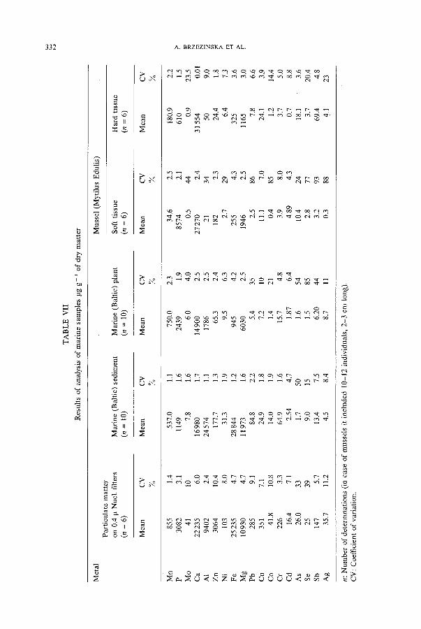
Table VII contains results obtained for the analysis of particulate matter and other samples from the Baltic sea, all using 'a' methods. (Methods 'a' must be used when no estimate is available of the trace element concentration ranges. If levels are within the needed range then methods 'b' can be employed for subsequent samples.)
2.4. B A C K G R O U N D CORRECTION
With our equipment and operating conditions, in some cases (e.g. river sediment) corrections had to be made to Cd and Pb values. These were corrected for the presence of A1, Fe, Ca, and Mg. When correction factors were used, the Cd value in river sediment (NBS 1645) and in marine sediment (NRC MESS-1 and BCSS-1), Pb content in pond sediment and Ni content in marine sediment appeared to be lower and consequently were within certified limits.
2.5. COMPARISON OF METHODS
The 'a' methods include evaporation steps during which some contamination or loss of metals may occur. Also, they are time consuming (tissue samples - 3 hr and plant samples and sediments - 1 0 hr). Thus 'b' methods should be used when levels of elements are high enough.
All sample solutions are made 6~o in a HNO3/HC104 solution (1 : 1). The 6~o acid solution used as blank contained 0.004 gg ml - a of Mo, 0.1 to 0.2 gg ml- 1 of Ca, 0.01 to 0.15 ~tg ml - i of Zn and 0.02 to 0.04 gg ml- 1 of Ni and Fe. The same teflon vessels should be used each time for blank determination.
Methods 'b' do not require evaporation steps for decomposition of animal and plant tissue and include only a short (1.5 to 2 hr) evaporation step for sediments digestion. This step is necessary when a quartz nebulizer is used. The effect of the level of HF on the nebulizer was assessed. A 20 ~o change in 1 ppm Si solution was observed when 3 ~o
3 3 2 A . B R Z E Z I N S K A E T A L .
r~
v
H
Z ,
"3"
~ Z
• -~ ,z/ II
;>
>
©
e-,
¢.,,I
=~ ,--' "r"
¢.) ~ . . o
,..Q
Z

ENVIRONMENTAL AND BIOLOGICAL SAMPLES 333
H F was used. Low levels (0.2 to 0.3~o) can be applied but to avoid problems 1 g of
H3BO 3 was added to samples and standards. A study is presently going on with a teflon cross flow nebulizer. Preliminary results
indicate that H3BO 3 is not necessary with plant samples. Blanks should be run in every vessel, from time to time, to guard against contamination.
Decomposition of plant material required more HC10 4 and some H F when SiO 2 is present. Typical blanks contained 0.005 ~tgml-1 of Mo, Cd, and Co, 0.01 to 0.02 gg m l - 1 of Cr, Cu, Pb, Ni, Zn, Mn, and Fe, and 0.1 to 0.2 gg ml - 1 of Ca, Mg,
A1. For sediments blanks were higher because of H3BO 3 content. Typical values were as follows: 0.06 to 0.08 jag ml -1 of Co, 0.01 to 0.02 tag ml -1 of P, Mn, Mo, Zn, Cu, Cr, Cd, 0.1 to 0.3 gg m l - 1 of Pb, Fe, Ni, Ca, and Mg and from 5 to 7 ~tg m l - 1 of A1.
In preliminary investigations, fusion methods were tried. The results, however, were
unsatisfactory (e.g. results were 20~o lower than acid digestion, for Ca, Mg, and Pb by
acid digestion methods). Also it was difficult to obtain good precision for most of the elements analyzed. Interferences caused by the presence of Na and Li must also be
corrected. High levels of dissolved salts can cause nebulizer problems.
3. Conclusions
It is difficult to cover 18 listed elements by one digestion procedure. However, methods
'a ' can be applied for many of the samples investigated. Methods 'b' are preferable when
applicable because fewer contamination problems are encountered and they are more rapid.
Acknowledgment
A. B. wishes to thank U N E S C O (contract No. SC/RP 560569) for financial assistance. This work was also partially supported by NSERC of Canada.
References
[1] Burman, J. O. and Bostr6m, K.: 1979, Anal. Chem. 51,516. [2] McLaren, J. W., Berman, S. S., Boyko, V. J., and Russell, D. S.: 1981, Anal. Chem. 53, 1802. [3] Dahlquist, R. L. and Knoll, J. W.: 1978, Appl. Spectros. 32, 1. [4] Ward, A. F., Marciello, L. F., Carrara, L., and Luciano, V. J.: 1980, Spectros. Letters 13, 803. [5] Jones, J. W., Capar, S. G., and O'Haver, T. C.: 1982, Analyst 107, 353. [6] McQuaker, N. R., Brown, D. F., and Kluckner, P. D.: 1979, Anal. Chem. 51, 1082. [7] Uchida, H., Nojiri, Y., Haraguchi, H., and Fuwa, K.: 1981, Anal. Chim. Acta 123, 57.