envelopment of human cytomegalovirus occurs by budding into golgi-derived vacuole
TRANSCRIPT
JOURNAL OF VIROLOGY, Mar. 2003, p. 3191–3203 Vol. 77, No. 50022-538X/03/$08.00�0 DOI: 10.1128/JVI.77.5.3191–3203.2003Copyright © 2003, American Society for Microbiology. All Rights Reserved.
Envelopment of Human Cytomegalovirus Occurs by Budding intoGolgi-Derived Vacuole Compartments Positive for gB, Rab 3,
Trans-Golgi Network 46, and Mannosidase IIM. Homman-Loudiyi,1 K. Hultenby,2 W. Britt,3 and C. Soderberg-Naucler1*
Department of Medicine, Center for Molecular Medicine, Karolinska Institute, SE 171 76 Stockholm,1 and Clinical ResearchCenter, Karolinska Institutet, Huddinge University Hospital, SE 141 86 Huddinge,2 Sweden, and
Departments of Pediatrics and Microbiology, The University of Alabama atBirmingham, Birmingham, Alabama 3523323
Received 30 October 2002/Accepted 26 November 2002
Although considerable progress has been made towards characterizing virus assembly processes, assignmentof the site of tegumentation and envelopment for human cytomegalovirus (HCMV) is still not clear. In thisstudy, we examined the envelopment of HCMV particles in human lung fibroblasts (HF) HL 411 and HL 19,human umbilical vein endothelial cells, human pulmonary arterial endothelial cells, and arterial smoothmuscle cells at different time points after infection by electron microscopy (EM), immunohistochemistry, andconfocal microscopy analysis. Double-immunofluorescence labeling experiments demonstrated colocalizationof the HCMV glycoprotein B (gB) with the Golgi resident enzyme mannosidase II, the Golgi marker TGN(trans-Golgi network) 46, and the secretory vacuole marker Rab 3 in all cell types investigated. Final envel-opment of tegumented capsids was observed at 5 days postinfection by EM, when tegumented capsids buddedinto subcellular compartments located in the cytoplasm, in close proximity to the Golgi apparatus. Immuno-gold labeling and EM analysis confirmed staining of the budding compartment with HCMV gB, Rab 3, andmannosidase II in HL 411 cells. However, the markers Rab 1, Rab 2, Rab 7, Lamp 1 (late endosomes andlysosomes), and Lamp 2 (lysosomes) neither showed specific staining of the budding compartment in theimmunogold labeling experiments nor colocalized with gB in the immunofluorescent colocalization experi-ments in any cell type studied. Together, these results suggest that the final envelopment of HCMV particlestakes place mainly into a Golgi-derived secretory vacuole destined for the plasma membrane, which mayrelease new infectious virus particles by fusion with the plasma membrane.
Human cytomegalovirus (HCMV) is a member of the her-pesvirus family, which includes members characterized by arestricted host range, the ability to establish latency, a longreproductive cycle, and a linear double-stranded DNA genomepackaged within an icosahedral capsid. Packaging of viralDNA into capsids occurs within the nucleus, whereupon nu-cleocapsids are transported out of the nucleus to the cytoplasmby an unknown pathway. The nuclear egress of herpes simplexvirus (HSV) has been suggested to involve a step of envelop-ment by budding through the inner leaflet of the nuclear mem-brane, followed by a de-envelopment when exiting through theperinuclear space (49). In the cytoplasm, CMV capsids start tosequentially become surrounded by a protein structure knownas the tegument or matrix. An envelope membrane, containingseveral virus-specific glycoproteins, in turn covers tegumentedHCMV nucleocapsids (42). This envelope consists of a lipidbilayer, which is similar in structure and composition to hostcell membranes (15). Although a number of studies have ex-amined the different steps in the HCMV assembly process,these studies have not conclusively identified the site of finalenvelope acquisition of the HCMV particles (17, 43–45, 47).
Most enveloped viruses, such as alpha-, orthomyxo-,
paramyxo-, rhabdo-, and retroviruses, acquire their lipid enve-lope by budding at the plasma membrane (15). Upon this typeof budding, mature virus particles are released directly into theextracellular space. In contrast, viruses that mature intracellu-larly bud into the lumen of intracellular compartments to ac-quire their final envelope (15, 56). Mature virus particles arethen transported within the cell in membrane vacuoles, whichupon fusion with the plasma membrane release virions extra-cellularly. For example, coronaviruses and poxviruses bud atthe endoplasmic reticulum (ER)-Golgi intermediate compart-ment (ERGIC) (28, 51), Bunyaviridae and rubella virus bud atthe Golgi complex (36), and HSV has been suggested to bud atthe inner nuclear membrane (8, 31, 55) or into cytoplasmicvacuoles (29). In the case of budding at the inner nuclearmembrane, subsequent maturation of the herpesvirus particleis believed to take place during passage of virus particlesthrough the ER (56) or the Golgi complex (24). In contrast,more recent studies on HSV morphogenesis suggest that nu-cleocapsids undergo a sequential envelopment and de-envel-opment crossing the nuclear membrane (5, 49), and thereafterthe capsid will receive its final envelope by budding into sub-cellular vacuoles in the cytoplasm (50). A number of studieshave presented evidence of different budding sites for HCMV.For example, structural electron microscopy (EM) studies havesuggested that capsids receive their final envelope by buddinginto early endosomes (60), or into trans-Golgi cisternae con-taining viral glycoproteins (27, 47). Such vacuoles containing
* Corresponding author. Mailing address: Department of Medicine,Center for Molecular Medicine, Karolinska Institutet, SE-171 76Stockholm, Sweden. Phone: 46-8-51779896. Fax: 46-8-313147. E-mail:[email protected].
3191
Dow
nloa
ded
from
http
s://j
ourn
als.
asm
.org
/jour
nal/j
vi o
n 23
Dec
embe
r 20
21 b
y 36
.72.
82.4
0.
mature virions have been suggested to release infectious virusparticles by excocytosis (27). Furthermore, studies using thefungal metabolite brefeldin A have demonstrated a cytoplas-mic accumulation of naked virus particles in HSV-infected (6)and HCMV-infected (10) cells. This finding supports the hy-pothesis that the final envelopment occurs in the cytoplasmand implies an important role of the Golgi apparatus in thisprocess. However, the site of the final envelopment for HCMVhas not been clearly defined by methods examining the originof the subcellular compartment for budding.
Since assembly processes are believed to be initiated byspecific interactions between the nucleocapsid and a cytoplas-mic extension of one or more of the viral glycoproteins (36,48), the site of virus maturation can be examined by determin-ing the accumulation of the viral glycoproteins in the buddingcompartment. To study the site of budding of HCMV particles,we performed EM and immunohistochemical analysis ofHCMV-infected human embryonic lung fibroblasts (HF) atdifferent time points after infection. Structural EM analysissuggested that tegumented virus particles bud into cytoplasmicvacuoles to acquire their final envelope. Colocalization studiesof HCMV glycoprotein B (gB) and different markers for spe-cific subcellular compartments were performed with HF, hu-man umbilical vein endothelial cells (HUVEC), human pul-monary arterial endothelial cells (HPAEC), and arterialsmooth muscle cells (ASMC) and revealed colocalization of gBwith the Golgi resident enzyme mannosidase II, TGN (trans-Golgi network) 46, and the Rab 3 GTPase, which is a specificmarker for secretory vacuoles. Immunogold labeling and EManalysis confirmed staining of the budding compartment andthe viral envelope with HCMV gB, mannosidase II, and Rab 3in HF. In contrast, lysosomal and endosomal markers were notdetected on the virus-containing vacuoles. These results sug-gest that HCMV buds into Golgi-derived vacuoles, which aredestined for the plasma membrane.
MATERIALS AND METHODS
Cells and viruses. HF HL 411 (kindly provided by V. Sundquist, KarolinskaInstitute, Stockholm, Sweden) and HL 19 were maintained in bicarbonate-freeminimal essential medium with Hanks salts (GIBCO BRL, Grand Island, N.Y.)supplemented with 25 mM HEPES, 10% heat inactivated fetal calf serum,L-glutamine (2 mM), penicillin (100 U/ml), and streptomycin (100 �g/ml)(GIBCO BRL) at 37°C with 5% CO2. The HF cells were used in experimentsuntil passage 20. The HF cells were infected with HCMV strain AD169 at amultiplicity of infection (MOI) of 1. The MOI was calculated as PFU � milli-liters/number of cells. The virus supernatant was collected at 7 or 10 dayspostinfection (dpi), cleared from cell debris by low-speed centrifugation, andfrozen at �70°C until used. Monolayers of HF grown in 96-well plates (Falcon)or on chamber slides (Nalge Nunc International) were infected with HCMVAD169 at an MOI of 1. Virus titers were determined as previously described(63).
HPAEC medium was obtained from Clonetics, San Diego, Calif. The cellswere maintained in EBM-2 (Clonetics) with growth factors according to themanufacturer’s instructions at 37°C with 5% CO2. At 1 h prior to infection, themedium was changed to endothelial basal medium with 5% fetal calf serum, andthe cells were cultured in this medium during the infection process. The cellswere cultured in 0.2% gelatin-coated 175-cm2 tissue culture flasks (Falcon) oreight-well chamber slides (Nalge Nunc International) and used until passage 8 inthe experiments.
HUVEC were cultured in EGM medium (Clonetics). At 1 h prior to infection,the medium was changed to endothelial basal medium with 5% fetal calf serum,and the cells were cultured in this medium during the infection process. The cellswere cultured in 175-cm2 tissue culture flasks (Falcon) and eight-well chamberslides (Nalge Nunc International) and used until passage 8. The cells were
infected at an MOI of 1 with an endothelium-adapted strain, TB40, kindlyprovided by G. Jahn and C. Sinzger, Department of Medical Virology, Universityof Tubingen, Tubingen, Germany.
Smooth muscle cells were isolated from aortic grafts obtained from patientsundergoing surgery for aortic aneurysm with insertion of aortic grafts or fromeither aortic grafts or grafts of the iliaca vessels from transplantation donors. Thecells were isolated by first removing the endothelial cells by cell scraping with asharp instrument. The vessel was then divided into two different layers, theinnermost layer (corresponding to the intima of the vessel) and the outermostlayer (corresponding to the vessel media), by gentle separation of the two layerswith a pair of tweezers. The aortic graft was cut into 1-mm pieces and culturedon cell culture dishes in SMGM 2 medium (Clonetics) with the addition ofSMGM 2 SingleQuots (human epidermal growth factor [0.5 �g/ml], 0.5 ml;insulin [5 mg/ml], 0.5 ml; human fibroblast growth factor b [1 �g/ml], 1 ml; fetalbovine serum, 25 ml; GA-1000, 0.5 ml) for about 2 weeks, until smooth musclecells had migrated from the explants. The cells were cultured in 175-cm2 cellculture flasks (Falcon). To determine the purity of the smooth muscle cellcultures, the cells were grown on chamber slides, fixed in 3% formaldehyde for45 min at 4°C, and permeabilized with 0.3% Triton X-100 for 5 min. Afterfixation, the cells were stained for smooth muscle cell-specific fluorescein iso-thiocyanate (FITC)-conjugated antiactin (Sigma) and evaluated by fluorescencemicroscopy. The cells were fixed with methanol-acetone (1:1) at 4°C for 5 min,stained with FITC-conjugated anti-von Willebrand factor and endothelial cell-specific von Willebrand factor, and evaluated by fluorescence microscopy. Iso-type controls for the antibodies were used in the experiments. The cultures were100% positive for antiactin antibody and negative for von Willebrand factor.
Antibodies. For immunohistochemical analysis, antibodies specific for themarkers Rab 1 to 6 (rabbit polyclonal immunoglobulin G), Rab 7, Lamp 1, andLamp 2 (goat polyclonal immunoglobulin G), purchased from Santa Cruz Bio-technology, Santa Cruz, Calif., were used as 1:100 dilutions (1:10 dilutions in theimmunogold experiments) from stocks of 200 �g/ml. TGN 46 (sheep anti-humanantibody; Serotec, Oxford, United Kingdom) was detected with FITC-labeledrabbit anti-sheep antibody (Dako A/S, Glostrup, Denmark). Polyclonal mouseanti-human antibody directed against mannosidase II was kindly provided byKelley Moreman and Marylin Farquhar, The University of Georgia, Athens.These antibodies were used in colocalization experiments with mouse or humananti-HCMV gB and detected with FITC- or rhodamine-phalloidin-conjugatedswine, goat, or mouse F(ab)�2 fragments against rabbit, goat, or mouse Ig,respectively; FITC-conjugated rabbit anti-mouse immunoglobulin; FITC-conju-gated goat F(ab)�2 anti-mouse antibody (Dako A/S); or phycoerythrin-conju-gated goat F(ab)�2 fragments against human immunoglobulin (Southern Bio-technology Associates Inc., Birmingham, Ala.). Secondary antibodies werediluted in DAKO antibody diluent (DAKO A/S) and used as 1:100 dilutions. Thefollowing mouse monoclonal antibodies (MAbs), produced by William Britt,were used for CMV protein localization studies with immunofluorescence de-tection at 1:100 dilutions: UL99 (pp28), UL83 (pp65), UL82 (pp71), UL32(pp150), UL55 (gB), UL75 (gH), and UL115 (gL) (44, 45).
Immunofluorescence staining of HCMV-infected cells. Monolayers of HF,HUVEC, HPAEC, or ASMC grown in 24- to 96-well tissue culture plates or on8-well chamber slides (Nalge Nunc International) were infected with HCMV atan MOI of 1 and fixed at 1, 3, 5, 7, or 9 dpi. Uninfected and infected cells werewashed in phosphate-buffered saline, fixed in either ice-cold methanol-acetoneor 3% paraformaldehyde for 15 min at room temperature (RT), and permeabil-ized by 0.3% Triton X-100 treatment for 5 min. Samples were incubated withSerum-Free Protein Block (DAKO A/S) for 10 min at RT before being washedand stained with antibodies diluted in antibody diluent (DAKO A/S) or appro-priate isotype controls. Both infected and uninfected cells were used in controlexperiments. Samples were mounted on glass slides by using fluorescent mount-ing medium containing 15 mM NaN3 (DAKO A/S). The secondary antibodieswere made in the same species when two different antibodies were stained forcolocalization experiments. For control purposes, the secondary antibody aloneor cells treated with human, mouse, rabbit, or goat serum (DAKO A/S) (incu-bation in 1 to 3% serum for 15 min at RT) were stained for false fluorescence andautofluorescence (same control with both infected and noninfected cells). 4,6-Diamide-2-phenylindol (Sigma-Aldrich, St. Louis, Mo.) at 4 ng/ml was used tostain the nucleus blue.
Fluorescence microscopy and image analysis. Fluorescence microscopy anal-ysis was performed with a DMRXA microscope (Leica Microsystems Gmbh,Wetzlar, Germany) equipped with a cooled charge-coupled device camera (mod-el S/N 370 KL 0565; Cooke Corporation, Auburn Hills, Mich.). Filter sets for4,6-diamide-2-phenylindol–Hoechst, FITC, Cy3, and Cy5 were obtained fromChroma Technology (Brattleboro, Vt.). The images were acquired and analyzedwith the image processing software SlideBook 2.1.5 (Intelligent Imaging Inno-
3192 HOMMAN-LOUDIYI ET AL. J. VIROL.
Dow
nloa
ded
from
http
s://j
ourn
als.
asm
.org
/jour
nal/j
vi o
n 23
Dec
embe
r 20
21 b
y 36
.72.
82.4
0.
vations Inc., Denver, Colo.). Images were postprocessed and mounted in Pho-toshop 5.0 (Adobe Systems Inc., San Jose, Calif.) and printed on a TektronixPhaser 860DP color printer (Xerox, Stamford, Conn.) or on a DS 8650 printer(Kodak, Rochester, N.Y.).
EM analysis. (i) Tissue preparation for structural EM analysis. To examinevirus-infected cells (MOI of 1) by EM, uninfected and HCMV-infected cellswere harvested at 1, 3, 5, 7, and 10 dpi and fixed in 2% glutaraldehyde in 0.1 Msodium cacodylate buffer–0.1 M sucrose with 3 mM CaCl2 (pH 7.4) at 4°C for
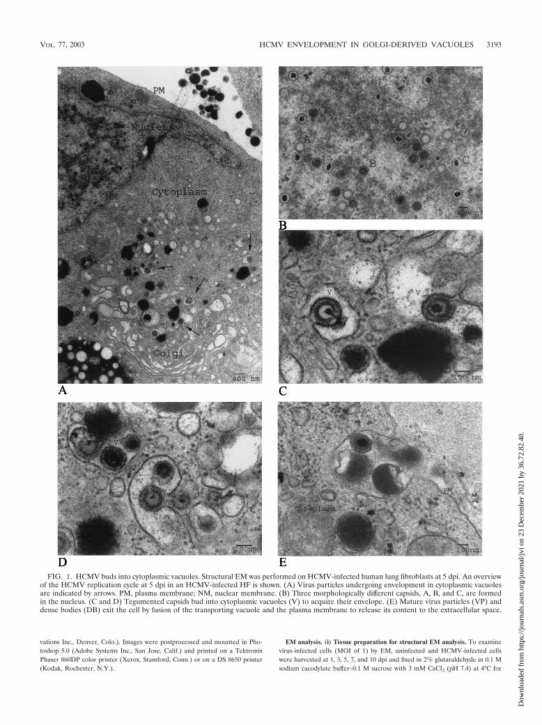
FIG. 1. HCMV buds into cytoplasmic vacuoles. Structural EM was performed on HCMV-infected human lung fibroblasts at 5 dpi. An overviewof the HCMV replication cycle at 5 dpi in an HCMV-infected HF is shown. (A) Virus particles undergoing envelopment in cytoplasmic vacuolesare indicated by arrows. PM, plasma membrane; NM, nuclear membrane. (B) Three morphologically different capsids, A, B, and C, are formedin the nucleus. (C and D) Tegumented capsids bud into cytoplasmic vacuoles (V) to acquire their envelope. (E) Mature virus particles (VP) anddense bodies (DB) exit the cell by fusion of the transporting vacuole and the plasma membrane to release its content to the extracellular space.
VOL. 77, 2003 HCMV ENVELOPMENT IN GOLGI-DERIVED VACUOLES 3193
Dow
nloa
ded
from
http
s://j
ourn
als.
asm
.org
/jour
nal/j
vi o
n 23
Dec
embe
r 20
21 b
y 36
.72.
82.4
0.
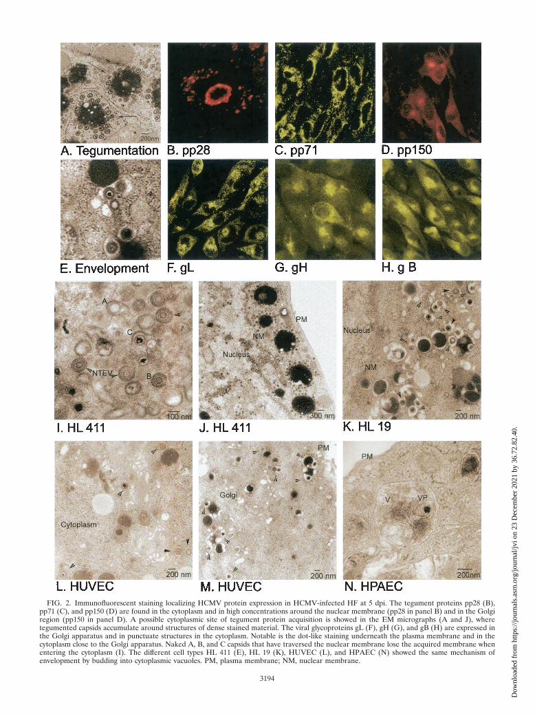
FIG. 2. Immunofluorescent staining localizing HCMV protein expression in HCMV-infected HF at 5 dpi. The tegument proteins pp28 (B),pp71 (C), and pp150 (D) are found in the cytoplasm and in high concentrations around the nuclear membrane (pp28 in panel B) and in the Golgiregion (pp150 in panel D). A possible cytoplasmic site of tegument protein acquisition is showed in the EM micrographs (A and J), wheretegumented capsids accumulate around structures of dense stained material. The viral glycoproteins gL (F), gH (G), and gB (H) are expressed inthe Golgi apparatus and in punctuate structures in the cytoplasm. Notable is the dot-like staining underneath the plasma membrane and in thecytoplasm close to the Golgi apparatus. Naked A, B, and C capsids that have traversed the nuclear membrane lose the acquired membrane whenentering the cytoplasm (I). The different cell types HL 411 (E), HL 19 (K), HUVEC (L), and HPAEC (N) showed the same mechanism ofenvelopment by budding into cytoplasmic vacuoles. PM, plasma membrane; NM, nuclear membrane.
3194
Dow
nloa
ded
from
http
s://j
ourn
als.
asm
.org
/jour
nal/j
vi o
n 23
Dec
embe
r 20
21 b
y 36
.72.
82.4
0.
20 h. Fixed cells were collected by scraping with a wooden peg and pelleted inEppendorf tubes. The pellets were washed in 0.15 M sodium cacodylate con-taining 3 mM CaCl2 (pH 7.4). Specimens were postfixed in 2% osmium tetroxidein 0.1 M cacodylate buffer containing 3 mM CaCl2 for 1 h at 4°C, dehydrated inethanol followed by acetone, and embedded in LX-112. Sections were cut, placedon Formvar-coated copper grids, and stained with uranyl acetate followed bylead citrate. Sections were examined in a calibrated Philips 420 electron micro-scope at 80 kV.
(ii) Immunohistochemical gold labeling of HCMV-infected cells. Monolayersof uninfected and HCMV-infected HF were washed with phosphate-bufferedsaline and fixed in 3% paraformaldehyde–0.1% glutaraldehyde in 0.1 M phos-phate buffer (PB) (pH 7.2) for 30 min at RT. Following fixation, cells werescraped with a wooden peg and pelleted in Eppendorf tubes. The pellet wasinfiltrated with 4% gelatin in 0.1 M phosphate buffer (pH 7.4) for 1 h, followedby fixation in the same fixative for 1 h at RT. The specimens were thereafterinfiltrated with 2.3 M sucrose and frozen in liquid nitrogen. Sectioning wasperformed in an ultracryotome (Leica UCT) at �90°C by the method previouslydescribed by Tokuyasu (59). The sections were washed in 0.1 M PB, blocked with10% normal goat serum for 10 min, and incubated with the MAbs at samedilutions as described above overnight at 4°C. Sections were rinsed in 0.1 M PBplus 0.1% normal goat serum. Bound antibodies were detected by secondary goatanti-mouse antibodies conjugated with 10-nm-diameter gold particles. After anadditional washing step with 0.1 M PB, the specimen was fixed in 2% glutaral-dehyde for 30 min and washed with distilled water four times for 1 min each,followed by final contrasting in 0.1 M uranyl acetate for 45 min.
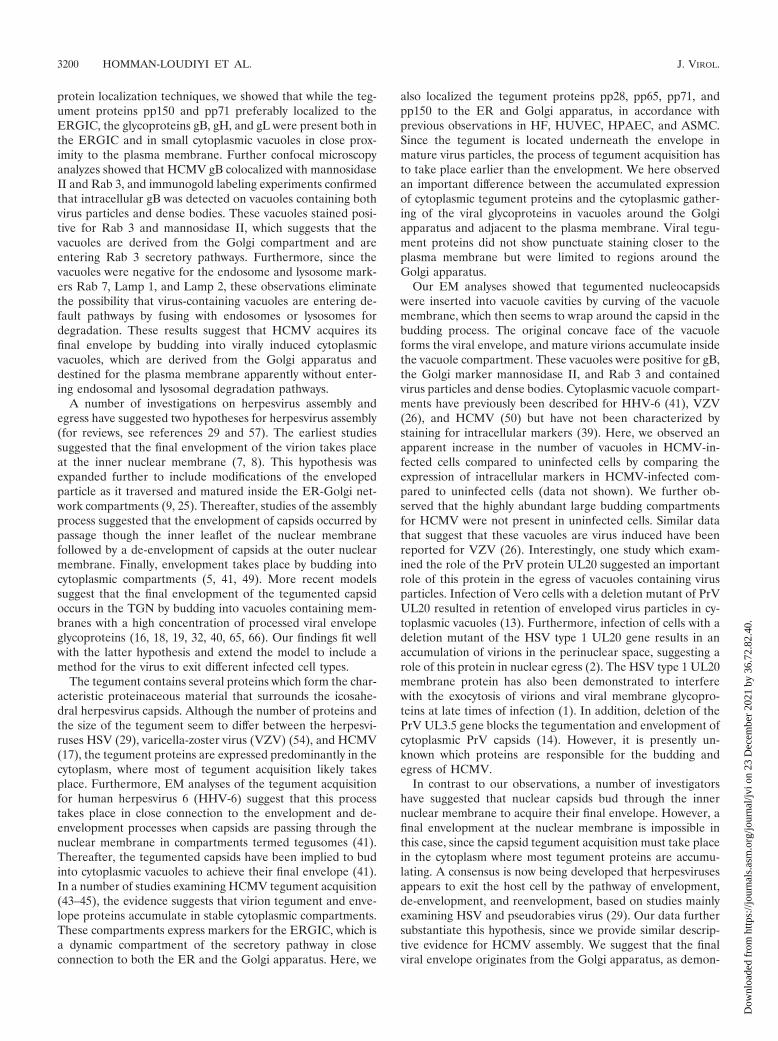
(iii) Quantification of immunogold labeling. Gold beads are the number ofgold particles (designated Au) counted on a selected micrograph or on a certainstructure seen in the micrograph, such as the envelope membrane of the virion,dense bodies, or vacuoles. The area of the micrograph where gold staining wascounted was measured in square micrometers. Background staining is given asAu per square micrometer. The quantification is based on the comparison be-tween the parameters Au per square micrometer on virus particles, Au persquare micrometer on dense bodies, and Au per square micrometer on virus-containing vacuoles in relation to the background staining. Several antibodies(e.g., Lamp 1, Lamp 2, Rab 4, and Rab 7) were not suitable or optimal for usein gold labeling analysis due to instability of the antibodies.
RESULTS
Structural EM analysis of HCMV-infected HF. The mech-anism and subcellular site of final envelopment of HCMV havepreviously not been well defined. In this study, we performedEM and immunohistochemical analysis with HCMV-infectedHF cells, at 1, 3, 5, 7, and 10 dpi as our model system, and inaddition, we examined HCMV-infected HUVEC, HPAEC,and ASMC harvested at 5, 7, and 9 dpi. As has previously beenreported, we observed the formation of three different capsidparticles in the nuclei of infected HF starting at 3 dpi, i.e., Acapsids (empty capsids without DNA), B capsids (capsids with
a translucent core), and C capsids (nucleocapsids containingDNA), all of which were approximately 100 nm in diameter(Fig. 1A and B and data not shown). DNA appeared to beencapsulated into empty A capsids in the nucleus, in accor-dance with the previous suggestion that A, B, and C capsidsmight reflect different maturity stages of the capsid (Fig. 1B).The number of nucleocapsids observed in the nucleus in-creased significantly from 5 dpi. Whether this accumulation ofnucleocapsids was due to high production or a limited releaseof the nucleocapsids from the nucleus remains to be investi-gated. Nontegumented enveloped virus particles (NTEV) un-dergoing de-envelopment were observed at the rim of thenuclear membrane (Fig. 2I), which further supports the samemechanism of action for the nuclear exit of HCMV nucleo-capsids as for HSV nucleocapsids, by envelopment and de-envelopment when traversing the nuclear membrane. All threecapsid forms were observed to exit the nucleus by the samemechanism (Fig. 2I). NTEV were observed in close associationwith the nuclear membrane in HL 411, HL 19, and HUVEC(data not shown). In the cytoplasm, an amorphous material,the tegument, surrounded nonenveloped A, B, and C capsids,giving the tegumented capsids a diameter of approximately 130nm (Fig. 2A and J and data not shown). The amorphousstructure, or the tegument, was never observed around capsidspresent in the nucleus. Furthermore, the majority of the tegu-mented capsids present in the cytoplasm were observed incytoplasmic clusters, surrounding vacuole-like structures (Fig.2A and J). Although tegumented A, B, and C capsids can beseen in the cytoplasm, the vast majority of capsids surroundingthe cytoplasmic clusters (Fig. 2A and J) were of the C type. Theacquisition of tegument proteins around capsids seems to be arapid process following nuclear exit, since only fully tegu-mented capsids were observed in the cytoplasm, with the ex-ception of NTEV that appeared to be in the process of loosinga possibly temporal nuclear membrane envelope.
HCMV acquires its envelope by budding into cytoplasmicvacuoles in close proximity to the Golgi apparatus. Abundantlarge intracellular vacuoles containing one or several virusparticles were observed in the cytoplasm of infected HL 411(Fig. 1C and D), HL 19 (Fig. 2K), HUVEC (Fig. 2L), andHPAEC (Fig. 2N) after 5 dpi by EM analysis. The final envel-opment of tegumented capsids appeared to take place throughbudding into vacuoles of different sizes, which were formed inclose proximity to the Golgi apparatus (Fig. 1A and Fig. 2M).The cytoplasmic vacuoles ranged in diameter from 300 to 600nm, with an average diameter of approximately 400 nm, andwere not observed in uninfected cells (data not shown). Thevacuoles appeared to fuse with the plasma membrane andreleased virions to the extracellular space (HF in Fig. 1E andHPAEC in Fig. 2N). Virus particles were observed neitherwithin the Golgi apparatus itself nor in association with thenuclear membrane. Tegumented capsids were never observedin the nucleus. Furthermore, dense bodies seemed to beformed in a similar way in HCMV-infected HF, where anamorphous electron-dense material became enveloped to formdense bodies of different sizes (200 to 500 nm) through bud-ding into the same type of vacuoles (Fig. 1D and E). Theseobservations suggest that the membranes of dense bodies andenveloped virus might have the same composition.
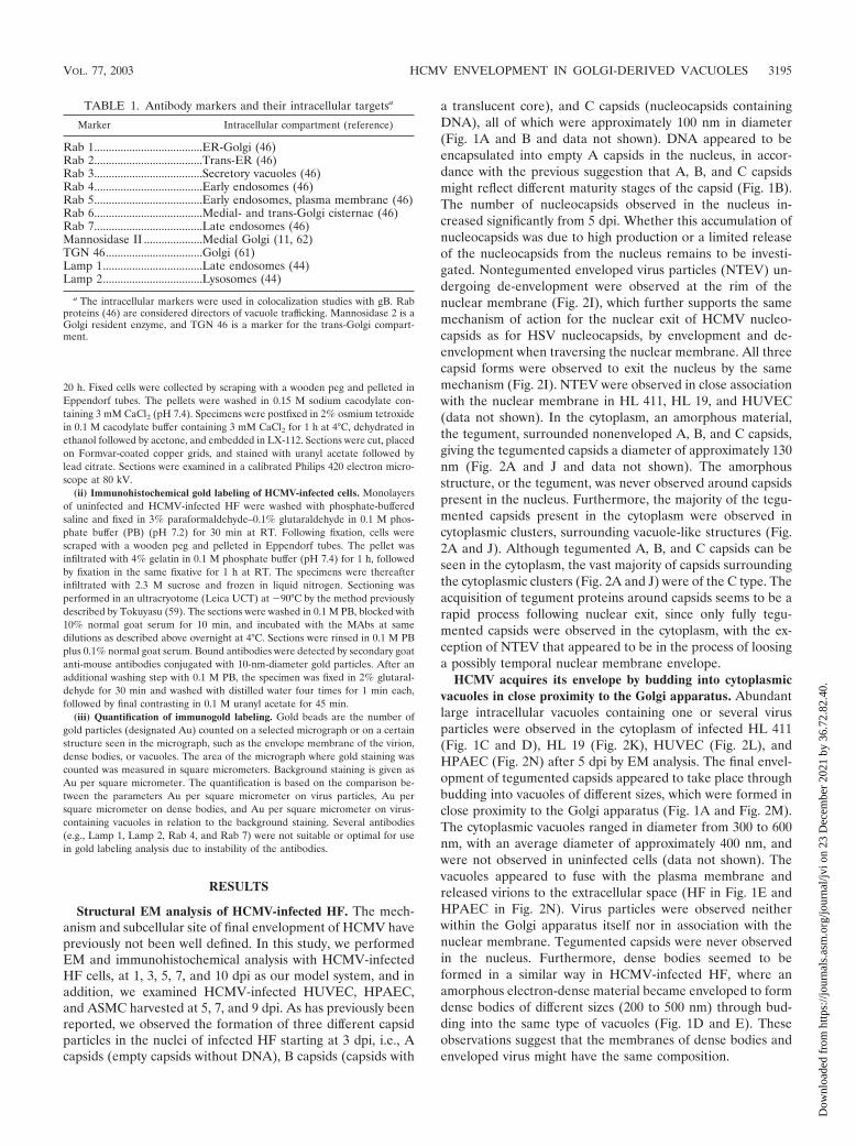
TABLE 1. Antibody markers and their intracellular targetsa
Marker Intracellular compartment (reference)
Rab 1.....................................ER-Golgi (46)Rab 2.....................................Trans-ER (46)Rab 3.....................................Secretory vacuoles (46)Rab 4.....................................Early endosomes (46)Rab 5.....................................Early endosomes, plasma membrane (46)Rab 6.....................................Medial- and trans-Golgi cisternae (46)Rab 7.....................................Late endosomes (46)Mannosidase II ....................Medial Golgi (11, 62)TGN 46.................................Golgi (61)Lamp 1..................................Late endosomes (44)Lamp 2..................................Lysosomes (44)
a The intracellular markers were used in colocalization studies with gB. Rabproteins (46) are considered directors of vacuole trafficking. Mannosidase 2 is aGolgi resident enzyme, and TGN 46 is a marker for the trans-Golgi compart-ment.
VOL. 77, 2003 HCMV ENVELOPMENT IN GOLGI-DERIVED VACUOLES 3195
Dow
nloa
ded
from
http
s://j
ourn
als.
asm
.org
/jour
nal/j
vi o
n 23
Dec
embe
r 20
21 b
y 36
.72.
82.4
0.
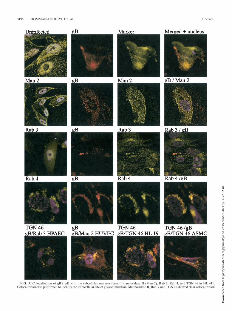
FIG. 3. Colocalization of gB (red) with the subcellular markers (green) mannosidase II (Man 2), Rab 3, Rab 4, and TGN 46 in HL 411.Colocalization was performed to identify the intracellular site of gB accumulation. Mannosidase II, Rab 3, and TGN 46 showed clear colocalization
3196 HOMMAN-LOUDIYI ET AL. J. VIROL.
Dow
nloa
ded
from
http
s://j
ourn
als.
asm
.org
/jour
nal/j
vi o
n 23
Dec
embe
r 20
21 b
y 36
.72.
82.4
0.
HCMV tegument proteins and glycoproteins localize to cy-toplasmic compartments in close proximity to the Golgi appa-ratus. To localize the site of viral capsid tegument acquisitionin the infected cells, MAbs reactive with the tegument proteinspp28, pp65, pp71, and pp150 were used to stain HCMV-in-fected HF, HUVEC, HPAEC, and ASMC. In these experi-ments, the viral tegument protein pp28 was present mainly inthe nuclear membrane and in large vacuole compartments inthe cytoplasm, which may correspond to the large sites ofaccumulation of electron-dense material (500 to 2,000 nm)that were observed in the cytoplasm by EM analysis (Fig. 2A).These round structures of large electron-dense material weregenerally surrounded by numerous tegumented capsids (Fig.2A). While the pp71 protein was strongly localized to thenuclear membrane (Fig. 2C), the pp150 protein was presentmainly in the cytoplasm, with a high concentration in the Golgiregion as well as in punctuate structures in close associationwith the cell nucleus and the Golgi apparatus (Fig. 2D). Whilepp28, pp71, and pp150 were detected exclusively in the cyto-plasm, pp65 was observed in the nucleus at 1 and 3 dpi and inboth the nucleus and the cytoplasm at 5 and 7 dpi (Fig. 2 anddata not shown). In contrast to the previously reported pres-ence of the HCMV tegument proteins pp28, pp71, and pp150in the nucleus (22), we did not observe either pp28, pp71, orpp150 in the nucleus at any time point examined between 1 and9 dpi. Further confocal microscopy analyses of the cellularlocalization of the HCMV envelope glycoproteins gB, gH, andgL demonstrated a staining pattern similar to that of the teg-ument proteins pp71 and pp150 in the cytoplasm, with a high-intensity staining pattern around, and in, the Golgi region. Inaddition, the envelope glycoproteins gB, gH, and gL werepresent in small vacuoles around the Golgi apparatus andunderneath the plasma membrane, which may correspond tothe small (300- to 600-nm) virus-containing vacuoles observedby EM analysis (Fig. 1C, D, and E and 2E). Furthermore, whileantibodies specific for gH and gL stained the nuclear mem-brane at early times in infection (Fig. 2F and G), gB was onlyrarely detected at the nuclear membrane (Fig. 2H). Thus, theseresults suggest that the tegumentation and envelopment ap-pear to be separate processes.
Confocal microscopy analyses demonstrate colocalization ofgB with Rab 3, TGN 46, and mannosidase II. To identify thesite of intracellular accumulation of the major HCMV envelopeglycoprotein gB by using specific intracellular markers, we per-formed colocalization experiments with antibodies directedagainst HCMV gB as well as markers for the following specificsubcellular compartments: Rab 1 (ER and Golgi complex), Rab 2(transitional ER and cis-Golgi network), Rab 3 (secretory vacu-oles), Rab 4 (early endosomes) Rab 5 (early endosomes), Rab 6(medial- and trans-Golgi cisternae), TGN 46 (trans-Golgi net-work), and mannosidase II (medial Golgi) (Table 1). The Golgiresident enzyme mannosidase II colocalized with HCMV gB incytoplasmic vacuoles, in close association with the plasma mem-
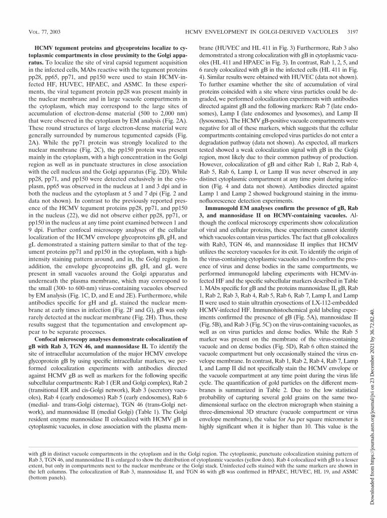
brane (HUVEC and HL 411 in Fig. 3) Furthermore, Rab 3 alsodemonstrated a strong colocalization with gB in cytoplasmic vacu-oles (HL 411 and HPAEC in Fig. 3). In contrast, Rab 1, 2, 5, and6 rarely colocalized with gB in the infected cells (HL 411 in Fig.4). Similar results were obtained with HUVEC (data not shown).To further examine whether the site of accumulation of viralproteins coincided with a site where virus particles could be de-graded, we performed colocalization experiments with antibodiesdirected against gB and the following markers: Rab 7 (late endo-somes), Lamp I (late endosomes and lysosomes), and Lamp II(lysosomes). The HCMV gB-positive vacuole compartments werenegative for all of these markers, which suggests that the cellularcompartments containing enveloped virus particles do not enter adegradation pathway (data not shown). As expected, all markerstested showed a weak colocalization signal with gB in the Golgiregion, most likely due to their common pathway of production.However, colocalization of gB and either Rab 1, Rab 2, Rab 4,Rab 5, Rab 6, Lamp I, or Lamp II was never observed in anydistinct cytoplasmic compartment at any time point during infec-tion (Fig. 4 and data not shown). Antibodies directed againstLamp 1 and Lamp 2 showed background staining in the immu-nofluorescence detection experiments.
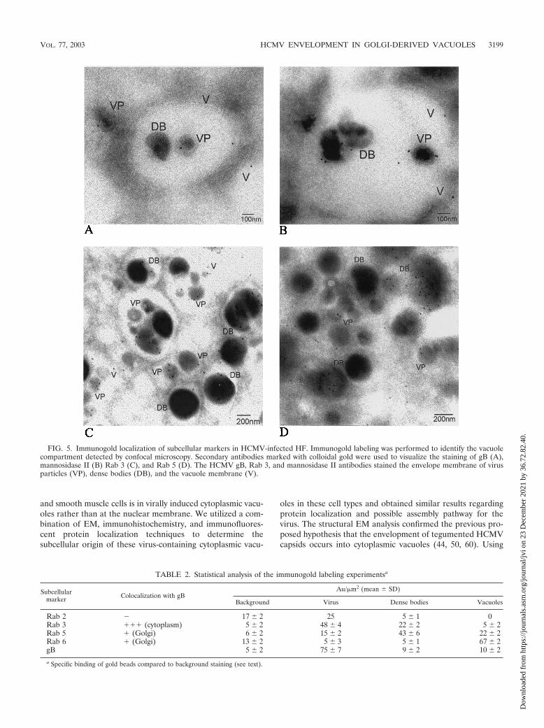
Immunogold EM analyses confirm the presence of gB, Rab3, and mannosidase II on HCMV-containing vacuoles. Al-though the confocal microscopy experiments show colocalizationof viral and cellular proteins, these experiments cannot identifywhich vacuoles contain virus particles. The fact that gB colocalizeswith Rab3, TGN 46, and mannosidase II implies that HCMVutilizes the secretory vacuoles for its exit. To identify the origin ofthe virus-containing cytoplasmic vacuoles and to confirm the pres-ence of virus and dense bodies in the same compartments, weperformed immunogold labeling experiments with HCMV-in-fected HF and the specific subcellular markers described in Table1. MAbs specific for gB and the proteins mannosidase II, gB, Rab1, Rab 2, Rab 3, Rab 4, Rab 5, Rab 6, Rab 7, Lamp I, and LampII were used to stain ultrathin cryosections of LX-112-embeddedHCMV-infected HF. Immunohistochemical gold labeling exper-iments confirmed the presence of gB (Fig. 5A), mannosidase II(Fig. 5B), and Rab 3 (Fig. 5C) on the virus-containing vacuoles, aswell as on virus particles and dense bodies. While the Rab 5marker was present on the membrane of the virus-containingvacuole and on dense bodies (Fig. 5D), Rab 6 often stained thevacuole compartment but only occasionally stained the virus en-velope membrane. In contrast, Rab 1, Rab 2, Rab 4, Rab 7, LampI, and Lamp II did not specifically stain the HCMV envelope orthe vacuole compartment at any time point during the virus lifecycle. The quantification of gold particles on the different mem-branes is summarized in Table 2. Due to the low statisticalprobability of capturing several gold grains on the same two-dimensional surface on the electron micrograph when staining athree-dimensional 3D structure (vacuole compartment or virusenvelope membrane), the value for Au per square micrometer ishighly significant when it is higher than 10. This value is the
with gB in distinct vacuole compartments in the cytoplasm and in the Golgi region. The cytoplasmic, punctuate colocalization staining pattern ofRab 3, TGN 46, and mannosidase II is enlarged to show the distribution of cytoplasmic vacuoles (yellow dots). Rab 4 colocalized with gB to a lesserextent, but only in compartments next to the nuclear membrane or the Golgi stack. Uninfected cells stained with the same markers are shown inthe left columns. The colocalization of Rab 3, mannosidase II, and TGN 46 with gB was confirmed in HPAEC, HUVEC, HL 19, and ASMC(bottom panels).
VOL. 77, 2003 HCMV ENVELOPMENT IN GOLGI-DERIVED VACUOLES 3197
Dow
nloa
ded
from
http
s://j
ourn
als.
asm
.org
/jour
nal/j
vi o
n 23
Dec
embe
r 20
21 b
y 36
.72.
82.4
0.
number of gold grains (Au) per square micrometer captured on atwo-dimensional level. Although Rab 3 is recycled between theplasma membrane and intracellular membranes, it was found tobe highly specific in its binding to the virus membrane, the vacuolecompartments containing enveloped virus particles, and densebodies (Fig. 5C).
DISCUSSION
The details of the cytoplasmic events during HCMV mor-phogenesis have been a matter of controversy. In this study, wepresent descriptive new evidence which suggest that the majorsite of HCMV envelopment in fibroblasts, endothelial cells,
FIG. 4. Colocalization of gB and the markers Rab 1, Rab 2, Rab 5, and Rab 6 in HCMV-infected HF cells at 5 dpi. Colocalization wasperformed to identify the intracellular site of gB accumulation. Rab 1, Rab 2, Rab 5, and Rab 6 did not show colocalization with gB. The fourmarkers demonstrated a weak colocalization with gB in the Golgi apparatus but not in any intracellular compartment.
3198 HOMMAN-LOUDIYI ET AL. J. VIROL.
Dow
nloa
ded
from
http
s://j
ourn
als.
asm
.org
/jour
nal/j
vi o
n 23
Dec
embe
r 20
21 b
y 36
.72.
82.4
0.
and smooth muscle cells is in virally induced cytoplasmic vacu-oles rather than at the nuclear membrane. We utilized a com-bination of EM, immunohistochemistry, and immunofluores-cent protein localization techniques to determine thesubcellular origin of these virus-containing cytoplasmic vacu-
oles in these cell types and obtained similar results regardingprotein localization and possible assembly pathway for thevirus. The structural EM analysis confirmed the previous pro-posed hypothesis that the envelopment of tegumented HCMVcapsids occurs into cytoplasmic vacuoles (44, 50, 60). Using
FIG. 5. Immunogold localization of subcellular markers in HCMV-infected HF. Immunogold labeling was performed to identify the vacuolecompartment detected by confocal microscopy. Secondary antibodies marked with colloidal gold were used to visualize the staining of gB (A),mannosidase II (B) Rab 3 (C), and Rab 5 (D). The HCMV gB, Rab 3, and mannosidase II antibodies stained the envelope membrane of virusparticles (VP), dense bodies (DB), and the vacuole membrane (V).
TABLE 2. Statistical analysis of the immunogold labeling experimentsa
Subcellularmarker Colocalization with gB
Au/�m2 (mean � SD)
Background Virus Dense bodies Vacuoles
Rab 2 � 17 � 2 25 5 � 1 0Rab 3 ��� (cytoplasm) 5 � 2 48 � 4 22 � 2 5 � 2Rab 5 � (Golgi) 6 � 2 15 � 2 43 � 6 22 � 2Rab 6 � (Golgi) 13 � 2 5 � 3 5 � 1 67 � 2gB 5 � 2 75 � 7 9 � 2 10 � 2
a Specific binding of gold beads compared to background staining (see text).
VOL. 77, 2003 HCMV ENVELOPMENT IN GOLGI-DERIVED VACUOLES 3199
Dow
nloa
ded
from
http
s://j
ourn
als.
asm
.org
/jour
nal/j
vi o
n 23
Dec
embe
r 20
21 b
y 36
.72.
82.4
0.
protein localization techniques, we showed that while the teg-ument proteins pp150 and pp71 preferably localized to theERGIC, the glycoproteins gB, gH, and gL were present both inthe ERGIC and in small cytoplasmic vacuoles in close prox-imity to the plasma membrane. Further confocal microscopyanalyzes showed that HCMV gB colocalized with mannosidaseII and Rab 3, and immunogold labeling experiments confirmedthat intracellular gB was detected on vacuoles containing bothvirus particles and dense bodies. These vacuoles stained posi-tive for Rab 3 and mannosidase II, which suggests that thevacuoles are derived from the Golgi compartment and areentering Rab 3 secretory pathways. Furthermore, since thevacuoles were negative for the endosome and lysosome mark-ers Rab 7, Lamp 1, and Lamp 2, these observations eliminatethe possibility that virus-containing vacuoles are entering de-fault pathways by fusing with endosomes or lysosomes fordegradation. These results suggest that HCMV acquires itsfinal envelope by budding into virally induced cytoplasmicvacuoles, which are derived from the Golgi apparatus anddestined for the plasma membrane apparently without enter-ing endosomal and lysosomal degradation pathways.
A number of investigations on herpesvirus assembly andegress have suggested two hypotheses for herpesvirus assembly(for reviews, see references 29 and 57). The earliest studiessuggested that the final envelopment of the virion takes placeat the inner nuclear membrane (7, 8). This hypothesis wasexpanded further to include modifications of the envelopedparticle as it traversed and matured inside the ER-Golgi net-work compartments (9, 25). Thereafter, studies of the assemblyprocess suggested that the envelopment of capsids occurred bypassage though the inner leaflet of the nuclear membranefollowed by a de-envelopment of capsids at the outer nuclearmembrane. Finally, envelopment takes place by budding intocytoplasmic compartments (5, 41, 49). More recent modelssuggest that the final envelopment of the tegumented capsidoccurs in the TGN by budding into vacuoles containing mem-branes with a high concentration of processed viral envelopeglycoproteins (16, 18, 19, 32, 40, 65, 66). Our findings fit wellwith the latter hypothesis and extend the model to include amethod for the virus to exit different infected cell types.
The tegument contains several proteins which form the char-acteristic proteinaceous material that surrounds the icosahe-dral herpesvirus capsids. Although the number of proteins andthe size of the tegument seem to differ between the herpesvi-ruses HSV (29), varicella-zoster virus (VZV) (54), and HCMV(17), the tegument proteins are expressed predominantly in thecytoplasm, where most of tegument acquisition likely takesplace. Furthermore, EM analyses of the tegument acquisitionfor human herpesvirus 6 (HHV-6) suggest that this processtakes place in close connection to the envelopment and de-envelopment processes when capsids are passing through thenuclear membrane in compartments termed tegusomes (41).Thereafter, the tegumented capsids have been implied to budinto cytoplasmic vacuoles to achieve their final envelope (41).In a number of studies examining HCMV tegument acquisition(43–45), the evidence suggests that virion tegument and enve-lope proteins accumulate in stable cytoplasmic compartments.These compartments express markers for the ERGIC, which isa dynamic compartment of the secretory pathway in closeconnection to both the ER and the Golgi apparatus. Here, we
also localized the tegument proteins pp28, pp65, pp71, andpp150 to the ER and Golgi apparatus, in accordance withprevious observations in HF, HUVEC, HPAEC, and ASMC.Since the tegument is located underneath the envelope inmature virus particles, the process of tegument acquisition hasto take place earlier than the envelopment. We here observedan important difference between the accumulated expressionof cytoplasmic tegument proteins and the cytoplasmic gather-ing of the viral glycoproteins in vacuoles around the Golgiapparatus and adjacent to the plasma membrane. Viral tegu-ment proteins did not show punctuate staining closer to theplasma membrane but were limited to regions around theGolgi apparatus.
Our EM analyses showed that tegumented nucleocapsidswere inserted into vacuole cavities by curving of the vacuolemembrane, which then seems to wrap around the capsid in thebudding process. The original concave face of the vacuoleforms the viral envelope, and mature virions accumulate insidethe vacuole compartment. These vacuoles were positive for gB,the Golgi marker mannosidase II, and Rab 3 and containedvirus particles and dense bodies. Cytoplasmic vacuole compart-ments have previously been described for HHV-6 (41), VZV(26), and HCMV (50) but have not been characterized bystaining for intracellular markers (39). Here, we observed anapparent increase in the number of vacuoles in HCMV-in-fected cells compared to uninfected cells by comparing theexpression of intracellular markers in HCMV-infected com-pared to uninfected cells (data not shown). We further ob-served that the highly abundant large budding compartmentsfor HCMV were not present in uninfected cells. Similar datathat suggest that these vacuoles are virus induced have beenreported for VZV (26). Interestingly, one study which exam-ined the role of the PrV protein UL20 suggested an importantrole of this protein in the egress of vacuoles containing virusparticles. Infection of Vero cells with a deletion mutant of PrVUL20 resulted in retention of enveloped virus particles in cy-toplasmic vacuoles (13). Furthermore, infection of cells with adeletion mutant of the HSV type 1 UL20 gene results in anaccumulation of virions in the perinuclear space, suggesting arole of this protein in nuclear egress (2). The HSV type 1 UL20membrane protein has also been demonstrated to interferewith the exocytosis of virions and viral membrane glycopro-teins at late times of infection (1). In addition, deletion of thePrV UL3.5 gene blocks the tegumentation and envelopment ofcytoplasmic PrV capsids (14). However, it is presently un-known which proteins are responsible for the budding andegress of HCMV.
In contrast to our observations, a number of investigatorshave suggested that nuclear capsids bud through the innernuclear membrane to acquire their final envelope. However, afinal envelopment at the nuclear membrane is impossible inthis case, since the capsid tegument acquisition must take placein the cytoplasm where most tegument proteins are accumu-lating. A consensus is now being developed that herpesvirusesappears to exit the host cell by the pathway of envelopment,de-envelopment, and reenvelopment, based on studies mainlyexamining HSV and pseudorabies virus (29). Our data furthersubstantiate this hypothesis, since we provide similar descrip-tive evidence for HCMV assembly. We suggest that the finalviral envelope originates from the Golgi apparatus, as demon-
3200 HOMMAN-LOUDIYI ET AL. J. VIROL.
Dow
nloa
ded
from
http
s://j
ourn
als.
asm
.org
/jour
nal/j
vi o
n 23
Dec
embe
r 20
21 b
y 36
.72.
82.4
0.
strated by the detection of mannosidase II and TGN 46 on thebudding compartment as well as on virus particles. The pre-sumed involvement of the Golgi complex in HCMV morpho-genesis is also supported by previous experiments using thefungal metabolite brefeldin A (10). Treatment of cells with thisdrug results in disintegration of the Golgi complex, and virus-infected cells treated with brefeldin A accumulate nonenvel-oped capsids in the cytoplasm. Our study presents the firstevidence of the presence of Golgi-specific markers on intra-cellular virus-containing vacuoles. Similar to the case forHCMV, other members of the herpesvirus family, such asHHV-6 and VZV, as well as other viruses, including bunyavi-ruses and coronaviruses, have also been suggested to utilizeGolgi-derived compartments for the final envelopment (re-viewed in reference 26).
The prerequisite for budding to take place is the accumula-tion of glycoproteins in the budding compartment. A numberof studies of intracellular trafficking of herpesvirus envelopeglycoproteins have demonstrated the retrieval or endocytosisof viral glycoproteins from the plasma membrane into thebudding compartment (4). HCMV gB is the major glycopro-tein present on HCMV particles, and the protein is essentialfor production of new viral progeny. A previous study hasfound that the majority of infectious intracellular virus is con-tained within gB-coated vacuoles (11). Studies of the intracel-lular transport of gB initially proposed that endocytosis bringsviral gB from the plasma membrane to a trans-Golgi vesicularcompartment where they are used for envelopment (38). How-
ever, later studies examining the retrieval of HCMV gB inastrocytoma cells (12, 23), a mutant PrV gB (33), or PrV gE(58) have shown that endocytosis is not required for virionincorporation of these glycoproteins into the final envelope.Interestingly, not only viral proteins are present in the virusenvelope. We and others have found that host cell-derivedproteins such as CD13 (52), �2-microglobulin (20), annexin II(64), a 45-kDa actin-related protein (3), and CD55 and CD59(53) are present in the viral envelope. The reason why the virusincorporates host cell-derived proteins in the envelope is un-known, but evidence suggest a role of some of these proteins inthe ability of the virus to avoid immune recognition (21).
Viral glycoproteins are processed through the Golgi net-work, where they are segregated into specific transport vacu-oles and dispatched to their final destinations, i.e., the plasmamembrane, secretory vacuoles, endosomes, or lysosomes (61).The secretory pathway of eukaryotic cells consists of a series ofcompartments that are interconnected by vesicular transportsteps. At present, three groups of intracellular vacuole coatshave been identified: membranes containing clathrin andplasma membrane-specific adaptation molecules that mediateendocytosis, membranes with TGN-specific adaptins that me-diate transport from the TGN to late endosomes, andcoatamer protein complex-containing coats found on Golgi-derived transport vacuoles (35). In this study, we identified theGolgi resident enzyme mannosidase II on the virus-containingvacuoles. Mannosidase II is present in the Golgi stack andprocesses mannoses on oligosaccharides to form a complex of
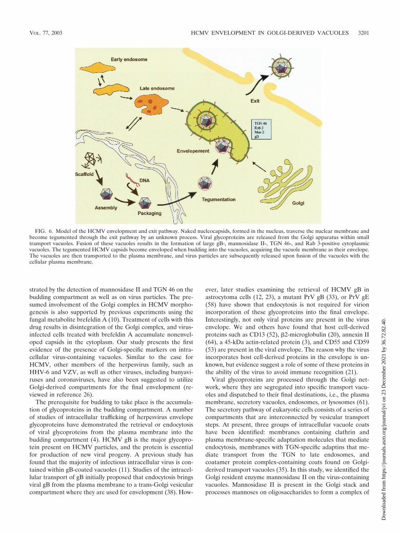
FIG. 6. Model of the HCMV envelopment and exit pathway. Naked nucleocapsids, formed in the nucleus, traverse the nuclear membrane andbecome tegumented through the exit pathway by an unknown process. Viral glycoproteins are released from the Golgi apparatus within smalltransport vacuoles. Fusion of these vacuoles results in the formation of large gB-, mannosidase II-, TGN 46-, and Rab 3-positive cytoplasmicvacuoles. The tegumented HCMV capsids become enveloped when budding into the vacuoles, acquiring the vacuole membrane as their envelope.The vacuoles are then transported to the plasma membrane, and virus particles are subsequently released upon fusion of the vacuoles with thecellular plasma membrane.
VOL. 77, 2003 HCMV ENVELOPMENT IN GOLGI-DERIVED VACUOLES 3201
Dow
nloa
ded
from
http
s://j
ourn
als.
asm
.org
/jour
nal/j
vi o
n 23
Dec
embe
r 20
21 b
y 36
.72.
82.4
0.
oligosaccharides that are resistant to attacks by endoglycosi-dase H. TGN 46 is a trans-Golgi marker that has been indi-cated to play a pivotal role in directing HCMV glycoproteins inthe secretory pathway (61). We further identified Rab 3 on thevirus-containing vacuole, which suggest that the vacuole mayenter a secretory pathway (37, 46). The Rab GTPases, whichbelong to a family of Ras-like enzymes, play key roles in thesecretory pathway and membrane trafficking and have beenused to identify specific vacuole membranes (34). Rab 3 is amarker for secretory vacuoles that are shuttled between theplasma membrane and the trans-Golgi compartment andwould therefore be optimal for the virus to utilize for transportout of the infected cell. In support of this hypothesis, Rab 3Dcycles between TGN 46-positive membranes and the plasmamembrane (61). A study on HSV type 1 in rat dorsal rootganglion neurons identified the Golgi markers giantin, man-nosidase II, and TGN 38 on cytoplasmic vacuoles, intracyto-plasmic virions, and extracellular virions (30), further strength-ening the hypothesis of a trans-Golgi origin of the cytoplasmicvacuoles utilized for HCMV envelopment.
In summary, our observations suggest the following modelfor HCMV assembly in fibroblasts, endothelial cells, andsmooth muscle cells (Fig. 6). Naked nucleocapsids, formed inthe nucleus, traverse the nuclear membrane and become tegu-mented through the exit pathway by an unknown sequentialprocess. Viral glycoproteins accumulate in Rab 3-, TGN 46-,and mannosidase II-positive Golgi-derived vacuoles. The tegu-mented nucleocapsids become enveloped by budding into thegB-, Rab3-, TGN 46-, and mannosidase II-positive vacuoles toacquire their final envelope. The vacuoles are then transportedto the plasma membrane in Rab 3-positive secretory vacuoles,and virus particles are subsequently released from the cells byfusion of the vacuoles with the plasma membrane. Furthermolecular studies have to be performed to reveal the molecu-lar events that govern the budding process of tegumentedHCMV capsids and identify the specific protein-protein inter-actions involved in the envelopment process.
ACKNOWLEDGMENTS
We thank Ingrid Jusinski for skillful assistance in EM sample prep-aration and sectioning, Birger Kristensson for kindly sharing his con-focal microscopy equipment, and Lars Branden for assistance with theconfocal microscopy analysis. We thank Erna Moller and Jay Nelsonfor helpful discussions and Erna Moller for carefully reading themanuscript. We thank Kelley Moreman and Marylin Farquhar, TheUniversity of Georgia, Athens, for providing the antibody against man-nosidase II; G. Jahn and C Sinzger, Department of Medical Virology,University of Tubingen, Tubingen, Germany, for providing the endo-thelium-adapted strain TB40; and Sara Gredmark, Department ofMedicine, Karolinska Institutet, Stockholm, Sweden, for providing theASMC.
This work was supported by grants from the Swedish Medical Re-search Council (12615-01A, 00793-36B, and K2001-16X-12615-04A),the Tobias Foundation (1313/98 and 33/02), the Swedish Children’sCancer Foundation (1998/065 and 01/046), the Swedish Society ofMedicine (1999-02-0347), the Heart and Lung Foundation (1999-41-305), and the Emil and Wera Cornells Foundation. C.S.-N. is a fellowof the Wenner-Gren Foundation, Sweden.
REFERENCES
1. Avitabile, E., P. L. Ward, C. Di Lazzaro, M. R. Torrisi, B. Roizman, and G.Campadelli-Fiume. 1994. The herpes simplex virus UL20 protein compen-sates for the differential disruption of exocytosis of virions and viral mem-brane glycoproteins associated with fragmentation of the Golgi apparatus.J. Virol. 68:7397–7405.
2. Baines, J. D., P. L. Ward, G. Campadelli-Fiume, and B. Roizman. 1991. TheUL20 gene of herpes simplex virus 1 encodes a function necessary for viralegress. J. Virol. 65:6414–6424.
3. Baldick, C. J., Jr., and T. Shenk. 1996. Proteins associated with purifiedhuman cytomegalovirus particles. J. Virol. 70:6097–6105.
4. Brideau, A. D., L. W. Enquist, and R. S. Tirabassi. 2000. The role of virionmembrane protein endocytosis in the herpesvirus life cycle. J. Clin. Virol.17:69–82.
5. Browne, H., S. Bell, T. Minson, and D. W. Wilson. 1996. An endoplasmicreticulum-retained herpes simplex virus glycoprotein H is absent from se-creted virions: evidence for reenvelopment during egress. J. Virol. 70:4311–4316.
6. Cheung, P., B. W. Banfield, and F. Tufaro. 1991. Brefeldin A arrests thematuration and egress of herpes simplex virus particles during infection.J. Virol. 65:1893–1904.
7. Darlington, R. W., and L. H. Moss III. 1969. The envelope of Herpesvirus.Prog. Med. Virol. 11:16–45.
8. Darlington, R. W., and L. H. Moss III. 1968. Herpesvirus envelopment.J. Virol. 2:48–55.
9. Di Lazzaro, C., G. Campadelli-Fiume, and M. R. Torrisi. 1995. Intermediateforms of glycoconjugates are present in the envelope of herpes simplexvirions during their transport along the exocytic pathway. Virology 214:619–623.
10. Eggers, M., E. Bogner, B. Agricola, H. F. Kern, and K. Radsak. 1992.Inhibition of human cytomegalovirus maturation by brefeldin A. J. Gen.Virol. 73:2679–2692.
11. Fish, K. N., W. Britt, and J. A. Nelson. 1996. A novel mechanism forpersistence of human cytomegalovirus in macrophages. J. Virol. 70:1855–1862.
12. Fish, K. N., C. Soderberg-Naucler, and J. A. Nelson. 1998. Steady-stateplasma membrane expression of human cytomegalovirus gB is determined bythe phosphorylation state of Ser900. J. Virol. 72:6657–6664.
13. Fuchs, W., B. G. Klupp, H. Granzow, and T. C. Mettenleiter. 1997. TheUL20 gene product of pseudorabies virus functions in virus egress. J. Virol.71:5639–5646.
14. Fuchs, W., B. G. Klupp, H. Granzow, H. J. Rziha, and T. C. Mettenleiter.1996. Identification and characterization of the pseudorabies virus UL3.5protein, which is involved in virus egress. J. Virol. 70:3517–3527.
15. Garoff, H., R. Hewson, and D. J. Opstelten. 1998. Virus maturation bybudding. Microbiol. Mol. Biol. Rev. 62:1171–1190.
16. Gershon, A. A., D. L. Sherman, Z. Zhu, C. A. Gabel, R. T. Ambron, andM. D. Gershon. 1994. Intracellular transport of newly synthesized varicella-zoster virus: final envelopment in the trans-Golgi network. J. Virol. 68:6372–6390.
17. Gibson, W. 1996. Structure and assembly of the virion. Intervirology 39:389–400.
18. Gong, M., and E. Kieff. 1990. Intracellular trafficking of two major Epstein-Barr virus glycoproteins, gp350/220 and gp110. J. Virol. 64:1507–1516.
19. Granzow, H., F. Weiland, A. Jons, B. G. Klupp, A. Karger, and T. C.Mettenleiter. 1997. Ultrastructural analysis of the replication cycle of pseu-dorabies virus in cell culture: a reassessment. J. Virol. 71:2072–2082.
20. Grundy, J. E., J. A. McKeating, and P. D. Griffiths. 1987. Cytomegalovirusstrain AD169 binds beta 2 microglobulin in vitro after release from cells.J. Gen. Virol. 68:777–784.
21. Hengel, H., W. Brune, and U. H. Koszinowski. 1998. Immune evasion bycytomegalovirus—survival strategies of a highly adapted opportunist. TrendsMicrobiol. 6:190–197.
22. Hensel, G., H. Meyer, S. Gartner, G. Brand, and H. F. Kern. 1995. Nuclearlocalization of the human cytomegalovirus tegument protein pp150(ppUL32). J. Gen. Virol. 76:1591–1601.
23. Jarvis, M. A., K. N. Fish, C. Soderberg-Naucler, D. N. Streblow, H. L.Meyers, G. Thomas, and J. A. Nelson. 2002. Retrieval of human cytomega-lovirus glycoprotein B from cell surface is not required for virus envelopmentin astrocytoma cells. J. Virol. 76:5147–5155.
24. Johnson, D. C., and P. G. Spear. 1982. Monensin inhibits the processing ofherpes simplex virus glycoproteins, their transport to the cell surface, and theegress of virions from infected cells. J. Virol. 43:1102–1112.
25. Johnson, D. C., and P. G. Spear. 1983. O-linked oligosaccharides are ac-quired by herpes simplex virus glycoproteins in the Golgi apparatus. Cell32:987–997.
26. Jones, F., and C. Grose. 1988. Role of cytoplasmic vacuoles in varicella-zoster virus glycoprotein trafficking and virion envelopment. J. Virol. 62:2701–2711.
27. Landini, M. P., B. Severi, L. Badiali, E. Gonczol, and G. Mirolo. 1987.Structural components of human cytomegalovirus: in situ localization of themajor glycoprotein. Intervirology 27:154–160.
28. Massalski, A., M. Coulter-Mackie, R. L. Knobler, M. J. Buchmeier, and S.Dales. 1982. In vivo and in vitro models of demyelinating diseases. V. Com-parison of the assembly of mouse hepatitis virus, strain JHM, in two murinecell lines. Intervirology 18:135–146.
29. Mettenleiter, T. C. 2002. Herpesvirus assembly and egress. J. Virol. 76:1537–1547.
3202 HOMMAN-LOUDIYI ET AL. J. VIROL.
Dow
nloa
ded
from
http
s://j
ourn
als.
asm
.org
/jour
nal/j
vi o
n 23
Dec
embe
r 20
21 b
y 36
.72.
82.4
0.
30. Miranda-Saksena, M., R. A. Boadle, P. Armati, and A. L. Cunningham.2002. In rat dorsal root ganglion neurons, herpes simplex virus type 1 tegu-ment forms in the cytoplasm of the cell body. J. Virol. 76:9934–9951.
31. Morgan, C., H. M. Rose, M. Holden, and E. P. Jones. 1954. Electron micro-scopic observations on the development of the herpes simplex virus. J. Exp.Med. 110:643–656.
32. Nii, S., M. Yoshida, F. Uno, T. Kurata, K. Ikuta, and K. Yamanishi. 1990.Replication of human herpesvirus 6 (HHV-6): morphological aspects. Adv.Exp. Med. Biol. 278:19–28.
33. Nixdorf, R., B. G. Klupp, A. Karger, and T. C. Mettenleiter. 2000. Effects oftruncation of the carboxy terminus of pseudorabies virus glycoprotein B oninfectivity. J. Virol. 74:7137–7145.
34. Novick, P., and P. Brennwald. 1993. Friends and family: the role of the RabGTPases in vesicular traffic. Cell 75:597–601.
35. Pelham, H. R., and S. Munro. 1993. Sorting of membrane proteins in thesecretory pathway. Cell 75:603–605.
36. Pettersson, R. F. 1991. Protein localization and virus assembly at intracellu-lar membranes. Curr. Top. Microbiol. Immunol. 170:67–106.
37. Pfeffer, S. R., A. B. Dirac-Svejstrup, and T. Soldati. 1995. Rab GDP disso-ciation inhibitor: putting rab GTPases in the right place. J. Biol. Chem.270:17057–17059.
38. Radsak, K., M. Eickmann, T. Mockenhaupt, E. Bogner, H. Kern, A. Eis-Hubinger, and M. Reschke. 1996. Retrieval of human cytomegalovirus gly-coprotein B from the infected cell surface for virus envelopment. Arch.Virol. 141:557–572.
39. Risco, C., and J. L. Carrascosa. 1999. Visualization of viral assembly in theinfected cell. Histol. Histopathol. 14:905–926.
40. Rixon, F. J., C. Addison, and J. McLauchlan. 1992. Assembly of envelopedtegument structures (L particles) can occur independently of virion matura-tion in herpes simplex virus type 1-infected cells. J. Gen. Virol. 73:277–284.
41. Roffman, E., J. P. Albert, J. P. Goff, and N. Frenkel. 1990. Putative site forthe acquisition of human herpesvirus 6 virion tegument. J. Virol. 64:6308–6313.
42. Roizman, B. 1996. Herpesviridae, p. 2221–2230. In B. N. Fields, D. M. Knipe,P. M. Howley, R. M. Chanock, J. L. Melnick, T. P Monath, B. Roizman, andS. E. Straus (ed.), Virology, 3rd ed. Lippincott-Raven, Philadelphia, Pa.
43. Sanchez, V., P. C. Angeletti, J. A. Engler, and W. J. Britt. 1998. Localizationof human cytomegalovirus structural proteins to the nuclear matrix of in-fected human fibroblasts. J. Virol. 72:3321–3329.
44. Sanchez, V., K. D. Greis, E. Sztul, and W. J. Britt. 2000. Accumulation ofvirion tegument and envelope proteins in a stable cytoplasmic compartmentduring human cytomegalovirus replication: characterization of a potentialsite of virus assembly. J. Virol. 74:975–986.
45. Sanchez, V., E. Sztul, and W. J. Britt. 2000. Human cytomegalovirus pp28(UL99) localizes to a cytoplasmic compartment which overlaps the endo-plasmic reticulum-Golgi-intermediate compartment. J. Virol. 74:3842–3851.
46. Schimmoller, F., I. Simon, and S. R. Pfeffer. 1998. Rab GTPases, directors ofvesicle docking. J. Biol. Chem. 273:22161–22164.
47. Severi, B., M. P. Landini, and E. Govoni. 1988. Human cytomegalovirusmorphogenesis: an ultrastructural study of the late cytoplasmic phases. Arch.Virol. 98:51–64.
48. Simons, K., and H. Garoff. 1980. The budding mechanisms of envelopedanimal viruses. J. Gen. Virol. 50:1–21.
49. Skepper, J. N., A. Whiteley, H. Browne, and A. Minson. 2001. Herpes simplexvirus nucleocapsids mature to progeny virions by an envelopment3deenvelopment3reenvelopment pathway. J. Virol. 75:5697–5702.
50. Smith, J. D., and E. De Harven. 1973. Herpes simplex virus and humancytomegalovirus replication in WI-38 cells. I. Sequence of viral replication.J. Virol. 12:919–930.
51. Sodeik, B., R. W. Doms, M. Ericsson, G. Hiller, C. E. Machamer, W. van ’tHof, G. van Meer, B. Moss, and G. Griffiths. 1993. Assembly of vacciniavirus: role of the intermediate compartment between the endoplasmic retic-ulum and the Golgi stacks. J. Cell Biol. 121:521–541.
52. Soderberg, C., T. D. Giugni, J. A. Zaia, S. Larsson, J. M. Wahlberg, and E.Moller. 1993. CD13 (human aminopeptidase N) mediates human cytomeg-alovirus infection. J. Virol. 67:6576–6585.
53. Spear, G. T., N. S. Lurain, C. J. Parker, M. Ghassemi, G. H. Payne, and M.Saifuddin. 1995. Host cell-derived complement control proteins CD55 andCD59 are incorporated into the virions of two unrelated enveloped viruses,human T cell leukemia/lymphoma virus type I (HTLV-I) and human cyto-megalovirus (HCMV). J. Immunol. 155:4376–4381.
54. Spengler, M., N. Niesen, C. Grose, W. T. Ruyechan, and J. Hay. 2001.Interactions among structural proteins of Varicella-zoster virus. Arch. Virol.17(Suppl.):71–79.
55. Stackpole, C. W. 1969. Herpes-type virus of the frog renal adenocarcinoma.I. Virus development in tumor transplants maintained at low temperature.J. Virol. 4:75–93.
56. Stephens, E. B., and R. W. Compans. 1988. Assembly of animal viruses atcellular membranes. Annu. Rev. Microbiol. 42:489–516.
57. Steven, A. C., and P. G. Spear. 1997. Herpesvirus capsid assembly andenvelopment, p. 312–351. In W. Chiu, R. M. Burnett, and R. Garcea (ed.),Structural biology of viruses. Oxford University Press, New York, N.Y.
58. Tirabassi, R. S., and L. W. Enquist. 1998. Role of envelope protein gEendocytosis in the pseudorabies virus life cycle. J. Virol. 72:4571–4579.
59. Tokuyasu, K. T. 1973. A technique for ultracryotomy of cell suspensions andtissues. J. Cell Biol. 57:551–565.
60. Tooze, J., M. Hollinshead, B. Reis, K. Radsak, and H. Kern. 1993. Progenyvaccinia and human cytomegalovirus particles utilize early endosomal cister-nae for their envelopes. Eur. J. Cell Biol. 60:163–178.
61. Traub, L. M., and S. Kornfeld. 1997. The trans-Golgi network: a late secre-tory sorting station. Curr. Opin. Cell Biol. 9:527–533.
62. Velasco, A., L. Hendricks, K. W. Moremen, D. R. Tulsiani, O. Touster, andM. G. Farquhar. 1993. Cell type-dependent variations in the subcellulardistribution of alpha-mannosidase I and II. J. Cell Biol. 122:39–51.
63. Wentworth, B. B., and L. French. 1970. Plaque assay of cytomegalovirusstrains of human origin. Proc. Soc. Exp. Biol. Med. 135:253–258.
64. Wright, J. F., A. Kurosky, E. L. Pryzdial, and S. Wasi. 1995. Host cellularannexin II is associated with cytomegalovirus particles isolated from culturedhuman fibroblasts. J. Virol. 69:4784–4791.
65. Zhu, Z., M. D. Gershon, Y. Hao, R. T. Ambron, C. A. Gabel, and A. A.Gershon. 1995. Envelopment of varicella-zoster virus: targeting of viral gly-coproteins to the trans-Golgi network. J. Virol. 69:7951–7959.
66. Zhu, Z., Y. Hao, M. D. Gershon, R. T. Ambron, and A. A. Gershon. 1996.Targeting of glycoprotein I (gE) of varicella-zoster virus to the trans-Golginetwork by an AYRV sequence and an acidic amino acid-rich patch in thecytosolic domain of the molecule. J. Virol. 70:6563–6575.
VOL. 77, 2003 HCMV ENVELOPMENT IN GOLGI-DERIVED VACUOLES 3203
Dow
nloa
ded
from
http
s://j
ourn
als.
asm
.org
/jour
nal/j
vi o
n 23
Dec
embe
r 20
21 b
y 36
.72.
82.4
0.