enthandanthdomainproteinsparticipateinap2-independent ... · research article...
TRANSCRIPT

RESEARCH ARTICLE
ENTH and ANTH domain proteins participate in AP2-independentclathrin-mediated endocytosisPaul T. Manna1,*, Catarina Gadelha2, Amy E. Puttick1 and Mark C. Field3,‡
ABSTRACTClathrin-mediated endocytosis (CME) is a major route of entry intoeukaryotic cells. A core of evolutionarily ancient genes encodesmany components of this system but much of our mechanisticunderstanding of CME is derived from a phylogenetically narrowsampling of a few model organisms. In the parasite Trypanosomabrucei, which is distantly related to the better characterised animalsand fungi, exceptionally fast endocytic turnover aids its evasion of thehost immune system. Although clathrin is absolutely essential for thisprocess, the adaptor protein complex 2 (AP2) has been secondarilylost, suggesting mechanistic divergence. Here, we characterise twophosphoinositide-bindingmonomeric clathrin adaptors, T. brucei (Tb)EpsinR and TbCALM, which in trypanosomes are represented bysingle genes, unlike the expansions present in animals and fungi.Depletion of these gene products reveals essential, but partiallyredundant, activities in CME. Ultrastructural analysis of TbCALM andTbEpsinR double-knockdown cells demonstrated severe defectsto clathrin-coated pit formation and morphology associated with adramatic inhibition of endocytosis. Depletion of TbCALM alone,however, produced a distinct lysosomal segregation phenotype,indicating an additional non-redundant role for this protein. Therefore,TbEpsinR and TbCALM represent ancient phosphoinositide-bindingproteins with distinct and vital roles in AP2-independent endocytosis.
KEY WORDS: Endocytosis, Evolution, Phosphoinositide,Trafficking, Trypanosome
INTRODUCTIONEndocytosis is an essential cellular process in eukaryotes. In animalcells, multiple endocytic pathways exist, but the best understoodrequires the vesicle coat protein clathrin. Intensive study in bothanimal and yeast systems has identified many, if not most, of theproteins involved in clathrin-mediated endocytosis (CME) (e.g.Borner et al., 2006; Borner et al., 2012). CME is evolutionarilyancient, with components of this pathway found throughout theeukaryotes, and most notably clathrin itself, with the exceptionof the extremely reduced genomes of microsporidia (Barlow et al.,2014), is present in all eukaryotic genomes so far examined.Recruitment of clathrin to the plasma membrane, concentration of
endocytic cargo and clathrin-coated pit (CCP) formation all dependon an array of accessory proteins or adaptors. The heterotetramericadaptor protein complex 2 (AP2) binds clathrin and functions as acentral adaptor hub in CME, mediating membrane recruitmentthrough phosphatidylinositol 4,5-bisphosphate [PtdIns(4,5)P2]binding, and cargo selection by recognition of endocytic motifson cargo proteins (e.g. [D/E]xxxL[LI] or Yxxɸ, where ɸ denotes alarge hydrophobic residue) (Lafer, 2002; Schmid et al., 2006;Jackson et al., 2010; McMahon and Boucrot, 2011). In animal cells,depletion of the AP2 complex is sufficient to block CCP formation,suggesting that CME is indeed dependent on AP2 (Boucrot et al.,2010).
Outside metazoa, the AP2 complex is consistently found tolocalisewith clathrin at the plasmamembrane (Elde et al., 2005), butits requirement in CME is less clear. Early studies in yeast found nosignificant endocytic defect upon depletion of the AP2 complex(Yeung et al., 1999; Huang et al., 1999), suggesting that there wasno absolute requirement for AP2 in CME.More recently, a potentialrole for the AP2 complex as a cargo-specific adaptor was identifiedin a yeast genetic screen for resistance to an endocytosed toxin(Carroll et al., 2009). However, the scale of involvement of AP2 inyeast CME appears to be highly reduced compared to animals.Similarly, studies in Dictyostelium find association of AP2 withclathrin structures at the plasma membrane (Sosa et al., 2012; Macroet al., 2012) but little effect of AP2 depletion on CME (Macro et al.,2012).
In addition to the heterotetrameric adaptor protein complexes,numerous monomeric clathrin adaptors are known. Among thesethe most widely conserved are phosphatidylinositol phosphate(PtdInsP)-binding proteins containing epsin N-terminal homology(ENTH) and AP180 N-terminal homology (ANTH) domains. TheENTH domain is present in metazoan epsins (Eps15-interacting)and EpsinR (epsin-related) proteins (CLINT1 in humans) alongwith their yeast counterparts ENT1–ENT5. The ANTH domain isfound in the clathrin assembly lymphoid myeloid leukaemia protein(CALM, also known as PICALM) and its neuronal-specifichomologue, adaptor protein of 180 kDa (AP180, also known asSNAP91), as well as in huntingtin-interacting protein 1 (HIP1) andHIP-related (HIP1R), and their yeast counterparts YAP180-1 andYAP180-2 and SLA2. In yeast and animals, the epsins function inCME, whereas epsinR localises primarily to the Golgi complex andmediates trafficking between endosomes and the trans-Golginetwork (TGN) (Kalthoff et al., 2002; Mills et al., 2003; Hirstet al., 2003). Outside of the opisthokonts, EpsinR is the solerepresentative of this protein family and in Trypanosoma bruceiplays a role in endocytosis from the plasma membrane (Gabernet-Castello et al., 2009). Likewise, CALM and AP180 represent theancestral ANTH domain proteins, with a single homologue presentin most eukaryotes (De Craene et al., 2012). AP180 has the ability torecruit clathrin to lipid bilayers in vitro (Ford et al., 2001) but againhomologues are dispensable for CME in many organisms. In yeast,Received 16 December 2014; Accepted 13 April 2015
1Department of Pathology, University of Cambridge, Tennis Court Road,Cambridge CB2 1QP, UK. 2School of Life Sciences, University of Nottingham,Queen’s Medical Centre, Nottingham NG7 2UH, UK. 3Division of BiologicalChemistry and Drug Discovery, University of Dundee, Dundee DD1 5EH, UK.*Present address: Cambridge Institute for Medical Research, University ofCambridge, Cambridge CB2 0XY, UK.
‡Author for correspondence ([email protected])
This is an Open Access article distributed under the terms of the Creative Commons AttributionLicense (http://creativecommons.org/licenses/by/3.0), which permits unrestricted use,distribution and reproduction in any medium provided that the original work is properly attributed.
2130
© 2015. Published by The Company of Biologists Ltd | Journal of Cell Science (2015) 128, 2130-2142 doi:10.1242/jcs.167726
Journal
ofCe
llScience

simultaneous depletion of EPN1 and EPN2, and both the AP180 orCALM homologues (YAP180-1 and YAP180-2) is sufficient toblock CME, suggesting a potential mechanism for AP2-independent CME involving redundant functions of ENTH andANTH domain proteins (Maldonado-Báez et al., 2008).Trypanosoma brucei, the causative agent of sleeping sickness
and nagana, is unusual in its ability to thrivewithin the mammalianbloodstream, successfully evading the host immune response.Key to this success is the dense protective surface coat ofglycosylphosphatidylinositol (GPI)-anchored variant surfaceglycoprotein (VSG). Regular antigenic variation of the VSG coatis coupled to extremely rapid endocytosis and removal of boundhost antibodies, allowing the parasite to reach extremely highlevels of parasitaemia (Barry andMcCulloch, 2001; Engstler et al.,2007; Manna et al., 2014). The high endocytic flux of bloodstreamform T. brucei depends entirely upon CME, which is the solemechanism of entry into the endomembrane system and is essentialto parasite viability (Allen et al., 2003). Although T. brucei has agenerally conventional endomembrane system, the molecularmechanisms underlying the earliest events in CCP formationappear highly divergent, and in particular T. brucei and its closerelatives have dispensed entirely with the AP2 complex (Berrimanet al., 2005; Field et al., 2007; Manna et al., 2013). A recentproteomic survey of clathrin-interacting proteins identified acohort of trypanosomatid-specific proteins, suggesting dynamic
evolution of this system (Adung’a et al., 2013), although there isclear maintenance of several conserved features, such as theinvolvement of EpsinR (Gabernet-Castello et al., 2009). Inaddition to EpsinR (Tb927.11.670), the trypanosome genomeencodes a single ANTH domain protein, T. brucei (Tb)CALM(Tb927.11.4800), which also retains features suggestive of a rolein CME. Here, we examine the contributions of TbEpsinR andTbCALM to endocytic activity in T. brucei and, by extension,further characterise the clearest example of wholly AP2-independent clathrin-mediated endocytosis.
RESULTSA reduced clathrin adaptor gene cohort in T. bruceiPrevious studies have provided a good understanding of the earlyemergence of CME and the broad conservation of many genefamilies involved. From these studies it emerged that T. bruceishows an unusual frequency of secondary loss of CME genes, likelythe result of selective pressure for very rapid endocytosis (Fieldet al., 2007). Owing to recent advances in our understanding of theearly stages of clathrin-coated vesicle formation and increasedavailability of eukaryotic genomes, we revisited and extended theseearlier analyses, focussing on genes involved in clathrin recruitmentand cargo selection (Fig. 1; supplementary material Table S1).
As previously reported, the T. brucei genome lacks all AP2complex subunits, as well as proposed endocytic initiators EPS15
Fig. 1. Phylogenetic distribution of early acting CME partners. Coulson plot demonstrating presence or absence of genes encoding early acting clathrin-associated proteins across a range of eukaryotes. Filled circles indicate genes identified with high confidence, open circles indicate genes not found, thegrey circle indicates a gene identified with low confidence. Rows are taxa and columns are predicted proteins. Supergroups are coloured for clarity. A conservedcore machinery is apparent, with an exceptional level of gene loss in T. brucei. Plot generated using Coulson Plot Generator (Field et al., 2013). CHC, clathrinheavy chain; CLC, clathrin light chain; β-arr, arrestin β.
2131
RESEARCH ARTICLE Journal of Cell Science (2015) 128, 2130-2142 doi:10.1242/jcs.167726
Journal
ofCe
llScience
and EPS15R (also known as EPS15L1 in mammals) and muniscinfamily proteins (Field et al., 2007; Manna et al., 2013). The loss ofEps15 in particular occurred at the base of the kinetoplastids andsuggests divergent mechanisms for endocytic pit initiationthroughout this clade. The precise role of the muniscins in CCPformation remains a matter of debate; however, it is worth notingthat true muniscins, that is proteins having both a mu homology anda BAR domain, are restricted to the opisthokonts (animals andfungi). Recently, the muniscin family has been shown to be theopisthokont-specific remnant of an ancient pan-eukaryotic adaptor-related protein complex named TSET, for which a detailedphylogenetic analysis has been reported (Hirst et al., 2014). Ouranalysis also highlights for the first time a further pan-eukaryoticCME component in the NECAP gene family. NECAP is also lostfrom the trypanosomatids, an event predating the loss of the AP2complex from salivarian trypanosomes, hinting at potentialrelaxation or alterations of AP2 function preceding its loss.Importantly, this analysis supports previous suggestions thatT. brucei has an unusually reduced cohort of endocytic clathrinadaptor and accessory genes, being restricted to one CALM and oneEpsinR homologue (Field et al., 2007).
TbCALM sequence conservation and endocytic localisationAlthough the trypanosome EpsinR homologue has been studied insome detail (Gabernet-Castello et al., 2009), the trypanosome
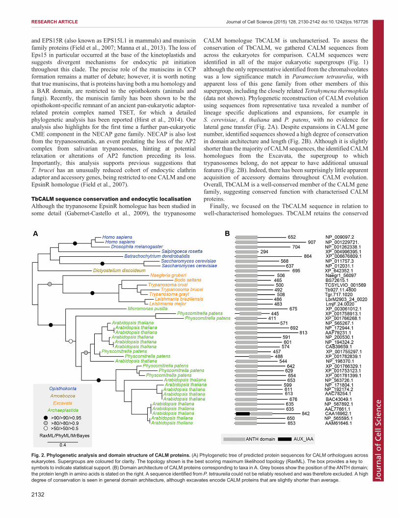
CALM homologue TbCALM is uncharacterised. To assess theconservation of TbCALM, we gathered CALM sequences fromacross the eukaryotes for comparison. CALM sequences wereidentified in all of the major eukaryotic supergroups (Fig. 1)although the only representative identified from the chromalveolateswas a low significance match in Paramecium tetraurelia, withapparent loss of this gene family from other members of thissupergroup, including the closely related Tetrahymena thermophila(data not shown). Phylogenetic reconstruction of CALM evolutionusing sequences from representative taxa revealed a number oflineage specific duplications and expansions, for example inS. cerevisiae, A. thaliana and P. patens, with no evidence forlateral gene transfer (Fig. 2A). Despite expansions in CALM genenumber, identified sequences showed a high degree of conservationin domain architecture and length (Fig. 2B). Although it is slightlyshorter than the majority of CALM sequences, the identified CALMhomologues from the Excavata, the supergroup to whichtrypanosomes belong, do not appear to have additional unusualfeatures (Fig. 2B). Indeed, there has been surprisingly little apparentacquisition of accessory domains throughout CALM evolution.Overall, TbCALM is a well-conserved member of the CALM genefamily, suggesting conserved function with characterised CALMproteins.
Finally, we focused on the TbCALM sequence in relation towell-characterised homologues. TbCALM retains the conserved
Fig. 2. Phylogenetic analysis and domain structure of CALM proteins. (A) Phylogenetic tree of predicted protein sequences for CALM orthologues acrosseukaryotes. Supergroups are coloured for clarity. The topology shown is the best scoring maximum likelihood topology (RaxML). The box provides a key tosymbols to indicate statistical support. (B) Domain architecture of CALM proteins corresponding to taxa in A. Grey boxes show the position of the ANTH domain;the protein length in amino acids is stated on the right. A sequence identified from P. tetraurelia could not be reliably resolved and was therefore excluded. A highdegree of conservation is seen in general domain architecture, although excavates encode CALM proteins that are slightly shorter than average.
2132
RESEARCH ARTICLE Journal of Cell Science (2015) 128, 2130-2142 doi:10.1242/jcs.167726
Journal
ofCe
llScience

domain architecture, consisting of an N-terminal ANTHdomain and a predicted disordered C-terminal domain (Fig. 3A).Within the ANTH domain a PtdIns(4,5)P2-binding motif is locatedin the loop joining helices 1 and 2. Sequence alignment ofTbCALM against several representative homologues demonstratesconservation of key residues within the PtdIns(4,5)P2-bindingmotif (Fig. 3B), suggesting conserved PtdIns(4,5)P2-bindingactivity for TbCALM. Furthermore, a trypanosome clathrin-binding motif, earlier identified in the clathrin-bindingTbEpsinR (Gabernet-Castello et al., 2009) is also found withinthe C-terminal disordered region of TbCALM, suggesting a rolein CME.
For localisation of TbCALM, the gene was tagged at itsendogenous locus with either three HA epitopes in tandem or GFP(Fig. 3; supplementary material Fig. S1). Both tags indicated thatTbCALM is restricted to the flagellar pocket region of the cell. Theflagellar pocket is a functional specialization of the trypanosomesurface membrane at the base of its flagellum, where all endocytosisand exocytosis take place. Flagellar pocket localization wasdemonstrated by colocalisation with a fluorescently labelled lectin,concanavalin A (ConA), a widely used marker for endocytosis inT. brucei, which accumulates in the flagellar pocket lumen andmembrane when incubated with cells at 4°C (Fig. 3C). The locationof TbCALM differs from the distribution of TbEpsinR, which
Fig. 3. Conservation of key sequence features and endocytic localisation in TbCALM. (A) Overview of CALM and AP180 proteins from human (Hs), yeast(Saccharomyces cerevisiae, Sc) and T. brucei. The extent of the ANTH domain is indicated (black box) along with the distribution of putative binding sites forclathrin and adaptors (black and grey bars). Clathrin and adaptor sites are well conserved along with general domain architecture. (B) Multiple sequencealignment of CALM proteins from across eukaryotes. Residues highlighted in dark grey are conserved in over 80% of the included sequences, light grey indicatesgreater than 60% conservation. Taxa are indicated to the left and horizontal bars above the sequence indicate the positions of secondary structural features (Fordet al., 2001). Residues identified as important for PtdIns(4,5)P2 binding are denoted with asterisks below the sequence. PtdIns(4,5)P2-binding residues are wellconserved. (C) Immunofluorescence localisation of endogenous-locus-tagged TbCALM–GFP in bloodstream form T. brucei. Incubation with ConA at 4°Cspecifically labels the flagellar pocket (green); anti-GFP staining (red) shows TbCALM–GFP colocalised with the flagellar pocket, consistent with an endocyticfunction. DNA is stained with DAPI to show the nucleus and kinetoplast (blue).
2133
RESEARCH ARTICLE Journal of Cell Science (2015) 128, 2130-2142 doi:10.1242/jcs.167726
Journal
ofCe
llScience

resides more extensively throughout the endocytic system, althoughsome of the protein is associated with clathrin-coated structures atand near the pocket (Gabernet-Castello et al., 2009).
PtdInsP-dependant membrane targeting of TbCALM andTbEpsinRWhilst the phosphoinositide composition of the bloodstream formtrypanosome flagellar pocket is unknown, a recent reportdemonstrated enrichment of PtdIns(4,5)P2 at the flagellar pocketof the insect stage parasite (Demmel et al., 2014). TbEpsinRand TbCALM show conservation of residues important forphosphoinositide binding, suggesting similar membrane targetingmechanisms to their opisthokont orthologues (Fig. 2; Gabernet-Castello et al., 2009).To assess the potential phosphoinositide-dependent membrane
targeting of trypanosome ENTH and ANTH domain proteins,mammalian expression constructs were made encoding TbCALMor TbEpsinR with C-terminal GFP fusions. TbCALM–GFPexpressed in COS-7 cells gave a largely perinuclear distributionwith some clear plasma membrane association (Fig. 4). TbEpsinR–GFP displayed more pronounced plasma membrane association,together with small cytoplasmic punctae (Fig. 4) colocalising withclathrin heavy chain, suggesting a location at clathrin-coatedvesicles (supplementary material Fig. S2). The plasma membranelocalisation of both proteins is suggestive of an interaction withPtdIns(4,5)P2 in line with the conserved PtdIns(4,5)P2-binding siteseen in TbCALM, but is somewhat unexpected for TbEpsinR,because its mammalian homologue shows selectivity for PtdIns(4)P
and a more perinuclear distribution (Kalthoff et al., 2002; Millset al., 2003; Hirst et al., 2003).
To directly address the role of PtdIns(4,5)P2 in the plasmamembrane recruitment of both TbCALM–GFP and TbEpsinR–GFP,we employed a rapamycin-inducible dimerisation system, leading tomembrane recruitment of an inositol polyphosphate 5-phosphatase(Inp45p), which selectively cleaves the phosphate at the 5 position ofplasma membrane PtdIns(4,5)P2 following rapamycin addition(Suh et al., 2006). As a control, we also expressed the pleckstrinhomology (PH) domain from phospholipase Cδ fused to GFP(PLCδ-PH–GFP). As expected the PLCδ-PH–GFP construct showedplasmamembrane localisation in the absence of rapamycin (Fig. 4C).When co-expressed with the membrane targeting Lyn11–FRBconstruct and CFP–Inp45p-FKBP, PLCδ-PH–GFP dissociated fromthe membrane following rapamycin treatment and PtdIns(4,5)P2depletion (Fig. 4C). Similarly, rapamycin treatment led to a loss ofboth TbCALM–GFP and TbEpsinR–GFP from the plasmamembrane (Fig. 4AB), supporting a specific role for PtdIns(4,5)P2in their membrane recruitment. It is, however, likely that additionalfactors control the distribution of these two proteins in T. brucei astheir localisations are distinct (Fig. 3 and Gabernet-Castello et al.,2009).
TbCALM and TbEpsinR are essential in T. bruceiWe next generated transgenic cell lines in which RNA interference(RNAi)-mediated depletion of TbCALM and TbEpsinR(individually or together) could be induced by the addition oftetracycline. Induction of RNAi in these cells causes a reduction inmRNA levels [assessed by quantitative real-time PCR (qRT-PCR);Fig. 5A] and an inhibition of proliferation (Fig. 5B). Theproliferative defect caused by silencing of TbCALM and/orTbEpsinR is accompanied by a marked cytokinesis block, as hasbeen seen previously with many gene products involved intrafficking, (Fig. 5C,D). TbCALM-knockdown (KD) cellsundergo several rounds of organelle duplication and accumulatemore than two nuclei and kinetoplasts (the region of thetrypanosome single mitochondrion that contains all mtDNA) – anabnormal state of the cell cycle termed >2N2K (Fig. 5C,D). Incontrast, TbEpsinR-KD cells show a block in cytokinesis after oneround of mitosis, accumulating mostly two nuclei and twokinetoplasts only (2K2N). Interestingly, silencing of both geneproducts mimics the effect seen for depletion of TbEpsinR(Fig. 5C), suggesting that the absence of TbEpsinR preventsadditional rounds of organelle replication.
TbCALM and TbEpsinR function synergistically in clathrin-mediated endocytosisDepletion of the clathrin heavy chain in T. brucei leads to a completeinhibition of endocytosis accompanied by a dramatic expansion ofthe flagellar pocket, a useful morphological marker for endocyticdysfunction in T. brucei (Allen et al., 2003). Therefore, to assess theeffects of TbCALM and TbEpsinR depletion upon CME, we firstexamined the distribution of clathrin heavy chain and theappearance of enlarged flagellar pockets at 48 h post-RNAiinduction. In control cells, clathrin antisera labels tubulovesicularstructures arranged between the cell posterior and the nucleus(Fig. 6A), the region containing the entire endosomal system(Morgan et al., 2001; Allen et al., 2003). A subset of clathrin-positive structures are found close to the kinetoplast. This cluster ofclathrin punctae represents structures at, or near to, the flagellarpocket, likely newly forming CCPs or early endocytic vesicles stillbearing a clathrin coat. TbCALM depletion had little effect on the
Fig. 4. PtdIns(4,5)P2 dependence of TbEpsinR and TbCALM membranetargeting. COS-7 cells grown on glass coverslips were transfected with (A)TbCALM–GFP, (B) TbEpsinR–GFP or (C) PLCδ-PH–GFP alone (left panels)or together with a bipartite, rapamycin inducible, PtdIns(4,5)P2 depletionsystem (centre and right panels). Addition of rapamycin (10 μM) (right panels)leads to specific depletion of plasma membrane PtdIns(4,5)P2, confirmed bythe dissociation of PLCδ-PH–GFP from the plasma membrane (C). Insets aremagnified images of the boxed regions encompassing plasma membrane.Scale bar: 10 μm. Both TbEpsinR and TbCALM show rapamycin-inducedmembrane dissociation consistent with some PtdIns(4,5)P2-binding activity.
2134
RESEARCH ARTICLE Journal of Cell Science (2015) 128, 2130-2142 doi:10.1242/jcs.167726
Journal
ofCe
llScience

distribution of clathrin (Fig. 6A), even in severely deformed cellsarising from the generalised cytokinesis block (data not shown).Effects of TbEpsinR depletion on clathrin distribution were subtleand in keeping with our previous study (Gabernet-Castello et al.,2009), as it became less clearly tubulovesicular in nature and morediffuse or cytosolic (Fig. 6A). Following depletion of bothTbEpsinR and TbCALM together a remarkable enlargement ofthe flagellar pocket (visible as phase-light vacuoles at the cellposterior) was observed. Enlarged flagellar pockets were seen in
over 40% of double KD cells versus 16% and 7% for TbEpsinR andTbCALM single KDs, respectively. Note, however, that theseobserved proportions are likely an underestimate of the totalphenotypic penetrance, as this effect is rapidly lethal, therebyremoving severely affected cells from the population.
Flagellar pocket enlargement is a good, although indirect,indicator of endocytic inhibition. To assay this directly, weexamined the uptake of two fluorescein-labelled endocyticmarkers, transferrin and ConA. In trypanosomes, transferrin is
Fig. 5. Proliferative and morphological defects following TbCALM and TbEpsinR depletion. (A) qRT-PCR analysis of TbCALM and TbEpsinR mRNAexpression following 48 h of RNAi induction (+Tet.) in single marker bloodstream-form cells harbouring RNAi constructs targeting either TbCALM, TbEpsinR orboth TbCALM and TbEpinR together (DKD). mRNA levels, normalised to β-tubulin, are expressed as relative expression compared to non-induced cells. Dataare expressed as mean±s.e.m. from three independent inductions. (B) Proliferation rate of RNAi-induced cells normalised to non-induced cells. Data are mean±s.e.m. from three independent inductions. (C) Cell cycle analysis following 48 h of RNAi induction. xK, number of kinetoplasts; yN, number of nuclei. Controlis a parental cell line, shown as black bars; TbCALM RNAi is shown as white bars; TbEpsinR RNAi is dark grey bars; double RNAi is light-grey bars. Data aremean±s.d. from two independent inductions with at least 100 cells counted per specimen per induction. (D) Morphology following depletion of TbCALM and/orTbEpsinR. DIC images of cells after 48 h of RNAi induction; DNA is stained with DAPI (blue).
2135
RESEARCH ARTICLE Journal of Cell Science (2015) 128, 2130-2142 doi:10.1242/jcs.167726
Journal
ofCe
llScience

taken up by a unique heterodimeric, GPI-anchored transferrinreceptor (TbTfR), which has no homology to the mammalianreceptor (Steverding, 2000). After a 45-min exposure to transferrin–FITC, control cells showed extensive uptake of the fluorophore, withseveral punctae visible between the nucleus and the kinteoplast(Fig. 6B,C). Depletion of either TbEpsinR or TbCALM had only asmall effect on transferrin uptake, whereas depletion of both togethercaused a strong reduction. To rule out specific effects upon TbTfRtrafficking, we examined the uptake of the mannose-binding lectinConA. At the trypanosome surface the major mannose-containingmolecule is VSG, which owing to its high packing density is likelyendocytosed non-concentratively through bulk uptake (Grünfelder
et al., 2003). Thus FITC–ConA uptake reports general clathrin-mediated endocytic activity. As seen for transferrin–FITC uptake,depletion of both TbEpsinR and TbCALM together led to a greaterinhibition of FITC–ConA uptake than depletion of either proteinalone (Fig. 6D,E).
When endocytosis is inhibited in trypanosomes, ConA stillaccumulates within the flagellar pocket. Following TbCALM andTbEpsinR co-depletion, the ConA–FITC signal was frequentlyrestricted to a single puncta adjacent to the kinetoplast, suggestingthat uptake might be stalled at the flagellar pocket. To test this,we examined the delivery of endocytosed ConA–FITC to laterendosomal compartments. After depletion of either TbCALM or
Fig. 6. Endocytic inhibition following TbCALM and TbEpsinR depletion. (A) Immunofluorescence analysis of clathrin heavy chain distribution (green) inparental (control) and TbCALM, TbEpsinR or double RNAi cell lines (DKD) after 48 h post-induction. Blue indicates DNA DAPI-stain. Note phase-light vacuolarstructures reminiscent of the big-eye phenotype observed following clathrin heavy chain depletion (Allen et al., 2003). (B) Uptake of Alexa-Fluor-488-conjugatedtransferrin in parental (control) and TbCALM, TbEpsinR or double (DKD) RNAi cell lines at 48 h post-induction. (C) Quantification of transferrin as in B. Endocyticinhibition following knockdown of TbEpsinR and TbCALM appears additive, suggestive of some redundancy. Results are mean±s.e.m. (n=50). (D) Uptake ofFITC-conjugated ConA in parental and TbCALM, TbEpsinR or double (DKD) RNAi cell lines after 48 h post-induction. (E) Quantification of ConA uptake shownin D. Results are mean±s.e.m. (n=50). Co-depletion of TbCALM and TbEpsinR leads to greater inhibition of ConA uptake than depletion of either protein alone.(F) Lysosomal (p67, red) delivery of ConA in knockdown cell lines after 48 h RNAi induction. Whereas uptake is reduced (D,E), ConA trafficking to the lysosomeappears largely unaffected in either TbCALM or TbEpsinR knockdown cells compared to parental control. However, some distension of the p67-positivecompartment is apparent following TbCALM depletion. In contrast, ConA uptake appears largely stalled at the flagellar pocket region in the double knockdown.(G) Quantification of ConA and p67 colocalisation following 48 h RNAi induction. Co-depletion of TbCALM and TbEpsinR greatly reduces lysosomal delivery ofConA. Results are mean±s.e.m. (n=50). Scale bars: 2 μm.
2136
RESEARCH ARTICLE Journal of Cell Science (2015) 128, 2130-2142 doi:10.1242/jcs.167726
Journal
ofCe
llScience

TbEpsinR alone, endocytosed ConA largely colocalised with p67,indicative of a presence in lysosomes, whereas depletion of bothTbCALM and TbEpsinR together greatly reduced the colocalisationof ConA and p67, supporting a block of ConA uptake from theflagellar pocket (Fig. 6F,G). Interestingly, given the role of CALMand AP180 in vacuolar maintenance in D. discoideum (Stavrou andO’Halloran, 2006), in many CALM-depleted cells the p67-labelledcompartment appeared distended when compared to control cells(Fig. 6F).
ENTH and ANTH protein depletion affects flagellar pocketfunctionTo further investigate the roles of TbCALM and TbEpsinR in earlyclathrin recruitment and vesicle formation, we examined RNAi celllines by fast isothermal fixation and transmission electronmicroscopy. The most apparent gross abnormality of cellsdepleted of the ENTH and ANTH proteins was the significant
increase in flagellar pocket size (Fig. 7A,C). This is consistent withthe compromised uptake of transferrin and ConA (Fig. 6).Interestingly, the endocytic block is not caused by a failure inrecruiting clathrin per se: clathrin-coated structures resemblingthose from control cells are visibly associated with the membrane ofenlarged pockets (Fig. 7C–E), either as CCPs or flat lattices (CCLs)(Fig. 7F). There was, however, a reduction in CCP density seenacross all RNAi cell lines (Fig. 7D). Close inspection of themorphology of clathrin-coated structures showed aberrant coated pitmorphology (Fig. 7B). These defects were less severe and lesscommonly observed in TbCALM-depleted cells, where pits werestill formed, although with irregular morphologies (Fig. 7B), whichis qualitatively similar to the defects reported following CALMdepletion inmammalian cells (Sahlender et al., 2013). In TbEpsinR-depleted cells, the clathrin-coated membrane regions no longerresembled true pits and instead formed large flat coated areasshowing modest curvature at their peripheries (Fig. 7B). This defect
Fig. 7. Defects at the flagellar pocket following ENTH and ANTH protein depletion. (A) Representative electron micrographs showing the great increase inflagellar pocket diameter following depletion of TbEpsinR, TbCALM or both together. (B) Representative micrographs of altered morphology of clathrin-coatedstructures in RNAi induced cells. Black arrowheads indicate clathrin assembly. (C–E) Bar graphs depict morphometric analysis for each cell line from culturesafter 48 h with or without tetracycline. Results are mean±s.e.m. (n=25); *P≤0.01, **P≤0.002, ***P≤0.0002.
2137
RESEARCH ARTICLE Journal of Cell Science (2015) 128, 2130-2142 doi:10.1242/jcs.167726
Journal
ofCe
llScience
was accentuated in tandem knockdown cells, with many instancesof extremely large flat clathrin-coated areas (Fig. 7B). Indeed in thetandem knockdown cells, the large accumulation of these aberrantstructures was reflected in an overall increase in the amount ofcoated membrane per section (Fig. 7E).The Golgi remained largely unaffected, despite the expected
importance of clathrin and TbEpsinR to its maintenance(supplementary material Fig. S3). Whereas superficially thisresult argues against a role for ENTH and ANTH domainproteins at the trypanosome Golgi, it is possible that the extremedependence of bloodstream-form parasites on clathrin function atthe flagellar pocket leads to cell death prior to an observable impacton Golgi morphology. Similarly, ablation of clathrin or clathrin-associated proteins (TbCAPs) in bloodstream-form parasites doesnot cause any obvious Golgi phenotype, whereas depletion in theless endocytically active insect form is associated with Golgihypertrophy (Allen et al., 2003; Adung’a et al., 2013).
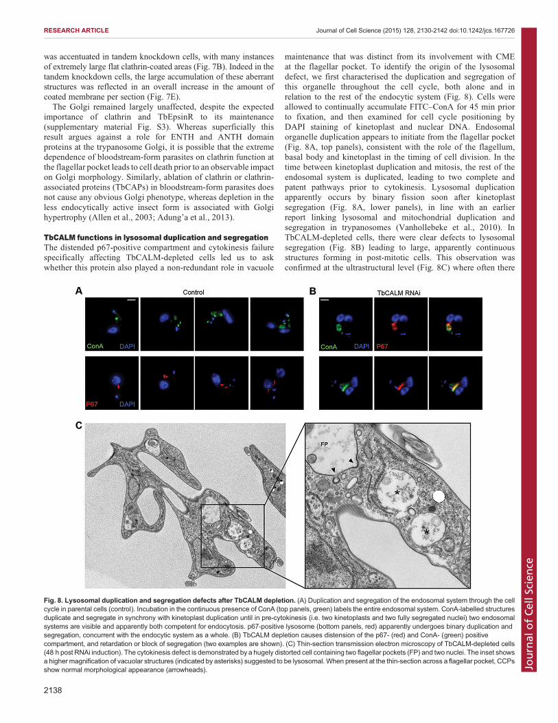
TbCALM functions in lysosomal duplication and segregationThe distended p67-positive compartment and cytokinesis failurespecifically affecting TbCALM-depleted cells led us to askwhether this protein also played a non-redundant role in vacuole
maintenance that was distinct from its involvement with CMEat the flagellar pocket. To identify the origin of the lysosomaldefect, we first characterised the duplication and segregation ofthis organelle throughout the cell cycle, both alone and inrelation to the rest of the endocytic system (Fig. 8). Cells wereallowed to continually accumulate FITC–ConA for 45 min priorto fixation, and then examined for cell cycle positioning byDAPI staining of kinetoplast and nuclear DNA. Endosomalorganelle duplication appears to initiate from the flagellar pocket(Fig. 8A, top panels), consistent with the role of the flagellum,basal body and kinetoplast in the timing of cell division. In thetime between kinetoplast duplication and mitosis, the rest of theendosomal system is duplicated, leading to two complete andpatent pathways prior to cytokinesis. Lysosomal duplicationapparently occurs by binary fission soon after kinetoplastsegregation (Fig. 8A, lower panels), in line with an earlierreport linking lysosomal and mitochondrial duplication andsegregation in trypanosomes (Vanhollebeke et al., 2010). InTbCALM-depleted cells, there were clear defects to lysosomalsegregation (Fig. 8B) leading to large, apparently continuousstructures forming in post-mitotic cells. This observation wasconfirmed at the ultrastructural level (Fig. 8C) where often there
Fig. 8. Lysosomal duplication and segregation defects after TbCALM depletion. (A) Duplication and segregation of the endosomal system through the cellcycle in parental cells (control). Incubation in the continuous presence of ConA (top panels, green) labels the entire endosomal system. ConA-labelled structuresduplicate and segregate in synchrony with kinetoplast duplication until in pre-cytokinesis (i.e. two kinetoplasts and two fully segregated nuclei) two endosomalsystems are visible and apparently both competent for endocytosis. p67-positive lysosome (bottom panels, red) apparently undergoes binary duplication andsegregation, concurrent with the endocytic system as a whole. (B) TbCALM depletion causes distension of the p67- (red) and ConA- (green) positivecompartment, and retardation or block of segregation (two examples are shown). (C) Thin-section transmission electron microscopy of TbCALM-depleted cells(48 h post RNAi induction). The cytokinesis defect is demonstrated by a hugely distorted cell containing two flagellar pockets (FP) and two nuclei. The inset showsa higher magnification of vacuolar structures (indicated by asterisks) suggested to be lysosomal. When present at the thin-section across a flagellar pocket, CCPsshow normal morphological appearance (arrowheads).
2138
RESEARCH ARTICLE Journal of Cell Science (2015) 128, 2130-2142 doi:10.1242/jcs.167726
Journal
ofCe
llScience
were multiple structures located in close apposition, againsuggesting errors to lysosomal segregation.
DISCUSSIONExtending our knowledge of conserved and novel cellular processesacross eukaryotic lineages is essential in providing new perspectivesand insights into how evolution has remodelled familiar pathways,adapting them to match specific selective pressures, and which mightbe relevant to disease-linked processes. Such studies can also identifyconserved core features, providing insights into fundamental aspectsof cell biology and the configuration of ancestral states. T. bruceiprovides an excellent example of this evolutionary adaptation, wherethe selective pressure for survival within the host bloodstream iscounteracted by extremely rapid endocytic flux, rapidly clearing theparasite surface of host antibodies (Engstler et al., 2007; Field andCarrington, 2009; Manna et al., 2014). This fast endocytic activity isessential and solely dependent upon clathrin (Allen et al., 2003). CMEwas a feature of the last eukaryotic common ancestor and iscorrespondingly found across all lineages. Historically, there hasbeen debate over the relative contribution of CME to total endocyticactivity in mammalian cells but a recent analysis suggests that nearlyall endocytic activity also relies upon clathrin in unperturbedmammalian cells (Bitsikas et al., 2014). Comparative genomicsdemonstrates an ancient and conserved core of widely distributedgenes together with animal and fungal specific innovations, largelycomprising cargo-specific adaptors (Field et al., 2007; Koumandouet al., 2013). Surprisingly, given their dependence upon CME forsurvival, T. brucei lacks a central conserved component, theheterotetrameric AP2 complex (Field et al., 2007; Manna et al.,2013), although it retains the clathrin-associated adaptor proteinsTbCALM and TbEpsinR.TbCALM and TbEpsinR represent members of two well-
conserved families with wide eukaryotic distributions. Both haveconserved domain architecture with their opisthokont orthologuesand sequence conservation within their respective ANTH andENTH domains, suggestive of conserved functions inphosphoinositide binding. PtdIns(4,5)P2 plays an important rolein endocytosis and flagellar pocket homeostasis in T. brucei,although the factors required to recognise this lipid are unknown(Demmel et al., 2014). Consistent with PtdIns(4,5)P2 at the flagellarpocket, we demonstrate phosphoinositide-dependent plasmamembrane targeting for both TbEpsinR and TbCALM whenexpressed in mammalian cells. Whereas yeast and mammalianEpsinRs preferentially bind to PtdIns(4)P and show strong Golgilocalization, we find that TbEpsinR has at least some bindingaffinity for plasma membrane PtdIns(4,5)P2 when expressed inCOS-7 cells. Close inspection of the conservation of residuesinvolved in phosphoinositide binding in the Epsins and EpsinRlends support to an intermediate phosphoinositide selectivity ofTbEpsinR (Gabernet-Castello et al., 2009). However a moredetailed functional analysis is required before firm conclusionscan be drawn with respect to the precise lipid specificities ofTbCALM and TbEpsinR in vivo. Nevertheless, we suggest that, inthe absence of the AP2 complex or other PtdIns(4,5)P2-sensingCCP nucleators such as EPS15, EPS15R or muniscins (Naslavskyet al., 2007; Stimpson et al., 2009), TbCALM and TbEpsinR areable to function as an important link between flagellar pocketphosphoinositides [likely PtdIns(4,5)P2] and CCP formation.Selection of cargoes for incorporation intoCCPs is usuallymediated
by an interaction of short linear motifs within the intracellular domainsof proteins with specific cargo or clathrin adaptors. These motif–adaptor interactions are well studied and include the interaction
of AP complexes with Yxxɸ and [D/E]xxxL[LI] motifs as well asGGA protein family interactions with DxxLL and ARH binding toFxNPxYmotifs (reviewed in Kelly andOwen, 2011). In contrast, bothEpsinR and CALM interact with specific soluble NSF attachmentprotein receptors (SNAREs) through relatively complex and specificfolded domain interactions (Miller et al., 2007; Miller et al., 2011).
So how then might this system function in T. brucei? Firstly, wecan disregard cargo adaptors of the GGA and phosphotyrosinebinding (PTB) domain types, such as ARH and DAB, as these arosewell after divergence of the excavate lineage and are absent from thegenomes of non-opisthokont organisms (Field et al., 2007). Thisleaves the ancient adaptors AP2, CALM and EpsinR. Although bothEpsinR and CALM are far more cargo selective than AP2, there isno evidence that cargo binding and clathrin recruitment are linked.In fact, two CALM mutants deficient for SNARE binding are stillable to drive correct CCP formation (Sahlender et al., 2013). In thisway, it is possible that T. brucei has adopted a predominantly cargo-independent means of CCP formation while retaining the importantdownstream ability to selectively sort specific SNAREs to theircorrect locations. This model suggests an uncoupling of CCPformation from cargo binding at the plasma membrane, which webelieve might underlie the fast kinetics of CME in T. brucei.
Importantly, loss ofAP2 from theAfrican trypanosome lineagewasconcurrent with emergence of the GPI-anchored, and thus non-AP2interacting, VSG coat, at least from the resolution available fromsequencedgenomes (Field et al., 2007;Manna et al., 2013).Given thatthe high rate of endocytosis in T. brucei underlies the removal of host-antibody-bound VSG from the surface, any concentration of non-VSG, AP2-interacting cargoes into nascent endocytic pits wouldlikely result in decreased efficiency of surface VSG removal.Additionally, the interaction of AP2 complexes with the membraneis further stabilised by cargo binding (Jackson et al., 2010). Thus, theefficiency of AP2-driven CCP formation would be predicted to beinversely related to the relative concentration ofVSGversus non-VSGcargoes. This suggests that abandoning the cargo- andAP2-dependentmechanism of CCP formation would aid in packaging more VSGinto each endocytic vesicle. Furthermore, the absence of AP2 mightbe fundamentally related to the extreme rapidity of CME intrypanosomes as AP2 appears not to be required in synaptic vesicleendocytosis, another example of a highly specialized and rapidendocytosis (Willox and Royle, 2012). The similarities between thesetwo systems are a potential example of convergent evolution, andindicate that AP2 probably provides a selective disadvantage incircumstances where speed is paramount. We are not, however,intending to draw other mechanistic parallels between neuronal ‘fastCME’ and T. bruceiCME, as there is no evidence to support multipledistinct modes of CME in T. brucei. Although necessarily highlyspeculative, these two lines of reasoning lead us to suggest that thecombination of superabundant GPI-anchored surface cargo andpressure for rapidity might underlie the unique configuration of theCME pathway in T. brucei.
In addition to its role in endocytic CCP formation, TbCALMappears to be important for proper duplication and segregation of thelysosome in T. brucei. As stated above, the SNARE traffickingfunction of human CALM can be perturbed independently from itsendocytic role (Sahlender et al., 2013). It is possible therefore that theendocytic effects of TbCALM depletion are masked by TbEpsinRfunctioning redundantly at the flagellar pocket, whereas a specific andnon-redundant SNARE sorting defect gives rise to the observedlysosomal phenotype. In Dictyostelium, CALM knockout leadsto deregulation of contractile vacuole size through VAMP7Bmis-sorting, resulting in endosomal fusion defects (Stavrou and
2139
RESEARCH ARTICLE Journal of Cell Science (2015) 128, 2130-2142 doi:10.1242/jcs.167726
Journal
ofCe
llScience
O’Halloran, 2006; Wen et al., 2009). A similar mechanism wouldseem to explain the observed lysosomal phenotype in TbCALM-depleted T. brucei. However, a direct assessment of the effects ofTbCALM depletion on SNARE sorting is needed to confirm thishypothesis, and presently there is little understanding of trypanosomeSNARE function (Murungi et al., 2014).In summary, the current study details partially redundant
functions for ENTH and ANTH domain protein homologues inCCP formation in T. brucei, allowing for rapid and AP2-independent CME. We have also confirmed an important andevolutionarily conserved role for membrane phosphoinositides andtheir adaptors in CME, extending beyond the requirement for AP2recruitment.
MATERIALS AND METHODSCell culture and transfectionBloodstream-form Trypanosoma brucei strain Lister 427 parasites werecultured in HMI-9 medium supplemented with 10% fetal bovine serum(Hirumi and Hirumi, 1989). For RNAi experiments, the tetracycline-responsive single-marker bloodstream-form cell line was maintained underG418 selection (Wirtz et al., 1999). For trypanosome transfections 3×10⁷–4×10⁷ cells in mid-log phase (1×10⁶ cells/ml) were transfected with 10 µg ofDNA using an AMAXA nucleofection system and the human T-cellnucleofection kit (Lonza). Stably transformed clonal cell lines were thenselected by limiting dilution in the presence of appropriate antibiotics.Antibiotics used were G418 (2 µg/ml), hygromycin B (5 µg/ml), puromycin(0.2 µg/ml). COS-7 cells were cultured in Dulbecco’s modified Eagle’smedium (DMEM) supplemented with 10% fetal bovine serum andtransiently transfected with Fugene HD reagent.
RNA interferenceFor TbCALM and TbEpsinR single knockdowns, suitable silencingfragments were identified by RNAit software (Redmond et al., 2003) andamplified by PCR from genomic DNA using Taq DNA polymerase.Primers for TbEpsinR RNAi were: forward, 5′-TTGTCGTGTCT-TCCAAGCTG-3′ and reverse, 5′-CATACGCTGTGCCTCAGAAA-3′,giving a 556-bp fragment. Primers for TbCALM were forward, 5′-TCT-TTGAGTCGCTGTTGGTG-3′ and reverse, 5′-TGAAGTTGTCGCCT-TCAGTG-3′, giving a 446-bp fragment. These gene fragments werecloned into the p2T7Ti:TAblue vector between opposing tetracycline-inducible T7 promoters to drive dsRNA expression (Alibu et al., 2005) insingle-marker bloodstream-form cells. For the TbEpsinR and TbCALMdouble knockdown, the TbEpsinR fragment was amplified using themodified forward primer 5′-GACCTAGCGTCTTGTCGTGTCTTCCA-AGCTG-3′ containing an Eam1105i site. After cloning into p2T7Ti:TAblue, the construct was digested with Eam1105i, blunted with T4DNA polymerase and T overhangs were added with Taq in the presenceof dTTP alone. The TbCALM RNAi fragment was then cloned into thisvector as above, generating a single construct expressing a dsRNAfragment targeting both TbEpsinR and TbCALM. For all cell lines,multiple clonal populations were obtained under hygromycin B and G418selection. Following RNAi induction with tetracycline (1 µg/ml),knockdown efficiency was assessed by qRT-PCR.
In situ tagging of TbCALM.GFP or 3×HA tags were introduced to the C-terminus of TbCALM byhomologous recombination of a tagging cassette into the genomic locususing the pMOTag system (Oberholzer et al., 2006). Forward taggingprimer, 5′-CGTCAGCATCATGGGGTCGAGGTAATTGCGGTAGCAA-TACTGTGGATCCGTTTAAGGATCTTTACGCGAGCCAGAAGGGA-GGCCAGGGTACCGGGCCCCCCCTCGAG-3′ and reverse tagging primer,5′-TAAGGACACAGTATTTTACCCAGACCCAACCACTGCACCAAC-ACACGACCTGAATAATTGGAAAACGTTTTCATCCTGCCACTCGA-TGGCGGCCGCTCTAGAACTAGTGGAT-3′ were used to amplify atagging cassette from either pMOT3G, bearing a GFP tag and G418selectable marker, or pMOT23H, bearing a 3×HA tag and puromycin
selectable marker. PCR purification and transfection were performed aspreviously described (Oberholzer et al., 2006). Correct integration of thetagging cassette was assessed by western blot.
Immunofluorescence of trypanosomesMid-log phase parasites harvested by centrifugation at 800 g, 4°C for 10 minwere washed once in chilled, serum-free HMI-9 and fixed in ice-cold 2%paraformaldehyde in phosphate-buffered saline (PBS). Cells were thenadhered to poly-L-lysine-coated slides and fixative removed by two washesin PBS. Adhered cells were permeabilised with 0.2% Triton X-100 in PBSfor 10 min and blocked in 20% fetal bovine serum plus 0.1% Triton X-100in PBS for 1 h. All antibody incubations were carried out for 1 h at roomtemperature in blocking solution. Polyclonal rabbit anti-clathrin heavy chainantibody (Morgan et al., 2001) was used at 1:2500. Polyclonal rabbit anti-GFP antibody, a kind gift from Mike P. Rout (Rockefeller University, NY,USA) was used at 1:2000. Mouse monoclonal anti-P67 was a kind gift fromJames D. Bangs (University at Buffalo, SUNY, NY, USA) and used at1:2000. Rat monoclonal anti-HA antibody (clone 3F10, Roche) was used at1:1000. Mouse monoclonal anti-clathrin X22 antibody was a kind gift fromMargaret Robinson (Cambridge Institute for Medical Research, Cambridge,UK). Appropriate Alexa-Fluor-conjugated secondary antibodies (LifeTechnologies) were used at 1:2000.
PtdIns(4,5)P2 depletion assayFor expression in COS-7 cells, the entire TbEpsinR open readingframe (ORF) was amplified from genomic DNA using forward primer5′-GTACGAGATCTATGTCATTTCCGACTTCTCTCC-3′ and reverseprimer 5′-GTACGGAATTCCTGACCTAACCGGCGACC-3′, and clonedinto pEGFP-N2 between the BglII and EcoRI sites. TbCALMwas amplifiedwith forward primer 5′-ACTTGGTCGACGGATGAACTCTAAAGACA-CGAATGAGTTG-3′ and reverse primer 5′-GCTATCCGCGGATCTG-GCCTCCCTTCTGGCT-3′, and cloned into pEGFP-N2 between the SalIand SacII sites. GFP–C1-PLCδ-PH, Lyn11-targeted FRB and CFP–Inp54pwere from Addgene (Cambridge, MA, USA; plasmids numbers 21179,20147 and 20155, respectively; Suh et al., 2006). COS-7 cells were seededonto glass coverslips and allowed to adhere overnight prior to transfectionwith either TbEpsinR–EGFP, TbCALM–EGFP or GFP–PLCδ-PH togetherwith Lyn11–FRB and CFP–Inp54p. At 48 h post-transfection cells wererinsed with PBS and incubated in serum-free DMEM with or withoutrapamycin (10 µM) for 30 min at 37°C. Cells were then rinsed with ice-coldPBS and fixed in 2% paraformaldehyde in PBS on ice for 10min. Coverslipswere mounted onto slides in Prolong Gold (Life Technologies) for imaging.
Endocytosis assaysEndocytic uptake was assayed as previously described (Gabernet-Castelloet al., 2009). Briefly, cells were washed in chilled, serum-free HMI-9 andresuspended at 107 cells/ml in chilled, serum-free HMI-9 plus 1% BSAcontaining either fluorescein-conjugated concanavalin A (5 μg/ml) orAlexa-Fluor-488-conjugated human transferrin (25 μg/ml). Cells were theneither kept on ice (0-min timepoint) or transferred to 37°C for the desiredtime before being washed three times in chilled PBS. Following the pulse,cells were fixed in ice-cold 2% paraformaldehyde in PBS and processed forimmunofluorescence as above. Samples were imaged under identicalacquisition settings and dye uptake was quantified using the publiclyavailable ImageJ software (National Institutes of Health, rsbweb.nih.gov/ij).
Fast, isothermal fixation and electron microscopyTo minimise perturbations to endocytosis due to live cell handling, the celllines analysed ultrastructurally were grown to mid-log phase and rapidlyfixed in culture by the addition of isothermal glutaraldehyde to the cultureflask, to a final concentration of 2.5%, as previously described (Gadelhaet al., 2009). The culture flask was gently rocked for 10 min at 37°C, afterwhich time fixed cells in medium were harvested by centrifugation at 800 gfor 10 min and resuspended in 2.5% glutaraldehyde in PBS for another 30min at room temperature. Fixed cells were post-fixed in 1% osmiumtetroxide in PBS for 30 min at room temperature, en-bloc-stained with 1%aqueous uranyl acetate, dehydrated through acetone and embedded in epoxy
2140
RESEARCH ARTICLE Journal of Cell Science (2015) 128, 2130-2142 doi:10.1242/jcs.167726
Journal
ofCe
llScience

resin. Ultra-thin sections (70 nm) were post-stained with 2% aqueous uranylacetate and lead citrate. For morphometric analysis, measurements onelectron micrographs were done using ImageJ. Bar graphs and tests in Fig. 7were performed using the statistical programming package ‘R’ (The RProject for Statistical Computing, r-project.org).
qRT-PCRRNA was isolated with an RNeasy Mini Kit (Qiagen, Manchester, UK)according to the manufacturer’s instructions. First strand cDNA wassynthesised from 1 μg total RNA using Superscript III reverse transcriptase(Invitrogen) with oligo(dT) primer. For qRT-PCR, the cDNA templatewas amplified using a iQ-SYBRGreen supermix (Bio-Rad) and aMini-Opticon Real-Time PCR system (Bio-Rad). Expression levels werenormalised to those of β-tubulin. Primers used were TbEpsinRqRTF,5′-CTCAATCACCACCTTTGTCG1-3′; TbEpsinRqRTR, 5′-TGTGCGATT-TGTTGTTCCAT; TbCALMqRTF, 5′-TCAAAACTTCTTCGGCCAAC-3′;TbCALMqRTR, 5′CCATGATGCTGACGAATCAC-3′; TbbTubqRTF, 5′-CAAGATGGCTGTCACCTTCA-3′; and TbbTubqRTR, 5′-GCCAGTGT-ACCA-GTGCAAGA.
Comparative genomics and phylogeneticsHomology searches were performed with BLAST and human and yeastgenome sequences as queries; retrieved sequences returning an e-valuebelow 1×10⁻³ were verified by reciprocal BLAST against the human oryeast databases as appropriate. Genome databases searched were: Homosapiens, Saccharomyces cerevisiae, Batrachochytrium dendrobatidis,Entamoeba histolytica, Dictyostelium discoideum, Arabidopsis thaliana,Physcomitrella patens, Micromonas pusilla, Cyanidioschizon merolae,Paramecium tetraurelia, Thalassiosira pseudonana, Phytophthoraramorum (NCBI, www.ncbi.nlm.nih.gov); Salpingoeca rosetta (Originsof Multicellularity database, Broad Institute, www.broadinstitute.org);Paramecium falciparum (PlasmoDB, www.plasmodb.org); Toxoplasmagondii (ToxoDB, www.toxodb.org); Naegleria gruberi (Joint GenomeInitiative, US Department of Energy, http://genome.jgi.doe.gov); Bodosaltans (GeneDB, www.genedb.org); Leishmania major, Trypanosomacruzi, Trypanosoma brucei (TriTrypDB, www.tritrypdb.org); andGiardialamblia (GiardiaDB, www.giardiadb.org). Where no orthologues wereidentified, further searches were carried out using relevant sequences fromclosely related taxa where available. Retrieved sequences were parsedthrough the NCBI conserved domain database (CDD) and HMMScan(HMMER, Janelia, http://hmmer.janelia.org) to identify conserveddomains. A candidate sequence was considered orthologous based uponthe presence of conserved domains, high scoring (e-value below 1×10⁻³)reciprocal BLAST returning the initial query sequence amongst the tophits, and inspection of protein sequence alignments (MAFFT) forconserved regions. Where these tests were not met, the query gene orgene family was considered not found within the target taxon. Forphylogenetic reconstruction, protein sequences were aligned usingMergeAlign (www.mergealign.appspot.com; Collingridge and Kelly,2012) and edited manually to remove gaps and poorly conservedregions. Phylogenetic trees were reconstructed using Bayesian(MrBayes) and maximum likelihood (RaxML, PhyML) approaches.PhyML was run through the South of France Bioinformatics Platformweb server (www.atgc-montpellier.fr/phyml). RaxML and MrBayes wererun through the Cyberinfrastructure for Phylogenetic Research (CIPRES)Science Gateway web server (www.phylo.org). MrBayes version 3.1.2,analyses were run using a mixed model for 1×10⁶ generations, withconvergence verified by standard deviation of split frequencies <0.05. Alltress before plateau were removed as burn-in. For ML approaches theappropriate model was assessed by protest v2.4 (http://darwin.uvigo.es/software/prottest2_server.html).
AcknowledgementsThe authors thank Margaret Robinson (Cambridge Institute for Medical Research)for comments on the manuscript and Lyn Carter (University of Cambridge) forassistance with ultramicrotomy.
Competing interestsThe authors declare no competing or financial interests.
Author contributionsM.C.F. conceived the study; P.T.M., C.G., A.E.P. performed the research; P.T.M.,C.G., M.C.F. analysed the data and wrote the manuscript.
FundingThis work was supported by the Wellcome Trust [grant number 090007/Z/09/Z toM.C.F.] and the Medical Research Council [grant number G0900255 to C.G. andM.C.F.]. Deposited in PMC for immediate release.
Supplementary materialSupplementary material available online athttp://jcs.biologists.org/lookup/suppl/doi:10.1242/jcs.167726/-/DC1
ReferencesAdung’a, V. O., Gadelha, C. and Field, M. C. (2013). Proteomic analysis of clathrin
interactions in trypanosomes reveals dynamic evolution of endocytosis. Traffic 14,440-457.
Alibu, V. P., Storm, L., Haile, S., Clayton, C. and Horn, D. (2005). A doublyinducible system for RNA interference and rapid RNAi plasmid construction inTrypanosoma brucei. Mol. Biochem. Parasit. 139, 75-82.
Allen, C. L., Goulding, D. and Field, M. C. (2003). Clathrin-mediated endocytosis isessential in Trypanosoma brucei. EMBO J. 22, 4991-5002.
Barlow, L. D., Dacks, J. B. and Wideman, J. G. (2014). From all to (nearly) none:tracing adaptin evolution in Fungi. Cell. Logist. 4, e28114.
Barry, J. D. and McCulloch, R. (2001). Antigenic variation in trypanosomes:enhanced phenotypic variation in a eukaryotic parasite. Adv. Parasitol. 49, 1-70.
Berriman, M., Ghedin, E., Hertz-Fowler, C., Blandin, G., Renauld, H.,Bartholomeu, D. C., Lennard, N. J., Caler, E., Hamlin, N. E., Haas, B. et al.(2005). The genome of the African trypanosome Trypanosoma brucei. Science309, 416-422.
Bitsikas, V., Corrêa, I. R., Jr and Nichols, B. J. (2014). Clathrin-independentpathways do not contribute significantly to endocytic flux. eLife 3, e03970.
Borner, G. H. H., Harbour, M., Hester, S., Lilley, K. S. and Robinson, M. S.(2006). Comparative proteomics of clathrin-coated vesicles. J. Cell Biol. 175,571-578.
Borner, G. H. H., Antrobus, R., Hirst, J., Bhumbra, G. S., Kozik, P., Jackson,L. P., Sahlender, D. A. and Robinson, M. S. (2012). Multivariate proteomicprofiling identifies novel accessory proteins of coated vesicles. J. Cell Biol. 197,141-160.
Boucrot, E., Saffarian, S., Zhang, R. and Kirchhausen, T. (2010). Roles of AP-2 inclathrin-mediated endocytosis. PLoS ONE 5, e10597.
Carroll, S. Y., Stirling, P. C., Stimpson, H. E. M., Giesselmann, E., Schmitt, M. J.and Drubin, D. G. (2009). A yeast killer toxin screen provides insights into a/btoxin entry, trafficking, and killing mechanisms. Dev. Cell 17, 552-560.
Collingridge, P. W. and Kelly, S. (2012). MergeAlign: improving multiple sequencealignment performance by dynamic reconstruction of consensus multiplesequence alignments. BMC Bioinformatics 13, 117.
DeCraene,J.-O.,Ripp,R., Lecompte,O., Thompson, J.D., Poch,O. andFriant, S.(2012). Evolutionary analysis of the ENTH/ANTH/VHS protein superfamily revealsa coevolution between membrane trafficking and metabolism. BMC Genomics13, 297.
Demmel, L., Schmidt, K., Lucast, L., Havlicek, K., Zankel, A., Koestler, T.,Reithofer, V., de Camilli, P. and Warren, G. (2014). The endocytic activity of theflagellar pocket in Trypanosoma brucei is regulated by an adjacentphosphatidylinositol phosphate kinase. J. Cell Sci. 127, 2351-2364.
Elde, N. C., Morgan, G., Winey, M., Sperling, L. and Turkewitz, A. P. (2005).Elucidation of clathrin-mediated endocytosis in tetrahymena reveals anevolutionarily convergent recruitment of dynamin. PLoS Genet. 1, e52.
Engstler, M., Pfohl, T., Herminghaus, S., Boshart, M., Wiegertjes, G.,Heddergott, N. and Overath, P. (2007). Hydrodynamic flow-mediated proteinsorting on the cell surface of trypanosomes. Cell 131, 505-515.
Field, M. C. and Carrington, M. (2009). The trypanosome flagellar pocket. Nat.Rev. Microbiol. 7, 775-786.
Field, M. C., Gabernet-Castello, C. and Dacks, J. B. (2007). Reconstructing theEvolution of the Endocytic System: Insights from Genomics and Molecular CellBiology Eukaryotic Membranes and Cytoskeleton, pp. 84-96. New York, NY:Springer.
Field, H. I., Coulson, R. M. R. and Field, M. C. (2013). An automated graphicstool for comparative genomics: the Coulson plot generator. BMC Bioinformatics14, 141.
Ford, M. G., Pearse, B. M., Higgins, M. K., Vallis, Y., Owen, D. J., Gibson, A.,Hopkins, C. R., Evans, P. R. and McMahon, H. T. (2001). Simultaneous bindingof PtdIns(4,5)P2 and clathrin by AP180 in the nucleation of clathrin lattices onmembranes. Science 291, 1051-1055.
Gabernet-Castello, C., Dacks, J. B. and Field, M. C. (2009). The single ENTH-domain protein of trypanosomes; endocytic functions and evolutionaryrelationship with epsin. Traffic 10, 894-911.
2141
RESEARCH ARTICLE Journal of Cell Science (2015) 128, 2130-2142 doi:10.1242/jcs.167726
Journal
ofCe
llScience

Gadelha, C., Rothery, S., Morphew,M.,McIntosh, J. R., Severs, N. J. andGull, K.(2009). Membrane domains and flagellar pocket boundaries are influenced by thecytoskeleton in African trypanosomes. Proc. Natl. Acad. Sci. USA 106,17425-17430.
Grunfelder, C. G., Engstler, M., Weise, F., Schwarz, H., Stierhof, Y.-D., Morgan,G. W., Field, M. C. and Overath, P. (2003). Endocytosis of aglycosylphosphatidylinositol-anchored protein via clathrin-coated vesicles,sorting by default in endosomes, and exocytosis via RAB11-positive carriers.Mol. Biol. Cell 14, 2029-2040.
Hirst, J., Motley, A., Harasaki, K., Peak Chew, S. Y. and Robinson, M. S. (2003).EpsinR: an ENTH domain-containing protein that interacts with AP-1. Mol. Biol.Cell 14, 625-641.
Hirst, J., Schlacht, A., Norcott, J. P., Traynor, D., Bloomfield, G., Antrobus, R.and Robinson, M. S. (2014). Characterization of TSET, an ancient andwidespread membrane trafficking complex. eLife 3, e02866.
Hirumi, H. and Hirumi, K. (1989). Continuous cultivation of Trypanosoma bruceiblood stream forms in a medium containing a low concentration of serum proteinwithout feeder cell layers. J. Parasitol. 75, 985-989.
Huang, K. M., D’Hondt, K., Riezman, H. and Lemmon, S. K. (1999). Clathrinfunctions in the absence of heterotetrameric adaptors and AP180-related proteinsin yeast. EMBO J. 18, 3897-3908.
Jackson, L. P., Kelly, B. T., McCoy, A. J., Gaffry, T., James, L. C., Collins, B. M.,Honing, S., Evans, P. R. and Owen, D. J. (2010). A large-scale conformationalchange couples membrane recruitment to cargo binding in the AP2 clathrinadaptor complex. Cell 141, 1220-1229.
Kalthoff, C., Groos, S., Kohl, R., Mahrhold, S. and Ungewickell, E. J. (2002).Clint: a novel clathrin-binding ENTH-domain protein at theGolgi.Mol. Biol. Cell 13,4060-4073.
Kelly, B. T. and Owen, D. J. (2011). Endocytic sorting of transmembrane proteincargo. Curr. Opin. Cell Biol. 23, 404-412.
Koumandou, V. L., Wickstead, B., Ginger, M. L., van der Giezen, M., Dacks, J. B.and Field, M. C. (2013). Molecular paleontology and complexity in the lasteukaryotic common ancestor. Crit. Rev. Biochem. Mol. Biol. 48, 373-396.
Lafer, E. M. (2002). Clathrin-protein interactions. Traffic 3, 513-520.Macro, L., Jaiswal, J. K. and Simon, S. M. (2012). Dynamics of clathrin-mediatedendocytosis and its requirement for organelle biogenesis in Dictyostelium. J. CellSci. 125, 5721-5732.
Maldonado-Baez, L., Dores, M. R., Perkins, E. M., Drivas, T. G., Hicke, L. andWendland, B. (2008). Interaction between Epsin/Yap180 adaptors and thescaffolds Ede1/Pan1 is required for endocytosis. Mol. Biol. Cell 19, 2936-2948.
Manna, P. T., Kelly, S. and Field, M. C. (2013). Adaptin evolution in kinetoplastidsand emergence of the variant surface glycoprotein coat in Africantrypanosomatids. Mol. Phylogenet. Evol. 67, 123-128.
Manna, P. T., Boehm, C., Leung, K. F., Natesan, S. K. and Field, M. C. (2014). Lifeand times: synthesis, trafficking, and evolution of VSG. Trends Parasitol. 30,251-258.
McMahon, H. T. and Boucrot, E. (2011). Molecular mechanism and physiologicalfunctions of clathrin-mediated endocytosis. Nat. Rev. Mol. Cell Biol. 12, 517-533.
Miller, S. E., Collins, B. M., McCoy, A. J., Robinson,M. S. andOwen, D. J. (2007).A SNARE-adaptor interaction is a new mode of cargo recognition in clathrin-coated vesicles. Nature 450, 570-574.
Miller, S. E., Sahlender, D. A., Graham, S. C., Honing, S., Robinson, M. S.,Peden, A. A. and Owen, D. J. (2011). The molecular basis for the endocytosis ofsmall R-SNAREs by the clathrin adaptor CALM. Cell 147, 1118-1131.
Mills, I. G., Praefcke, G. J. K., Vallis, Y., Peter, B. J., Olesen, L. E., Gallop, J. L.,Butler, P. J. G., Evans, P. R. and McMahon, H. T. (2003). EpsinR: an AP1/clathrin interacting protein involved in vesicle trafficking. J. Cell Biol. 160, 213-222.
Morgan, G. W., Allen, C. L., Jeffries, T. R., Hollinshead, M. and Field, M. C.(2001). Developmental and morphological regulation of clathrin-mediatedendocytosis in Trypanosoma brucei. J. Cell Sci. 114, 2605-2615.
Murungi, E., Barlow, L. D., Venkatesh, D., Adung’a, V. O., Dacks, J. B., Field,M. C. and Christoffels, A. (2014). A comparative analysis of trypanosomatidSNARE proteins. Parasitol. Int. 63, 341-348.
Naslavsky, N., Rahajeng, J., Chenavas, S., Sorgen, P. L. and Caplan, S. (2007).EHD1 and Eps15 interact with phosphatidylinositols via their Eps15 homologydomains. J. Biol. Chem. 282, 16612-16622.
Oberholzer, M., Morand, S., Kunz, S. and Seebeck, T. (2006). A vector series forrapid PCR-mediated C-terminal in situ tagging of Trypanosoma brucei genes.Mol. Biochem. Parasit. 145, 117-120.
Redmond, S., Vadivelu, J. and Field, M. C. (2003). RNAit: an automated web-based tool for the selection of RNAi targets in Trypanosoma brucei.Mol. Biochem.Parasit. 128, 115-118.
Sahlender, D. A., Kozik, P., Miller, S. E., Peden, A. A. and Robinson, M. S.(2013). Uncoupling the functions of CALM in VAMP sorting and clathrin-coated pitformation. PLoS ONE 8, e64514.
Schmid, E. M., Ford, M. G. J., Burtey, A., Praefcke, G. J. K., Peak-Chew, S.-Y.,Mills, I. G., Benmerah, A. and McMahon, H. T. (2006). Role of the AP2 beta-appendage hub in recruiting partners for clathrin-coated vesicle assembly. PLoSBiol. 4, e262.
Sosa, R. T., Weber, M. M., Wen, Y. and O’Halloran, T. J. (2012). A single β adaptincontributes to AP1 and AP2 complexes and clathrin function in Dictyostelium.Traffic 13, 305-316.
Stavrou, I. and O’Halloran, T. J. (2006). The monomeric clathrin assembly protein,AP180, regulates contractile vacuole size in Dictyostelium discoideum. Mol. Biol.Cell 17, 5381-5389.
Steverding, D. (2000). The transferrin receptor of Trypanosoma brucei. Parasitol.Int. 48, 191-198.
Stimpson, H. E. M., Toret, C. P., Cheng, A. T., Pauly, B. S. and Drubin, D. G.(2009). Early-arriving Syp1p and Ede1p function in endocytic site placement andformation in budding yeast. Mol. Biol. Cell 20, 4640-4651.
Suh, B.-C., Inoue, T., Meyer, T. and Hille, B. (2006). Rapid chemically inducedchanges of PtdIns(4,5)P2 gate KCNQ ion channels. Science 314, 1454-1457.
Vanhollebeke, B., Uzureau, P., Monteyne, D., Perez-Morga, D. and Pays, E.(2010). Cellular and molecular remodeling of the endocytic pathway duringdifferentiation of Trypanosoma brucei bloodstream forms. Eukaryot. Cell 9,1272-1282.
Wen, Y., Stavrou, I., Bersuker, K., Brady, R. J., De Lozanne, A. and O’Halloran,T. J. (2009). AP180-mediated trafficking of Vamp7B limits homotypic fusion ofDictyostelium contractile vacuoles. Mol. Biol. Cell 20, 4278-4288.
Willox, A. K. and Royle, S. J. (2012). Stonin 2 is amajor adaptor protein for clathrin-mediated synaptic vesicle retrieval. Curr. Biol. 22, 1435-1439.
Wirtz, E., Leal, S., Ochatt, C. and Cross, G. A. (1999). A tightly regulated inducibleexpression system for conditional gene knock-outs and dominant-negativegenetics in Trypanosoma brucei. Mol. Biochem. Parasitol. 99, 89-101.
Yeung, B. G., Phan, H. L. and Payne, G. S. (1999). Adaptor complex-independentclathrin function in yeast. Mol. Biol. Cell 10, 3643-3659.
2142
RESEARCH ARTICLE Journal of Cell Science (2015) 128, 2130-2142 doi:10.1242/jcs.167726
Journal
ofCe
llScience