enterobacteriaceae vibrio - weebly · members of the enterobacteriaceae are: •small gram negative...
TRANSCRIPT

•Enterobacteriaceae
•Vibrio
حامد الزعبي. د

Enterobacteriaceae
• Escherichia coli, proteus and klebsiella...
• Shigella
• Salmonella

Members of the Enterobacteriaceae are:
• small Gram negative straight rods.
• Some genera are motile by means of peritrichous flagella except Shigella and Klebsiella species which are non-motile.
• They are facultatively anaerobic and most species grow well at 37°C
• Divided into Lactose -fermenters and Lactose non-fermenters
• Catalase +ve & Oxidase –ve
• Many are normal flora

E. Coli
•1 Escherichia coli:
• Gram negative motile rods (peritrichous flagella)
• Lactose fermenting
• A normal flora of human and animal intestines:
• Antigenic and serological characters:
• Somatic O, and capsular (K) flagellar (H)antigen
• Approx. 100 K, 150 O, 50 H antigens


E. Coli • Virulence:
• Pili: colonising factors
• Polysaccharides Capsule: inhibits phagocytosis
• LPS endotoxin
• Some produce enterotoxins: heat labile and stable plasmid coded toxins
• Infections:
• 80% of UTI s cases
• Others (GIT, Respiratory, urinary, meningitis, sepsis)

E. Coli
1. Enterotoxigenic E. Coli
•Traveller’s diarrhoea and infantile Diarrhoea
•Usually self limiting watery diarrhoea but be careful of dehydration
•Pathogenesis:
•Adhesion to small intestine via pili > 2 toxins released :
•Antigenic heat labile and non antigenic heat stable
•secretion of water and chloride & decreased sodium absorption > watery diarrhoea

2. Enteropathogenic E. Coli:
• Infantile enteritis and diarrhoea• No toxin production• Adhesion and effacement to small intestinal epithelium
>microvilli damage impaired absorption and secretion
3. Enteroinvasive E. Coli:• The only non-motile and non-lactose fermenting
(Shigella like)• 2 types of toxins:• Invasion toxin (plasmid coded) • LPS and aerobactin (chromosomal mediated)> Bloody diarrhoea similar to Shigella

E. Coli 4. Verotoxigenic E. Coli:
• Verotoxin 1 (immunogenic) and verotoxin 2 (non immunogenic)
• Transmitted via contaminated food, water and person to person
• E. coli O157 is commonly transmitted from beef
• Clinically:
• Bloody diarrhoea and haemolytic uremic syndrome.
• Special media: Cefixime tellurite sorbitol MacConkey’s medium (CTSMAC) > colourless colonies due to absence of sorbitolfermentation in almost all cases (other E. Coli do ferment sorbitolresulting in pink colonies)

E. Coli
•Dx:
•Culture, gram stain and biochemical reactions
•Serotyping and toxin detection
•Rx and prevention:

• 2. Klebsiella
• Lactose fermenters
• UTI, Septicemia, Wounds.. Rare Meningitis.. Common Hospitalized patients.
• Encapsulated K. pneumoniae more pathogenic & Multidrug-resistant than other coliforms.. causes fatal pneumonia.. Common Outbreak Nosocomial infection

• 3. Proteus and Morganella species:
• Lactose -ve
• Low incidence in human & animal intestine.. cause about 3% of all UTIs, Less incidence Septicemia, Wounds, Nosocomial infection.
• Proteus & Morgenella spp. causes renal stones. All Urease positive..Providencia spp. 50%.

Shigella1. Shigella dysenteriae - serogroup A
2. Sh. flexneri - serogroup B
3. Sh. boydii - serogroup C
4. Sh. sonnei - serogroup D
• Non motile (No H antigen)and non lactose fermenter (sh. Sonnei is a late lactose fermenter)
• Grows at 37°C under aerobic and facultative anaerobic conditions
• We use MacConkey’s and Deoxycholate citrate agar (DCA)

Shigella

Shigella • Pathogenesis:
• Infects humans large intestine but not animals
• necrosis of intestinal mucosa are then sloughed and ulcers form.
• Low infectious dose of 10 organisms is enough
• Toxins:
• Endotoxin (LPS)
• Shiga toxin : It is an enterotoxin / neurotoxin and is thought to cause the HUS

Shigella • Clinically (bacillary dysentery, shigellosis):
• Faecal oral route of transmission (10 - 100 organism is enough)
• The incubation period is usually between 2 and 3 days, but may be as long as 8 days.
• The onset of symptoms is usually sudden, and frequently the initial symptom is abdominal colic.
• diarrhoea, fever, tenesmus and the frequent passage of small volumes of stool, predominantly consisting of bloody mucus.

Shigella • Complications such as :
• Strains of Sh. dysenteriae may cause haemolytic uraemicsyndrome, HUS, ( haemolytic anaemia, thrombocytopenia and acute renal failure).
Diagnosis:
• Stool sample
• Culture
• Antiserum on colonies (agglutination)• Treatment :
• supportive +- ampicillin or co -trimoxazole

Salmonella
• Salmonella: more than 2000 serotypes but Major pathogens are:
1. Salmonella enteritidis (S. Enteritidis)
2. Salmonella Typhimurium
3. Salmonella typhi and paratyphyi (A, B, C)
•
• Habitat:
• S. Enteritidis and Typhimurium: Gut of domestic animals, especially cattle and poultry
• S. typhi and Paratyphi: humans and primates

Salmonella • Characteristics:
• Motile, flagellated
• S. Typhi > capsulated
• Non-lactose fermenting
• Virulence factors:
• LPS Somatic antigen (O)
• Flagella (H) Antigen
• Vi antigen that codes for a polysaccharides capsule (only S. typhi)

Salmonella • Clinically:
1. Gastroenteritis or food poisoning: (diarrhea and vomiting)
• mainly S. Enteritidis and typhimurium, sometimes paratyphi
• faecal oral transmission via contaminated food (poultry, eggs, meat)

Salmonella 2. Enteric (typhoid) fever: S. Typhi and paratyphi:
• Human reservoir, person to person transmission through contaminated food or water
• Incubation period 7-21 days
• Picture:
• Fever+Nonspecific symptoms (FUO)
• Rose spots (transient mac.papular lesions on the trunk)
• Splenomegally and diarrhoea (late manifestation)

Salmonella • Course:
• 4 days if treated
• If untreated 3-4 weeks but might get complicated
• Complications:
• Relapse, GIT haemorrhages and perforations
• Sepsis > meningitis, osteomyelitis or pneumonia of recovered
• Chronic carrier state in 5% (stool secretion of organism for > 1 year, carried in the Gallblader)
• Blood, Stool and urine sample

Salmonella • Laboratory Diagnosis:
• The appropriate sample is plated on selentine broth and special media (MacConkey’s o,XLD media) at 37°C
(XLD = Xylose lysine deoxycholate agar )
• On XLD: Salmonellae metabolise thiosulfate to produce hydrogen sulfide, which leads to the formation of colonies with black centersand allows them to be differentiated from the similarly coloured Shigella colonies
• Latex agglutination on colonies to detect O and H reactivity

Salmonella / XLD

Salmonella • Rx:
• Food poisoning
• Supportive,
• Antibiotics if extreme ages, sickle cell or immunosuppressed
• Enteric fever:
• You must treat:
• ciprofloxacin , Azithromycin is an alternative
• Ceftriaxone for intravenous
• Prevention: hygiene, vaccine, chronic carriers?

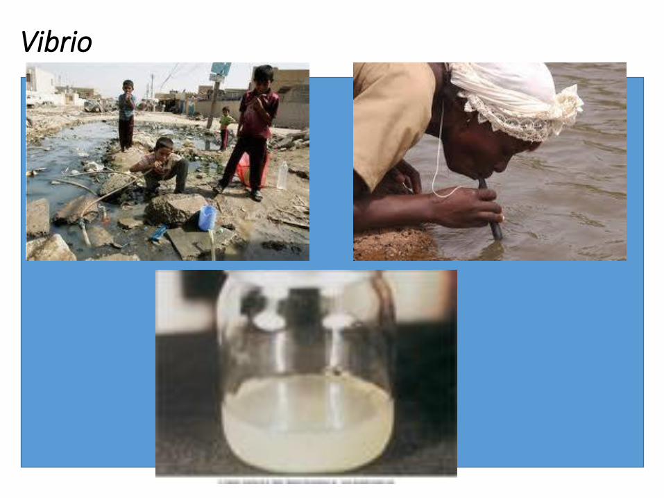
VibrioCharacteristics:
Gram negative rods or comma shaped
Motile by a flagellum
Optimum growth at alkaline pH (8-8.5)
Virulence:
LPS, Pili and cholera toxin (exotoxin)
The cholera toxin:
➢ Decreased NACL and water absorption
➢ Increased Cl and bicarbonates and water excretion

VibrioClassification:
Non-Halophilic: V. Cholera
Halophilic: V. Parahaemolyticus, V. Vulnificus
V. Cholera has 2 classification systems:
1. Serology (based on O1 antigen)
A. V. Cholera O1 antigen (3 isolates)
B. Non O1 antigen: Types 2-139
2. Biotyping (phage typing):
Classical V. Cholera
El Tor

VibrioClinically:
Faecal oral (contaminated water mainly)
IP : 6hrs – 5 days
Multiplies in small intestinal epithelium
Profuse diarrhoea with flecks of mucosa (rice diarrhoea fish odour )
Abdominal pain
Effortless vomiting
Fatal in 12 – 24 hrs
N.B asymptomatic carriers 1:100
Vibrio

VibrioDiagnosis:
Stool sample:
1. In peptone alkaline water (PAW) for 6 hrs
2. Take a loop from the PAW and grow on Thiosulphate Citrate Bile Sucrose (TCBS) which is green in colour
V. Cholera turns the green media into yellow as it ferments sucrose
3. Take a colonie and gram stain it: GNR comma shape, oxidase positive
4. Toxin can be detected by immunoassay
VibrioTreatment:
1. Fluids : 2.9gm Na Citrate, 2.6gm NaCl, 1.5 gmKCl and 13.5 gm glucose
2. Antibiotics to decrease the execretion of cholera into environment
Use Tetracycline
Alternatives: erythromycin, Ciprofloxacin, ceftriaxone
3. No antitoxin
Prevention:
Hygiene and clean water
Vaccine: Oral killed vaccine for O1 Ag type

Vibrio

Vibrio cholera / TCBS

The End