enhancing visual performance in individuals with cortical
TRANSCRIPT
Journal of Medical Hypotheses and Ideas (2015) 9, S8–S13
CORE Metadata, citation and similar papers at core.ac.uk
Provided by Elsevier - Publisher Connector
Available online at www.sciencedirect.com
Journal of Medical Hypotheses and Ideas
journal homepage: www.elsevier.com/locate/jmhi
REGULAR ARTICLE
Enhancing visual performance in individuals
with cortical visual impairment (homonymous
hemianopsia): Tapping into blindsight
* Corresponding author at: Alpert Medical School of Brown University/Rhode Island Hospital, Neuro-Ophthalmology Unit, Depart
Ophthalmology, 1 Hoppin Street, Suite 200, Providence, RI 02903, United States. Tel.: +1 401 444 6551; fax: +1 401 444 6587.
E-mail address: [email protected] (L.N. Johnson).
2251-7294 � 2016 Tehran University of Medical Sciences. Published by Elsevier Ltd.
This is an open access article under the CC BY-NC-ND license (http://creativecommons.org/licenses/by-nc-nd/4.0/).
URL: www.tums.ac.ir/english/
doi:http://dx.doi.org/10.1016/j.jmhi.2015.12.001
Faith A. Birnbauma, Steven A. Hackley
b, Lenworth N. Johnson
a,*
aNeuro-Ophthalmology Unit, Department of Ophthalmology, The Warren Alpert Medical School of BrownUniversity/Lifespan/Rhode Island Hospital, Providence, RI, United StatesbDepartment of Psychological Sciences of the University of Missouri Columbia, Columbia, MO, United States
Received 2 October 2015; revised 29 November 2015; accepted 15 December 2015Available online 22 January 2016
KEYWORDS
Blindsight;
Cortical blindness;
Homonymous hemianopsia;
Augmented virtual reality;
Vision restoration therapy
Abstract Homonymous hemianopsia is a type of cortical blindness in which vision is lost
completely or partially in the left half or the right half of the field of vision. It is prevalent in
approximately 12% of traumatic brain injury and 35% of strokes. Patients often experience
difficulty with activities such as ambulating, eating, reading, and driving. Due to the high prevalence
of homonymous hemianopsia and its associated difficulties, it is imperative to find methods for
visual rehabilitation in this condition. Traditional methods such as prism glasses can cause visual
confusion and result in patient noncompliance. There is a large unmet medical need for improving
this condition. In this article, we propose that modifying visual stimuli to activate non-cortical areas
of visual processing, such as lateral geniculate nucleus and superior colliculus, may result in
increased visual awareness. Presenting high contrast and low spatial frequency visual stimuli can
increase visual detection in patients with cortical blindness, a phenomenon known as blindsight.
Augmented virtual reality goggles have the potential to alter real-time visual input to high contrast
and low spatial frequency images, possibly improving visual detection in the blind hemifield and
providing an alternative therapy for homonymous hemianopsia.� 2016 Tehran University of Medical Sciences. Published by Elsevier Ltd. This is an open access article
under the CC BY-NC-ND license (http://creativecommons.org/licenses/by-nc-nd/4.0/).
ment of
Restoring vision homonymous hemianopsia blindsight S9
Introduction
Cortical visual impairment comprises a significant componentof strokes and traumatic brain injury. Cortical visual impair-
ment includes homonymous hemianopsia, in which vision islost completely or partially in the left half or the right half ofthe field of vision. Homonymous hemianopsia is prevalent in
approximately 12% of traumatic brain injury and 35% ofstrokes [1–3]. Individuals with this vision loss usually havedifficulties with activities of daily living such as ambulating,eating, reading, and driving [4,5]. Due to the high prevalence
of homonymous hemianopsia and its associated difficulties, itis imperative to find methods for visual rehabilitation in thiscondition. Traditional methods of visual rehabilitation for
homonymous hemianopsia include fitting spectacles withprisms to shift the visual field from the blind hemifield to theintact visual field. This is accomplished by placing the base of
the prism in the blind hemifield, which shifts the image towardthe apex of the prism into the intact hemifield. Many patientsdiscontinue treatment with prisms because the prisms may
induce visual confusion and double vision [1–4]. Another tech-nique used is to train individuals with hemianopsia to make
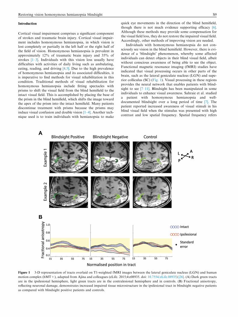
Figure 1 3-D representation of tracts overlaid on T1-weighted fMRI
motion complex (hMT+), adapted from Ajina and colleagues (eLife. 2
are in the ipsilesional hemisphere, light green tracts are in the contr
reflecting neuronal damage, demonstrates increased impaired tissue mi
as compared with blindsight positive patients and controls.
quick eye movements in the direction of the blind hemifield,though there is not much evidence supporting efficacy [6].Although these methods may provide some compensation for
the visual field loss, they do not restore the impaired visual field.Accordingly, other methods of improving vision are needed.
Individuals with homonymous hemianopsia do not con-
sciously see vision in the blind hemifield. However, there is evi-dence of a ‘blindsight’ phenomenon, whereby some affectedindividuals can detect objects in their blind visual field, albeit
without conscious awareness of being able to see the object.Functional magnetic resonance imaging (fMRI) studies haveindicated that visual processing occurs in other parts of thebrain, such as the lateral geniculate nucleus (LGN) and supe-
rior colliculus (SC) (Fig. 1). Visual processing in these regionsprovides the neural network that enables patients with blind-sight to see [7–11]. Blindsight has been manipulated in some
individuals to enhance visual awareness. Sahraie et al. studieda patient with homonymous hemianopsia and well-documented blindsight over a long period of time [7]. The
patient reported increased awareness of visual stimuli in hisblind visual field when the stimulus was presented with highcontrast and low spatial frequency. Spatial frequency refers
images between the lateral geniculate nucleus (LGN) and human
015;4:e08935. doi: 10.7554/eLife.08935) [26]. (A) Dark green tracts
alesional hemisphere and in controls. (B) Fractional anisotropy,
crostructure in the ipsilesional tract in blindsight negative patients

S10 F.A. Birnbaum et al.
to the level of detail in an image appearing within a degree ofthe visual field. Temporal frequency, the number of times astimulus is flashed within a second, also modulates detection.
Multiple studies have shown that within a temporal frequencyrange of 5–20 Hz (cycles/s), detection of visual stimuli in aforced-choice test is significantly better than chance [7–11].
The time of stimulus onset also affects the rate of detection.Patients with parietal lobe injury often cannot detect a visualstimulus in the neglected hemifield when it is presented simul-
taneously in the intact hemifield, but can detect the stimuluswhen it is presented by itself in the neglected hemifield only,a phenomenon known as visual extinction [12,13]. It is thoughtthat visual extinction reflects an attentional deficit as opposed
to primarily a sensory deficit, although this remains an area ofactive research [14]. Despite that visual extinction is primarilystudied in patients with hemi-neglect, patients with hemianop-
sia also can have hemi-neglect from injury to both the occipitaland parietal lobes [15]. Therefore, the relevant visual variablesfor increasing visual detection in hemianopsia are stimuli con-
trast, spatial frequency, temporal frequency and stimulus onsetasynchrony.
Hypothesis
We propose that real-time visual input can be altered to havehigh contrast, low spatial frequency, low temporal frequency,
and appropriate stimulus onset asynchrony using augmentedvirtual reality goggles (Fig. 2). Augmented virtual reality gog-gles, which overlay real-world input with a virtual display, havebeen developed in the past few years. Head-mounted displays
such as such as the Cast AR (http://technicalillusions.com/) or
Daqri Smart Helmet (http://hardware.daqri.com/smarthelmet/)
Figure 2 Testing of a subject with left homonymous hemianopsia vi
information – presented at a spatial frequency of 1 cycle/degree (1 Hz),
asynchrony of 300 ms – presented temporal of the fovea in the right
unfiltered visual information presented nasal of the fovea in the right
may offer potential solutions to modify visual stimuli in suchways as to allow people with hemianopsia to improve visualdetection. This new technology which has been marketed
toward business and gaming applications may be an idealmethod for visual rehabilitation for individuals with homony-mous hemianopsia. Weiskrantz’s group at Oxford University
[8], Huxlin’s group at the University of Rochester [10], andothers have studied visual detection with Gabor patches andcheckerboard reversal in patients with homonymous hemi-
anopsia. To our knowledge, no one has published informationon the detection rate of real time video images, the logical nextsteps. We expect the image detected in the blind hemifield willbe rudimentary, but this would be the first time that a true
extension of vision in the blind hemifield will be documentedusing blindsight (Fig. 3).
Evaluation of the hypothesis
The following research directions are recommended for theevaluation of our hypothesis:
(1) Baseline studies of people affected by homonymoushemianopsia to assess the best spatial frequency, tempo-
ral frequency, and stimulus onset asynchrony delayrequired for each individual for real visual stimuli(Fig. 1). If necessary, this could be performed by dis-playing video clips on a computer screen before using
augmented virtual reality goggles. Previously, temporalfrequencies of pattern-reversal at 10 Hz frequency havebeen documented to produce the highest rates of detec-
tion in hemianopsia [16–18]. A spatial frequency of 1cycle per degree for both images has been shown to elicitthe highest rates of detection among homonymous
sual field defect from right occipital stroke with augmented visual
temporal frequency of 10 cycles/degree (10 Hz), and stimulus onset
eye (and correspondingly, nasal of the fovea in the left eye), and
eye (and correspondingly, temporal of the fovea in the left eye).
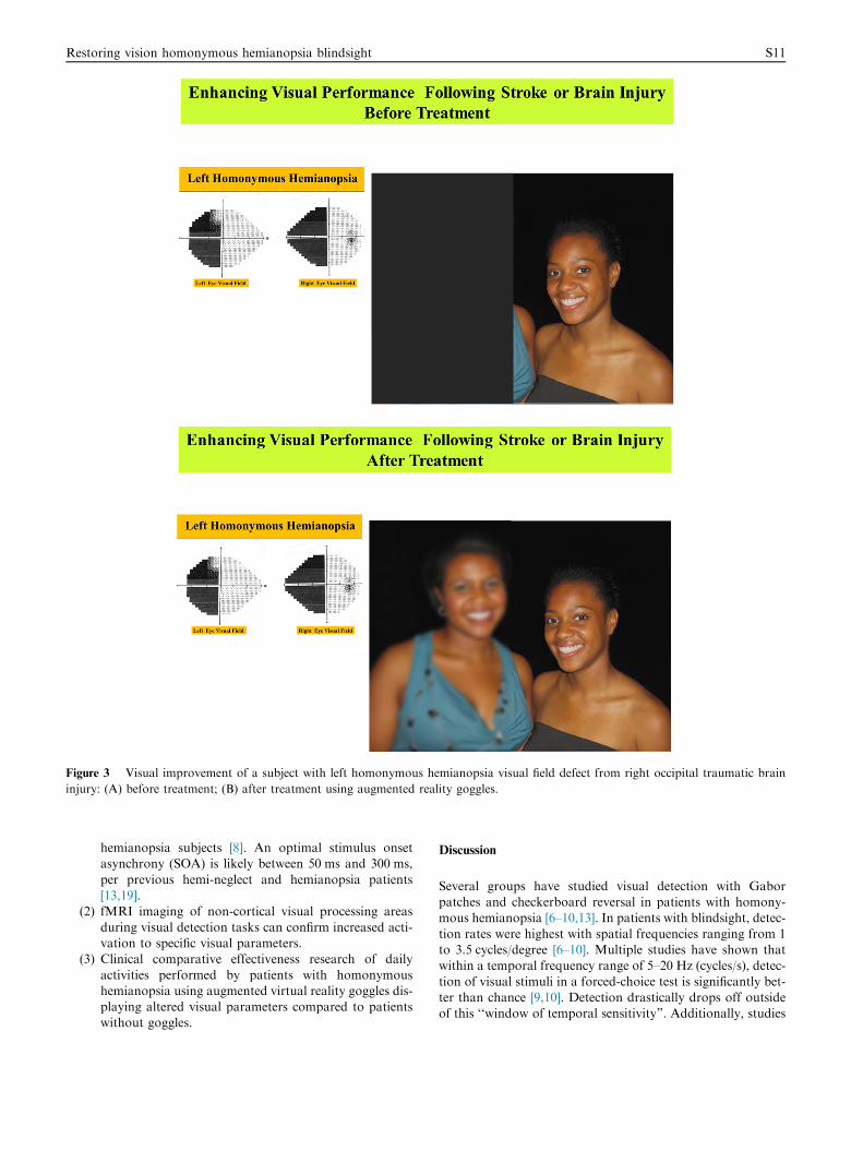
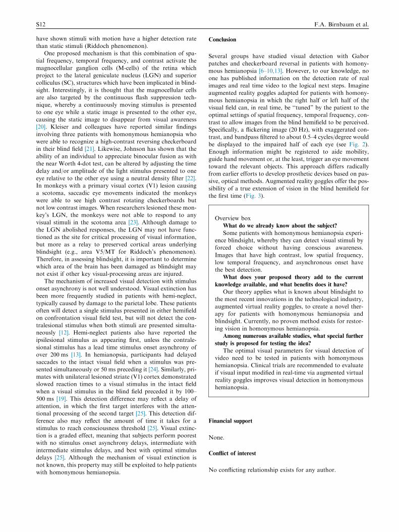
Figure 3 Visual improvement of a subject with left homonymous hemianopsia visual field defect from right occipital traumatic brain
injury: (A) before treatment; (B) after treatment using augmented reality goggles.
Restoring vision homonymous hemianopsia blindsight S11
hemianopsia subjects [8]. An optimal stimulus onsetasynchrony (SOA) is likely between 50 ms and 300 ms,
per previous hemi-neglect and hemianopsia patients[13,19].
(2) fMRI imaging of non-cortical visual processing areas
during visual detection tasks can confirm increased acti-vation to specific visual parameters.
(3) Clinical comparative effectiveness research of daily
activities performed by patients with homonymoushemianopsia using augmented virtual reality goggles dis-playing altered visual parameters compared to patientswithout goggles.
Discussion
Several groups have studied visual detection with Gaborpatches and checkerboard reversal in patients with homony-mous hemianopsia [6–10,13]. In patients with blindsight, detec-
tion rates were highest with spatial frequencies ranging from 1to 3.5 cycles/degree [6–10]. Multiple studies have shown thatwithin a temporal frequency range of 5–20 Hz (cycles/s), detec-
tion of visual stimuli in a forced-choice test is significantly bet-ter than chance [9,10]. Detection drastically drops off outsideof this ‘‘window of temporal sensitivity”. Additionally, studies
S12 F.A. Birnbaum et al.
have shown stimuli with motion have a higher detection ratethan static stimuli (Riddoch phenomenon).
One proposed mechanism is that this combination of spa-
tial frequency, temporal frequency, and contrast activate themagnocellular ganglion cells (M-cells) of the retina whichproject to the lateral geniculate nucleus (LGN) and superior
colliculus (SC), structures which have been implicated in blind-sight. Interestingly, it is thought that the magnocellular cellsare also targeted by the continuous flash suppression tech-
nique, whereby a continuously moving stimulus is presentedto one eye while a static image is presented to the other eye,causing the static image to disappear from visual awareness[20]. Kleiser and colleagues have reported similar findings
involving three patients with homonymous hemianopsia whowere able to recognize a high-contrast reversing checkerboardin their blind field [21]. Likewise, Johnson has shown that the
ability of an individual to appreciate binocular fusion as withthe near Worth 4-dot test, can be altered by adjusting the timedelay and/or amplitude of the light stimulus presented to one
eye relative to the other eye using a neutral density filter [22].In monkeys with a primary visual cortex (V1) lesion causinga scotoma, saccadic eye movements indicated the monkeys
were able to see high contrast rotating checkerboards butnot low contrast images. When researchers lesioned these mon-key’s LGN, the monkeys were not able to respond to anyvisual stimuli in the scotoma area [23]. Although damage to
the LGN abolished responses, the LGN may not have func-tioned as the site for critical processing of visual information,but more as a relay to preserved cortical areas underlying
blindsight (e.g., area V5/MT for Riddoch’s phenomenon).Therefore, in assessing blindsight, it is important to determinewhich area of the brain has been damaged as blindsight may
not exist if other key visual-processing areas are injured.The mechanism of increased visual detection with stimulus
onset asynchrony is not well understood. Visual extinction has
been more frequently studied in patients with hemi-neglect,typically caused by damage to the parietal lobe. These patientsoften will detect a single stimulus presented in either hemifieldon confrontation visual field test, but will not detect the con-
tralesional stimulus when both stimuli are presented simulta-neously [12]. Hemi-neglect patients also have reported theipsilesional stimulus as appearing first, unless the contrale-
sional stimulus has a lead time stimulus onset asynchrony ofover 200 ms [13]. In hemianopsia, participants had delayedsaccades to the intact visual field when a stimulus was pre-
sented simultaneously or 50 ms preceding it [24]. Similarly, pri-mates with unilateral lesioned striate (V1) cortex demonstratedslowed reaction times to a visual stimulus in the intact fieldwhen a visual stimulus in the blind field preceded it by 100–
500 ms [19]. This detection difference may reflect a delay ofattention, in which the first target interferes with the atten-tional processing of the second target [25]. This detection dif-
ference also may reflect the amount of time it takes for astimulus to reach consciousness threshold [25]. Visual extinc-tion is a graded effect, meaning that subjects perform poorest
with no stimulus onset asynchrony delays, intermediate withintermediate stimulus delays, and best with optimal stimulusdelays [25]. Although the mechanism of visual extinction is
not known, this property may still be exploited to help patientswith homonymous hemianopsia.
Conclusion
Several groups have studied visual detection with Gaborpatches and checkerboard reversal in patients with homony-
mous hemianopsia [6–10,13]. However, to our knowledge, noone has published information on the detection rate of realimages and real time video to the logical next steps. Imagine
augmented reality goggles adapted for patients with homony-mous hemianopsia in which the right half or left half of thevisual field can, in real time, be ‘‘tuned” by the patient to theoptimal settings of spatial frequency, temporal frequency, con-
trast to allow images from the blind hemifield to be perceived.Specifically, a flickering image (20 Hz), with exaggerated con-trast, and bandpass filtered to about 0.5–4 cycles/degree would
be displayed to the impaired half of each eye (see Fig. 2).Enough information might be registered to aide mobility,guide hand movement or, at the least, trigger an eye movement
toward the relevant objects. This approach differs radicallyfrom earlier efforts to develop prosthetic devices based on pas-sive, optical methods. Augmented reality goggles offer the pos-
sibility of a true extension of vision in the blind hemifield forthe first time (Fig. 3).
Overview boxWhat do we already know about the subject?
Some patients with homonymous hemianopsia experi-ence blindsight, whereby they can detect visual stimuli byforced choice without having conscious awareness.
Images that have high contrast, low spatial frequency,low temporal frequency, and asynchronous onset havethe best detection.
What does your proposed theory add to the current
knowledge available, and what benefits does it have?
Our theory applies what is known about blindsight to
the most recent innovations in the technological industry,augmented virtual reality goggles, to create a novel ther-apy for patients with homonymous hemianopsia andblindsight. Currently, no proven method exists for restor-
ing vision in homonymous hemianopsia.Among numerous available studies, what special further
study is proposed for testing the idea?
The optimal visual parameters for visual detection ofvideo need to be tested in patients with homonymoushemianopsia. Clinical trials are recommended to evaluate
if visual input modified in real-time via augmented virtualreality goggles improves visual detection in homonymoushemianopsia.
Financial support
None.
Conflict of interest
No conflicting relationship exists for any author.

Restoring vision homonymous hemianopsia blindsight S13
References
[1] Bruce B, Zhang X, Kedar S, Newman NJ, et al. Traumatic
homonymous hemianopia. J Neurol Neurosurg Psychiatry
2006;77:986–8.
[2] Ali M, Hazelton C, Lyden P, Pollock A, et al. Recovery from
poststroke visual impairment: evidence from a clinical trials
resource. Neurorehabil Neural Repair 2013;27:133–41.
[3] Gottlieb DD, Miesner N. Innovative concepts in hemianopsia
and complex visual loss–low vision rehabilitation for our older
population. Top Geriatr Rehabil 2004;20:212–22.
[4] Perez C, Chokron S. Rehabilitation of homonymous
hemianopia: insight into blindsight. Front Integr Neurosci
2014;8:1–12.
[5] de Haan GA, Heutink J, Melis-Dankers BJM, Brouwer WH,
et al. Difficulties in daily life reported by patients with
homonymous visual field defects. J Neuroophthalmol
2015;35:259–64.
[6] Grunda T, Marsalek P, Sykorova P. Homonymous hemianopia
and related visual defects: restoration of vision after a stroke.
Acta Neurobiol Exp (Wars) 2013;73:237–49.
[7] Sahraie A, Hibbard PB, Trevethan CT, Ritchie KL, et al.
Consciousness of the first order in blindsight. PNAS
2010;107:21217–22.
[8] Sahraie A, Trevethan CT, Weiskrantz L, Olson J, et al. Spatial
channels of visual processing in cortical blindness. Eur J
Neurosci 2003;18:1189–96.
[9] Sahraie A, Trevethan CT, MacLeod MJ. Temporal properties of
spatial channel of processing in hemianopia. Neuropsychologia
2008;46:879–85.
[10] Das A, Tadin D, Huxlin KR. Beyond blindsight: properties of
visual relearning in cortically blind fields. J Neurosci
2014;34:11652–64.
[11] Seifert D, Falter C, Strasburger H, Elliott MA. Bandpass
characteristics of high-frequency sensitivity and visual
experience in blindsight. Conscious Cogn 2010;19:144–51.
[12] Baylis GC, Limon SL, Baylis LL, Rorden C. Visual extinction
with double simultaneous stimulation: what is simultaneous?
Neuropsychologia 2002;40:1027–34.
[13] Rorden C, Mattingley JB, Karnath H, Driver J. Visual
extinction and prior entry: impaired perception of temporal
order with intact motion perception after unilateral parietal
damage. Neuropsychologia 1997;35:421–33.
[14] de Haan B, Karnath H, Driver J. Mechanisms and anatomy of
unilateral extinction after brain injury. Neuropsychologia
2012;1045–53.
[15] Meienberg O, Harrer M, Wehren C. Oculographic diagnosis of
hemineglect in patients with homonymous hemianopia. J Neurol
1986;233:97–101.
[16] Zeki S, ffytche, D.H. The Riddoch syndrome: insights into the
neurobiology of conscious vision. Brain 1998;121:25–45.
[17] Sinha P. Once blind and now they see: surgery in blind children
from India allows them to see for the first time and reveals how
vision works in the brain. Sci Am 2013;309:48–55.
[18] Balasubramanian V, Sterling P. Receptive fields and functional
architecture in the retina. J Physiol 2009;587:2753–67.
[19] Cowey A, Stoerig P, Le Mare C. Effects of unseen stimuli on
reaction times to seen stimuli in monkeys with blindsight.
Conscious Cogn 1998;7:312–23.
[20] Lin Z, He S. Seeing the invisible: the scope and limits of
unconscious processing in binocular rivalry. Prog Neurobiol
2009;87:195–211.
[21] Kleiser R, Wittsack J, Niedeggen M, Goebel R, et al. Is V1
necessary for conscious vision in areas of relative cortical
blindness? Neuroimage 2001;13:654–61.
[22] Johnson LN. The relative afferent pupillary defect and visual
cortex binocular cell activity: a novel method of fusion recovery
with Worth 4-dot test. Arch Ophthalmol 1996;114:171–5.
[23] Schmid MC, Mrowka SW, Turchi J, Saunders RC, et al.
Blindsight depends on the lateral geniculate nucleus. Nature
2010;466:373–7.
[24] Rafal R, Smith J, Krantz J, Cohen A, Brennan C.
Extrageniculate vision in hemianopic humans: saccade
inhibition by signals in the blind field. Science 1990;250:118–21.
[25] Cate A, Behrmann M. Spatial and temporal influences on
extinction. Neuropsychologia 2002;40:2206–25.
[26] Ajina S, Pestilli F, Rokem A, Kennard C, Bridge H. Human
blindsight is mediated by an intact geniculo-extrastriate
pathway. eLife 2015;4. http://dx.doi.org/10.7554/eLife.08935,
e08935.