enhancing the sensitivity of a surface plasmon resonance
TRANSCRIPT
RSC Advances
PAPER
Ope
n A
cces
s A
rtic
le. P
ublis
hed
on 1
7 D
ecem
ber
2019
. Dow
nloa
ded
on 1
2/14
/202
1 5:
49:3
0 A
M.
Thi
s ar
ticle
is li
cens
ed u
nder
a C
reat
ive
Com
mon
s A
ttrib
utio
n 3.
0 U
npor
ted
Lic
ence
.
View Article OnlineView Journal | View Issue
Enhancing the se
aInstitute of Advanced Technology, Universi
Selangor, Malaysia. E-mail: yapwingfen@upbDepartment of Physics, Faculty of Science
Serdang, Selangor, MalaysiacSynchrotron Light Research Institute, MaundWireless and Photonics Networks, Faculty o
43400 UPM Serdang, Selangor, Malaysia
Cite this: RSC Adv., 2019, 9, 41729
Received 13th September 2019Accepted 12th November 2019
DOI: 10.1039/c9ra07368j
rsc.li/rsc-advances
This journal is © The Royal Society o
nsitivity of a surface plasmonresonance-based optical sensor for zinc iondetection by the modification of a gold thin film
Wan Mohd Ebtisyam Mustaqim Mohd Daniyal,a Yap Wing Fen, *ab
Nur Ain Asyiqin Anas,a Nur Alia Sheh Omar,a Nur Syahira Md Ramdzan,b
Hideki Nakajimac and Mohd Adzir Mahdid
Surface plasmon resonance (SPR) sensors as novel optical sensors for the detection of a variety of analytes
have been receiving increasing attention and their sensitivity has become the research hotspot recently. In
this study, the sensitivity of an SPR optical sensor was enhanced by modifying a gold thin film with
a nanocrystalline cellulose (NCC)-based material for zinc ion (Zn2+) detection that exists in the
environment due to industrial processing. By replacing the gold thin film with a novel modified-gold thin
film, Zn2+ can be detected from the range of 0 to 10 ppm using SPR. It is believed that the Zn2+ may
interact with the negative charge molecules that exist on the modified-gold thin film, and this was
confirmed via X-ray photoelectron spectroscopy (XPS). Moreover, this modified-gold-SPR has a high
sensitivity of 1.892� ppm�1 up to 0.1 ppm with an enhanced detection of Zn2+ as low as 0.01 ppm. The
SPR results also followed the Langmuir isotherm model with a binding affinity of 1.927 � 103 M�1, which
further confirmed the sensitivity of the SPR sensor. In addition, using the modified-gold thin film, SPR
has a higher affinity towards Zn2+ compared to other metal ions, i.e. Ni2+, Fe2+, Cr2+, Mn2+, and Co2+.
1. Introduction
A biosensor or sensor is an analytical tool that is used for thedetermination of analytes based on a biocatalyst type ofsensors.1 A sensor can be dependent on magnetic, mechanical,electrical, and optical principles. Among all, optical sensors arethemost simpler in their setup for data acquisition and workingactivity. An optical sensor has many advantages over electricaland mechanical sensors, such as the optical sensor does notmodify nor destruct the measured and/or the surroundingenvironment.2 Basically, optical sensors require a recognitionelement that can interact specically with the desired targetanalytes and then, the sensor will detect the signal of thebinding event.3 For instance, reection interference spectros-copy (RIFS),4 surface-enhanced Raman scattering (SERS) spec-troscopy,5 and surface plasmon resonance (SPR) are opticalsensors that require a recognition element.6 SPR is one of thefavorable optical sensors that is widely used in sensing
ti Putra Malaysia, 43400 UPM Serdang,
m.edu.my
, Universiti Putra Malaysia, 43400 UPM
g, Nakhon Ratchasima, 30000, Thailand
f Engineering, Universiti Putra Malaysia,
f Chemistry 2019
biochemical reaction owing to its advantages such as low-cost,label-free, fast measurement, and simple sample preparation.7
The most common setup for SPR is by the Kretschmannconguration. When a p-polarized light is incident ontoa metallic thin lm (typically gold or silver) through the prism,the free electrons on the metal surface will excite and forma surface plasmon. At a certain angle, the surface plasmon willthen resonate with the incident light, thus reducing the inten-sity of the reected light.8 This angle is known as the resonanceangle. SPR is very sensitive towards the changes in the refractiveindex of the metal surface. Such change may result in a changein the resonance angle.9 However, SPR sensitivity is limited,where any solution with the same refractive index, such as a lowconcentration of the metal ion solution.10 Sensitivity is one ofthe most important features of a sensor where it is affected bythe bioreceptor, biomolecule immobilization procedure ortransduction method.11 Since the past two decades, SPR hasbeen extensively studied to enhance the optical sensorsensitivity.
One of the strategies to enhance the SPR sensitivity for themetal ion detection is by combining the SPR with other sensingmethods. For instance, Wang et al. in 2007 combined the SPRsensor with anodic stripping voltammetry (ASV) for sensingcopper, lead, and mercury ions, while Panta et al. in 2009combined the SPR with ASV and magnetohydrodynamic (MHD)convection for sensing mercury ions.12,13 Instead of combiningSPR with a different technique, researchers also studied the
RSC Adv., 2019, 9, 41729–41736 | 41729

Fig. 1 SPR optical sensor setup.
RSC Advances Paper
Ope
n A
cces
s A
rtic
le. P
ublis
hed
on 1
7 D
ecem
ber
2019
. Dow
nloa
ded
on 1
2/14
/202
1 5:
49:3
0 A
M.
Thi
s ar
ticle
is li
cens
ed u
nder
a C
reat
ive
Com
mon
s A
ttrib
utio
n 3.
0 U
npor
ted
Lic
ence
.View Article Online
sensitivity of SPR by using different light sources. Eum et al. in2003 modied an SPR sensor with near-infrared (NIR) lightsources and they reported that the modied SPR system wasable to detect potassium ions.14 In 2008, Chen et al. used whitelight sources for the SPR sensor where the detection of uranylions is based on a wavelength shi instead of a resonanceangle.15
Although the SPR sensitivity was proven to be enhancedeither by combining the SPR with other methods or by modi-fying the SPR sensor, the simplest way for the sensitivityenhancement is by modifying a gold thin lm with a sensingelement. As the SPR sensor works by measuring the refractiveindex changes in the vicinity of the gold thin lm in response tobiomolecular interactions, the modication of the gold thinlm surface will denitely enhance the SPR sensitivity. Studieson the SPR sensitivity enhancement by the modication of themetal thin lm for metal ion detection also has been conductedsince 2001.16–34 However, only a few studies for sensing zinc ionshas been conducted. For example, Wu and Lin in 2004 immo-bilized metallothionein onto a carboxymethylated dextranmatrix to be incorporated with SPR.35 They reported that theSPR system can be used to detect zinc as low as 0.13 ppm. In2011, Fen et al. introduced a chitosan layer on top of a gold thinlm using glutaraldehyde as the crosslinking agent.36 Fromtheir report, the SPR sensitivity for zinc ion detection wasenhanced as low as 0.5 ppm. In another interesting study,Sadrolhosseini et al., in 2013, used a polypyrrole–chitosan layerfor zinc ion detection using SPR.37 The binding interactions ofthe zinc ions with the active layer was monitored using theresonance angle shi where the lowest detection was 0.98 ppm.Fen et al. in 2015 again used chitosan and chitosan–tetrabu-tylthiuram disulphide for the detection of zinc ions using SPR.38
The active layer was deposited on the top of the gold layer viaa spin coating technique before combining with the SPR systemto monitor the zinc ion concentration. They found that the SPRsystem was able to detect zinc ion as low as 0.1 ppm. AlthoughSPR has been studied to detect zinc ion since 2004, the lowestconcentration detected was 0.1 ppm. Hence in this study, wemodied the gold thin lm with a nanocrystalline cellulose(NCC)-based composite that is believed to enhance the sensi-tivity of the SPR optical sensor for sensing zinc ions at a lowerconcentration owing to its toxic effect to human.
Aer iron, zinc is the second most abundant metal ion thatexists in the human body, which is about 2–3 g in total.39 Zinc isalso one of the essential trace elements in biological systems asit is involved in numerous aspects of cellular metabolism, playsa role in the human immune systems and development duringpregnancy, supports normal growth, and it is required fora normal sense of smell and taste.40,41 Moreover, zinc is esti-mated to bind about 10% of human proteins, and it is requiredfor the catalytic activity for more than 200 enzymes.42,43 Zincdeciency in the human body can cause damage to the immunefunction, delay growth, and cause a loss in appetite.44,45
Although zinc is very crucial in human biological systems, anexcess amount of zinc also proves to be lethal.46 At a higherlevel, zinc can cause health problems such as vomiting, nausea,stomach ache, anemia, and skin problems.47 The high level of
41730 | RSC Adv., 2019, 9, 41729–41736
zinc concentration in the environment is due to industrialsources, such as toxic waste sites, steel processing, wastecombustion, and mining. Furthermore, the zinc concentrationmagnied in tap water is due to the leaching of zinc from ttingand piping.48 Therefore, the detection of zinc at a low level isimportant for the continuous monitoring of environmentalwater.
2. Experimental details2.1. Modied-gold thin lm
All chemicals were purchased from Sigma Aldrich (St. Louis,MO, USA). In order to modify the gold thin lm with the NCC-based material, hexadecyltrimethylammonium bromide (CTA)was used for a slight modication of NCC. This modicationwas necessary in order to alter the NCC hydrophilicity proper-ties to make slightly hydrophobic.49 NCC was modied bydiluting 100 ml of NCC (0.1 wt% suspension) and mixed with0.1 wt% CTA. The mixture was then centrifuged for 10 minutesbefore being dispersed with graphene oxide (GO) in a 1 : 1volume ratio.
2.2. Surface plasmon resonance system
Our custom build SPR optical sensor system is based onKretschmann conguration that consists of a He–Ne laser beam(632.8 nm, 5 mW), optical chopper (SR 540), polarizer, steppermotor (Newport MM 3000), prism (n ¼ 1.77861), photodiodedetector, and lock-in amplier (SR 530), as shown in Fig. 1. Inorder to generate SPR, the incident light must be in a transversemagnetic mode as the electric eld is perpendicular to themetalthin lm that can be described by:
E. ¼ Eoðxþ izÞeiðkx�utÞe�k|z| (1)
where Eo is the amplitude, k is the wave vector, x and z are theunit vectors, and u is the angular optical frequency of theelectrical eld. When total internal reection occurred, surfaceplasmon was generated by the evanescent wave at the metal anddielectric interface. The surface plasmon wave vector, Ksp isdescribed by the following equation:
This journal is © The Royal Society of Chemistry 2019

Paper RSC Advances
Ope
n A
cces
s A
rtic
le. P
ublis
hed
on 1
7 D
ecem
ber
2019
. Dow
nloa
ded
on 1
2/14
/202
1 5:
49:3
0 A
M.
Thi
s ar
ticle
is li
cens
ed u
nder
a C
reat
ive
Com
mon
s A
ttrib
utio
n 3.
0 U
npor
ted
Lic
ence
.View Article Online
Ksp ¼ u
c
ffiffiffiffiffiffiffiffiffiffiffiffiffiffiffiffiffiffiffiffiffi�3132
31 þ 32
�s(2)
where u is the frequency, c is the light velocity, and 31 and 32 arethe dielectric constants for the surface-active and dielectricmedia, respectively. The dielectric constant can be described by:
3 ¼ n2 (3)
thus eqn (2) can be rewritten as:
Ksp ¼ u
c
ffiffiffiffiffiffiffiffiffiffiffiffiffiffiffiffiffiffiffiffiffiffiffiffiffi�n1
2n22
n12 þ n22
�s(4)
where n1, n2, and np are the refractive index of the gold layer,sample, and prism, respectively. SPR occurs when a componentof the incident light vector parallel to the prism/metal interface,Kx, given by:
Kx ¼�uc
�np sin qSPR (5)
was similar to the surface plasmon wave vector:
Ksp ¼ Kx (6)
with ffiffiffiffiffiffiffiffiffiffiffiffiffiffiffiffiffiffiffiffiffiffiffiffiffi�n1
2n22
n12 þ n22
�s¼ np sin qSPR (7)
The coupling of these two-wave vectors, Ksp, and Kx result ina sharp dip of the reectance at a resonance angle, qSPR. TheSPR optical sensor works by detecting the changes in the thinlm surface refractive index. Thus, the refractive index of thesample is:
n2 ¼ffiffiffiffiffiffiffiffiffiffiffiffiffiffiffiffiffiffiffiffiffiffiffiffiffiffiffiffiffiffiffiffiffiffiffiffiffin1
2np2 sin2
qSPR
n12 � np2 sin2qSPR
s(8)
Fig. 2 The SPR signal of deionized water using an unmodified goldthin film.
This journal is © The Royal Society of Chemistry 2019
In accordance with the boundary conditions for the electricaland magnetic elds at the interfaces between multilayers, thereection coefficient, r, can be expressed as:
r ¼ m21 þm22g2 �m11g0 �m12g2g0
m21 þm22g2 þm11g0 þm12g2g0
(9)
wheremij is the matrix transfer element. The transfer matrix canbe obtained from the relation between the electrical andmagnetic layers in the rst and the last layers.50 The reectivityof the multilayer system, R, is dened as the ratio of the energyreected at the surface to the energy of the incident, and can beexpressed as:
R ¼ rr* (10)
where it is the function of the refractive index of the sample, therefractive index and thickness of both gold and sensor layer.
To begin with, the SPR experiment was divided into twoparts. The rst part was to investigate the SPR signal using anunmodied gold thin lm to obtain the properties of the reso-nance angle. Then, the second part of the SPR experiment wasperformed using the modied-gold thin lm to determine thesensing ability. The SPR signal was investigated using both ofthe thin lms in contact with the deionized water and Zn2+ ofvarious concentrations, from 0.01 ppm to 10 ppm (prepared bydiluting 1000 ppm standard zinc solution with deionized waterusing the dilution formula M1V1 ¼ M2V2) injected into thehollow one by one. The SPR curve was taken aer Zn2+ wasinjected and le for 10 minutes in the cell.7 The SPR signalresults for both the modied and unmodied gold thin lmcontact with deionized water and Zn2+ have been discussed inthe next section.
2.3. X-ray photoelectron spectroscopy
X-ray photoelectron spectroscopy (XPS) was performed toinvestigate the possible interactions between the modied-goldthin lm with Zn2+. The XPS study was performed usingPHI5000 Versa Probe II, ULVAC-PHI Japan at the SUT-NANOTEC-SLRI Joint Research Facility, Synchrotron Light
Fig. 3 The SPR signal of Zn2+ (0.01–10 ppm) using an unmodified goldthin film.
RSC Adv., 2019, 9, 41729–41736 | 41731

Fig. 4 SPR signal of deionized water using themodified-gold thin film. Fig. 6 SPR signal of Zn2+ solution (0.1–10 ppm) using the modified-gold thin film.
RSC Advances Paper
Ope
n A
cces
s A
rtic
le. P
ublis
hed
on 1
7 D
ecem
ber
2019
. Dow
nloa
ded
on 1
2/14
/202
1 5:
49:3
0 A
M.
Thi
s ar
ticle
is li
cens
ed u
nder
a C
reat
ive
Com
mon
s A
ttrib
utio
n 3.
0 U
npor
ted
Lic
ence
.View Article Online
Research Institute (SLRI), Thailand. The XPS scans were recor-ded in the range of 0 to 1400 eV and tted by the Gaussian–Lorentzian curve tting program with a linear background foreach peak in order to determine the binding energies of variouselement core levels.
3. Results and discussion3.1. SPR sensor using an unmodied gold thin lm
For the rst part of the SPR experiment, the resonance angle fordeionized water in contact with an unmodied gold thin lmobtained was 53.66�, as shown in Fig. 2. The experiment thencontinued using Zn2+ solution. As shown in Fig. 3, the reso-nance angle of Zn2+ for all concentration remain the same as theresonance angle of deionized water, i.e. 53.66�. This resultmight be due to a small binding interaction amount of Zn2+
with the gold surface, thus does not change the optical prop-erties of the thin lm.51 Moreover, the refractive index of metalions at any concentration below 100 ppm is almost equal to therefractive index of deionized water.10
Fig. 5 SPR signal of Zn2+ solution (0–0.1 ppm) using the modified-gold thin film.
41732 | RSC Adv., 2019, 9, 41729–41736
3.2. SPR sensor using a modied-gold thin lm
For the second part of the SPR experiment, the resonance anglefor deionized water using a modied-gold thin lm found was54.65�, slightly different when using the unmodied gold thinlm, as shown in Fig. 4. The unequal resonance angle ofdeionized water might be due to the refractive index changeswhen the gold thin lm was immobilized with the NCC-basedmaterial.52 The SPR curves using the modied-gold thin lmfor sensing Zn2+ are shown in Fig. 5 and 6. The SPR curve shiedslightly from the deionized water at 0.01 ppm and shiedfurther at higher concentrations. The modied-gold thin lmachieved saturation aer the binding of Zn2+ that minimizedthe changes in the surface refractive index.53 Hence, the reso-nance angle of the SPR curves remains almost at the same at0.01 until 10 ppm and it can be concluded that the SPR usingthe modied-gold thin lm can be used to detect Zn2+ from 0.01up to 0.1 ppm. Zn2+ may bind with the negative charge thatexists on the thin lm surface forming a pair of shared electronsbetween the positive charge Zn2+, thus changing the opticalproperties of the thin lm.54 In order to investigate the existence
Fig. 7 Wide-scan XPS spectra for chemical compositions of themodified-gold thin film.
This journal is © The Royal Society of Chemistry 2019
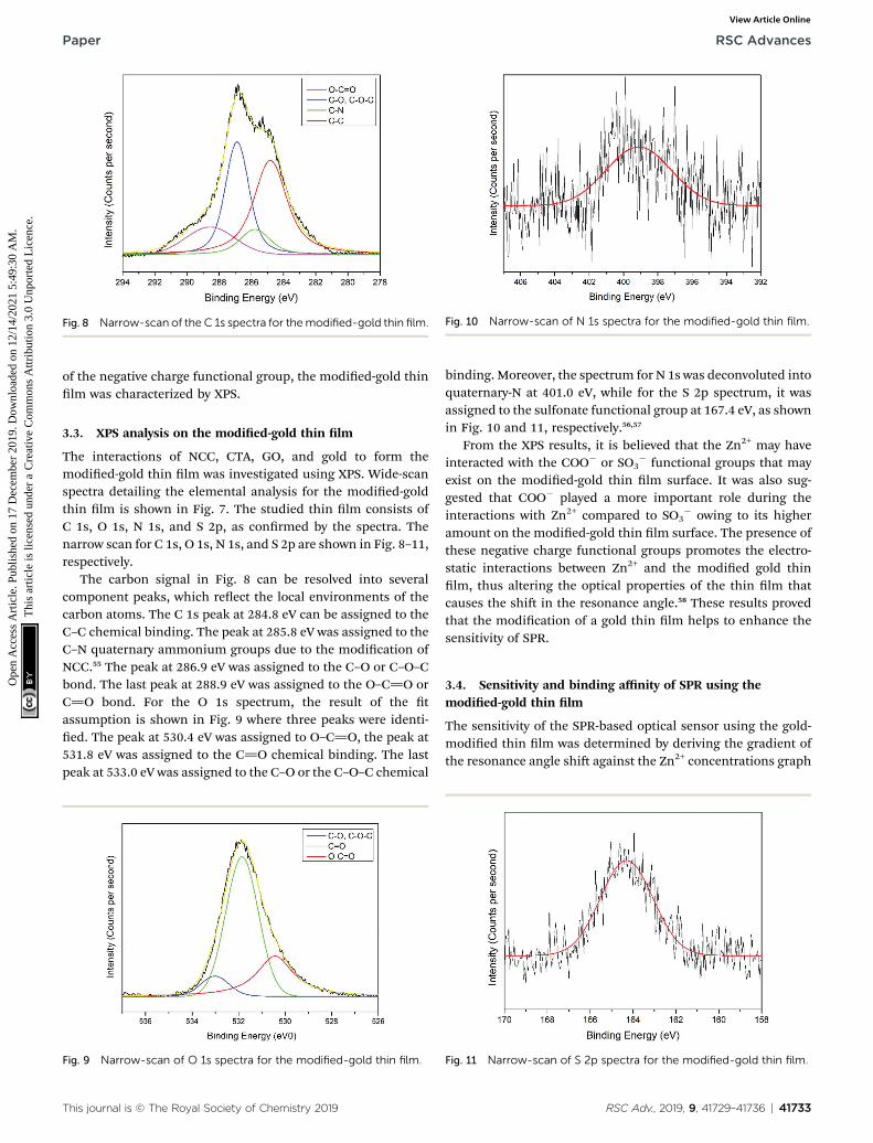
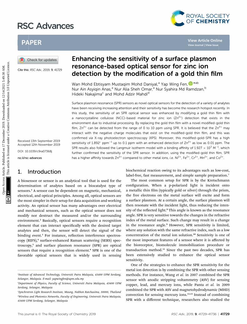
Fig. 10 Narrow-scan of N 1s spectra for the modified-gold thin film.Fig. 8 Narrow-scan of the C 1s spectra for themodified-gold thin film.
Paper RSC Advances
Ope
n A
cces
s A
rtic
le. P
ublis
hed
on 1
7 D
ecem
ber
2019
. Dow
nloa
ded
on 1
2/14
/202
1 5:
49:3
0 A
M.
Thi
s ar
ticle
is li
cens
ed u
nder
a C
reat
ive
Com
mon
s A
ttrib
utio
n 3.
0 U
npor
ted
Lic
ence
.View Article Online
of the negative charge functional group, the modied-gold thinlm was characterized by XPS.
3.3. XPS analysis on the modied-gold thin lm
The interactions of NCC, CTA, GO, and gold to form themodied-gold thin lm was investigated using XPS. Wide-scanspectra detailing the elemental analysis for the modied-goldthin lm is shown in Fig. 7. The studied thin lm consists ofC 1s, O 1s, N 1s, and S 2p, as conrmed by the spectra. Thenarrow scan for C 1s, O 1s, N 1s, and S 2p are shown in Fig. 8–11,respectively.
The carbon signal in Fig. 8 can be resolved into severalcomponent peaks, which reect the local environments of thecarbon atoms. The C 1s peak at 284.8 eV can be assigned to theC–C chemical binding. The peak at 285.8 eV was assigned to theC–N quaternary ammonium groups due to the modication ofNCC.55 The peak at 286.9 eV was assigned to the C–O or C–O–Cbond. The last peak at 288.9 eV was assigned to the O–C]O orC]O bond. For the O 1s spectrum, the result of the tassumption is shown in Fig. 9 where three peaks were identi-ed. The peak at 530.4 eV was assigned to O–C]O, the peak at531.8 eV was assigned to the C]O chemical binding. The lastpeak at 533.0 eV was assigned to the C–O or the C–O–C chemical
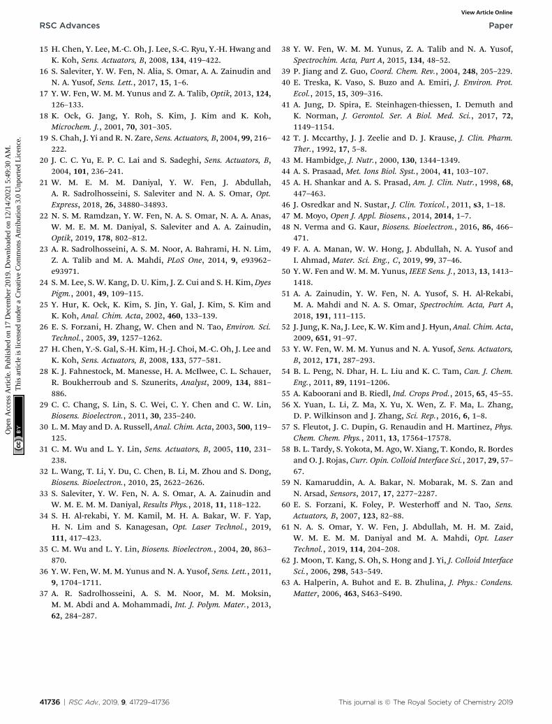
Fig. 9 Narrow-scan of O 1s spectra for the modified-gold thin film.
This journal is © The Royal Society of Chemistry 2019
binding. Moreover, the spectrum for N 1s was deconvoluted intoquaternary-N at 401.0 eV, while for the S 2p spectrum, it wasassigned to the sulfonate functional group at 167.4 eV, as shownin Fig. 10 and 11, respectively.56,57
From the XPS results, it is believed that the Zn2+ may haveinteracted with the COO� or SO3
� functional groups that mayexist on the modied-gold thin lm surface. It was also sug-gested that COO� played a more important role during theinteractions with Zn2+ compared to SO3
� owing to its higheramount on the modied-gold thin lm surface. The presence ofthese negative charge functional groups promotes the electro-static interactions between Zn2+ and the modied gold thinlm, thus altering the optical properties of the thin lm thatcauses the shi in the resonance angle.58 These results provedthat the modication of a gold thin lm helps to enhance thesensitivity of SPR.
3.4. Sensitivity and binding affinity of SPR using themodied-gold thin lm
The sensitivity of the SPR-based optical sensor using the gold-modied thin lm was determined by deriving the gradient ofthe resonance angle shi against the Zn2+ concentrations graph
Fig. 11 Narrow-scan of S 2p spectra for the modified-gold thin film.
RSC Adv., 2019, 9, 41729–41736 | 41733

Fig. 12 Comparison of the resonance angle shift for the modified-gold thin film with Zn2+ concentration (0.01–0.1 ppm).
RSC Advances Paper
Ope
n A
cces
s A
rtic
le. P
ublis
hed
on 1
7 D
ecem
ber
2019
. Dow
nloa
ded
on 1
2/14
/202
1 5:
49:3
0 A
M.
Thi
s ar
ticle
is li
cens
ed u
nder
a C
reat
ive
Com
mon
s A
ttrib
utio
n 3.
0 U
npor
ted
Lic
ence
.View Article Online
from 0.01 until 10 ppm. Overall, it could be seen that thesensitivity of SPR was enhanced by using the modied-goldthinlm. The sensitivity of the SPR using the modied-gold thinlm toward Zn2+ showed high sensitivity from 0.01 until0.1 ppm, while at a higher concentration the sensitivitydecreased. For further analysis, the data was plotted differentlyto obtain the best linear regression coefficient R2.
The plot to calculate the SPR sensitivity using the modied-gold thin lm from 0.01 until 0.1 ppm is shown in Fig. 12, and itcan be observed that the resonance angle shi increased line-arly with the Zn2+ concentration. The linear regression analysisof the graph produces the gradient of 1.892� ppm�1, which alsorepresents the sensor sensitivity with an R2 of 0.96. The SPR
Table 1 Comparison of the lowest detection of Zn2+ with various modi
Sensing layer
Polypyrrole–chitosanMMW chitosan (glutaraldehyde-crosslinked)Immobilize metallothionein onto a carboxymethylated dextran matrixChitosan and chitosan–tetrabutylthiuram disuldeModied-nanocrystalline cellulose/graphene oxide
Fig. 13 Comparison of the resonance angle shift for the modified-goldthin film with Zn2+ concentration (0.5–10 ppm).
41734 | RSC Adv., 2019, 9, 41729–41736
sensitivity may reach a saturated value at 0.5 until 10 ppm as thesensitivity of the SPR decreases down to 0.000131� ppm�1 withan R2 of 0.99, as shown in Fig. 13. Moreover, the efficiency ofthis SPR sensor was compared with other reported studies onthe Zn2+ ion detection using the SPR method, as summarized inTable 1. The NCC-based material that was used to modify thegold thin lm in this study has been proven to detect Zn2+ as lowas 0.01 ppm, which was much lower as compared to previouslyreported study, which was at 0.1 ppm. This result may be due tothe existence of different negative charge functional groups thatexist on the NCC-based composite, i.e., COO� and SO3
�. TheCOO� and SO3
�may have had higher electronegativity to attractZn2+ for electrostatic interactions, and hence enhance the SPRsensitivity as compared to the chitosan–tetrabutylthiuramdisulde that was reported to have only sulfur donor atoms.
Another important parameter of a sensor is the bindingaffinity. The binding affinity can be calculated by the Langmuirisotherm model with the following equation.59–61
Dq ¼ DqmaxC
1
Kþ C
(11)
where C is the Zn2+ concentration, Dqmax is the maximum SPRshi at saturation, and K is the binding affinity constant. Fig. 14shows the plot that tted the Langmuir model for both modi-ed and unmodied gold thin lms. The Dqmax of the curvetting SPR for the modied-gold thin lm was 0.2617�, slightlyhigher than the maximum angle of experimental value, i.e.,0.2536� with an R2 of 0.95.
The binding affinity was from the Langmuir model for Zn2+
towards the modied-gold thin lm also was calculated and thevalue of K obtained was 1.927� 103 M�1, while for Zn2+ towardsthe unmodied gold thin lm was 0.99 M�1.62,63 The higheraffinity of Zn2+ ions towards the modied gold thin lm provedthat the modication of the gold thin with the NCC-basedmaterial helps to improve the sensitivity of the SPR opticalsensor.
3.5. Affinity comparison of Zn2+ with other metal ions
Further investigation was performed to study the affinity ofother metal ions towards the modied-gold thin lm forcomparison with Zn2+. Fig. 15 depicts the shi in the resonanceangle comparison of Zn2+ with different metal ions includesNi2+, Fe2+, Cr2+, Mn2+, and Co2+. The concentration of Zn2+ wasset at a lower concentration, i.e., at 0.1 ppm while maintainingother metal ions concentration at 1 ppm. From Fig. 15, Zn2+ has
fication of the gold thin film
Lowest detection Reference
0.98 ppm 370.5 ppm 360.13 ppm 350.1 ppm 380.01 ppm This study
This journal is © The Royal Society of Chemistry 2019

Fig. 15 Affinity comparison of Zn2+ with other metal ions.
Fig. 14 Langmuir isotherm model of the resonance angle shift formodified and unmodified gold thin films in contact with Zn2+ ions.
Paper RSC Advances
Ope
n A
cces
s A
rtic
le. P
ublis
hed
on 1
7 D
ecem
ber
2019
. Dow
nloa
ded
on 1
2/14
/202
1 5:
49:3
0 A
M.
Thi
s ar
ticle
is li
cens
ed u
nder
a C
reat
ive
Com
mon
s A
ttrib
utio
n 3.
0 U
npor
ted
Lic
ence
.View Article Online
the highest shi in the resonance angle at 0.2536� even at theconcentration that is 10 times lower as compared to other metalions at a higher concentration, which were 0.151�, 0.0955�,0.0555�, 0.0097�, and 0.0096� for Ni2+, Fe2+, Cr2+, Mn2+, andCo2+, respectively. These results show that the SPR has a higheraffinity towards Zn2+ using the modied-gold thin lm.
4. Conclusions
In this study, the sensitivity of the SPR optical sensor wassuccessfully enhanced by modifying the gold thin lm witha nanocrystalline cellulose (NCC)-based material for Zn2+
detection. When the unmodied gold thin lm was tested withthe SPR in the rst part of the experiment, the resonance angleremained the same at 53.66� with all concentrations of Zn2+.The second part of the SPR experiment proved that the modi-cation of the gold thin lm enhanced the sensitivity of theSPR, where the optical sensor was able to detect Zn2+ as low as0.01 ppm. Also, the potential interactions of the Zn2+ with themodied-gold thin lm also were studied via X-ray photoelec-tron spectroscopy. From the XPS results, it was determined thatthe Zn2+ ions may have interacted with the negative chargefunctional groups contained on the modied thin lm surface,
This journal is © The Royal Society of Chemistry 2019
i.e., COO� and SO3�, via electrostatic interactions. Moreover,
the sensitivity of the SPR using the modied-gold thin lm wasalso calculated by comparing the resonance angle shi withconcentrations of Zn2+. The SPR using the modied-gold thinlm had a sensitivity of 1.892� ppm�1 with the lowest detectionof 0.01 ppm compared to other previous studies. Besides that,the modied-gold thin lm has a higher binding affinitycompared to the unmodied gold thin lm when calculatedusing the Langmuir isotherm model, i.e., 1.927 � 103 M�1 and0.99 M�1, respectively. By using the modied-gold thin lm, theSPR responses also had a higher affinity towards Zn2+ compareto Ni2+, Fe2+, Cr2+, Mn2+, and Co2+.
Conflicts of interest
There are no conicts of interest to declare.
Acknowledgements
This research work was funded by the Malaysian Governmentthrough Putra Grant Universiti Putra Malaysia (9627300 and9531500) and the Fundamental Research Grant Scheme (FRGS)(FRGS/1/2019/STG02/UPM/02/1).
Notes and references
1 N. Khansili, G. Rattu and P. M. Krishna, Sens. Actuators, B,2018, 265, 35–49.
2 C. Ciminelli, C. M. Campanella, F. D. Olio, C. E. Campanellaand M. N. Armenise, Prog. Quantum Electron., 2013, 37, 51–107.
3 X. Yan, H. Li and X. Su, Trends Anal. Chem., 2018, 103, 1–20.4 B. Schwarz, N. Schweizer, F. Proll, G. Proll and G. Gauglitz,Procedia Eng., 2010, 5, 906–909.
5 A. Santinom, M. A. Silva, J. E. L. Villa, R. J. Poppi, I. O. Mazaliand D. P. Santos, Vib. Spectrosc., 2018, 99, 34–43.
6 W. M. E. M. M. Daniyal, S. Saleviter and Y. W. Fen, Sens.Mater., 2018, 30, 2023–2038.
7 W. M. E. M. M. Daniyal, Y. W. Fen, J. Abdullah,A. R. Sadrolhosseini, S. Saleviter and N. A. S. Omar,Spectrochim. Acta, Part A, 2019, 212, 25–31.
8 P. Zhang, Y. P. Chen, W. Wang, Y. Shen and J. S. Guo, TrendsAnal. Chem., 2016, 85, 153–165.
9 N. F. Lokman, A. A. A. Bakar, F. Suja, H. Abdullah,W. B. W. A. Rahman, N. M. Huang and M. H. Yaacob, Sens.Actuators, B, 2014, 195, 459–466.
10 Y. W. Fen and W. M. M. Yunus, Opt. Photonics J., 2011, 1,116–123.
11 M. Majdinasab, M. Yaqub, A. Rahim, G. Catanante, A. Hayatand J. L. Marty, Sensors, 2017, 17, 17–37.
12 S. Wang, E. S. Forzani and N. Tao, Anal. Chem., 2007, 79,4427–4432.
13 Y. M. Panta, J. Liu, M. A. Cheney, S. W. Joo and S. Qian, J.Colloid Interface Sci., 2009, 333, 485–490.
14 N.-S. Eum, S.-H. Lee, D.-R. Lee, D.-K. Kwon, J.-K. Shin,J.-H. Kim and S.-W. Kang, Sens. Actuators, B, 2003, 96, 446–450.
RSC Adv., 2019, 9, 41729–41736 | 41735
RSC Advances Paper
Ope
n A
cces
s A
rtic
le. P
ublis
hed
on 1
7 D
ecem
ber
2019
. Dow
nloa
ded
on 1
2/14
/202
1 5:
49:3
0 A
M.
Thi
s ar
ticle
is li
cens
ed u
nder
a C
reat
ive
Com
mon
s A
ttrib
utio
n 3.
0 U
npor
ted
Lic
ence
.View Article Online
15 H. Chen, Y. Lee, M.-C. Oh, J. Lee, S.-C. Ryu, Y.-H. Hwang andK. Koh, Sens. Actuators, B, 2008, 134, 419–422.
16 S. Saleviter, Y. W. Fen, N. Alia, S. Omar, A. A. Zainudin andN. A. Yusof, Sens. Lett., 2017, 15, 1–6.
17 Y. W. Fen, W. M. M. Yunus and Z. A. Talib, Optik, 2013, 124,126–133.
18 K. Ock, G. Jang, Y. Roh, S. Kim, J. Kim and K. Koh,Microchem. J., 2001, 70, 301–305.
19 S. Chah, J. Yi and R. N. Zare, Sens. Actuators, B, 2004, 99, 216–222.
20 J. C. C. Yu, E. P. C. Lai and S. Sadeghi, Sens. Actuators, B,2004, 101, 236–241.
21 W. M. E. M. M. Daniyal, Y. W. Fen, J. Abdullah,A. R. Sadrolhosseini, S. Saleviter and N. A. S. Omar, Opt.Express, 2018, 26, 34880–34893.
22 N. S. M. Ramdzan, Y. W. Fen, N. A. S. Omar, N. A. A. Anas,W. M. E. M. M. Daniyal, S. Saleviter and A. A. Zainudin,Optik, 2019, 178, 802–812.
23 A. R. Sadrolhosseini, A. S. M. Noor, A. Bahrami, H. N. Lim,Z. A. Talib and M. A. Mahdi, PLoS One, 2014, 9, e93962–e93971.
24 S. M. Lee, S. W. Kang, D. U. Kim, J. Z. Cui and S. H. Kim, DyesPigm., 2001, 49, 109–115.
25 Y. Hur, K. Ock, K. Kim, S. Jin, Y. Gal, J. Kim, S. Kim andK. Koh, Anal. Chim. Acta, 2002, 460, 133–139.
26 E. S. Forzani, H. Zhang, W. Chen and N. Tao, Environ. Sci.Technol., 2005, 39, 1257–1262.
27 H. Chen, Y.-S. Gal, S.-H. Kim, H.-J. Choi, M.-C. Oh, J. Lee andK. Koh, Sens. Actuators, B, 2008, 133, 577–581.
28 K. J. Fahnestock, M. Manesse, H. A. McIlwee, C. L. Schauer,R. Boukherroub and S. Szunerits, Analyst, 2009, 134, 881–886.
29 C. C. Chang, S. Lin, S. C. Wei, C. Y. Chen and C. W. Lin,Biosens. Bioelectron., 2011, 30, 235–240.
30 L. M. May and D. A. Russell, Anal. Chim. Acta, 2003, 500, 119–125.
31 C. M. Wu and L. Y. Lin, Sens. Actuators, B, 2005, 110, 231–238.
32 L. Wang, T. Li, Y. Du, C. Chen, B. Li, M. Zhou and S. Dong,Biosens. Bioelectron., 2010, 25, 2622–2626.
33 S. Saleviter, Y. W. Fen, N. A. S. Omar, A. A. Zainudin andW. M. E. M. M. Daniyal, Results Phys., 2018, 11, 118–122.
34 S. H. Al-rekabi, Y. M. Kamil, M. H. A. Bakar, W. F. Yap,H. N. Lim and S. Kanagesan, Opt. Laser Technol., 2019,111, 417–423.
35 C. M. Wu and L. Y. Lin, Biosens. Bioelectron., 2004, 20, 863–870.
36 Y. W. Fen, W. M. M. Yunus and N. A. Yusof, Sens. Lett., 2011,9, 1704–1711.
37 A. R. Sadrolhosseini, A. S. M. Noor, M. M. Moksin,M. M. Abdi and A. Mohammadi, Int. J. Polym. Mater., 2013,62, 284–287.
41736 | RSC Adv., 2019, 9, 41729–41736
38 Y. W. Fen, W. M. M. Yunus, Z. A. Talib and N. A. Yusof,Spectrochim. Acta, Part A, 2015, 134, 48–52.
39 P. Jiang and Z. Guo, Coord. Chem. Rev., 2004, 248, 205–229.40 E. Treska, K. Vaso, S. Buzo and A. Emiri, J. Environ. Prot.
Ecol., 2015, 15, 309–316.41 A. Jung, D. Spira, E. Steinhagen-thiessen, I. Demuth and
K. Norman, J. Gerontol. Ser. A Biol. Med. Sci., 2017, 72,1149–1154.
42 T. J. Mccarthy, J. J. Zeelie and D. J. Krause, J. Clin. Pharm.Ther., 1992, 17, 5–8.
43 M. Hambidge, J. Nutr., 2000, 130, 1344–1349.44 A. S. Prasaad, Met. Ions Biol. Syst., 2004, 41, 103–107.45 A. H. Shankar and A. S. Prasad, Am. J. Clin. Nutr., 1998, 68,
447–463.46 J. Osredkar and N. Sustar, J. Clin. Toxicol., 2011, s3, 1–18.47 M. Moyo, Open J. Appl. Biosens., 2014, 2014, 1–7.48 N. Verma and G. Kaur, Biosens. Bioelectron., 2016, 86, 466–
471.49 F. A. A. Manan, W. W. Hong, J. Abdullah, N. A. Yusof and
I. Ahmad, Mater. Sci. Eng., C, 2019, 99, 37–46.50 Y. W. Fen and W. M. M. Yunus, IEEE Sens. J., 2013, 13, 1413–
1418.51 A. A. Zainudin, Y. W. Fen, N. A. Yusof, S. H. Al-Rekabi,
M. A. Mahdi and N. A. S. Omar, Spectrochim. Acta, Part A,2018, 191, 111–115.
52 J. Jung, K. Na, J. Lee, K. W. Kim and J. Hyun, Anal. Chim. Acta,2009, 651, 91–97.
53 Y. W. Fen, W. M. M. Yunus and N. A. Yusof, Sens. Actuators,B, 2012, 171, 287–293.
54 B. L. Peng, N. Dhar, H. L. Liu and K. C. Tam, Can. J. Chem.Eng., 2011, 89, 1191–1206.
55 A. Kaboorani and B. Riedl, Ind. Crops Prod., 2015, 65, 45–55.56 X. Yuan, L. Li, Z. Ma, X. Yu, X. Wen, Z. F. Ma, L. Zhang,
D. P. Wilkinson and J. Zhang, Sci. Rep., 2016, 6, 1–8.57 S. Fleutot, J. C. Dupin, G. Renaudin and H. Martinez, Phys.
Chem. Chem. Phys., 2011, 13, 17564–17578.58 B. L. Tardy, S. Yokota, M. Ago, W. Xiang, T. Kondo, R. Bordes
and O. J. Rojas, Curr. Opin. Colloid Interface Sci., 2017, 29, 57–67.
59 N. Kamaruddin, A. A. Bakar, N. Mobarak, M. S. Zan andN. Arsad, Sensors, 2017, 17, 2277–2287.
60 E. S. Forzani, K. Foley, P. Westerhoff and N. Tao, Sens.Actuators, B, 2007, 123, 82–88.
61 N. A. S. Omar, Y. W. Fen, J. Abdullah, M. H. M. Zaid,W. M. E. M. M. Daniyal and M. A. Mahdi, Opt. LaserTechnol., 2019, 114, 204–208.
62 J. Moon, T. Kang, S. Oh, S. Hong and J. Yi, J. Colloid InterfaceSci., 2006, 298, 543–549.
63 A. Halperin, A. Buhot and E. B. Zhulina, J. Phys.: Condens.Matter, 2006, 463, S463–S490.
This journal is © The Royal Society of Chemistry 2019