enhancer-trap targeting at the broad-complex locus of drosophila...
TRANSCRIPT

Enhancer-trap targeting at the Broad-Complex locus of Drosophila melanogaster Genevieve Gonzy-Tr4boul, Jean-Antoine Lepesant, and Jean Deutsch ~'e
Laboratoire de Biologie du D~veloppement, Institut Jacques Monod, Centre National de la Recherche Scientifique (CNRS) et Universit6 Paris 7-Denis Diderot, 75251 Paris CEDEX 05, France
Here, we describe the exact replacement of a defective unmarked P element by an enhancer-trap transposon marked by the miniwhite gene and carrying lacZ as a reporter gene. The original defective P e lement was located in an intron of the Broad-Complex (BRC), a key gene involved in metamorphosis. Replacement events resulted from conversions induced by the P-element transposase from a donor enhancer-trap element located on another chromosome. Six independent conversion events were selected. In all converted chromosomes, the enhancer-trap transposon was in the same orientation as the original P element. From the pattern of X-gal staining observed, lacZ expression likely reflects the regulatory influence of BRC enhancers on the convertant transposon. Reversion to wild type was achieved by excision of the enhancer-trap transposon. The six convertants were analyzed in detail at the nucleotide level. The occurrence of a polymorphism at position 33 of the P-element sequences led us to propose a conversion mechanism involving homologous P sequences for repair. This is in contrast to previously analyzed P-element transposase-induced conversion events and proposed models relying on sequence identity between genomic Drosophila sequences. The lack of any homology requirement other than between P element sequences means that our findings can be easily generalized. Targeting a marked P-element derivative at a precise site without loss or addition of genetic information makes it possible to exploit the hundreds of defective P elements scattered throughout the Drosophila genome by replacing them with engineered P elements, already available.
[Key Words: Enhancer-trap; P element; conversion; homologous recombination; Broad-Complex; Drosophila]
Received November 14, 1994; revised version accepted March 23, 1995.
For - 1 2 years, P-element derivatives have been used ex- tensively as vectors for germ-line transformation (Rubin and Spradling 1982) and enhancer trapping (O'Kane and Gehring 1987). Unl ike yeast molecular genetics with its system of transformation and mouse molecular genetics with its system of transgenesis, Drosophila molecular genetics suffers from the lack of a genetic system that permits easy selection of homologous recombinat ion events. In vitro-engineered P e lements containing Droso- phila sequences do not reach the original genomic site of these sequences but, rather, insert at any location in the genome, although some homing effects, not fully under- stood yet, have been reported in certain conditions (Fau- varque and Dura 1993; Hama et al. 1990). This random insert ion of P e lements generates position effects that result from unrelated f lanking sequences. The develop- ment of enhancer-detector e lements turned this draw- back into an advantage (for review, see Wilson et al. 1990). In these elements, a lacZ reporter gene is fused to
~Present address : l~volution Moldculaire, Universit6 Pierre et Marie Cu- rie, Bat B, 707, 75252 Paris CEDEX 05, France. 2Corresponding author.
a weak Drosophila-compatible promoter, the P e lement promoter itself. Insertion of the enhancer-detector ele- ment near f lanking Drosophila genomic enhancers can activate this promoter, thus allowing the screening of insertions of P e lements according to the pattern of ex- pression of a nearby gene, even when the insert ion itself does not lead to any phenotype. A huge number of en- hancer-trap transgenic lines has been produced, al lowing molecular and genetic analysis of many new genes.
Engels and co-workers made a breakthrough in the use of homologous recombinat ion as a tool for reverse genet- ics in Drosophila when they showed that the P-element transposase was able to induce conversion of Drosophila sequences. In their experiments, requirements for con- version, besides a source of P-element transposase, were (1) the presence of a P e lement at the converted acceptor site; and (2) the presence of donor Drosophila sequences homologous to the region of the acceptor site at an ec- topic locus, introduced by means of a P transgene, (En- gels et al. 1990, 1994; Gloor et al. 1991; Johnson-Schlitz and Engels 1993).
Here, we report a new development of P-element transposase-induced conversion: site-directed enhancer
GENES & DEVELOPMENT 9:1137-1148 �9 1995 by Cold Spring Harbor Laboratory Press ISSN 0890-9369/95 $5.00 1137
Cold Spring Harbor Laboratory Press on March 25, 2020 - Published by genesdev.cshlp.orgDownloaded from

Gonzy-Tt6boul et al.
trapping. As ment ioned above, the enhancer-trap device makes it very easy to describe the developmental pattern of expression of a gene. The potential use of this method would be greatly enhanced if the enhancer-trap transpo- son could be targeted to a chosen genomic site.
We chose the Broad-Complex (BRC) as a model sys- tem. Several investigators have shown that the BRC cor- responds to one of the early genes (Ashburner et al. 1974) involved in the modula t ion of the hormonal response during the third stage of larval development (for review, see Andres and T h u m m e l 1992). Molecular analysis has revealed that it comprises a single gene extending over 100 kb, giving rise to a large number of transcripts. The transcripts known to date can be classified into four fam- ilies: Each family possesses a single alternative 3' exon coding for a pair of zinc finger motifs, Z 1, Z2, Z3, and Z4 (DiBello et al. 1991; C. Bayer, pets. comm.)(see a sim- plified molecular map on Fig. 1). Although P /M dysgen- esis-induced mutants have been isolated (Belyaeva et al. 1989), so far no enhancer-trap insert ion has been located at the BRC. Such a tool could help to describe the pattern of expression of this complex gene and to analyze its genetic functions.
We obtained, at a manageable frequency, targeting of an enhancer-trap e lement to a P-defective element in- serted in an intron of the BR C. We analyzed six indepen- dent mutants at the nucleotide level and report molec- ular evidence to support that their occurrence is the re- sult of conversion events. The main difference between these conversion events and those analyzed by Engels and co-workers (Engels et al. 1990; Gloor et al. 1991) is that no homology was required between donor se- quences and genomic Drosophila sequences at the ac- ceptor site of the conversion: The sole homology was between P sequences themselves. Despite this striking difference, our results, wi th minor modifications, are in agreement with Engels' model for P-element trans- posase-induced conversion.
R e s u l t s
Genetic and molecular analysis of the p14 mutant
The init ial BRC allele used in this work, known previ-
ously as l(1)2Bc lp14 (Sampedro et al. 1989) or l(1)2Bc s (Lindsley and Z i m m 1992), is hereafter designated as pl 4. It was originally selected as a viable mu tan t unable to complement a deficiency that encompassed the BRC in a classic P / M dysgenesis mutagenesis screen wi th the -rr2 strain (Solovyeva and Belyaeva 1989). Southern blot- ting and in situ hybridizat ion on polytene chromosomes revealed that the original X chromosome (from a strain kindly provided by I. Zh imu lev and E. Belyaeva) con- tained, in addition to a P e lement inserted at the 2B5 location of the BRC, three other P e lements scattered at various locations along the X chromosome. Therefore, we generated a p14, ~/,7ce3 recombinant chromosome and checked, by both Southern blotting and in situ hy- bridization, that it contained a single P e lement located at 2B5. Strains carrying this chromosome were used in all of the following experiments.
Starting wi th this purified chromosome, we confirmed and extended prior genetic and molecular analyses of the p14 allele (Belyaeva et al. 1989; Sampedro et al. 1989). The BRC presents a genetic structure as complex as its molecular one: Four independent complementa t ion groups have been described, namely br, rbp, 2Bc, and 2Bd. In addition, two overlapping complementa t ion groups are known: npr, which overlaps all four previ- ously ment ioned groups; and 2Bab, which overlaps br and rbp only (Kiss et al. 1988; Belyaeva et al. 1989). The p14 mutant is homozygous and hemizygous viable and does not exhibit any typical BRC phenotype. The p14 allele is lethal over deficiencies uncovering the BRC and over npr amorphic alleles and can thus be classified as a bona fide BRC allele. It did, however, complement strong alleles belonging to the three complementa t ion groups, br, rbp, and 2Bc, as well as the unique hypomor- phic allele of the fourth group 2Bd (Table 1). Hence, it could not be assigned with certainty to any one of the known complementa t ion groups.
The p14 chromosome was shown previously to con- tain a P e lement located at the BRC locus (Sampedro et al. 1989)around coordinate 172 of the standard molecu- lar map of the locus (Chao and Guild 1986)(Fig. 1). By Southern analysis and polymerase chain reaction (PCR) amplif icat ion wi th primers complementary to the 31-bp
Figure 1. A simplified molecular map of the BRC locus [adapted from DiBello et al. 11991) and C. Bayer {pers. comm.)]. Two identified promoters are indicated, at posi- tions 120 and 165 of the molecular map IChao and Guild 1986). Only the major forms of messages are drawn. (Solid boxes) Translated regions; [open boxesl untrans- lated regions. {Z1-Z4) Pairs of zinc finger motifs. The insertion point of the p 14 P el- ement is indicated by a solid triangle.
I I I00
p120
r I
ER
p165 ATG
r ? 1 I 1 I A I I I 1
150 I I 2 0 0 k b
p14 core ZI
Z4
Z2
Z3
1138 GENES & DEVELOPMENT
Cold Spring Harbor Laboratory Press on March 25, 2020 - Published by genesdev.cshlp.orgDownloaded from

Enhancer-trap targeting at Broad-Complex
Table 1. Genetic analysis of P[Zw] mutants
Stage of Mutants Eye color lethality npr 6 br a8 rbp 5 2Be 2 p14 2Bd ~ 2Bab s Df(I)S39
p14 white viable 0/48 108/75 52/29 17/12 a M 1 orange prepupal 0/43 0/66 1/75 90/66 b 120/151 ~
(1.36) (0.79) M2 orange prepupal 0/146 2/113 0/147 61/80 89/95 r
(0.76) (0.94) M3 orange prepupal 0/41 2/66 1/61 47/63 58/59 c
(0.751 (0.98) M4 bright red prepupal 0/38 0/53 0/24 28/38 39/104 r
(0.74} (0.37) M5 deep orange prepupal 0/54 1/47 0/50 29/60 74/86 c
(0.48) (0.86) M6 orange prepupal 0/42 0/53 1/66 41/89 82/123 c
(0.46) (0.67) M7 orange prepupal 0/64 0/33 0/49 49/63 47/92
(0.78) (0.51) M8 orange third larval 0/123 0/101 0/153 0/118 5/104 d
(0.05}
86/79 99/79 (1.2)
0/67 0/72
Crosses were performed at 25~ between mutant males (first column) and females (first line) carrying the indicated BRC allele over the FM6, I(I) balancer as for lethal alleles or homozygous females as for viable alleles (i.e. p14 and 2Bd~). The ratios are the number of complementing Bar ~ females over the number of sibling Bar females, except in crosses involving the viable alleles. In this case the ratio is the number of complementing females over the number of sibling males. Crosses performed at 29~ gave essentially the same results (not shown). ~Figures from a similar cross with the 2Bd allele: 44/50. bFigures from a similar cross with the 2Bd allele: 93/55. ~10% to 25% of the complementing females showed malformed third legs. dAI1 complementing females had malformed third legs.
t e rmina l inver ted repeats shared by P e lements , the length of the p14 P e l e m e n t was e s t ima ted to be 0.85 kb. It was located precise ly w i t h i n a 565-bp P s t I - C l a I frag- m e n t (Fig. 2) whose wi ld- type sequence was de te rmined (Fig. 3). By use of the appropriate pr imers located w i t h i n the P e l e m e n t and the f l ank ing genomic sequences (Fig. 3), j unc t ion f ragments were ampl i f ied from the m u t a n t D N A and sequenced. The p14 P e l e m e n t was inser ted 399 bp d o w n s t r e a m from the PstI site and 166 bp up- s t ream from the ClaI si te or iented in the same 5'---~ 3' d i rec t ion as the B R C t ranscr ipt . An 8-bp A T C T A G C G target sequence was found dupl ica ted on each side of the t ransposon. Both ends of the p14 P e l e m e n t were com- plete: 109 bp on the 5' side and 123 bp on the 3' side were
sequenced and found to be ident ica l to the wi ld type (O'Harc and Rubin 1983).
The p14 c h r o m o s o m e was subjected to reverse mu- tagenesis w i t h P[A2-3](99B) as a source of t ransposase . Rever tan ts were selected tha t ful ly c o m p l e m e n t e d the amorphic npr r' allele and exhib i ted a wi ld- type mo lecu l a r pa t tern w i t h i n the l imi t s of de tec t ion of Sou the rn anal- ysis.
It can be conc luded from these resul t s tha t the defec- t ive P e l e m e n t is responsible for the h y p o m o r p h i c p 1 4
m u t a t i o n of the B R C . This e l e m e n t is amenab le to P-el- e m e n t t ransposase- induced modi f ica t ions . It is inser ted in a noncod ing region, not far d o w n s t r e a m from a k n o w n promoter . Because the p14 m u t a n t is viable, no extra
PlZwl E S S E SH
P S ' ~ ~ lacZ miniwhi~ amp ori I > I
H S ..J . . . . , .
L... . ._.. . . . . 5' 3' _.....-.-"""
p165 ~ ~ ~ ~ ........ ~ ~ """
I I I I II I I I
165
ATG ES II #
180 lkb G probe .,
Figure 2. Molecular structure of the p14 and convertant alleles. A restriction map of the genomic sequences of the BRC from co- ordinates 165 to 180 is shown. The molec- ular structure of the p14 and P[Zw] ele- ments are drawn above the insertion site. (E) EcoRI; (C) ClaI; (H)HindIII; (Ps) PstI; (S) SalI. (Solid boxes) P-element sequences; (open box) lacZ sequences; (striped box) white sequences; (thin line) plasmid se- quences. (G probe) BRC genomic probe used in Southern blots.
GENES & DEVELOPMENT 1139
Cold Spring Harbor Laboratory Press on March 25, 2020 - Published by genesdev.cshlp.orgDownloaded from

Gonzy-Tr~boul et al.
a) restriction map of the Sall-Clal fragment
p!65
1S E H S CH EH E E ES
S r / ~ l v Ps I I
AI A2 A3[A4A5 (
deleted i ~ 7 6 ~ ~ ~ 200bp 3'
TI T2 p 14, PlZwl
b) partial nucleotide sequence of the Sall-Clal fragment
Sad I
GTCGACAGCGACGCGACGGT CGCAGAGAGCAGC T T T T T GAT GAGAAAT GC TAATACAT TG 60 CAGCGTGCGT TTGTGTGCATGTATGTGTGTCAAT TGATCGCGAGTGTATGTGTGCCACAT 120 TGCGCAAAATGGTCTATAAATTAATGAGCTGGCCACGCACATACACATGTGCACAATCGT 180 ACACACGAACGCC TAT GTAAAC GCATCTCCATATCAATATG TGCGATGCGAATGGAAAC G 240 GGAAT TGGAAT GGGAC T GGAAC TGGAAGGCAG TCGAGC TAAAAGCAAAAGCAAGAGGCAA 300 ACAT GC TAAATCC C CAT GTAATGC GAAT TAAAT T GTG T GGAGACGAGGGGCGGCAGAAGA 360 AAGGGGAGTGCAGCTGAC TTTCGCTTTGCATTCGCTCTTCTTCTTTT TTCTTC TTCATCG 420
CCTTCGATAGGCTTCTGTGCTTCCACTTATTTACTTTCCACT ..... ca 500 nt .. 462
#4 PstI
.......... CTGCAGGCGGCGGAGAGGGCACAACGCAACACTTATCGGTTACCGGGCGA 50 AGAAAAGCTCGCGATAATGGCCGCAATCGCCGTCACACTTTGGAGTTGCACGAATCCCAA 110 AACGAAACGGAACGCACTGCCGGACGATGAGCGGAGGAGGAGGGGCGGCGGGTGGAACGC 170 T TGCCAAGT TCAAAGACC T CAATGACAGGGGCAAAAAT CAACGCCACAACAC CACGATCA 230 CT CAC T T CAC T TACT T T C TC T T T T GC T GGGAAGAGACACCGAAAACACAAGTAAATAGCA 290 CCCAACAAAAAATAGAAACAAC TC CAAAT CCGTGGAGGGATAGGAACCAAT CC GAT C T GC 350 GAACTAAAGCAAACTAAGCAACGTTGGAAAATGTACGCGCGATCTAC-CGCAAAACTAT TC 410 GCAATTCCACTCGCGGTCTTTCCATTTGATTTGACTCAAGAGAAGTGCTTCAAAAC TTAC 470 ATGGC TAGTTCACGTACCCTGCACGATAGAGGGGCATGCTGGCACATTGGTCTGTAGC TA 530 GCAGCATGTAACGTCATAGCATATTACGCATCGAT 565
ClaI
c) sequence of the junctions in the convertants
in M5, M6
T
GTACGCGCGATCTAGCGcatgatgaaataacataaggtggtcccgtcgaAagccgaagctt .....
• P[Zw]. ca 10kb... gcc_gacgggaccaccttatgttatttcatcatgATCTAGCGCAAAAC • .
( -+
deleted in M5
Figure 3. Molecular analysis of the insertion site. (A) A mo- lecular map of the SalI-ClaI fragment of the BRC in which the p 14 and convertant elements were inserted. Restriction sites are the same as in Fig. 2, with the addition of PvuII (Pv). (B) Partial nucleotide sequence of this fragment. 462 nucleotides was de- termined from the SalI site, then -500 nucleotides undeter- mined until the PstI site. The sequence of the PstI-ClaI frag- ment (565 nucleotides) was determined on both strands. The 8-bp target sequence of the p14 and the P[Zw] converted ele- ments is in boldface type. The left breakpoint of the genomic deletion (at nucleotide 425 downstream from the SalI site), as- sociated with the conversion in the 2Bab 1° (M6) allele is indi- cated by an arrowhead. (C) Nucleotide sequence of the junction between BRC genomic sequences and P-element sequences in the convertants. The 5' and 3' junction sequences are given for the 2Bab 8 allele (mutants M1-M4). The 12 nucleotides deleted at the 3' end in the 2Bab 9 (MS) allele are indicated. The right breakpoint of the deletion at the 5' end of the 2Bab 1° (M6) allele is indicated by an arrowhead. BRC sequences are in uppercase, and the target sequence is in boldface type. P-element sequences are in lowercase; the 31-nucleotide terminal repeats are under- lined. The A {in M1-M4) or T (in M5 and M6) nucleotide at position 33 is in italics and uppercase lettering.
wild-type copy of the BRC is needed in the genome. All of these features made the p14 allele a good candidate for
the introduction of an enhancer-trap P e lement into the BRC locus.
Screening for a targeted enhancer-trap P e lement
A strain was constructed carrying the p14 P e lement on the X chromosome and an enhancer-trap element, here- after designated as P[Zw], on a Cy-marked second chro- mosome. Both elements can be mobil ized by the pres- ence of transposase. At the BRC locus, the mutagenic events resulting from the mobil izat ion of these trans- posons may be (1) excision of the original p 14 P e lement leading to either reversion, internal rearrangement, or deletion of f lanking genomic sequences; (2) transposit ion of the P[Zw] element, of the p14 P element, or of both; (3) conversion of the p14 P e lement to the P[Zw] element. In addition, both elements may transpose anywhere else in the genome.
We guessed that the replacement of the p14 P e lement by the P[Zw] transposon would lead to a stronger BRC phenotype, such as broad wings, malformed legs, re- duced number of bristles on the palpus and stemites, or lethality. Lethality, and mutan t phenotypes as well, can be rescued by crossing the progeny from the P-element transposase-induced mutagenesis wi th whi te attached-X females carrying on the Y chromosome a duplication of the tip of the X, including a wild-type copy of the BRC lOCUS.
The mating scheme is described in Figure 4. Five hun- dred seventy-four independent dysgenic l ines were estab- lished, from which 544 independent eye-colored non- Curly males were selected for possible transposit ion (or conversion) events affecting the P[Zw] e lement (see Ma- terials and methods). Mutant lines underwent a series of genetic tests: a test for X-linkage of the P[Zw] transpo- son, a test for lethal i ty or a visible phenotype by crossing with an attached-X strain devoid of the duplication, and a test for BRC al le l ism by complementa t ion testing wi th the amorphic npr 6 allele.
From 155 X-linked transposition events, eight inde- pendent mutants were recovered, corresponding to lethal mutat ions in the region covered by the duplication. All turned out to be BRC mutants . No viable BRC mutan ts were recovered. The genetic analysis of these eight lethal mutants is detailed in Table 1. One mutan t {M8) is lethal at the third larval stage, does not complement alleles from any of the three complementa t ion groups, br, rbp, and 2Be, and therefore belongs to the npr c~np lemen ta - tion group. The seven other mutan ts are lethal at the prepupal stage, complement 2Bc alleles but not br nor rbp ones, and thus belong to the 2Bab partially noncom- plement ing group.
Molecular analysis of the mu tan t s
DNA from each of the eight mutants was extracted from hemizygous male larvae and from sibling attached-X fe- males, then digested wi th three different enzymes {EcoRI, HindIII, and SalI). Southern blots were hybrid- ized wi th probes specific for (1) a 4.8-kb fragment of wild-
1140 GENES & DEVELOPMENT
Cold Spring Harbor Laboratory Press on March 25, 2020 - Published by genesdev.cshlp.orgDownloaded from

Enhancer-trap targeting at Broad-Complex
C(1)DX y w f p14 w CyO PIZwI Dr [a2-3]
~ " + +
I - Dp(1;Dy2r67g
Selection for altered eye-color, Dr+, Cy+ 1
males of various genotypes
C(1)DX y w f
f ,
Dp(1;Y)y2Y67g
Selection for X-linked P[Zw] J
p14 w P[Zw]
Dp(l;Y)y2u
C(1)DX y w f
Selection for lethality 1
Figure 4. Genetic screening of convertants. G o males possess both the target p14 and P[Zw] transposons and the P[A2-3] source of transposase. Among their G~ progeny, males with col- ored eyes and non-Curly wings were selected: They originated from a transposition or conversion event affecting P[Zw]. The second step allows the recovery of P[Zw] bearing X chromo- somes. The expected conversion might have occurred among these. At the third step, males were successively crossed first with attached-X females carrying the y~Y67g duplication cov- ering the BRC, then with females without duplication, to check for lethality.
type B R C sequences encompassing the p14 insert ion site (G probe, Fig. 2); (2) the complete P element; and (3) plasmid sequences present in the P[Zw] transposon.
In five of the eight mutants (M1-MS, Table 2) a strik- ingly constant pattern was observed. This pattern strictly corresponded to that expected for the exact re- placement, wi th in the l imi ts of a Southern analysis, of the p14 P e lement by the P[Zw] transposon in the same orientation (Fig. 2). Additional evidence in favor of the integrity of internal P[Zw] sequences was the function- ality of l a c Z (as shown by X-gal staining; see below), w h i t e (as shown by eye phenotype), and plasmid se- quences (as shown by plasmid rescue, see below). We interpret these mutan ts to result from five independent conversion events. Whatever the actual molecular mech- anism, the replacement of a defective P e lement by an
enhancer trap at the same site can be considered to be a conversion. In two of the mutan ts (M1 and M2), South- ern analysis indicated the presence of a single P element , whereas in the other three (M3-MS), addit ional bands appeared wi th the P-element-specific probe. These sig- nals could be easily interpreted as result ing from addi- tional mutagenic events: a p14 X-linked transposit ion in the M3 mutant , and additional X-linked P[Zw] transpo- sitions for the M4 and M5 mutants . By Southern probing, these other P e lements were shown to be located outside a 4.8-kb fragment encompassing the p14 site and were not localized more precisely. A sixth mutant , M6, ap- peared to be identical to the other convertants at the 3' junction while differing at the 5' junction (Fig. 3). We conclude that this mu tan t resulted from an imperfect conversion associated wi th a 880-bp deletion of genomic sequences on the 5' side.
These six conversions were analyzed at the nucleot ide level. From M1 genomic DNA, four different plasmids comprising the 5' and the 3' genomic sequences f lanking the transposon were rescued. On the 5' side, 0.4 kb of genomic sequence was cloned from a PstI digestion and 0.45 kb from a BgIII digestion. On the 3' side, 0.55 kb was cloned from an EcoRI digestion and 3.2 kb from a SacII one.
These plasmids enabled us to sequence both junct ions of the P[Zw] e lement present in the M1 mutant . Geno- mic sequence 1231, 230 bp) was determined ups t ream and downstream of the insertion site, respectively. These sequences were identical to the wild-type B R C sequence, and the same 8-bp target site, as in the p14 chromosome, was found (Fig. 3b). Thus, in the M1 mu- tant, the P[Zw] e lement is inserted at exactly the same nucleotide as the original p14. Internal to the transpo- son, a 303-nucleotide-long sequence identical to the Car- negie vector P-element sequences [Rubin and Spradling 1983) was determined on the 3' side. It is noteworthy
Table 2. Molecular features of the eight lethal BRC mutants
Nucleotide at Event at the position 33 of
Mutants p14 site the P element Other events
M1 conversion A M2 conversion A M3 conversion A
M4 conversion A
M5 3' incomplete T conversion, 12-bp deletion
M6 5' incomplete T conversion 880-bp deletion
M7 rearrangement
M8 7.4-kb deletion
X-linked p14 transposition
another X-linked P[Zw] element
another X-linked P[Zw] element
X-linked P[Zw] element
X-linked P[Zw] element
GENES & DEVELOPMENT 1141
Cold Spring Harbor Laboratory Press on March 25, 2020 - Published by genesdev.cshlp.orgDownloaded from
Gonzy-Tr~boul et al.
that the HindIII site specific to the P[Zw] transposon was found in M 1 at the expected position. Two hundred sev- enty-five nucleotides of the P e lement was determined on the 5' side, and found to be identical to the corre- sponding wild-type P-element sequences (O'Hare and Rubin 1983). Significantly, an A was found at position 33 (taking + 1 as the first nucleotide of the P-element se- quence), as in the pl 4 P element, whereas a T is present in the original P[Zw] at this position (Table 2). The orig- inal P[Zw] is a derivative of the Carnegie series of vectors that share this single sequence difference from wild-type P e lements (Rubin and Spradling 1983). Thus, the P[Zw] transposon present at coordinate 172 in the M1 mutan t bears, at posit ion 33, a nucleotide specific to the previous p14 P-element.
The junctions between the inserted transposon and ge- nomic sequences were determined at the nucleotide level in the five other convertants through direct se- quencing of PCR amplif icat ion products wi th pairs of P-element-specific and BRC-specific primers (Fig. 3). M2 to M4 transposons were found to be identical to M1 (Ta- ble 2). The M5 element revealed two peculiarities: At the 5' end, al though the junction between genomic and P-el- ement sequences was found to be identical to the other mutants, including the 8-bp target sequence, it possessed a T at position 33 of the P-element sequence, typical of a P[Zw] element. At the 3' end, a small 12-bp deletion was present, which included the last 4 bp of the P element and the 8-bp genomic repeat (Table 2; Fig. 3c).
The breakpoints of the deletion associated with the imperfect conversion of M6 were determined at the nu- cleotide level. About 880 bp of genomic sequence and 21 bp of P-element sequence were deleted on the 5' side of the transposon (Figs. 2 and 3). The 5' genomic 8-bp repeat was included wi th in the deletion. A T nucleotide was found at position 33 of the P-element sequence, indicat- ing a P[Zw] origin (Table 2). On the 3' side, the genomic 8-bp-specific target sequence was conserved, indicating again that this conversion event occurred precisely at the p14 site.
The structure of the last two M7 and M8 mutants was also investigated. In each one a P[Zw] transposon was present on the X chromosome but was located outside a 4.8-kb region encompassing the p14 site. Their BRC mu- tant phenotype is at tr ibutable to an insert ion of unre- lated sequences (M7) or a 7-kb deletion of BRC genomic sequences (M8) at the p14 site.
Reversion of the P/Zw] insertion
Considering that the original p14 mutan t was homozy- gous viable and that the convertants were all lethal, we wondered whether the lethal i ty was at tr ibutable only to the presence of the P[Zw] transposon at the same site or whether it could be the result of another mutagenic event elsewhere on the 100-kb BRC locus that may have been overlooked by our molecular analysis. To this end, an exper iment was designed to reverse the insertion of the P[Zw] transposon.
The single P[Zw] e lement in the M1 strain was mobi-
lized in the presence of the P-element transposase. From 35 independent white-eyed viable males recovered, 33 were shown by genetic and Southern analysis to retain various lengths of P[Zw] sequences, and only two were indis t inguishable from the wild type. The nucleot ide se- quence of the fragment overlapping the previous P[Zw] insert ion site was determined for one of them: It was identical to the wild type. These results demonstra te that insert ion of the P[Zw] e lement at this site induces the lethal i ty by itself.
The M6 mutan t is deleted at its 5' side from the 8-bp genomic repeat, and 21 of 31 bp of the P-element in- verted repeat. Hence, we predicted that this e lement should not be able to mobilize. In the presence of the P/A2-3](99B) source of transposase, nei ther excision nor autosomal transposition was detected among >3600 chromosomes scored.
The enhancer-trap expression pattern
The lacZ expression pattern was studied during devel- opment in heterozygous females and hemizygous males carrying the M1 chromosome, bearing a single P[Zw] el- ement because of a complete and faithful conversion. X-Gal staining was observed in salivary glands, fat body, Malpighian tubules, gut, and brain during the third in- star. Very specific traits, such as dotted expression in maxil lary palps in late pupae, in agreement wi th the re- duced bristles on palpus (rbp) phenotype (Kiss et al. 1988), and expression in ovarian follicular cells, in agree- ment with the sterili ty and egg shell defects observed in some BRC mutants (Mazina et al. 1991; Huang and Orr 1992), were observed (a detailed account wil l be pub- lished elsewhere). Considering what is presently known from molecular studies on the BRC temporal and tissu- lar expression (Huet et al. 1993; Karim et al. 1993; Emery et al. 1994}, the observed lacZ pattern in convertants can be interpreted as a partial image of the BRC expression. Hence, it can be assumed that in directed conversion, lacZ expression is driven by neighboring genomic con- troling e lements as it is in enhancer trapping.
D i s c u s s i o n
The genetic nature of the P/Zw] convertants and of the original p14 allele
The M1 (perfect conversion), M5 (12 bp deleted on 3' side), and M6 (880 bp deleted on the 5' side) mutan t s do not complement strong br and rbp alleles, whereas they complement 2Bc alleles. Hence, they define three new 2Bab alleles, which will be named 2Bab s, 2Bab 9, and 2Bab m, respectively.
The p14 allele was originally classified as a hypomor- phic 2Bd allele (Belyaeva et al. 1989) or as a weak 2Bc allele (Sampedro et al. 1989). Our genetic analysis of the p14 interactions wi th other BRC alleles disfavors the assumption that it belongs to either the 2Bc or the 2Bd complementa t ion groups but does not clearly discrimi- nate between the other BRC groups. The three conver-
1142 GENES & DEVELOPMENT
Cold Spring Harbor Laboratory Press on March 25, 2020 - Published by genesdev.cshlp.orgDownloaded from

Enhancer-trap targeting at Broad-Complex
tant alleles 2Bab 8 to 2Bab I~ due to insertions and dele- tions at exactly the same site as the p14 P element, ex- hibit a slight genetic interaction in combination with p14 (Table 1). This led us to classify the p14 mutant as a weak 2Bab allele and, hence, to rename it 2Bab z.
Insertions at position 172 of the BRC yield various phenotypic effects, ranging from no effect at all (P[Zw] pseudorevertants; see above), no visible phenotype but lack of complementation with a npr 6 allele (2BabZ[pl4]), to prepupal lethality (2Bab 8 to 2BabZ~ Therefore, inser- tion per se at this position does not yield a BRC muta- tion. This is in agreement with the fact that the insertion site is located in an intron (DiBello et al. 1991). Still, in P[Zw] convertants, the inserted sequences disturb BRC expression. Sampedro et al. (1989)proposed that the lack of complementation observed with the 2BabZ(pl 4) allele could result from partial premature termination of BRC transcription at P sequences. Similarly, taking into ac- count that the P-element transcription-termination sig- nals are weakly efficient (Karess and Rubin 1984), intro- duction of additional termination signals brought in by the P[Zw] element may cause lethality by preventing downstream transcription of the BRC locus. Alterna- tively, the genetic effect of insertions could be attribut- able to the introduction of new splice sites. These two hypotheses are not mutually exclusive.
Whatever the actual molecular mechanism may be, it is not clear why the br and rbp functions are impaired and not the 2Bc function because the known promoters and the first (noncoding) exons of the major classes of messages are located upstream of the P[Zw] insertion site. The presence of the transposon could preferentially disturb the splicing pattern of the br and rbp RNA classes (see Fig. 1). Alternatively, it could be assumed that the concentration level of the br and rbp classes of BRC proteins is more crucial than that of the 2Bc class.
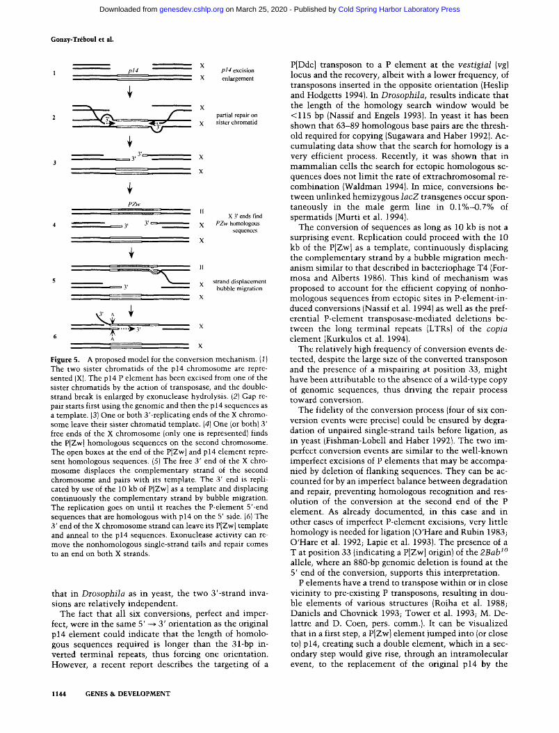
The mechanism of conversion
Engels and co-workers (Engels et al. 1990; Gloor et al. 1991), proposed that P-element transposition occurs through a mechanism similar to the repair of radiation- induced double-strand breaks (Resnick 1976), or gene conversion in yeast (Szostak et al. 1983). The transposase would cut both DNA strands of the chromosome, the P element would be excised from its genomic site, and exonucleases present in the nucleus would enlarge the break. Then the gap could be repaired by copying the missing sequences either from the sister chromatid, hence restoring a copy of the original P element, or from the homologous chromosome, yielding a wild-type copy of the genomic sequences (Engels et al. 1990; Johnson- Schlitz and Engels 1993; Nassif and Engels 1993), or even from homologous sequences inserted at an ectopic site of the genome (Gloor et al. 1991; Nassif et al. 1994). This model was supported by genetic experiments from the Engels' group and by the molecular properties of the P-el- ement transposase (Kaufman and Rio 1992). It led to the prediction that it should be possible to introduce dele-
tions or insertions by conversion to the excision site, a prediction that has been confirmed (Johnson-Schlitz and Engels 1993; Nassif et al. 1994).
According to Engels' model, it is assumed that the double-strand break occurs at or near the terminal re- peats of the P element, thus initiating transposition. The repair process leading to precise or imprecise excision, and synthesis of a new copy of the transposon or conver- sion, is primed by use of the homology between the par- tially degraded strands and the genomic sequences flank- ing the transposon.
The type of conversion events reported in the present work did not require homology between the donor se- quences and the genomic sequences at the converted site: The unique homologous sequences between donor and acceptor sites were located inside and not outside the transposon and were the P-element sequences them- selves. Apparently, the simplest way to reconcile our results with Engels' model is to assume that the double- strand breaks induced by the P-element transposase are internal to the P element. However, although not ex- cluded, this additional assumption is unnecessary, and the data can well be accounted for by the model, as il- lustrated in Figure 5. The resolution of the apparent dis- crepancy is based on the involvement of three templates during the repair process.
Because the 2BabZ(pl4) mutation is viable, the muta- genic males containing both the p14 P element and the source of transposase did not need to, and did not, con- tain an additional copy of the BRC chromosomal region anywhere in the genome. After excision, a repair process leading to conversion could only initiate at G2, after rep- lication, on the sister chromatid. During this repair, the replicating strands copy P-element sequences. At that point, homologous sequences can be found at the ectopic site of the enhancer-trap donor transposon. The conver- sion process is thus initiated. A second change of partner is needed to resolve the conversion. This will create a mismatch at position 33 of the heteroduplex, illustrated by the two facing noncomplementary A bases on the diagram in Figure 5. This mismatch would be repaired further.
The presence of this polymorphic site allowed us to detect chimeric molecules composed of parts of both the p14 and the P[Zw] elements among the convertants. This observation strongly supports the mechanism that we propose. The involvement of three template strands of DNA could well account for certain complex conversion events selected by Gloor et al. (1991). In yeast, plasmid- mediated induction of recombination was shown to in- volve tripartite events between one plasmid and two chromosomes (Silberman and Kupiec 1994).
In addition, the difference between the type of conver- sion event discussed here and those reported by Engels and co-workers (Engels et al. 1990; Gloor et al. 1991) is similar to ends-in versus ends-out recombination in yeast. In ends-in (O type), the regions of homology be- tween the partners are located on one and the other side of the double-strand cuts; in ends-out (f~ type), they are located inside (Hastings et al. 1993). Our results suggest
GENES & DEVELOPMENT 1143
Cold Spring Harbor Laboratory Press on March 25, 2020 - Published by genesdev.cshlp.orgDownloaded from
Gonzy-Tr~boul et al.
p14
, , , , ,
. . . . .
3 ' r . - , ,
. 3 '
. .
PZw : i~ i i ........ 7
3 " - - " f ' ' "~ 3 '
X p14 excision
X enlargement
X partial repair on
X sister chromatid
1I x 3' ends find
X PZw homologous sequences
' ' X
.............. "" ~ . ' ' II r . . . . . . . , , , ~
5 ~ X strand displacement 3' bubble migration
X
, x
6 ^
X
Figure 5. A proposed model for the conversion mechanism. (1) The two sister chromatids of the p14 chromosome are repre- sented (X). The p14 P element has been excised from one of the sister chromatids by the action of transposase, and the double- strand break is enlarged by exonuclease hydrolysis. (2) Gap re- pair starts first using the genomic and then the p 14 sequences as a template. (3) One or both 3'-replicating ends of the X chromo- some leave their sister chromatid template. (4) One (or both) 3' free ends of the X chromosome (only one is represented) finds the P[Zw] homologous sequences on the second chromosome. The open boxes at the end of the P[Zw] and p14 element repre- sent homologous sequences. (5) The free 3' end of the X chro- mosome displaces the complementary strand of the second chromosome and pairs with its template. The 3' end is repli- cated by use of the 10 kb of P[Zw] as a template and displacing continuously the complementary strand by bubble migration. The replication goes on until it reaches the P-element 5'-end sequences that are homologous with p14 on the 5' side. (6) The 3' end of the X chromosome strand can leave its P[Zwl template and anneal to the p14 sequences. Exonuclease activity can re- move the nonhomologous single-strand tails and repair comes to an end on both X strands.
that in D r o s o p h i l a as in yeast, the two 3 '-strand inva- sions are relatively independent.
The fact that all six conversions, perfect and imper- fect, were in the same 5' ~ 3' orientation as the original p14 element could indicate that the length of homolo- gous sequences required is longer than the 31-bp in- verted terminal repeats, thus forcing one orientation. However, a recent report describes the targeting of a
P[Ddc] transposon to a P e lement at the v e s t i g i a l (vg}
locus and the recovery, albeit wi th a lower frequency, of transposons inserted in the opposite orientat ion (Heslip and Hodgetts 1994). In D r o s o p h i l a , results indicate tha t the length of the homology search window would be <115 bp (Nassif and Engels 1993). In yeast it has been shown that 63-89 homologous base pairs are the thresh- old required for copying (Sugawara and Haber 1992). Ac- cumulat ing data show that the search for homology is a very efficient process. Recently, it was shown that in m a m m a l i a n ceils the search for ectopic homologous se- quences does not l imit the rate of ex t rachromosomal re- combinat ion (Waldman 1994). In mice, conversions be- tween unlinked hemizygous l a c Z transgenes occur spon- taneously in the male germ line in 0.1%-43.7% of spermatids (Murti et al. 1994).
The conversion of sequences as long as 10 kb is not a surprising event. Replication could proceed with the 10 kb of the P[Zw] as a template, cont inuously displacing the complementary strand by a bubble migrat ion mech- anism similar to that described in bacteriophage T4 (For- mosa and Alberts 1986). This kind of mechan i sm was proposed to account for the efficient copying of nonho- mologous sequences from ectopic sites in P-element-in- duced conversions (Nassif et al. 1994) as well as the pref- erential P-element t ransposase-mediated deletions be- tween the long terminal repeats (LTRs) of the c o p i a
element (Kurkulos et al. 1994). The relatively high frequency of conversion events de-
tected, despite the large size of the converted transposon and the presence of a mispairing at position 33, might have been attributable to the absence of a wild-type copy of genomic sequences, thus driving the repair process toward conversion.
The fidelity of the conversion process (four of six con- version events were precise) could be ensured by degra- dation of unpaired single-strand tails before ligation, as in yeast (Fishman-Lobell and Haber 1992). The two im- perfect conversion events are similar to the wel l -known imperfect excisions of P elements that may be accompa- nied by deletion of flanking sequences. They can be ac- counted for by an imperfect balance between degradation and repair, preventing homologous recognition and res- olution of the conversion at the second end of the P element. As already documented, in this case and in other cases of imperfect P-element excisions, very little homology is needed for ligation (O'Hare and Rubin 1983; O'Hare et al. 1992; Lapie et al. 1993). The presence of a T at position 33 (indicating a P[Zw] origin) of the 2 B a b 1~
allele, where an 880-bp genomic deletion is found at the 5' end of the conversion, supports this interpretation.
P elements have a trend to transpose wi th in or in close vicinity to pre-existing P transposons, resulting in dou- ble elements of various structures (Roiha et al. 1988; Daniels and Chovnick 1993; Tower et al. 1993; M. De- lattre and D. Coen, pets. comm.). It can be visualized that in a first step, a P[Zw] element jumped into (or close to) p14, creating such a double element, which in a sec- ondary step would give rise, through an int ramolecular event, to the replacement of the original p14 by the
1144 GENES & D E V E L O P M E N T
Cold Spring Harbor Laboratory Press on March 25, 2020 - Published by genesdev.cshlp.orgDownloaded from

Enhancer-trap targeting at Broad-Complex
P[Zw] element. However, we did not find any double element at the p14 site.
Replacements of a defective P element at the vg gene by various P[Ddc] and a P[Zw] donors were recently re- ported [Heslip and Hodgetts (1994); Staveley et al. (1994), respectively]. Thus, P-element replacement is not a spe- cific property of either the P[Zw] donor or p14 target element that we used. Previously described P-element t ransposase- induced events (Salz et al. 1987; Geyer et al. 1988) may also be wel l accoun ted for by the present model (Fig. 5).
Perspec t i ve s
The targeted convers ion of an u n m a r k e d P e l e m e n t by an enhancer- t rap t ransposon at reasonable f requencies opens in te res t ing possibil i t ies. It becomes possible to combine the advantages of the two me thods proposed for P-e lement mutagenes i s : sho tgun mutagenes i s w i th the B i r m 2 c h r o m o s o m e con ta in ing 17 defect ive e l emen t s (Robertson et al. 1988), and single-copy mutagenesis , w i th marked e l emen t s (Cooley et al. 1989). Shotgun mu- tagenesis is more eff icient in t e rms of the n u m b e r of inser t ions per gamete, a l lowing PCR screening of an in- sert in a single gene of interest , (Ballinger and Benzer 1989; Kaiser and Goodwin 1990; S6galat et al. 1992), whereas the use of engineered e l emen t s great ly facili- tates loca l iza t ion of the inser t ion, de t e rmina t i on of the pa t te rn of expression by X-gal s taining, c loning by plas- mid rescue, and the recovery of nu l l m u t a n t s by imper- fect excision. Similarly, it has been proposed recent ly to make use of the great var ie ty of P-e lement inser ts avail- able in wi ld- type Drosoph i la strains (Clark et al. 1994). The usefulness of this procedure wil l be greatly im- proved if, in a second step, wi ld- type P e l emen t s are re- placed by enhancer - t rap ones.
Given the increas ing var ie ty of new P-element-der ived tools for D r o s o p h i l a genetics, such as the F L P / F R T sys- tem, the U A S U G A L 4 system, the presence of ovo ~ on autosomes, and the pale t te of hundreds of P e lements already local ized th roughou t the genome, it would be easier to target one of these newly engineered e l emen t s to a site of interest , by use of an outdated, but precisely located, P e lement , as a recipient .
Given the complex i ty and the huge size of the BRC, the d issec t ion of its var ious func t ions and regula tory se- quences could be more amenab le to si te-directed en- hancer t rapping than classical t ransgenesis .
Materials and methods
Genetic nomenclature
Genetic nomenclature follows Lindsley and Zimm (1992) ex- cept that l(1)2Bab, l(1)2Bc, and l(1)2Bd were simplified as 2Bab, 2Bc, and 2Bd, respectively.
Drosophila stocks
The original p14 mutant was isolated and kindly provided by I. Zhimulev (Solovyeva and Belyaeva 1989). The C(1)DX y w f / w
v l(1)44ts/Y stock was used to collect virgins (Busson et al. 1983). The C(1)DX y w f / w v l(1)44tS/yeY67g19.1 was con- structed in our laboratory. The rearranged Y chromosome of this stock was provided by I. Zhimulev. The X-transposed frag- ment extends from 1A1 to 2B18-19, thus covering the BRC lo- cus at 2B5 (Belyaeva et al. 1980). The BRC alleles were gener- ously provided by J. Fristrom (br 28, 2BabS), G. Guild (rbp a, rpb s, 2Bdl), and I. Zhimulev [Df(1)S39, npr 6, 2Bc 1, 2Bc 2] and are de- scribed in Lindsley and Zimm (1992) except for the br e8 allele, which corresponds to an insertion of a truncated P element in a coding exon (DiBello et al. 1991) and for the 2Bab 5 allele which bears an inversion whose breakpoint separates the promoters from the coding exons (C. Bayer, pers. comm.). The following alleles are considered to be amorphic: npr 6 (Belyaeva et al. 1981), 2Bc I (Kiss et al. 1988), 2Bc 2, (B. Sebban, J. Deutsch, and G. Gonzy-Trdboul, unpubl.), and bl ~8, 2Bab s (C. Bayer, pets. comm.). The balancer chromosomes used throughout the crosses were FM6, I(I)69 i (Belyaeva et al. 1980) or Binsn (Lind- sley and Zimm 1992). The stable source of transposase was on a Dr, P/A2-3](99B) chromosome (Robertson et al. 1988). The CyO, P[Zw] chromosome was constructed and kindly provided by J.-M. Dura {Laboratoire de Biologie Cellulaire IV; Universit6 Paris-Sud, Orsay, France). The P[Zw] element came from mobi- lization of an enhancer-trap element constructed by E. Bier {Bier et al. 1989). Fly stocks were raised at 20~ or 25~ and crossed at 25~ or 29~ on a standard corn meal/sugar/yeast/agar me- dium.
Genetic screens
The mating scheme for selecting convertants is described in detail in Results and in Figure 4. Five hundred seventy-four Go w p14/yeY67g19.1; CyO P[Zw]/+ ; Dr P[za2-3](99B)/+ males were individually crossed to C(1)DX y w f /y2Y6 7gl 9.1 females. In 471 crosses, at least one eye-colored, Cy +, Dr + male ap- peared, tracing a transposition or conversion event. As many as 544 independent males could be selected from these crosses. This figure is higher than the actual number of favorable crosses, because in some crosses males of various colors ap- peared. In this case, a representative of each color was picked up. They were kept and qualified as independent only when supported by further genetic analysis. By use of attached X, a simple genetic analysis could discriminate between autosomal and X-linked transposition or conversion. Autosomal lines were discarded. When a double event affected both an autosome and the sex chromosome, the two transposons were separated by segregation. Because only attached X and X balancers were used in our genetic analysis, multiple events affecting only the X chromosome could not segregate. One hundred and fifty-five independent X-linked lines were analyzed.
To mobilize the p14 element 160 Go males carrying the p14 allele on the X chromosome and the Sb, P/a2-3]{99B) source of transposase on the third chromosome were individually crossed with C(1)DXy wf/yeY67g19.1 females. Ten Gl Sb + males were individually recovered from each cross and were expanded by crossing with the same C(1)DX y w f/y2Y67g19.1 females. The crosses were brooded 5 days at 25~ after which time the male parents were individually crossed with C(1)DX y w f l y females to screen for viability. Among 1523 viable males without any BRC phenotype, 50 (progeny of 10 different Go males) were sub- mitted to molecular analysis. Among these, seven (progeny of five different Go males) could be classified as true revertants because of the precise excision of the p14 element, within the limits of a Southern analysis.
To screen for 2Bab 8 (M 1) revertants, a similar experiment was performed: females carrying the novel convertant chromosome,
GENES & DEVELOPMENT 1145
Cold Spring Harbor Laboratory Press on March 25, 2020 - Published by genesdev.cshlp.orgDownloaded from

Gonzy-Tr6boul et al.
that is, 2Bab 8 w (this work), balanced with FM6, 1(1)69 j were mass-mated to +/y2Y67gl 9.1; Dr P/A2-3](99B) males. Sixty-five phenotypically (B + Dr) Go males of the offspring were recovered and individually mated with C(1)DX females. Thirty-five inde- pendent viable (white Dr + ) progeny males were expanded and crossed with npr 6 mutant females to test for genetic reversion.
DNA extraction and Southern analysis
Hemizygous males were collected as wandering third instar lar- vae. DNA was extracted according to Lapie et al. (1993). South- ern blotting was performed according to Sambrook et al. (1989).
Plasmid probes
The pw25.7bwc plasmid, used as a P-element-specific probe, was kindly provided by D. Anxolab4hhre (Institut J. Monod, Paris, France). It bears the HindIII(39)-AvaII(2882) fragment of the P element and no genomic sequences. The pLB1 plasmid was constructed by cloning in a Bluescript vector (Stratagene) a 4.8-kb SalI(170.5)-SalI(176) fragment [coordinates from Chao and Guild (1986)] taken from the P205 BRC clone of the ~, Charon4 library kindly provided by I. Zhimulev (Belyaeva et al. 1987). It was used as a BRC-specific probe (G probe on Fig. 3). Plasmids were labeled by nick translation (Rigby et al. 1977) to a specific activity of 1 x 10 ~ to 2x 10 ~ cpm/~g.
In situ hybridization
In situ hybridization on polytene chromosomes was performed according to Ronsseray et al. (1991) with the p~25.1 plasmid as a probe.
Molecular analysis of the P/Zw] conversion mutants
Cloning by plasmid rescue was performed on M 1 DNA digested with EcoRI, SaclI, PstI, or BglII, according to Pirrotta (1986). Four different plasmids were obtained by this method: pLB27 and pLB28 carried the 3' junction, pLB29 and pLB30, the 5' junction.
DNA corresponding to a single fly was amplified in a 20-~1 volume over 30 cycles with 2% formamide in the mixture (Sarkar et al. 1990). The sizes of the amplified products were determined from 1.5% agarose gel electrophoresis by comparing them with products amplified from the pLB27 plasmid (down- stream product) or from the pLB29 one tupstream product) with the same pair of primers. Direct sequencing of the PCR products was performed essentially according to Dod4 et al. (1990), with 10% formamide in the sequencing mixture (Zhang et al. 1991).
P-specific primers used for PCR were T1 189-108), 5'-CGTC- CGCACACAACCTTTCC-3', and T2 (2785-2804), 5'-TCGCT- GTCTCACTCAGACTC-3'. BRC-specific primers are listed from 5' to 3' according to the BRC transcription orientation (see Fig. 3b). A1, 5'-GTATGTGTGCCACATTGCGC-3'; A2, 5'-GC- TCTAGAGGAGGAGGGGCGGCGGG-3'; A3, 5'-TCCGTGG- AGGGATAGGAACC-3'; A4, 5'-CCAATGTGCCAGCATGC- CCC-3'; and A5, 5'-CTGCGCCCAGAATCGATGCG3'.
BRC primers A1, A2, and A5, were used for amplification; A3 and A4 for the subsequent sequencing. The size of the p14 ele- ment was estimated by amplification with a single primer com- plementary to the P-element terminal repeats (positions 3-31).
Histochemical staining
Third-instar larvae were staged on standard corn meal medium containing 0.05% bromophenol blue. [3-Galactosidase activity
was tested in larvae and pupae according to Lemaitre and Coen (1991) and in adult ovaries according to Lemaitre et al. (1993).
A c k n o w l e d g m e n t s
We thank the students of the Module de Dynamique du G4- nome de l'Universit6 Pierre et Marie Curie for their help in genetic screening, C. Rigolot for her technical assistance in se- quencing, and P. Feynerol and K. Taalba for their help in main- taining fly stocks. We are very grateful to C. Bayer, J. Fristrom, I. Emery, and G. Guild for sharing results before publication, to G. Guild, C. Bayer, and I. Zhimulev for sending DNAs, to I. Zhimulev and E. Belyeva, C. Bayer, J. Fristrom, and G. Guild for sending fly stocks and to C. Bayer, D. Coen, M. Delattre, and F. Schweisguth for stimulating discussions and suggestions. We are particularly grateful to J. Haber, J.-L. Rossignol, F. Schweis- guth, and A. Kropfinger for their reading and comments on the manuscript, with special thanks to Cindy Bayer. This work was supported by the CNRS and grant 6294 from the Association pour la Recherche sur le Cancer to J.-A. L.
The publication costs of this article were defrayed in part by payment of page charges. This article must therefore be hereby marked "advertisement" in accordance with 18 USC section 1734 solely to indicate this fact.
R e f e r e n c e s
Andres, A.I. and C.S. Thummel. 1992. Hormones, puffs and flies: The molecular control of metamorphosis by ecdysone. Trends Genel. 8: 132-138.
Ashburner, M., C. Chihara, P. Meltzer, and G. Richards. 1974. Temporal control of puffing activity in polytcne chromo- somes. Coht Sprin~g Harbor Syrup. Quant. Biol. 38: 655-662.
Ballinger, D.G. and S. Benzer. 1989. Targeted gene mutations in Drosophila. Proc. Natl. Acad. Sci. 86: 9402-9406.
Belyaeva, E.S., M.G. Aizenzon, V.F. Semeshin, I. Kiss, K. Koc- zka, E.M. Baricheva, T.D. Gorelova, and I.F. Zhimulev. 1980. Cytogenetic analysis of the 2B3-4-2B 11 region of X chromo- some of Drosophila melanogaster, I: Cytology of the region and mutant comptementation groups. Chromosoma 81: 281-306.
Belyaeva, E.S., I.E. Vlassova, Z.M. Biyasheva, V.T. Kakpakov, G. Richards, and I.F. Zhimulev. 1981. Cytogenetic analysis of the 2B3-4-2B11 region of the X chromosome of Drosophila melanogaster. II. Changes in the 20-OH ecdysone puffing caused by genetic defects of puff 2B5. Chromosoma 84: 207- 219.
Belyaeva, E.S., M.O. Protopopov, E.M. Baricheva, V.F. Se- meshin, M.L. Izquierdo, and I.F. Zhimulev. 1987. Cytoge- netic analysis of the 2B3-4-2B11 region of the X chromosome of Drosophila melanogaster, VI: Molecular and cytological mapping of the ecs locus and the 2B puff. Chromosoma 95: 295-310.
Belyaeva, E.S., M.O. Protopopov, E.B. Dubrovsky, and I.F. Zhimulev. 1989. Cytogenetic analysis of ecdysteroid action. In Ecdysone: From chemistry to mode of action (ed. J. Kool- man), pp. 368-376. Georg Thieme Verlag, Stuttgart, Ger- many.
Bier, E., H. Vaessin, S. Sheperd, K. Lee, K. McCall, S. Barbel, L. Ackerman, R. Carretto, T. Uemura, E. Grell, L.Y. Jan, and Y.N. Jan. 1989. Searching for pattern and mutation in the Drosophila genome with a P-lacZ vector. Genes & Dev. 3: 1273-1287.
Busson, D., M. Gans, K. Komotopoulou, and M. Masson. 1983. Genetic analysis of three dominant female sterile mutations
1146 GENES & DEVELOPMENT
Cold Spring Harbor Laboratory Press on March 25, 2020 - Published by genesdev.cshlp.orgDownloaded from

Enhancer-trap targeting at Broad-Complex
located on the X chromosome of Drosophila melanogaster. Genetics 105: 309-325.
Chao, A.T. and G.M. Guild. 1986. Molecular analysis of the ecdysterone-inducible 2B "early" puff in Drosophila mela- nogaster. EMBO J. 5: 143-150.
Clark, A.G., S. Silveria, W. Meyers, and C.H. Langley. 1994. Nature screen: An efficient method for screening natural populations of Drosophila for targeted P-element insertions. Proc. Natl. Acad. Sci. 91: 719-722.
Cooley, L., C. Berg, R. Kelley, D. McKearin, and A. Spradling. 1989. Identification and cloning Drosophila genes by single P element insertiona] mutagenesis. Prog. Nucleic Acids Res. 36: 99-109.
Daniels, S.B. and A. Chovnick. 1993. P-Element transposition in Drosophila melanogaster--an analysis of sister-chromatid pairs and the formation of intragenic secondary insertions during meiosis. Genetics 133: 623-636.
DiBello, P.R., D.A. Withers, C.A. Bayer, J.W. Fristrom, and G.M. Guild. 1991. The Drosophila Broad-Complex encodes a fam- ily of related, zinc finger proteins. Genetics 129: 385-397.
Dod6, C., J. Rochette, and R. Krishnamoorthy. 1990. Locus as- signment of human a globin by selective amplification and direct sequencing. Br. /. Haematol. 76" 275-281.
Emery, I.F., V. Bedian, and G.M. Guild. 1994. Differential ex- pression of the Broad-Complex transcription factors may forecast tissue-specific developmental fates during Droso- phila metamorphosis. Development 120: 3275-3287.
Engels, W.R., D.M. Johnson-Schlitz, W.B. Eggleston, and J. Sved. 1990. High-frequency P element loss in Drosophila is ho- molog dependent. Cell 62: 515-525.
Engels, W.R., C.R. Preston, and D.M. Johnson-Schlitz. 1994. Long-range cis preference in DNA homology search over the length of a Drosophila chromosome. Science 263: 1623- 1625.
Fauvarque, M.O. and J.M. Dura. 1993. polyhomeotic regulatory sequences induce developmental regulator-dependent varie- gation and targeted P-element insertions in Drosophila. Genes & Dev. 7: 1508-1520.
Fishman-Lobell, J. and J.E. Haber. 1992. Removal of non-homol- ogous DNA ends in double-strand break recombination: The role of the yeast ultraviolet repair gene RADI. Science 258: 480-484.
Formosa, T. and B.M. Alberts. 1986. DNA synthesis dependent on genetic recombination: Characterization of a reaction catalyzed by purified bacteriophage T4 proteins. Cell 47: 793-806.
Geyer, P.K., K.L. Richardson, V.G. Cortes, and M.M. Green. 1988. Genetic instability in Drosophila melanogaster: P-el- ement mutagenesis by gene conversion. Proc. Natl. Acad. Sci. 85: 6455-6459.
Gloor, G.B., N.A. Nassif, D.M. Johnson-Schlitz, C.R. Preston, and W.R. Engels. 1991. Targeted gene replacement in Droso- phila via P element-induced gap repair. Science 253:1110- 1117.
Hama, C., Z. Ali, and T.B. Komberg. 1990. Region-specific recom- bination and expression are directed by portions of the Droso- phila engrailed promoter. Genes & Dev. 4: 1079-1093.
Hastings, P.J., C. McGill, B. Shafer, and J.N. Strathern. 1993. Ends-in vs. ends-out recombination in yeast. Genetics 135: 973-980.
Heslip, T.R. and R.B. Hodgetts. 1994. Targeted transposition at the vestigial locus of Drosophila melanogaster. Genetics 1 3 8 : 1 1 2 7 - 1 1 3 5 .
Huang, R.Y. and W.C. Orr. 1992. Broad-Complex function dur- ing oogenesis in Drosophila melanogaster. Dev. Genet. 13: 277-288.
Huet, F., C. Ruiz, and G. Richards. 1993. Puffs and PCR: The in vivo dynamics of early gene expression during ecdysone re- sponses in Drosophila. Development 118: 613-627.
Johnson-Schlitz, D.M. and W.R. Engels. 1993. P-element-in- duced interallelic gene conversion of insertions and dele- tions in Drosophila melanogaster. Mol. Cell. Biol. 13: 7006- 7018.
Kaiser, K. and S.F. Goodwin. 1990. "Site-selected" transposon mutagenesis of Drosophila. Proc. Natl. Acad. Sci. 87: 1686- 1690.
Karess, R.E. and G.M. Rubin. 1984. Analysis of P transposable element function in Drosophila. Cell 38: 135-146.
Karim, F.D., G.M. Guild, and C.S. Thummel. 1993. The Droso- phila Broad-Complex plays a key regulatory role in control- ling ecdysone-regulated gene expression at the onset of metamorphosis. Development 118: 977-988.
Kaufman, P.D. and D.C. Rio. 1992. P-element transposition in vitro proceeds by a cut-and-paste mechanism and uses GTP as a cofactor. Cell 69: 27-39.
Kiss, I., A.H. Beaton, J. Tardiff, D. Fristrom, and J.W. Fristrom. 1988. Interactions and developmental effects of mutations in the Broad-Complex of Drosophila melanogaster. Genetics 118: 247-259.
Kurkulos, M., J.M. Weinberg, D. Roy, and S.M. Mount. 1994. P-element mediated in vivo deletion analysis of white-apri- cot: Deletions between direct repeats are strongly favored. Genetics 136:1001-1011.
Lapie, P., F. Nasr, J.A. Lepesant, and J. Deutsch. 1993. Deletion scanning of the regulatory sequences of the Fbpl gene of D. melanogaster using P transposase-induced deficiencies. Ge- netics 135: 801-816.
Lemaitre, B. and D. Coen. 1991. P regulatory products repress in vivo the P promoter activity in P-lacZ fusion genes. Proc. Natl. Acad. Sci. 88: 4419-4423.
Lemaitre, B., S. Ronsseray, and D. Coen. 1993. Maternal repres- sion of the P-element promoter in the germline of Droso- phila melanogaster--a model for the P cytotype. Genetics 135: 149-160.
Lindsley, D.L. and G. Zimm. 1992. The genome of Drosophila melanogaster. Academic Press, San Diego, CA.
Mazina, O., E.S. Belyaeva, and I.F. Zhimulev. 1991. Cytogenet- ical analysis of the 2B3-4-2B 11 region of the X chromosome of Drosophila melanogaster. VII: Influence of the ecs locus on female fertility. Mol. Gen. Genet. 225: 99-105.
Murti, J.R., M. Bumbulis, and J.C. Schimenti. 1994. Gene con- version between unlinked sequences in the germline of mice. Genetics 137: 837-843.
Nassif, N. and W. Engels. 1993. DNA homology requirements for mitotic gap repair in Drosophila. Proc. Natl. Acad. Sci. 90: 1262-1266.
Nassif, N., J. Penney, S. Pal, W.R. Engels, and G.B. Gloor. 1994. Efficient copying of nonhomologous sequences from ectopic sites via P-element-induced gap repair. Mol. Cell. Biol. 14: 1613-1625.
O'Hare, K. and G.M. Rubin. 1983. Structures of P transposable elements and their sites of insertion and excision in the Drosophila melanogaster genome. Cell 34: 25-35.
O'Hare, K., A. Driver, S. McGrath, and D.M. Johnson-Schiltz. 1992. Distribution and structure of cloned P-elements from the Drosophila melanogaster P-strain ~r. Genet. Res. 60: 33- 41.
O'Kane, C.J. and W.I. Gehring. 1987. Detection in situ of geno- mic regulatory elements in Drosophila. Proc. Natl. Acad. Sci. 84: 9123-9127.
Pirrotta, V. 1986. Cloning Drosophila genes. In Drosophila: A practical approach (ed. D.B. Roberts), pp. 83-110. IRL Press,
GENES & DEVELOPMENT 1147
Cold Spring Harbor Laboratory Press on March 25, 2020 - Published by genesdev.cshlp.orgDownloaded from

Gonzy-Tr6boul et al.
Oxford, UK. Resnick, M.A. 1976. The repair of double strand breaks in DNA:
A model involving recombination. J. Theor. Biol. 59: 97- 106.
Rigby, P.W.J., M. Diekmann, C. Rhodes, and P. Berg. 1977. La- belling deoxyribonucleic acid to high specificity in vitro by nick translation with DNA polymerase I. J. Mol. Biol. 113: 237-251.
Robertson, H.M., C.R. Preston, R.W. Phillis, D.M. Johnson- Schlitz, W.K. Benz, and W.R. Engels. 1988. A stable genomic source of P element transposase in Drosophila melano- gaster. Genetics 118: 461-470.
Roiha, H., G.M. Rubin, and K. O'Hare. 1988. P element inser- tions and rearrangements at the singed locus of Drosophila melanogaster. Genetics 119: 75-83.
Ronsseray, S., M. Lehmann, and D. Anxolab4hhre. 1991. The maternally inherited regulation of P elements in Drosophila melanogaster can be elicited by two copies at cytological site 1A on the X chromosome. Genetics 129: 501-512.
Rubin, G.M. and A.C. Spradling. 1982. Genetic transformation of Drosophila with transposable element vectors. Science 218: 348-353.
1983. Vectors for P element-mediated gene transfer in Drosophila. Nucleic Acids Res. 11: 6341-6351.
Salz, H.K., T.W. Cline, and P. Schedl. 1987. Functional changes associated with structural alterations induced by mobiliza- tion of a P element inserted in the sex-lethal gene of Droso- phila. Genetics 117: 221-231.
Sambrook, J., F.F. Fritsch, and T. Maniatis. 1989. Molecular cloning: A laboratory manual. Cold Spring Harbor Labora- tory Press, Cold Spring Harbor, New York.
Sampedro, J., J. Galceran, and M. Izquierdo. 1989. Mutation mapping of the 2B5 ecdysone locus in Drosophila melano- gaster reveals a long-distance controlling element. MoI. Cell. Biol. 9:3588-3591.
Sarkar, G., S. Kalpener, and S.S. Sommer. 1990. Formamide can dramatically improve the specificity of PCR. Nucleic Acids Res. 24: 7465.
S4galat, L., R. Perichon, J.P. Bouly, and ].A. Lepesant. 1992. The Drosophila pourquoi-pas?/wings-down zinc finger protein: Oocyte nucleus localization and embryonic requirement. Genes & Dev. 6: 1019-1029.
Silberman, R. and M. Kupiec. 1994. Plasmid-mediated induc- tion of recombination in yeast. Genetics 137: 41--48.
Solovyeva, I.V. and E.S. Belyaeva. 1989. Cytogenetic analysis of the region 2B1,2-2B9,10 of the X chromosome of Drosophila melanogaster. VIII. Genetic analysis of mutations obtained in the system of P-M hybrid dysgenesis. Genetika 25: 1209- 1217.
Staveley, B.E., R.B. Hodgetts, S.L. O'Keefe, and J.B. Bell. 1994. Targeting of an enhancer trap to vestigial. Dev. Biol. 165: 290-293.
Sugawara, N. and J.E. Haber. 1992. Characterization of double- strand break-induced recombination: Homology require- ments and single-stranded DNA formation. Mol. Cell. Biol. 12: 563-575.
Szostak, J.W., T.L. Orr-Weaver, and R.J. Rothstein. 1983. The double strand repair model for recombination. Ceil 33: 25- 35.
Tower, J., G.H. Karpen, N. Craig, and A.C. Spradling. 1993. Preferential transposition of Drosophila P elements to nearby chromosomal sites. Genetics 133: 347-359.
Waldman, A.S. 1994. The search for homology does not limit the rate of extrachromosomal homologous recombination in mammalian cells. Genetics 136: 597-605.
Wilson, C., H.J. Bellen, and W.J. Gehring. 1990. Position effects
on eucaryotic gene expression. Annu. Rev. Cell Biol. 6: 679- 714.
Zhang, W., G. Hu, and A. Deisseroth. 1991. Improvement of PCR sequencing by formamide. Nucleic Acids Res. 19: 6649.
1148 GENES & DEVELOPMENT
Cold Spring Harbor Laboratory Press on March 25, 2020 - Published by genesdev.cshlp.orgDownloaded from

10.1101/gad.9.9.1137Access the most recent version at doi: 9:1995, Genes Dev.
G Gonzy-Tréboul, J A Lepesant and J Deutsch melanogaster.Enhancer-trap targeting at the Broad-Complex locus of Drosophila
References
http://genesdev.cshlp.org/content/9/9/1137.full.html#ref-list-1
This article cites 64 articles, 40 of which can be accessed free at:
License
ServiceEmail Alerting
click here.right corner of the article or
Receive free email alerts when new articles cite this article - sign up in the box at the top
Copyright © Cold Spring Harbor Laboratory Press
Cold Spring Harbor Laboratory Press on March 25, 2020 - Published by genesdev.cshlp.orgDownloaded from