enhancement of diosgenin distribution in the skin by
TRANSCRIPT

36 Vol. 36, No. 1Biol. Pharm. Bull. 36(1) 36–40 (2013)
© 2013 The Pharmaceutical Society of Japan
Regular Article
Enhancement of Diosgenin Distribution in the Skin by Cyclodextrin Complexation Following Oral AdministrationMasaki Okawara, Yoshihiro Tokudome, Hiroaki Todo, Kenji Sugibayashi, and Fumie Hashimoto*Faculty of Pharmaceutical Sciences, Josai University; 1–1 Keyakidai, Sakado, Saitama 350–0295, Japan.Received May 21, 2012; accepted October 4, 2012
Orally administrated diosgenin, a steroidal saponin found in several plants including Dioscorea villosa, recovers skin thickness reduced in ovariectomized mice, and plays an important role in the treatment of hyperlipidemia. Thus, diosgenin is an active element of cosmeceutical and dietary supplements. However, we have already elucidated that the skin distribution and absolute oral bioavailability of diosgenin is very low. The aim of this study is to evaluate the efficacy of diosgenin–cyclodextrin (CD) complexes in improving the skin concentration of diosgenin. The formation of the CD complex was indicated by powder X-ray diffrac-tion (XRD), differential scanning calorimetry (DSC), and scanning electron microscope (SEM) studies. Oral administration of the diosgenin/β-CD complex resulted in a significant enhancement in terms of the skin distribution of diosgenin, maximum plasma level (Cmax), area under the plasma concentration–time curve (AUC), and absolute oral bioavailability over those of the drug alone. These results suggest that the inclusion complex of diosgenin/β-CD can be used to improve low skin content of diosgenin.
Key words diosgenin; skin distribution; cyclodextrin; intravenous administration; oral administration
Diosgenin (Fig. 1) is a steroid sapogenin which can be found in several species of the genus Dioscorea villosa, Costus speciosus, and Trigonella foenum greaecum.1) Previ-ous investigations have shown that diosgenin was effective in treating various diseases such as hyperglycemia and hy-perlipidemia.2,3) It was established as a starting material for the production of steroidal hormones in the pharmaceutical industry.4) Diosgenin has been applied to hormone replace-ment therapy in menopausal women.5,6) Estrogen enhanced the proliferation of estrogen-dependent cancer cells,7) whereas di-osgenin inhibited the proliferation of breast cancer cells.8) Oral administration of diosgenin improved reduced skin thickness in ovariectomized mice.9) In B16 melanoma cells, it inhibits melanogenesis by activating the phosphatidylinositol-3-kinase pathway.10) Thus, diosgenin was noted as an active element of cosmeceutical and dietary supplements. Furthermore, dios-genin was expected for systemic action by oral administration. However, only a few reports have investigated the disposition and skin distribution of diosgenin.
In our previous study, the skin distributions and absolute oral bioavailability of diosgenin was very low.11) Diosgenin aqueous solubility was found to be 0.95 µg/mL (calculated using Advance Chemistry Development Software V8.14 for Solaris). This low oral bioavailability may be caused by dios-genin’s low solubility in water.
Cyclodextrins (CDs) show a remarkable ability to form inclusion complexes with diverse lipophilic molecules that fit inside the cavity. This phenomenon modifies solubility, dis-solution rates, and bioavailability of guest molecules. Inclu-sion complexation of a number of drugs with β-CD has been reported.12–14)
In the present study, we prepared and characterized com-plexes of diosgenin with several CDs. The effects of prepared CD inclusion complexes in the oral administration of diosgen-in were investigated on skin content and oral bioavailability of diosgenin.
MATERIALS AND METHODS
Materials Diosgenin was purchased from Sigma-Aldrich (St. Louis, MO, U.S.A.). Polyoxyethylene hydrogenated castor oil 60 (HCO-60) was supplied from Nikko Chemicals (Tokyo, Japan). Sodium pentobarbital was obtained from Kyoritsu Sei-yaku Co. (Tokyo, Japan). 6-Methyl diosgenin and other chemi-cals were obtained from Wako Pure Chemical Industries, Ltd. (Osaka, Japan).
Phase Solubility Study Solubility studies were performed as described earlier.15) Diosgenin was dissolved in methanol and dispensed into each test tube. Methanol was removed by evaporation in a 40°C heating block. Different concentrations of α, β, and γ-CD solutions (2 mL) were added to 0.2 mg of diosgenin. Each CD concentrations were ranging from 0 to 10 mm. These sample solutions were shaken in an incubator at 37°C for 24 h. The solution was filtered using 0.2 µm mem-brane filters. Diosgenin content was determined by a LC/MS system. Separation was achieved by a Michrom Biosources Inc. MXY01-01 (Auburn, CA, U.S.A.) with a Tosoh TSK gel ODS-100V column (2.0×50 mm, 3 µm) (Tokyo, Japan) at room temperature. The mobile phase consisted of methanol (90%) and H2O (10%) containing 10 mm ammonium acetate. Flow rate was set to 150 µL/min and detection was carried out
* To whom correspondence should be addressed. e-mail: [email protected]
Fig. 1. Molecular Structure of DiosgeninThe authors declare no conflict of interest.

January 2013 37
using a Thermo Fisher LCQ DECA XPPlus mass spectrometer (Waltham, MA, U.S.A.).
Preparation of Mixture and Complex In previous study, it was found that diosgenin and β-CD form 1 : 2 complexes.11) The complexes were prepared by co precipitate method. Briefly, diosgenin (1.6 mmol) and each CD (3.2 mmol) were shaking in 100 mL water at 37°C for 24 h. The solid was col-lected, dried, and ground into a fine powder using a mortar and pestle. The physical mixture of diosgenin and each CD was prepared by mixing individual components in the mor-tar and pestle in 1 : 2 molar ratios of diosgenin and each CD. These samples were subsequently analyzed by powder X-ray diffraction (XRD), differential scanning calorimetry (DSC) and scanning electron microscope (SEM).
Powder X-Ray Diffraction Analysis XRD patterns of di-osgenin, CDs, physical mixtures and complexes were recorded on a Rigaku Mini FlexII (Tokyo, Japan) using Ni-filtered, CuKα radiation, a voltage of 30 kV, and a 15 mA current. The instrument was operated with a scanning rate of 2 degree/min over the 2θ range of 0–30 degree.
Differential Scanning Calorimetry Analysis DSC spectra of diosgenin, CDs, physical mixtures and complexes were obtained using a Rigaku Thermo plus EVO DSC 8230 (Tokyo, Japan). Samples (4 to 5 mg) were weighed in crimped aluminum pans and heated from 50 to 230°C, at a constant scanning rate of 10°C/min with nitrogen purging (50 mL/min).
Scanning Electron Microscope The morphology of diosgenin, CDs, physical mixtures and complexes were de-termined using a Hitachi High-Technologies S-3000N (Tokyo, Japan), operated at an accelerating voltage of 15 kV. Samples were mounted onto aluminum stub 5×5 mm graphite tape and sputter-coated with gold.
Animals Mail Wistar rats (200 to 250 g) were provided from Japan SLC (Hamamatsu, Shizuoka, Japan). Animals were housed under a 12 h light and dark cycle in a tempera-ture controlled room (25°C). They had free access to food and water. All procedures were approved by the Ethics Committee of Josai University (Sakado, Saitama, Japan) in accordance with the National Institute of Health (Tokyo, Japan).
Pharmacokinetic Studies Intravenous and oral adminis-tration studies were performed to compare the pharmacoki-netic parameters and bioequivalence of diosgenin. Rats were fasted overnight for at least 12 h prior to dosing. Diosgenin and its CD complex were dissolved or suspended in physiolog-ical saline containing 1% HCO-60. In intravenous administra-tion, 60 µg/mL of diosgenin solution was prepared and 120 µg/kg of diosgenin was injected into the tail vein. In oral admin-istration, 50 mg/mL of diosgenin suspension was prepared and 100 mg/kg of diosgenin was administrated. In the physical mixture group, the diosgenin suspension and each CD solution were simultaneously administered. Skin samples were col-lected at 4, 6, and 8 h after oral administration. At time range from 0 to 120 h, blood was collected in heparinized tubes by tail vein and separated by centrifugation immediately. Each sample was stored at −30°C until analyzed.
Analytical Procedure For the analysis of diosgenin in skin and plasma samples, 6-methyl diosgenin was used as an internal standard. Skin samples (1 cm2) were minced and sonicated for 20 min in methanol. Plasma samples were added to three folds of methanol and mixed by a vortex mixer for 1 min. These samples were centrifuged at 15000×g for 5 min.
Analysis was achieved by a LC/MS system.Pharmacokinetics and Statistical Analysis Pharmacoki-
netics analysis was performed with least squares methods. The area under the plasma concentration–time curve (AUC) was calculated by the liner trapezoidal rule. The absolute bioavail-ability, was determined as AUC oral/AUC intravenous, using mean AUC values for oral and intravenous doses. To assess the significance of differences between two groups, Dennett’s multiple comparison tests was used. A p value of less than 0.05 was termed significant.
RESULTS AND DISCUSSION
Phase Solubility Study Figure 2 shows the phase solu-bility diagrams obtained for diosgenin against each α, β and γ-CD concentration in distilled water, where the solubility curves can be classified as type Ap, suggesting high order complexation. The stability constants (K11 and K12) were de-fined by Eqs. 1 and 2 where [S] and [L] represent molar con-centrations of free diosgenin and CD, respectively. The S0 is the solubility of diosgenin in the absence of CD. The Seq and Leq are total concentration of diosgenin and CD, respectively. The stability constants were determined by analyzing Eqs. 3 and 4 by the nonlinear least square method at 0–10 mm of each CD.16)
11 0[ ] / [ ]K SL S L= (1)
12 2[ ] / [ ][ ]K SL SL L= (2)
eq 0 2[ ] [ ]S S SL SL= + + (3)
eq 2[ ] [ ] 2[ ]L L SL SL= + + (4)
These results indicate that the small cavity size of α-CD resulted in low inclusion of diosgenin (K11=100 m−1 and K12=860 m−1), whereas β-CD (K11=5680 m−1 and K12=1160 m−1) and γ-CD (K11=4550 m−1 and K12=510 m−1) have large cavity sizes, which enhance effective interactions with diosgenin.
Fig. 2. Phase Solubility Diagrams of Diosgenin/α-CD (a), Diosgenin/β-CD (b), and Diosgenin/γ-CD (c) at 37°C
38 Vol. 36, No. 1
Binding of diosgenin with γ-CD was less than that with β-CD, suggesting that the cavity size of γ-CD is too large for dios-genin.17)
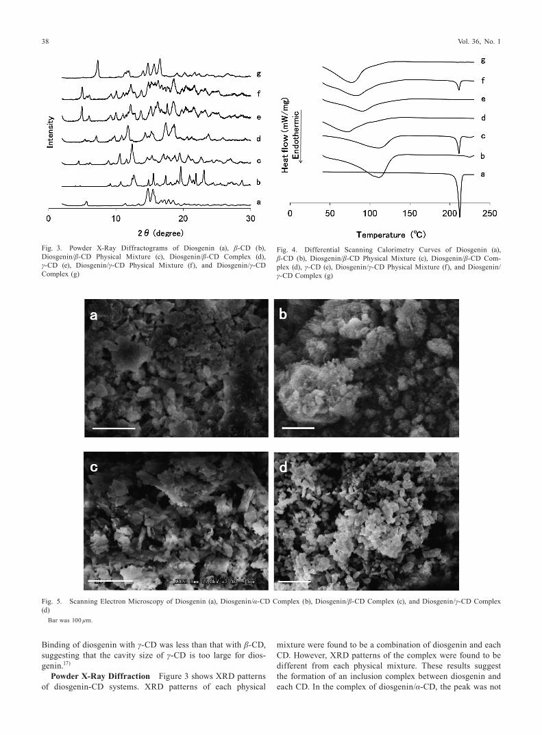
Powder X-Ray Diffraction Figure 3 shows XRD patterns of diosgenin-CD systems. XRD patterns of each physical
mixture were found to be a combination of diosgenin and each CD. However, XRD patterns of the complex were found to be different from each physical mixture. These results suggest the formation of an inclusion complex between diosgenin and each CD. In the complex of diosgenin/α-CD, the peak was not
Fig. 3. Powder X-Ray Diffractograms of Diosgenin (a), β-CD (b), Diosgenin/β-CD Physical Mixture (c), Diosgenin/β-CD Complex (d), γ-CD (e), Diosgenin/γ-CD Physical Mixture (f), and Diosgenin/γ-CD Complex (g)
Fig. 4. Differential Scanning Calorimetry Curves of Diosgenin (a), β-CD (b), Diosgenin/β-CD Physical Mixture (c), Diosgenin/β-CD Com-plex (d), γ-CD (e), Diosgenin/γ-CD Physical Mixture (f), and Diosgenin/γ-CD Complex (g)
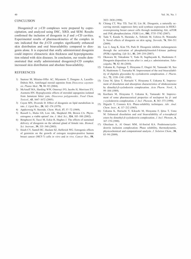
Fig. 5. Scanning Electron Microscopy of Diosgenin (a), Diosgenin/α-CD Complex (b), Diosgenin/β-CD Complex (c), and Diosgenin/γ-CD Complex (d)
Bar was 100 µm.

January 2013 39
different from the diosgenin/α-CD physical mixture (data not shown).
Differential Scanning Calorimetry The thermograms of diosgenin–CD systems are shown in Fig. 4. DSC thermograms revealed the endothermic peak of diosgenin at 210°C. With in-creasing ratios of β or γ-CD in the complex, the diosgenin en-dothermic peak shifted lower (data not shown). Thermograms of the diosgenin/β-CD (1 : 2) and diosgenin/γ-CD (1 : 2) com-plexes showed an absence of the characteristic endothermic peak of the drug.11) This indicates that the inclusion complex has a 1 : 2 (diosgenin/β or γ-CD) formula.
Scanning Electron Microscope In order to investigate whether complexations with each CD could have some influ-ence on particle morphology, SEM was performed for dios-genin and each complex (Fig. 5). It is clear that diosgenin and the diosgenin/α-CD complex have similar crystals. SEM images of each physical mixture have diosgenin and each CD crystal. On the other hand, β and γ-CD complexes presented a parallelogram shape.
Skin Distribution of Diosgenin In Fig. 6, skin distribu-tions of diosgenin were shown at 4, 6, and 8 h after oral ad-ministration of diosgenin, physical mixture, and complex in rats. After the oral dose, the peak level in skin was observed at 6 h. Significant enhancements were found at 4 and 6 h in the β-CD complex and β-CD physical mixture groups than the drug alone. After oral administration, however, high di-osgenin concentrations were obtained in skin, indicating that orally dosed diosgenin was probably distributed through the systemic circulation to skin.
Concentrations of Diosgenin in Plasma Figure 7a shows the mean diosgenin concentration–time profile after intrave-nous administration of diosgenin at a dose of 10 mg/kg in rats. Furthermore, Fig. 7b shows the results of oral administra-tion of diosgenin alone and formulation at a dose of 100 mg/kg. Obtained pharmacokinetic parameters are summarized in Table 1. In diosgenin/β-CD complex groups, significant en-hancements were found in Cmax, AUC, and absolute oral bio-availability compared to drug alone. These data suggest that diosgenin solubility was increased in the intestine because of inclusion complex formation.
Fig. 6. Skin Diosgenin Levels after Oral Doses of Diosgenin and Its β or γ-CD Complexes to Rats
Mean±S.E., n=3–4, * p<0.05 compared to diosgenin.
Fig. 7. Plasma Diosgenin Levels after Intravenous (a) and Oral (b) Doses of Diosgenin and Its β or γ-CD Complexes to Rats
Symbols: square, diosgenin; open circle, diosgenin/β-CD physical mixture; circle, diosgenin/β-CD complex; open triangle, diosgenin/γ-CD physical mixture; triangle, diosgenin/γ-CD complex. Mean±S.E., n=4–8, * p<0.05 compared to diosgenin.
Table 1. Pharmacokinetic Parameters of Diosgenin and Its β or γ-CD Complexes Following Oral Administration to Rats
AUC oral (µg·h/mL)
Cmax (µg/mL)
Bioavailability (%)
β Complex 424.6±37.2* 14.7±2.2* 35.5±3.2*γ Complex 129.7±56.2 2.9±1.3 11.1±4.8β Mixture 91.9±31.1 3.0±1.0 8.0±2.7γ Mixture 34.6±9.8 1.5±0.6 3.2±1.0Diosgenin 53.1±5.8 1.7±0.2 4.3±0.5Mean±S.E., n=4–8, * p<0.05 compared to diosgenin.
40 Vol. 36, No. 1
CONCLUSION
Diosgenin/β or γ-CD complexes were prepared by copre-cipitation, and analyzed using DSC, XRD, and SEM. Results confirmed the inclusion of diosgenin in β and γ-CD cavities. Experimental results of pharmacokinetics of the complex in rats indicated that the β-CD complex significantly enhanced skin distribution and oral bioavailability compared to dios-genin alone. It is expected that orally administrated diosgenin could improve climacteric skin thickness and hyperpigmenta-tion related with skin diseases. In conclusion, our results dem-onstrated that orally administrated diosgenin/β-CD complex increased skin distribution and absolute bioavailability.
REFERENCES
1) Sautour M, Mitaine-Offer AC, Miyamoto T, Dongmo A, Lacaille-Dubois MA. Antifungal steroid saponins from Dioscorea cayenen-sis. Planta Med., 70, 90–92 (2004).
2) McAnuff MA, Harding WW, Omoruyi FO, Jacobs H, Morrison EY, Asemota HN. Hypoglycemic effects of steroidal sapogenins isolated from Jamaican bitter yam, Dioscorea polygonoides. Food Chem. Toxicol., 43, 1667–1672 (2005).
3) Cayen MN, Dvornik D. Effect of diosgenin on lipid metabolism in rats. J. Lipid Res., 20, 162–174 (1979).
4) Applezweig N. Steroids. Chem. Week, 17, 57–72 (1969). 5) Russell L, Hicks GS, Low AK, Shepherd JM, Brown CA. Phyto-
estrogens: a viable option? Am. J. Med. Sci., 324, 185–188 (2002). 6) Benghuzzi H, Tucci M, Eckie R, Hughes J. The effects of sustained
delivery of diosgenin on the adrenal gland of female rats. Biomed. Sci. Instrum., 39, 335–340 (2003).
7) Hsieh CY, Santell RC, Haslam SZ, Helferich WG. Estrogenic effects of genistein on the growth of estrogen receptor-positive human breast cancer (MCF-7) cells in vitro and in vivo. Cancer Res., 58,
3833–3838 (1998). 8) Chiang CT, Way TD, Tsai SJ, Lin JK. Diosgenin, a naturally oc-
curring steroid, suppresses fatty acid synthase expression in HER2-overexpressing breast cancer cells through modulating Akt, mTOR and JNK phosphorylation. FEBS Lett., 581, 5735–5742 (2007).
9) Tada Y, Kanda N, Haratake A, Tobiishi M, Uchiwa H, Watanabe S. Novel effects of diosgenin on skin aging. Steroids, 74, 504–511 (2009).
10) Lee J, Jung K, Kim YS, Park D. Diosgenin inhibits melanogenesis through the activation of phosphatidylinositol-3-kinase pathway (PI3K) signaling. Life Sci., 81, 249–254 (2007).
11) Okawara M, Tokudome Y, Todo H, Sugibayashi K, Hashimoto F. Diosgenin disposition in rats after i.v. and p.o. administration. Yaku-zaigaku, 70, 82–86 (2010).
12) Uekama K, Fujinaga T, Hirayama F, Otagiri M, Yamasaki M, Seo H, Hashimoto T, Tsuruoka M. Improvement of the oral bioavailabil-ity of digitalis glycosides by cyclodextrin complexation. J. Pharm. Sci., 72, 1338–1341 (1983).
13) Ueno M, Ijitsu T, Horiuchi Y, Hirayama F, Uekama K. Improve-ment of dissolution and absorption characteristics of ubidecarenone by dimethyl-β-cyclodextrin complexation. Acta Pharm. Nord., 1, 99–104 (1989).
14) Kurihara M, Hirayama F, Uekama K, Yamasaki M. Improve-ment of some pharmaceutical properties of nocloprost by β- and γ-cyclodextrin complexation. J. Incl. Phenom., 8, 363–373 (1990).
15) Higuchi T, Connors KA. Phase-solubility techniques. Adv. Anal. Chem. Instr., 4, 117–212 (1965).
16) Uekama K, Horiuchi Y, Kikuchi M, Hirayama F, Ijitsu T, Ueno M. Enhanced dissolution and oral bioavailability of α-tocopheryl esters by dimethyl-β-cyclodextrin complexation. J. Incl. Phenom., 6, 167–174 (1988).
17) Ghuzlaan A, Al Omari MM, Al-Sou'od KA. Prednisone/cyclo-dextrin inclusion complexation: Phase solubility, thermodynamic, physicochemical and computational analysis. J. Solution Chem., 38, 83–94 (2009).