enhanced expression of dna polymerase eta … expression of dna polymerase eta contributes to...
TRANSCRIPT
Enhanced expression of DNA polymerase etacontributes to cisplatin resistance of ovarian cancerstem cellsAmit Kumar Srivastavaa, Chunhua Hana, Ran Zhaoa, Tiantian Cuia, Yuntao Daib, Charlene Maoc, Weiqiang Zhaod,Xiaoli Zhange, Jianhua Yuc,f, and Qi-En Wanga,c,1
aDepartment of Radiology, bDepartment of Molecular Virology, Immunology and Medical Genetics, cComprehensive Cancer Center, dDepartment ofPathology, eCenter for Biostatistics, and fDivision of Hematology/Oncology, The Ohio State University, Columbus, OH 43210
Edited by Stephen J. Lippard, Massachusetts Institute of Technology, Cambridge, MA, and approved February 27, 2015 (received for review November 7, 2014)
Cancer stem cells (CSCs) with enhanced tumorigenicity and chemo-resistance are believed to be responsible for treatment failureand tumor relapse in ovarian cancer patients. However, it is stillunclear how CSCs survive DNA-damaging agent treatment. Here,we report an elevated expression of DNA polymerase η (Pol η) inovarian CSCs isolated from both ovarian cancer cell lines and pri-mary tumors, indicating that CSCs may have intrinsically enhancedtranslesion DNA synthesis (TLS). Down-regulation of Pol η blockedcisplatin-induced CSC enrichment both in vitro and in vivo throughthe enhancement of cisplatin-induced apoptosis in CSCs, indicatingthat Pol η-mediated TLS contributes to the survival of CSCs uponcisplatin treatment. Furthermore, our data demonstrated a deple-tion of miR-93 in ovarian CSCs. Enforced expression of miR-93 inovarian CSCs reduced Pol η expression and increased their sensi-tivity to cisplatin. Taken together, our data suggest that ovarianCSCs have intrinsically enhanced Pol η-mediated TLS, allowingCSCs to survive cisplatin treatment, leading to tumor relapse.Targeting Pol η, probably through enhancement of miR-93 ex-pression, might be exploited as a strategy to increase the efficacyof cisplatin treatment.
cancer stem cell | cisplatin | translesion synthesis | DNA polymerase eta |miR-93
Ovarian cancer is the most lethal malignancy of the femalereproductive tract with a 5-y survival rate of only 27% in
advanced stages (1). The American Cancer Society estimatesthat in 2014, about 21,980 new cases of ovarian cancer will bediagnosed and 14,270 women will die of ovarian cancer in theUnited States (1). The mainline treatment of ovarian cancer iscytoreductive surgery followed by platinum (Pt)-based chemo-therapy (2). Chemotherapy with Pt is initially effective for mostpatients. However, the majority eventually becomes refractory toPt treatment, and around 70% of patients have tumor relapses(3). Poor understanding of the underlying mechanisms of thisacquired drug resistance and tumor relapse poses a critical cancerresearch challenge.cis-diamminedichloroplatinum(II) (cisplatin), the first mem-
ber of Pt-based chemotherapeutic agents, has been widely usedto treat various malignant tumors, including ovarian cancer (4).Mechanistically, cisplatin reacts with DNA bases to cross-linkadjacent purines. These cross-links block DNA replication andinvoke apoptosis in rapidly dividing cells (5). Thus, the prefer-ential activation of the DNA damage responses, especially theefficient removal of these DNA lesions, or prompt rescue of thereplication, will prevent replication fork collapse and promotesurvival of the cells upon cisplatin treatment, eventually leadingto cisplatin resistance. The cisplatin-induced DNA cross-linksare primarily removed by the nucleotide excision repair (NER)pathway (6) or bypassed during replication through translesionDNA synthesis (TLS) (7–10). TLS is mediated by specializedDNA polymerases (Pols), which are characterized by low fidelityand an ability to replicate across certain types of damaged sites in
template DNA with the assistance of monoubiquitylated pro-liferating cell nuclear antigen (ub-PCNA) (11). TLS rescues cellsfrom the collapse of the replication fork and thus is believed tocontribute to the development of cisplatin resistance (8, 12–17).It has been increasingly evident that heterogeneous ovarian
cancers contain a subpopulation of cancer stem cells (CSCs) withenhanced tumorigenicity and chemoresistance. These CSCs arebelieved to be responsible for treatment failure and tumor re-lapse. Ovarian CSCs have been successfully isolated, based onthe expression of distinctive cell surface markers CD44 andCD117 (18, 19), their ability to efflux the Hoechst 33342 fluo-rescent dye (Side population, SP) (20), the activity of ALDH(21), and their ability to grow as floating spheres in serum-freemedium (19). The CD44+CD117+ cells, SP cells, ALDH+ cells,and spheroid cells isolated from both ovarian cancer cell linesand primary human ovarian tumors fulfill all currently acceptedcriteria for the existence of a subpopulation of tumor-initiatingcells (19, 22, 23). Most importantly, these CSCs also demonstrateincreased cisplatin resistance. However, it is still unclear howCSCs survive cisplatin treatment. In this study, we demonstratedthat the expression level of TLS Pol η is higher in ovarian CSCsisolated from both cancer cell lines and primary tumors than thebulk cancer cells. Down-regulation of Pol η expression blockedcisplatin-induced enrichment of the CSC population, throughfacilitating the killing of CSCs by cisplatin. Mechanistic in-vestigation demonstrated that decreased expression of miR-93 inovarian CSCs contributes, at least partially, to the enhancedexpression of Pol η. Taken together, our study suggests that Pol
Significance
Cancer stem cells (CSCs) exhibit enhanced chemo/radiotherapyresistance, and their survival following cancer treatment is be-lieved to be responsible for tumor recurrence and metastasis.Thus, understanding the mechanisms through which CSCs sur-vive conventional chemotherapy is essential for identification ofnew therapeutic strategies to prevent tumor relapse. Our find-ings that ovarian CSCs survive cisplatin treatment through ele-vated expression of polymerase η represent an opportunityto eradicate CSCs and improve the survival of ovarian cancerpatients. In addition, identification of miR-93 as the regulatorof polymerase η expression provides a target to increase theefficacy of cisplatin treatment.
Author contributions: A.K.S. and Q.-E.W. designed research; A.K.S., C.H., R.Z., T.C., Y.D.,and W.Z. performed research; Y.D. contributed new reagents/analytic tools; A.K.S., C.M.,X.Z., J.Y., and Q.-E.W. analyzed data; and Q.-E.W. wrote the paper.
The authors declare no conflict of interest.
This article is a PNAS Direct Submission.1To whom correspondence should be addressed. Email: [email protected].
This article contains supporting information online at www.pnas.org/lookup/suppl/doi:10.1073/pnas.1421365112/-/DCSupplemental.
www.pnas.org/cgi/doi/10.1073/pnas.1421365112 PNAS | April 7, 2015 | vol. 112 | no. 14 | 4411–4416
GEN
ETICS

η-mediated TLS could be a target to facilitate the eradication ofovarian CSCs by cisplatin.
ResultsReduced DNA Damage Formation and Enhanced DNA Repair CapacityWere Not Consistently Found in Ovarian CSCs upon CisplatinTreatment. Inefficient formation of DNA lesions and enhancedDNA repair have been implicated in cancer therapy resistance inCSCs (24). We sought to determine whether ovarian CSCs are alsoresistant to the formation of cisplatin-induced DNA lesions andexhibit enhanced DNA repair capacity. CD44+CD117+ cells wereisolated from ovarian cancer cell lines 2008 and C13 (25, 26) byusing fluorescence-activated cell sorting (FACS) and have beenconsidered CSCs based on their characteristics (19, 27). The MTT[3-(4,5-dimethylthiazol-2-yl)-2,5-diphenyl tetrazolium bromide] cellviability assay confirmed the cisplatin resistance of these CD44+
CD117+ cells compared with their corresponding unsorted cells (SIAppendix, Fig. S1 A and B). However, we were unable to demon-strate a consistently reduced formation of 1,2-intrastrand cross-links (Pt-GG) in ovarian CSCs compared with the unsorted bulkcancer cells following the same amount of cisplatin treatment (SIAppendix, Fig. S2 A and C). No significant reduction of Pt-GGformation was found in another CSC population isolated fromovarian cancer cell line SKOV3 based on the spheroid formation(27, 28) (SI Appendix, Fig. S2 B and D). The removal rates of Pt-GG in these cells were further analyzed after equivalent amounts ofPt-GG were generated. Again, we failed to find an enhanced ca-pacity of 2008-CSCs, C13-CSCs, and SKOV3-CSCs to removecisplatin-induced DNA lesions (SI Appendix, Fig. S2 E–G). Giventhat cisplatin can induce other DNA lesions besides Pt-GG (29), weperformed inductively coupled plasma mass spectrometry to de-termine the Pt content in DNA to account for all DNA-Pt adducts.Similarly, neither significant reduction of DNA-Pt adducts norsignificant increase of the removal rate of DNA-Pt adducts wasfound in these CSCs compared with their corresponding bulkcancer cells (SI Appendix, Fig. S3 A–F). Taken together, these datasuggest that inefficient DNA lesion formation and enhanced DNArepair capacity are not likely to be the cause of increased cisplatinresistance in ovarian CSCs. It is worth noting that even more DNA-Pt adducts were formed in C13-CSCs than their corresponding bulkcancer cells (SI Appendix, Figs. S2 A and C and S3B). The differ-ences in the formation of DNA lesion and in DNA repair capacityamong these ovarian CSCs suggest the complexity of cisplatin re-sistance, and there may likely be a distinct major mechanism forcisplatin resistance in ovarian CSCs with different backgrounds.
Expression of TLS Pol η Is Elevated in Ovarian CSCs. One of theimportant cell survival mechanisms following cisplatin treatmentis TLS, which is mediated by specialized DNA Pols (11). Theanalyses of expression levels of various TLS Pols in ovarian CSCsdemonstrated that POLH mRNA (encoding Pol η) is highlyexpressed in 2008-CSCs, C13-CSCs, and SKOV3-CSCs (Fig.1 A–C and SI Appendix, Fig. S4). Interestingly, the POLHmRNA levels in 2008, 2008-CSCs, C13, and C13-CSCs correlatewell with cisplatin sensitivity (SI Appendix, Figs. S1 vs. S5). Wewished to extend these observations to the CSCs isolated fromprimary human ovarian tumors. Ovarian serous adenocarcino-mas were disaggregated and subjected to different growth con-ditions, either for regular monolayer adherent growth of tumorcells or for the selection of self-renewing, nonadherent sphe-roids, which have been demonstrated to be CSCs (19). Dis-aggregated tumor cells from five patients were able to formspheroids under CSC-selective culture conditions (SI Appendix,Fig. S6A). The spheroids displayed significantly enhanced ex-pression of Nanog, a marker of CSCs, compared with the bulktumor cells (SI Appendix, Fig. S6B). Most importantly, thespheroid cells isolated from four out of five primary ovariantumors also exhibited significantly increased expression level
of POLH mRNA (Fig. 1D). In support of this finding, we alsodemonstrated an enhanced protein level of Pol η in 2008-CSCs,C13-CSCs, and SKOV3-CSCs compared with their correspondingbulk cancer cells (Fig. 1E). In addition, an elevated basal level ofub-PCNA was also revealed in these CSCs (Fig. 1F). Theseobservations suggest an enhanced TLS activity in ovarian CSCsmainly mediated by elevated expression of Pol η.
Pol η Is Required for Cisplatin-Induced Enrichment of the CSCPopulation. Cisplatin treatment efficacy is inversely correlatedto the expression level of Pol η in various cancers (30–32). Todetermine whether Pol η down-regulation affects the efficacy ofcisplatin treatment in ovarian cancers, we established a 2008 cellline with Pol η stable knockdown and generated xenografts byinjecting cells s.c. into Athymic nude mice. Upon tumor pre-sentation, mice were chronically treated with cisplatin six timesduring a period of 74 d. As shown in SI Appendix, Fig. S7 A and B,POLH-deficient transplants exhibited an enhanced response tocisplatin relative to POLH-proficient controls, indicating thatdown-regulation of Pol η is also able to sensitize ovarian cancerto cisplatin.Cisplatin treatment is capable of inducing enrichment in a
population of cells with CSC properties, probably due to killingof cisplatin-sensitive bulk cancer cells and survival of cisplatin-resistant CSCs (33, 34). We sought to assess the contribution ofPol η to cisplatin-induced enrichment of CSCs. In 2008, C13, andSKOV3 cells, treatment with cisplatin did increase the percent-age of the CSC population defined as CD44+CD117+ cells.Down-regulation of Pol η expression by transient transfection ofPOLH siRNA into these cells reduced the cisplatin-enrichedCSC population (Fig. 2 A–F and SI Appendix, Fig. S8 A–C).Consistently, treatment with cisplatin failed to enrich the CSCpopulation in a 2008 cell line with POLH stable knockdown (SIAppendix, Fig. S9 A and B). We further validated this finding ina xenograft model with in vivo cisplatin treatment. Mice bearingPOLH-proficient or POLH-deficient xenografts (Fig. 3A) weretreated with cisplatin twice. Similar to the chronic treatment, this
Fig. 1. Enhanced TLS in ovarian CSCs. (A-C) Expression of various TLS Pols inovarian cancer cell lines 2008 (A), C13 (B), and SKOV3 (C), as well as theircorresponding CSC populations were determined using quantitative RT-PCR(qRT-PCR). n = 3; Bar, SD; **, P < 0.01. (D) The mRNA expression level of POLHwas determined in primary tumor cells isolated from five freshly removedovarian tumors and their corresponding spheroid cells, using qRT-PCR. n = 3;Bar, SD. Analysis by the linear mixed model indicates that POLH expressionincreased significantly in spheroid cells compared with bulk cancer cells (P <0.0001). (E) Protein levels of Nanog and Pol η in ovarian cancer cell lines andtheir corresponding CSC populations were determined using immunoblotting.(F) Monoubiquitylated PCNA in ovarian cancer cell lines and their corre-sponding CSC populations were determined using immunoblotting. Resultsshown here are from one of three experiments with identical results.
4412 | www.pnas.org/cgi/doi/10.1073/pnas.1421365112 Srivastava et al.
short-term cisplatin treatment also inhibited the growth of allxenografts, with POLH-deficient tumors exhibiting a less pro-gressive growth dynamics and more significant tumor regressionfollowing treatments (Fig. 3B), in comparison with POLH-pro-ficient transplants. The tumor cells were isolated 2 d after thesecond treatment and subjected to FACS to determine the per-centage of CD44+CD117+ cells. As expected, in vivo cisplatintreatment enriched CSCs in the POLH-proficient, but not POLH-deficient xenografts (Fig. 3 C and D). Taken together, these datasuggest Pol η plays a critical role in reducing the efficacy of cis-platin to shrink ovarian tumors and in the enrichment of CSCpopulation upon cisplatin treatment.
Down-Regulation of TLS Pol η Sensitizes Ovarian CSCs to CisplatinTreatment. To assess the contribution of Pol η to cisplatin re-sistance in CSCs, the expression of Pol η in 2008-CD44+ CD117+,C13-CD44+ CD117+, and SKOV3-spheroid cells was knockeddown with either POLH siRNA or shRNA, and the cell sensitivityto cisplatin was determined. Down-regulation of Pol η promotedcisplatin-induced cell killing in all these ovarian CSC populations,with 2.1–8.3-fold reduction of IC50 (Fig. 4 A–C and SI Appendix,Fig. S10 A and B). Annexin V staining and FACS analysis ofSKOV3-spheroid cells further revealed that the number of apo-ptotic cells increased in POLH–down-regulated cells uponcisplatin treatment (Fig. 4D). All these data suggest that Polη-mediated TLS facilitates CSCs to survive cisplatin treatment,leading to an enrichment of the CSC population.
Decreased Expression of miR-93 Is Responsible for the Increased Pol ηExpression in Ovarian CSCs. Micro RNAs (miRNAs) can be dif-ferentially expressed in CSCs and regulate their characteristics(35). By using the web-based algorithms miRDB and miRanda,we identified two miRNAs that have high potential to bindto 3′-UTR of POLH (miR-93 and miR-20b) (Fig. 5A and SIAppendix, Fig. S11A). qRT-PCR analyses demonstrated thatmiR-93 level was significantly lower in all CSC populations de-rived from three ovarian cancer cell lines compared with theircorresponding bulk cancer cells (Fig. 5 B and C and SI Appendix,Fig. S12A). The CSC populations derived from primary tumorsexhibited significantly lower miR-93 level compared with theircorresponding bulk tumor cells as well (Fig. 5D). However, alower level of miR-20b was only found in C13 and SKOV3 CSCs(SI Appendix, Figs. S11 B and C and S12B), and no significantdifference for miR-20b expression was found in the CSCpopulation and bulk cancer cells derived from primary tumors
(SI Appendix, Fig. S11D). Interestingly, those CSC populations(OV-7, OV-9, OV-10, and OV-11) showing lower levels of miR-93also exhibited higher levels of POLH (Figs. 1D vs. 5D). These datasuggest an inverse correlation between miR-93 and POLH ex-pression levels in ovarian CSCs and indicate that miR-93 mightregulate the expression of POLH.To establish the regulatory role of miR-93 in POLH expres-
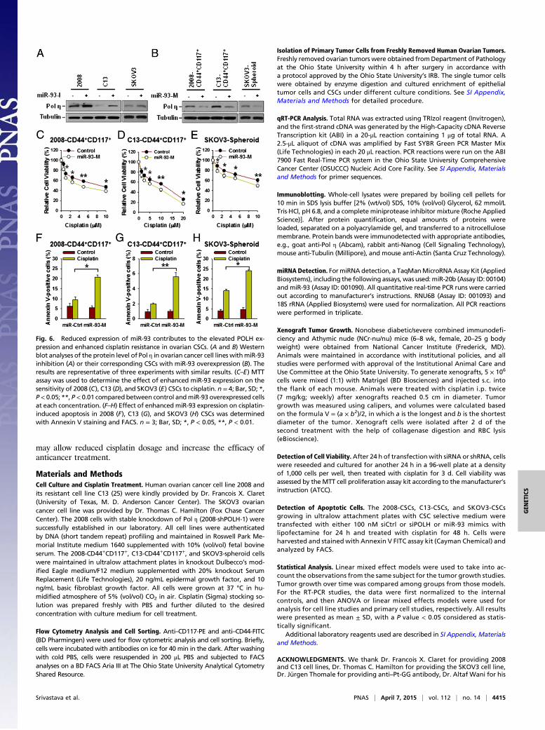
sion, 2008, C13, and SKOV3 cells were transfected with miR-93inhibitors, whereas 2008-CD44+CD117+, C13-CD44+CD117+,and SKOV3-spheroid cells were transfected with miR-93 mimics.qRT-PCR analyses demonstrated that down-regulation of miR-93 in 2008 and C13 cells enhanced the POLH mRNA levels (SIAppendix, Fig. S13 A and B), whereas overexpression of miR-93in 2008-CSCs and C13-CSCs reduced the POLH mRNA levels(SI Appendix, Fig. S13 C and D). Furthermore, the inhibitoryeffect of miR-93 on the protein level of Pol η was also revealedby immunoblotting analyses (Fig. 6 A and B). In contrast, trans-fection of miR-20b mimics did not have influence on the expres-sion of Pol η in these CSCs (SI Appendix, Fig. S11E). Collectively,these data suggest that reduced expression of miR-93 contributesto the elevated expression of POLH in ovarian CSCs.Given that miR-93 down-regulates Pol η expression, overex-
pression of miR-93 should be able to enhance the sensitivity ofovarian CSCs to cisplatin treatment by decreasing the Pol η level.Indeed, transfection of miR-93 mimics into 2008-CD44+CD117+,C13-CD44+CD117+, and SKOV3-spheroid cells increased cis-platin-induced cell death and cellular apoptosis (Fig. 6 C–Hand SI Appendix, Fig. S14 A–C), indicating that enforced miR-93expression sensitizes ovarian CSCs to cisplatin treatment.
DiscussionTLS is believed to contribute to the development of cisplatin re-sistance because TLS is able to rescue the cells from the collapseof the replication fork induced by DNA intrastrand cross-linksfollowing cisplatin treatment (8, 12–17). However, it is still unclear
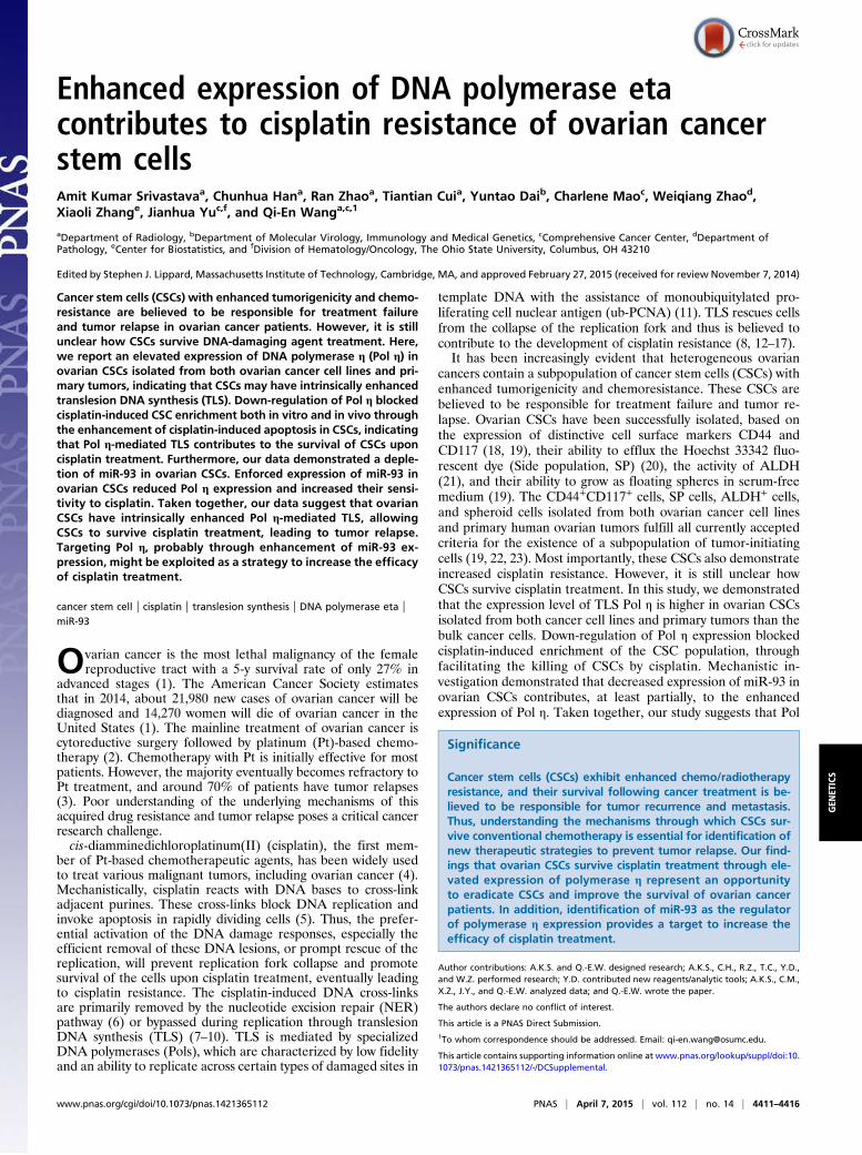
Fig. 2. Down-regulation of Pol η in ovarian cancer cells blocked cisplatin-in-duced enrichment of CSCs in vitro. (A, C, and E) Ovarian cancer cell lines 2008(A), C13 (C), and SKOV3 (E) were transiently transfected with either POLHsiRNA or control siRNA for 24 h, and the expression of Pol η was determinedusing immunoblotting. (B, D, and F) The siRNA transfected 2008 (B), C13 (D),and SKOV3 (F) cells were treated with cisplatin for 3 d and stained with anti–CD44-FITC and anti–CD117-PE antibodies. The percentage of CD44+CD117+
cells was analyzed using FACS. n = 3; Bar, SD; *, P < 0.05; **, P < 0.01.
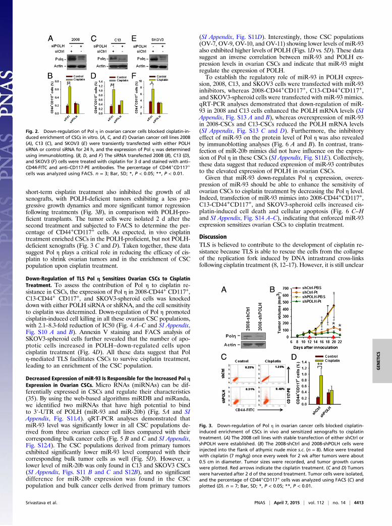
Fig. 3. Down-regulation of Pol η in ovarian cancer cells blocked cisplatin-induced enrichment of CSCs in vivo and sensitized xenografts to cisplatintreatment. (A) The 2008 cell lines with stable transfection of either shCtrl orshPOLH were established. (B) The 2008-shCtrl and 2008-shPOLH cells wereinjected into the flank of athymic nude mice s.c. (n = 8). Mice were treatedwith cisplatin (7 mg/kg) once every week for 2 wk after tumors were about0.5 cm in diameter. Tumor sizes were recorded, and tumor growth curveswere plotted. Red arrows indicate the cisplatin treatment. (C and D) Tumorswere harvested after 2 d of the second treatment. Tumor cells were isolated,and the percentage of CD44+CD117+ cells was analyzed using FACS (C) andplotted (D). n = 7; Bar, SD; *, P < 0.05; **, P < 0.01.
Srivastava et al. PNAS | April 7, 2015 | vol. 112 | no. 14 | 4413
GEN
ETICS

whether the cisplatin-resistant property of CSCs is also due toenhanced TLS activity. TLS allows the DNA replication machin-ery to bypass an unrepaired DNA damage site using special pol-ymerases (11, 36). Among many polymerases tested in vitro,the Y-family DNA Pol η is the most efficient and accurate atbypassing Pt-GG lesions (10, 37, 38). Pol η down-regulation resultsin increased sensitivity to cisplatin (13), whereas the increased Polη level reduces the effectiveness of chemotherapy and the survivaltime of patients with non–small-cell lung cancer or metastaticgastric adenocarcinoma (30). Our mice xenografts study alsodemonstrated that down-regulation of Pol η significantly enhancedthe response of ovarian tumor xenografts to cisplatin treatment.To further support this notion, we analyzed the public database ofgene expression arrays (free online software Kaplan-Meier Plot-ter: www.kmplot.com) and found that the higher POLH mRNAexpression in ovarian tumors is negatively correlated with the out-come of patients (SI Appendix, Fig. S15 A and B). In contrast, wedid not find such a correlation between REV1 or REV3L mRNAexpression level and the overall survival of patients (SI Appendix,Fig. S15 C and D), although it has been reported that REV1 orREV3L depletion sensitized lymphoma to cisplatin (14), andREV3L depletion sensitized lung adenocarcinoma to cisplatin (17).From the in vivo xenografts study, we noticed that the regrowth
rate of xenografts after each cisplatin treatment is lower in Polη–down-regulated ovarian tumor xenografts than controls. Giventhat CSCs are believed to be responsible for the initiation andregrowth of tumors, our data indicate that down-regulation of Polη may facilitate the eradication of ovarian CSCs by cisplatin. In-deed, we did demonstrate an elevated expression of Pol η at bothmRNA and protein levels in ovarian CSC populations and a pro-tective role of Pol η in ovarian CSCs upon cisplatin treatment.Thus, Pol η must be a critical contributor to the chemoresistantproperty of CSCs, and inhibition of the Pol η-mediated TLSpathway in CSCs would be a promising therapeutic strategy topromote the eradication of CSCs by cisplatin.Our data also demonstrated that the elevated Pol η expression
in ovarian CSCs can be attributed to the reduced expression ofmiR-93. miR-93 belongs to the miR106b-25 cluster that has beenreported to be overexpressed in different types of cancer, such asgastric, prostate, and pancreatic neural endocrine tumors, as wellas neuroblastoma and multiple myeloma (39). However, highlydepleted miR-93 was found in mouse mammary stem cellscharacterized with ALDH+ (40) and breast CSCs characterized bytheir expression of ALDH+ or CD44+CD24− (41). The role of
miR-93 in tumor growth is still controversial. miR-93 is able toenhance cell survival, promote sphere formation, and augmenttumor growth by targeting integrin-β8 in the glioblastoma U87 cellline (42). In contrast, miR-93 was reported to inhibit tumor growthand metastasis by decreasing the CSC population in SUM159breast cancer cells (41). Our data also showed a decreased level ofmiR-93 in CSCs isolated from both ovarian cancer cell lines andprimary ovarian tumors and demonstrated that low miR-93 level iscritical to the cisplatin resistance property of ovarian CSCs. Wefor the first time to our knowledge showed an inverse correlationbetween miR-93 and POLH expression in various ovarian CSCs.The use of miR-93 mimics led to attenuated Pol η expression inovarian CSCs, whereas the use of miR-93 inhibitors caused sig-nificant increase in Pol η expression in bulk ovarian cancer cells.Thus, miR-93 is able to target POLH 3′-UTR and inhibit theexpression of Pol η. As a result, the low level of miR-93 causesenhanced expression of Pol η, which facilitates TLS to promoteCSCs survival after cisplatin treatment. Collectively, the findingsimplicate a miR-93–Pol η axis affecting the survival of ovarianCSCs upon cisplatin treatment. However, it is also worth notingthat miR-93 can target several important pathways, such as STAT3and AKT pathways (41). Given that elevated expression of STAT3and AKT3 is associated with cisplatin resistance in various cancers(43, 44), it is possible that the low level of miR-93 also contributesto cisplatin resistance through affecting other critical pathways.miR-93 is located at chromosome 7q22, in the intron 13 of the
host gene minichromosome maintenance 7 (MCM7), where theyare cotranscribed in the context of MCM7 primary transcript (39).Although MCM7 overexpression has been identified in varioustumors and considered a bad prognostic indicator in prostatecancer (45, 46), MCM7 expression is lower in various CSCs, in-cluding SP of human lung cancer cells (47) and prostate cancercells (48), as well as ALDH+ breast cancer cells (41). In addition,an analysis of 12 publically available microarray datasets revealeda down-regulation of the MCM7 gene in various cancer stem-likecells in all datasets, although only three of them showed significantchange (SI Appendix, Table S1). Thus, this reduced expression ofMCM7, probably due to relative quiescence of CSCs (49, 50), couldbe a contributor to the decreased expression of miR-93 in CSCs.In summary, our data demonstrated a previously unidentified
mechanism of cisplatin resistance in ovarian CSCs. A low level ofmiR-93 in ovarian CSCs enhances the expression of Pol η, whichpromotes the bypass of cisplatin-induced, unrepaired DNA intra-strand cross-links, leading to the elevated survival of CSCs.Therefore, Pol η inhibitors could be exploited as chemotherapy-enhancing agents. A combination of cisplatin and Pol η inhibitors
Fig. 4. Down-regulation of Pol η-sensitized CSCs to cisplatin treatment.(A–C ) The 2008-CD44+CD117+ (A), C13-CD44+CD117+ (B), and SKOV3-spheroid (C) cells were transfected with either POLH siRNA or control siRNA,followed by treatment with cisplatin for 3 d. Cell survival was determinedusing the MTT assay. n = 4; Bar, SD; *, P < 0.05; #, P < 0.01 compared betweencontrol and POLH knockdown cells at each time point. (D) SKOV3-spheroidcells were transfected with either control or POLH siRNA for 48 h and thentreated with cisplatin for 24 h. Apoptotic cells were stained with Annexin Vand detected by FACS. n = 3; Bar, SD; *, P < 0.05.
Fig. 5. Reduced miR-93 expression in ovarian CSCs. (A) Predicted binding ofmiR-93 to the 3′UTR of POLH. (B and C) Expression of miR-93 was determinedin various ovarian cancer cell lines and their corresponding CSCs by qRT-PCR.n = 3; Bar, SD; **, P < 0.01. (D) Expression of miR-93 in bulk primary tumor cellsand their corresponding spheroid cells was analyzed using qRT-PCR. n = 3, Bar,SD. Analysis indicates that there was significantly decreased miR-93 expressionin the spheroid cells compared with bulk cells (P = 0.047).
4414 | www.pnas.org/cgi/doi/10.1073/pnas.1421365112 Srivastava et al.
may allow reduced cisplatin dosage and increase the efficacy ofanticancer treatment.
Materials and MethodsCell Culture and Cisplatin Treatment. Human ovarian cancer cell line 2008 andits resistant cell line C13 (25) were kindly provided by Dr. Francois X. Claret(University of Texas, M. D. Anderson Cancer Center). The SKOV3 ovariancancer cell line was provided by Dr. Thomas C. Hamilton (Fox Chase CancerCenter). The 2008 cells with stable knockdown of Pol η (2008-shPOLH-1) weresuccessfully established in our laboratory. All cell lines were authenticatedby DNA (short tandem repeat) profiling and maintained in Roswell Park Me-morial Institute medium 1640 supplemented with 10% (vol/vol) fetal bovineserum. The 2008-CD44+CD117+, C13-CD44+CD117+, and SKOV3-spheroid cellswere maintained in ultralow attachment plates in knockout Dulbecco’s mod-ified Eagle medium/F12 medium supplemented with 20% knockout SerumReplacement (Life Technologies), 20 ng/mL epidermal growth factor, and 10ng/mL basic fibroblast growth factor. All cells were grown at 37 °C in hu-midified atmosphere of 5% (vol/vol) CO2 in air. Cisplatin (Sigma) stocking so-lution was prepared freshly with PBS and further diluted to the desiredconcentration with culture medium for cell treatment.
Flow Cytometry Analysis and Cell Sorting. Anti–CD117-PE and anti–CD44-FITC(BD Pharmingen) were used for flow cytometric analysis and cell sorting. Briefly,cells were incubated with antibodies on ice for 40 min in the dark. After washingwith cold PBS, cells were resuspended in 200 μL PBS and subjected to FACSanalyses on a BD FACS Aria III at The Ohio State University Analytical CytometryShared Resource.
Isolation of Primary Tumor Cells from Freshly Removed Human Ovarian Tumors.Freshly removed ovarian tumors were obtained fromDepartment of Pathologyat the Ohio State University within 4 h after surgery in accordance witha protocol approved by the Ohio State University’s IRB. The single tumor cellswere obtained by enzyme digestion and cultured enrichment of epithelialtumor cells and CSCs under different culture conditions. See SI Appendix,Materials and Methods for detailed procedure.
qRT-PCR Analysis. Total RNA was extracted using TRIzol reagent (Invitrogen),and the first-strand cDNA was generated by the High-Capacity cDNA ReverseTranscription kit (ABI) in a 20-μL reaction containing 1 μg of total RNA. A2.5-μL aliquot of cDNA was amplified by Fast SYBR Green PCR Master Mix(Life Technologies) in each 20 μL reaction. PCR reactions were run on the ABI7900 Fast Real-Time PCR system in the Ohio State University ComprehensiveCancer Center (OSUCCC) Nucleic Acid Core Facility. See SI Appendix, Materialsand Methods for primer sequences.
Immunoblotting. Whole-cell lysates were prepared by boiling cell pellets for10 min in SDS lysis buffer [2% (wt/vol) SDS, 10% (vol/vol) Glycerol, 62 mmol/LTris·HCl, pH 6.8, and a complete miniprotease inhibitor mixture (Roche AppliedScience)]. After protein quantification, equal amounts of proteins wereloaded, separated on a polyacrylamide gel, and transferred to a nitrocellulosemembrane. Protein bands were immunodetected with appropriate antibodies,e.g., goat anti-Pol η (Abcam), rabbit anti-Nanog (Cell Signaling Technology),mouse anti-Tubulin (Millipore), and mouse anti-Actin (Santa Cruz Technology).
miRNA Detection. FormiRNAdetection, a TaqManMicroRNAAssay Kit (AppliedBiosystems), including the following assays, was used:miR-20b (Assay ID: 00104)and miR-93 (Assay ID: 001090). All quantitative real-time PCR runs were carriedout according to manufacturer’s instructions. RNU6B (Assay ID: 001093) and18S rRNA (Applied Biosystems) were used for normalization. All PCR reactionswere performed in triplicate.
Xenograft Tumor Growth. Nonobese diabetic/severe combined immunodefi-ciency and Athymic nude (NCr-nu/nu) mice (6–8 wk, female, 20–25 g bodyweight) were obtained from National Cancer Institute (Frederick, MD).Animals were maintained in accordance with institutional policies, and allstudies were performed with approval of the Institutional Animal Care andUse Committee at the Ohio State University. To generate xenografts, 5 × 10⁶
cells were mixed (1:1) with Matrigel (BD Biosciences) and injected s.c. intothe flank of each mouse. Animals were treated with cisplatin i.p. twice(7 mg/kg; weekly) after xenografts reached 0.5 cm in diameter. Tumorgrowth was measured using calipers, and volumes were calculated basedon the formula V = (a × b2)/2, in which a is the longest and b is the shortestdiameter of the tumor. Xenograft cells were isolated after 2 d of thesecond treatment with the help of collagenase digestion and RBC lysis(eBioscience).
Detection of Cell Viability.After 24 h of transfection with siRNA or shRNA, cellswere reseeded and cultured for another 24 h in a 96-well plate at a densityof 1,000 cells per well, then treated with cisplatin for 3 d. Cell viability wasassessed by theMTT cell proliferation assay kit according to the manufacturer’sinstruction (ATCC).
Detection of Apoptotic Cells. The 2008-CSCs, C13-CSCs, and SKOV3-CSCsgrowing in ultralow attachment plates with CSC selective medium weretransfected with either 100 nM siCtrl or siPOLH or miR-93 mimics withlipofectamine for 24 h and treated with cisplatin for 48 h. Cells wereharvested and stained with Annexin V FITC assay kit (Cayman Chemical) andanalyzed by FACS.
Statistical Analysis. Linear mixed effect models were used to take into ac-count the observations from the same subject for the tumor growth studies.Tumor growth over time was compared among groups from those models.For the RT-PCR studies, the data were first normalized to the internalcontrols, and then ANOVA or linear mixed effects models were used foranalysis for cell line studies and primary cell studies, respectively. All resultswere presented as mean ± SD, with a P value < 0.05 considered as statis-tically significant.
Additional laboratory reagents used are described in SI Appendix, Materialsand Methods.
ACKNOWLEDGMENTS. We thank Dr. Francois X. Claret for providing 2008and C13 cell lines, Dr. Thomas C. Hamilton for providing the SKOV3 cell line,Dr. J}urgen Thomale for providing anti–Pt-GG antibody, Dr. Altaf Wani for his
Fig. 6. Reduced expression of miR-93 contributes to the elevated POLH ex-pression and enhanced cisplatin resistance in ovarian CSCs. (A and B) Westernblot analyses of the protein level of Pol η in ovarian cancer cell lines withmiR-93inhibition (A) or their corresponding CSCs with miR-93 overexpression (B). Theresults are representative of three experiments with similar results. (C–E) MTTassay was used to determine the effect of enhanced miR-93 expression on thesensitivity of 2008 (C), C13 (D), and SKOV3 (E) CSCs to cisplatin. n = 4; Bar, SD; *,P< 0.05; **, P< 0.01 compared between control andmiR-93 overexpressed cellsat each concentration. (F–H) Effect of enhanced miR-93 expression on cisplatin-induced apoptosis in 2008 (F), C13 (G), and SKOV3 (H) CSCs was determinedwith Annexin V staining and FACS. n = 3; Bar, SD; *, P < 0.05, **, P < 0.01.
Srivastava et al. PNAS | April 7, 2015 | vol. 112 | no. 14 | 4415
GEN
ETICS
generous support throughout the course of this work, and Dr. RobertSnapka and Edie Yamasaki for their reading and review of the manuscript.
This work was supported by National Institutes of Health R01 CA151248 (toQ.-E.W.) and P30 CA016058 (to OSUCCC core facility).
1. Siegel R, Ma J, Zou Z, Jemal A (2014) Cancer statistics, 2014. CA Cancer J Clin 64(1):9–29.
2. Pfisterer J, Ledermann JA (2006) Management of platinum-sensitive recurrent ovariancancer. Semin Oncol 33(2, Suppl 6):S12–S16.
3. Agarwal R, Kaye SB (2003) Ovarian cancer: Strategies for overcoming resistance tochemotherapy. Nat Rev Cancer 3(7):502–516.
4. Cohen SM, Lippard SJ (2001) Cisplatin: From DNA damage to cancer chemotherapy.Prog Nucleic Acid Res Mol Biol 67:93–130.
5. Jung Y, Lippard SJ (2007) Direct cellular responses to platinum-induced DNA damage.Chem Rev 107(5):1387–1407.
6. Reed E (1998) Platinum-DNA adduct, nucleotide excision repair and platinum basedanti-cancer chemotherapy. Cancer Treat Rev 24(5):331–344.
7. Chaney SG, Campbell SL, Bassett E, Wu Y (2005) Recognition and processing of cis-platin- and oxaliplatin-DNA adducts. Crit Rev Oncol Hematol 53(1):3–11.
8. Mamenta EL, et al. (1994) Enhanced replicative bypass of platinum-DNA adducts incisplatin-resistant human ovarian carcinoma cell lines. Cancer Res 54(13):3500–3505.
9. Reardon JT, Vaisman A, Chaney SG, Sancar A (1999) Efficient nucleotide excision re-pair of cisplatin, oxaliplatin, and Bis-aceto-ammine-dichloro-cyclohexylamine-plati-num(IV) (JM216) platinum intrastrand DNA diadducts. Cancer Res 59(16):3968–3971.
10. Bassett E, et al. (2004) The role of DNA polymerase eta in translesion synthesis pastplatinum-DNA adducts in human fibroblasts. Cancer Res 64(18):6469–6475.
11. Shachar S, et al. (2009) Two-polymerase mechanisms dictate error-free and error-prone translesion DNA synthesis in mammals. EMBO J 28(4):383–393.
12. Zhao Y, et al. (2012) Structural basis of human DNA polymerase η-mediated chemo-resistance to cisplatin. Proc Natl Acad Sci USA 109(19):7269–7274.
13. Chen YW, Cleaver JE, Hanaoka F, Chang CF, Chou KM (2006) A novel role of DNApolymerase eta in modulating cellular sensitivity to chemotherapeutic agents. MolCancer Res 4(4):257–265.
14. Xie K, Doles J, Hemann MT, Walker GC (2010) Error-prone translesion synthesis me-diates acquired chemoresistance. Proc Natl Acad Sci USA 107(48):20792–20797.
15. Sharma S, Shah NA, Joiner AM, Roberts KH, Canman CE (2012) DNA polymerase ζ isa major determinant of resistance to platinum-based chemotherapeutic agents. MolPharmacol 81(6):778–787.
16. Lin X, Okuda T, Trang J, Howell SB (2006) Human REV1 modulates the cytotoxicity andmutagenicity of cisplatin in human ovarian carcinoma cells. Mol Pharmacol 69(5):1748–1754.
17. Doles J, et al. (2010) Suppression of Rev3, the catalytic subunit of Polzeta, sensitizesdrug-resistant lung tumors to chemotherapy. Proc Natl Acad Sci USA 107(48):20786–20791.
18. Alvero AB, et al. (2009) Molecular phenotyping of human ovarian cancer stem cellsunravels the mechanisms for repair and chemoresistance. Cell Cycle 8(1):158–166.
19. Zhang S, et al. (2008) Identification and characterization of ovarian cancer-initiatingcells from primary human tumors. Cancer Res 68(11):4311–4320.
20. McAuliffe SM, et al. (2012) Targeting Notch, a key pathway for ovarian cancerstem cells, sensitizes tumors to platinum therapy. Proc Natl Acad Sci USA 109(43):E2939–E2948.
21. Silva IA, et al. (2011) Aldehyde dehydrogenase in combination with CD133 definesangiogenic ovarian cancer stem cells that portend poor patient survival. Cancer Res71(11):3991–4001.
22. Dalerba P, Cho RW, Clarke MF (2007) Cancer stem cells: Models and concepts. AnnuRev Med 58:267–284.
23. Clarke MF, et al. (2006) Cancer stem cells—perspectives on current status and futuredirections: AACR Workshop on cancer stem cells. Cancer Res 66(19):9339–9344.
24. Morrison R, et al. (2011) Targeting the mechanisms of resistance to chemotherapyand radiotherapy with the cancer stem cell hypothesis. J Oncol 2011:941876.
25. Andrews PA, Velury S, Mann SC, Howell SB (1988) cis-Diamminedichloroplatinum(II)accumulation in sensitive and resistant human ovarian carcinoma cells. Cancer Res48(1):68–73.
26. Delmastro DA, Li J, Vaisman A, Solle M, Chaney SG (1997) DNA damage inducible-gene expression following platinum treatment in human ovarian carcinoma cell lines.Cancer Chemother Pharmacol 39(3):245–253.
27. Han C, et al. (2014) DDB2 suppresses tumorigenicity by limiting the cancer stem cell
population in ovarian cancer. Mol Cancer Res 12(5):784–794.28. Tirino V, et al. (2013) Cancer stem cells in solid tumors: An overview and new ap-
proaches for their isolation and characterization. FASEB J 27(1):13–24.29. Kartalou M, Essigmann JM (2001) Mechanisms of resistance to cisplatin. Mutat Res
478(1–2):23–43.30. Ceppi P, et al. (2009) Polymerase eta mRNA expression predicts survival of non-small
cell lung cancer patients treated with platinum-based chemotherapy. Clin Cancer Res
15(3):1039–1045.31. Teng KY, et al. (2010) DNA polymerase η protein expression predicts treatment re-
sponse and survival of metastatic gastric adenocarcinoma patients treated with
oxaliplatin-based chemotherapy. J Transl Med 8:126.32. Zhou W, et al. (2013) Expression of DNA translesion synthesis polymerase η in head
and neck squamous cell cancer predicts resistance to gemcitabine and cisplatin-based
chemotherapy. PLoS ONE 8(12):e83978.33. Abubaker K, et al. (2013) Short-term single treatment of chemotherapy results in the
enrichment of ovarian cancer stem cell-like cells leading to an increased tumor bur-
den. Mol Cancer 12:24.34. Bertolini G, et al. (2009) Highly tumorigenic lung cancer CD133+ cells display stem-like
features and are spared by cisplatin treatment. Proc Natl Acad Sci USA 106(38):
16281–16286.35. Liu C, Tang DG (2011) MicroRNA regulation of cancer stem cells. Cancer Res 71(18):
5950–5954.36. Fischhaber PL, Friedberg EC (2005) How are specialized (low-fidelity) eukaryotic pol-
ymerases selected and switched with high-fidelity polymerases during translesion
DNA synthesis? DNA Repair (Amst) 4(2):279–283.37. Vaisman A, Masutani C, Hanaoka F, Chaney SG (2000) Efficient translesion replication
past oxaliplatin and cisplatin GpG adducts by human DNA polymerase eta. Bio-
chemistry 39(16):4575–4580.38. Masutani C, Kusumoto R, Iwai S, Hanaoka F (2000) Mechanisms of accurate trans-
lesion synthesis by human DNA polymerase eta. EMBO J 19(12):3100–3109.39. Petrocca F, Vecchione A, Croce CM (2008) Emerging role of miR-106b-25/miR-17-92
clusters in the control of transforming growth factor beta signaling. Cancer Res
68(20):8191–8194.40. Ibarra I, Erlich Y, Muthuswamy SK, Sachidanandam R, Hannon GJ (2007) A role for
microRNAs in maintenance of mouse mammary epithelial progenitor cells. Genes Dev
21(24):3238–3243.41. Liu S, et al. (2012) MicroRNA93 regulates proliferation and differentiation of normal
and malignant breast stem cells. PLoS Genet 8(6):e1002751.42. Fang L, et al. (2011) MicroRNA miR-93 promotes tumor growth and angiogenesis by
targeting integrin-β8. Oncogene 30(7):806–821.43. Ji T, et al. (2013) Abrogation of constitutive Stat3 activity circumvents cisplatin re-
sistant ovarian cancer. Cancer Lett 341(2):231–239.44. Gagnon V, Mathieu I, Sexton E, Leblanc K, Asselin E (2004) AKT involvement in cis-
platin chemoresistance of human uterine cancer cells. Gynecol Oncol 94(3):785–795.45. Levesque MH, El-Alfy M, Berger L, Labrie F, Labrie C (2007) Evaluation of AIbZIP and
Cdc47 as markers for human prostatic diseases. Urology 69(1):196–201.46. Laitinen S, et al. (2008) EZH2, Ki-67 and MCM7 are prognostic markers in prostatec-
tomy treated patients. Int J Cancer 122(3):595–602.47. Ho MM, Ng AV, Lam S, Hung JY (2007) Side population in human lung cancer
cell lines and tumors is enriched with stem-like cancer cells. Cancer Res 67(10):
4827–4833.48. Singh S, et al. (2012) Chemoresistance in prostate cancer cells is regulated by miRNAs
and Hedgehog pathway. PLoS ONE 7(6):e40021.49. Forsburg SL (2008) The MCM helicase: linking checkpoints to the replication fork.
Biochem Soc Trans 36(Pt 1):114–119.50. Nguyen LV, Vanner R, Dirks P, Eaves CJ (2012) Cancer stem cells: An evolving concept.
Nat Rev Cancer 12(2):133–143.
4416 | www.pnas.org/cgi/doi/10.1073/pnas.1421365112 Srivastava et al.