engineered recombinant protein products of the avian
TRANSCRIPT
RESEARCH Open Access
Engineered recombinant protein productsof the avian paramyxovirus type-1nucleocapsid and phosphoprotein genesfor serological diagnosisNa Zhao, Christian Grund, Martin Beer and Timm C. Harder*
Abstract
Background: Virulent Newcastle disease virus (NDV, avian Avulavirus-1, APMV-1) induces a highly contagious andlethal systemic disease in gallinaceous poultry. APMV-1 antibody detection is used for surveillance and to controlvaccination, but is hampered by cross-reactivity to other subtypes of avian Avulaviruses. Data are lacking concerningthe applicability of NDV V proteins as differential diagnostic marker to distinguish vaccinated from virus-infected birds(DIVA strategy).
Methods: Full length and C-terminally truncated nucleocapsid (NP) protein, and the unique C-terminal regions of thephospho- (P) and V proteins of the NDV LaSota strain were bacterially expressed as fusion proteins with themultimerization domain of the human C4 binding protein, and used as diagnostic antigens in indirect ELISA.
Results: When used as diagnostic antigen in indirect ELISAs, recombinant full-length proved to be a sensitivetarget to detect seroconversion in chickens after APMV-1 vaccination and infection, but revealed some degreeof cross reactivity with sera raised against other APMV subtypes. Cross reactivity was abolished but also sensitivitydecreased when employing a C-terminal fragment of the NP of NDV as diagnostic antigen. Antibodies to the NDV Vprotein were mounted in poultry following NDV infection but also, albeit at lower rates and titers, after vaccinationwith attenuated NDV vaccines. V-specific seroconversion within the flock was incomplete and titers inindividual bird transient.
Conclusions: Indirect ELISA based on bacterially expressed recombinant full-length NP compared favorablywith a commercial NDV ELISA based on whole virus antigen, but cross reactivity between the NP proteins ofdifferent APMV subtypes could compromise specificity. However, specificity increased when using a less conservedC-terminal fragment of NP instead. Moreover, a serological DIVA strategy built on the NDV V protein was notfeasible due to reduced immunogenicity of the V protein and frequent use of live-attenuated NDV vaccines.
Keywords: Newcastle disease virus, Recombinant protein, Subtype-specific serology
* Correspondence: [email protected] Federal Research Institute for Animal Health, Friedrich-Loeffler-Institut,Institute of Diagnostic Virology, Suedufer 10, 17493 Greifswald, Germany
© The Author(s). 2018 Open Access This article is distributed under the terms of the Creative Commons Attribution 4.0International License (http://creativecommons.org/licenses/by/4.0/), which permits unrestricted use, distribution, andreproduction in any medium, provided you give appropriate credit to the original author(s) and the source, provide a link tothe Creative Commons license, and indicate if changes were made. The Creative Commons Public Domain Dedication waiver(http://creativecommons.org/publicdomain/zero/1.0/) applies to the data made available in this article, unless otherwise stated.
Zhao et al. Virology Journal (2018) 15:8 DOI 10.1186/s12985-018-0924-8

BackgroundNewcastle Disease (ND) is caused by infection of chickenswith virulent strains of the avian paramyxovirus serotype-1(APMV-1, syn. ND virus, NDV). ND, together with highlypathogenic avian influenza, are among the most dreadedviral infections of gallinaceous poultry worldwide [1].Rapid spread of a highly lethal, pantropic disease ensues inaffected flocks after incursion of virulent (syn. Velogenic)NDV. Consequently, ND is an O.I.E.-notifiable disease ofpoultry. The virus is enzootic in many regions of the worldand continues to threaten industrial production as well asbackyard poultry holdings. Vaccination with inactivatedadjuvanted or, alternatively, live virus vaccines featuringattenuated (lentogenic) APMV-1 strains such as LaSota orB1 is widely used to prevent ND [2, 3].Avian avulaviruses are members of the Avulavirus
genus within the Paramyxoviridae family. To date atleast fifteen serotypes (APMV-1 to APMV-15) have beenidentified [4–11]. The virus possesses a single stranded,negative-sense, non-segmented RNA genome, whichencodes six structural proteins in following order:nucleocapsid protein (NP), phosphoprotein (P), matrixprotein (M), fusion protein (F), hemagglutinin-neuraminidase protein (HN), and the large (L) polymer-ase protein [12, 13]. The NP protein is a highlyconserved and the most abundant viral proteinexpressed in infected cells, and induces a stronghumoral (non-neutralizing) and cellular immuneresponse in the infected host and also followingvaccination with inactivated virus [3, 14–16].The P protein plays an important role in the viral tran-
scription and replication, and it is associated with thenucleocapsid in the virion [17, 18]. Two additional pro-teins, V and putative W, are predicted to be producedfrom P gene by mRNA editing post transcription [19–23].The product that ensues by insertion into the nascent PmRNA of one non-template G residue at position 401 (+2reading frame) is referred to as the V protein [21].Addition of two untemplated G’s at the polymerase slip-ping point of the P gene would generate a third proteinspecies, the W protein, from the P gene [21, 23]. Thus, allthree P gene-derived proteins have a commonN-terminus, but vary at their carboxyl termini both inlength and amino acid composition. The V protein ofNDV harbors 106 amino acids in its unique C-terminalpart (LaSota strain). Similar to other viruses in theparamyxoviridae family, the V protein is found to be azinc-finger domain protein and appears to function as avirulence factor by antagonizing, in a strain-specific man-ner, components of the host innate immunity, in particu-lar the interferon system [24, 25]. However, very little isknown about the immunogenicity of the V protein.Serological assays to detect ND-specific antibodies can
be used for demonstration of lack of exposure of a flock
to NDV, and for assessment of vaccination efficacy. Thehemagglutination inhibition (HI) test is a standardmethod and widely used for NDV antibody detection,although it may lack in sensitivity and is time-consum-ing [26–28]. Several ELISA formats have been developedas an alternative for conventional HI test in flock screeningapproaches. Their sensitivity, and easy standardizationmake them suitable for high throughput screening [28–34]. ELISA formats based on whole virus and recombinantviral proteins expressed in baculovirus or bacterial systemsas the coating antigen have been reported [28, 29, 35–38].Recombinant NP protein in particular has been used forthe development of indirect ELISAs (iELISA) [14, 35].However, the NP protein is highly conserved among avianparamyxoviruses, and serologic assays building on recom-binant NDV NP may be compromised by cross reactionsbetween various APMV serotypes.To overcome limitations of full length NDV NP as the
antigen in ELISA format, we hypothesized that use ofless conserved NP and P protein fragments would enablea more serotype-specific distinction. The objective of thisstudy was to explore the suitability of truncated NP and Pproteins as diagnostic antigens for detection and differenti-ation of avian paramyxovirus-specific antibodies. Moreover,assuming that a humoral immune response against the Vprotein would be enhanced by active viral replication inthe host and expression of V in infected cells, V protein-specific antibodies might be used to distinguish infectedbirds from those that are seropositive after vaccinationwith inactivated or attenuated vaccines. Therefore, anotherobjective was to explore and evaluate the diagnostic feasi-bility of using the unique portion of V protein in an indir-ect ELISA-based DIVA (differentiating infected fromvaccinated animals) strategy.
MethodsVirus propagationAPMV-1 vaccine strain LaSota (LS; class II, genotype 2)[39] and APMV-8 strain goose/Delaware/1053/76 weregrown in the allantoic cavity of embryonated chickeneggs as detailed elsewhere [28].
Cloning and bacterial expression of recombinant proteinsThe pET19b vector and Rosettagami E. coli cells (bothNovagen, Darmstadt, Germany) were used for expres-sion of recombinant NDV proteins. The full length ORFencoding the nucleocapsid protein (NP) of NDV LaSotawas cloned into pET19b downstream of the T7promoter as shown in Fig. 1a. A hexa-histidine- and anAvi-tag were positioned in-frame and N-terminally ofthe ORF [40]. Similarly, the sequences encoding theunique parts of the P and V (Pu, Vu) downstream of theRNA editing site of the P gene were cloned into pET19b(Fig. 1b). In addition, the C-terminal 99 amino acids of
Zhao et al. Virology Journal (2018) 15:8 Page 2 of 12

NDV LaSota NP (NPct), and the C-terminal 81 aminoacids of the NP of APMV-8 were cloned as shown inFig. 1c. A heptamerization fragment of the human C4binding protein (C4BP) was positioned between the tagsand the NDV sequences [41, 42]. SGS-linker sequenceswere placed between C4BP and the virus-specificsequences to ensure unrestrained folding of the latter.Plasmid pBirCam (Avidity, Aurora, U.S.A) over-express-ing the bacterial biotin ligase BirA was co-transformedinto strain Rosettagami to ensure cotranslational mono-biotinylation of the recombinant APMV protein fragmentsat their Avi-tag [43]. Ampicillin and chloramphenicol wereused for selection. Presence of both plasmids was con-firmed by plasmid-insert-specific PCRs.Induction of expression was achieved in TYH
medium supplemented with IPTG (1.5 mM) and D(+)-biotin (50 μM). After culturing for 4 h at 37 °C,cells were pelleted and lysed using ultrasonic disrup-tion as previously described [43] to liberate inclusion
bodies (ICs) into which recombinant proteins hadsequestered.
Purification and reconstitution of recombinant proteinsfrom bacterial inclusion bodiesPurified ICs were solubilized in 6 M guanidinium-HCl.A set of refolding buffers was used in a stepwise solubil-isation strategy using the ProteoStat kit (Enzo, Lörrach,Germany) to determine the most appropriate buffer con-ditions to fold back the proteins into an antigenicallyauthentic structure while keeping it solubilized.Antigenic properties in ELISA were assayed using poly-clonal antibodies raised in chickens. Final proteinconcentration in refolding buffer was determined usinga coomassie protein assay kit (ThermoScientific,Rockford, IL, U.S.A.) and proteins were stored at 4 °Cuntil further use.
Production of antiseraGalline hyperimmune sera against reference isolates ofAPMV serotype-1(NDV strain Ulster, pigeon paramyxo-virus, class I isolate APMV-1/Mallard/Germany/R2481/2007), and other serotypes-2 (APMV-2/chicken/Califor-nia/Yucaipa/56), −3a (APMV-3/turkey/England/1087/82),−3b (APMV-3/Parakeet/Netherlands/449/75), −4 (APMV-4/duck/Hong Kong/D3/75), −6 (APMV-6/Dk/HK/77), −7(APMV-7/dove/Tennessee/4/75), −8 (APMV-8/goose/Delaware/1053/46), and −9 (APMV-9/duck/New York/22/78) had been generated in chickens by boosterimmunization using egg-grown virus adjuvant with in-complete Freund’s adjuvant. SPF chicken sera were sam-pled from chickens hatched at the FLI from SPF eggs(Lohmann Tierzucht, Cuxhaven, Germany). All animal ex-periments had received full legal approval by the animalwelfare committee of the German Federal State ofMecklenburg-Vorpommern (LALLF M-V/TSD/7221.3–2.5-004/10; LALLF M-V/TSD/7221.3–2.5-010/10).
Western blottingProtein samples were separated by SDS-PAGE in 12.5%gels. For denaturing conditions samples were heated inLaemmli loading buffer containing dithioerythritol (DTE).Proteins were also separated under non-denaturing condi-tions in 10% PAGE without SDS and DTE. Proteins weretransferred onto nitrocellulose membranes by semi-dryblotting at 0.8 mA/cm2 for 1 h (PerfectBlueTM ElectroBlotter, Peqlab, Erlangen, Germany). Membranes wereblocked with 5% skim milk powder in Tris-buffered saline(TBS) supplemented with 0.05% (v/v) Tween (TBST) for1 h. Specific antibodies and sera were appropriately dilutedin TBST and incubated on the membranes for 1 h at roomtemperature. Membranes were washed three times withTBST and incubated for another hour with POD-labeledsecondary antibody. Blots were washed again for three
Fig. 1 Schematic presentation of constructs used for generation ofpET19b plasmids and T7- driven expression of NDV proteins inRosettagami E. coli cultures. All sketches show the protein C-terminus. aDesign of full-length expression of the nucleocapsid gene of NDV strainLaSota, with N-terminal hexahistidin ([H] 6) and AVI tags. b Expression oftruncated and frame-edited versions of the P gene expressing uniqueC-termini of the two P gene-derived proteins P and V as fusion proteinswith the heptamerization domain of the human C4 binding protein(C4BP), and tags. An SGS-linker separates virus-specific from fused proteinsequences. c Expression cassette of C-terminally truncated versions ofthe NP protein of NDV LaSota or APMV-8, respectively
Zhao et al. Virology Journal (2018) 15:8 Page 3 of 12
times, then incubated with substrate SuperSignal™West PicoChemiluminescent substrate solution (ThermoScientific,Braunschweig, Germany) before being analyzed with an im-aging system (VersaDoc, Bio-Rad). Photos were edited withrespect to contrast and brightness and composite blots wereassembled using cut-out lanes (GIMP software).
Indirect ELISARecombinant proteins solubilized in the respectiverefolding buffers were further diluted to a concentrationof 5 μg/ml using bicarbonate coating buffer (pH 9.6) ofwhich 100 μl per well were used to coat Maxisorb ELISAplates (NUNC, Thermofisher, Braunschweig, Germany)overnight at 4 °C. After washing, wells were blocked by5% skim milk-TBST) for 2 h at room temperature andthen washed four times with TBST [44]. Individual serawere diluted 1:200 (for the NP iELISA) or 1:500 (otherantigens) in appropriate sample dilution buffer. Serawere incubated at room temperature for 1 h. Wells werewashed again four times with TBST before 100 μl of ap-propriately diluted goat-anti-chicken or -turkey IgYPOD conjugate (Dianova, Hamburg, Germany) wasadded for 1 h at room temperature. Antibody was re-moved and after a final washing cycle with TBST, 50 μlof chromogenic TMB substrate was added. OD450values were measured after 10 min of incubation andaddition of 50 μl of 1 N H2SO4 to each well. Resultswere calculated and expressed in S/P units:
ODTest‐ODBackgroundODPositive control‐ODBackground
� 100 ¼ S=P
Determination of cut-off values for indirect ELISAsMaxisorb ELISA plates were coated with 0.5 μg ofrecombinant protein per well and then blocked. A predi-lution step of sera, as outlined above, was required forbackground reduction. Species-specific anti-IgY POD--conjugates were used for detection of bound antibodies.A total of 51 sera from SPF chickens were examined todetermine cut-off values for the different recombinantantigens in this indirect ELISA format. None of thesesera tested positive in a commercial indirect NDV ELISA(see below). Mean extinctions and standard deviationswere calculated (see Additional file 1). Serum S185, ahyperimmune serum raised in chicken against inacti-vated NDV strain Ulster, served as standard positivecontrol for assays employing NP and P antigens. SerumS880/Hu295 was used as standard positive control for Vprotein-specific assays since this serum had been ob-tained at day 28 after immunization with live-attenuatedNDV LaSota vaccine and was shown to harbor V-specific antibodies in Western blotting (Fig. 2c). An SPFchicken serum was chosen as standard negative control.
These control sera were used as standards in each of theassays to determine threshold OD values and to calculateS/P ratios. The S/P mean values of all 51 SPF sera plus 2SD were chosen as cut-offs in the examination of chickenfield sera by indirect ELISAs. Additional file 1 presentsexact cut-off values for each assay.
Fig. 2 Assembled Western blot assays of monobiotinylated recombinantproteins of NDV LaSota. Panels a and b were stained using amonoclonal antibody specific for biotin. For panel c a chicken serum(S880/Hu295) was used that was obtained at day 28 after immunizationwith live-attenuated NDV LaSota vaccine. Panels a and c were run underdenaturing conditions while panel b was obtained after proteinseparation in native PAGE. M – marker lane, NP – full lengthnucleocapsid protein; NPct -C-terminal fragment of nucleocapsid protein;Pu – unique C-terminal part of phosphoprotein; Vu – unique C-terminalpart of V protein; C4BP – heptamerization domain of human C4 bindingprotein; N – Rosettagami E. coli lysate. Molecular weights of monomericrecombinant proteins (panels a, c) and multimeric conglomerates (panelb) are presented to the left of the marker lane
Zhao et al. Virology Journal (2018) 15:8 Page 4 of 12
Commercial ELISA for detection of NDV antibodiesA commercial indirect NDV-ELISA (Flock Chek NDV)based on complete ND virions was purchased fromIDEXX (Ludwigsburg, Germany). Instructions of themanufacturer were followed exactly using either the ver-sion for chickens or turkeys as appropriate.
Hemagglutination inhibition assay (HI)HI assays were performed according to O.I.E. recom-mendations essentially as described [45]. Four hemagglu-tinating units of egg-grown APMV-1 or APMV-8 viruseswere used throughout. All sera had been heat-inactivated for 30 min at 56 °C.
Origin of field seraA total of 150 chicken and 147 turkey sera submitted forroutine avian influenza or ND serodiagnostic investiga-tions originated from various poultry holdings inGermany. It should be noted that, in Germany, NDVvaccination is compulsory for all gallinaceous poultryincluding those kept in small backyard flocks.
Origin of experimental infection seraAPMV-1A total of 10 chickens hatched from eggs of NDV vacci-nated hens received immunization on day 14 post hatchapplying live-attenuated vaccine based on NDV strain“Clone 30” (Table 1, vaccinated group). An additional sixhatch mates were used as controls and did not receive
vaccination. At day 29 post vaccination i.e. day 43 posthatch all chickens were challenged with the velogenicNDV strain “Herts 33/56” and serum samples were takenat days 0, 3, 7, 14, 21, and 28 post challenge.
APMV-8Fifteen chickens were immunized with wild-type APMV-8strain APMV-8/goose/Delaware/1053/76 (GenBank acc.no. FJ619036) one day after hatch and challenged withAPMV-8 on day 21 post vaccination. as described in detailby Grund et al. [46]. Samples used here for serological an-alyzes were taken on days 21 post vaccination and 14 postchallenge. Both animal experiments received full legal ap-proval by the animal welfare committee of the FederalState of Mecklenburg-Vorpommern (LALLF M-V/TSD/7221.3–1.1-053/10).
ResultsProduction of recombinant proteinsRecombinant proteins were designed to represent thoseamino acid sequences of the APMV-1 P gene productswhich are unique (“u”) to P and V. Since this resulted,especially for V, in comparatively small peptides (Pu 27.6kD; Vu 11.7 kD), the heptamerization domain of thehuman C4 binding protein (C4BP) was fused as a carriermodule in order to facilitate handling as well as properpresentation of the specific peptide antigens (Fig. 1). Thesame strategy was used to express the C-terminal 99amino acids of the LaSota NP protein (LS-NPct) of
Table 1 Serology of chickens challenged with velogenic NDV strain Herts following vaccination with live-attenuated NDV vaccine
DPC Group Serological assays
CommercialNDV iELISA
HI iELISA-NPct iELISA-Pu iELISA-Vu
0 DPC vaccinated 8/101 8/10 9/10 9/10 2/10
control 0/6 0/6 0/6 0/6 0/6
3 DPC vaccinated 9/10 9/10 9/10 7/10 2/10
control 0/6 0/6 0/6 0/6 0/6
7 DPC vaccinated 10/10 10/10 10/10 8/10 7/10
control 4/4 4/4 4/4 2/4 2/4
14 DPC vaccinated 10/10 10/10 10/10 8/10 7/10
control 4/4 4/4 4/4 4/4 4/4
21 DPC vaccinated 9/9 8/9 9/9 8/9 7/9
control 4/4 4/4 4/4 4/4 4/4
28 DPC vaccinated 6/6 5/6 6/6 4/6 4/6
control 4/4 4/4 4/4 4/4 2/4
Vaccinated group – Ten chickens each were vaccinated with NDV strain “Clone 30”Control group – Six chickens did not receive vaccination before challengeDPC – days postchallenge; challenge infection with NDV strain Herts33 was carried out on day 21 post vaccination, i.e. day 0 DPC characterizes the status at day21 post vaccinationHI – hemagglutination inhibition assay against NDV LaSota antigen1N/M – numbers of seropositive/total chickens
Zhao et al. Virology Journal (2018) 15:8 Page 5 of 12

APMV-1 which is much less conserved among APMVserotypes compared to the full-length NP protein (seeAdditional file 2). Also, the NPct of an APMV-8 strainwas expressed as an example of a non-NDV APMV sero-type. In addition, full length APMV-1 NP was expressedbut without the C4BP fusion. All constructs harboredhexahistidine and AVI tags [47] to facilitate downstreamprocessing (Fig. 1). The AVI tag was used for co-translational mono-biotinylation in E. coli in which BirA,a biotin ligase, was overexpressed.Probing refolded recombinant proteins by Western blot
analysis under reducing conditions with a monoclonalantibody against biotin yielded specific bands in the sizerange calculated from the amino acid sequence for NDVfull-length NP as well as LS-NPct, Pu, Vu (Fig. 2a). TheRosettagami E.coli lysate and recombinant heptameriza-tion domain of the human C4 binding protein expressedwithout any APMV-specific peptides were used as con-trols (Fig. 2a, lanes “N and C4BP”). When separatingunder non-denaturing conditions, proteins that containedthe C4BP domain assembled into multimeric complexes.Their molecular weight measured under native PAGEconditions were approximately sevenfold the calculatedvalue of their corresponding monomeric protein (Fig. 2b,lane “NPct, Pu, Vu and C4BP”). This indicated functionalityof the heptamerization domain of C4BP. Intriguingly,apart from the monomers, also dimers and oligomers ofthe full-length NP protein were observed as well (Fig. 2b,lane “NP”) demonstrating that the refolded full-length NPhas the ability of spontaneously forming multimerswithout the C4BP multimerization domain helper.Immunogenicity of the APMV-specific peptide part
was confirmed, by a chicken serum (S880/Hu295)obtained at day 28 after immunization with live-attenu-ated NDV LaSota vaccine. Strong responses to fulllength NDV NP, NPct and Pu as well as weak reactivityto Vu protein, visible as faint band at the expectedsize, proved that the major epitopes were preservedand accessible in the recombinant proteins after therefolding procedure (Fig. 2c). The results indicate thatexpressed recombinant proteins constitute suitableantigens for testing NDV specific antibodies in aviansera.
Use of recombinant NDV proteins in indirect ELISAs(iELISA)Indirect ELISA assays for all four recombinantNDV-antigens and an APMV-8 NPct antigen wereestablished, applying a total of 51 sera from SPFchickens to determine cut-off values (see Additionalfile 1). A defined positive serum (S880/Hu295) and aselected SPF-chicken serum served as standards ineach of the assays in order to determine validity(threshold OD values) and to calculate S/P ratios.
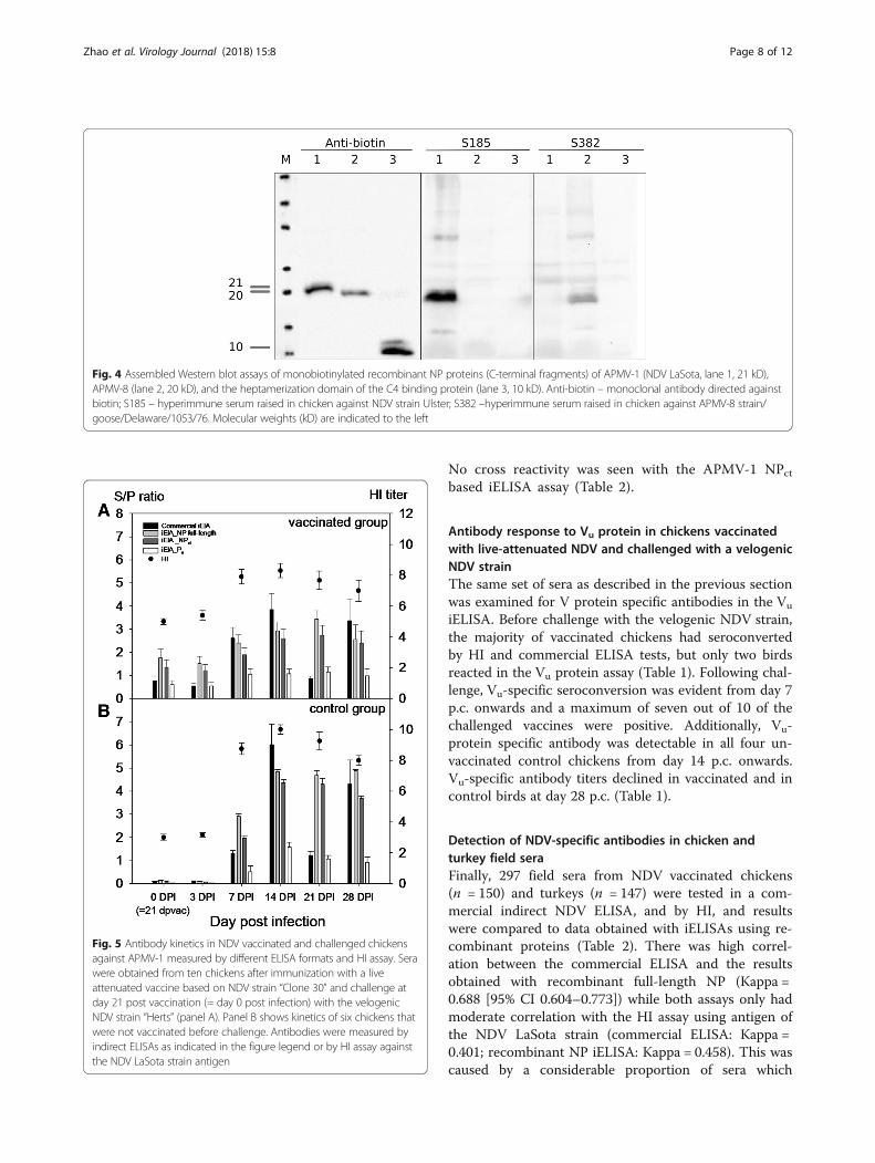
Analytical specificity of recombinant NDV NP- and P-genederived proteins in iELISA with sera raised againstdifferent avian avulavirus subtypesRecombinant iELISAs were used to test chickenhyperimmune sera raised against inactivated antigens ofnine APMV serotypes; for APMV-1 and APMV-3 serawere raisted against each of 3 (APMV-1 class II/Ulster,PPMV, APMV-1 class I) and 2 (APMV-3/England;APMV-3/Netherlands) strains, respectively. Other serawere raised against APMV-2, −4, −6, −7, −8, −9. A com-mercial NDV ELISA based on virion-preparations asantigen was used as a control. In this ELISA, only 2 outof 3 APMV-1 sera reacted (Fig. 3a) but a hyperimmuneserum raised against APMV-1 class I was not detected.In addition, APMV-6, 7 and 9 hyperimmune serashowed clear cross reactivity (dark greyed columns inFig. 3), whereas the APMV-4 hyperimmune serum re-vealed marginal reactivity, but was considered APMV-1positive by internal standards of the commercial ELISA.When using recombinant full length LaSota NP protein,positive reactions were obtained with all APMV-1 sera,including the APMV-1 class I serum. However, crossreactivity of APMV-6 and -9 specific sera remained (Fig.3a). In the NDV NPct based iELISA, in contrast, onlyNDV-specific sera raised against APMV-1 class II viruses(NDV LaSota, and the pigeon paramyxovirus type-1)reacted while all cross reactions were abolished (Fig. 3b,left panel). No cross reactivity was observed with aserum specific for APMV-1 of class I. Conversely, by theAPMV-8 NPct iELISA, the corresponding homologousantiserum was detected while cross reactions were lim-ited to an APMV-7 serum only. Serotype specificity ofNPct antigens to corresponding APMV-1 and APMV-8hyperimmune sera was also confirmed in compositeWestern blot assays (Fig. 4).Similar to the recombinant full-length NP iELISA,
cross reactivity of serotype-specific APMV sera wasobserved also for the iELISA when using the Pufragmentas the coating protein: Sera specific for APMV-3b(Netherlands), −4, −7, and −9 revealed reactivity abovethreshold level against the LaSota Pu protein (Fig. 3c).
Detection of NDV NP and P antibodies in vaccinated/challenged chickensTen chickens were immunized with a live-attenuatedvaccine based on NDV strain “Clone 30” [48]. Six SPFchickens were used as controls. At day 21 postvaccination, all chickens were challenged with thevelogenic NDV strain “Herts” and serum samples ofsurviving chickens were taken at days 0 (= day 21post vaccination), 3, 7, 14, 21, and 28 postchallenge(p.c.). Results obtained with these sera iniELISAs using recombinant NPct and Pu proteins were
Zhao et al. Virology Journal (2018) 15:8 Page 6 of 12
compared to commercial NDV ELISA and to HI titersagainst the LaSota strain of NDV. HI titers higherthan 1:8 were considered positive.As shown in Table 1, NDV antibodies were detected
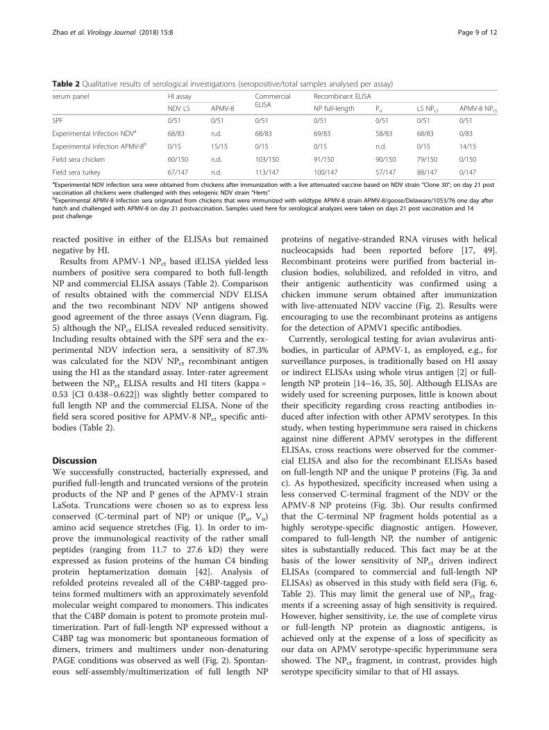
in a majority of the vaccinated chickens at day 0 postchallenge by HI, commercial ELISA and indirect NPctELISA assays, whereas a lower number of birds werepositive by the Pu ELISA assay. Moreover, in theunvaccinated control group, all chicken sera testednegative at day 0 p.c. and had seroconverted until7 days p.c. in HI, commercial ELISA and indirect
NPct ELISA assays, but complete seroconversion ofthe control group to Pu was delayed to day 14 p.c.(Table 1). This indicates that the iELISA based on thePu protein is less sensitive than the other assays. Thedevelopment of antibody titers as measured by thedifferent ELISAs and compared to HI titers of thevaccinated and control groups before and afterchallenge is shown in Fig. 5.When testing sera from fifteen chickens immunized
with wild-type APMV-8 strain, 14 out of 15 serumsamples were positive in the APMV-8 NPct iELISA.
Fig. 3 Use of NP and P gene products of APMV-1 and APMV-8 (NP) as diagnostic antigens in indirect ELISA compared to a commercial NDV ELISA.NDV commercial iELISA and Full length NP of NDV strain LaSota (NDV LS full-length NP) iELISA were showed in panel a; C-terminal fragment of theNDV (NDV LS NPct) and the APMV-8 (APMV-8 NPct) NP protein based iELISA were presented in panel b; The truncated P (NDV LS Pu) iELISA were in thepanel c. The chicken hyperimmune sera raised against avian avulavirus subtypes 1 through 9. 1 – APMV-1 class I, 1′ – APMV-1 class II,LaSota, 1″ – APMV-1 class II, pigeon paramyxovirus, other serotypes as indicated. Dotted line indicates cut-off. Dark grey bars indicateunspecific reactivity
Zhao et al. Virology Journal (2018) 15:8 Page 7 of 12
No cross reactivity was seen with the APMV-1 NPctbased iELISA assay (Table 2).
Antibody response to Vu protein in chickens vaccinatedwith live-attenuated NDV and challenged with a velogenicNDV strainThe same set of sera as described in the previous sectionwas examined for V protein specific antibodies in the Vu
iELISA. Before challenge with the velogenic NDV strain,the majority of vaccinated chickens had seroconvertedby HI and commercial ELISA tests, but only two birdsreacted in the Vu protein assay (Table 1). Following chal-lenge, Vu-specific seroconversion was evident from day 7p.c. onwards and a maximum of seven out of 10 of thechallenged vaccines were positive. Additionally, Vu-protein specific antibody was detectable in all four un-vaccinated control chickens from day 14 p.c. onwards.Vu-specific antibody titers declined in vaccinated and incontrol birds at day 28 p.c. (Table 1).
Detection of NDV-specific antibodies in chicken andturkey field seraFinally, 297 field sera from NDV vaccinated chickens(n = 150) and turkeys (n = 147) were tested in a com-mercial indirect NDV ELISA, and by HI, and resultswere compared to data obtained with iELISAs using re-combinant proteins (Table 2). There was high correl-ation between the commercial ELISA and the resultsobtained with recombinant full-length NP (Kappa =0.688 [95% CI 0.604–0.773]) while both assays only hadmoderate correlation with the HI assay using antigen ofthe NDV LaSota strain (commercial ELISA: Kappa =0.401; recombinant NP iELISA: Kappa = 0.458). This wascaused by a considerable proportion of sera which
Fig. 4 Assembled Western blot assays of monobiotinylated recombinant NP proteins (C-terminal fragments) of APMV-1 (NDV LaSota, lane 1, 21 kD),APMV-8 (lane 2, 20 kD), and the heptamerization domain of the C4 binding protein (lane 3, 10 kD). Anti-biotin – monoclonal antibody directed againstbiotin; S185 – hyperimmune serum raised in chicken against NDV strain Ulster; S382 –hyperimmune serum raised in chicken against APMV-8 strain/goose/Delaware/1053/76. Molecular weights (kD) are indicated to the left
Fig. 5 Antibody kinetics in NDV vaccinated and challenged chickensagainst APMV-1 measured by different ELISA formats and HI assay. Serawere obtained from ten chickens after immunization with a liveattenuated vaccine based on NDV strain “Clone 30” and challenge atday 21 post vaccination (= day 0 post infection) with the velogenicNDV strain “Herts” (panel A). Panel B shows kinetics of six chickens thatwere not vaccinated before challenge. Antibodies were measured byindirect ELISAs as indicated in the figure legend or by HI assay againstthe NDV LaSota strain antigen
Zhao et al. Virology Journal (2018) 15:8 Page 8 of 12
reacted positive in either of the ELISAs but remainednegative by HI.Results from APMV-1 NPct based iELISA yielded less
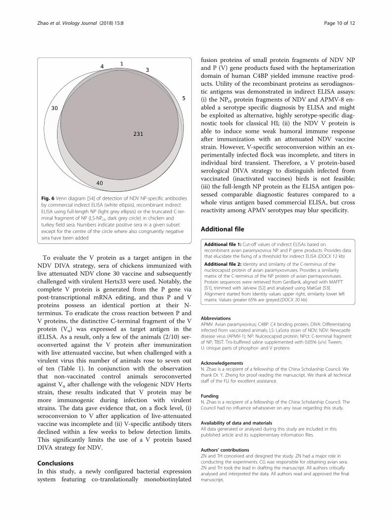
numbers of positive sera compared to both full-lengthNP and commercial ELISA assays (Table 2). Comparisonof results obtained with the commercial NDV ELISAand the two recombinant NDV NP antigens showedgood agreement of the three assays (Venn diagram, Fig.5) although the NPct ELISA revealed reduced sensitivity.Including results obtained with the SPF sera and the ex-perimental NDV infection sera, a sensitivity of 87.3%was calculated for the NDV NPct recombinant antigenusing the HI as the standard assay. Inter-rater agreementbetween the NPct ELISA results and HI titers (kappa =0.53 [CI 0.438–0.622]) was slightly better compared tofull length NP and the commercial ELISA. None of thefield sera scored positive for APMV-8 NPct specific anti-bodies (Table 2).
DiscussionWe successfully constructed, bacterially expressed, andpurified full-length and truncated versions of the proteinproducts of the NP and P genes of the APMV-1 strainLaSota. Truncations were chosen so as to express lessconserved (C-terminal part of NP) or unique (Pu, Vu)amino acid sequence stretches (Fig. 1). In order to im-prove the immunological reactivity of the rather smallpeptides (ranging from 11.7 to 27.6 kD) they wereexpressed as fusion proteins of the human C4 bindingprotein heptamerization domain [42]. Analysis ofrefolded proteins revealed all of the C4BP-tagged pro-teins formed multimers with an approximately sevenfoldmolecular weight compared to monomers. This indicatesthat the C4BP domain is potent to promote protein mul-timerization. Part of full-length NP expressed without aC4BP tag was monomeric but spontaneous formation ofdimers, trimers and multimers under non-denaturingPAGE conditions was observed as well (Fig. 2). Spontan-eous self-assembly/multimerization of full length NP
proteins of negative-stranded RNA viruses with helicalnucleocapsids had been reported before [17, 49].Recombinant proteins were purified from bacterial in-clusion bodies, solubilized, and refolded in vitro, andtheir antigenic authenticity was confirmed using achicken immune serum obtained after immunizationwith live-attenuated NDV vaccine (Fig. 2). Results wereencouraging to use the recombinant proteins as antigensfor the detection of APMV1 specific antibodies.Currently, serological testing for avian avulavirus anti-
bodies, in particular of APMV-1, as employed, e.g., forsurveillance purposes, is traditionally based on HI assayor indirect ELISAs using whole virus antigen [2] or full-length NP protein [14–16, 35, 50]. Although ELISAs arewidely used for screening purposes, little is known abouttheir specificity regarding cross reacting antibodies in-duced after infection with other APMV serotypes. In thisstudy, when testing hyperimmune sera raised in chickensagainst nine different APMV serotypes in the differentELISAs, cross reactions were observed for the commer-cial ELISA and also for the recombinant ELISAs basedon full-length NP and the unique P proteins (Fig. 3a andc). As hypothesized, specificity increased when using aless conserved C-terminal fragment of the NDV or theAPMV-8 NP proteins (Fig. 3b). Our results confirmedthat the C-terminal NP fragment holds potential as ahighly serotype-specific diagnostic antigen. However,compared to full-length NP, the number of antigenicsites is substantially reduced. This fact may be at thebasis of the lower sensitivity of NPct driven indirectELISAs (compared to commercial and full-length NPELISAs) as observed in this study with field sera (Fig. 6,Table 2). This may limit the general use of NPct frag-ments if a screening assay of high sensitivity is required.However, higher sensitivity, i.e. the use of complete virusor full-length NP protein as diagnostic antigens, isachieved only at the expense of a loss of specificity asour data on APMV serotype-specific hyperimmune serashowed. The NPct fragment, in contrast, provides highserotype specificity similar to that of HI assays.
Table 2 Qualitative results of serological investigations (seropositive/total samples analysed per assay)
serum panel HI assay CommercialELISA
Recombinant ELISA
NDV LS APMV-8 NP full-length Pu LS NPct APMV-8 NPct
SPF 0/51 0/51 0/51 0/51 0/51 0/51 0/51
Experimental Infection NDVa 68/83 n.d. 68/83 69/83 58/83 68/83 0/83
Experimental Infection APMV-8b 0/15 15/15 0/15 0/15 n.d. 0/15 14/15
Field sera chicken 60/150 n.d. 103/150 91/150 90/150 79/150 0/150
Field sera turkey 67/147 n.d. 113/147 100/147 57/147 88/147 0/147aExperimental NDV infection sera were obtained from chickens after immunization with a live attenuated vaccine based on NDV strain “Clone 30”; on day 21 postvaccination all chickens were challenged with thes velogenic NDV strain “Herts”bExperimental APMV-8 infection sera originated from chickens that were immunized with wildtype APMV-8 strain APMV-8/goose/Delaware/1053/76 one day afterhatch and challenged with APMV-8 on day 21 postvaccination. Samples used here for serological analyzes were taken on days 21 post vaccination and 14post challenge
Zhao et al. Virology Journal (2018) 15:8 Page 9 of 12
To evaluate the V protein as a target antigen in theNDV DIVA strategy, sera of chickens immunized withlive attenuated NDV clone 30 vaccine and subsequentlychallenged with virulent Herts33 were used. Notably, thecomplete V protein is generated from the P gene viapost-transcriptional mRNA editing, and thus P and Vproteins possess an identical portion at their N-terminus. To eradicate the cross reaction between P andV proteins, the distinctive C-terminal fragment of the Vprotein (Vu) was expressed as target antigen in theiELISA. As a result, only a few of the animals (2/10) ser-oconverted against the V protein after immunizationwith live attenuated vaccine, but when challenged with avirulent virus this number of animals rose to seven outof ten (Table 1). In conjunction with the observationthat non-vaccinated control animals seroconvertedagainst Vu after challenge with the velogenic NDV Hertsstrain, these results indicated that V protein may bemore immunogenic during infection with virulentstrains. The data gave evidence that, on a flock level, (i)seroconversion to V after application of live-attenuatedvaccine was incomplete and (ii) V-specific antibody titersdeclined within a few weeks to below detection limits.This significantly limits the use of a V protein basedDIVA strategy for NDV.
ConclusionsIn this study, a newly configured bacterial expressionsystem featuring co-translationally monobiotinylated
fusion proteins of small protein fragments of NDV NPand P (V) gene products fused with the heptamerizationdomain of human C4BP yielded immune reactive prod-ucts. Utility of the recombinant proteins as serodiagnos-tic antigens was demonstrated in indirect ELISA assays:(i) the NPct protein fragments of NDV and APMV-8 en-abled a serotype specific diagnosis by ELISA and mightbe exploited as alternative, highly serotype-specific diag-nostic tools for classical HI; (ii) the NDV V protein isable to induce some weak humoral immune responseafter immunization with an attenuated NDV vaccinestrain. However, V-specific seroconversion within an ex-perimentally infected flock was incomplete, and titers inindividual bird transient. Therefore, a V protein-basedserological DIVA strategy to distinguish infected fromvaccinated (inactivated vaccines) birds is not feasible;(iii) the full-length NP protein as the ELISA antigen pos-sessed comparable diagnostic features compared to awhole virus antigen based commercial ELISA, but crossreactivity among APMV serotypes may blur specificity.
Additional file
Additional file 1: Cut-off values of indirect ELISAs based onrecombinant avian paramyxovirus NP and P gene products. Provides datathat elucidate the fixing of a threshold for indirect ELISA (DOCX 12 kb)
Additional file 2: Identity and similarity of the C-terminus of thenucleocapsid protein of avian paramyxoviruses. Provides a similaritymatrix of the C-terminus of the NP protein of avian parmayxoviruses.Protein sequences were retrieved from GenBank, aligned with MAFFT[51], trimmed with Jalview [52] and analysed using MatGat [53].Alignment started from Identity values upper right, similarity lower leftmatrix. Values greater 65% are greyed.(DOCX 20 kb)
AbbreviationsAPMV: Avian paramyxovirus; C4BP: C4 binding protein; DIVA: Differentiatinginfected from vaccinated animals; LS: LaSota strain of NDV; NDV: Newcastledisease virus (APMV-1); NP: Nulceocapsid protein; NPct: C-terminal fragmentof NP; TBST: Tris-buffered saline supplemented with 0.05% (v/v) Tween;U: Unique parts of phosphor- and V proteins
AcknowledgementsN. Zhao is a recipient of a fellowship of the China Scholarship Council. Wethank Dr. Y. Zheng for proof reading the manuscript. We thank all technicalstaff of the FLI for excellent assistance.
FundingN. Zhao is a recipient of a fellowship of the China Scholarship Council. TheCouncil had no influence whatsoever on any issue regarding this study.
Availability of data and materialsAll data generated or analysed during this study are included in thispublished article and its supplementary information files.
Authors’ contributionsZN and TH conceived and designed the study. ZN had a major role inconducting the experiments. CG was responsible for obtaining avian sera.ZN and TH took the lead in drafting the manuscript. All authors criticallyanalysed and interpreted the data. All authors read and approved the finalmanuscript.
Fig. 6 Venn diagram [54] of detection of NDV NP-specific antibodiesby commercial indirect ELISA (white ellipsis), recombinant indirectELISA using full-length NP (light grey ellipsis) or the truncated C-ter-minal fragment of NP (LS-NPct, dark grey circle) in chicken andturkey field sera. Numbers indicate positive sera in a given subsetexcept for the centre of the circle where also congruently negativesera have been added
Zhao et al. Virology Journal (2018) 15:8 Page 10 of 12
Ethics approvalAll studies involving animals have received appropriate legal approval ofindependent ethic committees as detailed in the Method section.
Consent for publicationNot applicable.
Competing interestsThe authors declare that they have no competing interests.
Publisher’s NoteSpringer Nature remains neutral with regard to jurisdictional claims inpublished maps and institutional affiliations.
Received: 24 November 2017 Accepted: 8 January 2018
References1. Alexander DJ, Aldous EW, Fuller CM. The long view: a selective review of 40
years of Newcastle disease research. Avian pathology : journal of the WVPA.2012;41(4):329–35.
2. Senne DA, King DJ, Kapczynski DR. Control of Newcastle disease byvaccination. Dev Biol. 2004;119:165–70.
3. Kapczynski DR, Afonso CL, Miller PJ. Immune responses of poultry toNewcastle disease virus. Dev Comp Immunol. 2013;41(3):447–53.
4. Miller PJ, Afonso CL, Spackman E, Scott MA, Pedersen JC, Senne DA, BrownJD, Fuller CM, Uhart MM, Karesh WB, et al. Evidence for a new avianparamyxovirus serotype 10 detected in rockhopper penguins from theFalkland Islands. J Virol. 2010;84(21):11496–504.
5. Briand FX, Henry A, Massin P, Jestin V. Complete genome sequence of anovel avian paramyxovirus. J Virol. 2012;86(14):7710.
6. Terregino C, Aldous EW, Heidari A, Fuller CM, De Nardi R, Manvell RJ, BeatoMS, Shell WM, Monne I, Brown IH, et al. Antigenic and genetic analyses ofisolate APMV/wigeon/Italy/3920-1/2005 indicate that it represents a newavian paramyxovirus (APMV-12). Arch Virol. 2013;158(11):2233–43.
7. Karamendin K, Kydyrmanov A, Seidalina A, Asanova S, Sayatov M,Kasymbekov E, Khan E, Daulbayeva K, Harrison SM, Carr IM, et al. Completegenome sequence of a novel avian Paramyxovirus (APMV-13) isolated froma wild bird in Kazakhstan. Genome announcements. 2016;4(3):e00167–16.
8. Thampaisarn R, Bui VN, Trinh DQ, Nagai M, Mizutani T, Omatsu T, KatayamaY, Gronsang D, Le DHT, Ogawa H, et al. Characterization of avianparamyxovirus serotype 14, a novel serotype, isolated from a duck fecalsample in Japan. Virus Res. 2017;228:46–57.
9. Goraichuk I, Sharma P, Stegniy B, Muzyka D, Pantin-Jackwood MJ, GerilovychA, Solodiankin O, Bolotin V, Miller PJ, Dimitrov KM, et al. Complete genomesequence of an avian Paramyxovirus representative of putative newserotype 13. Genome announcements. 2016;4(4):e00729–16.
10. Thomazelli LM, de Araujo J, Fabrizio T, Walker D, Reischak D, Ometto T,Barbosa CM, Petry MV, Webby RJ, Durigon EL. Novel avian paramyxovirus(APMV-15) isolated from a migratory bird in South America. PLoS One. 2017;12(5):e0177214.
11. Lee HJ, Kim JY, Lee YJ, Lee EK, Song BM, Lee HS, Choi KS. A novel avianParamyxovirus (putative serotype 15) isolated from wild birds. FrontMicrobiol. 2017;8:786.
12. Krishnamurthy S, Samal SK. Nucleotide sequences of the trailer,nucleocapsid protein gene and intergenic regions of Newcastle diseasevirus strain Beaudette C and completion of the entire genome sequence. JGen Virol. 1998;79(Pt 10):2419–24.
13. Lamb RP, G: Paramyxoviridae: the viruses and their replication; 2007.14. Makkay AM, Krell PJ, Nagy E. Antibody detection-based differential ELISA for
NDV-infected or vaccinated chickens versus NDV HN-subunit vaccinatedchickens. Vet Microbiol. 1999;66(3):209–22.
15. Panshin A, Shihmanter E, Weisman Y, Orvell C, Kydyrmanov A, Asanov N,Daulbaeva K, Sayatov M, Lipkind M. Antigenic characterization of thenucleocapsid protein of Newcastle disease virus by means of a new panelof monoclonal antibodies. Comp Immunol Microbiol Infect Dis. 2000;23(3):209–20.
16. Ahmad-Raus R, Ali AM, Tan WS, Salleh HM, Eshaghi M, Yusoff K. Localizationof the antigenic sites of newcastle disease virus nucleocapsid using a panelof monoclonal antibodies. Res Vet Sci. 2009;86(1):174–82.
17. Errington W, Emmerson PT. Assembly of recombinant Newcastle diseasevirus nucleocapsid protein into nucleocapsid-like structures is inhibited bythe phosphoprotein. J Gen Virol. 1997;78(Pt 9):2335–9.
18. Kho CL, Tan WS, Tey BT, Yusoff K. Regions on nucleocapsid protein ofNewcastle disease virus that interact with its phosphoprotein. Arch Virol.2004;149(5):997–1005.
19. Hausmann S, Garcin D, Delenda C, Kolakofsky D. The versatility ofParamyxovirus RNA polymerase stuttering. J Virol. 1999;73(7):5568–76.
20. Jordan IK, Sutter BA, McClure MA. Molecular evolution of theParamyxoviridae and Rhabdoviridae multiple-protein-encoding P gene. MolBiol Evol. 2000;17(1):75–86.
21. McGinnes L, McQuain C, Morrison T. The P protein and the nonstructural38K and 29K proteins of Newcastle disease virus are derived from the sameopen reading frame. Virology. 1988;164(1):256–64.
22. Samson AC, Levesley I, Russell PH. The 36K polypeptide synthesized inNewcastle disease virus-infected cells possesses properties predicted for thehypothesized ‘V’ protein. J Gen Virol. 1991;72(Pt 7):1709–13.
23. Steward M, Vipond IB, Millar NS, Emmerson PT. RNA editing in Newcastledisease virus. J Gen Virol. 1993;74(Pt 12):2539–47.
24. Huang Z, Krishnamurthy S, Panda A, Samal SK. Newcastle disease virus Vprotein is associated with viral pathogenesis and functions as an alphainterferon antagonist. J Virol. 2003;77(16):8676–85.
25. Alamares JG, Elankumaran S, Samal SK, Iorio RM. The interferon antagonisticactivities of the V proteins from two strains of Newcastle disease viruscorrelate with their known virulence properties. Virus Res. 2010;147(1):153–7.
26. Czifra G, Nilsson M, Alexander DJ, Manvell R, Kecskemeti S, Engstrom BE.Detection of PMV-1 specific antibodies with a monoclonal antibodyblocking enzyme-linked immunosorbent assay. Avian pathology : journal ofthe WVPA. 1996;25(4):691–703.
27. Xu H, Lohr J, Greiner M. The selection of ELISA cut-off points for testingantibody to Newcastle disease by two-graph receiver operatingcharacteristic (TG-ROC) analysis. J Immunol Methods. 1997;208(1):61–4.
28. Hauslaigner R, Sonnenburg J, Kothlow S, Kaspers B, Staubach C, Grund C.Evaluation of an indirect enzyme-linked immunosorbent assay to study thespecific humoral immune response of Muscovy ducks (Cairina Moschata) anddomestic geese (Anser Anser Var. Domestica) after vaccination against Newcastledisease virus. Avian pathology : journal of the WVPA. 2009;38(2):89–95.
29. Das M, Kumar S. Recombinant phosphoprotein based single serum dilutionELISA for rapid serological detection of Newcastle disease virus. J VirolMethods. 2015;225:64–9.
30. Chaka H, Goutard F, Gil P, Abolnik C, Servan de Almeida R, Bisschop S,Thompson PN. Serological and molecular investigation of Newcastle diseasein household chicken flocks and associated markets in eastern Shewa zone,Ethiopia. Trop Anim Health Prod. 2013;45(3):705–14.
31. de Sousa RLM, Montassier HJ, Pinto AA. Detection and quantification ofantibodies to Newcastle disease virus in ostrich and Rhea sera using a liquidphase blocking enzyme-linked Immunosorbent assay. Clin Diagn LabImmunol. 2000;7(6):940–4.
32. Hemmatzadeh F, Kazemimanesh M. Detection of specific antigens ofNewcastle disease virus using an absorbed western blotting method.Iranian journal of veterinary research. 2017;18(2):92–6.
33. Kho CL, Mohd-Azmi ML, Arshad SS, Yusoff K. Performance of an RT-nestedPCR ELISA for detection of Newcastle disease virus. J Virol Methods. 2000;86(1):71–83.
34. de Oliveira ES, Silva KR, Fernando FS, Goncalves MC, Fernandes CC, BorziMM, dos Santos RM, Tamanini Mde L, Montassier Mde F, Montassier HJ. Aliquid-phase-blocking concanavalin a enzyme-linked immunosorbent assayfor the detection of antibodies against Newcastle disease virus in serum offree-ranging pigeons. Journal of veterinary diagnostic investigation : officialpublication of the American Association of Veterinary LaboratoryDiagnosticians, Inc. 2013;25(6):720–6.
35. Errington W, Steward M, Emmerson PT. A diagnostic immunoassay forNewcastle disease virus based on the nucleocapsid protein expressed by arecombinant baculovirus. J Virol Methods. 1995;55(3):357–65.
36. Mohan CM, Dey S, Rai A, Kataria JM. Recombinant haemagglutininneuraminidase antigen-based single serum dilution ELISA for rapidserological profiling of Newcastle disease virus. J Virol Methods. 2006;138(1–2):117–22.
37. Miers LA, Bankowski RA, Zee YC. Optimizing the enzyme-linkedimmunosorbent assay for evaluating immunity of chickens to Newcastledisease. Avian Dis. 1983;27(4):1112–25.
Zhao et al. Virology Journal (2018) 15:8 Page 11 of 12
38. Rivetz B, Weisman Y, Ritterband M, Fish F, Herzberg M. Evaluation of a novelrapid kit for the visual detection of Newcastle disease virus antibodies.Avian Dis. 1985;29(4):929–42.
39. Diel DG, da Silva LH, Liu H, Wang Z, Miller PJ, Afonso CL. Genetic diversityof avian paramyxovirus type 1: proposal for a unified nomenclature andclassification system of Newcastle disease virus genotypes. Infection,genetics and evolution : journal of molecular epidemiology andevolutionary genetics in infectious diseases. 2012;12(8):1770–9.
40. Beckett D, Kovaleva E, Schatz PJ. A minimal peptide substrate in biotinholoenzyme synthetase-catalyzed biotinylation. Protein science : apublication of the Protein Society. 1999;8(4):921–9.
41. Hofmeyer T, Schmelz S, Degiacomi MT, Dal Peraro M, Daneschdar M, ScrimaA, van den Heuvel J, Heinz DW, Kolmar H. Arranged sevenfold: structuralinsights into the C-terminal oligomerization domain of human C4b-bindingprotein. J Mol Biol. 2013;425(8):1302–17.
42. Shinya E, Owaki A, Norose Y, Sato S, Takahashi H. Quick method ofmultimeric protein production for biologically active substances such ashuman GM-CSF (hGM-CSF). Biochem Biophys Res Commun. 2009;386(1):40–4.
43. Zhao N, Lange E, Kubald S, Grund C, Beer M, Harder TC. Distinction ofsubtype-specific antibodies against European porcine influenza viruses byindirect ELISA based on recombinant hemagglutinin protein fragment-1.Virol J. 2013;10:246.
44. Postel A, Ziller M, Rudolf M, Letzel T, Ehricht R, Pourquier P, Dauber M,Grund C, Beer M, Harder TC. Broad spectrum reactivity versus subtypespecificity-trade-offs in serodiagnosis of influenza a virus infections bycompetitive ELISA. J Virol Methods. 2011;173(1):49–59.
45. Grund S, Adams O, Wahlisch S, Schweiger B. Comparison ofhemagglutination inhibition assay, an ELISA-based micro-neutralizationassay and colorimetric microneutralization assay to detect antibodyresponses to vaccination against influenza a H1N1 2009 virus. J VirolMethods. 2011;171(2):369–73.
46. Grund C, Steglich C, Huthmann E, Beer M, Mettenleiter TC, Romer-Oberdorfer A. Avian paramyoxvirus-8 immunization reduces viral sheddingafter homologous APMV-8 challenge but fails to protect against Newcastledisease. Virol J. 2014;11:179.
47. Rigaut G, Shevchenko A, Rutz B, Wilm M, Mann M, Seraphin B. A genericprotein purification method for protein complex characterization andproteome exploration. Nat Biotechnol. 1999;17(10):1030–2.
48. Steglich C, Grund C, Ramp K, Breithaupt A, Hoper D, Keil G, Veits J, Ziller M,Granzow H, Mettenleiter TC, et al. Chimeric newcastle disease virus protectschickens against avian influenza in the presence of maternally derived NDVimmunity. PLoS One. 2013;8(9):e72530.
49. Bulavaite A, Lasickiene R, Vaitiekaite A, Sasnauskas K, Zvirbliene A. Synthesisof human parainfluenza virus 2 nucleocapsid protein in yeast asnucleocapsid-like particles and investigation of its antigenic structure. ApplMicrobiol Biotechnol. 2016;100(10):4523–34.
50. Pinette MM, Rodriguez-Lecompte JC, Pasick J, Ojkic D, Leith M, SudermanM, Berhane Y. Development of a duplex fluorescent microsphereimmunoassay (FMIA) for the detection of antibody responses to influenza aand newcastle disease viruses. J Immunol Methods. 2014;405:167–77.
51. Katoh K, Asimenos G, Toh H. Multiple alignment of DNA sequences withMAFFT. Methods in molecular biology (Clifton, NJ). 2009;537:39–64.
52. Waterhouse AM, Procter JB, Martin DM, Clamp M, Barton GJ. Jalview version2–a multiple sequence alignment editor and analysis workbench.Bioinformatics. 2009;25(9):1189–91.
53. Campanella JJ, Bitincka L, Smalley J. MatGAT: an application that generatessimilarity/identity matrices using protein or DNA sequences. BMCbioinformatics. 2003;4:29.
54. Rodgers PFJ, Stapleton G, Howse J. Drawing area-proportional venn-3diagrams with convex polygons. In: 2010; Portland, OR, USA: DiagrammaticRepresentation and Inference, 6th International Conference, Diagrams 2010;2010.
• We accept pre-submission inquiries
• Our selector tool helps you to find the most relevant journal
• We provide round the clock customer support
• Convenient online submission
• Thorough peer review
• Inclusion in PubMed and all major indexing services
• Maximum visibility for your research
Submit your manuscript atwww.biomedcentral.com/submit
Submit your next manuscript to BioMed Central and we will help you at every step:
Zhao et al. Virology Journal (2018) 15:8 Page 12 of 12