endovascular brain intervention and mapping in a dog
TRANSCRIPT

Biomed Pap Med Fac Univ Palacky Olomouc Czech Repub. 2014 Jun; 158(2):221-226.
221
Endovascular brain intervention and mapping in a dog experimental model using magnetically-guided micro-catheter technology
Tomas Karaa,b, Pavel Leinvebera, Michal Vlasinc, Pavel Juraka,d, Miroslav Novaka, Zdenek Novake, Jan Chrastinae, Krzysztof Czechowicza, Milos Belehrada, Samuel J. Asirvathamb
Aim. Despite the substantial progress that has been achieved in interventional cardiology and cardiac electrophysi-ology, endovascular intervention for the diagnosis and treatment of central nervous system (CNS) disorders such as stroke, epilepsy and CNS malignancy is still limited, particularly due to highly tortuous nature of the cerebral arterial and venous system. Existing interventional devices and techniques enable only limited and complicated access espe-cially into intra-cerebral vessels. The aim of this study was to develop a micro-catheter magnetically-guided technology specifically designed for endovascular intervention and mapping in deep CNS vascular structures.Methods. Mapping of electrical brain activity was performed via the venous system on an animal dog model with the support of the NIOBE II system.Results. A novel micro-catheter specially designed for endovascular interventions in the CNS, with the support of the NIOBE II technology, was able to reach safely deep intra-cerebral venous structures and map the electrical activity there. Such structures are not currently accessible using standard catheters. Conclusion. This is the first study demonstrating successful use of a new micro-catheter in combination with NIOBE II technology for endovascular intervention in the brain.
Key words: central nervous system, magnetically-guided endovascular interventions, mapping, NIOBE
Received: June 25, 2012; Accepted: July 19, 2012; Available online: November 12, 2012http://dx.doi.org/10.5507/bp.2012.076
aInternational Clinical Research Center - Department of Cardiovascular Diseases, St Anne's University Hospital in Brno and Masaryk University, Brno, Czech Republic bDivision of Cardiovascular Diseases, Mayo Clinic, Rochester, MN, USAcSchool of Veterinary Medicine, University of Veterinary and Pharmaceutical Sciences Brno, Brno dInstitute of Scientific Instruments of the Czech Academy of Sciences, BrnoeDepartment of Neurosurgery, St Anne's University Hospital in Brno and Masaryk University, BrnoCorresponding author: Samuel J. Asirvatham, e-mail: [email protected]
INTRODUCTION
Within the last twenty years, significant progress in the diagnostics and treatment of major cardiovascular diseases has been achieved particularly due to tremen-dous progress in interventional cardiology and cardiac electrophysiology. Introduction of direct percutaneous coronary interventions has completely changed the man-agement and treatment of acute coronary syndromes and ST-segment elevation myocardial infarction in particular, demonstrating clear benefit in comparison with throm-bolytic therapy1-3. Similar progress has been achieved in invasive cardiac electrophysiology. Development of novel methods and technologies for electrical and magnetic mapping, hybrid imaging, and ablations introduced the novel concept of non-pharmacological treatment of both supraventricular and ventricular cardiac arrhythmias, pro-viding clear benefit for many indications in comparison to standard pharmacological therapy3-5.
Furthermore, progress in interventional cardiology and electrophysiology has provided new opportunities for the treatment of structural heart diseases, such as aortic stenosis, mitral regurgitation, patent foramen ovale, hy-pertrophic cardiomyopathy, and many others. Combining
interventional cardiology and electrophysiological tech-niques provides the necessary basis for the treatment of pharmacoresistant hypertension using renal sympathetic denervation6-8. Similarly, novel bio- and nanotechnologies for the treatment of cardiovascular disease will begin a new era of cardiovascular regenerative medicine9,10.
Novel technologies are being developed to make both coronary and non-coronary cardiac interventions and methods for cardiac electrophysiology and pacing even safer and more effective. One such example is technology for magnetically-guided endovascular and intra-cardiac procedures. Remote magnetic navigation systems, such as the NIOBE II technology, allow for very precise three-di-mensional navigation of interventional devices (catheter, guidewire) (ref.11,12). The precise positioning of the tip of the interventional device is achieved with two permanent magnets which directly control the tip of a remotely di-rected interventional device. Thus, movement of specially designed catheters can be very precisely controlled by the physician. Changing the vector of the magnetic field allows rapid and safe navigation of the catheters into posi-tions that are challenging to achieve by traditional manual or even robotic control11-14.
Biomed Pap Med Fac Univ Palacky Olomouc Czech Repub. 2014 Jun; 158(2):221-226.
222
So far, magnetically-guided coronary and intracardiac procedures have only been used for the treatment of car-diovascular diseases and substantial progress in interven-tional cardiology and cardiac electrophysiology has been made. However, the use of endovascular intervention for the diagnosis and treatment of CNS disorders, such as stroke, epilepsy and CNS malignancy, is still limited. One reason for this is that the cerebral arterial and venous system is highly circuitous. Existing interventional devices and techniques enable only limited and complicated ac-cess, especially into intra-cerebral vessels
On the other hand, magnetically-guided interventions may represent new hope for interventions within the CNS, as they have the ability to safely navigate catheters even into extremely complex vascular structures. However, the catheters used for cardiovascular interventions are too large and too rigid to be used for interventions in the CNS.
To the best of our knowledge, there is no catheter technology yet available that is designed for specifically magnetically-guided interventions within the CNS.
The aim of our study was to develop a micro-catheter magnetically-guided technology that would be specifically designed for endovascular interventions, mapping in deep CNS vascular structures, and the testing of micro-catheter technology on an experimental animal dog model.
Such technology, if successfully developed, would represent a novel approach to for the diagnostics and treatment of the most devastating CNS disorders, such
as stroke and epilepsy, with potentially even broader ap-plications, including neurostimulation and implantable monitoring of CNS functions.
METHODS
The experiments were performed at the Cardiovascular Animal Research Center in the Laboratory for Advanced Cardiovascular and CNS Interventions at the School of Veterinary Medicine, University of Veterinary and Pharmaceutical Sciences Brno (Brno, Czech Republic).
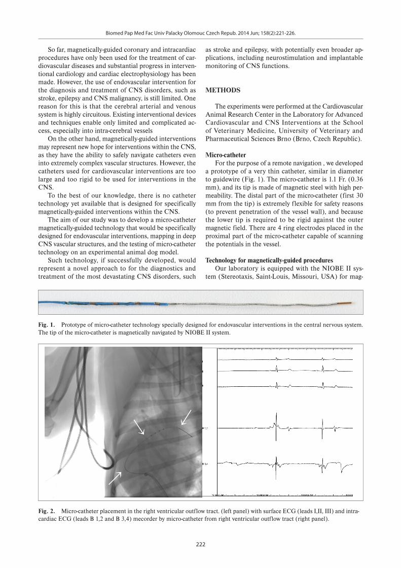
Micro-catheter For the purpose of a remote navigation , we developed
a prototype of a very thin catheter, similar in diameter to guidewire (Fig. 1). The micro-catheter is 1.1 Fr. (0.36 mm), and its tip is made of magnetic steel with high per-meability. The distal part of the micro-catheter (first 30 mm from the tip) is extremely flexible for safety reasons (to prevent penetration of the vessel wall), and because the lower tip is required to be rigid against the outer magnetic field. There are 4 ring electrodes placed in the proximal part of the micro-catheter capable of scanning the potentials in the vessel.
Technology for magnetically-guided procedures Our laboratory is equipped with the NIOBE II sys-
tem (Stereotaxis, Saint-Louis, Missouri, USA) for mag-
Fig. 1. Prototype of micro-catheter technology specially designed for endovascular interventions in the central nervous system. The tip of the micro-catheter is magnetically navigated by NIOBE II system.
Fig. 2. Micro-catheter placement in the right ventricular outflow tract. (left panel) with surface ECG (leads I,II, III) and intra-cardiac ECG (leads B 1,2 and B 3,4) mecorder by micro-catheter from right ventricular outflow tract (right panel).
Biomed Pap Med Fac Univ Palacky Olomouc Czech Repub. 2014 Jun; 158(2):221-226.
223
netic navigation in combination with AXIOM ARTIS DFC Digital Angiography System (Siemens, Erlangen, Germany).
Signal Acquisition Electrocardiogram (ECG) and electroencephalogram
(EEG) signals were recorded by the Prucka EP system (General Electric, Milwakee, Wiconsin, USA) and by ANNALab MI-3 technology (St. Anne’s University Hospital in Brno and Institute of Scientific Instruments of Czech Academy of Sciences, Brno, Czech Republic ) that was developed by our team as an experimental system for comprehensive invasive and non-invasive cardiac and CNS electrophysiology.
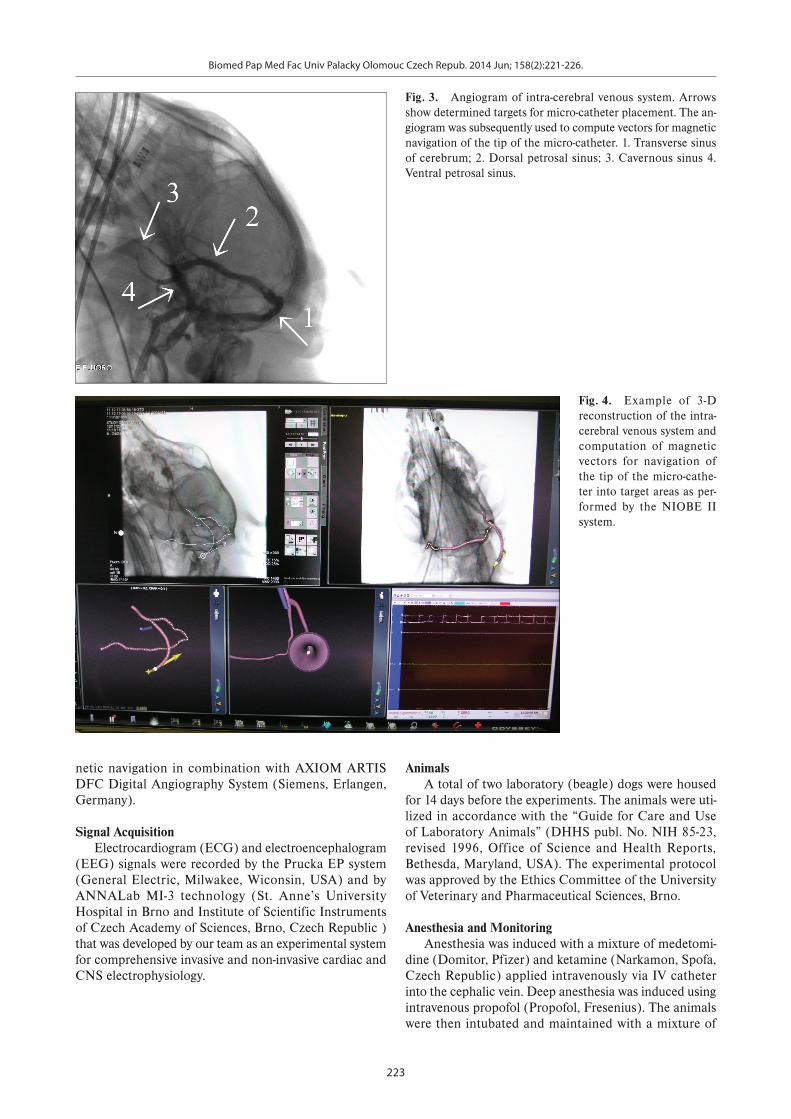
Fig. 3. Angiogram of intra-cerebral venous system. Arrows show determined targets for micro-catheter placement. The an-giogram was subsequently used to compute vectors for magnetic navigation of the tip of the micro-catheter. 1. Transverse sinus of cerebrum; 2. Dorsal petrosal sinus; 3. Cavernous sinus 4. Ventral petrosal sinus.
Animals A total of two laboratory (beagle) dogs were housed
for 14 days before the experiments. The animals were uti-lized in accordance with the “Guide for Care and Use of Laboratory Animals” (DHHS publ. No. NIH 85-23, revised 1996, Office of Science and Health Reports, Bethesda, Maryland, USA). The experimental protocol was approved by the Ethics Committee of the University of Veterinary and Pharmaceutical Sciences, Brno.
Anesthesia and Monitoring Anesthesia was induced with a mixture of medetomi-
dine (Domitor, Pfizer) and ketamine (Narkamon, Spofa, Czech Republic) applied intravenously via IV catheter into the cephalic vein. Deep anesthesia was induced using intravenous propofol (Propofol, Fresenius). The animals were then intubated and maintained with a mixture of
Fig. 4. Example of 3-D reconstruction of the intra-cerebral venous system and computation of magnetic vectors for navigation of the tip of the micro-cathe-ter into target areas as per-formed by the NIOBE II system.

Biomed Pap Med Fac Univ Palacky Olomouc Czech Repub. 2014 Jun; 158(2):221-226.
224
oxygen and isoflurane (Forane, Fresenius) at the range of 1% to 1.5% of isoflurane. IV infusion was applied (lactat-ed Ringer solution, Baxter) using linear perfusor (Braun Perfusor S, B.Braun, Germany) at a rate of 10 mL/kg/h. The heart rate (HR) was measured by ECG electrodes applied on the thorax, SpO2 by a sensor applied on the tongue, and respiratory rate (RR) electronically based on thorax-impedance changes. All parameters were sampled and saved by Datex – Ohmeda vital monitor.
Interventions The experiment was planned and conducted as an
acute experiment followed by euthanasia. Each procedure was performed under deep anesthesia to prevent pain and suffering of the animal.
Femoral access was secured by surgical dissection of
left groin and separate cannulation of the left femoral artery and vein. A 6-Fr. sheath was inserted into each vessel as a portal entrance for the catheter.
RESULTS
Testing of micro-catheter in the heartInitially, the micro-catheter was inserted via the ve-
nous portal into the femoral vein and through the sinus venosus and navigated down the right atrium and into the right ventricle by using the support of NIOBE II system. This “cardio” part of our experiment was designed to test the mechanical and electrical characteristics of our micro-catheter prototype, particularly the remote navigation of the tip and electrical signal acquisition. The catheter tip was successfully placed into the preselected locations
Fig. 5. Example of successful micro-catheter placement into a palatine plexus via ventral petrosal sinus with mapping of electrical activity of the brain.
Fig. 6. Example of successful micro-catheter placement into a cavernous sinus with mapping of electrical activity of the brain. Note the tortuosity of venous circulation that was overcome by our technology.

Biomed Pap Med Fac Univ Palacky Olomouc Czech Repub. 2014 Jun; 158(2):221-226.
225
within the right heart (high right atrium, right ventricle outflow tract, right ventricle apex, and annulus tricuspida-lis) and recorded intracardiac electrocardiogram (ECG) signals of comparable quality to standard catheters (Fig. 2).
Testing of micro-catheter in the brainAfter successful mechanical and electrical testing of
our catheter in the heart, the second part of the experi-ment began with the objective of reaching complex venous structures in the brain and obtaining endovascular record-ing of electrical activity of the brain, using the support of the NIOBE II system.
The injection of contrast dye into the main venous sinuses was performed (Fig. 3) and two images with a contrast medium (RAO 47° and LAO 37°) were stored in special Niobe II software as shown in Fig. 4. Both main venous sinuses and peripheral venous structures were determined as locations for micro-catheter place-ment. Subsequently, the 3-D navigation vessel model was computed by the NIOBE system according to our speci-fications. Parameters for magnetic navigation were set, allowing navigation of the tip of the micro-catheter via tor-tuosities of the venous cerebral circulation and reaching the predetermined locations in the brain, including deep vein structures. Subsequently, the endovascular navigation of the micro-catheter into CNS structures was initiated by gently pushing the micro-catheter and changing the outer magnetic field. With support of the NIOBE II system, successful navigation of the micro-catheter was performed into the vena cava cranialis, vena jugularis communis, vena maxilaris, sinus temporalis, sinus sigmoideus, sinus petrosus ventralis, sinus cavernosus, and sinus intercav-ernosus without evidence of venous dissection, acute thromboembolism, or extravascular bleeding during the procedure (Fig. 4 and Fig. 5.)
Electrical activity of the brain was successfully mea-sured via micro-catheter from intracerebral venous loca-tions (Fig. 4 and Fig. 5).
DISCUSSION
To our knowledge, this is the first study demonstrating the ability to reach deep areas of rather complicated ca-nine brain vascularity using the percutaneous endovascu-lar approach. For these first in living dog experiments, our original micro-catheter, in combination with the NIOBE II system, was capable of safely reaching deep venous structures in the brain of the dog which are not accessible using standard techniques, and successfully measuring the electrical signals there. Our group has previously shown that successful EEG recording and radiofrequency abla-tion can be performed using an endovascular approach in an experimental pig model15. Given the fact that the struc-ture of the venous system in the canine brain is even more complex and fragile than in primates, including humans, our newly developed micro-catheter can be navigated, in combination with the NIOBE II system, to final loca-
tions using an external magnetic field, thus representing a potentially new approach for semi-invasive diagnostics and treatment of epilepsy and stroke. Modified versions of the catheter can also be used to damage tumor and other pathological tissues by delivering radiofrequency energy, or for neurostimulation and/or implantable monitoring of CNS functions.
LIMITATIONS
Despite promising results provided by our pilot experi-ment, it is evident that further testing, particularly on a non-human primate model will be needed to confirm the safety and efficacy of our micro-catheter technology. It is also likely that our prototype micro-catheter will need to be developed for endovascular arterial access due to the smaller diameter of cerebral arteries.
CONCLUSION
Our micro-catheter, specially designed for endovas-cular interventions in the CNS with support of NIOBE II technology, was capable of safely reaching deep intra-cerebral venous structures and mapping electrical activity. Such structures have not been accessible with standard catheters. This is the first study demonstrating the suc-cessful use of the NIOBE II technology for endovascular interventions in the brain. Further experiments are need-ed to confirm the safety and efficacy of this new approach for the endovascular treatment of CNS disorders.
ABBREVIATIONS
CNS, central nervous system; ECG, electrocardio-gram; EEG, electroencephalogram; RR, respiratory rate; HR, heart rate
ACKNOWLEDGEMENTS
This study was supported by grants of IGA of Ministry of Health No. NS 10099/2008, European Regional Development Fund - Project FNUSA-ICRC (No. CZ.1.05/1.1.00/02.0123) and European Community's Seventh Framework Programme (FP7/2007-2013) under grant agreement No. 238802 (IIIOS Project).
Authors would like to acknowledge Michal Kuna, MSc and Vlastimil Vondra, PhD for their essential help with the study and development of ANNALab MI-3 tech-nology.
CONFLICT OF INTEREST STATEMENT
The authors stated that there are no conflicts of inter-est regarding the publication of this article.

Biomed Pap Med Fac Univ Palacky Olomouc Czech Repub. 2014 Jun; 158(2):221-226.
226
Disclosures for Samuel Asirvatham: Honoraria/Consulting - Abiomed, Biotronik, Boston Scientific, Medtronic, Spectranetics, St. Jude, Sanofi-Aventis, Stereotaxis. Co-patent holder - may receive future royal-ties from - Nevro: Use of nerve signal modulation to treat central, autonomic, and peripheral nervous system dis-orders, including pain; Aegis: Appendage ligation; ATP: Atrial fibrillation ablation and coagulum reduction during ablation
REFERENCES
1. Widimský P, Budesínský T, Vorác D, Groch L, Zelízko M, Aschermann M, Branny M, St'ásek J, Formánek P; 'PRAGUE' Study Group Investigators. Long distance transport for primary angioplasty vs immediate thrombolysis in acute myocardial infarction. Final results of the randomized national multicentre trial--PRAGUE-2. Eur Heart J. 2003;24(1):94-104.
2. Widimský P, Groch L, Zelízko M, Aschermann M, Bednár F, Suryapranata H. Multicentre randomized trial comparing transport to primary angioplasty vs immediate thrombolysis vs combined strategy for patients with acute myocardial infarction presenting to a community hospital without a catheterization laboratory. The PRAGUE study. Eur Heart J. 2000;21(10):823-31.
3. Braunwald E. The rise of cardiovascular medicine. Eur Heart J 2012 Apr;33(7):838-45.
4. Calkins H, Kuck KH, Cappato R, Brugada J, Camm AJ, Chen SA, Crijns HJ, Damiano RJ Jr, Davies DW, DiMarco J, Edgerton J, Ellenbogen K, Ezekowitz MD, Haines DE, Haissaguerre M, Hindricks G, Iesaka Y, Jackman W, Jalife J, Jais P, Kalman J, Keane D, Kim YH, Kirchhof P, Klein G, Kottkamp H, Kumagai K, Lindsay BD, Mansour M, Marchlinski FE, McCarthy PM, Mont JL, Morady F, Nademanee K, Nakagawa H, Natale A, Nattel S, Packer DL, Pappone C, Prystowsky E, Raviele A, Reddy V, Ruskin JN, Shemin RJ, Tsao HM, Wilber D. 2012 HRS/EHRA/ECAS expert consensus statement on catheter and surgical ablation of atrial fibrillation: recommendations for patient selection, proce-dural techniques, patient management and follow-up, definitions, endpoints, and research trial design. J Interv Card Electrophysiol 2012;33(2):171-257.
5. Aliot EM, Stevenson WG, Almendral-Garrote JM, Bogun F, Calkins CH, Delacretaz E, Della Bella P, Hindricks G, Jaïs P, Josephson ME, Kautzner J, Kay GN, Kuck KH, Lerman BB, Marchlinski F, Reddy V, Schalij MJ, Schilling R, Soejima K, Wilber D. EHRA/HRS Expert Consensus on Catheter Ablation of Ventricular Arrhythmias: devel-oped in a partnership with the European Heart Rhythm Association (EHRA), a Registered Branch of the European Society of Cardiology (ESC), and the Heart Rhythm Society (HRS); in collaboration with the American College of Cardiology (ACC) and the American Heart Association (AHA). Heart Rhythm 2009;6(6):886-933.
6. Krum H, Schlaich M, Whitbourn R, Sobotka PA, Sadowski J, Bartus K, Kapelak B, Walton A, Sievert H, Thambar S, Abraham WT, Esler M. Catheter-based renal sympathetic denervation for resistant hyper-tension: a multicentre safety and proof-of-principle cohort study. Lancet 2009; 373:1275-81.
7. Simplicity HTN-1 Investigators 2011. Catheter-based renal sympa-thetic denervation for resistant hypertension: durability of blood pressure reduction out to 24 months. Hypertension 2011;57:911-7.
8. Simplicity HTN-2 Investigators, Esler MD, Krum H, Sobotka PA, Schlaich MP, Schmieder RE, Bohm M. Renal sympathetic denervation in patients with treatment-resistant hypertension (The Symplicity HTN-2 Trial): a randomised controlled trial. Lancet 2010; 376:1903-9.
9. Pislaru SV, Harbuzariu A, Gulati R, Witt T, Sandhu NP, Simari RD, Sandhu GS. Magnetically targeted endothelial cell localization in stented vessels. J Am Coll Cardiol 2006;48(9):1839-45.
10. Wang X, Jameel MN, Li Q, Mansoor A, Qiang X, Swingen C, Panetta C, Zhang J. Stem cells for myocardial repair with use of a transarterial catheter. Circulation 2009;120(11 Supl):238-46.
11. Malcolme-Lawes L, Kanagaratnam P. Robotic navigation and abla-tion. Minerva Cardioangiol 2010;58(6):691-9.
12. Nazarian S. New technologies and therapies for cardiac arrhythmias. Minerva Cardioangiol 2010;58(6):731-40.
13. Kiemeneij F, Patterson MS, Amoroso G, Laarman G, Slagboom T. Use of the Stereotaxis Niobe magnetic navigation system for percutane-ous coronary intervention: results from 350 consecutive patients. Catheter Cardiovasc Interv 2008;71(4):510-6.
14. Buergler JM, Alam S, Spencer W, Kleiman NS, Melendez Y, Franklin J, Nagueh SF. Initial experience with alcohol septal ablation using a novel magnetic navigation system. J Interv Cardiol 2007;20(6):559-63.
15. Henz BD, Friedman PA, Bruce CJ, Holmes DR Jr, Okumura Y, Johnson SB, Packer DL, Asirvatham SJ. Successful radiofrequency ablation of the cerebral cortex in pigs using the venous system: possible impli-cations for treating CNS disorders. Epilepsy Res 2008;80(2-3):213-8.