endotoxin stimulates expression of the murine urokinase receptor
TRANSCRIPT

American Journal ofPathology, Vol. 147, No. 3, September 1995Copyright t American Societyfor Investigative Pathology
Endotoxin Stimulates Expression of the MurineUrokinase Receptor Gene in Vivo
Fanny Almus-Jacobs, Nissi Varki,Michael S. Sawdey, and David J. LoskutoffFrom the Department of Vascular Biology, The ScrippsResearch Institute, LaJolla, California
The regulation of urokinase receptor (u-PAR)gene expression during endotoxemia was stud-ied in vivo with a murine model systenm Northernblot analysis demonstrated relatively bigb levelsofu-PAR mRNA in mouseplacenta, witb interme-diate levels in lung and spleen and very low levelsin heart and kidney. No u-PAR mRNA could bedetected in liver, gut, tbymus, brain, or skeletalmuscle. Intraperitoneal injection of endotoxin(lipopolysaccbaride) increased the steady-statelevels ofu-PAR mRNA in most tissues examinedThe greatest induction (sevenfold) was observedin the lung at I hour after injection. The celularlocalization ofu-PAR mRNA was assessed by insitu hybridization. In control mice, u-PAR mRNAwas detectedprimarily in alveolar macrophagesof the lung and lymphocytes of the spleen andthymus, although a specific signal was alsopresent in other ceU types. In general, endotbelalceUs lacked detectable u-PAR mRNA. The induc-tion of u-PAR mRNA by lipopo(ysaccharide wasapparent within 30 minutes and was localixed totissue macrophages, lymphocytes, and endothe-lial ceUs lining arteries and veins. At later times(1 to 3 hours), specialized epitbelial ceUspresent in gastrointestinal tract, bile ducts, anduterus were also positive for u-PAR mRNA. In-duction of u-PAR in vivo by lipopolysaccharidemay facilitate the extravasation and migrationof leukocytes during inflammation. (Am JPathol 1995, 147:688-698)
Plasminogen activators (PAs) are serine proteasesthat catalyze the conversion of plasminogen intoplasmin. Plasmin plays a central role in thrombolysisand in a variety of other processes.1 Two PAs havebeen identified, including the urokinase type (u-PA)
and tissue type (t-PA), which are produced by dis-tinct genes. Urokinase accumulates on the surfaceon many cells bound to the u-PA receptor (u-PAR).2Plasminogen also binds to cell surfaces via a recep-tor,3 and its interaction with receptor-bound u-PAresults in the formation of plasmin. This surface-associated plasmin is able to degrade proteins of theextracellular matrix and basement membranes.These interactions localize extracellular proteolyticactivity during tumor cell invasion, tissue remodeling,and cell migration.45 Recently, u-PAR was impli-cated as a critical component of the metastatic pro-cess.6 The human,78 murine,9 and bovine10 u-PARcDNAs have been isolated and completely se-quenced. The human u-PAR protein has been puri-fied11 and appears to be the single chain glycopro-tein of Mr 50,000 to 60,000. The deduced amino acidsequence of human u-PAR predicts a polypeptide of313 amino acids composed of three 90-amino-acid,cysteine-rich repeats. The first repeat contains theu-PA binding region.12 The attachment site for theglycosyl-phosphatidylinositol (GPI) anchor is locatedat the end of the third repeat and is sensitive to aphosphatidylinositol-specific phopholipase C. u-PARis deficient on peripheral blood monocytes and gran-ulocytes from patients with paroxysmal nocturnal he-moglobinuria, an acquired disorder characterized bya defect in the membrane attachment of proteinsnormally anchored by GPI.13 This deficiency inu-PAR may be related to the high incidence of ve-nous thrombosis in these patients. The unique do-main structure of u-PAR, and the fact that it is aGPI-anchored protein, suggests that it is a memberof the T cell activating proteins or Ly-6 superfamily. 14
Murine u-PAR resembles human u-PAR with re-spect to ligand-binding characteristics, molecularmass, glycosylation pattern, and GPI anchorage.9
Supported by grant HL47819 (to DJL) from the National Institutes ofHealth.Accepted for publication May 19, 1995.
Address reprint requests to Dr. David J. Loskutoff, Department ofVascular Biology (VB-3), The Scripps Research Institute, 10666North Torrey Pines Rd., La Jolla, CA 92037.
688
Murine Urokinase Receptor Gene Expression 689AJP September 1995, Vol. 14 7, No. 3
The amino acid sequence deduced from the murinecDNA is 62% identical with the human u-PAR se-quence, and this homology increases to 70% homol-ogy when conservative amino acid substitutions aretaken into account. Interestingly, a second form ofmurine u-PAR was detected by cDNA cloning9 andseems to arise from alternative mRNA splicing. Thisform, designated mu-PAR-2, codes for a form of thereceptor that lacks the sequence for GPI additionand thus is thought to be a soluble form that issecreted from the cells. The first 133 residues of themu-PAR-2 protein are homologous to human u-PAR,but the next 66 residues appear to be unique. Re-cently, a variant form of human u-PAR mRNA (u-PAR-2) was identified15 and is likely to represent afunctional analogue of mouse u-PAR-2.
In cells in culture, u-PAR synthesis is regulated bya diverse group of agents, including inflammatorycytokines, growth factors, hormones, tumor promot-ers, thrombin, and secondary mediators such asprotein kinase C and cAMP. For example, interferon,tumor necrosis factor, and u-PA itself have beenreported to stimulate the expression of u-PAR onhuman monocytes,16 whereas interleukin (IL)-2 andphorbol myristate acetate increased u-PAR expres-sion in T lymphocytes.17'18 Basic fibroblast growthfactor was shown to increase u-PAR expression byhuman vascular cells.19 Despite these results, little isknown about the tissue distribution and regulation ofthe u-PAR gene in vivo. In the present work, we haveemployed a murine model system to identify thetissues and cells that constitutively synthesize u-PARmRNA and to analyze changes in its expressionduring endotoxemia. Our results demonstrate thatLPS induces u-PAR expression in a variety of tissuesand cells in vivo. The induction of u-PAR may facili-tate extravasation and migration of leukocytes duringinflammation and contribute to the pathogenesis ofgram-negative sepsis.
Materials and Methods
Cloning of Murine u-PAR cDNA with theReverse-Transcriptase Polymerase ChainReaction (RT-PCR)The human u-PAR cDNA, kindly provided by Dr.E. K. 0. Kruithof (Centre Hospitalier UniversitaireVaudois, Lausanne, Switzerland), was used as aprobe to identify murine cells producing u-PAR. Inthese experiments, RNA samples isolated eitherfrom different murine cell lines (eg, Balb-3T3 fibro-blasts, hepatoma 1-6 cells, and RAW 264.7 cells) or
from various murine tissues were initially analyzed byNorthern blotting. RNA extracted from human U937cells (human hystiocytic lymphoma) was used as apositive control. A murine monocyte-macrophageline transformed by Abelson leukemia virus (RAW264.7, ATCC TIB 71, American Tissue Culture Col-lection, Rockville, MD) and known to constitutivelyexpress u-PA activity20 exhibited the strongest hy-bridization signal for u-PAR mRNA. It was thereforeselected as the source of murine RNA for PCR. TwoPCR primers were synthesized based on the pub-lished sequence of murine u-PAR9 and designed fordirectional cloning with different protuding termini asfollows: 5' primer (5'-GGGAGCTCCTGCAGTGCAT-GCAGTGTGAGA-3') and 3' primer (5'-CCTCTA-GAAGTCAGGTCCAGAGGAGGACGC-3'). RT-PCRwas performed with the Gene Amp RNA kit (Perkin-Elmer Cetus, Norwalk, CT) according to the manu-facturer's instructions for 35 cycles. The amplifiedfragment was resolved by agarose gel electrophore-sis and had the predicted size (1.0 kb). The PCRproduct was purified, digested with Sacl and Xbaland subcloned into an ampicillin-resistant plasmidvector (pGEM-3Z; Promega, Madison, WI). Restric-tion analysis of the resulting plasmid DNA indicatedan insert of the predicted size, and sequence anal-ysis demonstrated identity with murine u-PAR 1.9 ThecDNA includes bp 80 to bp 997 from the publishedsequence.
Animal Protocols and Tissue PreparationAdult male or female CB6.F1 mice (BALB/c/Byj xC57B16/j; Jackson Laboratories, Bar Harbor, ME, orScripps Clinic Rodent Breeding Colony), aged 6 to 8weeks, were used for all of the experiments. Lipo-polysaccharide (LPS) from Escherichia co/i serotype0111 :B4, (Sigma Chemical Co., St. Louis, MO) wasresuspended in sterile saline (Baxter, Deerfield, IL),and 50 ,ug (approximately 2 mg/kg) was injectedintraperitoneally into mice anesthetized by inhalationof metofane (methoxyflurane, Pitman-Moore, Munde-lein, IL). Control mice were anesthetized and in-jected with saline alone. At different time intervalsafter injection, mice were anesthetized by metofaneinhalation and killed by cervical dislocation. Tissueswere rapidly removed by standard dissection tech-niques. The placenta employed for these experi-ments was purchased (Harlam Bioproducts for Sci-ence, Indianapolis, IN). For Northern blot analysis,tissues were minced and stored in liquid nitrogenuntil RNA extraction. For in situ hybridization, thetissues were immersed in cold 4% (w/v) paraformal-dehyde in Dulbecco's phosphate-buffered saline
690 Almus-Jacobs et alAJP September 1995, Vol. 147, No. 3
(Whittaker Bioproducts, Walkersville, MD) and fixedat 40C overnight. The fixed tissues were then embed-ded in paraffin blocks and sectioned at 2 to 5 ,umthickness with a microtome. The sections weremounted onto polylysine slides and stored at roomtemperature until in situ hybridization analysis.
Preliminary dose-response experiments were per-formed to determine the optimal amount of LPS. Micewere injected with increasing amounts of LPS (from0.6 to 100 ,ug), and 1 hour later, RNA was extractedfrom lung and spleen and resolved by Northernblot analysis. The maximal response for inductionof u-PAR mRNA was achieved with 50 ,ug, andthis amount was thus employed for most of theexperiments.
Northern Blot AnalysisFor each Northern blot experiment, tissues were ob-tained from three different animals and analyzedindependently. Total RNA was extracted from thefrozen tissues by the acid guanidinium-thiocyanate-phenol-chloroform method21 and the concentrationof RNA was determined by sample absorbance at260 nm. Total RNA was analyzed for u-PAR mRNAby Northern blotting essentially as described.22Briefly, the RNA (20 jig) was fractionated by electro-phoresis under denaturing conditions on a 1.5%agarose gel, stained with ethidium bromide, andtransferred overnight onto nylon membranes (Bio-trans ICN, Irvine, CA). The blots were prehybridizedfor 30 minutes in 50 mmol/L Pipes buffer, pH 6.8,containing 100 mmol/L NaCI, 20 mmol/L Na2HPO4,30 mmol/L NaH2PO4, 1 mmol/L EDTA, and 5% so-dium dodecyl sulfate, and then were hybridized inthe same solution for 16 hours at 650C, with 106cpm/ml murine u-PAR cDNA probe. The probe wasradiolabeled by employing a random primer labelingkit (Boehringer-Mannheim Biochemical, Indianapo-lis, IN) in the presence of a-[32P]dGTP (3000 Ci/mmol, Amersham, Arlington Heights, IL). After hy-bridization, the blots were washed four times for 15minutes each with prewarmed (650C) 0.67X stan-dard saline citrate (SSC) containing 5% sodium do-decyl sulfate and then subjected to autoradiographicanalysis with Kodak XAR-5 film and intensifyingscreens at -700C for varying times. The resultingautoradiograms were quantified by densitometricscanning by using an LKB Ultrascan XL laser den-sitometer (LKB, Bromma, Sweden). To verify thatdensitometric measurements were within the linearrange, total RNA from the lung and spleen of LPS-treated mice was prepared, serially diluted, andanalyzed by Northern blotting. The densitometric
values obtained from this calibration curve demon-strated linearity. To assess variability in sample load-ing, Northern blots were rehybridized with a 32p_labeled 18S rat cDNA probe.23 Despite somevariability in sample loading, similar results were ob-tained for all tissues in three independent experi-ments. Northern blot analysis of total RNA frommouse tissues with the murine u-PAR cDNA proberevealed the presence of a single transcript of ap-proximately 1.4 kb as estimated with a 0.16 to 1.77kb RNA ladder from GIBCO BRL (Gaithersburg,MD).
Riboprobe Preparation and in SituHybridizationThe pGEM-3Z vector carrying the murine u-PARcDNA insert was linearized with the restriction endo-nucleases EcoRI or Hindlll and employed as a tem-plate for the synthesis of antisense or sense ribo-probes, respectively. In vitro transcription reactionswere performed with SP6 or T7 RNA polymerases(Promega) in the presence of [35S]UTP (Amer-sham, 1000 Ci/mmol). The DNA templates wereremoved by digestion with RNAse-free DNAse(RQ1 DNAse, Promega) for 15 minutes at 370Cand the riboprobes purified by phenol extractionand ethanol precipitation.To study the cellular localization of u-PAR mRNA,
in situ hybridization was performed essentially asdescribed.24 Paraffin-embedded tissue sectionswere treated sequentially with xylene (three times for5 minutes each), 2X SSC (1X SSC is 150 mmol/LNaCI, 15 mmol/L sodium citrate, pH 7.0) containing10 mmol/L 2-mercaptoethanol and 1 mmol/L EDTA(10 minutes), paraformaldehyde (10 minutes at 40C),and proteinase K (1 ,ug/ml in 300 mmol/L NaCI, 10mmol/L Tris-HCI, pH 8.0). All washes and incuba-tions were performed at room temperature unlessotherwise specified. The slides were then prehybrid-ized for 2 hours in 100 ,ul of prehybridization buffer(50% w/v) formamide, 0.3 mmol/L NaCI, 20 mmol/LTris-HCI, pH 8.0, 5 mmol/L EDTA, 0.02% polyvi-nylpyrrolidone, 0.02% Ficoll, 0.02% bovine serumalbumin, 10% (w/v) dextran sulfate, and 10 mmol/Ldithiothreitol) at 420C. Prehybridization buffer (20 ,ul)containing 2.5 mg/ml yeast tRNA and 600,000 cpm35S-labeled riboprobe was added, and the slideswere hybridized at 550C overnight. After hybridiza-tion, the slides were treated with 2X SSC containing1 mmol/L EDTA (twice for 10 minutes each), RNAseA (20 ,ug/ml in 500 mmol/L NaCI and 10 mmol/LTris-HCI; 30 minutes), 2X SSC containing 10 mmol/L

Murine Urokinase Receptor Gene Expression 691AJP September 1995, Vol. 147, No. 3
2-mercaptoethanol and 1 mmol/L EDTA (twice for 10minutes each), 0.1X SSC containing 10 mmol/L2-mercaptoethanol and 1 mmol/L EDTA (2 hours at600C), and 0.5X SSC (twice for 10 minutes each).Finally, the slides were dehydrated by immersion in agraded alcohol series containing 0.3 mmol/LCH3COONH4, dried, coated with NTB2 emulsion(Kodak, Rochester, NY) diluted 1:2 in distilled water,and exposed in the dark at 40C for 2, 6, and 12weeks. Slides were then developed for 2 minutes inD19 developer (Kodak), fixed, washed in water(three times for 5 minutes each), and counterstainedwith hematoxylin and eosin by standard procedures.Positive hybridization signals appeared under micro-scopic observation as green grains (epilumines-cence) or as dark grains (bright field). No specificsignal could be detected in parallel sections hybrid-ized with a sense riboprobe as a control for nonspe-cific hybridization. Because in situ hybridization is nota very quantitative technique, care was taken tominimize variations caused by the handling of thetissues or tissue sections. For example, in all in-stances in which tissues from control and LPS-treated animals were compared, the tissues wereisolated, processed, and hybridized in parallel withthe same buffers, probe mixtures, emulsions, andwhere possible, exposure times. In general, quanti-tative conclusions were based on results obtainedby Northern blot analysis.
Results
Tissue Distribution of Murine u-PAR mRNATo examine constitutive expression of the murineu-PAR gene in vivo, total RNA was extracted fromtissues of CB6 mice and analyzed by Northern blot-ting with a murine u-PAR cDNA probe. Results froma representative animal are shown in Figure 1A. Pla-centa showed by far the highest concentration ofu-PAR mRNA, followed by spleen, lung, heart, andkidney. No u-PAR mRNA could be detected in liver,gut, thymus, brain, or skeletal muscle.To compare the distribution of u-PAR with its Ii-
gand u-PA, the blot was rehybridized with a murineu-PA cDNA probe (Figure 1 B). The kidney, whichhad relatively low levels of u-PAR mRNA, showed thehighest concentration of u-PA mRNA, in agreementwith previous observations.25'26 The placenta, whichhad high levels of u-PAR mRNA and exhibits strongimmunohistochemical staining for u-PA protein,27also had relatively high levels of u-PA mRNA. Inter-estingly, significant levels of u-PA mRNA were de-tected in the thymus, and this tissue lacked detect-
a a
A.
uPAR -
Dl
UPA
C.18S -
Figure 1. Relative level of expression of u-PAR and u-PA mRNAs inmurine tissues. Total RNA was preparedfrom the indicated tissues asdescribed in Materials and Methods, and 20 ,ug were analyzed byNorthern blotting with 32P-labeled murine cDNA probes to u-PAR(A) or to u-PA (B). The two autoradiograms were exposedfor 4 days.To assess variability in sample loading, the blot shown in (A) wasrebybridized with a 32P-labeled rat 18S cDNA probe (C).
able u-PAR mRNA. Comparison of Figure 1, A and B,reveals that expression of the receptor and its ligandis not necessarily coincident in the tissues studied.
Regulation of Murine u-PAR GeneExpression by LPSExperiments were performed to determine whetheru-PAR gene expression was altered in vivo in re-sponse to endotoxin. Mice were injected intraperito-neally either with saline (controls) or with saline con-taining 50 ,ug of LPS (2.0 mg/kg). At various timesafter injection, selected tissues were removed andanalyzed for u-PAR mRNA by Northern blotting. Re-sults from a representative experiment are shown inFigure 2. LPS treatment increased the steady-statelevels of u-PAR mRNA in most tissues, with maximalinduction observed at 1 to 3 hours. The autoradio-graphic signals for u-PAR and 18S rRNA were quan-titated by scanning densitometry (see Materials andMethods) and the values obtained for u-PAR mRNAnormalized to those of 18S rRNA. Maximal inductionsof 7-, 5-, 3-, and 2.5-fold were observed in the lung,kidney, spleen, and heart, respectively. The concen-tration of u-PAR mRNA began to decrease after 1 to3 hours, reaching baseline levels by 24 hours in mostinstances. Similar results were obtained for all fourorgans in three independent experiments. In sepa-rate experiments (not shown), the tissues were har-vested 15 and 30 minutes after LPS injection andanalyzed as above by Northern blotting. Althoughincreased expression of u-PAR mRNA was detectedat 30 minutes in the lung, no increase was evident at

692 Almus-Jacobs et alAJP September 1995, Vol. 14 7, No. 3
Kidney Gut Thymus Liver
A 1 3 6 8 12 16 24 1 3 6 8 12 16 Ti (hr)
uPAR -
^ 1 3 6 8 12 16 24 X 1 3 6 a 12 16 24 Tinme (hrs)
uPAR- -
Blas-
Figure 2. Changes in the concentration ofu-PAR mRNA in response toLPS. Mice were injected intraperitoneally with 50 ,tg ofLPS, and at theindicated times, tissues were removed and total RNA (20 p-g) wasanalyzed by Northern blotting. A: Changes in the concentration ofu-PAR mRNA in lung, kidney, spleen, and heart at various times. B:Variability in sample loading as detected by rebybridizing the spleenand heart blots in A with a 32P-labeled 18S cDNA probe. The autora-diograms were exposedfor 4 to 6 days.
this early time in kidney, spleen, and heart. Upregu-lation of u-PAR mRNA also was observed in liver andgut, and possibly in the thymus and brain as well(Figure 3). In these instances, the level of induction
_ + - + + _ +Figure 3. Induction of u-PAR mRNA in the brain, gut, thymus, andliver by LPS. Mice were injected with 50 ,ug of LPS and 3 hours later,tissues were removed and total RNA (20 Ag) was analyzed by Northernblotting. -, control; +, LPS. The autoradiogramsforliverandgut were
exposed for 9 days whereas those for brain and thymus were forexposedfor 12 days.
was not possible to quantitate by densitometricscanning as the control values were very low or notdetected (Figures 1 and 3). However, a positive sig-nal was detected in the gut 1 hour after LPS treat-ment (not shown). Comparison of the 1- and 3-hourtime points revealed that LPS induced a 3-fold in-crease in PAI-1 mRNA during this interval. Thus, thedifference between the u-PAR mRNA levels in thegut of untreated controls (not detected by densito-metric analysis) and the 3-hour sample must begreater than 3-fold.
Figure 4. Localization of u-PAR mRNA in cells of the lung. A: Sectionfrom a normal lung hybridized with the u-PAR antisense riboprobe. Examplesof cells specifically expressing u-PAR mRNA are indicated by the arows. In general, u-PAR mRNA was detected on cells bordering the alveolar (a)space, whereas bronchial epithelium (e) was negative. Magnification, X250. B: Section of lung from an animal treated with endotoxin for 1 hour
showing that the same cells express mRNA. X250. C: A high power, bright-field view of lung from the LPS-treated mouse shown in B. X 1000. The
arrowspoint to cells at the edge ofthe alveoli. In A to C, the slides were exposedfor 6 weeks. D: Sectionfrom the lung of the endotoxin-treated mouse,
hybridized with a u-PAR sense riboprobe and exposedfor 12 weeks. X250.
A Luna Brain#~~~~~~~~~~~~~~~~~~~~~~~~~~I .
i4
I
I
Murine Urokinase Receptor Gene Expression 693AJP September 1995, Vol. 147, No. 3
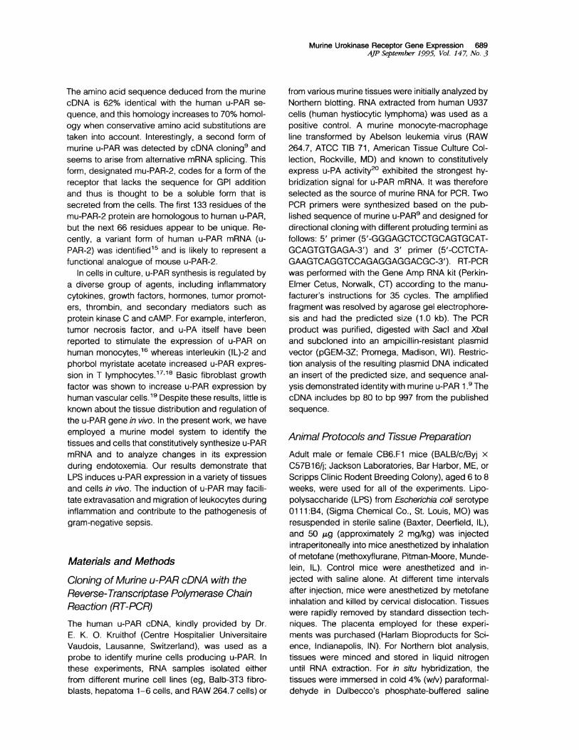
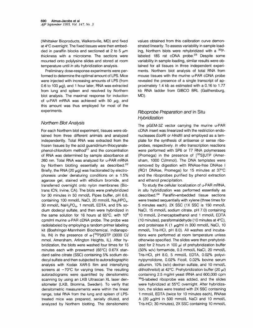
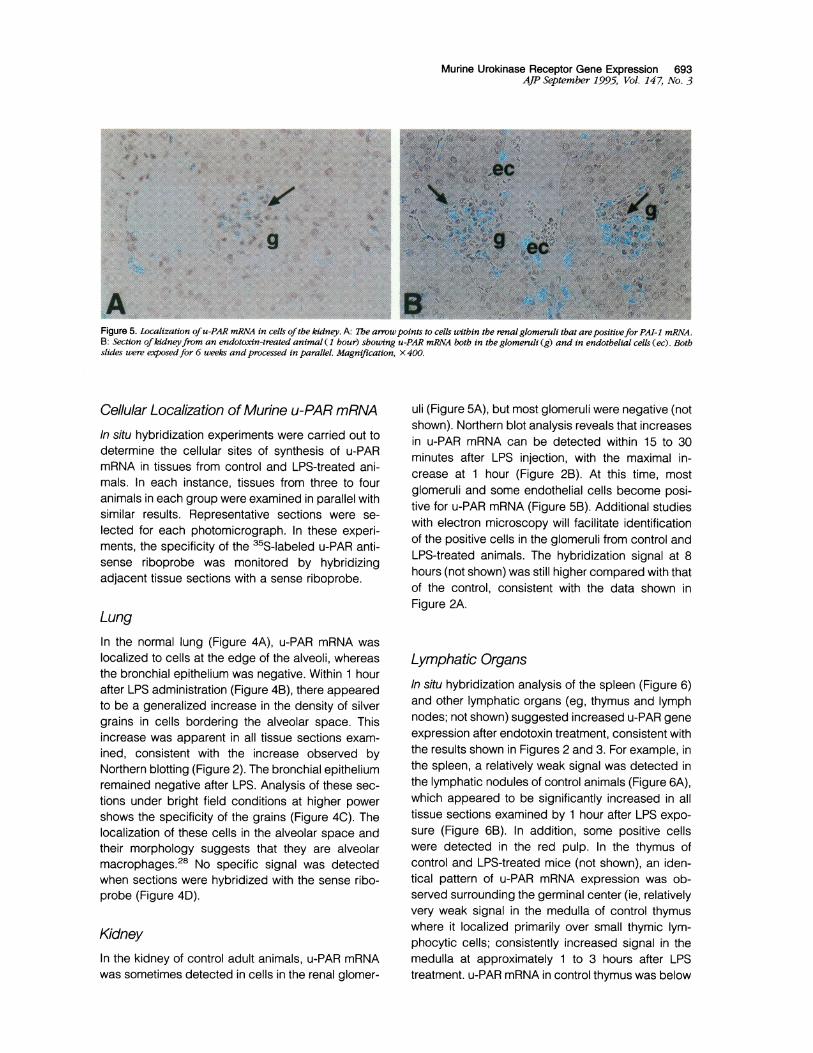
Figure 5. Localization ofu-PAR mRNA in cells ofthe kidney. A: The arrowpoints to cells within the renal glomeruli that arepositivefor PAI-1 mRNA.B: Section ofkidneyfrom an endotoxin-treated animal (1 hour) showing u-PAR mRNA both in the glomeruli (g) and in endothelial cells (ec). Bothslides were exposedfor 6 weeks andprocessed in parallel. Magnification, X 400.
Cellular Localization of Murine u-PAR mRNAIn situ hybridization experiments were carried out todetermine the cellular sites of synthesis of u-PARmRNA in tissues from control and LPS-treated ani-mals. In each instance, tissues from three to fouranimals in each group were examined in parallel withsimilar results. Representative sections were se-lected for each photomicrograph. In these experi-ments, the specificity of the 35S-labeled u-PAR anti-sense riboprobe was monitored by hybridizingadjacent tissue sections with a sense riboprobe.
LungIn the normal lung (Figure 4A), u-PAR mRNA waslocalized to cells at the edge of the alveoli, whereasthe bronchial epithelium was negative. Within 1 hourafter LPS administration (Figure 4B), there appearedto be a generalized increase in the density of silvergrains in cells bordering the alveolar space. Thisincrease was apparent in all tissue sections exam-ined, consistent with the increase observed byNorthern blotting (Figure 2). The bronchial epitheliumremained negative after LPS. Analysis of these sec-tions under bright field conditions at higher powershows the specificity of the grains (Figure 4C). Thelocalization of these cells in the alveolar space andtheir morphology suggests that they are alveolarmacrophages.28 No specific signal was detectedwhen sections were hybridized with the sense ribo-probe (Figure 4D).
KidneyIn the kidney of control adult animals, u-PAR mRNAwas sometimes detected in cells in the renal glomer-
uli (Figure 5A), but most glomeruli were negative (notshown). Northern blot analysis reveals that increasesin u-PAR mRNA can be detected within 15 to 30minutes after LPS injection, with the maximal in-crease at 1 hour (Figure 2B). At this time, mostglomeruli and some endothelial cells become posi-tive for u-PAR mRNA (Figure 5B). Additional studieswith electron microscopy will facilitate identificationof the positive cells in the glomeruli from control andLPS-treated animals. The hybridization signal at 8hours (not shown) was still higher compared with thatof the control, consistent with the data shown inFigure 2A.
Lymphatic OrgansIn situ hybridization analysis of the spleen (Figure 6)and other lymphatic organs (eg, thymus and lymphnodes; not shown) suggested increased u-PAR geneexpression after endotoxin treatment, consistent withthe results shown in Figures 2 and 3. For example, inthe spleen, a relatively weak signal was detected inthe lymphatic nodules of control animals (Figure 6A),which appeared to be significantly increased in alltissue sections examined by 1 hour after LPS expo-sure (Figure 6B). In addition, some positive cellswere detected in the red pulp. In the thymus ofcontrol and LPS-treated mice (not shown), an iden-tical pattern of u-PAR mRNA expression was ob-served surrounding the germinal center (ie, relativelyvery weak signal in the medulla of control thymuswhere it localized primarily over small thymic lym-phocytic cells; consistently increased signal in themedulla at approximately 1 to 3 hours after LPStreatment. u-PAR mRNA in control thymus was below

694 Almus-Jacobs et alAJP September 1995, Vol. 147, No. 3
Figure 6. Distribution of u-PAR mRNA in the spleen. A: Section of control mouse spleen shouwng positive cells in the lymphatic nodules (in).Magnification, X 250. B: Representative section demonstrating a more intense hybridization signal in this area 1 hour after LPS. X250. Both slideswere processed in parallel and exposedfor 6 weeks. Arrows indicate e-xamples ofpositive cells.
the limits of detection by Northern blot analysis (Fig-ures 1A and 3).The observations that the lymphoid areas of
spleen, the medulla of the thymus, and lymph nodes(not shown) throughout the body were positive foru-PAR raises the possibility that the common celltype responsive to LPS in these tissues is the lym-phocyte. Additional immunohistochemical studiesare needed to verify this hypothesis.
Heart and Blood VesselsIn the control myocardium, no specific u-PAR mRNAsignal was detected in myocytes or veins (Figure 7A)or in endothelium of blood vessels including aorta(Figure 7B). In contrast, after LPS treatment, some ofthe cardiac myocytes (Figure 7C) and endotheliumof the heart vasculature (Figure 7, D and E) becamepositive. The endothelium of large vessels (eg, aorta;not shown), as well as endothelium lining arteries,veins, and capillaries of a number of organs includ-ing the kidney (Figure 5B), brain, and liver (notshown) were also positive after LPS treatment.Smooth muscle cells do not appear to express de-tectable amounts of u-PAR mRNA.
GutNorthern blot analysis revealed that endotoxin alsoinduced u-PAR mRNA in the gut of mice (Figure 3),and the cells producing it in the absence and pres-ence of endotoxin appear to be largely the same(Figure 8). For example, in control gut (Figure 8A),u-PAR was observed in cells lining the outer epithe-lial luminal surface as well as in cells located towardthe core of the villus. The basal epithelial cells hadvery low levels of u-PAR mRNA (not shown). After
LPS treatment, u-PAR mRNA was detected primarilyover nuclei of these same cells (Figure 8, B and C)and in the basal cells of the crypt (not shown).
LiverAlthough the liver of control animals did not containdetectable u-PAR mRNA (Figure 1), it was detectedin livers from LPS-treated animals (Figure 3). Threehours after LPS treatment, u-PAR mRNA was de-tected primarily in the biliary duct epithelium (Figure9A) and in scattered cells localized in the sinusoidalspaces. Additional immunohistochemical staining isrequired to identify these latter cells.
Epitheliumu-PAR mRNA was detected in epithelium of a num-ber of organs from untreated control mice, includingthose in the urinary bladder, liver gallbladder, andgut (not shown). However, u-PAR mRNA was notdetected in epithelial cells of the lung, uterus, biliaryducts, and Bowman's capsule of the kidney. Theepithelium appears to demonstrate tissue-specificsensitivity to LPS as the bile ducts of the liver (Figure9A) and uterus epithelium (Figure 9B) were inducedby LPS treatment, whereas epithelial cells from thelung and kidney were not (not shown).
BrainIn the brain (not shown), the hypothalamus and cer-ebellum exhibited a weak hybridization signal foru-PAR mRNA, which did not increase after LPS ex-posure, consistent with the Northern blots (Figure 3).The meningeal layer also appeared to express u-PAR mRNA and u-PAR mRNA was detected in cap-

Murine Urokinase Receptor Gene Expression 695AJP September 1995, Vol. 147, No. 3
Figure 7. Localization ofu-PAR mRNA in the heart. A: Sectionfrom thebeart qf a control animal showing the absence of specific signal foru-PAR mRNA in the myocardium and veins (v0. Magnification, X250.B: Section showing lack of specfi'c signalfor u-PAR mRNA in a sectionofcontrol aorta. X250. C: Sectionfrom beart 1 hour afterLPS treatmentshowing the presence of detectable u-PAR mRNA in myocytes (arrows).X250. D: Section from hear 1 hourafterITPS reatment showing positiveadipose tissue (ad) andpositive endothelium (e) lining an a?tely. X 250.E: Section sbowing positivity ofthe endothelium (e) lining a veinfrom thebeart 3 hours after LPS exposure. X400. All slides were processed inparallel and exposed.for 6 wceeks.
illaries, probably originating from endothelial cellsand oligodendroglial cells (macrophage origin).
Adipose Tissue, Skin, and Skeletal MuscleVery few u-PAR mRNA-positive cells were detectedin adipose tissue from untreated mice. However, theamount of u-PAR mRNA-positive cells appeared toincrease 1 to 3 hours after LPS treatment (Figure 7D).Establishing the identity of the positive cells in theadipose tissue of LPS-treated mice will require addi-tional studies. No u-PAR mRNA was detected in skinand skeletal muscle from control and LPS-treatedanimals (not shown).
DiscussionIn this report, Northern blot and in situ hybridizationexperiments were performed to investigate the nor-mal tissue distribution and regulation of u-PAR geneexpression in vivo. The cDNA probes employed forthese studies should detect both u-PAR-1 and u-PAR-2 as the first 480 bp of these cDNAs are iden-tical. Northern blot analysis demonstrated relativelyhigh levels of u-PAR mRNA in mouse placenta, withintermediate levels in the lung and spleen and verylow levels in the heart and kidney (Figure 1A). Nou-PAR mRNA could be detected in the liver, gut,thymus, brain, or skeletal muscle. This distribution

696 Almus-Jacobs et alAJP September 1995, Vol. 147, No. 3
Figure 8. Distribution ofu-PAR mRNA in the gut. A: Sectionfrom control gut. A positive signal can be detected over scattered cells lining the outerepithelial luminal surface(ls) as well as on cells at the core(c) ofthe villus. B: Section 3 hours after LPS treatment showing PAI-1 mRNA positive cellsin the core of the villus and in scattered epithelial cells. Arrows indicate examples ofpositive cells. Slides were exposedfor 6 weeks andprocessed inparallel. X 400.
was distinctly different from that of u-PA (Figure 1 B).Thus, expression of the receptor and its ligand is notnecessarily coincident in the tissues studied.
Endotoxin is a component of the cell walls ofgram-negative bacteria that activates many of thecellular phases of acute inflammation.29 The effectsof endotoxin are mediated in part by tumor necrosisfactor-a, IL-1, IL-6, and other cytokines. Althoughcytokines regulate u-PAR synthesis in culturedcells,16-18 little is known about their effects, or that ofLPS itself, on u-PAR synthesis in vivo. Figures 2 and3 demonstrate that LPS administration induces arapid but transient increase in u-PAR in a variety ofmurine tissues. For example, administration of sub-lethal doses of LPS leads to an increase in the steady-state levels of u-PAR mRNA in the lung (7-fold), kidney(5-fold), spleen (3-fold), and heart (2.5-fold) (Figure 2),with a peak at 1 hour. u-PAR mRNA in the gut and liver,and possibly in the thymus and brain, also appeared tobe upregulated by LPS (Figure 3).
The widespread induction of u-PAR mRNA by LPSsuggested that common vascular cells might be in-volved. To investigate this possibility, the cellularlocalization of u-PAR mRNA in tissues from controland LPS-treated mice was assessed by in situ hy-bridization with 35S-labeled sense and antisense u-PAR riboprobes. In control mice, u-PAR mRNA wasdetected primarily in alveolar macrophages of thelung (Figure 4A), in macrophages and lymphocytesof the spleen (Figure 6A), and in lymphocytes of thethymus (not shown). In addition, a relatively weak butconsistently detected signal was observed in someglomeruli of the kidney of control animals (Figure 5A)and in a variety of epithelia (not shown). In general,u-PAR mRNA was not detected in endothelial cells ofcontrol tissue (Figure 7B) and no specific signal wasapparent in hybridizations with the 35S-labeledsense probe (Figure 4D).The primary cellular response to LPS was local-
ized to tissue macrophages and lymphocytes (Fig-
Figure 9. u-PAR gene expression in the epithelium after LPS treatment. A: u-PAR mRNA localization in the epithelium (ep) of the biliary duct (bd)of the liver after 1 hour after LPS treatment. B: u-PAR mRNA localization in the uterus epithelium (ep) 1 hour after LPS treatment. Both slides wereexposedfor 6 weeks. x 400.

Murine Urokinase Receptor Gene Expression 697AJP September 1995, Vol. 147, No. 3
ures 4B and 6B). Only rare neutrophils showed pos-itivity in control or LPS-exposed animals, and thesignal was relatively weak. Interestingly, u-PARmRNA was also markedly enhanced by LPS in theendothelial cells of arteries, veins, and capillaries ofa number of organs, including brain and liver (notshown) and blood vessels from the heart (Figure 7, Dand E) and kidney (Figure 5B). Some myocytes in theheart expressed u-PAR mRNA in response to LPS(Figure 7C) and cells within most glomeruli of thekidneys showed a marked induction of u-PAR mRNA(Figure 4B). Additional studies are required to iden-tify the specific cell type(s) in the kidney and spleenthat express u-PAR mRNA in response to LPS. LPSalso markedly induced u-PAR gene expression in avariety of specialized epithelial cells including thosepresent in bile ducts (Figure 9A) and uterus (Figure8B). Finally, comparison of the results shown in Fig-ures 4A, 5A, and 6A with those in Figures 4B, 5B,and 6B reveal that LPS frequently induced u-PARmRNA in the same cells that were expressing it in theuntreated controls.The complex effect of endotoxin on mammalian
systems includes the activation of the immune sys-tem. The ability of macrophages and T lymphocytesto extravasate and reach inflammatory sites is a ba-sic function of cellular immunity and may require thepronounced and rapid induction of the urokinasereceptor. For example, antisense nucleotides to u-PAR completely inhibited chemotaxis of humanmonocytes in vitro.30 Moreover, it was recently shownthat u-PAR is present in large human granular lym-phocytes and in a small subset of T cells (CD3+).Treatment of T cells with IL-2 (a T-cell-derived lym-phokine) caused a large increase in urokinase bind-ing.18 Thus, induction of u-PAR by LPS may promotelocal proteolysis and facilitate the extravasation oflymphocytes and monocytes to sites of injury.
Induction of u-PAR may also contribute to theimbalance in the coagulation and fibrinolytic systemsfrequently observed during gram-negative sepsis.For example, endotoxin appears to activate the co-agulation system in vivo through the induction of tis-sue factor and downregulation of thrombomodulin.31At the same time, endotoxin suppresses the fibrino-lytic system through the induction of type 1 plasmin-ogen activator inhibitor (PAI-1) and the inhibition of
32,3t-PA gene expression. 3 Collectively, thesechanges would be expected to create a potent pro-coagulant state in vivo, and disseminated intravas-cular coagulation is one of the primary life-threaten-ing consequences of endotoxemia. Although it is notyet clear whether induction of u-PAR would alsoinfluence these events, it seems likely that the in-
crease in the receptor and its ligand may promotefibrinolysis and the inflammatory state in general.u-PAR appears to participate in the retraction ofendothelium in vitro.34 This change may influenceendothelial permeability, an important process in thepassage of circulating cells through the vessel wall.
In summary, this study provides the first demon-stration that u-PAR mRNA is induced during endo-toxemia in vivo. LPS caused rapid induction of u-PARin vascular cells including endothelial cells, in cellsinvolved in the inflammatory/immune response, andin epithelial cells. These changes in u-PAR may pro-mote and contribute to the general imbalance invascular homeostasis associated with gram-nega-tive sepsis.
AcknowledgmentsWe thank T. Thinnes for technical assistance and J.Lapan and T. Stanford for secretarial assistance.
References
1. Scully M: Plasminogen activator-dependent peri-cellular proteolysis. Br J Haematol 1991, 70:537-543
2. Ellis V, Dano K: Plasminogen activation by receptor-bound urokinase. Semin Thromb Hemost 1991, 17:194-200
3. Plow E, Miles L: Plasminogen receptors in the media-tion of pericellular proteolysis. Cell Differ Dev 1990,32:293-298
4. Bianchi E, Cohen R, Thor A, Todd R, Mizukami I, Law-rence D, Ljung B, Shuman M, Smith H: The urokinasereceptor is expressed in invasive breast cancer but notin normal breast tissue. Cancer Res 1994, 54:861-866
5. Estreicher A, Muhlhauser J, Carpentier J, Orci L, Vas-salli D: The receptor for urokinase type plasminogenactivator polarizes expression of the protease to theleading edge of migrating monocytes and promotesdegradation of enzyme inhibitor complexes. J Cell Biol1990, 111:783-792
6. Crowley C, Cohen R, Lucas B, Liu G, Shuman M,Levinson A: Prevention of metastasis by inhibition of theurokinase receptor. Proc Natl Acad Sci USA 1993,90:5021-5025
7. Roldan A, Cubellis V, Masucci M, Behrendt N, Lund L,Dano K, Appella E, Blasi F: Cloning and expression ofthe receptor for human urokinase plasminogen activa-tor, a central molecule in cell surface, plasmin depen-dent proteolysis. EMBO J 1990, 9:467-474
8. Min H, Semnani R, Mizukami I, Watt K, Todd R, Liu D:cDNA for Mo3, a monocyte activation antigen, encodesthe human receptor for urokinase plasminogen activator.J Immunol 1992, 148:3636-3642
9. Kristensen P, Eriksen J, Blasi F, Dano K: Two alter-

698 Almus-Jacobs et alAJP September 1995, Vol. 147, No. 3
natively spliced mouse urokinase receptor mRNAs withdifferent histological localization in the gastrointestinaltract. J Cell Biol 1991, 115:1763-1771
10. Kratzschmar J, Haendler B, Kojima S, Rifkin D,Schleuning W: Bovine urokinase-type plasminogen ac-tivator and its receptor: cloning and induction by reti-noic acid. Gene 1993, 125:177-183
11. Behrent N, Ronne E, Ploug H, Petri T, Lober D, NielsenLS, Schleuning W, Blasi F, Appella E, Dano K: Thehuman receptor for urokinase plasminogen activator:NH2-terminal amino acid sequence and glycosylationvariants. J Biol Chem 1990, 265:6453-6460
12. Ploug M, Behrendt N, Lober D, Dano K: Protein struc-ture and membrane anchorage of the cellular receptorfor urokinase-type plasminogen activator. SeminThromb Hemost 1991, 17:183-193
13. Ploug M, Plesner T, Ronne E, Ellis V, Hoyer-Hansen G,Hansen N, Dano K: The receptor for urokinase-typeplasminogen activator is deficient on peripheral bloodleukocytes in patients with paroxysmal nocturnal he-moglobinuria. Blood 1992, 70:1447-1455
14. Palfree R: The urokinase-type plasminogen activatorreceptor is a member of the Ly-6 superfamily. ImmunolToday 1991, 12:170-171
15. Pyke C, Eriksen J, Solberg H, Schnack Nielsen B, Kris-tensen P, Lund L, Dano K: An alternatively spliced variantof mRNA for the human receptor for urokinase plasmin-ogen activator. FEBS Lett 1993, 326:69-74
16. Kirchheimer J, Nong Y, Remold H: IFN-,y, tumor necro-sis factor-a, and urokinase regulate the expression ofurokinase receptors on human monocytes. J Immunol1988, 141:4229-4234
17. Nykjaer A, Moller B, Todd R, Christensen T, AndreasenP, Gliemann J, Petersen C: Urokinase receptor: anactivation antigen in human T lymphocytes. J Immunol1994, 152:505-516
18. Nykjaer A, Petersen C, Moller B, Andreasen P, Gli-emann J: Identification and characterization of uroki-nase receptors in natural killer cells and T-cell-derivedlymphokine activated killer cells. FEBS Lett 1992, 300:13-17
19. Mignatti P, Mazzieri R, Rifkin D: Expression of the uroki-nase receptor in vascular endothelial cells is stimulatedby basic fibroblast growth factor. J Cell Biol 1991,113:1193-1201
20. Cassady A, Stacey K, Nimmo K, Murphy K, von derAhe D, Pearson D, Botteri F, Nagamine Y, Hume D:Constitutive expression of the urokinase plasminogenactivator gene in murine RAW 264 macrophages in-volves distal and 5' non-coding sequences that areconseved between mouse and pig. Nucleic Acids Res1991, 19:6839-6847
21. Chomczynski P, Sacchi N: Single-step method of RNA
isolation by acid guanidinium thiocynate-phenol-chlo-roform extraction. Anal Biochem 1987, 162:156-159
22. Schneiderman J, Sawdey M, Keeton M, Bordin G,Bernstein E, Dilley R, Loskutoff D: Increased type 1plasminogen activator inhibitor gene expression in ath-erosclerotic human arteries. Proc Natl Acad Sci USA1992, 89:6998-7002
23. Mroczka D, Cassidy B, Busch H, Rothblum L: Char-acterization of rat ribosomal DNA. J Mol Biol 1984,174:141-162
24. Wilcox J, Gee C, Roberts J: In situ cDNA:mRNAhybridization: development of a technique to measuremRNA levels in individual cells. Methods Enzymol1986, 124:510-533
25. Kristensen P, Eriksen J, Dano K: Localization of uro-kinase-type plasminogen activator messenger RNAin the normal mouse by in situ hybridization. J Histo-chem Cytochem 1991, 39:341-349
26. Sappino A, Huarte J, Vassalli D, Belin D: Sites of syn-thesis of urokinase and tissue-type plasminogen acti-vators in murine kidney. J Clin Invest 1991, 87:962-970
27. Larsson L, Skriver L, Nielsen L, Grondahl-Hansen J,Kristensen P, Dano K: Distribution of urokinase-typeplasminogen activator immunoreactivity in the mouse.J Cell Biol 1984, 98:894-903
28. Chapman H, Bertozzi P, Sailor L, Nusrat A: Alveolarmacrophage urokinase receptors localize enzyme ac-tivity to the cell surface. Am J Physiol 1990, 259:432-438
29. Raetz C: Biochemistry of endotoxins. Annu Rev Bio-chem 1990, 59:129-170
30. Gyetko M, Todd R, Wilkinson C, Sitrin R: The urokinasereceptor is required for human monocyte chemotaxis invitro. J Clin Invest 1994, 93:1380-1387
31. Nawroth P, Handley D, Esmon C, Stern D: lnterleukin-1induces endothelial cell procoagulant while suppress-ing cell surface anticoagulant activity. Proc Natl AcadSci USA 1986, 83:3460-3464
32. Schleef R, Bevilacqua M, Sawdey M, Gimbrone M Jr,Loskutoff D: Cytokine activation of vascular endo-thelium: effects on tissue-type plasminogen activatorand type 1 plasminogen activator inhibitor. J Biol Chem1988, 263:5797-5803
33. Suffredini A, Harpel P, Parrillo J: Promotion and subse-quent inhibition of plasminogen activation after admin-istration of intravenous endotoxin to normal subjects. NEngl J Med 1989, 320:1165-1172
34. Conforti G, Dominguez-Jimenez C, Ronne E, Hoyer-Hansen G, Dejana E: Cell-surface plasminogen activa-tion causes a retraction of in vitro cultured human um-bilical vein endothelial cell monolayer. Blood 1994, 83:994-1005