endometrial hyperplasia & carcinomaendometrial hyperplasia & carcinoma * david engle, md,...
TRANSCRIPT
huntsvillehospital.org
Endometrial Hyperplasia & Carcinoma *
David Engle, MD, MS, FACOGAssociate Professor, Dept. of OB/GYN
University of Alabama
Division of Gynecologic Oncology Huntsville Hospital
*Based on APGO Student objectives 10th Ed
huntsvillehospital.org
Learning Objectives Endometrial Hyperplasia & Endometrial Cancer
Risk factorsDescribe symptoms and physical findingsCauses, diagnosis, and management of
postmenopausal bleeding
huntsvillehospital.org
Signs & Symptoms
Vaginal BleedingPost menopausal bleeding Even a drop should be worked upApproximately 10% caused by cancer
Abnormal Uterine Bleeding (pre & peri-menopausal)
Especially with risk factors
huntsvillehospital.org
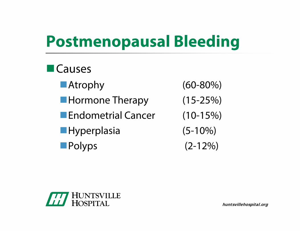
Postmenopausal Bleeding
CausesAtrophy (60-80%)Hormone Therapy (15-25%)Endometrial Cancer (10-15%)Hyperplasia (5-10%)Polyps (2-12%)
huntsvillehospital.org
Postmenopausal Bleeding
80-90% of Endometrial Cancer present with postmenopausal bleeding or discharge
huntsvillehospital.org
Postmenopausal Bleeding
Work-UpHistoryPhysical-examAssessment of endometriumTrans-vaginal Ultrasound (saline infused may
help)Endometrial sampling (biopsy, D&C)
huntsvillehospital.org
Postmenopausal Bleeding Evaluation Recommendations
Any vaginal bleeding in a postmenopausal woman requires assessment to exclude malignancy.
Women with postmenopausal uterine bleeding may be assessed initially with either endometrial biopsy or transvaginalultrasonography; this initial evaluation does not require performance of both tests.
When endometrial biopsy is performed and tissue is reported as insufficient for diagnosis, some further investigation is necessary and transvaginal ultrasonography may be performed.
When transvaginal ultrasonography is performed for patients with postmenopausal bleeding and an endometrial thickness of less than or equal to 4 mm is found, endometrial sampling is not required.
huntsvillehospital.org
Evaluation Recommendations (ACOG-C.O. #440 9/09)
Endometrial thickness of greater than 4 mm in a patient with postmenopausal bleeding should trigger alternative evaluation (such as sonohysterography, office hysteroscopy, or endometrial biopsy), as should an inability to adequately visualize thickness.
Meaningful assessment of the endometrium by ultrasonography is not possible in all patients. In such cases, alternative assessment should be completed.
When bleeding persists despite negative initial evaluations, additional assessment usually is indicated.
The significance of an endometrial thickness of greater than 4 mm in an asymptomatic, postmenopausal patient has not been established, and this finding need not routinely trigger evaluation.

huntsvillehospital.org
Postmenopausal Bleeding
Endometrial samplingIn office biopsyDone with Endometrial Pipelle
Operative biopsyD&C For patients unable to tolerate or undergo an in
office biopsy Gold standard
huntsvillehospital.org
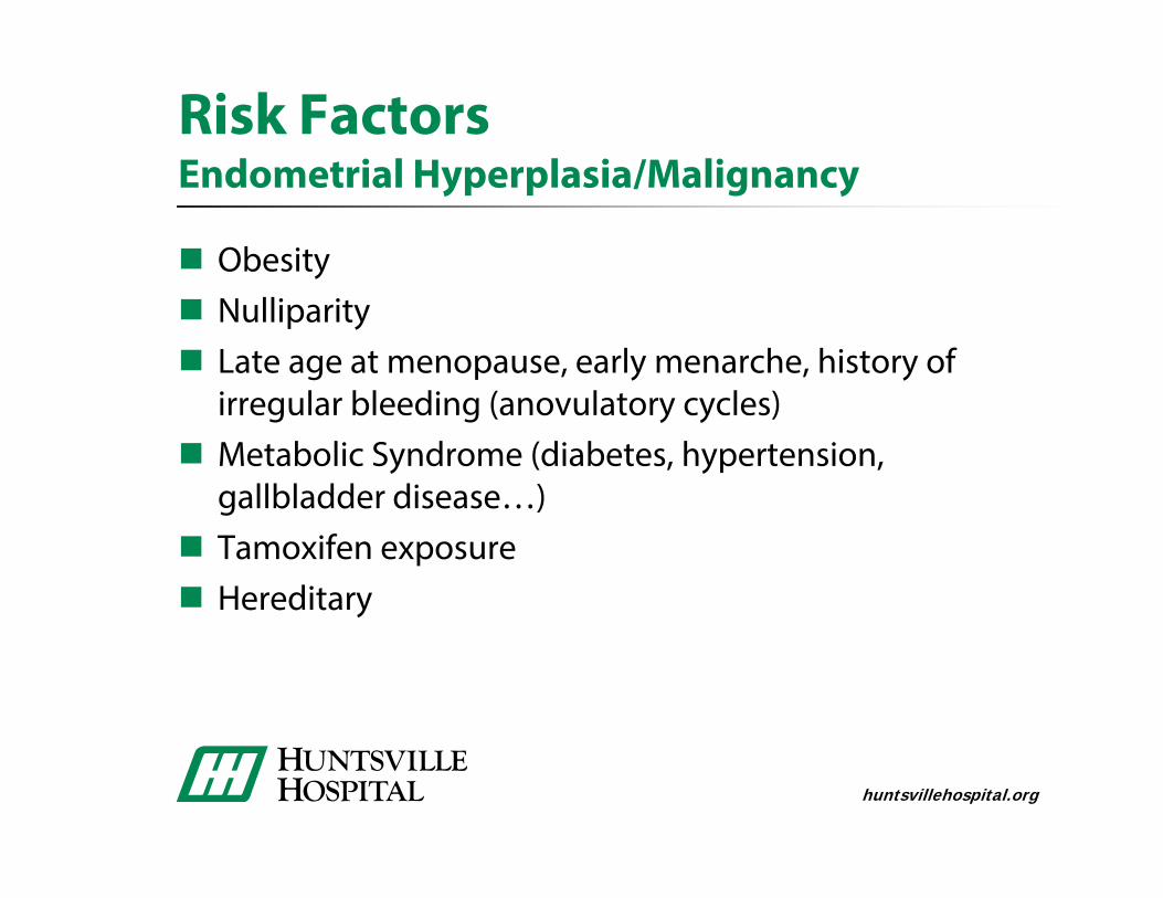
Risk Factors Endometrial Hyperplasia/Malignancy
Obesity Nulliparity Late age at menopause, early menarche, history of
irregular bleeding (anovulatory cycles) Metabolic Syndrome (diabetes, hypertension,
gallbladder disease…) Tamoxifen exposure Hereditary
huntsvillehospital.org
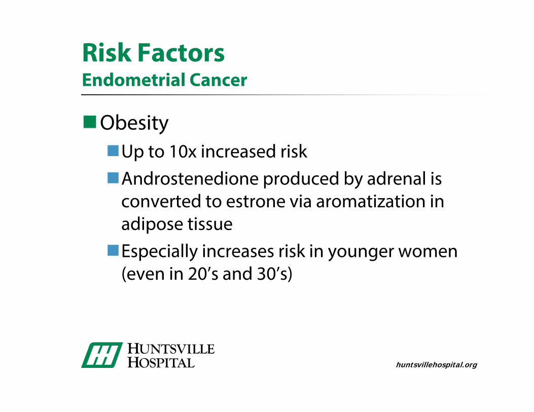
Risk FactorsEndometrial Cancer
ObesityUp to 10x increased riskAndrostenedione produced by adrenal is
converted to estrone via aromatization in adipose tissueEspecially increases risk in younger women
(even in 20’s and 30’s)
huntsvillehospital.org
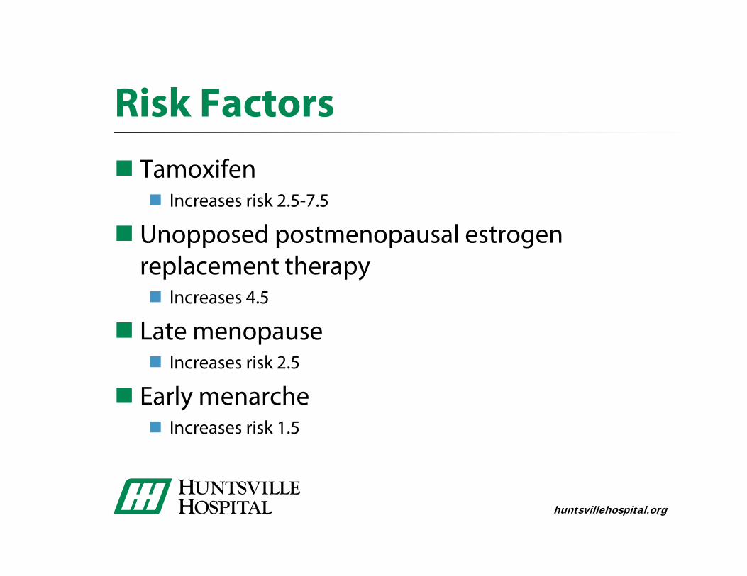
Risk Factors Tamoxifen
Increases risk 2.5-7.5
Unopposed postmenopausal estrogen replacement therapy Increases 4.5
Late menopause Increases risk 2.5
Early menarche Increases risk 1.5
huntsvillehospital.org
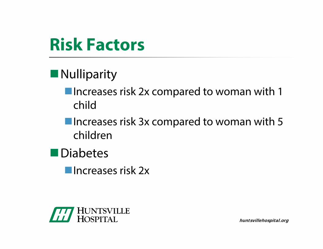
Risk Factors
NulliparityIncreases risk 2x compared to woman with 1
childIncreases risk 3x compared to woman with 5
children
DiabetesIncreases risk 2x
huntsvillehospital.org
Risk Factors
Polycystic ovarian diseaseIncreases risk but amount unknown
HypertensionDoes not increase risk when controlled for
other factors
huntsvillehospital.org
Protective Factors
Current types of oral contraceptivesDecrease risk up to 50%
huntsvillehospital.org
Endometrial Hyperplasia Precursor lesion for endometrial
adenocarcinoma Types of hyperplasia
Simple hyperplasia Complex hyperplasia Nuclear Atypia (+/-)
Atypical Complex Hyperplasia has a >40%of having a synchronous endometrial adenocarcinoma
huntsvillehospital.org
Endometrial Hyperplasia
Treatment Based on patients age, future fertility
desire, co-morbidities…Hormonal (progesterone)Surgical (hysterectomy)
huntsvillehospital.org
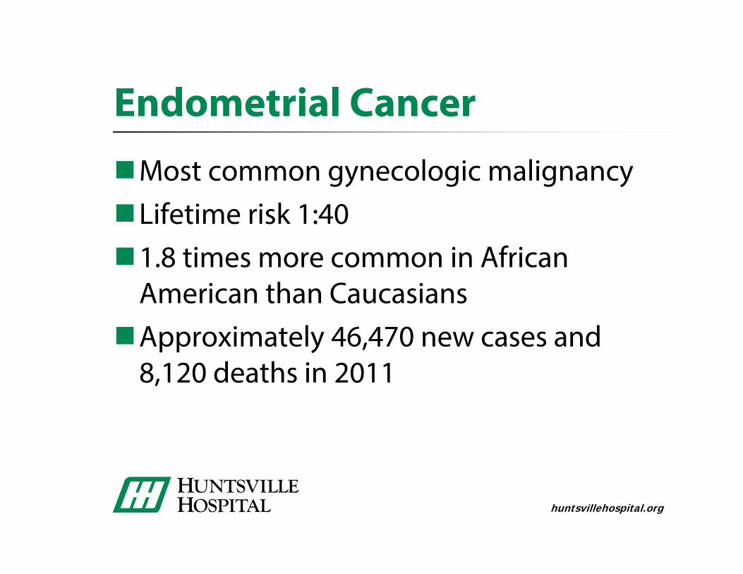
Endometrial Cancer
Most common gynecologic malignancyLifetime risk 1:401.8 times more common in African
American than CaucasiansApproximately 46,470 new cases and
8,120 deaths in 2011
huntsvillehospital.org
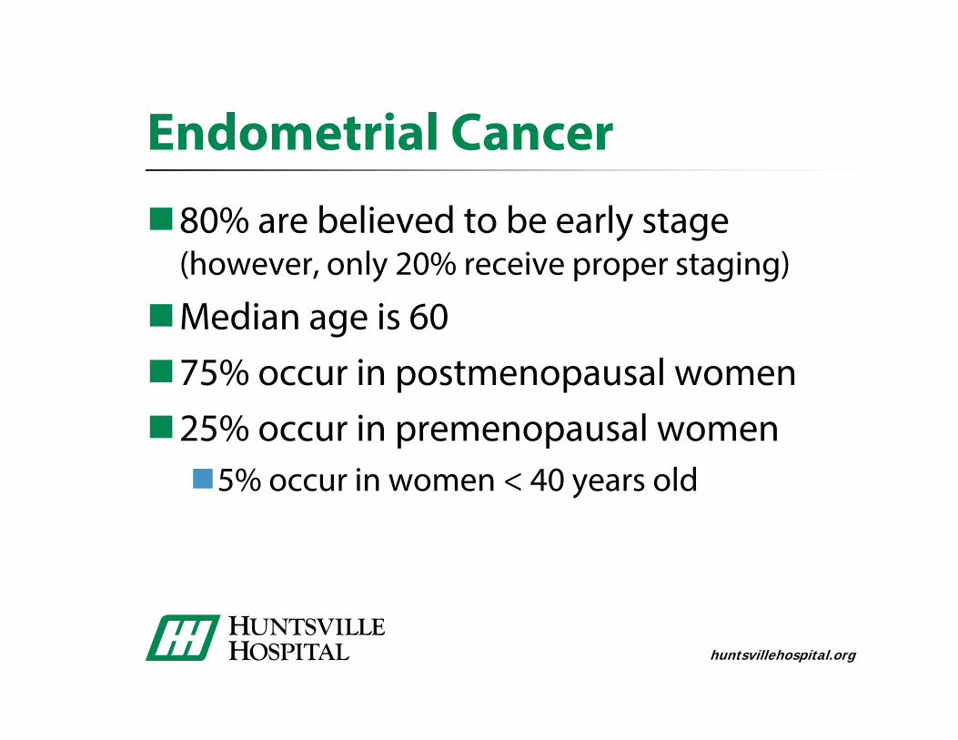
Endometrial Cancer
80% are believed to be early stage (however, only 20% receive proper staging)
Median age is 6075% occur in postmenopausal women25% occur in premenopausal women5% occur in women < 40 years old

huntsvillehospital.org
Types of Endometrial Cancer Epithelial Tumors Type I (estrogen dependent)
70-80% all cases Example- endometrioid
Type II (estrogen Independent)10-20% all cases Example- papillary serous (UPSC)
Mesenchymal Tumors Sarcoma (rare)
huntsvillehospital.org
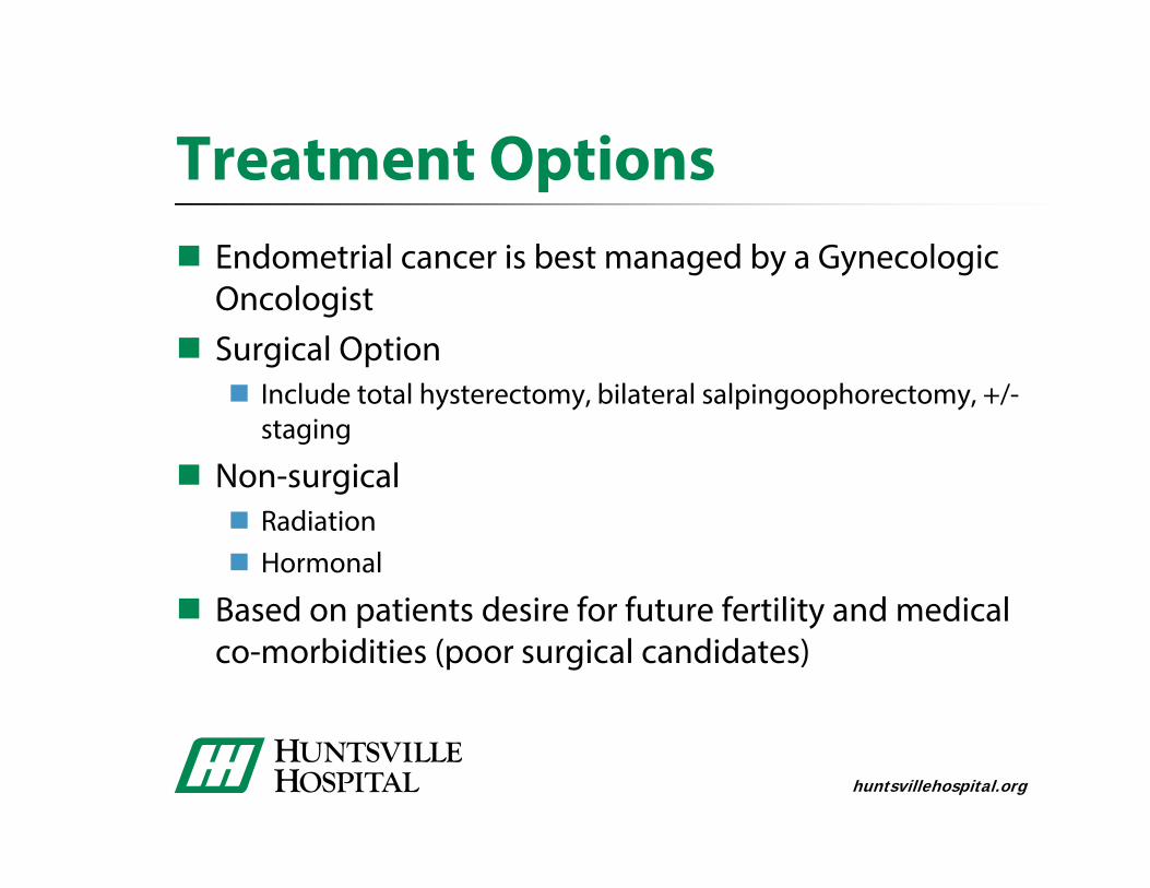
Treatment Options Endometrial cancer is best managed by a Gynecologic
Oncologist Surgical Option
Include total hysterectomy, bilateral salpingoophorectomy, +/-staging
Non-surgical Radiation Hormonal
Based on patients desire for future fertility and medical co-morbidities (poor surgical candidates)

huntsvillehospital.org
Endometrial Cancer Staging Staging is surgical Pelvic and para-aortic lymphadenectomy
May be able to omit staging and avoid increased post operative morbidity in a selection of low grade minimally invasive tumor This decision is best made by a Gynecologic
Oncologist
huntsvillehospital.org
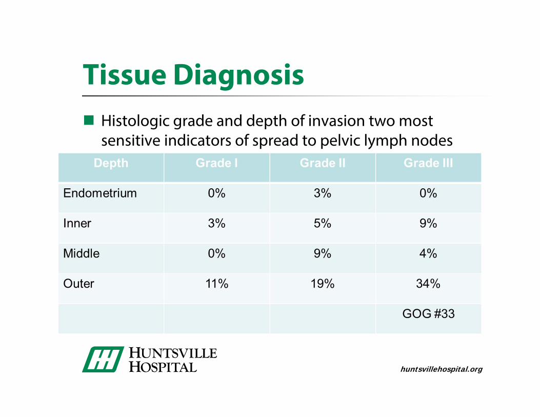
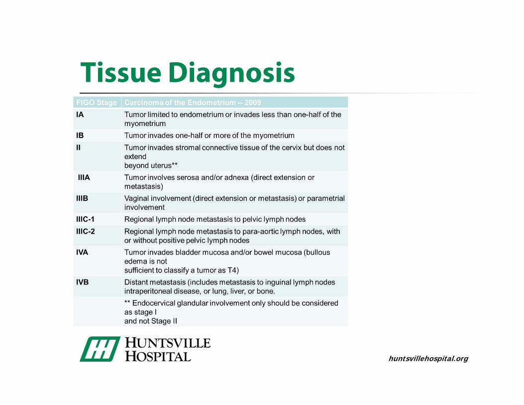
Tissue Diagnosis Histologic grade and depth of invasion two most
sensitive indicators of spread to pelvic lymph nodes
huntsvillehospital.org
Tissue Diagnosis