endokrin
TRANSCRIPT
ENDOCRINE SYSTEM
Dr.REISNA REFIANA
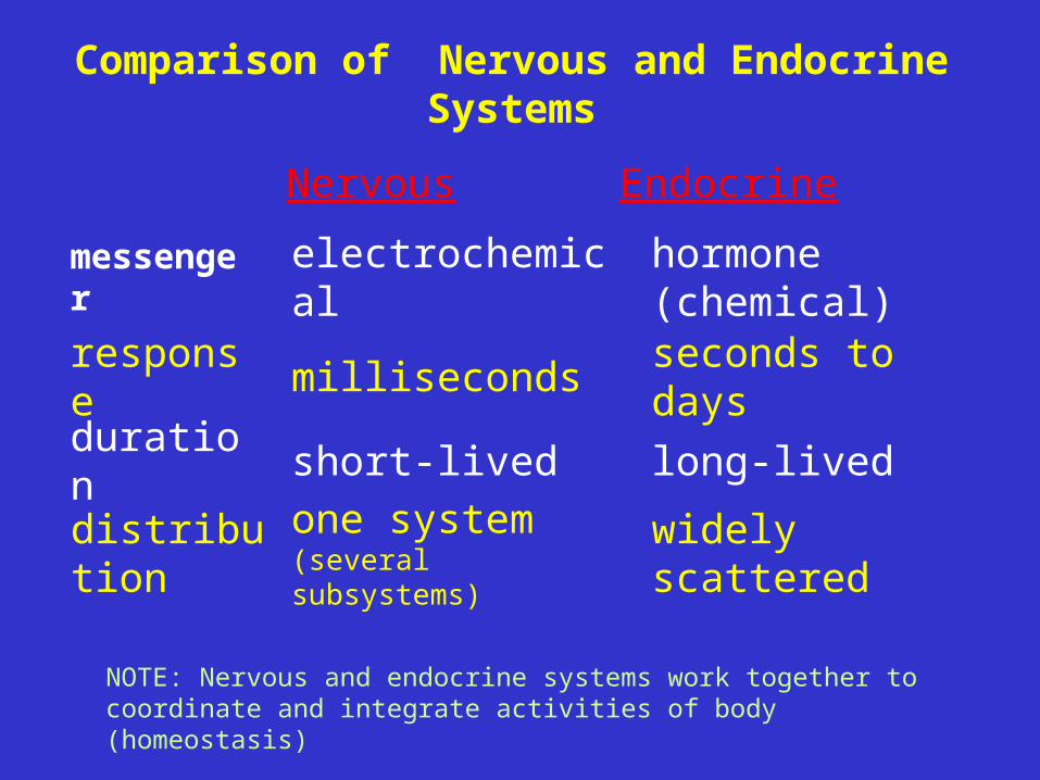
Comparison of Nervous and Endocrine Systems
NOTE: Nervous and endocrine systems work together to coordinate and integrate activities of body (homeostasis)
Nervous Endocrine
messenger electrochemicalhormone (chemical)
response milliseconds seconds to days
duration short-lived long-lived
distribution one system (several subsystems)
widely scattered
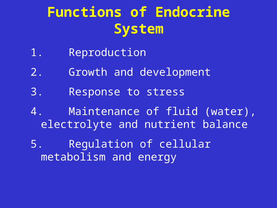
Functions of Endocrine System
1.Reproduction
2.Growth and development
3.Response to stress
4.Maintenance of fluid (water), electrolyte and nutrient balance
5.Regulation of cellular metabolism and energy
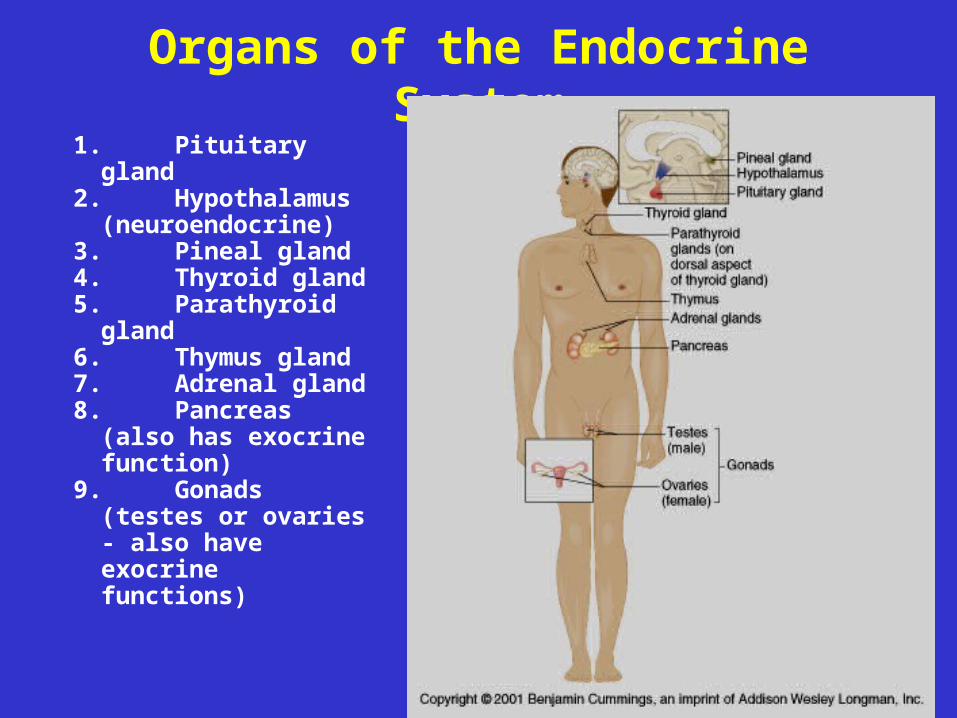
Organs of the Endocrine System
1. Pituitary gland2. Hypothalamus
(neuroendocrine)3. Pineal gland4. Thyroid gland5. Parathyroid
gland6. Thymus gland7. Adrenal gland8. Pancreas (also
has exocrine function)
9. Gonads (testes or ovaries - also have exocrine functions)
Topics• Hormone – Types– Modes of Action– Target cell activation– Control
• Specific glands, their hormones, and disorders– Pituitary– Thyroid– Parathyroid– Adrenal– Pancreas– Thymus– Gonads (testes and ovaries)
• General Adaptation Syndrome
Hormones• chemicals • secreted by endocrine gland cells into blood
(by way of interstitial fluid)• regulate metabolic functions of other cells
(called target cells)• carried to all cells, but action is specific to cells
that have receptors for the hormone– specificity of body’s response to hormone
depends on how many cells have the receptor (highly specific if few cells respond [e.g., ACTH]; diffuse action if many respond [e.g., thyroxine])
Chemical Types of Hormones• Amino-acid based (amino acids, short or long
peptides, proteins)– e.g., insulin, growth hormone, prolactin
• Steroids - lipid derivatives of cholesterol– e.g., hormones from gonads (testosterone,
estrogen)– e.g., hormones from adrenal cortex
(adrenocortical hormones)• Eicosanoids - locally-secreted, locally-acting
hormones secreted by all cell membranes (e.g., prostaglandins, which increase blood pressure and contribute to uterine contraction)
Types of Changes in Target Cells
• plasma membrane permeability changes (opening of protein channels; may change membrane potential)
• activation of genes for increased protein synthesis, including enzymes
• activation or deactivation of enzymes already present
• secretion of cellular products• stimulation of cell division (mitosis)
Mechanisms of Action
• action in target cell depends on receptor• receptor may be:
– in plasma membrane° second messenger mechanisms° used by most amino acid-based
hormones (water soluble)– intracellular (in cytoplasm or nucleus)
° direct gene activation° used by steroids and thyroid hormones
(lipid soluble)
Mechanisms of Action: Steroids• bind to intracellular receptors• hormone diffuses through plasma membrane
and makes its way to nucleus– > where it binds with intracellular receptor
to form hormone-receptor complex– > hormone-receptor complex interacts with
chromatin (DNA) to affect gene activity (turn genes on or off)
– > synthesis of mRNA – > synthesis of protein
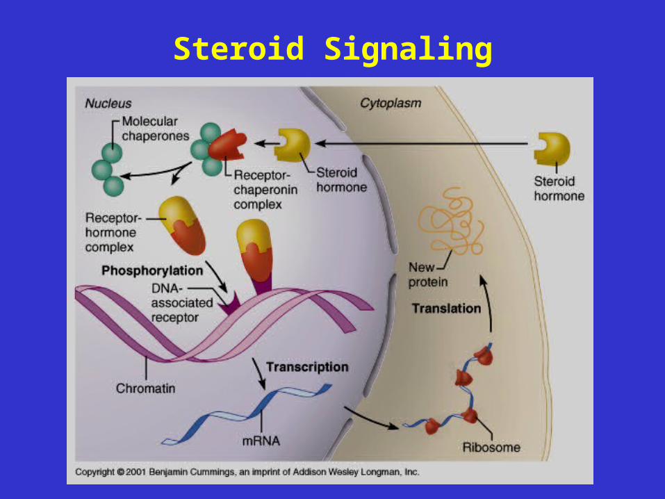
Steroid Signaling
Mechanism of Action:Thyroid Hormone
• similar to mechanism for steroid hormones
• diffuses across plasma membrane
– diffuses into nucleus where it interacts with intracellular receptors to activate genes for proteins (enzymes) involved in cellular respiration (glycolysis)
– also, binds to receptors at mitochondria to activate genes for proteins involved in cellular respiration (Krebs cycle and electron transport chain)
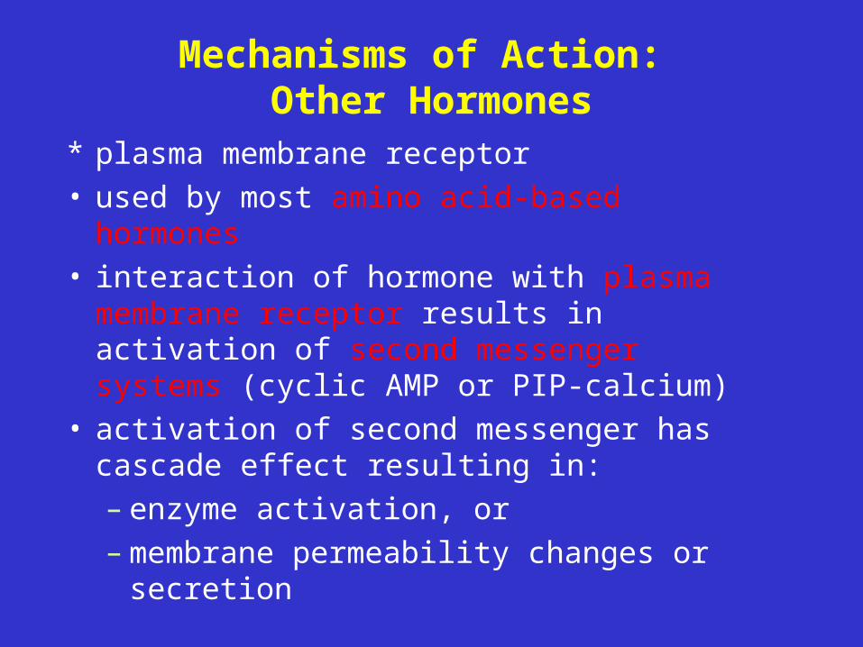
Mechanisms of Action: Other Hormones
* plasma membrane receptor• used by most amino acid-based hormones • interaction of hormone with plasma
membrane receptor results in activation of second messenger systems (cyclic AMP or PIP-calcium)
• activation of second messenger has cascade effect resulting in: – enzyme activation, or – membrane permeability changes or
secretion
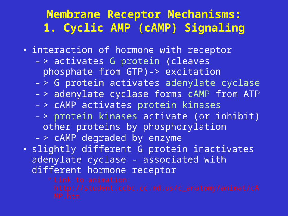
Membrane Receptor Mechanisms:1. Cyclic AMP (cAMP) Signaling
• interaction of hormone with receptor – > activates G protein (cleaves phosphate from
GTP)-> excitation– > G protein activates adenylate cyclase– > adenylate cyclase forms cAMP from ATP – > cAMP activates protein kinases– > protein kinases activate (or inhibit) other
proteins by phosphorylation– > cAMP degraded by enzyme
• slightly different G protein inactivates adenylate cyclase - associated with different hormone receptor
° Link to animation: http://student.ccbc.cc.md.us/c_anatomy/animat/cAMP.htm
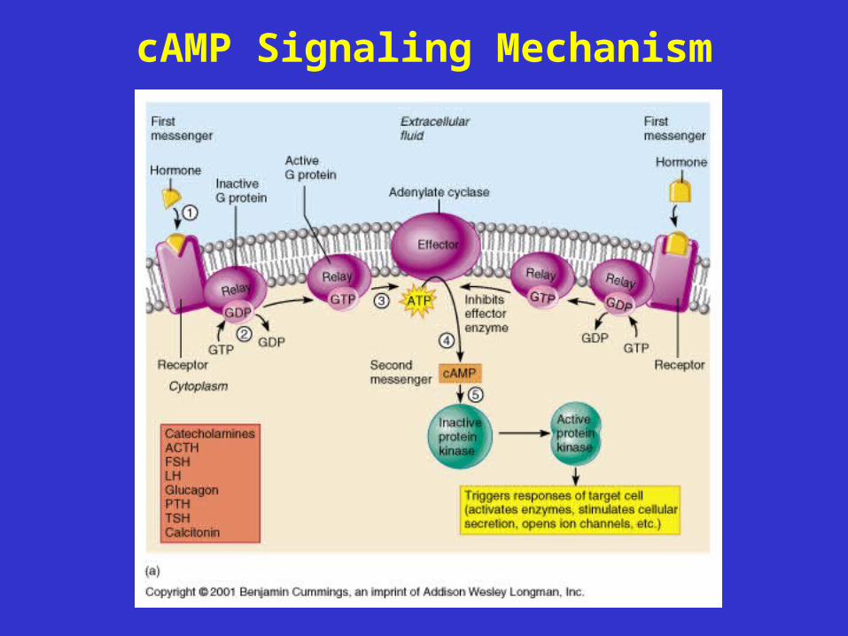
cAMP Signaling Mechanism
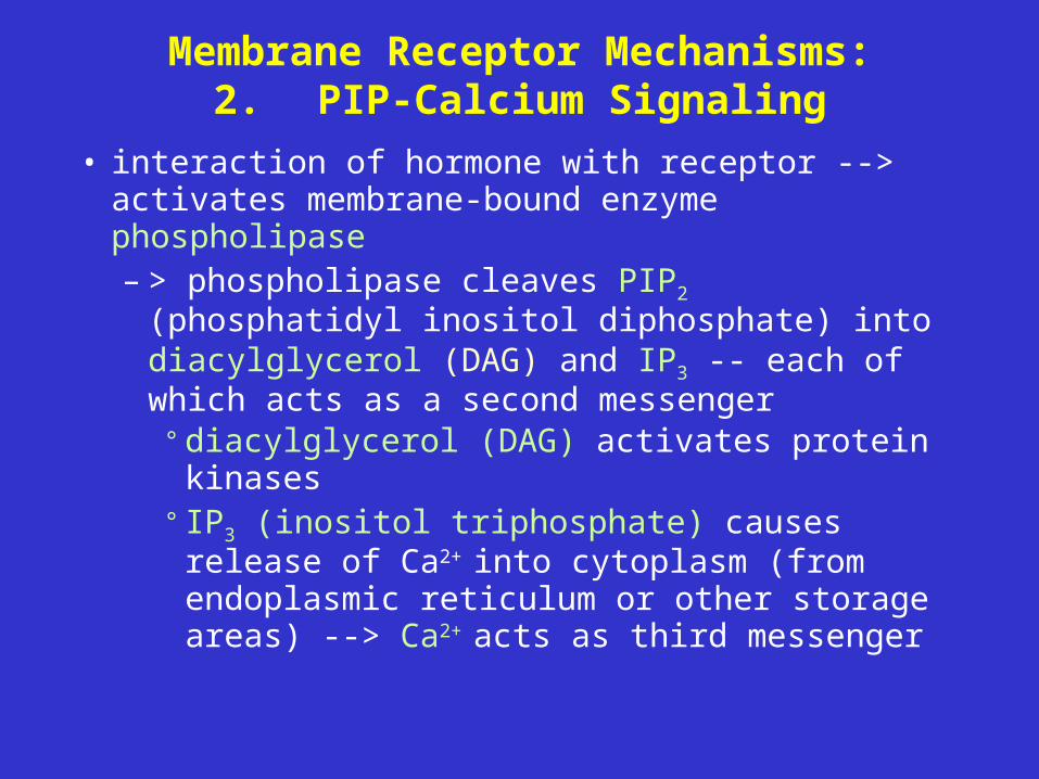
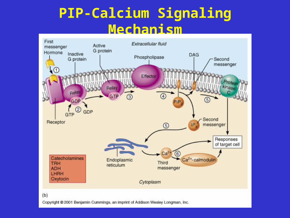
Membrane Receptor Mechanisms:2. PIP-Calcium Signaling
• interaction of hormone with receptor --> activates membrane-bound enzyme phospholipase– > phospholipase cleaves PIP2 (phosphatidyl
inositol diphosphate) into diacylglycerol (DAG) and IP3 -- each of which acts as a second messenger
° diacylglycerol (DAG) activates protein kinases
° IP3 (inositol triphosphate) causes release of Ca2+ into cytoplasm (from endoplasmic reticulum or other storage areas) --> Ca2+
acts as third messenger
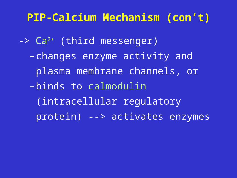
PIP-Calcium Mechanism (con’t)
-> Ca2+ (third messenger)
– changes enzyme activity and plasma
membrane channels, or
– binds to calmodulin (intracellular
regulatory protein) --> activates
enzymes
PIP-Calcium Signaling Mechanism
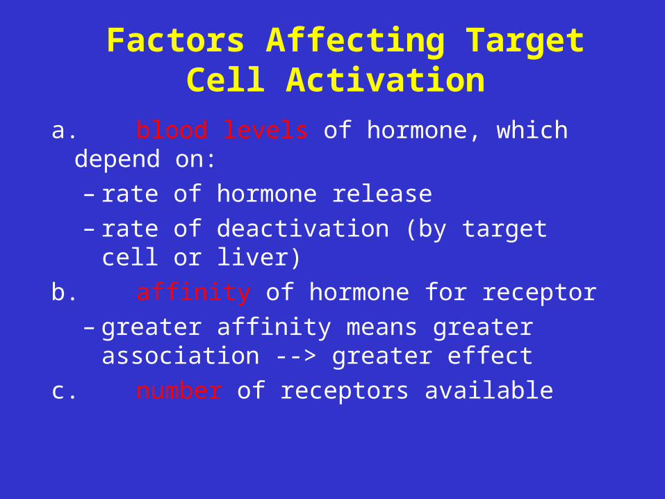
Factors Affecting Target Cell Activation
a.blood levels of hormone, which depend on:– rate of hormone release– rate of deactivation (by target cell or liver)
b.affinity of hormone for receptor– greater affinity means greater association
--> greater effect
c. number of receptors available
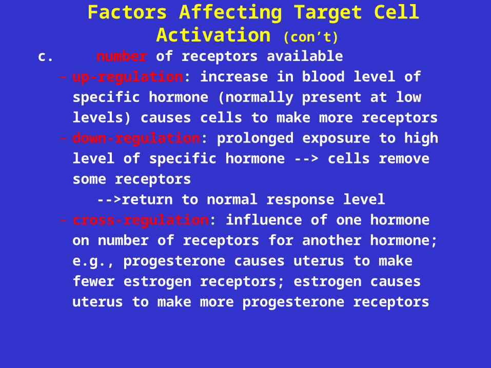
Factors Affecting Target Cell Activation (con’t)
c. number of receptors available– up-regulation: increase in blood level of
specific hormone (normally present at low
levels) causes cells to make more receptors– down-regulation: prolonged exposure to high
level of specific hormone --> cells remove
some receptors
-->return to normal response level– cross-regulation: influence of one hormone on
number of receptors for another hormone; e.g.,
progesterone causes uterus to make fewer
estrogen receptors; estrogen causes uterus to
make more progesterone receptors
Hormone Removal
• hormones may be:
– degraded by specific enzymes within target cells;
– removed from blood by kidneys (excreted in urine)
– degraded by liver (excreted in urine and feces)
• half-life - time for 1/2 of hormone to be removed (from
a fraction of a minute to 30 minutes)
• onset - time from release to action (minutes [amino
acid-based] to days [steroids])
• duration of action - how long the effects last (~20
minutes to several hours)
Control of Hormone Release
• Humoral control
• Neural control
• Hormonal control
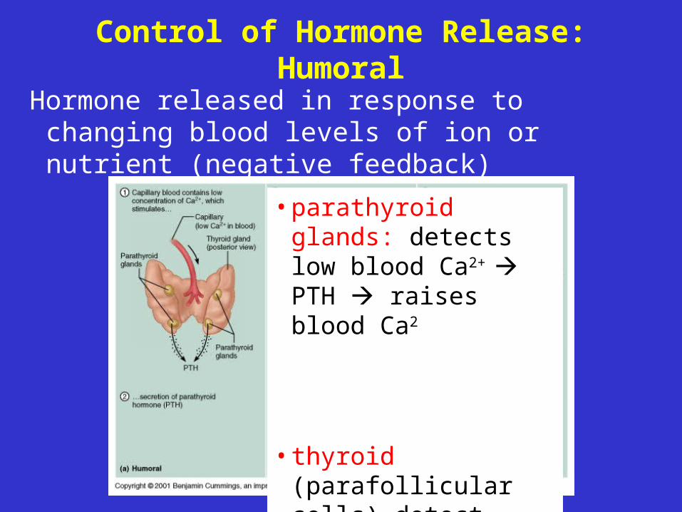
Control of Hormone Release: Humoral
Hormone released in response to changing blood levels of ion or nutrient (negative feedback)
• parathyroid glands: detects low blood Ca2+
PTH raises blood Ca2
• thyroid (parafollicular cells) detect high blood Ca2+-->calcitonin-->decrease blood Ca2+
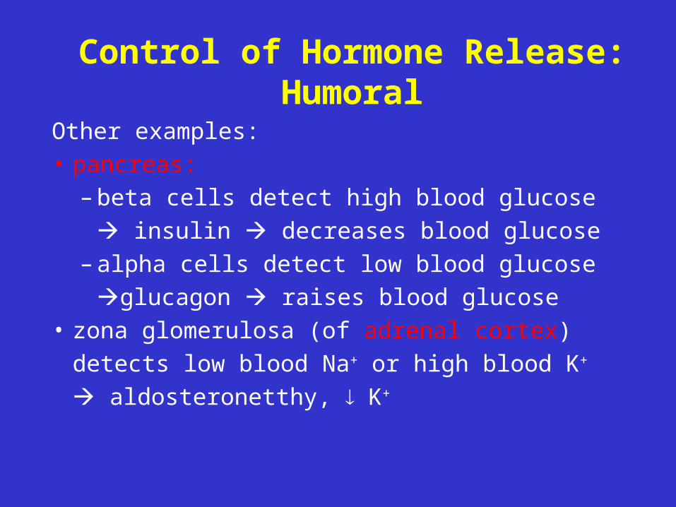
Control of Hormone Release: Humoral
Other examples:• pancreas:
– beta cells detect high blood glucose
insulin decreases blood glucose– alpha cells detect low blood glucose
glucagon raises blood glucose• zona glomerulosa (of adrenal cortex)
detects low blood Na+ or high blood K+
aldosteronetthy, K+
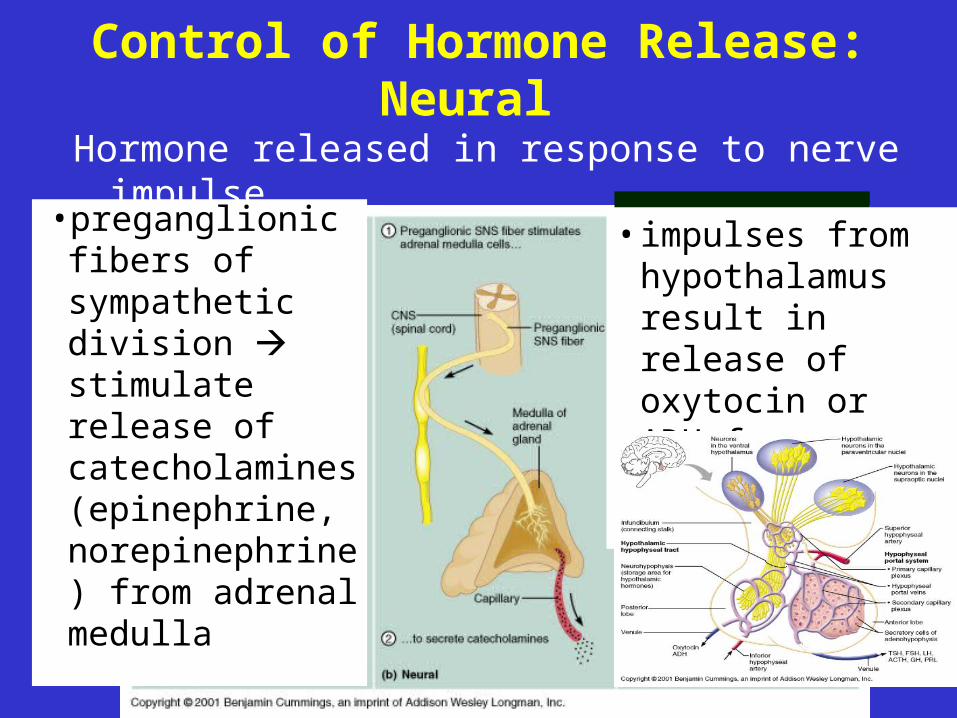
Control of Hormone Release: Neural
Hormone released in response to nerve impulse
• preganglionic fibers of sympathetic division stimulate release of catecholamines (epinephrine, norepinephrine) from adrenal medulla
• impulses from hypothalamus result in release of oxytocin or ADH from posterior pituitary

Control of Hormone Release: Hormonal
Hormone produced by one endocrine gland (or hypothalamus) affects secretion of hormone by another endocrine gland
• hypothalamus acts as overall coordinator
releases regulatory hormones (releasing hormones or inhibitory hormones) affects anterior pituitary
• anterior pituitary, when stimulated, secretes hormones that affect other glands (e.g., TSH [thyroid stimulating hormone] stimulates release of thyroid hormones from thyroid gland)
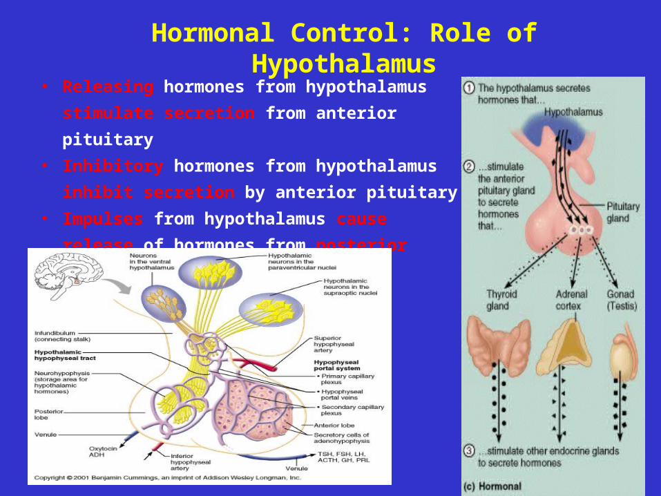
Hormonal Control: Role of Hypothalamus
• Releasing hormones from hypothalamus
stimulate secretion from anterior pituitary
• Inhibitory hormones from hypothalamus
inhibit secretion by anterior pituitary
• Impulses from hypothalamus cause release
of hormones from posterior pituitary
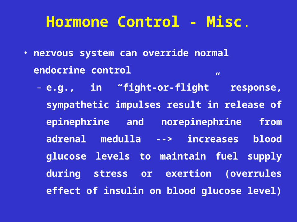
Hormone Control - Misc.
• nervous system can override normal endocrine
control
– e.g., in “fight-or-flight” response, sympathetic
impulses result in release of epinephrine and
norepinephrine from adrenal medulla -->
increases blood glucose levels to maintain fuel
supply during stress or exertion (overrules effect
of insulin on blood glucose level)
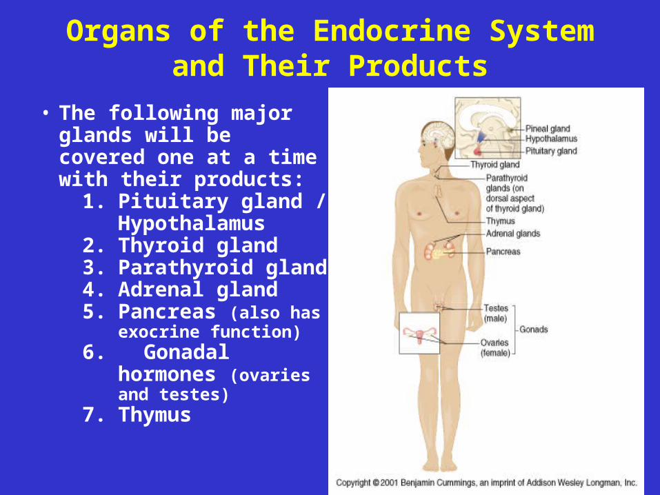
Organs of the Endocrine System and Their Products
• The following major glands will be covered one at a time with their products:
1. Pituitary gland / Hypothalamus
2. Thyroid gland3. Parathyroid gland4. Adrenal gland5. Pancreas (also has
exocrine function)6. Gonadal hormones
(ovaries and testes)7. Thymus
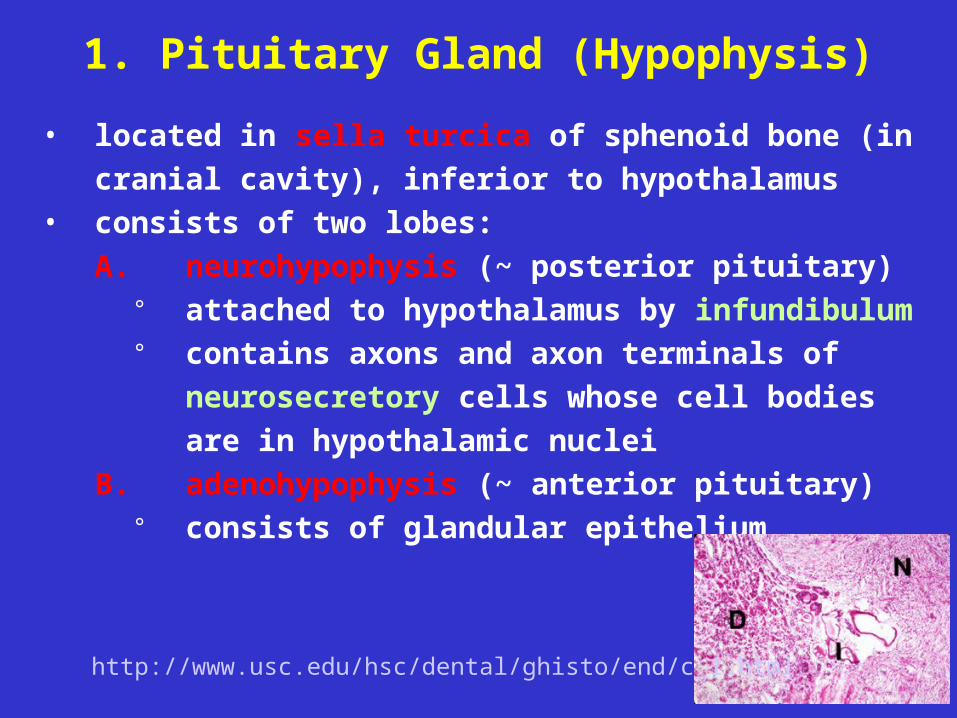
1. Pituitary Gland (Hypophysis)
• located in sella turcica of sphenoid bone (in cranial
cavity), inferior to hypothalamus• consists of two lobes:
A. neurohypophysis (~ posterior pituitary)° attached to hypothalamus by infundibulum° contains axons and axon terminals of
neurosecretory cells whose cell bodies are in
hypothalamic nuclei
B. adenohypophysis (~ anterior pituitary)° consists of glandular epithelium
http://www.usc.edu/hsc/dental/ghisto/end/c_1.html
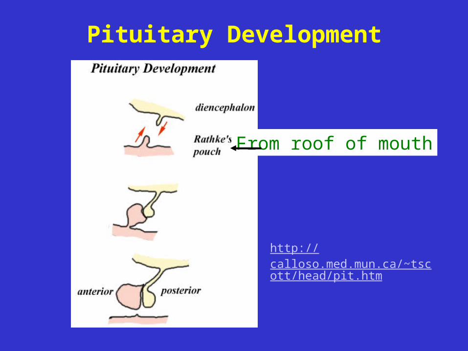
Pituitary Development
From roof of mouth
http://calloso.med.mun.ca/~tscott/head/pit.htm
A. Neurohypophysis (Posterior Pituitary)
• consists of nerve fibers (axons of neurosecretory
cells with cell bodies in hypothalamus) and
pituicytes (glial cells that support nerve fibers)
• acts primarily as a storage and releasing area for
hormones actually made in hypothalamic nuclei
• hormones released in response to impulses from
hypothalamus (neural control)
• hormones are short amino acid chains (peptides)
– oxytocin
– antidiuretic hormone (ADH or “vasopressin”)
A. Neurohypophysis : Oxytocin (OT)
• action, in pregnant or nursing women:
– stimulates contraction of smooth muscle of
uterine wall during labor and delivery
– stimulates ejection of milk in lactating mothers
• action, in men and non-pregnant women, may be
involved in sexual arousal and orgasm
A. Neurohypophysis : Oxytocin (OT)
• control:
– during labor/delivery, positive feedback:
stretching of uterus/cervix --> sensory
impulses to hypothalamus --> increased
secretion of OT --> increased contraction
– suckling: sucking of infant on breast -->
sensory to hypothalamus --> oxytocin release
--> release of milk
A. Neurohypophysis: Antidiuretic Hormone (ADH)
• action: antidiuretic hormone (ADH) directly
affects blood pressure - acts as powerful
vasoconstrictor --> increases blood pressure
(hence name “vasopressin”)
* action: affects water balance (indirect affect
on blood pressure) - acts on tubules of kidney
to increase reabsorption of water less
water lost in urine
A. Neurohypophysis: ADH
• disorders:
– hyposecretion due to damage of
hypothalamic nucleus or neurohypophysis--
> diabetes insipidus - excessive urine
production (polyuria) and thirst
– hypersecretion --> SIADH (syndrome of
inappropriate ADH secretion) - water
retention, headache, cerebral edema,
weight gain, hypoosmolarity
Antidiuretic Hormone (ADH): Control• neural control: increased electrolyte (NaCl)
concentration --> affects (supraoptic) nucleus in hypothalamus --> impulse to neurohypophysis --> release of ADH --> increased water reabsorption --> decrease in electrolyte concentration
• other stimuli: pain, low BP, morphine, barbiturates, nicotine, aldosterone (hormone from adrenal cortex - hormonal control)
• inhibition: alcohol (results in more urine production and, potentially, dehydration)
• diuretic drugs - some act to supress ADH secretion; used to treat hypertension and congestive heart failure
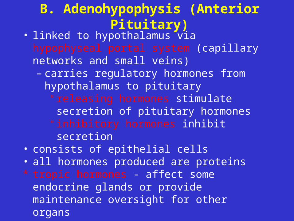
B. Adenohypophysis (Anterior Pituitary)• linked to hypothalamus via hypophyseal
portal system (capillary networks and small veins)– carries regulatory hormones from
hypothalamus to pituitary° releasing hormones stimulate secretion
of pituitary hormones° inhibitory hormones inhibit secretion
• consists of epithelial cells• all hormones produced are proteins* tropic hormones - affect some endocrine
glands or provide maintenance oversight for other organs
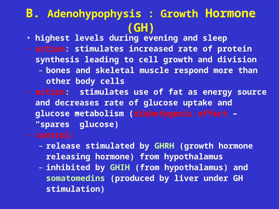
B. Adenohypophysis : Growth Hormone (GH)
• highest levels during evening and sleep• action: stimulates increased rate of protein synthesis
leading to cell growth and division– bones and skeletal muscle respond more than
other body cells• action: stimulates use of fat as energy source and
decreases rate of glucose uptake and glucose metabolism (diabetogenic effect – “spares” glucose)
• control:– release stimulated by GHRH (growth hormone
releasing hormone) from hypothalamus– inhibited by GHIH (from hypothalamus) and
somatomedins (produced by liver under GH stimulation)
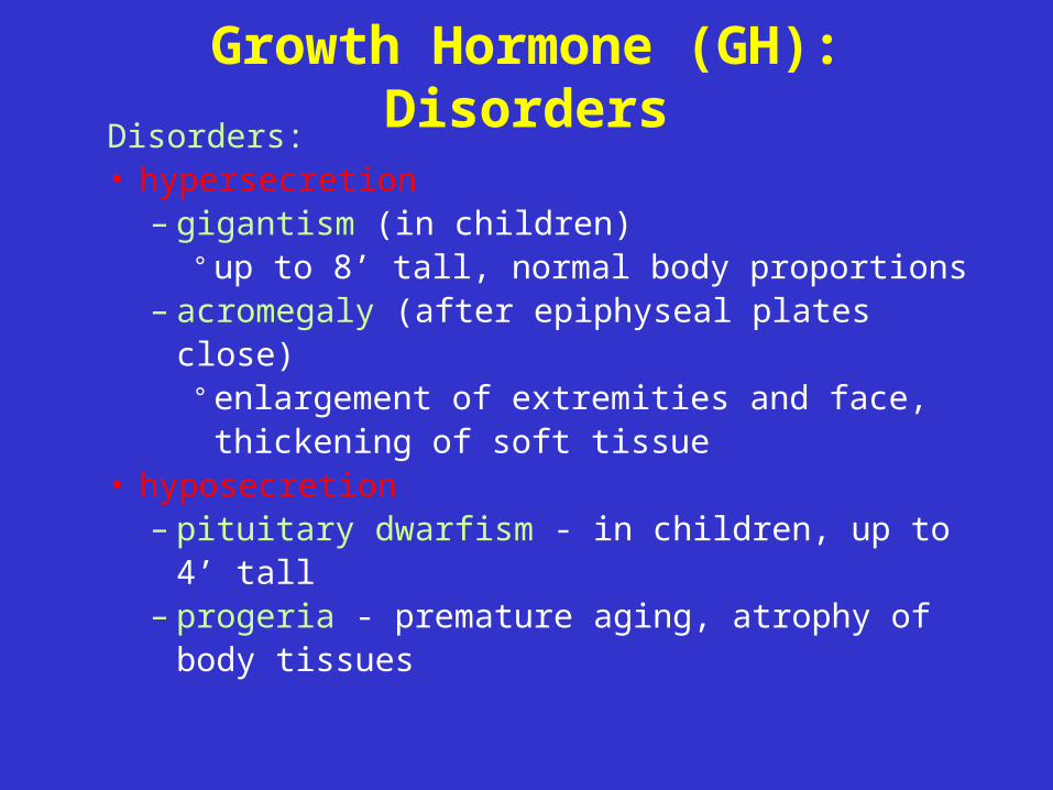
Growth Hormone (GH): DisordersDisorders:• hypersecretion
– gigantism (in children)° up to 8’ tall, normal body proportions
– acromegaly (after epiphyseal plates close)° enlargement of extremities and face,
thickening of soft tissue• hyposecretion
– pituitary dwarfism - in children, up to 4’ tall– progeria - premature aging, atrophy of
body tissues
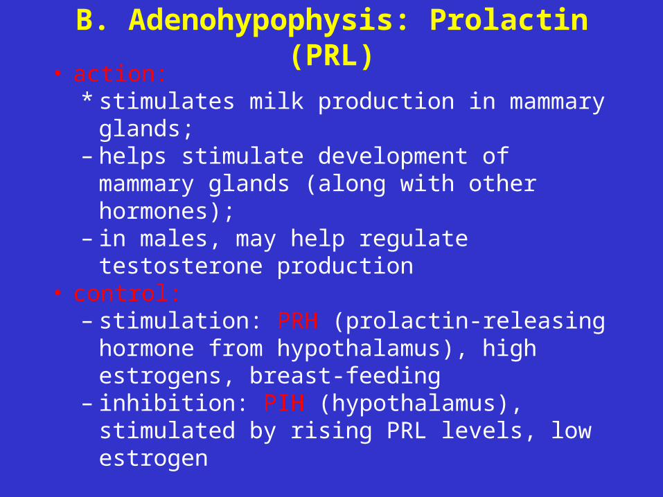
B. Adenohypophysis: Prolactin (PRL)• action:
* stimulates milk production in mammary glands;
– helps stimulate development of mammary glands (along with other hormones);
– in males, may help regulate testosterone production
• control: – stimulation: PRH (prolactin-releasing
hormone from hypothalamus), high estrogens, breast-feeding
– inhibition: PIH (hypothalamus), stimulated by rising PRL levels, low estrogen
B. Adenohypophysis : Prolactin (PRL)
Disorders
• hyperprolactinemia = hypersecretion due to
adenohypophyseal tumors; results in
galactorrhea, lack of menses and infertility in
women, impotence in men
B. Adenohypophysis: Thyroid-Stimulating Hormone (TSH)
• TSH = thyrotropin– action:
° stimulates secretion of hormones from
thyroid gland (T4 and T3); also stimulates
development of thyroid in youth– control:
° release stimulated by TRH (thyroid
releasing hormone from hypothalamus)° inhibited by rising levels of thyroid
hormones and by GHIH
B. Adenohypophysis: Adrenocorticotropic hormone (ACTH)
• ACTH=corticotropin
• action: stimulates release of hormones from
adrenal cortex
• control:
– release stimulated by CRH (corticotropin-
releasing hormone from hypothalamus)
– release inhibited by rising levels of
glucocorticoids from adrenal cortex
B. Adenohypophysis:Gonadotropins
• regulate activity and secretion by gonads (testes in males; ovaries in females)
• control: – stimulated by GnRH (gonadotropin-
releasing hormone from hypothalamus)– release of GnRH is inhibited by rising
levels of estrogens, progestins and androgens (testosterone)
• two important hormones° FSH° LH
Gonadotropins:Follicle-Stimulating Hormone (FSH)
• action:– females (ovaries) - stimulates development
of ovarian follicles and estrogen production – males (testes) - stimulates sperm
production and development• inhibited by inhibin and testosterone from
testes (feedback to hypothalamus and
anterior pituitary) and estrogen, progesterone
and inhibin from ovaries (feedback to anterior
pituitary)
Gonadotropins:Luteinizing Hormone (LH)
• LH=lutropin– action:
° females (ovaries) - induces ovulation;
stimulates secretion of estrogens and
progestins (e.g., progesterone) ° males (testes) - stimulates production of
androgens (e.g., testosterone )– inhibited by estrogen, progesterone and
inhibin form ovaries (feedback to anterior
pituitary) and by inhibin and testosterone from
testes (feedback to hypothalamus and anterior
pituitary)
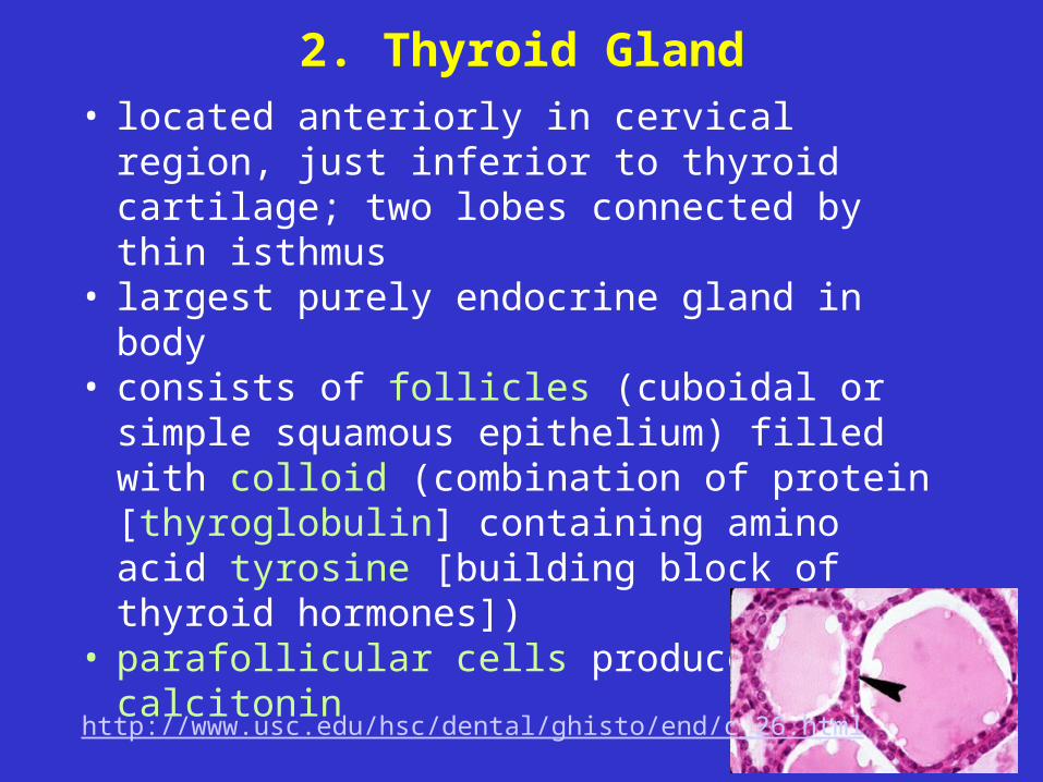
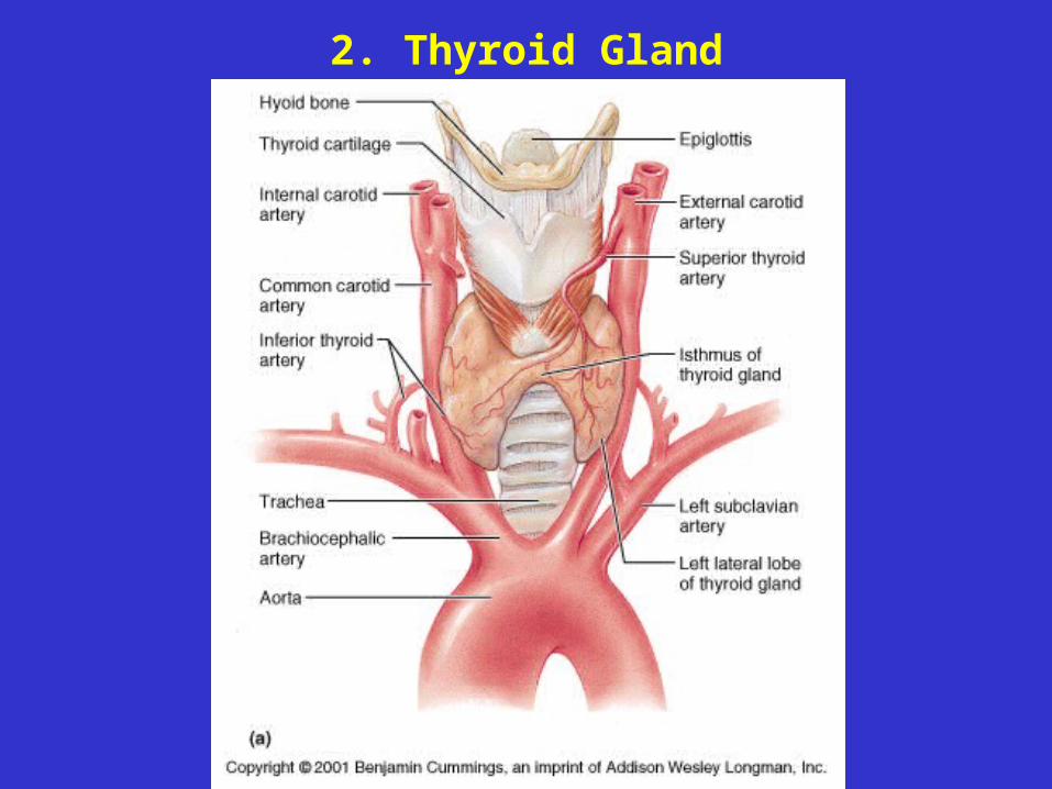
2. Thyroid Gland• located anteriorly in cervical region, just
inferior to thyroid cartilage; two lobes connected by thin isthmus
• largest purely endocrine gland in body• consists of follicles (cuboidal or simple
squamous epithelium) filled with colloid (combination of protein [thyroglobulin] containing amino acid tyrosine [building block of thyroid hormones])
• parafollicular cells produce calcitonin
http://www.usc.edu/hsc/dental/ghisto/end/c_26.html
2. Thyroid Gland: T4 and T3
• hormones based on amino acid tyrosine (differ in number of iodine ions)
– thyroxine (tetraiodothyronine [T4]) and
– triiodothyronine (T3)
• T3 is 10x more active, but less common (T4
accounts for about 90% of all thyroid hormone)
• much T4 converted to T3 by liver, kidneys,
some other tissues

2. Thyroid Gland: T4 and T3
• affect metabolic rate of every cell in the body,
except brain, spleen, testes, uterus and
thyroid gland
– affect other activities within these organs
and glands
• readily cross membranes (diffuse through
plasma membrane to bind to mitochondrial
receptors and receptors in nucleus)
2. Thyroid Gland
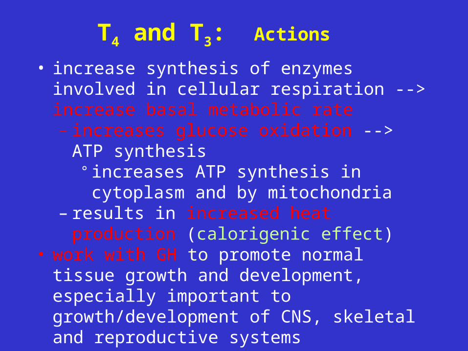
T4 and T3: Actions
• increase synthesis of enzymes involved in cellular respiration --> increase basal metabolic rate– increases glucose oxidation --> ATP
synthesis° increases ATP synthesis in cytoplasm
and by mitochondria– results in increased heat production
(calorigenic effect)• work with GH to promote normal tissue growth
and development, especially important to growth/development of CNS, skeletal and reproductive systems
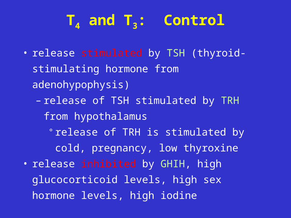
T4 and T3: Control
• release stimulated by TSH (thyroid-
stimulating hormone from adenohypophysis)
– release of TSH stimulated by TRH from
hypothalamus
° release of TRH is stimulated by cold,
pregnancy, low thyroxine
• release inhibited by GHIH, high glucocorticoid
levels, high sex hormone levels, high iodine
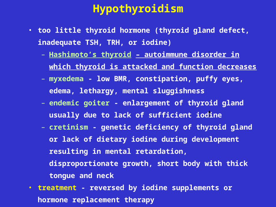
Hypothyroidism
• too little thyroid hormone (thyroid gland defect, inadequate
TSH, TRH, or iodine)
– Hashimoto’s thyroid – autoimmune disorder in which
thyroid is attacked and function decreases
– myxedema - low BMR, constipation, puffy eyes, edema,
lethargy, mental sluggishness
– endemic goiter - enlargement of thyroid gland usually
due to lack of sufficient iodine
– cretinism - genetic deficiency of thyroid gland or lack of
dietary iodine during development resulting in mental
retardation, disproportionate growth, short body with
thick tongue and neck
• treatment - reversed by iodine supplements or hormone
replacement therapy
Hyperthyroidism• too much thyroid hormone (thyrotoxicosis)
– Grave’s disease - autoimmune disease in which abnormal antibodies similar to TSH mimic its function and continuously stimulate release of thyroid hormones; results in high BMR, sweating, rapid heart rate, weight loss, restlessness, mood shifts, fatigues easily, limited energy; also toxic goiter
– exophthalmos - protrusion of eyeballs, fibrous tissue become edematous (swollen)
• treatments - removal of thyroid gland or irradiation– patient must be on synthetic thyroid hormone the
rest of his/her life
2. Thyroid Gland: Calcitonin (CT)
• polypeptide produced by parafollicular cells
• actions: decreases blood calcium levels by:
– stimulating osteoblasts (Ca2+ uptake and
incorporation into bone)
– inhibiting osteoclast activities (osteoclasts break
down bone matrix releasing calcium)
• control: responds directly to blood calcium levels
• very rapid effect
• probably more important during childhood when it
stimulates bone growth
• important because at high blood Ca2+, membranes
become less permeable to Na+
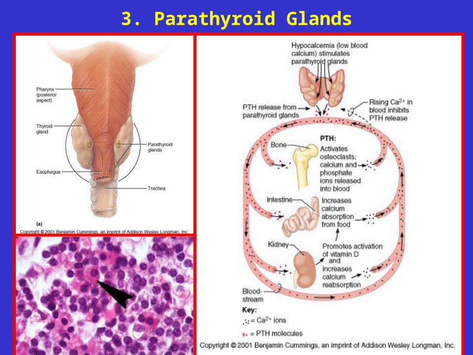
3. Parathyroid Glands
• 2 paired structures on posterior of thyroid gland• oxyphyil cells - function unknown• chief cells secrete parathyroid hormone (PTH;
protein)• actions: increases blood Ca2+ by:
– stimulating osteoclast activity (which break down bone matrix) while inhibiting osteoblasts (which form bone matrix)
– stimulating increased reabsorption of Ca2+ by kidney
– indirectly stimulating increased absorption of Ca2+ by small intestine by causing secretion of calcitrol form kidneys
Hyperparathyroidism
• rare; caused by parathyroid gland tumor
• results in hypercalcemia (excess Ca2+
levels in blood) --> depression of nervous
system (because of effect on sodium
permeability), abnormal reflexes, skeletal
muscle weakness, nausea, vomiting,
kidney stones, calcium deposits in soft
tissues; bones become soft
Hypoparathyroidism
• trauma to or removal of parathyroid
gland
• results in hypocalcemia (low blood Ca2+)
--> neurons become too excitable -->
muscle tetany --> spasms/cramps -->
respiratory paralysis --> death
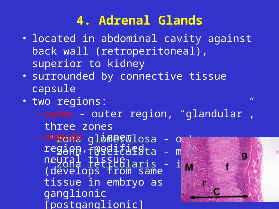
4. Adrenal Glands• located in abdominal cavity against back wall
(retroperitoneal), superior to kidney• surrounded by connective tissue capsule• two regions:
– cortex - outer region, “glandular”, three zones° zona glomerulosa - outer zone° zona fasciculata - middle zone° zona reticularis - inner zone
– medulla - inner region, modified neural tissue (develops from same tissue in embryo as ganglionic [postganglionic] neurons of sympathetic division)
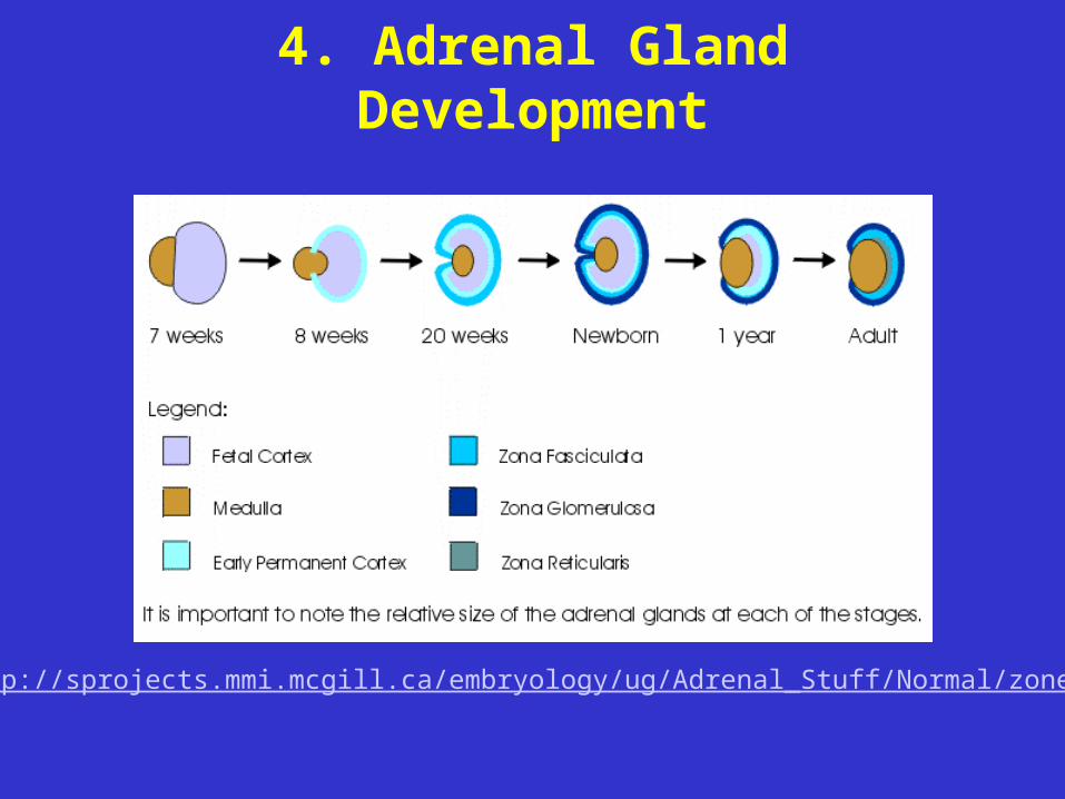
4. Adrenal Gland Development
http://sprojects.mmi.mcgill.ca/embryology/ug/Adrenal_Stuff/Normal/zones.html
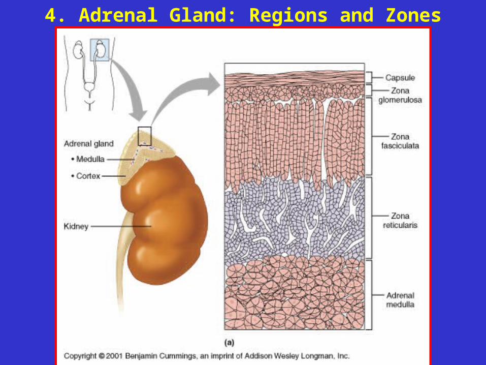
4. Adrenal Gland: Regions and Zones
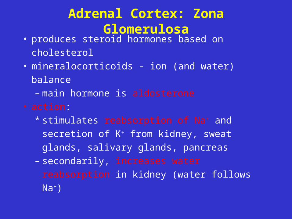
Adrenal Cortex: Zona Glomerulosa
• produces steroid hormones based on
cholesterol• mineralocorticoids - ion (and water) balance
– main hormone is aldosterone• action:
* stimulates reabsorption of Na+ and
secretion of K+ from kidney, sweat glands,
salivary glands, pancreas – secondarily, increases water reabsorption
in kidney (water follows Na+)
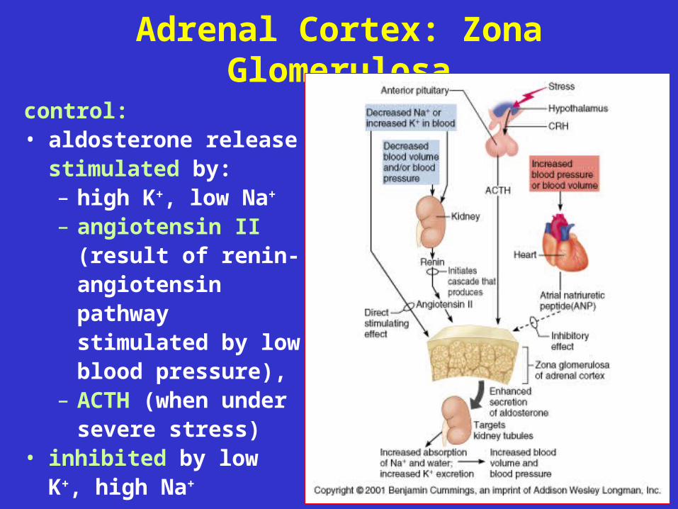
Adrenal Cortex: Zona Glomerulosa
control: • aldosterone release
stimulated by:– high K+, low Na+
– angiotensin II (result of renin-angiotensin pathway stimulated by low blood pressure),
– ACTH (when under severe stress)
• inhibited by low K+, high Na+
Adrenal Cortex: Zona Glomerulosa
Disorders:• aldosteronism = hypersecretion (adrenal
tumor)→ increased water and Na+ reabsorption -->
hypertension, edema; → loss of K+ --> disruption of neural and
muscle function
Adrenal Cortex: Zona Glomerulosa
Disorders:• Addison’s Disease = hyposecretion
glucocorticoids and mineralocorticoids– results in decreased Na+ and water
reabsorption, increased blood K+ --> low blood volume --> hypotension, dehydration;
– changes in membrane potentials --> disruption in neural and muscular function
– also decreased cortisol secretion by zona fasciculata --> decreased blood glucose levels (especially during prolonged stress)
Adrenal Cortex: Zona Fasciculata• glucocorticoids - effects on glucose metabolism• main hormone is cortisol (hydrocortisone)• actions:
– maintains blood glucose levels, especially in times of stress, by:
° promoting gluconeogenesis (making new glucose in liver) and use of alternative fuels by other cells (saves glucose for the brain)
– anti-inflammatory decrease immune response
* can be used clinically to treat allergic reactions (e.g., poison ivy), rheumatoid arthritis
Adrenal Cortex: Zona Fasciculata
• Control
– stimulated by ACTH
– inhibited by cortisol (inhibits secretion of CRH
from hypothalamus)
– blood levels peak in the morning
Disorders:
• Addison’s Disease
- hyposecretion of glucocorticoids and
mineralocorticoids
Zona Fasciculata: Cushing’s Disease
• hypersecretion of glucocorticoids • caused by hypersecretion of ACTH due to tumor in
ZF, pituitary, lungs, kidneys, or pancreas• suppresses glucose metabolism resulting in
– hyperglycemia (elevated glucose= steroid diabetes),
– stimulates lipid metabolism (weight loss), – loss of muscle and bone mass,– “buffalo neck” and “moon face” (fat
redistribution),– anti-inflammatory effects (mask infection)– water and salt retention (effect of aldosterone
hypersecretion --> water retention --> hypertension)
Adrenal Cortex: Zona Reticularis• gonadocorticoids
• most are androgens (“male” sex hormones) -
converted to testosterone; small amounts of
estrogens
• actions: may contribute to onset of puberty (levels
rise between 7 and 13 years of age; exact function
compared to hormones from ovaries or testes
unclear)
• control: stimulated by ACTH
Adrenal Cortex: Zona Reticularis
• hypersecretion results in:
– masculinization and masculine pattern of hair
distribution in females
– in males - rapid maturation of reproductive
organs, secondary sex characteristics;
hypersecretion of estrogens causes feminization
and gynecomastia (enlarged breasts)
Adrenal Medulla
• chromaffin cells (modified neurons - arise from
same embryonic tissue as postganglionic
neurons of sympathetic division)
• catecholamines - epinephrine (~80%), norepi
(NE)
• control: secretion stimulated by preganglionic
fibers of sympathetic nerves during flight-or-
fight response
Adrenal Medulla
• actions:
– epinephrine (more potent) - increases HR
(beta receptors), bronchodilation (in lungs),
increased blood glucose (breakdown of
glycogen in liver and skeletal muscle, and
breakdown of adipose tissue)
– NE - peripheral vasoconstriction -->
increased BP
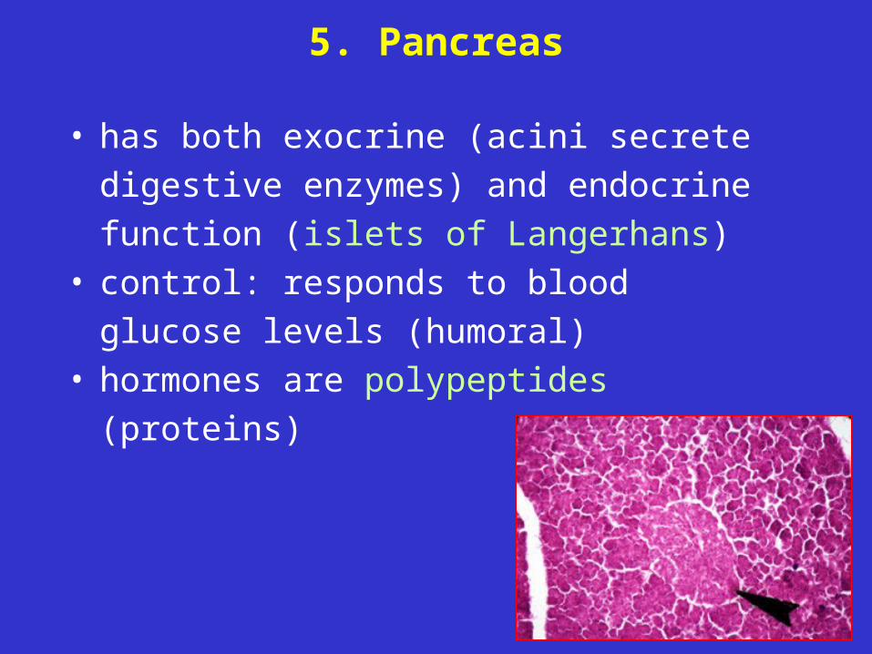
5. Pancreas
• has both exocrine (acini secrete digestive
enzymes) and endocrine function (islets of
Langerhans)• control: responds to blood glucose levels
(humoral)• hormones are polypeptides (proteins)
5. Pancreas
• major cell types
– alpha cells secrete glucagon
– beta cells secrete insulin
– delta cells secrete somatostatin (which inhibits
insulin and glucagon secretion, and decrease
fat absorption in intestines)
– F cells regulate exocrine function of pancreas
(secrete pancreatic polypeptide)
5. Pancreas: Glucagon
• actions: hyperglycemic (increases blood glucose)– stimulates formation and release of glucose
from liver (main target)° glycogenolysis - breakdown of glycogen
(storage form of glucose)° gluconeogenesis - formation of glucose
from noncarbohydrate molecules (e.g., amino acids, glycerol, lactic acid)
– stimulates glycogenolysis in skeletal muscle– stimulates triglyceride breakdown in adipose
tissue (fat mobilization)
5. Pancreas: Glucagon
• control:
– secreted in response to low blood sugar,
rising amino acid levels in blood
– inhibited by increased blood glucose and
by somatostatin
5. Pancreas: Insulin
• actions: hypoglycemic (lowers blood glucose)– increases transport of glucose into muscle
and fat cells (NOTE: does not increase uptake by brain, liver, or kidney)
– inhibits breakdown of glycogen and formation of glucose from amino acids or fatty acids (inhibits glycogenolysis and gluconeogenesis)
– promotes formation of glycogen (liver, skeletal muscles), protein synthesis (muscle), and fat synthesis and storage (adipose)
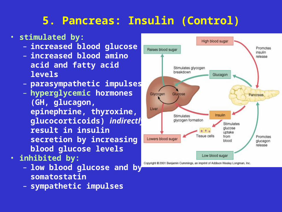
5. Pancreas: Insulin (Control)
• stimulated by: – increased blood glucose– increased blood amino
acid and fatty acid levels– parasympathetic impulses– hyperglycemic hormones
(GH, glucagon, epinephrine, thyroxine, glucocorticoids) indirectly result in insulin secretion by increasing blood glucose levels
• inhibited by: – low blood glucose and by
somatostatin– sympathetic impulses
5. Pancreas: Insulin - Disorders: Diabetes Mellitus (DM)
• hyposecretion (or hypoactivity) of insulin• body cells not stimulated to take up glucose• hyperglycemia (excess blood glucose)
– very high glucose --> nausea --> fight-or-flight response --> secretion of hyperglycemic hormones (epi, NE [adrenal medulla], glucocorticoids [adrenal cortex]) --> stimulates gluconeogenesis, lipolysis, glycogenolysis --> adds to already high glucose
– not all sugar reabsorbed from urine --> glucose lost in urine (glucosuria) --> increased water loss --> excessive urine production (polyuria) and excessive thirst (polydipsia)
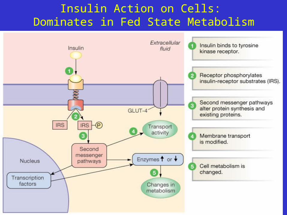
Insulin Action on Cells: Dominates in Fed State Metabolism
Insulin Action on Cells: Dominates in Fed State Metabolism
5. Pancreas: Insulin - Diabetes Mellitus
• cells use fats as energy source (due to poor
glucose uptake)• hyperglycemic hormones stimulate fat
mobilization --> fats in blood (lipidemia) -->
increase in lipid metabolites in blood (ketone
bodies, which are strong organic acids) -->
decrease blood pH (ketoacidosis) and ketone
bodies in urine (ketonuria)– decreased blood pH --> severe depression of
nervous system --> deep breathing --> diabetic
coma --> death• polyphagia (excessive hunger) - final sign, due to
use of fats and proteins as energy sources
Type I Diabetes mellitus• also called insulin-dependent diabetes
(IDDM; formerly juvenile onset diabetes)• onset is sudden, usually before age 15• may be due to autoimmune attack of proteins
in beta cells (see “A Closer Look”, p. 640-641)
• result is lack of insulin activity• lipidemia (high blood lipid content) and
increased cholesterol lead to long-term vascular problems (arteriosclerosis, strokes, heart attacks, renal shutdown, gangrene, blindness)
• treated with insulin injections or pancreatic islet transplant (newer technique)
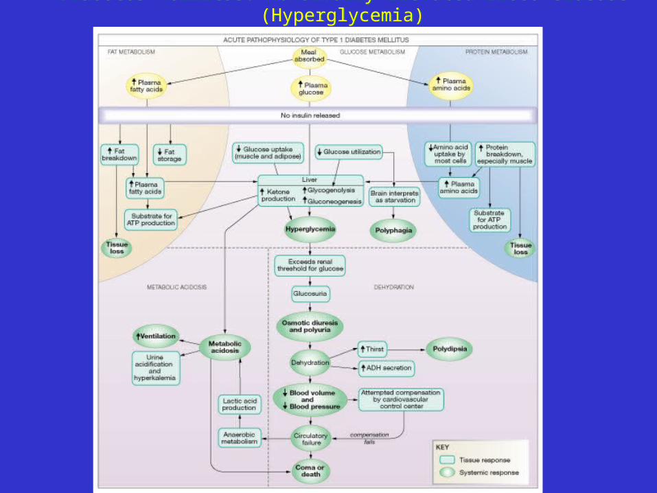
Diabetes Mellitus: Abnormally Elevated Blood Glucose (Hyperglycemia)Diabetes Mellitus: Abnormally Elevated Blood Glucose (Hyperglycemia)
Type II Diabetes Mellitus• non-insulin-dependent (NIDDM; formerly
mature-onset diabetes)• usually starts after age 40• insulin levels are normal or elevated, but
peripheral tissue become less sensitive to it • 25-30% of Americans carry gene that
predisposes them to NIDDM, more likely in over-weight people (~90% of cases)– adipose cells secrete tumor necrosis factor
alpha that depresses production of protein needed for glucose uptake
• often controllable with diet and exercise
Hyperinsulinism
• excess of insulin (usually from injection of excess)
• causes hypoglycemia --> secretion of hyperglycemic hormones (to raise blood glucose) - low glucose to brain --> anxiety, nervousness, tremors, weakness --> eventually, disorientation, convulsions, death due to “insulin shock”
• treated by providing sugar source