endodontic - nzse.org.nz 40.pdf · amna siddiqui has written a very comprehensive review on...
TRANSCRIPT

New Zealand Endodontic Journal Vol 40 September 2009 Page 1
EndodonticJournalVol 40 SEPTEMBER 2009 ISSN 0114-7722
New
Zea
land
Contents
2 Editorial Notes
3 President’s Report Sara Jardine
4 Editorial Tina Hauman 5 Endodontic Implications of Bisphosphonate-associated Osteonecrosis of the Jaws: A Review Jack Lin
12 Systemic Complications of Endodontic Infections Amna Siddiqui
23 Radix Entomolaris – A Case Report Poonam Verma
25 News from the School
26 Minutes of the Annual General Meeting
New Zealand Societyof Endodontics (Inc)
PresidentSara JardinePO Box 7788Wellesley StreetAuckland
SecretaryMike Jameson2 Granville TerraceBelleknowesDunedin
TreasurerDeborah CreaghCMC HouseLevel 989 Courtenay PlaceWellington
Journal EditorTina HaumanPO Box 647Dunedin
Front Cover: Radix Entomolaris

Page 2 New Zealand Endodontic Journal Vol 40 September 2009
Editorial Notices
The New Zealand Endodontic Journal is published twice yearly and sent free to members of the New Zealand Society of Endodontics (Inc). The subscription rates for membership of the Society are $35 per annum in New Zealand or $45 plus postage for overseas members. Graduates of the University of Otago School of Dentistry enjoy complimentary membership for the first year after graduation. Subscription inquiries should be sent to the Honorary Secretary, Dr Mike Jameson, 2 Granville Terrace, Dunedin. Contributions for inclusion in the Journal should be sent to the Editor, Tina Hauman, PO Box 647, Dunedin. Deadline for inclusion in the May or November issue is the first day of the preceding month. All expressions of opinion and statements of fact are published on the authority of the writer under whose name they appear and are not necessarily those of the New Zealand Society of Endodontics, the Editor or any of the Scientific Advisers.
Information for AuthorsThe Editor welcomes original articles, review articles, case reports, views and comments, correspondence, announcements and news items. The Editor reserves the right to edit contributions to ensure conciseness, clarity and consistency to the style of the Journal. Contributions will normally be subjected to peer review. It is the wish of the Editor to encourage practitioners and others to submit material for publication. Assistance with word processing and photographic and graphic art production will be available to authors.
ArrangementArticles should be typewritten on one side of A4 paper with double spacing and 3cm margins. The author’s name should appear under the title and name and postal address at the end of the article. If possible, the manu-script should also be submitted on computer disc, either Macintosh or PC compatible.
ReferencesReferences cited in the text should be placed in parenthesis stating the authors’ names and date, eg (Sundqvist & Reuterving 1980). At the end of the article references should be listed alphabetically giving surnames and initials of all authors, the year, the full title of the article, name of periodical, volume number and page numbers.
The form of reference to a journal article is:Sundqvist G, Reuterving C-O (1980) Isolation of
Actinomyces israelii from periapical lesion. Journal of Endodontics 6, 602-6.
The form of reference to a book is:Trowbridge HO, Emling RC (1993) Inflammation,
4th edn, pp 51-7. Chicago, USA: Quintessence Publishing Company Inc.
IllustrationsIllustrations should be submitted as clear drawings, black & white or colour photographs and be preferably of column width. Radiographs are acceptable. However a black & white photograph is preferred. Illustrations must be numbered to match the text and bear the author’s name and an indication of the top edge on the back. Legends are required for all illustrations and should be typewritten on a separate page.
New Zealand Endodontic Journal Vol 40 September 2009 Page 3
Hi members and welcome to spring! The year is once again flying by with the NZDA branch conference done and dusted along with another AGM. Many thanks to those half dozen members that managed to drag themselves away from lunch and attend. Our long serving and hard working secretary Mike Jameson has resigned from his post. Thank you so much Mike for all your hard work and advice over the past years. Mike has organised us into more regular executive teleconferences and helped put our governance on track.
It looks like all of Hani’s hard work has come to fruition with the launching of the NZSE website. I urge you all to take the opportunity to check it out. It really does look great and easy to navigate around. The journal will be available on the site to members only, along with information about up coming meetings etc. The address is www.nzse.org.nz so get surfing.
We are trying to update our data base with the email addresses of all of our members to allow emailing of notices of up coming meetings, subs etc. Could you please let Deb Creagh know when you send in your subs your email address, if you have not already so.Please mark in your dairy for the 2nd Trans Tasman Endodontic Congress in Christchurch on 4-6th November 2010. I know its over a year away but it will come around soon enough. Speakers are yet to be finalised but if the Hobart meeting last year is anything to go by I am sure you will not want to miss it. Thank you all once again for all your input and for the committee for giving up their time to help make the society run smoothly.
Thanks again Tina for all you hard work with the journal.
Happy spring … summer is nearly here!
All the best, Sara Jardine
President’s Report

Page 4 New Zealand Endodontic Journal Vol 40 September 2009
EditorialDear members,
This edition is dedicated to some medical conditions and therapies that may influence or effect endodontic treatment or may occur as a complication of endodontic therapy.
Bisphosphonate-associated osteonecrosis has been a hot topic over the past 2-3 years in the dental literature. The review by Jack Lin revisits bisphosphonates and their uses with a definite slant towards the implications of this condition on endodontics and the important role of endodontics in patients on oral bisphosphonate therapy.
Amna Siddiqui has written a very comprehensive review on possible systemic complica-tions of endodontic treatment. Although most of these complications are very rare it is important to be aware of these systemic conditions and when to anticipate it or when to act. She included the guidelines and antibiotic regimes for prophylaxis in patients with heart conditions and for those with prosthetic joints.
The case report by Poonam Verma reviews radix entomolaris, an anatomical variant in lower molars, and not uncommonly seen in New Zealand.
I wish to thank our postgraduate students for their valuable contributions.
Tina Hauman

New Zealand Endodontic Journal Vol 40 September 2009 Page 5
Endodontic Implications ofBisphosphonate-associated Osteonecrosis of the Jaws
A REVIEW
Jack Lin
Bisphosphonates The bisphosphonates were first discovered during the middle part of the 19th century. They were used as corrosion inhibitors or as complexing agents in the textile, fertilizer, and oil industries. Bisphosphonates have been developed as a drug and used clinically during the past 30 years (Fleisch 1998).
Chemistry and ClassesThe structure of bisphosphonates is based on the pyrophosphate being covalently linked to a carbon atom (Figure 1).
Figure 1: Chemical structure of pyrophosphate and bisphosphonates (Fleisch 1998).
The P-C-P structure allows a great number of possible variations, which depends on the side chains R1 and R2 coupled to the central carbon atom or by modification of the phosphate groups. There are only a few commercially available bisphosphonates for the treatment of bone disease Each bisphosphonate has its own chemical, physicochemical, and biological characteristics, which implies that it is impossible to extrapolate the results of one compound with respect to its actions and apply it to another (Fleisch 1998).
Mechanism of ActionsBisphosphonates are potent inhibitors of osteo-
clastic activity (Licata 2005). In addition, bisphosphonates reduce recruitment of osteoclasts and induce osteoblasts to produce an osteoclast-inhibiting factor (Hughes et al. 1989; Vitte et al. 1996). Both mechanisms lead to a reduction in bone resorption and consequently a decrease in bone turnover (Fleisch 2002).
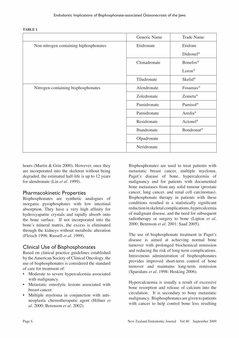
There are currently two classes of bisphosphonate (Table 1):• Non-nitrogen-containing bisphosphonates• Nitrogen-containing bisphosphonates
The non-nitrogen-containing bisphosphonates are metabolised by osteoclasts to form the adenosine triphosphate analogues. These metabolites inhibit the adenosine diphosphate/adenosine triphosphate (ADP/ATP) translocase in the mitochondria, resulting in the inhibition of cell function and the induction of apoptosis (Fleisch 1998; Russell et al. 1999; Lehenkari et al. 2002).
The nitrogen-containing bisphosphonates are more potent than the non-nitrogen containing bisphosphonates due to the fact that osteoclasts cannot metabolise the nitrogen side-chain. Nitrogen-containing bisphosphonates are taken up by osteoclasts during bone resorption. Within the cells, they inhibit the enzyme, farnesyl diphosphonate synthase, which is part of the mevalonate pathway of cholesterol synthesis (Green 2004). The loss of protein prenylation, results in deregulation of intracellular transport, cytoskeletal organization, and cell proliferation thus leading to the inhibition of osteoclast function and cell death (apoptosis).
The half-life of bisphosphonates in the circulation is quite short, ranging from thirty minutes to two
Page 6 New Zealand Endodontic Journal Vol 40 September 2009
TABLE 1
hours (Martin & Grin 2000). However, once they are incorporated into the skeleton without being degraded, the estimated half-life is up to 12 years for alendronate (Lin et al. 1999).
Pharmacokinetic PropertiesBisphosphonates are synthetic analogues of inorganic pyrophosphates with low intestinal absorption. They have a very high affinity for hydroxyapatite crystals and rapidly absorb onto the bone surface. If not incorporated into the bone’s mineral matrix, the excess is eliminated through the kidneys without metabolic alteration (Fleisch 1998; Russell et al. 1999).
Clinical Use of BisphosphonatesBased on clinical practice guidelines established by the American Society of Clinical Oncology, the use of bisphosphonates is considered the standard of care for treatment of:• Moderate to severe hypercalcemia associated
with malignancy.• Metastatic osteolytic lesions associated with
breast cancer.• Multiple myeloma in conjunction with anti-
neoplastic chemotheraputic agent (Hillner et al. 2000; Berenson et al. 2002).
Generic Name Trade Name
Non-nitrogen containing biphosphonates Etidronate Etidrate
Didronel®
Clonadronate Bonefos®
Loron®
Tiludronate Skelid®
Nitrogen-containing bisphosphonates Alendronate Fosamax®
Zoledronate Zometa®
Pamidronate Pamisol®
Pamidronate Aredia®
Residronate Actonel®
Ibandronate Bondronat®
Olpadronate
Neridronate
Endodontic Implications of Bisphosphonate-associated Osteonecrosis of the Jaws
Bisphosphonates are used to treat patients with metastatic breast cancer, multiple myeloma, Paget’s disease of bone, hypercalcemia of malignancy and for patients with documented bone metastases from any solid tumour (prostate cancer, lung cancer, and renal cell carcinomas). Bisphosphonate therapy in patients with these conditions resulted in a statistically significant reduction in skeletal complications, hypercalcemia of malignant disease, and the need for subsequent radiotherapy or surgery to bone (Lipton et al. 2000; Berenson et al. 2001; Saad 2005).
The use of bisphosphonate treatment in Paget’s disease is aimed at achieving normal bone turnover with prolonged biochemical remission and reducing the risk of long-term complications. Intravenous administration of bisphosphonates provides improved short-term control of bone turnover and maintains long-term remission (Sparidans et al. 1998; Hosking 2006).
Hypercalcaemia is usually a result of excessive bone resorption and release of calcium into the circulation. It is secondary to bony metastatic malignancy. Bisphosphonates are given to patients with cancer to help control bone loss resulting
New Zealand Endodontic Journal Vol 40 September 2009 Page 7
from metastatic skeletal lesions (Rogers et al. 1997; Lin et al. 1999). They reduce skeletally-related events associated with multiple myeloma and metastatic solid tumors in the bones. In addition to the anti-resorption effects, studies suggest that bisphosphonates have several anti-tumor effects. These include induction of tumor cell apoptosis, inhibition of tumor cell adhesion to the extracellular matrix, and inhibition of tumor invasion (Berenson et al. 1998; Santini et al. 2003; Green 2004). Intravenous infusion of bisphosphonates improves bio-availability, do not produce gastrointestinal side effects, and is well tolerated by the patient. Bisphosphonates have become a standard therapy in the management of patients with multiple myeloma and metastatic bone diseases.
Osteoporosis is the most common cause of fractures in the elderly. It is defined as a reduction in bone mass caused by the imbalance between physiological bone destruction and formation. It is aggravated by estrogen deficiency and other factors. Bisphosphonates are the most effective inhibitors of bone resorption. They decrease bone turnover, increase bone mineral density and reduce the risk of osteoporotic fractures in the spine (Watts 1998; Rodan & Reszka 2003). Orally administered bisphosphonate preparations are also potent osteoclast inhibitors. They are, however, not as efficient as intravenous derivatives in the treatment of malignant osteolytic disease. They are only indicated for the treatment of osteoporosis.
Adverse Effects of BisphosphonatesGenerally, bisphosphonates are well tolerated, with predictable side effects including elevated serum creatinine, transient low-grade fever, fatigue, arthralgia, nausea, and increased bone pain, giving these drugs a safety profile that is well accepted among physicians (Conte & Guarneri 2004). However, bisphosphonates are now known to have a low incidence of a serious adverse effect. Long-term use of these medications has been associated with osteonecrosis of the jaws (Marx 2003; Ruggiero et al. 2004).
Osteonecrosis of the JawsRecently, an increasing number of cases of bisphosphonate-associated osteonecrosis of the jaws have been reported. However, the
Endodontic Implications of Bisphosphonate-associated Osteonecrosis of the Jaws
exact mechanism that leads to the induction of the condition is unknown. Bone remodelling is a normal physiologic function. It removes microdamage and replaces damaged bone with new elastic osseous tissue (Ott 2005). Bisphosphonates inhibit osteoclast function, prevent bone turnover and have anti-angiogenic properties (Rogers et al. 1997; Fleisch 2002). Ischaemia of the jaws, possibly due to alternations in the normal bone homeostatic mechanism, has been reported in several patients with bisphosphonate-associated osteonecrosis (Wood et al. 2002). As a result, suppressed bone turnover will accumulate microdamage and reduction in bone strength (Mashiba et al. 2000; Odvina et al. 2005; Ott 2005). When microdamage is not repaired it sets the stage for osteomyelitis and eventually osteonecrosis.
The potency of bisphosphonates is dependent on the uptake and retention of the drug in the bone in each individual. The skeleton most likely acts as a reservoir for bisphosphonates. This will influence the adsorption and desorption of bisphosphonates from the bone surfaces (Bukowski et al. 2005). The effects of bisphosphonates seem to persist for extended periods, and this could explain why osteonecrosis appears after long-term treatment, and even in cases in which bisphosphonate treatment was discontinued (Ruggiero et al. 2004; Woo et al. 2005).
Predisposing Risk Factors The predisposing risk factors for bisphosphonate-associated osteonecrosis have not been identified. However, Migliorati et al. (2005) have classified the potential risk factors into two groups:• Systemic factors (American Dental Association
2006)• Local factors Systemic FactorsNinety-four percent of patients affected by bisphosphonate-associated osteonecrosis have metastatic bone disease and received nitrogen-containing bisphosphonate treatment intravenously (Woo et al. 2006). These include systemic factors such as:• Type and total dose of bisphosphonates.• Presence of diabetes mellitus.• Overall tumour burden and stage of disease.• Extent of skeletal involvement.

Page 8 New Zealand Endodontic Journal Vol 40 September 2009
hard tissue healing after tooth extraction (Marx 2003; Ruggiero et al. 2004; Migliorati et al. 2005). Symptoms may occur spontaneously. The most common complaint is the sudden presence of intraoral discomfort due to soft tissues traumatised by the rough edges of necrotic bone (Migliorati et al. 2006).
In the early stages, patients are usually asymptomatic. Radiographic findings are variable with no specific diagnostic characteristics (Nishimura et al. 1982). Later on, patients may develop pain due to secondary infection. The gingival or mucosal tissues surrounding necrotic bone are usually inflamed and sensitive to palpation (Migliorati et al. 2006). The necrotic process could extend to the periodontium, resulting in increased tooth mobility and the need for additional extractions (Marx et al. 2005).
In severe cases, it can cause intense pain, extensive destruction of bone, sinus tracts present to the skin surface and affect the sensory innervation, or even result in jaw fracture (Ruggiero et al. 2004; Hellstein & Marek 2005; Migliorati et al. 2005). The diagnosis of bisphosphonate-associated necrosis is based on the medical and dental history and clinical observation of each individual.
Management RecommendationsThe management of bisphosphonate-associated osteonecrosis of the jaws represents an additional challenge to professionals. Currently, there is no known effective treatment for the condition (Carter et al. 2005). The treatment of patients receiving oral or intravenous bisphosphonate therapy is principally preventive in nature. The need for the patient to be dentally fit and be prepared to maintain this state for life should form part of treatment informed consent. Other considerations involve modification of the dental treatment plan for a patient taking bisphosphonates and a protocol for the patients who develop bisphosphonate-associated osteonecrosis.
The recommendations are based on the expert panel outlining the recommendations for the management of bisphosphonate-associated osteonecrosis of the jaws (American Dental Association 2006), and literature reviews (Migliorati et al. 2005; Woo et al. 2006)
Endodontic Implications of Bisphosphonate-associated Osteonecrosis of the Jaws
• Patient’s overall systemic health.• Degree of immunosuppression.• Patient’s history of stem cell transplantation.• Peripheral vascular disease.• Patient’s current and historical use of other
medications such as chemotherapeutic agents or corticosteroids.
It is important to note that patients with multiple myeloma are treated with other antiangiogenic agents such as thalidomide, glucocorticoids and bortezomib (Munshi et al. 1999; Clerc et al. 2003; Hussein 2004; Chauhan et al. 2005). Other factors may play a role, but the extent of their influence remains to be determined.
Local Factors• Dental extractions• Surgical bone manipulation• Trauma to oral tori• Trauma from dentures• Dental infection• Poor oral health
The most important predisposing local factors for the development of bisphosphonate-associated osteonecrosis of the jaw are history of trauma, dental surgery or dental infection. Reports have identified dental extraction as a predisposing factor for osteonecrosis. Ill-fitting prosthodontic appliances can also lead to chronic irritation and initiation of this pathological process. However, spontaneous exposures and necrosis of the alveolar bone, commonly occurring in the lingual surface of the posterior mandible and area of thin mucosa have also been reported (Marx 2003; Ruggiero et al. 2004).
A suggested mechanism for bisphosphonate-associated osteonecrosis of the mandible or maxilla is physiological microdamage in bones with compromised biomechanical properties. Trauma, dental surgery, and infections increase the demand for an osseous turnover that exceeds the capacity of the hypodynamic bone. This results in repeated inflammation and necrosis (Marx 2003).
Clinical Signs and SymptomsSymptoms may be negligible, mild or severe. The most common clinical history associated with bisphosphonate-associated osteonecrosis is patients experiencing absent or delayed soft and

New Zealand Endodontic Journal Vol 40 September 2009 Page 9
reconstruction should be discussed with the patient.
Intravenous Bisphosphonate Therapy (High Risk)Patients exposed to intravenous bisphosphonates are at risk to develop osteonecrosis. The recommendations are the same as above with the following additions:• Tooth extractions and/or other oral surgical
procedures with exposure or manipulation of bone, should be avoided
• Non-surgical endodontics (with or without decoronation) should be performed instead of tooth extraction whenever possible
• Any elective dental procedure requiring bone healing should be avoided
Management of Patients with Bisphosphonate-associated OsteonecrosisFor these patients recommendations should include:• Non-surgical approach to prevent further bone
injury• Perform conservative bone debridement
with copious irrigation to reduce sharp bone spicules
• Coverage of exposed bone with removable appliance to prevent secondary trauma
• Meticulous oral hygiene and daily antiseptic rinses
• Systemic analgesic as required• Systemic antibiotics as required• Regular monitoring• Suggest discontinuation of bisphosphonate
therapy until osteonecrosis heals or until the underlying disease require treatment again
Surgical intervention does not appear to eliminate the osteonecrotic process (Migliorati et al. 2005). In addition, hyperbaric oxygen therapy, extensive antibiotic treatment or the topical use of mouth rinses do not appear to promote healing in these cases (Ruggiero et al. 2004; Carter et al. 2005; Hellstein & Marek 2005). The difficulty in treating this condition is that debridement cannot be carried out on uninvolved bone and may actually cause further exposure of the bone (Marx 2003). Surgical intervention ranges from simple debridement or curettage to radical resection, depending on the extent of necrotic bone. However, if large volume
Endodontic Implications of Bisphosphonate-associated Osteonecrosis of the Jaws
Recommendations for Patients prior to Commencement of Bisphosphonates Therapy• A complete dental and radiographic
examination is recommended, in order to determine the periodontal and endodontic status of the remaining dentition
• All dental prosthesis should be checked to prevent soft tissue trauma
• Teeth with poor prognosis or poor periodontal health should be extracted, mucoperiosteal flaps, and intramedullary bone manipulations should be performed prior to commencement of the intravenous bisphosphonate therapy
• Provide preventive dental education, combining excellent oral hygiene and routine dental care as needed
• Patient should be aware of the importance of potential problems relating to bisphosphonate therapy.
Oral Bisphosphonate Therapy(Low Risk)Patients receiving oral bisphosphonates are at very low ri sk (estimated at 0.7 cases per 100,000) of developing bisphosphonate-associated osteo-necrosis (American Dental Association 2006). The recommendations for these patients are:• A comprehensive recent medical history before
commencing any dental treatment• Identifying the risk factors in the medical
history• Routine preventive dentistry and exams• Prophylaxis and oral hygiene instructions
should be given• Avoid intrusion into the biological width or
disruption of submucosal periosteum during dental procedures
• Maintenance of dental prosthesis to eliminate risk of soft tissue trauma
• Devise treatment plans to minimize surgery both in the short-term and long-term
No dental procedures are absolutely contra-indicated. However, before undertaking any invasive procedure involving manipulation of bone or periosteum, patients should again be informed about the complications of oral bisphosphonate therapy and the risk of bisphosphonate-associated osteonecrosis. Alternatively, treatment options such as endodontics rather than extraction and bridges and partial dentures versus implant
Page 10 New Zealand Endodontic Journal Vol 40 September 2009
Endodontic Implications of Bisphosphonate-associated Osteonecrosis of the Jaws
ConclusionIt is important for dental professionals to become familiar with this condition of bisphosphonate-associated osteonecrosis. Any history of cancer or osteoporosis treated with bisphosphonates should be carefully documented. At present, recommendation guidelines are based on individual experiences in management of bisphosphonate-associated osteonecrosis. Endodontics can be a valuable alternative treatment for pulpitis or apical periodontitis.
ReferencesAmerican Dental Association (2006) Dental management of
patients receving oral bisphosphonate therapy. Journal of the American Dental Association 137, 1144-50.
Berenson JR, Lichtenstein A, Porter L, Dimopoulos MA, Bordoni R, George S, Lipton A, Keller A, Ballester O, Kovacs M, Blacklock H, Bell R, Simeone JF, Reitsma DJ, Heffernan M, Seaman J, Knight RD (1998) Long-term pamidronate treatment of advanced multiple myeloma patients reduces skeletal events. Myeloma Aredia Study Group. Journal of Clinical Oncology 16, 593-602.
Berenson JR, Rosen LS, Howell A, Porter L, Coleman RE, Morley W, Dreicer R, Kuross SA, Lipton A, Seaman JJ (2001) Zoledronic acid reduces skeletal-related events in patients with osteolytic metastases. Cancer 91, 1191-200.
Berenson JR, Hillner BE, Kyle RA, Anderson K, Lipton A, Yee GC, Biermann JS, American Society of Clinical Oncology Bisphosphonates Expert Panel (2002) American Society of Clinical Oncology clinical practice guidelines: the role of bisphosphonates in multiple myeloma. Journal of Clinical Oncology 20, 3719-36.
Bukowski JF, Dascher CC, Das H (2005) Alternative bisphosphonate targets and mechanisms of action. Biochemical & Biophysical Research Communications 328, 746-50.
Carter G, Goss AN, Doecke C (2005) Bisphosphonates and avascular necrosis of the jaw: a possible association. Medical Journal of Australia 182, 413-5.
Chauhan D, Hideshima T, Mitsiades C, Richardson P, Anderson KC (2005) Proteasome inhibitor therapy in multiple myeloma. Molecular Cancer Therapeutics 4, 686-92.
Clerc D, Fermand JP, Mariette X (2003) Treatment of multiple myeloma. Joint, Bone, Spine: Revue du Rhumatisme 70, 175-86.
Conte P, Guarneri V (2004) Safety of intravenous and oral bisphosphonates and compliance with dosing regimens. Oncologist 9 Suppl 4, 28-37.
Fleisch H (1998) Bisphosphonates: mechanisms of action. Endocrine Reviews 19, 80-100.
Fleisch H (2002) Development of bisphosphonates. Breast Cancer Research 4, 30-4.
Green JR (2004) Bisphosphonates: preclinical review. Oncologist 9 Suppl 4, 3-13.
Hellstein JW, Marek CL (2005) Bisphosphonate osteochemonecrosis (bis-phossy jaw): is this phossy jaw of the 21st century? Journal of Oral & Maxillofacial Surgery 63, 682-9.
debridement becomes necessary, the goal should be to remove as little bone as possible. Removal of the bone with minimal epithelial manipulation associated with topical and systemic antibiotics seem to be the treatment modality of choice (Leite et al. 2006).
Endodontic ImplicationNon-surgical endodontics instead of tooth extraction should be performed whenever possible, even if the tooth is non-restorable. Endodontic surgical procedures are not recommended.
Pain related to osteonecrosis or a secondary infection due to exposed bone, may mimic pain of endodontic origin and should be considered in the differential diagnosis (Migliorati et al. 2005). The diagnosis of dental pulp status should include history of pain, clinical examination, sensibility tests, and radiological examination, and not as the outcome of one specific test.
Endodontic ProceduresIf endodontic treatment is indicated, procedures should be performed with care to avoid trauma to the surrounding periodontal tissues. Rubber dam placement with clamps should avoid impinging gingival tissue or a modified isolation technique (split dam technique) should be considered. Procedural errors resulting in periodontal tissue damage (perforation or apical foramen damage) can be prevented by improved knowledge of root canal anatomy, careful instrumentation, correct working length measurement, and using an operating microscope and electronic apex locater.
A final restoration should be carried out after root canal treatment. However, subgingival matrix band placement should be avoided. A decoronation procedure should be considered for teeth with extensive coronal destruction, subgingival margin, or if not restorable. The tooth can be left with a permanent seal or prepared as an over-denture abutment.
Overall, care should be taken with any procedures that invade the biological width. Violation of the connective tissue portion of biological width appears to be the area where the risk of osteonecrosis becomes a major concern for patients receiving bisphosphonate therapy.

New Zealand Endodontic Journal Vol 40 September 2009 Page 11
Endodontic Implications of Bisphosphonate-associated Osteonecrosis of the Jaws
bisphosphonate treatment. Lancet Oncology 7, 508-14.Munshi NC, Barlogie B, Desikan KR, Wilson C (1999) Novel
approaches in myeloma therapy. Seminars in Oncology 26, 28-34.
Nishimura Y, Yakata H, Kawasaki T, Nakajima T (1982) Metastatic tumours of the mouth and jaws. A review of the Japanese literature. Journal of Maxillofacial Surgery 10, 253-8.
Odvina CV, Zerwekh JE, Rao DS, Maalouf N, Gottschalk FA, Pak CY (2005) Severely suppressed bone turnover: a potential complication of alendronate therapy. Journal of Clinical Endocrinology & Metabolism 90, 1294-301.
Ott SM (2005) Long-term safety of bisphosphonates. Journal of Clinical Endocrinology & Metabolism 90, 1897-9.
Rodan GA, Reszka AA (2003) Osteoporosis and bisphosphonates. Journal of Bone & Joint Surgery - American Volume 85-A Suppl 3, 8-12.
Rogers MJ, Watts DJ, Russell RG (1997) Overview of bisphosphonates. Cancer 80, 1652-60.
Ruggiero SL, Mehrotra B, Rosenberg TJ, Engroff SL (2004) Osteonecrosis of the jaws associated with the use of bisphosphonates: a review of 63 cases. Journal of Oral & Maxillofacial Surgery 62, 527-34.
Russell RG, Croucher PI, Rogers MJ (1999) Bisphosphonates: pharmacology, mechanisms of action and clinical uses. Osteoporosis International 9 Suppl 2, S66-80.
Saad F (2005) Clinical benefit of zoledronic acid for the prevention of skeletal complications in advanced prostate cancer. Clinical Prostate Cancer 4, 31-7.
Santini D, Vespasiani Gentilucci U, Vincenzi B, Picardi A, Vasaturo F, La Cesa A, Onori N, Scarpa S, Tonini G (2003) The antineoplastic role of bisphosphonates: from basic research to clinical evidence. Annals of Oncology 14, 1468-76.
Sparidans RW, Twiss IM, Talbot S (1998) Bisphosphonates in bone diseases. Pharmacy World & Science 20, 206-13.
Vitte C, Fleisch H, Guenther HL (1996) Bisphosphonates induce osteoblasts to secrete an inhibitor of osteoclast-mediated resorption. Endocrinology 137, 2324-33.
Watts NB (1998) Treatment of osteoporosis with bisphosphonates. Endocrinology & Metabolism Clinics of North America 27, 419-39.
Woo SB, Hande K, Richardson PG (2005) Osteonecrosis of the jaw and bisphosphonates. New England Journal of Medicine 353, 99-102; discussion 99-.
Woo SB, Hellstein JW, Kalmar JR (2006) Systematic review: bisphosphonates and osteonecrosis of the jaws. Annals of Internal Medicine 144, 753-61.
Wood J, Bonjean K, Ruetz S, Bellahcene A, Devy L, Foidart JM, Castronovo V, Green JR (2002) Novel antiangiogenic effects of the bisphosphonate compound zoledronic acid. Journal of Pharmacology & Experimental Therapeutics 302, 1055-61.
Hillner BE, Ingle JN, Berenson JR, Janjan NA, Albain KS, Lipton A, Yee G, Biermann JS, Chlebowski RT, Pfister DG, American Society of Clinical Oncology Bisphosphonates Expert Panel (2000) American Society of Clinical Oncology guideline on the role of bisphosphonates in breast cancer. Journal of Clinical Oncology 18, 1378-91.
Hosking D (2006) Pharmacological therapy of Paget’s and other metabolic bone diseases. Bone 38, S3-7.
Hughes DE, MacDonald BR, Russell RG, Gowen M (1989) Inhibition of osteoclast-like cell formation by bisphosphonates in long-term cultures of human bone marrow. Journal of Clinical Investigation 83, 1930-5.
Hussein MA (2004) New treatment strategies for multiple myeloma. Seminars in Hematology 41, 2-8.
Lehenkari PP, Kellinsalmi M, Napankangas JP, Ylitalo KV, Monkkonen J, Rogers MJ, Azhayev A, Vaananen HK, Hassinen IE (2002) Further insight into mechanism of action of clodronate: inhibition of mitochondrial ADP/ATP translocase by a nonhydrolyzable, adenine-containing metabolite. Molecular Pharmacology 61, 1255-62.
Leite AF, Figueiredo PT, Melo NS, Acevedo AC, Cavalcanti MG, Paula LM, Paula AP, Guerra EN (2006) Bisphosphonate-associated osteonecrosis of the jaws. Report of a case and literature review. Oral Surgery Oral Medicine Oral Pathology Oral Radiology & Endodontics 102, 14-21.
Licata AA (2005) Discovery, clinical development, and therapeutic uses of bisphosphonates. Annals of Pharmacotherapy 39, 668-77.
Lin JH, Russell G, Gertz B (1999) Pharmacokinetics of alendronate: an overview. International Journal of Clinical Practice Supplement 101, 18-26.
Lipton A, Theriault RL, Hortobagyi GN, Simeone J, Knight RD, Mellars K, Reitsma DJ, Heffernan M, Seaman JJ (2000) Pamidronate prevents skeletal complications and is effective palliative treatment in women with breast carcinoma and osteolytic bone metastases: long term follow-up of two randomized, placebo-controlled trials. Cancer 88, 1082-90.
Martin T, Grin V (2000) Bisphosphonates - mechanisms of action. Australian Prescriber 23, 130-2.
Marx RE (2003) Pamidronate (Aredia) and zoledronate (Zometa) induced avascular necrosis of the jaws: a growing epidemic. Journal of Oral & Maxillofacial Surgery 61, 1115-7.
Marx RE, Sawatari Y, Fortin M, Broumand V (2005) Bisphosphonate-induced exposed bone (osteonecrosis/osteopetrosis) of the jaws: risk factors, recognition, prevention, and treatment. Journal of Oral & Maxillofacial Surgery 63, 1567-75.
Mashiba T, Hirano T, Turner CH, Forwood MR, Johnston CC, Burr DB (2000) Suppressed bone turnover by bisphosphonates increases microdamage accumulation and reduces some biomechanical properties in dog rib.[see comment]. Journal of Bone & Mineral Research 15, 613-20.
Migliorati CA, Schubert MM, Peterson DE, Seneda LM (2005) Bisphosphonate-associated osteonecrosis of mandibular and maxillary bone: an emerging oral complication of supportive cancer therapy. Cancer 104, 83-93.
Migliorati CA, Siegel MA, Elting LS (2006) Bisphosphonate-associated osteonecrosis: a long-term complication of
Page 12 New Zealand Endodontic Journal Vol 40 September 2009
Systemic Complications of Endodontic Infections
Amna Siddiqui
IntroductionEndodontic infection comprises microbial infection of the root canal system and is the major etiologic factor in apical periodontitis (Kakehashi et al. 1965, Sundqvist 1976, Möller et al. 1981). When properly managed, these infections are resolved in over 90% of cases (Sjögren et al. 1990). Untreated or persistent infections can continue as chronic asymptomatic lesions. The host immune response plays an important role in localizing these infections (Stashenko et al. 1998). However, under certain circumstances, these infections may break through the bony, muscular, and mucosal barriers and spread into contiguous anatomical spaces. This may result in serious systemic complications (Walsh 1997). Although these complications are uncommon, failure or delay in achieving accurate diagnosis and initiating prompt treatment often leads to catastrophic events. The very first description of the interaction between oral infections and systemic health appeared in Ancient Egypt (Baron 1999). In 1911 the focal infection theory, stating that microorganisms from a focus of infection disseminate to other parts of the body and cause serious systemic disease, was proposed by William Hunter. He attributed many human diseases to oral sepsis surrounding ill-fitting bridgework (Hunter 1911). This theory lacked scientific evidence, and has never been proven (Pallasch and Wahl 2003). However, over the last two decades there has been increasing interest in the impact of oral health on general health. Bacteraemia of dental origin has often been linked to cases of infective endocarditis or prosthetic joint infections (Jacobsen and Murray 1980, Waldman et al. 1997, Fouad 2009). The role of oral infections, particularly periodontal infections, as a predisposing factor for the development of systemic disease or as an aggravating factor in existing systemic conditions have also been investigated [reviewed in (Li et al. 2000, Caplan 2004, Meurman et al. 2004, Mealey and Rose 2008)]. Evidence-based literature on the
effect of endodontic infections on systemic health is limited.
This review will highlight the possible systemic complications resulting from direct or haematogenous spread of endodontic infections. Furthermore, ongoing research investigating pulpal and periapical infections as a predisposing factor for coronary heart diseases and their effect on diabetes will be reviewed. Systemic conditions that can complicate the healing or adversely affect the outcome of endodontic infections are not included in this text. Ludwig’s AnginaLudwig’s angina is a rapidly expanding, diffuse cellulitis that bilaterally affects the submandibular, submental and sublingual spaces resulting in a hard “wood-like” swelling. It is named after the German physician, Wilhelm Friedrick Ludwig, who first described it in 1836 (Burk and Ludovici 1939). The word angina comes from the Latin word angere meaning “to strangle” and reflects the potentially life-threatening nature of this condition. Extension of the abscess from periapical infection of the mandibular second or third molars is the most common cause of developing Ludwig’s angina (Kurien et al. 1997). Other causes include tonsillitis, otitis media, mandibular fractures, and infections from foreign body or secondary to oral malignancies (Finch et al. 1980). Any age group can be affected but it is relatively uncommon in children (Kurien et al. 1997).
Patients with Ludwig’s angina usually present with tender, non-fluctuating, hard bilateral suprahyoid swelling. The mouth hangs open due to the swollen tongue, contacting the palate, with oedema of the floor of the mouth. Other clinical features include pain and swelling of the neck, dyspnoea, dysphagia, malaise, fever, and chills (Sethi and Stanley 1994, Ferrera et al. 1996,
New Zealand Endodontic Journal Vol 40 September 2009 Page 13
dyspnoea, stiff neck, pyrexia, pain, sepsis, crepitus, trismus, swelling of floor of the mouth, and swelling on the side of the neck (Pearse 1983, Sakamoto et al. 2000, Sandner et al. 2007). However, diagnosis of DNM based on history and clinical examination alone is difficult because early symptoms are vague. Use of CECT along with the clinical examination increased the sensitivity of diagnosis from 55% to 95% (Miller et al. 1999). These scans provide precise information on the extent of the infection, directing the optimal surgical approach for an effective drainage. Treatment of DNM cases is based on airway management, aggressive surgical intervention, and broad spectrum antibiotic coverage (Estrera et al. 1983, Sakamoto et al. 2000, Sandner et al. 2007).
Necrotizing Fasciitis Necrotizing fasciitis is an acute polymicrobial infection that spreads rapidly along the facial planes resulting in extensive necrosis of fascia, subcutaneous tissues, skin, and possibly muscular tissue. It has a high mortality rate, if not treated promptly and aggressively (Roberson et al. 1996). This type of infection was initially described as “hospital gangrene” by Joseph Jones, a confederate army surgeon, during the American Civil War in 1871 [cited in (Rapoport et al. 1991, Stoykewych et al. 1992, Lin et al. 2001)]. The term necrotizing fasciitis was coined by Wilson in 1952 due to characteristic fascial necrosis associated with the disease (Wilson 1952).
This condition is commonly seen in the abdomen, perineum, and extremities whereas head and neck involvement is relatively rare and frequently related to dental infections (Rapoport et al. 1991, Tung-Yiu et al. 2000, Whitesides et al. 2000, Umeda et al. 2003, Fenton et al. 2004, Farrier et al. 2007, Quereshy et al. 2009). Periapical infections of the mandibular molars have been reported to be the most common cause of this disease in the head and neck region (Tung-Yiu et al. 2000, Umeda et al. 2003). Necrotizing fasciitis may affect patients of any age with a slight male predilection (Whitesides et al. 2000, Lin et al. 2001, Umeda et al. 2003, Fenton et al. 2004, Farrier et al. 2007, Quereshy et al. 2009). The disease can occur in healthy individuals but the prognosis is worse in patients with an underlying condition. A number of predisposing factors such as diabetes, malnutrition, alcoholism, obesity, renal failure,
Systemic Complications of Endodontic Infections
Jiménez et al. 2004). Besides the clinical findings, contrast-enhanced computed tomography (CECT) is reported to be a very efficient diagnostic tool (Marioni et al. 2008).
Untreated Ludwig’s angina can lead to mediastinitis, empyema and pericarditis (Ferrera et al. 1996). The mortality rate is reported to be 8-10% (Sethi and Stanley 1994, Kurien et al. 1997) mainly due to the acute obstruction of the airways (Har-El et al. 1994). Thus, airway management is the most important step in the treatment followed by administration of broad spectrum intravenous antibiotics and surgical intervention. Descending Necrotizing MediastinitisThe first modern account on descending necrotizing mediastinitis (DNM) was given by Pearse in 1983. DNM is uncommon yet it is the most lethal form of mediastinitis having a mortality rate of about 40%. This acute polymicrobial infection has a fulminating course often leading to death (Estrera et al. 1983).
Approximately 70% of the reported cases of DNM resulted from spread of dental infections; especially from periapical abscesses of mandibular molars (Estrera et al. 1983). Thorough knowledge of the facial spaces and their relationship to the neck and the mediastinum is essential for the proper diagnosis and management. Three anatomical pathways exist thorough which infection may spread to the mediastinum along the facial spaces: pretracheal route, lateral pharyngeal route, and retropharyngeal-retrovisceral route [reviewed in (Moncada et al. 1978, Estrera et al. 1983, Pearse 1983)]. Early diagnosis and aggressive management is crucial to reduce the morbidity and mortality (Moncada et al. 1978, Estrera et al. 1983, Miller et al. 1999, Sandner et al. 2007). Estrera et al (1983) suggested the following criteria for the diagnosis of DNM: 1) clinical evidence of severe oropharyngeal infection, 2) demonstration of characteristic radiographic features of mediastinitis, 3) documentation of necrotizing mediastinal infection at the operation and/or post-mortem examination, 4) and estab-lishment of the relationship of oropharyngeal infection, with the development of the necrotizing mediastinal process.
Clinical signs and symptoms include dysphagia,
Page 14 New Zealand Endodontic Journal Vol 40 September 2009
and infection with human immunodeficiency virus have been associated with an increased risk of developing the disease (Whitesides et al. 2000, Lin et al. 2001, Quereshy et al. 2009).
Because of the progressive course of necrotizing fasciitis, early recognition and prompt diagnosis is the most important step in the management of this potentially fatal disease (Stoykewych et al. 1992, Tung-Yiu et al. 2000, Farrier et al. 2007). Clinical signs and symptoms are initially non-specific and could easily be mistaken for a routine dental infection (Umeda et al. 2003, Fenton et al. 2004). Fever, Dysphagia, increasing pain, trismus, paresthesia, and dyspnea are often reported. In the early stages the skin is warm, tender, tense, and shiny due the underlying oedema with no clear demarcation between normal and affected skin. It is recommended to mark the extent of the borders or the periphery of the suspected tissue involvement with a pen so that the progression of the disease can be monitored later (Fenton et al. 2004).
As the disease progresses, the pathognomic sign of necrotizing fasciitis appears as a dusky discoloration of the skin appearing as small purplish patches with ill-defined borders. Eventually, blisters or bullae form with the underlying skin becoming necrotic and blue in colour. The localised skin necrosis is secondary to thrombosis of the blood vessels passing through necrotic fascia. The underlying subcutaneous destruction creates an ideal medium for bacterial growth and if left untreated, will progress to frank cutaneous gangrene. Crepitus is a common finding due to the presence of gas-forming organisms. However, the absence of crepitus does not rule out gas formation since gas can accumulate in areas inaccessible to palpation (Stoykewych et al. 1992, Whitesides et al. 2000, Fenton et al. 2004, Farrier et al. 2007). Computed tomography (CT) scans serve as a useful tool in diagnosing necrotizing fasciitis particularly in the early stages. Gas formation in the soft tissue as well as the extent of the infection and involvement of the anatomic structures can easily be determined on these scans (Becker et al. 1997, Umeda et al. 2003). Once the diagnosis is made, immediate treatment is necessary to minimize mortality (Stoykewych et al. 1992, Umeda et al. 2003). The management of necrotizing fasciitis consists of extensive
surgical debridement, broad spectrum antibiotic coverage, and supportive therapy (Fenton et al. 2004, Quereshy et al. 2009). Reports on adjunctive therapies such as hyperbaric oxygen (Riseman et al. 1990, Langford et al. 1995) and intravenous immunoglobulins (Cawley et al. 1999) have shown promising results in improving treatment outcome. However, the efficacy of these adjunctive therapies needs to be further evaluated in well conducted clinical trials.
Necrotizing fasciitis of the head and neck is uncommon but is a potentially fatal disease. It can result in life-threatening complications such as mediastinits, brain abscesses, systemic toxicity, and multisystem organ failure. Since many cases have been reported to be originating from periapical infections, it is important for the endodontist to be familiar with the disease and to recognise it early in its course to minimize mortality (Stoykewych et al. 1992, Umeda et al. 2003, Fenton et al. 2004). Orbital InfectionsExtension of dental infections to involve orbital spaces is extremely rare, with a prevalence of 1.3% (Blake et al. 2006). The cardinal signs of an orbital infection are impairment of visual acuity, proptosis, pain, and limited ocular motility. Abscesses that extend to the posterior orbital space can be life threatening, because the infection can spread through the optic canal and ophthalmic veins to the meninges and the brain (Kim et al. 2007). The routes of infectious extension may be summarised as follows: (1) the roots of maxillary premolar and molar teeth may lie very close to the maxillary sinus. Maxillary sinusitis may result from extension of maxillary molar or premolar infection or from perforation of the sinus floor during extraction of diseased maxillary teeth. Sinusitis may then extend into the orbit to cause orbital cellulitis. (2) infection of maxillary incisors or canines may spread through local tissue planes over the maxilla, resulting in swelling of the upper lip, canine fossa, and periorbital tissue. Retrograde spread into the orbit may then occur through the valveless anterior facial, angular, and ophthalmic veins. (3) infection of anterior maxillary teeth may also spread as a subperiosteal abscess to the anterior surface of the maxilla to involve the orbit. (4) infection of the posterior maxillary teeth, most
Systemic Complications of Endodontic Infections

New Zealand Endodontic Journal Vol 40 September 2009 Page 15
commonly the third molar, may spread posteriorly into the pterygopalatine and infratemporal fossae. The infection may then extend into the orbit through the inferior orbital fissure (Robin et al. 1996).
Once an orbital space abscess is recognized, early diagnosis and institution of multidisciplinary management, involving dentists, ophthalmologists, otolaryngologists and oral and maxillofacial surgeons is necessary to reduce morbidity and mortality. The treatment includes intravenous broad spectrum antibiotics, surgical incision and drainage of the subperiosteal or intraorbital abscess, and eradication of the primary source of infection (Blake et al. 2006). Cavernous sinus thrombosis, cerebral abscess, meningitis and blindness are rare but potential complications in the extension of periorbital abscesses (Miller and Kassebaum 1995).
Cavernous Sinus ThrombosisTwo cavernous sinuses are situated on either side of the sella turcica (Marinkovica et al. 2001). When a thrombus is formed at some point in the facial venous system, it can undergo retrograde spread towards the cavernous sinus giving rise to thrombosis. Two routes exist for spread of infection to the cavernous sinus, either anteriorly via angular and ophthalmic veins or posteriorly through the pterygoid venous plexus. Dental infections rarely results in cavernous sinus thrombosis (CST) (Ebright et al. 2001, Jiménez et al. 2004). Signs and symptoms include eye pain, sensitivity of the eyeball to pressure, high fever, chills, headache, nausea, vomiting, supraorbital paresthesia, ophthalmoplegia, photophobia, palpebral oedema, ptosis, and retinal bleeding (Ferrera et al. 1996, Ebright et al. 2001). CT and magnetic resonance imaging are valuable diagnostic tools in cases of CST. Treatment consists of antibiotics, anticoagulants, and occasionally surgical aspiration (Ebright et al. 2001).
Brain AbscessBrain abscess is a rare, but life threatening infection in which a localized area of suppuration develops within the brain parenchyma. The most common sites involved are the temporal lobes (42%), followed by the cerebellum (30%) (Yang 1981). The clinical presentation of brain abscess
is influenced by a number of factors including the size and location of the abscess, the virulence of the infecting organisms, and the presence of underlying systemic conditions. Most patients suffer from headaches or lethargy, but fewer than half of them have fever, focal neurologic signs, increased intracranial pressure, or altered mental status (Li et al. 1999).
Dental infections have been occasionally reported to cause brain abscess, but these reports lack any scientific evidence (Aldous et al. 1987, Schuman and Turner 1994). Corson and co-workers (2001) have stressed the importance of using sound microbiological methodology to precisely identify microbial isolates in both oral and cranial sites in order to confirm any link between dental infections and brain abscess. Infective EndocarditisInfective endocarditis is a bacterial infection of the heart valves and the epithelial lining (endocardium). It can occur when bacteria in the bloodstream lodge on abnormal heart valves or damaged heart tissue. Patients with certain pre-existing heart defects are at high risk for developing endocarditis when bacteraemia occurs. The disease rarely occurs in people with normal hearts (Li et al. 2000) and can clinically be divided into acute and subacute forms. Acute infective endocarditis is usually caused by highly virulent organisms, such as Staphylococcus aureus, attacking a healthy heart to create a rapidly progressive disease which usually results in death within a few weeks or months. Subacute infective endocarditis is usually caused by less virulent organisms, characteristically organisms that belong to the viridans group of streptococci. It is a more insidious disease characterized by low-grade fever, anaemia, and debility. Untreated subacute infective endocarditis may persist for three to six months and cause death due to valvular dysfunction and congestive heart failure, renal complications, or progressive debility caused by the infection (Cawson 1981, Skaug 2003). A number of oral pathogens have been implicated in the aetiology of infective endocarditis (Lockhart and Durack 1999). The most common pathogens are viridans streptococci (50-63%) followed by Staphylococci (25-26%). Among the oral viridians streptococci associated with infective endocarditis, Streptococcus mitis and
Systemic Complications of Endodontic Infections
Page 16 New Zealand Endodontic Journal Vol 40 September 2009
Streptococcus sanguis dominate and account for more than two-thirds of the registered cases. Bate et al (2000) have identified virulence genes in endodontic bacteria critical in the pathogenesis of infective endocarditis, such as those for fibrinogen-binding protein and fibronectin-binding protein. Transient bacteraemia from dental infections or dental procedures may place susceptible patients, with predisposing cardiac conditions, at risk for developing the disease. Whyman and MacFadyen (1994) reported a case of bacterial endocarditis in an 11 year-old patient with a history of congenital heart disease. A periapical abscess, as a result of dens-in-dente of the upper left lateral incisor, was suggested as the source of bacteraemia. Overinstrumentation during conventional root canal treatment can cause transient bacteraemia (Baumgartner et al. 1976). Interestingly, Debelian et al (1995) reported that bacteraemia could occur even if the instrumentation was confined to the root canals.
It is controversial whether any prophylaxis is needed for the prevention of infective endocarditis, in bacteraemia resulting from endodontic manipulations (Roberts 1999, Brincat et al. 2006). The magnitude of bacteraemia needed to cause experimental infective endocarditis in animal models was shown to be much higher than that resulting from dental procedures. Hundreds of poorly documented case reports linked dental procedures to infective endocarditis, yet none of them demonstrated a causal relationship (Lockhart et al. 2007). Furthermore, the disease is much more likely to result from cumulative exposure to random bacteraemia associated with daily activities, such as tooth brushing, flossing and chewing, than to dental procedures. Patients at high risk of developing infective endocarditis or any other disease that could get worse as a result of bacteraemia are encouraged to maintain optimal oral health and meticulous oral hygiene (Roberts 1999). There is still no scientific evidence to prove that antibiotic prophylaxis is required prior to root canal treatment, or dental procedures in general, for the prevention of infective endocarditis in patients with underlying cardiac conditions (Brincat et al. 2006, Lockhart et al. 2007). The risk of antibiotic-associated adverse events exceeds the benefit, if any, for prophylactic
antibiotic therapy. Nonetheless, practitioners are bound to current guidelines and medico-legal considerations. The recent guidelines by the American Heart Association (AHA) are outlined in Table 1 (Wilson et al. 2007).
TABLE 1: Cardiac conditions associated with the highest risk of adverse outcome from endocarditis for which prophylaxis with dental procedures is reasonable
• Prosthetic cardiac valve or prosthetic material used for cardiac valve repair
• Previous IE• Congenital heart disease (CgHD)*
– Unrepaired cyanotic CgHD, including palliative shunts and conduits
– Completely repaired congenital heart defect with prosthetic material or device, whether placed by surgery or by catheter intervention, during the first 6 months after the procedure†
– Repaired CgHD with residual defects at the site or adjacent to the site of a prosthetic patch or prosthetic device (which inhibit endothelialization)
• Cardiac transplantation recipients who develop cardiac valvulopathy
* Except for the conditions listed above, antibiotic prophylaxis is no longer recommended for any other form of CgHD.† Prophylaxis is reasonable because endothelialization of prosthetic matherial occurs within 6 months after the procedure.(Adapted from Wilson et al. 2007)
TABLE 2: Dental procedures for which endocarditis prophylaxis is recommended
All dental procedures that involve manipulation of gingival tissue or the periapical region of teegth or perforation of the oral mucvosa*
*The following procedures and events do not need prophylaxis:• routine anaesthetic injections through non-infexted
tissue• taking dental radiographs• placement of removable prosthodontic or orthodonic
appliances• adjustment of orthodontic appliances• placement of orthodontic brackets• shedding of deciduous teeth• bleeding from trauma to the lips or oral mucosa
* (Adapted from Ellis-Pegier et al. 2008)
The main difference found in the guidelines of the National Heart Foundation of New Zealand for prevention of infective endocarditis is the inclusion of rheumatic heart disease (RHD) to the list in table 1. This is due to the fact that RHD remains a major cause of morbidity and mortality in New Zealand, especially in young Maori and Pacific people (Ellis-Pegier et al. 2008). Dental procedures that might put susceptible patients at
Systemic Complications of Endodontic Infections
New Zealand Endodontic Journal Vol 40 September 2009 Page 17
risk of developing infective endocarditis as well as those not requiring antibiotic cover are listed in table 2. For the recommended antibacterial regimen in New Zealand, refer to table 3.
Infection of Prosthetic JointsProsthetic replacement of large joints such as the hip, knee, elbow, and shoulder is becoming increasingly common, especially in developed countries with an ageing population. Serious morbidity can result if these joints become infected. Prosthetic joint infections can be classified into early and late onset. Early-onset infection may occur from contamination of the surgical site during placement of the prosthesis. Late-onset infection occurs at least three months after surgery (Tong and Rothwell 2000). Dental procedures resulting in bacteraemia have been implicated in late prosthetic joint infection in studies lacking scientific evidence (Jacobsen and Murray 1980, Waldman et al. 1997). There are no controlled prospective studies or well-documented cases that have determined that root canal treatment or other dental procedures increase the probability of prosthetic joint infections (Lockhart et al.
TABLE 3: Antibacterial regimen for dental procedures
• Orally, 1 hour before the procedure, or• IV, just before the procedure, or• IM, 30 minutes before the procedureAdminister parenterally if unable to take medication orally; administer IV if IV access is readily available.
For penicillin allergy or if a penicillin or cephalosporin-group antibiotic is taken more than once in the previous month (including those on long-term penicillin prophylaxis for rheumatic fever):Clindamycin 600mg (child: 15mg/kg up to 600mg),administered• Orally, 1 hour before the procedure, or• IV, over at least 30 minutes, just before the procedure,
or• IM, 30 minutes before the procedure orClarithromycin 500mg (child: 15mg/kg up to 500kg) orally, 1 hour before the procedure
Clindamycin is not available in syrup form in New Zealand.Beware potential interactions between clarithromycin and other medications.
If the antibacterial agent is inadvertently not administered before the procedure, it may be administered up to 2 hours after the procedure.
IV: intravenous, IM: intramuscular(Adapted from Ellis-Pegier et al. 2008)
TABLE 4: Patients who are at potentially increased risk of haematogenous spread of late prosthetic joint replacement infection
• Inflammatory arthropathies, e.g. rheumatoid arthritis, SLE
• Immuno-compromised and immune-suppressed• Diabetes mellitus• Steroid replacement therapy• Malnourishment• Haemophilia• Previously infected prosthetic joints• Prosthetic joint replacement surgery within the past 2
years
(Adapted from New Zealand Dental Association 2003)
TABLE 5: Dental procedures creating a bacteraemia of sufficient magnitude to justify antibiotic prophylaxis
• In general, any procedure that causes bleeding from the gingiva, mucosa or bone
• Periodontal procedures, including probing, scaling, root planing and surgery
• Endodontic instrumentation or surgery beyond the apex• Application of matrix bands below the gingival margin• Subgingival placement of gingival retraction cords/
strips• Placement of orthodontic bands, but not brackets• Intraligamentary local anaesthetic injections• Reimplantation of avulsed teeth and repositioning of
teeth after trauma• Oral surgical procedures, including biopsy procedures
and raising of mucosal flaps• Surgical drainage of dental abscesses• Extraction of teeth
(Adapted from New Zealand Dental Association 2003)
TABLE 6: Antibacterial recommendations for dentally-induced bacteraemia in patients at potentially increased risk of developing late prostetic joint replacement infection
Standard:Oral amoxycillin 2.0g one hour before procedure and oral amoxycillin 1.0g six hours later
Penicillin Allergy:• Penicillin allergy Oral cefuroxime acetil 1.0g one
hour before procedure and oral cefuroxime axetil 1.0g six hours later
• Oral clindamycin 300mg one hour before procedure and oral clindamycin 150mg six hours later
• Oral clarithromycin 500mg one hour before proce-dure. No subsequent dose recommended.
(Adapted from New Zealand Dental Association 2003)
2007). The risk of developing infection as a result of bacteraemia from common daily activities is higher than from dental procedures as discussed previously.
Systemic Complications of Endodontic Infections
Page 18 New Zealand Endodontic Journal Vol 40 September 2009
The American Dental Association and American Academy of Orthopaedic Surgeons (ADA/AAOS) (2003) stated that routine antibiotic prophylaxis is not indicated for most dental patients with total joint replacements. However, it is advisable to consider premedication of a small group of patients who might be at potential risk of experiencing haematogenous joint infection. Similar recommendation have also been adopted by the New Zealand Dental Association (NZDA) (2003) and by the New Zealand Orthopaedic Association (NZOA) (Tong and Theis 2008) for high risk patients (Table 4) receiving certain dental treatments (Table 5). It has also been emphasized that patients scheduled for joint replacement must be free of oral pathology and maintain good oral hygiene. ADA and AAOS follow the same regimen as advised by the AHA for prevention of infective endocarditis (American Dental Association and American Academy of Orthopaedic Surgeons 2003, Wilson et al. 2007). NZDA and NZOA follow the old regimen suggested by the New Zealand Heart Foundation (Table 6) (New Zealand Dental Association 2003).
A recent survey involving orthopaedic surgeons in New Zealand showed that 90% of the respondents did not follow the guidelines of NZOA and considered antibiotic prophylaxis necessary as long as a prosthetic joint was present. Only 4.4% thought that antibiotic prophylaxis is required up to 2 years. The majority of clinicians followed the AHA guidelines regarding the dose of the antibiotics and about 56% of them did not recommend a 6-hour postoperative dose (Tong and Theis 2008).
Coronary Heart Disease Coronary heart disease (CHD) refers to diminished blood and oxygen supply to the heart and surrounding tissue due to the narrowing of coronary arteries. CHD is known to be the leading cause of death in industrialized countries, killing more than seven million people per year. It was ranked fifth by the World Health Organization in 1990, in terms of disability and is expected to rank first by the year 2020 (Lopez and Murray 1998).
The role of inflammation in the pathogenesis of atherosclerosis and subsequent CHD has been examined by several researchers (Spodick 1985, Ridker et al. 2000, Hansson 2005). A variety of inflammatory markers for CHD including
C-reactive protein, fibrinogen, and leukocyte counts were found in significantly higher levels in cases of severe gingivitis or periodontitis (Kweider et al. 1993). Molecular mimicry has also been suggested as a link between periodontal infections and CHD [reviewed in (Seymour et al. 2009)]. Thus, an association between periodontal infections and CHD has been suggested (Li et al. 2000, Janket et al. 2004, Meurman et al. 2004, Seymour et al. 2009). Mattila et al (1989, 1993, 1995) found a relationship between overall poor dental health and coronary atherosclerosis/myocardial infarction. A prospective study performed on 9,760 subjects in the United States demonstrated an association between periodontal disease and an increased risk of coronary artery disease (DeStefano et al. 1993). A recent meta-analysis also indicated that the prevalence and incidence of CHD is significantly increased in the presence of periodontitis (Bahekar et al. 2007).
A number of studies addressing a possible association between oral health and CHD have also investigated the influence of presence or absence of periapical lesions (Mattila et al. 1993, Willershausen et al. 2009). However, the role of endodontic infections in these studies remains uncertain. Exclusive association between endodontic pathosis and CHD is only reported in a few studies. A cross-sectional Scandinavian study of 1056 Swedish women aged 38-84 years reported no statistically significant relationship between the frequency of apical lesions and the presence of CHD (Frisk et al. 2003). A large longitudinal study evaluated the effects of endodontic treatment (as a surrogate variable for pulpal disease) on CHD and suggested a possible modest association between pulpal inflammation and CHD (Joshipura et al. 2006). Caplan et al (2006) found a significant association between the incident of periapical lesions and subsequent CHD in men younger than 40 years. The inconsistent findings in these studies could be due to poor control of confounding factors or restriction of the sample to a specific gender. Although it is too early to confirm any association between endodontic infections and CHD, the possibility of such association cannot be completely excluded. This especially applies to cases of acute endodontic infections based on the findings of Kettering and Torabinejad (1984), who reported increased systemic inflammatory markers in cases of acute apical abscess.
Systemic Complications of Endodontic Infections
New Zealand Endodontic Journal Vol 40 September 2009 Page 19
Knowledge on the relationship between endodontic infections and CHD, or systemic disease in general, is still in its infancy. More epidemiological data are clearly needed to determine if specific associations can be established. Furthermore, better models need to be developed in order to identify the mechanisms of disease, the pathogenicity of the involved endodontic microorganisms, and the contribution of host factors (Caplan 2004, Fouad 2009).
Diabetes MellitusDiabetes is a clinical syndrome characterised by abnormalities in carbohydr`ate and lipid metabolism that results either from decreased production or action of insulin. It is commonly categorized as Type 1 diabetes (formerly called insulin-dependent diabetes), or Type 2 diabetes (formerly known as non insulin-dependent diabetes). Type 1 results from destruction of Beta cells in the islets of Langerhans in the pancreas due to autoimmune, genetic, or environmental causes, and represents 5-10% of all diabetes. Type 2 diabetes results from insulin resistance and is the most prevalent form. It is associated with increased age, obesity, lack of exercise, race and ethnicity. Diabetes can affect the functions of the immune system resulting in delayed healing and compromised immune function. Diabetic patients are more prone to bacterial and opportunistic infections (Mealey and Ocampo 2007, Fouad 2009).
Recent periodontal literature suggests that there is a two-way relationship between diabetes mellitus and periodontal disease (Grossi and Genco 1998). Diabetes can exaggerate the periodontal condition and periodontal disease may worsen the existing diabetes. Elevated systemic inflammation associated with periodontal inflammation may alter the glycemic control in diabetics. Periodontal therapy decreases the intraoral bacterial burden and reduces periodontal inflammation which in turn can reduce the systemic inflammation. Evidence suggests that periodontal therapy is associated with improved glycemic control in many patients with both diabetes and periodontal diseases (Stewart et al. 2001, Mealey and Rose 2008).
Diabetes Mellitus, mainly of long duration, have been associated with poor outcome of endodontic infection and increased prevalence
of apical periodontitis (Falk et al. 1989, Iwama et al. 2003, Segura-Egea et al. 2005). Moreover, a high percentage of acute pulpal or periodontal infections was reported in diabetic patients (Ueta et al. 1993).
There is a lack of studies exploring the effect of endodontic infection on existing diabetes. However, acute endodontic infections producing a systemic response may exacerbate existing diabetes. Preliminary investigation by Fouad et al (2003) suggests increased prevalence of virulent endodontic pathogens in root canals of diabetic patients. These pathogens can result in more severe infections and consequently more severe inflammation. Thus, a two-way relationship between endodontic infections and diabetes cannot be denied. Further studies are needed to confirm the existence of such a relationship. Until then it is advised to control any dental infections by appropriate treatment to minimize the risk of adverse consequences.
ConclusionFulminant systemic infections are rare sequelae of acute endodontic infections. These conditions have a high morbidity and mortality. Proper clinical assessment, selective diagnostic imaging, appropriate antibiotic therapy, prompt airway control, and timely surgical intervention are critical for patient survival. Thorough knowledge of the anatomical pathways along which spread of infection occurs is very important.
There is a lack of sound scientific evidence to link infective endocarditis or prosthetic joint infections to bacteraemia of endodontic origin. Antibiotic prophylaxis prior to the endodontic treatment for prevention of these conditions should not be used routinely but be reserved for selective cases. It is important for clinicians to regularly update their knowledge on current guidelines for antibiotic prophylaxis.
Recently there has been a rising debate on the effect of oral infections on general health. Periodontal disease has been associated with several systemic diseases, but no cause-effect relationship has been confirmed to date. Some investigations have implied the same links for endodontic infections. Further studies are needed to determine the role of endodontic infection in predisposing or aggravating systemic diseases. Treatment of all
Systemic Complications of Endodontic Infections

Page 20 New Zealand Endodontic Journal Vol 40 September 2009
endodontic pathosis and maintenance of good oral hygiene is recommended to limit any possible adverse consequences on the general health.
ReferencesAldous JA, Powell GL, Stensaas SS (1987) Brain abscess
of odontogenic origin: Report of case. Journal of the American Dental Association 115, 861-863.
American Dental Association and American Academy of Orthopaedic Surgeons (2003) Advisory Statement: Antibiotic prophylaxis for dental patients with total joint replacements. Journal of the American Dental Association 134, 895-898.
Bahekar AA, Singh S, Saha S, Molnar J, Arora R (2007) The prevalence and incidence of coronary heart disease is significantly increased in periodontitis: A meta-analysis. American Heart Journal 154, 830-837.
Baron P (1999) The development of dentistry, 1000-2000. Lancet 354 Suppl, SIV11.
Bate AL, Ma JK, Pitt Ford TR (2000) Detection of bacterial virulence genes associated with infective endocarditis in infected root canals. International Endodontic Journal 33, 194-203.
Baumgartner JC, Heggers JP, Harrison JW (1976) The incidence of bacteremias related to endodontic procedures I. Non surgical endodontics. Journal of Endodontics 2, 135-140.
Becker M, Zbären P, Hermans R et al. (1997) Necrotizing fasciitis of the head and neck: Role of CT in diagnosis and management. Radiology 202, 471-476.
Blake FAS, Siegert J, Wedl J, Gbara A, Schmelzle R (2006) The acute orbit: Etiology, diagnosis, and therapy. Journal of Oral and Maxillofacial Surgery 64, 87-93.
Brincat M, Savarrio L, Saunders W (2006) Endodontics and infective endocarditis – is antimicrobial chemo-prophylaxis required? International Endodontic Journal 39, 671-682.
Burk J, Ludovici A (1939) A translation together with biography of Wilhem F. von Ludwig. Bulletin of the history of medicine 7, 1115-1126.
Caplan DJ (2004) Epidemiologic issues in studies of association between apical periodontitis and systemic health. Endodontic Topics 8, 15-35.
Caplan DJ, Chasen JB, Krall EA et al. (2006) Lesions of endodontic origin and risk of coronary heart disease. Journal of Dental Research 85, 996-1000.
Cawley MJ, Briggs M, Haith LR et al. (1999) Intravenous immunoglobulin as adjunctive treatment for streptococcal toxic shock syndrome associated with necrotizing fasciitis: Case report and review. Pharmacotherapy 19, 1094-1098.
Cawson RA (1981) Infective endocarditis as a complication od dental treament. British Dental Journal 151, 409-414.
Corson MA, Postlethwaite KP, Seymour RA (2001) Are dental infections a cause of brain abscess? Case report and review of the literature. Oral Diseases 7, 61-65.
Debelian GJ, Olsen I, Tronstad L (1995) Bacteremia in conjunction with endodontic therapy. Endodontics and Dental Traumatology 11, 142-149.
DeStefano F, Anda RF, Kahn HS, Williamson DF, Russell CM (1993) Dental disease and risk of coronary heart disease and mortality. British Medical Journal 306, 688-691.
Ebright JR, Pace MT, Niazi AF (2001) Septic thrombosis of the cavernous sinuses. Archives of Internal Medicine 161, 2671-2676.
Ellis-Pegier R, Sharpe N, Everts R et al. (2008). Guidline for prevention of infective endocarditis associated with dental and other medical interventions. Auckland: The National Heart Foundation of New Zealand.
Estrera AS, Landay MJ, Grisham JM, Sinn DP, Platt MR (1983) Descending necrotizing mediastinitis. Surgery, Gynecology and Obstetrics 157, 545-552.
Falk H, Hugoson A, Thorstensson H (1989) Number of teeth, prevalence of caries and periapical lesions in insulin-dependent diabetics. Scandinavian Journal of Dental Research 97, 198-206.
Farrier JN, Kittur MA, Sugar AW (2007) Necrotising fasciitis of the submandibular region; a complication of odontogenic origin. British Dental Journal 202, 607.
Fenton CC, Kertesz T, Baker G, Sándor GKB (2004) Necrotizing fasciitis of the face: A rare but dangerous complication of dental infection. Journal of the Canadian Dental Association 70, 611-615.
Ferrera PC, Busino LJ, Snyder HS (1996) Uncommon complications of odontogenic infections. The American Journal of Emergency Medicine 14, 317-322.
Finch R, Snider G, Sprinkle P (1980) Ludwig’s angina: Medical emergency medicine. Journal of the American Medical Association 243, 1171-1173.
Fouad AF (2009). Endodontic infections and systemic complications. In: Fouad AF, ed. Endodontic Microbiology, 1st edn; pp. 320-338. Ames, Iowa, USA: Wiley-Blackwell.
Fouad AF, Kum KY, Clawson ML et al. (2003) Molecular characterization of the presence of Eubacterium spp and Streptococcus spp in endodontic infections. Oral Microbiology and Immunology 18, 249-255.
Frisk F, Hakeberg M, Ahlqwist M, Bengtsson C (2003) Endodontic variables and coronary heart disease. Acta Odontologica Scandinavica 61, 257-262.
Grossi SG, Genco RJ (1998) Periodontal disease and diabetes mellitus: a two-way relationship. Annals of Periodontology 3, 51-61.
Hansson GK (2005) Inflammation, atherosclerosis, and coronary heart disease. New England Journal of Medicine 352, 1684-1695.
Har-El G, Aroesty JH, Shaha A, Lucente FE (1994) Changing trends in deep neck abscess: A retrospective study of 110 patients. Oral Surgery, Oral Medicine, Oral Pathology 77, 446-450.
Hunter W (1911) The role of oral sepsis and of antisepsis in medicine. Lancet 1, 79-86.
Iwama A, Nishigaki N, Nakamura K et al. (2003) The effect of high sugar intake on the development of periradicular lesions in rats with type 2 diabetes. Journal of Dental Research 82, 322-325.
Jacobsen PL, Murray W (1980) Prophylactic coverage of dental patients with artificial joints. A retrosepective analysis of thirty three infections in hip prosthesis. Oral Surgery, Oral Medicine, Oral Pathology 50, 130-133.
Janket SJ, Qvarnstrom M, Meurman JH, Baird AE, Nuutinen P, Jones JA (2004) Asymptomatic dental score and prevalent coronary heart disease. Circulation 109, 1095-1100.
Jiménez Y, Bagán JV, Murillo J, Poveda R (2004) Odontogenic infections. Complications. Systemic manifestations. Medicina Oral, Patologia Oral y Cirugia Bucal 9 Suppl,
Systemic Complications of Endodontic Infections

New Zealand Endodontic Journal Vol 40 September 2009 Page 21
143-147.Joshipura KJ, Pitiphat W, Hung H-C, Willett WC, Colditz GA,
Douglass CW (2006) Pulpal inflammation and incidence of coronary heart disease. Journal of Endodontics 32, 99-103.
Kakehashi S, Stanley HR, Fitzgerald RJ (1965) The effects of surgical exposures of dental pulps in germ-free and conventional laboratory rats. Oral Surgery, Oral Medicine, Oral Pathology 20, 340-349.
Kettering JD, Torabinejad M (1984) Concentrations of immune complexes, IgG, IgM, IgE, and C3 in patients with acute apical abscesses. Journal of Endodontics 10, 417-421.
Kim I-K, Kim J-R, Jang K-S, Moon Y-S, Park S-W (2007) Orbital abscess from an odontogenic infection. Oral Surgery, Oral Medicine, Oral Pathology, Oral Radiology, and Endodontology 103, e1-e6.
Kurien M, Mathew J, Job A, Zachariah N (1997) Ludwig’s angina. Clinical Otolaryngology and Allied Sciences 22, 263-365.
Kweider M, Lowe GD, Murray GD, Kinane DF, McGowan DA (1993) Dental disease, fibrinogen and white cell count; links with myocardial infarction? Scottish Medical Journal 38, 73-74.
Langford FPJ, Moon RE, Stolp BW, Scher RL (1995) Treatment of cervical necrotizing fasciitis with hyperbaric oxygen therapy. Otolaryngology - Head and Neck Surgery 112, 274-278.
Li X, Kolltveit KM, Tronstad L, Olsen I (2000) Systemic diseases caused by oral infection. Clinical Microbiology Reviews 13, 547-558.
Li X, Tronstad L, Olsen I (1999) Brain abscesses caused by oral infection. Dental Traumatology 15, 95-101.
Lin C, Yeh FL, Lin JT et al. (2001) Necrotizing fasciitis of the head and neck: An analysis of 47 cases. Plastic and Reconstructive Surgery 107, 1684-1693.
Lockhart PB, Durack DT (1999) Oral microflora as a cause of endocarditis and other distant site infections. Infectious Disease Clinics of North America 13, 833-850.
Lockhart PB, Loven B, Brennan MT, Fox PC (2007) The evidence base for the efficacy of antibiotic prophylaxis in dental practice. Journal of the American Dental Association 138, 458-474.
Lopez AD, Murray CCJL (1998) The global burden of disease, 1990-2020. Nature Medicine 4, 1241-1243.
Marinkovica S, Gibob H, Vucevicc R, Petrovic P (2001) Anatomy of the cavernous sinus region. Journal of Clinical Neuroscience 8, 78-81.
Marioni G, Rinaldi R, Staffieri C et al. (2008) Deep neck infection with dental origin: Analysis of 85 consecutive cases (2000-2006). Acta Oto-Laryngologica 128, 201-206.
Mattila KJ, Nieminen MS, Valtonen VV et al. (1989) Association between dental health and acute myocardial infarction. British Medical Journal 298, 779-781.
Mattila KJ, Valle MS, Nieminen MS, Valtonen VV, Hietaniemi KL (1993) Dental infections and coronary atherosclerosis. Atherosclerosis 103, 205-211.
Mattila KJ, Valtonen VV, Nieminen M, Huttunen JK (1995) Dental Infection and the risk of new coronary events: prospective study of patients with documented coronary artery disease. Clinical Infectious Diseases 20, 588-592.
Mealey BL, Ocampo GL (2007) Diabetes mellitus and periodontal disease. Periodontology 2000 44, 127-153.
Mealey BL, Rose LF (2008) Diabetes mellitus and
inflammatory periodontal diseases. Compendium of Continuing Education in Dentistry 29, 402-413.
Meurman JH, Sanz M, Janket SJ (2004) Oral health, atherosclerosis, and cardiovascular disease. Critical Reviews in Oral Biology and Medicine 15, 403-413.
Miller EH, Kassebaum DK (1995) Managing periorbital space abscess. Secondary to dentoalveolar abscess. Journal of the American Dental Association 126, 469-472.
Miller WD, Furst IM, Sandor GK, Keller MA (1999) A prospective, blinded comparison of clinical examination and computed tomography in deep neck infections. Laryngoscope 109, 1873-1879.
Möller AJ, Fabricius L, Dahlén G, Ohman AE, Heyden G (1981) Influence on periapical tissues of indigenous oral bacteria and necrotic pulp tissue in monkeys. Scandinavian Journal of Dental Research 89, 475-484.
Moncada R, Warpeha R, Pickleman J et al. (1978) Mediastinitis from odontogenic and deep cervical infection. Anatomic pathways of propagation. Chest 73, 497-500.
New Zealand Dental Association (2003) Code of Practice: Antibiotic prophylaxis for dental treatment of patients with prosthetic joint replacements. New Zealand Dental Journal 99, 63-64.
Pallasch TJ, Wahl MJ (2003) Focal infection: new age or ancient history? Endodontic Topics 4, 32-45.
Pearse HE (1983) Mediastinitis following cervical suppuration. Annals of Surgery 108, 588-611.
Quereshy FA, Baskin J, Barbu AM, Zechel MA (2009) Report of a case of cervicothoracic necrotizing fasciitis along with a current review of reported cases. Journal of Oral and Maxillofacial Surgery 67, 419-423.
Rapoport Y, Himelfarb MZ, Zikk D, Bloom J (1991) Cervical necrotizing fasciitis of odontogenic origin. Oral Surgery, Oral Medicine, Oral Pathology 72, 15-18.
Ridker PM, Hennekens CH, Buring JE, Rifai N (2000) C-reactive protein and other markers of inflammation in the prediction of cardiovascular disease in women. New England Journal of Medicine 342, 836-843.
Riseman JA, Zamboni WA, Curtis A, Graham DR, Konrad HR, Ross DS (1990) Hyperbaric oxygen therapy for necrotizing fasciitis reduces mortality and the need for debridements. Surgery 108, 874-850.
Roberson JB, Harper JL, Jauch EC (1996) Mortality associated with cervicofacial necrotizing fasciitis. Oral Surgery, Oral Medicine, Oral Pathology, Oral Radiology, and Endodontology 82, 264-267.
Roberts GJ (1999) Dentists are innocent! “Everyday” bacteremia is the real culprit: a review and assessment of the evidence that dental surgical procedures are a principal cause of bacterial endocarditis in children. Pediatric Cardiology 20, 317-325.
Robin MD, Gerald JH, Russell SG (1996). Infections of the orbit. In: Khalid FT and Robert AH, eds. Infections of The Eye, 2nd edn; pp. 531-550. Boston: Little Brown.
Sakamoto H, Aoki T, Kise Y, Watanabe D, Sasaki J (2000) Descending necrotizing mediastinitis due to odontogenic infections. Oral Surgery, Oral Medicine, Oral Pathology, Oral Radiology, and Endodontology 89, 412-419.
Sandner A, Borgermann J, Kosling S, Silber R-E, Bloching MB (2007) Descending necrotizing mediastinitis: Early detection and radical surgery are crucial. Journal of Oral and Maxillofacial Surgery 65, 794-800.
Schuman NJ, Turner JE (1994) Brain abscess and dentistry: A review of the literature. Quintessence International 25, 411-413.
Systemic Complications of Endodontic Infections

Page 22 New Zealand Endodontic Journal Vol 40 September 2009
Segura-Egea JJ, Jimenez-Pinzon A, Rios-Santos JV, Velasco-Ortega E, Cisneros-Cabello R, Poyato-Ferrera M (2005) High prevalence of apical periodontitis amongst type 2 diabetic patients. International Endodontic Journal 38, 564-569.
Sethi DS, Stanley RE (1994) Deep neck abscesses--changing trends. Journal of Laryngology and Otology 108, 138-143.
Seymour GJ, Ford PJ, Cullinan MP, Leishman S, West MJ, Yamazaki K (2009) Infection or inflammation: The link between periodontal and cardiovascular diseases. Future Cardiology 5, 5-9.
Sjögren U, Hägglund B, Sundqvist G, Wing K (1990) Factors affecting the long-term results of endodontic treatment. Journal of Endodontics 16, 498-504.
Skaug N (2003). Systemic complications of endodontic infections. In: Bergenholtz G, Hørsted-Bindslev P and Reit C, eds. Textbook of Endodontology, 1st edn; pp. 145-155. Oxford: Blackwell Munksgaard.
Spodick DH (1985) Inflammation and the onset of myocardial infarction. Annals of Internal Medicine 102, 699-702.
Stashenko P, Teles R, D’Souza R (1998) Periapical inflammatory responses and their modulation. Critical Reviews in Oral Biology and Medicine 9, 498-521.
Stewart JE, Wager KA, Friedlander AH, Zadeh HH (2001) The effect of periodontal treatment on glycemic control in patients with type 2 diabetes mellitus. Journal of Clinical Periodontology 28, 306-310.
Stoykewych AA, Beecroft WA, Cogan AG (1992) Fatel necrotizing fasciits of dental origin. Journal of the Canadian Dental Association 58, 59-62.
Sundqvist G (1976). Bacteriological studies of necrotic dental pulps (odontological dissertation no.7) Umeå, Sweden: University of Umeå.
Tong D, Theis J-C (2008) Antibiotic prophylaxis and invasive dental treatment in prosthetic joint patients. The New Zealand Medical Journal 121, 45-52.
Tong DC, Rothwell BR (2000) Antibiotic prophylaxis in dentistry: a review and practice recommendations. Journal of the American Dental Association 131, 366-374.
Tung-Yiu W, Jehn-Shyun H, Ching-Hung C, Hung-An C (2000) Cervical necrotizing fasciitis of odontogenic origin: A report of 11 cases. Journal of Oral and Maxillofacial Surgery 58, 1347-1352.
Ueta E, Osaki T, Yoneda K, Yamamoto T (1993) Prevalence of diabetes mellitus in odontogenic infections and oral candidiasis: an analysis of neutrophil suppression. Journal of Oral Pathology the Medicine 22, 168-174.
Umeda M, Minamikawa T, Komatsubara H, Shibuya Y, Yokoo S, Komori T (2003) Necrotizing fasciitis caused by dental infection: A retrospective analysis of 9 cases and a review of the literature. Oral Surgery, Oral Medicine, Oral Pathology, Oral Radiology, and Endodontology 95, 283-290.
Waldman BJ, Mont MA, Hungerford DS (1997) Total knee arthroplasty infections associated with dental procedures. Clinical Orthopaedics and Related Research, 164-172.
Walsh LJ (1997) Serious complications of endodontic infections: Some cautionary tales. Australian Dental Journal 42, 156-159.
Whitesides L, Cotto-Cumba C, Myers RAM (2000) Cervical necrotizing fasciitis of odontogenic origin: A case report and review of 12 cases. Journal of Oral and Maxillofacial Surgery 58, 144-151.
Whyman RA, MacFadyen EE (1994) Dens in dente associated with infective endocarditis. Oral Surgery, Oral Medicine, Oral Pathology 78, 47-50.
Willershausen B, Kasaj A, Willershausen I et al. (2009) Association between chronic dental infection and acute myocardial infarction. Journal of Endodontics 35, 626-630.
Wilson B (1952) Necrotizing fasciitis. The American Surgeon 18, 416-431.
Wilson W, Taubert KA, Gewitz M et al. (2007) Prevention of infective endocarditis: Guidelines from the American Heart Association. Circulation 116, 1736-1754.
Yang SY (1981) Brain abscess: A review of 400 cases. Journal of Neurosurgery 55, 794-799.
Systemic Complications of Endodontic Infections

New Zealand Endodontic Journal Vol 40 September 2009 Page 23
A prerequisite for disinfection of the root canal is the location and identification of the canal anatomy for a particular tooth. The knowledge of root canal anatomy is the rationale for a predictable treatment outcome. An awareness and understanding of the clinician to the presence of unusual root canal morphology can contribute to the success of root canal treatment.
Mandibular molars usually have two roots. However, occasionally three roots are present with two or three canals in the mesial and one, two, or three canals in the distal root. This supernumerary root, located distolingually in mandibular molars, is called radix entomolaris (RE). An additional root at the mesiobuccal side is called the radix paramolaris.
Case Report A 50-year-old Asian female presented at the Urgent Care Unit in the School of Dentistry with pain in the lower left quadrant. On a periapcal radiograph widening of the periapical periodontal ligament space was noted in the lower left first molar. Pulpectomy was attempted and the mesiobuccal and mesiolingual canals were located but the distal canal could not be found.
She was referred to the postgraduate clinic for endodontic treatment. On modifying the access cavity, four distinct canal orifices were found. Initial negotiation of the root canals was performed with a K-file ISO no 15. Initially the second distolingual canal was thought to be a second canal in the distal root but the different access inclination and the radiograph confirmed the presence of RE. The length of the four canals was determined electronically using an apex locator and the chemo-mechanical preparation was performed with ProFiles (Dentsply Mallifer, Ballaigues, Switzerland), 2.5% sodium hypochlorite and RC PrepTM (Premier Products Company, Plymouth
Preoperative
Radix Entomolaris – A Case Report
Poonam Verma
Preoperative
Intraoperative Postoperative
6 months review
Preoperative
Intraoperative Postoperative
6 months review
Preoperative
Intraoperative Postoperative
6 months review
Preoperative
Intraoperative Postoperative
6 months review
Intraoperative
Postoperative
6 months’ review
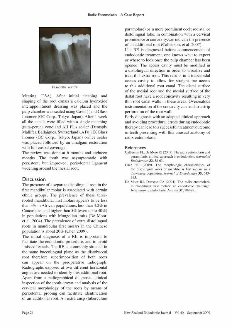
Page 24 New Zealand Endodontic Journal Vol 40 September 2009
Radix Entomolaris – A Case Report
18 months’ review
18 months review
i
Discussion
The presence of a separate distolingual root in the first mandibular molar is associated
with certain ethnic groups. The prevalence of these three-rooted mandibular first molars
appears to be less than 3% in African populations, less than 4.2% in Caucasians, and higher
than 5% (even up to 40%) in populations with Mongolian traits (De Moor, et al. 2004). The
prevalence of extra distolingual roots in mandibular first molars in the Chinese population is
about 20% (Chen 2009).
The initial diagnosis of a RE is important to facilitate the endodontic procedure, and
to avoid ‘missed’ canals. The RE is commonly situated in the same buccolingual plane as the
distobuccal root therefore superimposition of both roots can appear on the preoperative
radiograph. Radiographs exposed at two different horizontal angles are needed to identify this
additional root. Apart from a radiographical diagnosis, clinical inspection of the tooth crown
and analysis of the cervical morphology of the roots by means of periodontal probing can
facilitate identification of an additional root. An extra cusp (tuberculum paramolare) or a
more prominent occlusodistal or distolingual lobe, in combination with a cervical prominence
or convexity, can indicate the presence of an additional root (Calberson, et al. 2007).
If a RE is diagnosed before commencement of endodontic treatment, one knows what
to expect or where to look once the pulp chamber has been opened. The access cavity must be
modified in a distolingual direction in order to visualize and treat this extra root. This results
in a trapezoidal access cavity to allow for straight-line access to this additional root canal.
The distal surface of the mesial root and the mesial surface of the distal root have a root
concavity resulting in very thin root canal walls in these areas. Overzealous instrumentation
of the concavity can lead to a strip perforation of the root wall.
Early diagnosis with an adapted clinical approach and avoiding procedural errors
during endodontic therapy can lead to a successful treatment outcome in teeth presenting with
this unusual anatomy of radix entomolaris.
Meeting, USA). After initial cleaning and shaping of the root canals a calcium hydroxide interappointment dressing was placed and the pulp chamber was sealed using Cavit ( )and Glass Ionomer (GC Corp., Tokyo, Japan). After 1 week all the canals were filled with a single matching gutta-percha cone and AH Plus sealer (Dentsply Mallifer, Ballaigues, Switzerland). A Fuji IX Glass Inomer (GC Corp., Tokyo, Japan) orifice sealer was placed followed by an amalgam restoration with full cuspal coverage.The review was done at 6 months and eighteen months. The tooth was asymptomatic with persistent, but improved, periodontal ligament widening around the mesial root.
DiscussionThe presence of a separate distolingual root in the first mandibular molar is associated with certain ethnic groups. The prevalence of these three-rooted mandibular first molars appears to be less than 3% in African populations, less than 4.2% in Caucasians, and higher than 5% (even up to 40%) in populations with Mongolian traits (De Moor, et al. 2004). The prevalence of extra distolingual roots in mandibular first molars in the Chinese population is about 20% (Chen 2009).The initial diagnosis of a RE is important to facilitate the endodontic procedure, and to avoid ‘missed’ canals. The RE is commonly situated in the same buccolingual plane as the distobuccal root therefore superimposition of both roots can appear on the preoperative radiograph. Radiographs exposed at two different horizontal angles are needed to identify this additional root. Apart from a radiographical diagnosis, clinical inspection of the tooth crown and analysis of the cervical morphology of the roots by means of periodontal probing can facilitate identification of an additional root. An extra cusp (tuberculum
paramolare) or a more prominent occlusodistal or distolingual lobe, in combination with a cervical prominence or convexity, can indicate the presence of an additional root (Calberson, et al. 2007).If a RE is diagnosed before commencement of endodontic treatment, one knows what to expect or where to look once the pulp chamber has been opened. The access cavity must be modified in a distolingual direction in order to visualize and treat this extra root. This results in a trapezoidal access cavity to allow for straight-line access to this additional root canal. The distal surface of the mesial root and the mesial surface of the distal root have a root concavity resulting in very thin root canal walls in these areas. Overzealous instrumentation of the concavity can lead to a strip perforation of the root wall. Early diagnosis with an adapted clinical approach and avoiding procedural errors during endodontic therapy can lead to a successful treatment outcome in teeth presenting with this unusual anatomy of radix entomolaris.
ReferencesCalberson FL, De Moor RJ (2007). The radix entomolaris and
paramolaris: clinical approach in endodontics. Journal of Endodontics 33, 58-63.
Chen YC (2009). The morphologic characteristics of the distolingual roots of mandibular first molars in a Taiwanese population. Journal of Endodontics 35, 643-645.
De Moor RJ, Deroose CA (2004). The radix entomolaris in mandibular first molars: an endodontic challenge. International Endodontic Journal 37, 789-99.

New Zealand Endodontic Journal Vol 40 September 2009 Page 25
Congratulations
News from the School
Congratulations to final year Clinical Doctorate in Endodontics student Artika Patel, who won the prize for Best Student Presenter at the Faculty of Dentistry Research Day held in March. Artika’s presentation was titled ‘Scanning electron microscopy study of root-end cavities following laser irradiation’. Her project was supervised by Tina Hauman, Nick Chandler and Jonathan Leichter. The research was supported by a Dentsply Research Grant and the award was presented by Mr Steve Freeman, the General Manager of 3M Healthcare.
Page 26 New Zealand Endodontic Journal Vol 40 September 2009
New Zealand Society of Endodontics
Minutes of the Annual General Meeting
Held at the Wellington Convention Centre, Wellingtonon Friday, 21 August 2009 – 1.00pm
Present: Sara Jardine (Chair) Deborah Creagh, Ted Bealing, Denis Beale, Richard Ellis, Jaqui Pahl, Mike Jameson
Apologies: Charlie Meade, Hani Naoum, Philip Chong
Minutes: The minutes of the Annual General Meeting held in Rotorua at the NZDA Regional Conference on 12 September 2008 and circulated at the meeting were accepted as a true and correct record with the amendment of recording the apologies received from Sara Jardine and Mike Jameson.
Matters Arising: There has been no progress with the purchase of the Gutmann ‘problem solving’ book.
Correspondence: Nil
President’s Report: To be published in the New Zealand Endodontic Journal.
Treasurer’s Report: To be published in the New Zealand Endodontic Journal. There is a balance of $120,868.32 in the savings account on term deposit. There is a $4,000 balance in the current account.
Subscriptions: The subscription will remain unchanged at $40.00
Election of Officers: It was resolved that the current committee would be re-elected with the exception of the secretary Mike Jameson who had earlier notified his intention to stand down this year. It was suggested that a new secretary be co-opted from the committee.
General Business: Membership: There have been 6 resignations from the Society through retirement and there have been 12 new members.
E-mail database: The NZSE now has an email database of members, thanks to
Deborah Creagh. A new system using the database will be implemented for tracking payment of subscriptions. The database should also help to reduce costs and improve communication with members.
Website: The meeting acknowledged with appreciation the work of Hani Noum on the website. The address is: www.nzse.org.nz
The possibility of the Journal being available online via the website, was discussed.
The meeting closed at 1.30pm.

New Zealand Endodontic Journal Vol 40 September 2009 Page 27

Page 28 New Zealand Endodontic Journal Vol 40 September 2009