endodontic assistance - · pdf fileendodontic assistance ... such clinical signs as...
TRANSCRIPT
CHAPTER 7
ENDODONTIC ASSISTANCE
DENTAL SPECIALTY OFENDODONTICS
Before major advances in the treatment of diseasesof the dental pulp and periapical tissues were made,dentists extracted many teeth needlessly. Endodonticsis the dental specialty primarily concerned with thesediseases. In some dental clinics, an endodontist isassigned exclusively to this specialty. Often, some ofthe restorative dentists spend part of their time seeingpatients who require endodontic treatment, also knownas root canal therapy. As a basic dental assistant, youmust be familiar with the following aspects ofendodontics:
Functions, causes, and diagnosis
Types of procedures
Steps in pulpectomy and root canal treatment
Steps in apicoectomy and associated procedures
You must also be able to identify:
Endodontic instruments
Endodontic materials
Endodontic equipment
When involved with endodontic procedures, youmust follow BUMEDINST 6600.10, Dental InfectionControl Program. Strict compliance to steriletechnique, sterilization, and disinfection is absolutelyessential in endodontic treatment.
FUNCTION
The primary purpose of endodontics is thetreatment of diseases of the pulp and periapical tissues.The goal of this treatment is to retain the natural teethrather than extract them. Often, the endodonticpatient’s initial appointment is of an urgent naturebecause of the associated pain or infection.Understanding the causes of pulp disease and how adiagnosis is reached will increase your ability to be aneffective endodontic assistant.
CAUSES
The dental pulp can be injured in several ways.Some injured teeth can be treated and returned to
normal. Other injured pulpal tissue may undergonecrosis (die) after the slightest injury. Some of themost common causes of injury to the pulp includedental caries (covered in Dental Technician, Volume 1,NAVEDTRA 12572, chapter 5, "Oral Pathology"),traumatic blows to the teeth, pulp exposure (covered inDental Technician, Volume 1, NAVEDTRA 12572,chapter 6, "Emergency Treatment of Oral Diseases andInjuries"), chemical irritation, and thermal irritation.
Traumatic Blows
Traumatic blows to the teeth can result fromsituations such as common household accidents, autocollisions, or athletic injuries. A sharp blow to one ormore teeth can result in fracture of the crown or root, oreven the avulsion (forcefully knocked out of thesocket) of the complete tooth, cutting off the bloodflow to the pulp.
Chemical Irritation
Chemical irritation after placement of certainchemical substances commonly used in restorativeprocedures can cause pulp injury or death. Anothercause of chemical irritation is a faulty restoration,which allows oral fluids to leak between the restorationand dentin.
Thermal Irritation
Thermal irritation can cause pulp injury andpatients will experience discomfort when they inhalecold air through their mouths. If metallic restorativematerials are placed close to the pulp, the patient w-illexperience thermal irritation.
DIAGNOSIS
The diagnosis of pulp and periapical conditionsmust precede the treatment. Endodontic diagnosis is aresult of the skillful use and interpretation of severalmethods. Some of the more common methods arediscussed in the paragraphs that follow.
7-1
Dental History
The patient's dental history is a valuable aid to thedentist. It provides communication between the dentistand the patient, and allows the dentist to trace thehistory of the complaint through symptoms describedby the patient. Often, patients reveal valuableinformation regarding previous injuries to the teeth,even though they may have occurred many yearsearlier.
Clinical Examination
A clinical examination of the oral cavity allows thedentist to visually inspect the patient's mouthproviding clues to the nature of the patient's problems.Such clinical signs as discoloration of the teeth, crownfracture, gross caries, swelling, abnormal soft tissue,and a draining abscess can be identified during aclinical examination.
Radiographs
Radiographs of the teeth and bone are one of themost valuable diagnostic tools the dentist has toevaluate structures that cannot be seen by clinicalexamination, such as the pulp and periapical tissues.The presence of bone loss in the periapical area inresponse to a necrotic pulp can be detected on aradiograph as a dark area surrounding the apex of theroot. The presence of this dark area, or radiolucency,on a dental radiograph is an important feature used todiagnose pulp and periapical disease. Periapicalpathology appears as radiolucencies on a radiograph.
Radiographs can also reveal possible causes ofpulpal injury before bone resorption occurs. Rootfractures, deep caries, and previous pulp exposures aresome examples of possible causes of pupal injurydetected on a radiograph. An accurate radiograph canreveal root length, abnormal root curvature, andabnormal calcification, which is helpful information indetermining if the tooth can be treated endodontically.A radiograph, properly exposed and processed, canlast indefinitely and provide a permanent record of thecondition of the patient and be used for futurereference. Comparison of the initial radiographs withpostoperative radiographs is a valuable index todetermine if the treatment was successful.
Pulp Testers
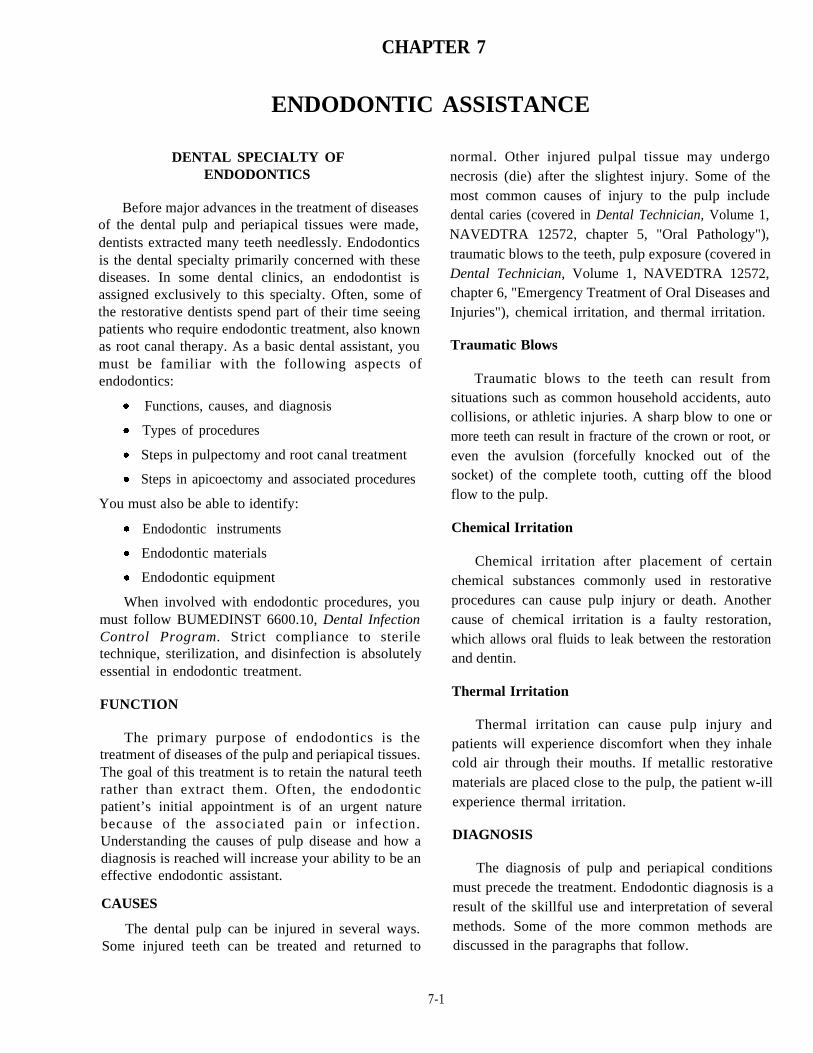
Two of the common pulp testers used primarily todetermine whether the pulp is vital or necrotic
(nonvital) are shown in figure 7-1. Electric current isused to stimulate nerve fibers in the pulp through thedentin layer. General information about the status ofthe pulp is obtained by comparing the response of asuspected tooth with that of a normal tooth (controltooth) of the same type on the opposite side of themouth. The amount of current delivered to a tooth isindicated by a numerical scale. The patient holds theends of the probe to complete the circuit. Highernumbers on the scale indicate that more current isdelivered to the tooth. As the current is increasedgradually, the patient is instructed to let go of the probewhenever a sensation is first detected within the tooth.
Generally, the sensation is described as a slighttingling or warm feeling. The number at which aresponse occurs is recorded and compared with the testresults of the control tooth. A tooth with a necrotic pulpwill not respond to even the most intense electricalstimulation. A dying pulp can produce a variety ofresponses, depending on the state of the pulp at thetime of the test. However, the number readings arerelative and cannot be used to diagnose vital pulp.
Thermal Sensitivity Test
The thermal sensitivity test exposes a tooth toextremes in temperature and provides an accuratemethod of identifying the problem tooth, as well asdetermining the status of its pulp. The two mostcommon diagnostic tests are cold and heat.
COLD TEST.—The cold test can be done easilyby placing a cylinder or stick of ice on the tooth. First,the suspected tooth is isolated and dried, then the icestick, held in a gauze square, is applied to the cervicalarea of the tooth. Healthy teeth will respond positivelyto a cold stimulus, but the sensitivity should resolvequickly. If the pulp is inflamed, the patient will
Figure 7-1.—Two common pulp testers.
7-2
experience a lingering sensation to cold. Other coldtest materials that can be used are ethyl chloride andskin refrigerants.
HEAT TEST.—The heat test consists of isolatingthe suspected tooth with a rubber dam and applying awarm liquid (hot water or coffee) to the tooth. Thewarm liquid should not be hotter than 140°F andshould not burn your skin. If the tooth reacts with apainful response that lingers a few seconds after theheat is removed, pulpitis may be present. If the patientexperiences a violent pain reaction to the heat and isrelieved by a cold application, the pulp is irreversiblyinflamed and will need a root canal. If the patientexperiences no response to heat or cold, the pulp isnecrotic.
Percussion
Percussion is the gentle tapping of the crown of thetooth with the finger or the end of a mirror handle todetermine the presence of periapical inflammation. If apatient has an acute inflammation at the apex of theroot, percussion stimulates the already inflamed areaand pain results. An abnormal dull sound may signify aroot that has attached to the bone. Several normal,opposing, and adjacent teeth should be checked forcomparison.
Palpation
Palpation is the application of the finger with lightpressure to areas of the mouth to detect normal orabnormal tissue. Swelling, pain, and degree of rigidityof tissues are determined by palpation. When usingpalpation in the diagnosis of periapical diseases, thefingers are pressed gently against the soft tissueoverlying the bone and apexes of the teeth to comparethe tissues.
Mobility Test
The mobility test is done by moving the toothbetween the handles of 2 instruments. Abnormalmobility of a tooth when compared to healthy teethsignifies temporary or permanent loss of supportingalveolar bone or trauma. Mobility of the tooth tends toincrease if an infection or injury is long standing andhas affected the supporting periodontium tissues.
Selective Anesthesia
Selective anesthesia can be of assistance if thepatient cannot accurately determine which teeth are the
source of discomfort. If other diagnostic tests havenarrowed the choice down to two teeth, one tooth canbe anesthetized to determine if the pain disappears. Ifthe pain does not disappear until the second tooth isanesthetized, the second tooth is the probable source.Selective anesthesia is most effective when the choiceis between a maxillary and a mandibular tooth.
Transillumination
Transillumination uses fiber optic lighting toallow an intense, concentrated light to pass through thetooth from the lingual to the facial aspect. This is donemost effectively on anterior teeth because of theirstructure and location in the arch. The light transmitsthrough the enamel and dentin, permitting thedetection of caries or a fractured crown.
TYPES OF PROCEDURES
There are several types of endodontic procedures.The more common procedures include pulp capping,pulpotomy, pulpectomy, and root canal therapy.Occasionally other procedures such as incision anddrainage, apicoectomy, periapical curet tage,retrograde filling, root amputation, and bleaching ofteeth are indicated.
PULP CAPPING
In an attempt to protect the pulp against additionalinjury and stimulate pulp regeneration, an applicationof protective dressing, such as calcium hydroxide, isplaced over an exposed or nearly exposed vital pulp.This treatment is referred to as pulp capping. Whenthe pulp is exposed mechanically during toothpreparation, placing a pulp cap directly over theexposed pulp is referred to as a direct pulp cap. If deepcaries are present and a danger of exposing the pulpexists, placing a pulp cap over a layer of remainingdentin is termed an indirect pulp cap. If pulp cappingin not effective, the pulp can be treated withendodontic therapy.
PULPOTOMY AND PULPECTOMY
A pulpotomy is the surgical removal of the coronalpart (pulp chamber) of an exposed vital pulp. The pulpis retained in root canals with the exposed endscovered with applications of calcium hydroxide, zincoxide and eugenol, and zinc phosphate cement topreserve its vitality and function. If indicated, rootcanal treatment is completed at a later date.
7-3
The most common endodontic procedure is thepulpectomy, which is the removal of the entire pulp(chamber and canal). After removal of the pulp, rootcanal therapy is performed.
ROOT CANAL THERAPY
This t r ea tmen t cons i s t s o f the in t e rna ldebridement, cleaning, shaping, and permanent fillingof the root canal system. During the therapy, the dentistmay place medications and temporary filling material.The therapy may vary slightly because of the type oftooth and number of canals in the tooth.
INCISION AND DRAINAGE
An acute periapical abscess may indicate a needfor incision and drainage to eliminate the infectionalong with endodontic treatment. Incision anddrainage can be effective when the swelling andinfection are localized in the alveolus with a clearlydefined point on the surface of the mucosa. Endodontictreatment Should be initiated at the same appointmentto remove the necrotic infected pulp. Although theperiapical abscess usually is accompanied by severepain, it is not advisable to inject a local anestheticsolution directly into the infected area when drainingthe abscess because of the danger of spreading theinfection. Instead, block anesthesia and infiltrationaway from the infected area. Local anesthesia may notbe as effective because of changes in the pH of thetissues in the presence of the infection. The patientmust be informed to expect momentary discomfortwhen the area is lanced, but the pain is immediatelyand significantly reduced after the incision is made andthe exudate (pus) is expressed. If indicated, a drain isplaced to provide short term drainage and to preventthe opening from closing prematurely until theinfected area drains. The dentist may prescribeantibiotics. Once the infection is controlled and theswelling and tenderness subside, the dentist will treatthe tooth endodontically.
APICOECTOMY AND PERIAPICALCURETTAGE
An apicoectomy (root end resection) is the surgicalremoval of the apical portion of the tooth through asurgical opening made in the overlying bone andgingival tissues. An apicoectomy usually is performedin conjunction with periapical curettage after the bodyfails to heal after endodontic treatment. Periapicalcurettage is the surgical removal of apically inflamed
tissue associated with the tooth through an openingmade in the overlying bone and gingival tissues.Treatment is limited to curettage of the area to removeall diseased material. Conditions that may indicate theneed for an apicoectomy include:
Persistent, local infection following endodontictreatment.
Canal filling materials or medications extrudedinto the periapical tissue.
A broken instrument lodged in the canalpreventing complete filling.
Obstruction caused by a calcified root canal.
Extreme curvature of the canal preventingaccess to the apex of the root.
Root canals that are unfilled or debrided.
RETROGRADE FILLING
This is a method of sealing the apical end of theroot canal by placing a restoration in the root apex.This is usually done in conjunction with theapicoectomy. Superortho-ethoxybenzoic acid (EBA)cement or an intermediate restorative material such asZinc Oxide and Eugenol (ZOE) is used as the fillingmaterial because they will not react with any moisturethat may be present in the root canal. Some dentistsprefer to use composite filling material.
ROOT AMPUTATIONS
Occasionally, a multirooted tooth requiringendodontic treatment may have a root that isimpossible to obtain an adequate apical seal or isaffected by periodontal disease. When the other rootsof the teeth are treatable, rather than extracting theentire tooth, the untreatable root is amputated andremoved. The opening to which the amputated rootwas attached is sealed with amalgam similar to that ofan apicoectomy procedure. The retained section of thetooth is treated endodontically before amputation.
BLEACHING OF DISCOLORED TEETH
The use of chemical agents may be used to removediscoloration from the crowns of vital or nonvitalteeth. Nonvital teeth may discolor because of pupalhemorrhage into the dentinal tubules after traumaticinjury of the tooth, or from the use of medications thatcause staining when used in endodontic therapy. Insuch cases, the appearance of the discolored teeth maybe improved dramatically by bleaching the tooth.
7-4

STEPS IN PULPECTOMY AND ROOTCANAL TREATMENT
As in all efficient assisting, you will need toanticipate the dentist's needs. In endodontics, your dutiesconsist of such tasks as performing infection controlprocedures, preparing for the treatment, aiding in theplacement of the rubber clam, irrigating and aspirating toflush the area, mixing materials, and passing instruments.You will need to have knowledge of the treatmentprocedure and sequence to effectively anticipate thedentist's needs and to schedule appointments.
APPOINTMENT SCHEDULING
Root canal therapy may take one or moreappointments based on the number of canals andseverity of infection. Before a canal can be filled, thecanals must be completely cleaned. Filling the canalwhile infective organisms are still present may result innon-healing. A patient suffering from an acuteperiapical abscess may experience severe pain. Thepain is due to inflammation in the pulp canal, and/orperiapical tissues. The pressure, and therefore the pain,is relieved during the first step of endodontics when thepulp canal is opened. Once the pulp canal is opened,broaches can be used to remove intact pulp tissue fromthe canal. The canal is then irrigated, and debrided withfiles and reamers. Dry the canal and place smallmedicated cotton pellets into the pulp chamber to helpclear up the infection. Then, the dentist may place atemporary restoration.
During a second appointment, if necessary, thetemporary restoration is removed, the canals irrigated,and root canal reamers and files are used to enlarge,shape, and smooth the pulp canal. If infectioncontinues to be a problem, placement of medicationinto the canal, and placing a temporary restoration willbe required. Schedule the patient for anotherappointment. When all instrumentation is completeand infection is eliminated, gutta-percha is placed intothe canals with a sealer that acts as a cement. Then atemporary restoration can be placed.
After root canal treatment is completed, apermanent restoration is placed, usually at a laterappointment. At this time, the tooth may be evaluatedfor possible prosthodoiltics treatment to replace therestoration with an artificial crown.
During all appointments, use a rubber dam toisolate the tooth, prevent contamination of the rootcanal, and prevent the small endodontic instrumentsfrom going down the patient's throat.
ENDODONTIC MATERIALS
The main materials used in root canal therapy arevarious liquid antiseptics, paste, paper points,gutta-percha points, and sealers. The dentist uses theseto treat and fill a properly prepared root canal fromwhich the pulp has been removed.
Paper Points
Paper points are primarily used during thetreatment phase of endodontics to dry out root canals.They are highly absorbent, rolled sterile paper that arelong and narrow with a tapered point to fit into the rootcanal. Paper points are available in assorted sizes, fromcoarse to X-fine, depending on the size of the canal intowhich they are being inserted.
Root Canal Restorative Materials
Root canal restorative materials are used to fill thepreviously prepared root canals and complete the rootcanal or endodontic therapy. Root canal restorativematerials consist of tapered gutta-percha points in avariety of sizes and root canal sealers or cements. Agood root canal restorative material should beinsoluble in tissue fluids, opaque to the passage ofX-rays, easy to remove, nonirritating to periapicaltissues, nonabsorbent, and dimensionally stable afterits insertion into a root canal.
GUTTA-PERCHA. —Gutta-percha is used as atemporary restoration and as a root canal restorativematerial. Gutta-percha is the refined, coagulated,milky exudate of certain trees. It is pink or gray incolor, softens when heated, and is easily molded.When it is cool, it maintains its shape well.Gutta-percha points have been a choice for root canalrestorative materials for many years. The manyadvantages of the material are as follows:
High thermal expansion
Will not shrink unless used with a solvent
Radiopaque
Can be kept sterile in an antiseptic solution
Resistant to moisture and bacteriostatic
A poor heat conductor
The disadvantages of gutta-percha are as follows:
Shrinks when used with a solvent
Is not always easily inserted into the root canal
7-5
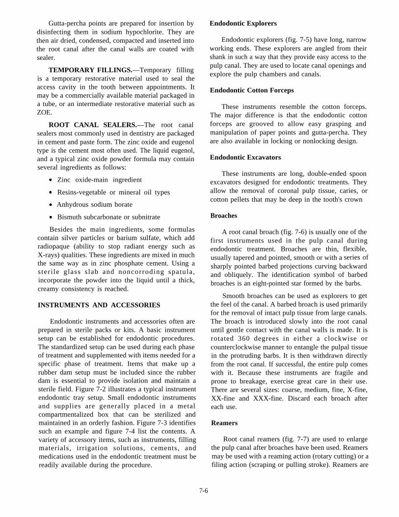
Gutta-percha points are prepared for insertion by Endodontic Explorersdisinfecting them in sodium hypochlorite. They arethen air dried, condensed, compacted and inserted intothe root canal after the canal walls are coated withsealer.
TEMPORARY FILLINGS.—Temporary fillingis a temporary restorative material used to seal theaccess cavity in the tooth between appointments. Itmay be a commercially available material packaged ina tube, or an intermediate restorative material such asZOE.
ROOT CANAL SEALERS.—The root canalsealers most commonly used in dentistry are packagedin cement and paste form. The zinc oxide and eugenoltype is the cement most often used. The liquid eugenol,and a typical zinc oxide powder formula may containseveral ingredients as follows:
Zinc oxide-main ingredient
Resins-vegetable or mineral oil types
Anhydrous sodium borate
Bismuth subcarbonate or subnitrate
Besides the main ingredients, some formulascontain silver particles or barium sulfate, which addradiopaque (ability to stop radiant energy such asX-rays) qualities. These ingredients are mixed in muchthe same way as in zinc phosphate cement. Using aster i le glass s lab and noncorroding spatula,incorporate the powder into the liquid until a thick,creamy consistency is reached.
INSTRUMENTS AND ACCESSORIES
Endodontic instruments and accessories often areprepared in sterile packs or kits. A basic instrumentsetup can be established for endodontic procedures.The standardized setup can be used during each phaseof treatment and supplemented with items needed for aspecific phase of treatment. Items that make up arubber dam setup must be included since the rubberdam is essential to provide isolation and maintain asterile field. Figure 7-2 illustrates a typical instrumentendodontic tray setup. Small endodontic instrumentsand supplies are generally placed in a metalcompartmentalized box that can be sterilized andmaintained in an orderly fashion. Figure 7-3 identifiessuch an example and figure 7-4 list the contents. Avariety of accessory items, such as instruments, fillingmaterials, irrigation solutions, cements, andmedications used in the endodontic treatment must bereadily available during the procedure.
Endodontic explorers (fig. 7-5) have long, narrowworking ends. These explorers are angled from theirshank in such a way that they provide easy access to thepulp canal. They are used to locate canal openings andexplore the pulp chambers and canals.
Endodontic Cotton Forceps
These instruments resemble the cotton forceps.The major difference is that the endodontic cottonforceps are grooved to allow easy grasping andmanipulation of paper points and gutta-percha. Theyare also available in locking or nonlocking design.
Endodontic Excavators
These instruments are long, double-ended spoonexcavators designed for endodontic treatments. Theyallow the removal of coronal pulp tissue, caries, orcotton pellets that may be deep in the tooth's crown
Broaches
A root canal broach (fig. 7-6) is usually one of thefirst instruments used in the pulp canal duringendodontic treatment. Broaches are thin, flexible,usually tapered and pointed, smooth or with a series ofsharply pointed barbed projections curving backwardand obliquely. The identification symbol of barbedbroaches is an eight-pointed star formed by the barbs.
Smooth broaches can be used as explorers to getthe feel of the canal. A barbed broach is used primarilyfor the removal of intact pulp tissue from large canals.The broach is introduced slowly into the root canaluntil gentle contact with the canal walls is made. It isrotated 360 degrees in either a clockwise orcounterclockwise manner to entangle the pulpal tissuein the protruding barbs. It is then withdrawn directlyfrom the root canal. If successful, the entire pulp comeswith it. Because these instruments are fragile andprone to breakage, exercise great care in their use.There are several sizes: coarse, medium, fine, X-fine,XX-fine and XXX-fine. Discard each broach aftereach use.
Reamers
Root canal reamers (fig. 7-7) are used to enlargethe pulp canal after broaches have been used. Reamersmay be used with a reaming action (rotary cutting) or afiling action (scraping or pulling stroke). Reamers are
7-6
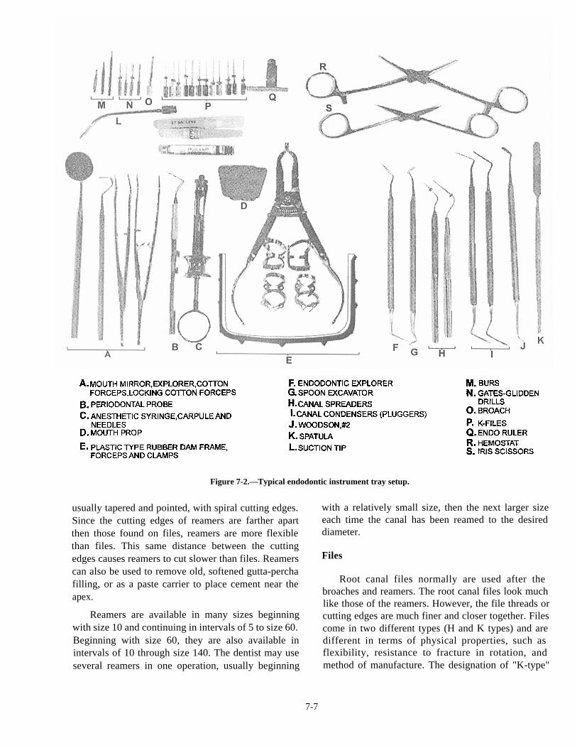
Figure 7-2.—Typical endodontic instrument tray setup.
usually tapered and pointed, with spiral cutting edges.Since the cutting edges of reamers are farther apartthen those found on files, reamers are more flexiblethan files. This same distance between the cuttingedges causes reamers to cut slower than files. Reamerscan also be used to remove old, softened gutta-perchafilling, or as a paste carrier to place cement near theapex.
Reamers are available in many sizes beginningwith size 10 and continuing in intervals of 5 to size 60.Beginning with size 60, they are also available inintervals of 10 through size 140. The dentist may useseveral reamers in one operation, usually beginning
with a relatively small size, then the next larger sizeeach time the canal has been reamed to the desireddiameter.
Files
Root canal files normally are used after thebroaches and reamers. The root canal files look muchlike those of the reamers. However, the file threads orcutting edges are much finer and closer together. Filescome in two different types (H and K types) and aredifferent in terms of physical properties, such asflexibility, resistance to fracture in rotation, andmethod of manufacture. The designation of "K-type"
7-7
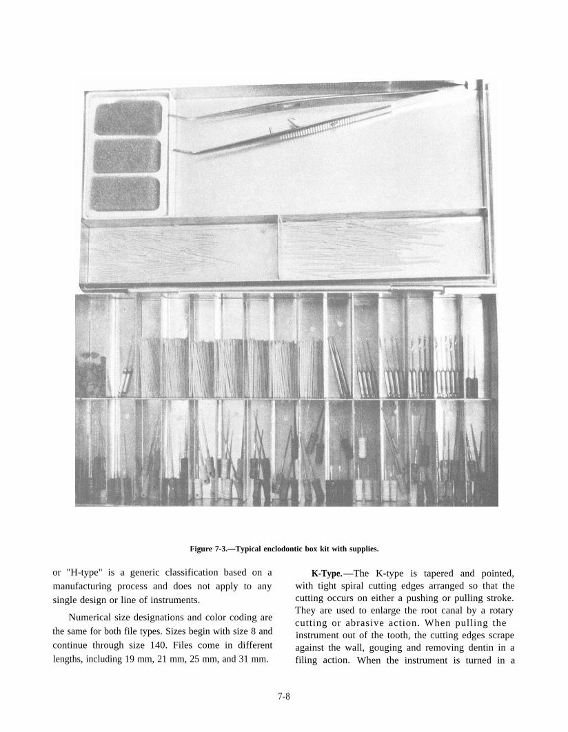
Figure 7-3.—Typical enclodontic box kit with supplies.
or "H-type" is a generic classification based on a
manufacturing process and does not apply to any
single design or line of instruments.
Numerical size designations and color coding are
the same for both file types. Sizes begin with size 8 and
continue through size 140. Files come in different
lengths, including 19 mm, 21 mm, 25 mm, and 31 mm.
K-Type.—The K-type is tapered and pointed,with tight spiral cutting edges arranged so that thecutting occurs on either a pushing or pulling stroke.They are used to enlarge the root canal by a rotarycutting or abrasive action. When pulling theinstrument out of the tooth, the cutting edges scrapeagainst the wall, gouging and removing dentin in afiling action. When the instrument is turned in a
7-8
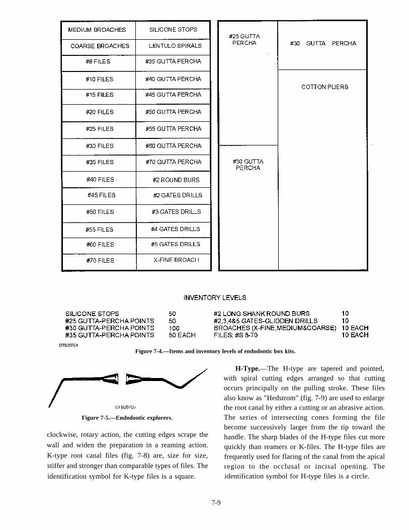
Figure 7-4.—Items and inventory levels of endodontic box kits.
Figure 7-5.—Endodontic explorers.
clockwise, rotary action, the cutting edges scrape the
wall and widen the preparation in a reaming action.
K-type root canal files (fig. 7-8) are, size for size,
stiffer and stronger than comparable types of files. The
identification symbol for K-type files is a square.
H-Type.—The H-type are tapered and pointed,with spiral cutting edges arranged so that cuttingoccurs principally on the pulling stroke. These filesalso know as "Hedstrom" (fig. 7-9) are used to enlargethe root canal by either a cutting or an abrasive action.The series of intersecting cones forming the filebecome successively larger from the tip toward thehandle. The sharp blades of the H-type files cut morequickly than reamers or K-files. The H-type files arefrequently used for flaring of the canal from the apicalregion to the occlusal or incisal opening. Theidentification symbol for H-type files is a circle.
7-9
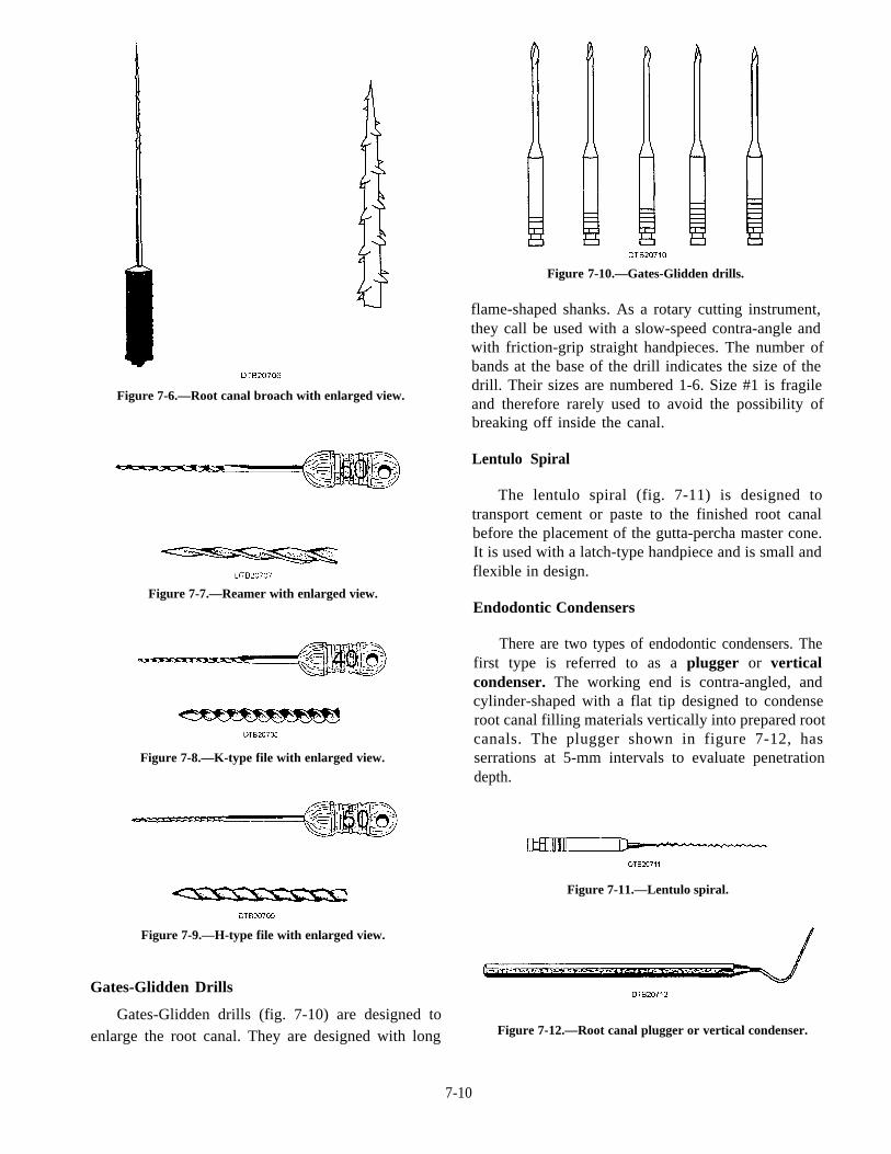
Figure 7-6.—Root canal broach with enlarged view.
Figure 7-7.—Reamer with enlarged view.
Figure 7-8.—K-type file with enlarged view.
Figure 7-10.—Gates-Glidden drills.
flame-shaped shanks. As a rotary cutting instrument,they call be used with a slow-speed contra-angle andwith friction-grip straight handpieces. The number ofbands at the base of the drill indicates the size of thedrill. Their sizes are numbered 1-6. Size #1 is fragileand therefore rarely used to avoid the possibility ofbreaking off inside the canal.
Lentulo Spiral
The lentulo spiral (fig. 7-11) is designed totransport cement or paste to the finished root canalbefore the placement of the gutta-percha master cone.It is used with a latch-type handpiece and is small andflexible in design.
Endodontic Condensers
There are two types of endodontic condensers. Thefirst type is referred to as a plugger or verticalcondenser. The working end is contra-angled, andcylinder-shaped with a flat tip designed to condenseroot canal filling materials vertically into prepared rootcanals. The plugger shown in figure 7-12, hasserrations at 5-mm intervals to evaluate penetrationdepth.
Figure 7-9.—H-type file with enlarged view.
Gates-Glidden Drills
Gates-Glidden drills (fig. 7-10) are designed toenlarge the root canal. They are designed with long
Figure 7-11.—Lentulo spiral.
Figure 7-12.—Root canal plugger or vertical condenser.
7-10
The second type of endodontic condenser is calleda spreader. The root canal spreader (fig. 7-13) has acontra-angled working end that tapers to a point(compared to the flat tip of a plugger). This instrumentis single ended. Spreaders are designed to condenseroot canal filling materials horizontally against thewall of the prepared root canal.
plugger. The finger spreader has a pointed end, thefinger plugger has a flat end.
Finger spreaders and finger pluggers have a handlelike a file and a smooth working end like a spreader or
Endodontic Measuring Gauges
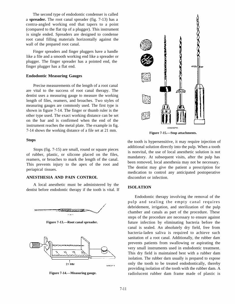
are vital to the success of root canal therapy. Thedentist uses a measuring gauge to measure the workinglength of files, reamers, and broaches. Two styles ofmeasuring gauges are commonly used. The first type isshown in figure 7-14. The finger or thumb ruler is theother type used. The exact working distance can be seton the bar and is confirmed when the end of theinstrument reaches the metal plate. The example in fig.7-14 shows the working distance of a file set at 21 mm.
Precise measurements of the length of a root canal
Stops
Stops (fig. 7-15) are small, round or square piecesof rubber, plastic, or silicone placed on the files,reamers, or broaches to mark the length of the canal.This prevents injury to the apex of the root andperiapical tissues.
ANESTHESIA AND PAIN CONTROL
A local anesthetic must be administered by thedentist before endodontic therapy if the tooth is vital. If
Figure 7-13.—Root canal spreader.
Figure 7-14.—Measuring gauge.
Figure 7-15.—Stop attachments.
the tooth is hypersensitive, it may require injection ofadditional solution directly into the pulp. When a toothis nonvital, the use of local anesthetic solution is notmandatory. At subsequent visits, after the pulp hasbeen removed, local anesthesia may not be necessary.The dentist may give the patient a prescription formedication to control any anticipated postoperativediscomfort or infection.
ISOLATION
Endodontic therapy involving the removal of thepu lp and sea l ing the empty cana l r equ i re sdebridement, irrigation, and sterilization of the pulpchamber and canals as part of the procedure. Thesesteps of the procedure are necessary to ensure againstfuture infection by eliminating bacteria before thecanal is sealed. An absolutely dry field, free frombacteria-laden saliva is required to achieve suchsanitation of a root canal. Additionally, the rubber damprevents patients from swallowing or aspirating thevery small instruments used in endodontic treatment.This dry field is maintained best with a rubber damisolation. The rubber darn usually is prepared to exposeonly the tooth to be treated endodontically, therebyproviding isolation of the tooth with the rubber dam. Aradiolucent rubber dam frame made of plastic is
7-11
commonly used and saves valuable time whenexposing radiographs. Metal rubber dam frames mustbe taken off while exposing radiographs. Remember arisk of contamination can occur while the frame is off.
OPENING THE PULP CHAMBER ANDCANALS
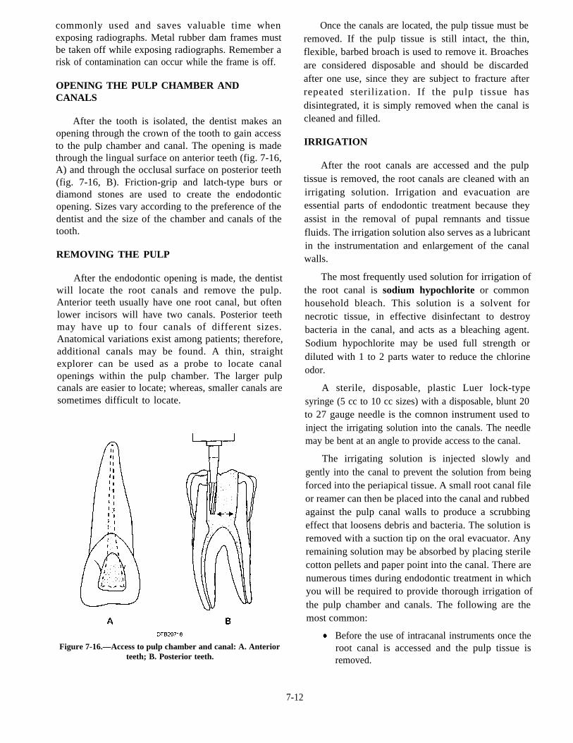
After the tooth is isolated, the dentist makes anopening through the crown of the tooth to gain accessto the pulp chamber and canal. The opening is madethrough the lingual surface on anterior teeth (fig. 7-16,A) and through the occlusal surface on posterior teeth(fig. 7-16, B). Friction-grip and latch-type burs ordiamond stones are used to create the endodonticopening. Sizes vary according to the preference of thedentist and the size of the chamber and canals of thetooth.
REMOVING THE PULP
After the endodontic opening is made, the dentistwill locate the root canals and remove the pulp.Anterior teeth usually have one root canal, but oftenlower incisors will have two canals. Posterior teethmay have up to four canals of different sizes.Anatomical variations exist among patients; therefore,additional canals may be found. A thin, straightexplorer can be used as a probe to locate canalopenings within the pulp chamber. The larger pulpcanals are easier to locate; whereas, smaller canals aresometimes difficult to locate.
Figure 7-16.—Access to pulp chamber and canal: A. Anteriorteeth; B. Posterior teeth.
Once the canals are located, the pulp tissue must beremoved. If the pulp tissue is still intact, the thin,flexible, barbed broach is used to remove it. Broachesare considered disposable and should be discardedafter one use, since they are subject to fracture afterrepeated sterilization. If the pulp tissue hasdisintegrated, it is simply removed when the canal iscleaned and filled.
IRRIGATION
After the root canals are accessed and the pulptissue is removed, the root canals are cleaned with anirrigating solution. Irrigation and evacuation areessential parts of endodontic treatment because theyassist in the removal of pupal remnants and tissuefluids. The irrigation solution also serves as a lubricantin the instrumentation and enlargement of the canalwalls.
The most frequently used solution for irrigation ofthe root canal is sodium hypochlorite or commonhousehold bleach. This solution is a solvent fornecrotic tissue, in effective disinfectant to destroybacteria in the canal, and acts as a bleaching agent.Sodium hypochlorite may be used full strength ordiluted with 1 to 2 parts water to reduce the chlorineodor.
A sterile, disposable, plastic Luer lock-typesyringe (5 cc to 10 cc sizes) with a disposable, blunt 20to 27 gauge needle is the comnon instrument used toinject the irrigating solution into the canals. The needlemay be bent at an angle to provide access to the canal.
The irrigating solution is injected slowly andgently into the canal to prevent the solution from beingforced into the periapical tissue. A small root canal fileor reamer can then be placed into the canal and rubbedagainst the pulp canal walls to produce a scrubbingeffect that loosens debris and bacteria. The solution isremoved with a suction tip on the oral evacuator. Anyremaining solution may be absorbed by placing sterilecotton pellets and paper point into the canal. There arenumerous times during endodontic treatment in whichyou will be required to provide thorough irrigation ofthe pulp chamber and canals. The following are themost common:
Before the use of intracanal instruments once theroot canal is accessed and the pulp tissue isremoved.
7-12
Before the instrumentation of a previouslyopened pulp cavity to remove food particles andsaliva.
At intervals during instrumentation, often aftereach size file is used.
At the completion of canal instrumentation,before placement of medication.
When using root canal preparation type.
CANAL CLEANSING AND SHAPING
Canal cleansing and shaping is the progressiveelimination of organic and inorganic debris within theroot canal by mechanical instruments. As part of thecleansing process, the canals are enlarged and shapedwith endodontic files and reamers. Filing shapes thewalls of the root canal so that they are smooth withspecific size and shape.
The filing procedure begins by first establishingthe approximate or estimated length of the root canal.Accurately determining the length of the tooth is vitalto successful endodontic treatment. Failure todetermine an accurate length may lead to apicalperforation and overextension, with increasedpostoperative pain or incomplete instrumentation andunderfilling.
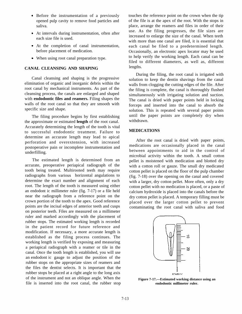
The estimated length is determined from anaccurate, preoperative periapical radiograph of thetooth being treated. Multirooted teeth may requireradiographs from various horizontal angulations todetermine the exact number and alignment of eachroot. The length of the tooth is measured using eitheran endodont ic millimeter ruler (fig. 7-17) or a file heldnear the radiograph from a reference point on thecrown portion of the tooth to the apex. Good referencepoints are the incisal edges of anterior teeth and cuspson posterior teeth. Files are measured on a millimeterruler and marked accordingly with the placement ofrubber stops. The estimated working length is recordedin the patient record for future reference andmodification. If necessary, a more accurate length isestablished as the filing process continues. Theworking length is verified by exposing and measuringa periapical radiograph with a reamer or tile in thecanal. Once the tooth length is established, you will usean endodont ic gauge to adjust the position of therubber stops on the appropriate sizes of reamers andthe files the dentist selects. It is important that therubber stops be placed at a right angle to the long axisof the instrument and not an oblique angle. When thefile is inserted into the root canal, the rubber stop
touches the reference point on the crown when the tipof the file is at the apex of the root. With the stops in
lengths.
place, arrange the reamers and files in order of theiruse. As the filing progresses, the file sizes areincreased to enlarge the size of the canal. When teethwith more than one canal are filed, it is essential thateach canal be filed to a predetermined length.Occasionally, an electronic apex locator may be usedto help verify the working length. Each canal can befiled to different diameters, as well as, different
During the filing, the root canal is irrigated withsolution to keep the dentin shavings from the canalwalls from clogging the cutting edges of the file. Afterthe filing is complete, the canal is thoroughly flushedsimultaneously with irrigating solution and suction.The canal is dried with paper points held in lockingforceps and inserted into the canal to absorb thesolution. This is repeated with several paper pointsuntil the paper points are completely dry whenwithdrawn.
MEDICATIONS
After the root canal is dried with paper points,medications are occasionally placed in the canalbetween appointments to aid in the control ofmicrobial activity within the tooth. A small cottonpellet is moistened with medication and blotted drywith a cotton roll or gauze. The small dry medicatedcotton pellet is placed on the floor of the pulp chamber(fig. 7-18) over the opening on the canal and coveredwith a larger, dry cotton pellet. More often, only a drycotton pellet with no medication is placed, or a paste ofcalcium hydroxide is placed into the canals before thedry cotton pellet is placed. A temporary filling must beplaced over the larger cotton pellet to preventcontaminating the root canal with saliva and food
Figure 7-17.—Estimated working distance using anendodontic millimeter ruler.
7-13
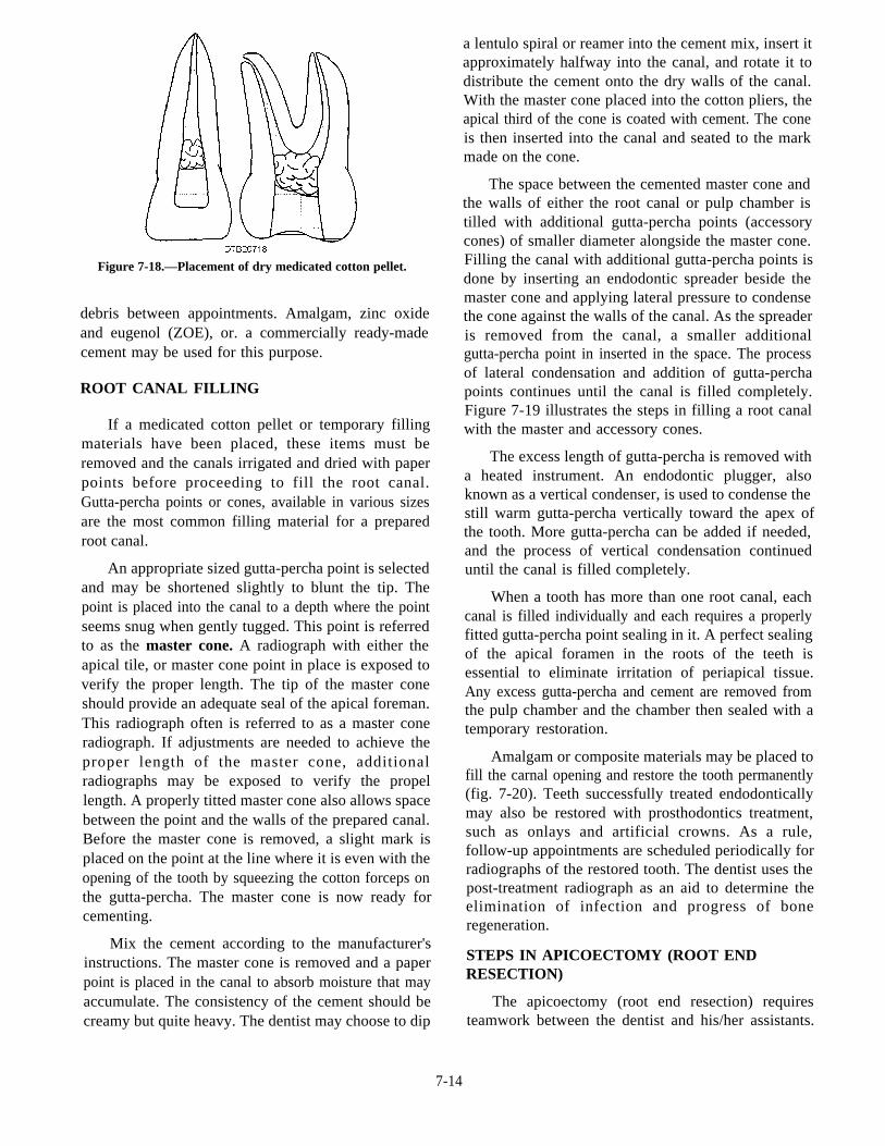
Figure 7-18.—Placement of dry medicated cotton pellet.
debris between appointments. Amalgam, zinc oxideand eugenol (ZOE), or. a commercially ready-madecement may be used for this purpose.
ROOT CANAL FILLING
If a medicated cotton pellet or temporary fillingmaterials have been placed, these items must beremoved and the canals irrigated and dried with paperpoints before proceeding to fill the root canal.Gutta-percha points or cones, available in various sizesare the most common filling material for a preparedroot canal.
An appropriate sized gutta-percha point is selectedand may be shortened slightly to blunt the tip. Thepoint is placed into the canal to a depth where the pointseems snug when gently tugged. This point is referredto as the master cone. A radiograph with either theapical tile, or master cone point in place is exposed toverify the proper length. The tip of the master coneshould provide an adequate seal of the apical foreman.This radiograph often is referred to as a master coneradiograph. If adjustments are needed to achieve theproper length of the master cone, additionalradiographs may be exposed to verify the propellength. A properly titted master cone also allows spacebetween the point and the walls of the prepared canal.Before the master cone is removed, a slight mark isplaced on the point at the line where it is even with theopening of the tooth by squeezing the cotton forceps onthe gutta-percha. The master cone is now ready forcementing.
Mix the cement according to the manufacturer'sinstructions. The master cone is removed and a paperpoint is placed in the canal to absorb moisture that mayaccumulate. The consistency of the cement should becreamy but quite heavy. The dentist may choose to dip
a lentulo spiral or reamer into the cement mix, insert itapproximately halfway into the canal, and rotate it todistribute the cement onto the dry walls of the canal.With the master cone placed into the cotton pliers, theapical third of the cone is coated with cement. The coneis then inserted into the canal and seated to the markmade on the cone.
The space between the cemented master cone andthe walls of either the root canal or pulp chamber istilled with additional gutta-percha points (accessorycones) of smaller diameter alongside the master cone.Filling the canal with additional gutta-percha points isdone by inserting an endodontic spreader beside themaster cone and applying lateral pressure to condensethe cone against the walls of the canal. As the spreaderis removed from the canal, a smaller additionalgutta-percha point in inserted in the space. The processof lateral condensation and addition of gutta-perchapoints continues until the canal is filled completely.Figure 7-19 illustrates the steps in filling a root canalwith the master and accessory cones.
The excess length of gutta-percha is removed witha heated instrument. An endodontic plugger, alsoknown as a vertical condenser, is used to condense thestill warm gutta-percha vertically toward the apex ofthe tooth. More gutta-percha can be added if needed,and the process of vertical condensation continueduntil the canal is filled completely.
When a tooth has more than one root canal, eachcanal is filled individually and each requires a properlyfitted gutta-percha point sealing in it. A perfect sealingof the apical foramen in the roots of the teeth isessential to eliminate irritation of periapical tissue.Any excess gutta-percha and cement are removed fromthe pulp chamber and the chamber then sealed with atemporary restoration.
Amalgam or composite materials may be placed tofill the carnal opening and restore the tooth permanently(fig. 7-20). Teeth successfully treated endodonticallymay also be restored with prosthodontics treatment,such as onlays and artificial crowns. As a rule,follow-up appointments are scheduled periodically forradiographs of the restored tooth. The dentist uses thepost-treatment radiograph as an aid to determine theelimination of infection and progress of boneregeneration.
STEPS IN APICOECTOMY (ROOT ENDRESECTION)
The apicoectomy (root end resection) requiresteamwork between the dentist and his/her assistants.
7-14
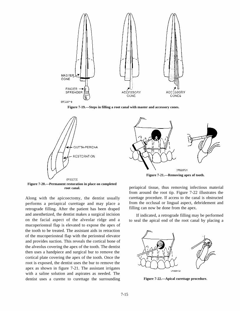
Figure 7-19.—Steps in filling a root canal with master and accessory cones.
Figure 7-20.—Permanent restoration in place on completedroot canal.
Along with the apicoectomy, the dentist usuallyperforms a periapical curettage and may place aretrograde filling. After the patient has been drapedand anesthetized, the dentist makes a surgical incisionon the facial aspect of the alveolar ridge and amucoperiosteal flap is elevated to expose the apex ofthe tooth to be treated. The assistant aids in retractionof the mucoperiosteal flap with the periosteal elevatorand provides suction. This reveals the cortical bone ofthe alveolus covering the apex of the tooth. The dentistthen uses a handpiece and surgical bur to remove thecortical plate covering the apex of the tooth. Once theroot is exposed, the dentist uses the bur to remove theapex as shown in figure 7-21. The assistant irrigateswith a saline solution and aspirates as needed. Thedentist uses a curette to curettage the surrounding
Figure 7-21.—Removing apex of tooth.
periapical tissue, thus removing infectious materialfrom around the root tip. Figure 7-22 illustrates thecurettage procedure. If access to the canal is obstructedfrom the occlusal or lingual aspect, debridement andfilling can now be done from the apex.
If indicated, a retrograde filling may be performedto seal the apical end of the root canal by placing a
Figure 7-22.—Apical curettage procedure.
7-15
restoration in the root apex. The dentist may use an hemostatic agents are carefully removed to avoidultrasonic or a high-speed handpiece with a bur to dropping scraps or cement into the incision. The site
prepare the blunted apex for the filling. When the may be irrigated and aspirated again.
preparation is complete, the surgical site is irrigatedcarefully with saline solution and aspirated until it is
A radiograph is exposed to determine the absence
dried thoroughly. Hemostatic agents such as bovineof any filling particles in the tissue at the surgical site.
collagen or ferric sulfate may be placed to controlWhen it is determined that the filling is satisfactory andthat all particles of the filling material are removed,
bleeding and to catch scraps during the placement and sutures are placed to close the incision. The surgicalcondensation of the root end filling material. portion of the apicoectomy is done quickly. The longerIntermediate restorative material such as ZOE, or the patient is subjected to a surgical procedure, thesuper EBA cement (ethoxybenzoic acid) is mixed and more likely it is that there will be swelling andplaced into the recessed preparation of the root apex. discomfort. Follow-up appointments usually areThe retrograde filling is condensed and smoothed even scheduled periodically for radiographs of the restoredwith the tip of the amputated root surface. The tooth.
7-16