endocrine system. nervous vs. endocrine systems nervous controlling system releases chemical...
TRANSCRIPT

ENDOCRINE SYSTEM

Nervous Vs. Endocrine Systems• NERVOUS
• controlling system• releases chemical
messengers-neurotransmitters
• regulated by negative feedback
• goal-Homeostasis• effects appear fast
– can produce effects in milliseconds
• effects-short duration– useful for crisis
control
• ENDOCRINE
• controlling system• releases chemical
messengers-hormones • regulated by negative
feedback• goal-Homeostasis• effects appear slow
– require hours, months or years
• effects-long lasting– regulates long
acting changes such as metabolic activity

Endocrine Communication• changes cellular operations by changing
• types of enzymes or proteins
• quantities of enzymes or proteins
• activities of enzymes or structural proteins
• accomplishes with hormones
• substances released from one tissue and transported via blood stream to have effects on distant tissues

Endocrine System• composed of endocrine glands
– small, ductless glands• widely scattered throughout
body• release secretions-hormones into
circulatory system• hormones make contact with all
cell types• influence only target cells
– contain specific protein receptors on membranes or inside cell to which hormones can bind
• some endocrine glands are discrete organs– pituitary & thyroid
• others contain discrete areas of endocrine tissue within their structure– islet of Langerhans in
pancreas

Chemical Classes of Hormones
• Lipid Soluble– Steroids– Thyroid Hormone– Nitric Oxide
• Water Soluble– Amines– Peptides– Proteins– Eicosanoid

Steroid Hormones• derived from
cholesterol• Estrogen• Progesterone• Testosterone• Adrenocortical
hormones• transported bound to
blood transport proteins– keeps them in
circulation longer

Thyroid Hormone• Made by adding
Iodine to the amino acid tyrosine
• Soluble in lipid due to the ring structure

Peptide/Protein Hormones• Peptide
–chains of 3 to 49 amino acids
• Protein
–chains of 50 -200 amino acids

Amines• made from
amino acids
• retain an amino group
• epinephrine
• norepinephrineEpinephrine

Eicosanoids• derived from
fatty acid• Arachidonic acid• Prostaglandins• Leukotrienes• Local hormones

Mechanisms of Action • Hormones change cellular operations• by stimulating synthesis of enzymes or structural proteins• by activating genes• by changing rate of transcription or translation• by turning existing enzymes on or off• come into contact with all types of body cells• only have effects at particular target cells• target cells have receptors for particular hormones• found on cell membrane or inside cell

Lipid Soluble Hormones- Direct Gene Activation
• soluble in lipid• can cross cell
membrane• bind to receptors on
inside of cell• in cytoplasm or
nucleus• forming hormone-
receptor complex

Direct Gene Activation Steps• 1-hormone diffuses from
blood to cell• 2-hormone enters cell• 3-hormone binds to
receptor forming receptor-hormone complex
• 4-alters gene expression-mRNA is made
• 5-mRNA leaves nucleus-directs protein synthesis
• 6-new protein alters metabolic activities of other protein

Water Soluble Hormones-Second Messenger Mechanism
• cannot enter target cells– must stimulate target cells indirectly
• second-messenger system– receptors embedded in cell membrane
• Hormone-first messenger– responsible for appearance of second
messenger-produces effects of hormone• Second messengers include
– cAMP– cGMP– Ca++
• second messenger activates or inhibits cofactors which changes rate of metabolic reactions

Second Messenger Mechanism• hormone binds to plasma
membrane receptor• activates G protein-
regulatory molecule• activated G protein
activates adenlyate cyclase
• Activatged adenylate cyclase converts ATP to cAMP (cyclic AMP)
• cAMP is the second messenger
• activates protein kinase• catalyzes reactions• degraded by
phosphodiesterase


Control of Endocrine Activity
• neural impulses
• hormones
• humoral stimuli
–changes in blood chemistry
–changes in extracellular body fluids

Neural Control• nerve fibers stimulate
hormone release• neuroendocrine system• Example-milk-let down
reflex• stimulation of udder
neural signals spinal cord brain secretory neurons in posterior pituitary oxytocin releasedblood flows through venous system to heartlungs back to heart arterial systemuddersmilk let down
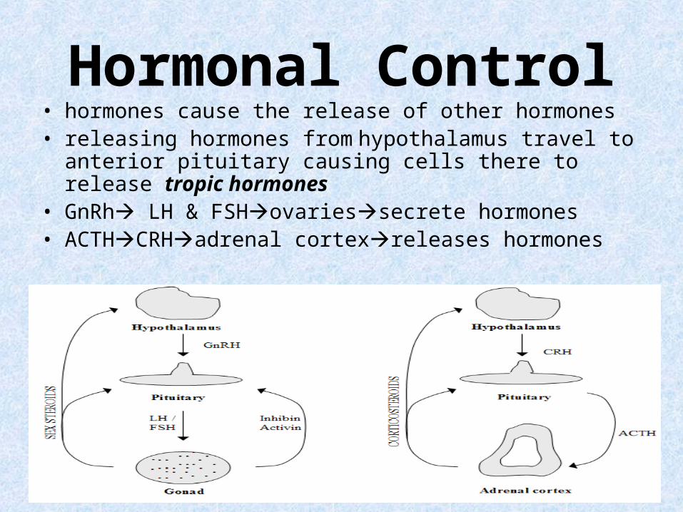
Hormonal Control• hormones cause the release of other hormones• releasing hormones from hypothalamus travel to anterior
pituitary causing cells there to release tropic hormones• GnRh LH & FSHovariessecrete hormones• ACTHCRHadrenal cortexreleases hormones

Humoral Control• changing blood levels of ions & nutrients cause
hormones to be released• parathyroid gland monitors Ca++ concentration• when low secretes parathyroid hormoneCa++
increases• insulin is released by pancreas due to humoral
stimulus of too much glucose in blood


Hypothalamus• forms floor & walls of
third ventricle of brain• responsible for
regulation of primitive body functions:
• reproduction, hunger & thirst
• many functions are carried out by pituitary gland
• master gland

Hypothalamus-Pituitary• hypothalamus secretes
regulatory hormones (releasing hormones) which control endocrine cells in anterior lobe of pituitary
• also manufactures hormones which travel via hypothalamus-hypophyseal tract to be stored in posterior pituitary
• hypothalamus has direct neural control over endocrine cells in adrenal medulla via sympathetic nervous system– neuroendocrine reflex

Hypophyseal PortalSystem• Adenohypohysis has no
direct connection to hypothalamus
• connected by hypophyseal portal system
• typically arteries take blood from heart to capillaries & veins from capillaries to heart
• portal system-blood flows from one capillary network into portal vein then to secondary capillary network before returning to heart.
• arrangement ensures all blood entering portal vessels will reach target cells before entering general circulation

Hypothalamic Hormones• 2 neurohormones
– oxytocin & antidiruretic hormones– stored in pituitary
• 5 regulating hormones-releasing hormones (RH)– stimulate synthesis & secretion of other
hormones in anterior pituitary
• 2 inhibiting hormones (IH)– inhibit synthesis & secretion of other
hormones from anterior pituitary

Hypothalamic Control of Anterior Pituitary
• negative feedback loops• Hypothalamus
releasing hormones adenohypophysis tropic or trophic hormone stimulate other endocrine glands or tissues
• hormones from other endocrine glands feedback to turn off their own production

Pituitary Gland-Hypophysis• located in sella turcica-
depression in sphenoid bone
• size & shape of a pea• connects to
hypothalamus by infundibulum
• composed of two parts: • Adenohypothesis • Neurohypothesis
– separate functions– separate anatomy

Anterior Pituitary Gland• three discrete regions • pars distalis
– largest & most anterior
• pars tuberalis– wraps around
infundibulum
• pars intermedia– narrow band bordering
the posterior pituitary lobe
– atrophies before birth

Growth Hormone-Somatotropin• secreted by somatotropes• promote protein synthesis• influence carbohydrate metabolism by
decreasing glucose uptake• promote production of somatomedins or IGFs-
Insulin like growth factors• stimulate uptake of amino acids & incorporation
into proteins• overall increase growth rate of skeleton &
skeletal muscles during childhood & teenage years
• in adults help maintain muscle & bone mass & help to promote healing & tissue repair

Growth Hormone Secretion• GHRH (growth
hormone releasing hormone) stimulates release of GH
• GHIH (growth hormone inhibiting factor) or somatostatin inhibits release
• GHIH-triggered by feedback of GH & IGFs
• as GH increases-inhibits its own release

Growth Hormone

TSH-Thyroid Stimulating Hormone
• TRH (thyrotropin releasing hormone) is made by hypothalamus
thyrotrope cells of anterior pituitary TSHthyroid gland thyroid hormone
• thyroid hormone feeds backs on TRH & TSHinhibits secretion

ACTH-Corticotrophin• released from
corticotrope cells of anterior pituitary due to stimulation from CRH (corticotropin releasing hormone) made by hypothalamus
• ACTHadrenal cortex• glucocorticorticoids
(especially cortisol) • glucocorticoids feedback
to block secretion of CRH & ACTH

Gonadotropins-FSH & LH• regulate activities of gonads• hypothalamus releases gonadotropin
releasing factors (GnRH)anterior pituitaryLH & FSH
• FSH (follicle stimulating hormone) promotes follicle development in females & with LH stimulates estrogen secretion
• in males FSH stimulates sperm differentiation
• production of GnRH is inhibited by inhibin– released by cells in gonads
• LHinduces ovulation & promotes progestin secretion by ovaries
• in males LH-also called ICSH or interstitial cell stimulating hormone stimulates interstitial cells of testesandrogens
• gonadal hormones feedback to suppress FSH & LH release

Prolactin-PRL• released from anterior
pituitary due to stimulation by PRH made by hypothalamus
• PRL targets lactotropes in mammary glandsmilk production
• PRL helps regulate androgen production in males by making interstitial cells more sensitive to LH.
• hypothalamusPIH or prolactin inhibiting hormone prevents prolactin secretion
• circulating prolactin stimulates PIH production & inhibits PRH

MSH-Melanocyte Stimulating Hormone
• made by pars intermedia
• stimulates melanocytes to make melatin
• no circulating MSH in adults
• melanin receptors in the brain suggests it is involved in brain activity
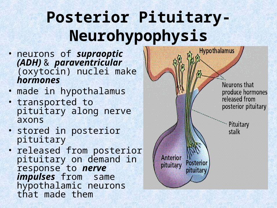
Posterior Pituitary-Neurohypophysis
• neurons of supraoptic (ADH) & paraventricular (oxytocin) nuclei make hormones
• made in hypothalamus• transported to pituitary
along nerve axons• stored in posterior pituitary• released from posterior
pituitary on demand in response to nerve impulses from same hypothalamic neurons that made them

ADH-Vasopression or Antidiuretic Hormone
• involved in water balance• released in response to increases in
electrolyte concentrations in blood• neurons responsible for release-
osmoreceptors• solutes concentrated
osmoreceptors excite supraoptic nucleisynthesis & release of ADH
• Target-kidney tubulesreabsorb water from urineless urineblood volume increasesblood pressure increases
• solute concentration decreases, osmoreceptorsend ADH release
• high concentrations of ADH produce vasoconstriction primarily of visceral blood vessels-thus it is named vasopressin

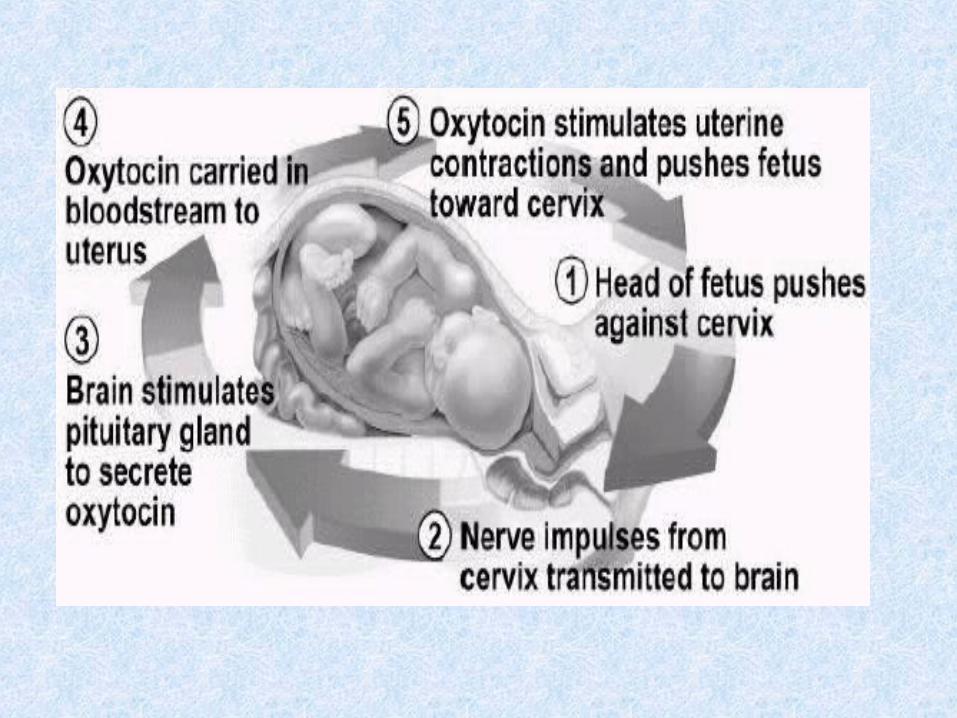
Oxytocin• produces smooth muscle
contraction in uterus & breasts• released in high amounts during
childbirth & when nursing• uterus stretchesimpulse to
hypothalamusoxytoxin made & released from posterior pituitary bloodcontractions increase-positive feed back control
• also acts for milk ejection via neuroendocrine reflex-milk let down reflex
• suckling (stimulus)sensory neurons hypothalamusocytoxin releasedmyoepithelial cellsrelease milk
• also believed to be cuddle hormone which promotes nurturing & affectionate or bonding behavior

Pituitary Gland Review• secretes nine
peptide hormones
• 7 from anterior pituitary
• 2 from posterior pituitary

Thyroid Gland • largest endocrine
gland• located in anterior
neck• 2 lobes connected by
an isthmus• contains large number
of follicles• lined by simple
cuboidal epithelium or follicular cells
• follicle cells surround a follicular cavity which stores colloid
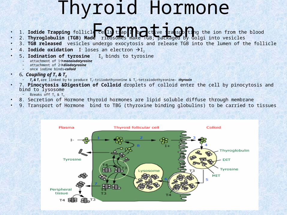
Thyroid Hormone Formation• 1. Iodide Trapping follicle cells trap I- by active transporting the ion from the blood• 2. Thyroglobulin (TGB) Made ribosomes make TGB; packaged by Golgi into vesicles• 3. TGB released vesicles undergo exocytosis and release TGB into the lumen of the follicle• 4. Iodide oxidation I- loses an electron I2
• 5. Iodination of tyrosine I2 binds to tyrosine– attachment of 1monoiodotyrosine– attachment of 2diiodotyrosine– once iodine binds-colloid
• 6. Coupling of T1 & T2– T2 & T1 are linked by to produce T3-triiodothyronine & T4-tetraiodothyronine- thyroxin
• 7. Pinocytosis &Digestion of Colloid droplets of colloid enter the cell by pinocytosis and bind to lysosome
– Breaks off T3 & T4
• 8. Secretion of Hormone thyroid hormones are lipid soluble diffuse through membrane• 9. Transport of Hormone bind to TBG (thyroxine binding globulins) to be carried to tissues

Release of Thyroid Hormone• thyroxine levels fall
hypothalamus TRH pituitaryTSH thyroid cells thyroid hormones
• 90% is T4-major thytoid hormone
• 10% is T3
• T3 is more potent and responsible for effects of thyroid hormone

Functions of Thyroid Hormone• major metabolic hormones• Increase BMR (basal metabolic rate) which
raises use of oxygen• calorigenic effect-increases heat production• involved in long-term regulation of metabolic
turnover• effects almost every cell in body except-adult
brain, spleen, testes, uterus and thyroid

Control of Thyroid Hormone

Parafollicular Cells of Thyroid• secrete calcitonin• lowers blood Ca++ levels• Ca++ homeostasis is
essential for nerve transmission, muscle contraction & blood clotting
• too lowNa permeability increasescells become more excitable
• calcium levels too high, hypercalcemiaNa permeability decreases membranes less excitable
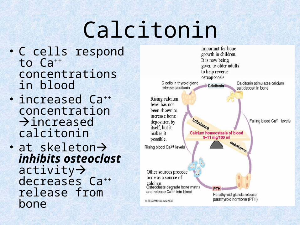
Calcitonin• C cells respond to
Ca++ concentrations in blood
• increased Ca++ concentration increased calcitonin
• at skeleton inhibits osteoclast activity decreases Ca++ release from bone
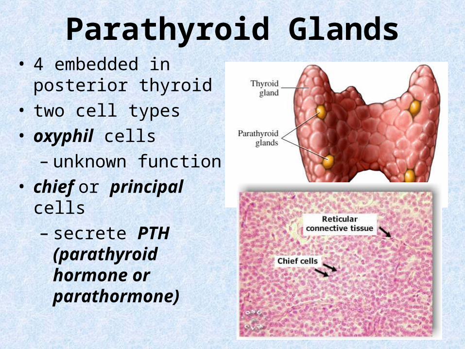
Parathyroid Glands• 4 embedded in
posterior thyroid• two cell types• oxyphil cells
– unknown function • chief or principal cells
– secrete PTH (parathyroid hormone or parathormone)

Parathyroid Hormone Targets• single most important agent in
controlling Ca++ balance• chief cells monitor Ca levels• lowered blood Ca++-
hypocalcemia• stimulates secretion• increases Ca++ in blood by
stimulating skeleton, kidneys & intestines
• SkeletonPTH stimulates osteoclasts to digest bone matrix releases Ca++
• KidneysPTH enhances reabsorption of Ca++
• PTH increases Ca++ by enhancing synthesis of calcitriol by the kidney which enhances Ca++ absorption by gut


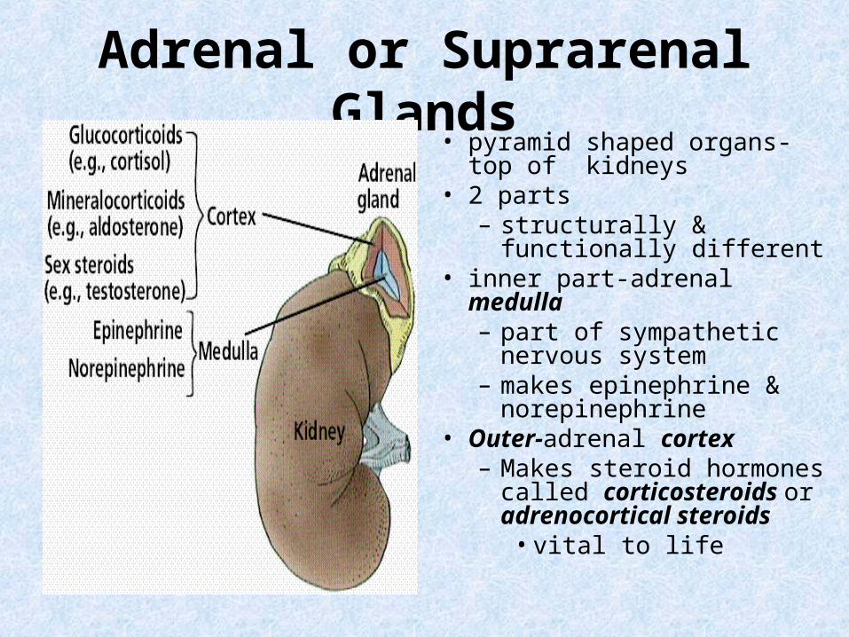
Adrenal or Suprarenal Glands• pyramid shaped organs-top
of kidneys• 2 parts
– structurally & functionally different
• inner part-adrenal medulla– part of sympathetic
nervous system– makes epinephrine &
norepinephrine• Outer-adrenal cortex
– Makes steroid hormones called corticosteroids or adrenocortical steroids
• vital to life


Adrenal Cortex• 3 regions or zones• Zona Glomerulosa
– outer region– composed of globular cell
clusters called glomeruli– mineralocorticoids
• Zona Fasciculata– middle zone– cells form in linear cords– glucocorticoids
• Zona reticularis– inner zone– cells form net like
arrangement– gonadocorticoids
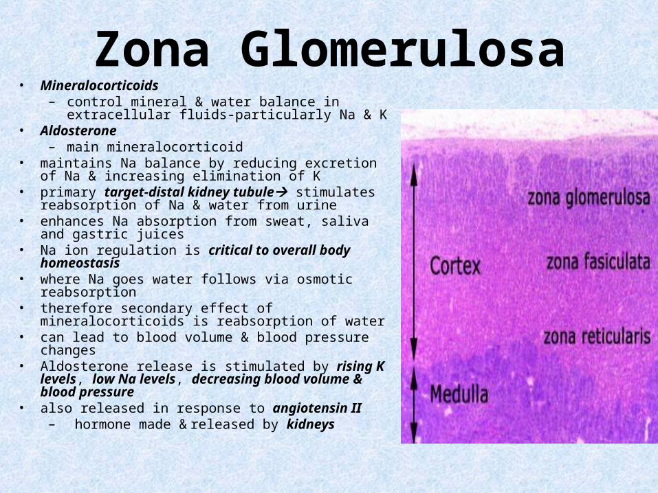
Zona Glomerulosa• Mineralocorticoids
– control mineral & water balance in extracellular fluids-particularly Na & K
• Aldosterone– main mineralocorticoid
• maintains Na balance by reducing excretion of Na & increasing elimination of K
• primary target-distal kidney tubule stimulates reabsorption of Na & water from urine
• enhances Na absorption from sweat, saliva and gastric juices
• Na ion regulation is critical to overall body homeostasis
• where Na goes water follows via osmotic reabsorption
• therefore secondary effect of mineralocorticoids is reabsorption of water
• can lead to blood volume & blood pressure changes• Aldosterone release is stimulated by rising K levels,
low Na levels, decreasing blood volume & blood pressure
• also released in response to angiotensin II– hormone made & released by kidneys


Zona Fasciculata• produce glucocorticoids
– influence glucose metabolism• important in helping to resist stressors• absolutely essential to life• main one-cortisol or hydrocortisone• primary metabolic effect is gluconeogenesis\
– formation of glucose from non-carbohydrate sources
• helps body adapt to intermittent food intake by keeping blood sugar levels constant
• does so by breaking down adipose tissuesfatty acids & proteinsamino acids.
• glucocoricoids enhance epinephrine’s vasoconstrictive effectsrise in blood pressure and circulatory efficiency
• helps maintain blood volume by preventing a shift of water into tissue cells
• Too muchdepresses cartilage & bone formation, inhibits inflammation, depresses immune system, and promotes changes in cardiovascular, neural and gastrointestinal functions
• inhibit activities of WBCs

Cortisol Regulation• regulated by negative
feedback• CRH from hypothalamus
ACTH from anterior pituitaryzona fasciculatacortisol
• increased cortisol hypothalamus & anterior pituitary prevents CRH release & ACTH production

Zona Reticularis• Gonadocorticoi
ds• DHEA-
dehydroepiandrosterone
• sustains libidio (sex drive)
• release stimulated by ACTH

Adrenal Medulla• secretes
catecholamines-epinephrine & norepinephrine and dopamine
• made by chromaffin cells• sympathetic activity
increases rate of release• Stress & exercise
hypothalamusmedullahormones fight or flight reaction
• Increase in heart rate and blood pressure

Pancreas• elongated, spongy mixed
gland• exocrine (98%) & endocrine
(2%) functions• located partially behind
stomach in abdomen• exocrine cells are found in
clusters called pancreatic acini
• secrete alkaline, enzyme rich fluid used in digestion
• endocrine pancreas is found scattered throughout gland in groups of cell clusters called Islets of Langerhans

Islets of Langerhans• Alpha cells
– secrete glucagon– hyperglycemic effect
• increases blood glucose levels by increasing glycogenolysis in liver
• Beta cells– secrete insulin– hypoglycemic effect
• Decreases blood glucose
• Delta cells– synthesize somatostatin when
blood glucose, fatty acids and amino acid levels rise after eating
– inhibits digestive functions in blood & pancreas
– suppresses release of glucagon & insulin by the neighboring alpha alpha & beta cells.
• F Cells– secrete pancreatic polypeptide– Inhibits somatostatin

Glucagon• secretion prompted by humoral
stimuli– falling blood sugar levels & rising
amino acid levels• secretion suppressed by rising
blood sugar• in liver& skeletal muscle
stimulates break down of glycogen into glucose- glycogenolysis
• stimulates break down of triglycerides in adipose tissue
• stimulates glucose production from lactic acid & other non-carbohydrate sources-gluconeogenesis in liver
• Resultless glucose use & more glucose releaseincreases blood glucose

Insulin• decreases blood glucose
levels by increasing rate of glucose uptake & use
• one effect is to enhance glucose absorption & utilization
• accelerates glucose use & enhances ATP production
• stimulates glycogen formation in skeletal muscle and liver cells
• stimulates amino acid absorption and protein synthesis
• stimulates triglyceride formation in adipose tissue


Gonads• Testes
• Androgens
–testosterone-major one
• Ovaries
• Estrogens

Testosterone Functions• maturation of
reproductive organs
• appearance & maintenance of secondary sex characteristics
• effects metabolic activities
• stimulates protein synthesis & muscle growth

Estrogen Functions• maturation of
reproductive organs
• Development & maintenance of secondary sex characteristics


Pineal Gland• tiny, pine coned shaped
structure hanging from roof of third ventricle
• secretes melatonin at night• rises & falls in diurnal cycle• peaks at night inducing sleep• lowest during day• pineal receives information
regarding intensity & duration of day light via retinasuprachiasmatic nucleus in hypothalamuspineal glandmelatonin
• responsible for setting circadian rhythm or daily-cycles

Endocrine Tissues of Other Organs• Heart-atrium
– ANP-atrial natriuretic peptide• promotes loss of Na & water at kidneys & inhibits renin release & secretion of ADH &
aldosterone• net result is reduction of blood volume & pressure.
• Skin-keratinocytes– Vitamin D3-first step in production of calcitriol
• hormone that raises blood calcium levels• Stomach & Small Intestine
– Enteroendocrine cells secrete about 10 enteric hormones• Coordinate different regions & glands of digestive system with each other
• Kidney– Erythropoietin
• released in response to low O2 levels stimulates RBCs production– renin
• cleaves angiotensinogenangiotensin II adrenal cortexaldosterone increases blood volume & pressure
• Thymus– thymopoietins & thymosins
• important in development of T lymphocytes important in the immune response• Adipose tissue
– Leptin-appetite control
.