ems system for moody county, south dakota › resources › documents › sdaa...ems system for...
TRANSCRIPT


EMS System for Moody County, South Dakota Medical Control Board Treatment Protocols
Mission Statement
We are committed to providing the highest standard of care to the residents and visitors of
Moody County. Using evidence-based medicine, we will meet the health care needs of our
community while continuing to improve the overall health of all our stakeholders.
Introduction
This protocol manual represents the basic foundation for clinical medical standards for the
combined Emergency Medical Services (EMS) system of Moody County in South Dakota. The
EMS system activates when a stakeholder in our community dials 911 and reports any type of
medical emergency. The stakeholder communicates with the Emergency Medical Dispatcher,
who then alerts Police and/or Fire Department First Responders and then the Ambulance. If
transport occurs, the EMS system concludes with further evaluation and continued treatment at
many of our local Emergency Rooms.
These protocols are designed to assist the care provider in the treatment of both medical and
traumatic patents. The protocols are meant as guidelines but remember treatment should never be
limited by protocols. Always do what is best for your patient. Because medicine is
continuously changing, these dynamic protocols will be frequently reviewed and updated to meet
the needs of our stakeholders, using current evidence-based medical and nationally recognized
best practices to guide patient treatments.
By signing below, you have acknowledged receiving these protocols and accept the responsibly
of knowing, understanding, and practicing in accordance with these protocols.
Welcome to Moody County EMS-Progressive, Compassionate, Community-focused, Committed
to Excellence.
Name___________________________________________ Date_______________
___________________________________________
Employee Signature
____________________________________________ ______________________________
Ambulance Supervisor Medical Director

EMS System for Moody County, South Dakota Medical Control Board Treatment Protocols
Index of Protocols
SECTION 1 GENERAL ASSESSMENT & GENERAL SUPPORTIVE CARE
1A Medical General Assessment – Adult & Pediatric
1B Trauma General Assessment – Adult & Pediatric
1C General Supportive Care – Adult & Pediatric
1D Trauma and Hypovolemic Shock Supportive Care – Adult & Pediatric
1E Neonatal Resuscitation- Pediatric
SECTION 2 AIRWAY
2A Airway Assessment – Adult & Pediatric
2B Airway Establishment/Obstruction Management – Adult & Pediatric
2C Airway Suctioning – Adult & Pediatric
2D Bag Valve Mask (BVM) Management – Adult & Pediatric
2E Supraglottic Airways – Adult & Pediatric
2F Oral Intubation – Adult
2G Medication Assisted Intubation – Adult
2H Nasal Intubation – Adult
2I Cricothyrotomy – Adult
2J Confirmation of Endotracheal Airway Placement – Adult & Pediatric
2K Stoma/Tracheostomy Management – Adult & Pediatric
SECTION 3 PULMONARY/RESPIRATORY
3A Respiratory Arrest – Adult & Pediatric
3B Dyspnea – Uncertain Etiology – Adult & Pediatric
3C Dyspnea – Asthma – Adult & Pediatric
3D Dyspnea – Chronic Obstructive Pulmonary Disease (COPD) – Adult
3E Dyspnea – Congestive Heart Failure (CHF) – Adult & Pediatric
3F Pulse Oximetry – Adult & Pediatric
3G Waveform Capnography – Adult & Pediatric
3H Oxygen Administration – Adult & Pediatric
3I Nebulization Therapy – Adult & Pediatric
3J Continuous Positive Airway Pressure (CPAP) Ventilation – Adult

EMS System for Moody County, South Dakota Medical Control Board Treatment Protocols
SECTION 4 CARDIAC ARREST
4A Resuscitation (CPR) – Adult & Pediatric
4B Resuscitation Team Roles – Adult & Pediatric
4C Automated External Defibrillation (AED) – Adult & Pediatric
4D Manual Defibrillation – Adult & Pediatric
4E Asystole – Adult & Pediatric
4F Ventricular Fibrillation/Ventricular Tachycardia – Adult & Pediatric
4G Pulseless Electrical Activity (PEA) – Adult & Pediatric
4H Specific Causes of Cardiac Arrest – Adult & Pediatric
4I Post Cardiac Arrest Treatment – Adult & Pediatric
4J “Do Not Resuscitate” (DNR)/South Dakota Care One, Futility of Resuscitation Initiation,
&Termination of Resuscitation – Adult & Pediatric
4K Intra-Arrest Wakefulness – Adult
4L LUCAS Activation
SECTION 5 CARDIAC (NON-ARREST)
5A Chest Pain – Uncertain Etiology – Adult & Pediatric
5B Acquiring & Transmitting 12-Lead ECGs – Adult & Pediatric
5C Acute Coronary Syndrome – Adult
5D Bradycardia – Adult & Pediatric
5E Transcutaneous Pacing – Adult & Pediatric
5F Tachycardia – Stable – Adult & Pediatric
5G Tachycardia – Unstable – Adult & Pediatric
5H Synchronized Cardioversion – Adult & Pediatric
5I Pacemaker Management – Adult & Pediatric
5J Implantable Cardioverter/Defibrillator (ICD) Management –Adult & Pediatric
5K Premature Ventricular Contractions – Adult & Pediatric
SECTION 6 NEUROLOGIC/ALTERED MENTAL STATUS
6A Stroke – Adult & Pediatric
6B Altered Mental Status – Adult & Pediatric
6C Glucometry (Blood Glucose Determination) – Adult & Pediatric
6D Seizure – Adult & Pediatric
6E Syncope – Adult & Pediatric
6F Dystonic Reactions – Adult & Pediatric

EMS System for Moody County, South Dakota Medical Control Board Treatment Protocols
SECTION 7 PSYCHIATRIC/BEHAVIORAL DISORDERS
7A Behavioral Disorder – Adult & Pediatric
7B Physical Restraint – Adult & Pediatric
7C Chemical Restraint – Adult & Pediatric
7D Emergency Mental Hold Issues – Adult & Pediatric
SECTION 8 TOXICOLOGIC/POISONINGS
8A Poisonings – General Management – Adult & Pediatric
8B Toxidromes – Adult & Pediatric
8C National Poison Control Center Use
8D Acute Allergic Reactions – Adult & Pediatric
8E Snakebites (Crotalinae Envenomation) – Rattlesnakes, Copperheads, & Moccasins –
Adult & Pediatric
8F Bee/Wasp Stings (Hymenopera Envenomation) – Adult & Pediatric
8G Hazardous Materials Response
8H Hydrofluoric Acid – Adult & Pediatric
SECTION 9 MEDICAL
9A Abdominal Pain/Nausea/Vomiting/Diarrhea – Adult & Pediatric
9B Sepsis– Adult & Pediatric
9C Epistaxis – Adult & Pediatric
9D Pain Management (Acute Onset & Chronic Type) – Adult & Pediatric
9E Dialysis Related Issues – Adult & Pediatric
9F Infectious Disease Precaution Recommendations – EMS Professionals
9G Post-Exposure Prophylaxis Recommendations – EMS Professionals
9H Vascular Access – Intravenous – Adult & Pediatric
9I Vascular Access – Intraosseous – Adult & Pediatric
9J Indwelling Central Vascular Device Management – Adult & Pediatric
9K Medication Administration – Adult & Pediatric
9Ka – Intravenous/Intraosseous – Adult & Pediatric
9Kb – Intramuscular/Subcutaneous–Adult & Pediatric
9Kc – Intranasal – Adult & Pediatric
9Kd – Sublingual/Oral – Adult & Pediatric
9Ke – Ocular – Adult & Pediatric
9Kf – Intravascular Infusion Management – Adult & Pediatric
9L Nasogastric/Orogastric Tube – Adult
9M Suspected Abuse or Neglect – Adult & Pediatric

EMS System for Moody County, South Dakota Medical Control Board Treatment Protocols
SECTION 10 TRAUMA
10A Head/Neck/Spine Injury – Adult & Pediatric
10B Eye Injury – Adult & Pediatric
10C Dental Injury/Pain – Adult & Pediatric
10D Chest/Abdomen/Pelvis Injury – Adult & Pediatric
10E Needle Thoracostomy (Tension Pneumothorax Decompression) – Adult & Pediatric
10F Chest Tube Monitoring – Adult & Pediatric
10G Extremity/Amputation Injury – Adult & Pediatric
10H Tourniquet – Adult & Pediatric
10I Hemostatic Agents – Adult & Pediatric
10J Compartment/Crush Injury Syndrome – Adult & Pediatric
10K Burns – Adult & Pediatric
10L Conductive Energy Weapon Related Management – Adult & Pediatric
10M “Less Lethal” Weapon Related Management – Adult & Pediatric
10N Splinting of Injuries – Adult & Pediatric
10Na – Spinal Motion Restriction – Adult & Pediatric
10Nb – Extremity – Adult & Pediatric
10O Blast Injury – Adult & Pediatric
SECTION 11 ENVIRONMENTAL
11A Heat Illness – Adult & Pediatric
11B Cold Illness/Injury – Adult & Pediatric
11C Electrical/Lightning Injury – Adult & Pediatric
11D Water Submersion Events – Adult & Pediatric
SECTION 12 FIREGROUND-RELATED
12A Fire Ground Rehabilitation Concepts – Adult
12B Smoke Inhalation – Adult & Pediatric
12C Carbon Monoxide – Adult & Pediatric
12D Hyperbaric Oxygen Therapy Considerations – Adult & Pediatric
12E Cyanide – Adult & Pediatric
SECTION 13 OBSTETRIC/GYNECOLOGIC
13A Childbirth – Routine
13B Childbirth – Complicated
13C Complications of Pregnancy – Adult
13D Vaginal Bleeding/Discharge – Adult & Pediatric
13E Pelvic Pain – Adult & Pediatric
13F Sexual Assault – Adult & Pediatric
EMS System for Moody County, South Dakota Medical Control Board Treatment Protocols
SECTION 14 RESPONSES, SCENE ISSUES & PATIENT TRANSPORTATION
14A Staging Considerations
14B Actions to Preserve Crime Scenes
14C Other Health Care Professionals on Emergency Scene(s)
14D Informed Patient Consent/Refusal
14E On-Line Medical Control Physicians
14F Helicopter EMS (HEMS) Considerations
14G Patient Prioritization
14H Radio Report Communications
14I Interfacility Transfers
SECTION 15 MASS CASUALTY/DISASTER/TERRORIST EVENTS
15A Multiple Patient Scenes/Mass Casualty Event Concepts
15B Chemical Weapons
15C Nerve Agents
15D Biological Weapons
15E Radiological Weapons
15F Nuclear Weapons
SECTION 17 RESERVED FOR AGENCY SPECIFIC USE
16A Helmet Removal
16B Controlled Substance Handling & Documentation
SECTION 17 DRUG SUMMARIES
17A Adenosine (Adenocard®)
17B Albuterol (Proventil®)
17C Amiodarone (Cordarone®)
17D Aspirin
17E Atropine Sulfate
17F Calcium Gluconate
17G Dextrose (50% as D50, 25% as D25, 10% as D10)
17H Diazepam (Valium®)
17I Diltiazem (Cardizem®)
17J Diphenhydramine (Benadryl®)
17K Dopamine (Intropin®)
17L Duodote® Auto-injector
17M Epinephrine 1:1,000 & 1:10,000
17N Epinephrine Auto-Injector (EpiPen®)

EMS System for Moody County, South Dakota Medical Control Board Treatment Protocols
SECTION 17 DRUG SUMMARIES (CONTINUED)
17O Fentanyl (Sublimaze®)
17P Glucagon
17Q Glucose (Oral)
17R Haloperidol (Haldol®)
17S Ipratropium Bromide (Atrovent®)
17T Lidocaine 2% Intravascular (Xylocaine®)
17U Lorazepam (Ativan®)
17V Magnesium Sulfate
17W Methylprednisolone (Solu-Medrol®)
17X Midazolam (Versed®)
17Y Morphine Sulfate
17Z Naloxone (Narcan®)
17AA Nitroglycerin – Nitroglycerin (Nitrolingual®)
17DD Ondansetron (Zofran®)
17CC Pralidoxine Chloride (2-Pram)
17DD Sodium Bicarbonate
17EE Tranexamic Acid (TXA, Cyclokapron®)
17FF Xopenex (Levabuterol HCL®)
SECTION 18 APPENDICES
18A – Approved Abbreviations
Adopted on April 24, 2017

EMS System for Moody County, South Dakota Medical Control Board Treatment Protocols
In general, approach the assessment of medical (non-trauma) patients, in A-B-C order:
Airway: Evaluate the patency and mechanics of the airway. Is the patient able to oxygenate
and ventilate? Rapid intervention may be required during the assessment phase if airway
patency and protection is compromised.
Breathing: Expose the chest as required to accurately assess the mechanics of respiration
(taking into account patient privacy/modesty if in public location). Note the rate, depth, and
pattern of respirations and if any degree of respiratory distress or effort. Auscultate breath
sounds bilaterally.
Liberally obtain pulse oximetry readings and in patients with respiratory difficulties,
waveform capnography readings (if equipped, **Mandatory use if the patient is
intubated). Circulation: The adequacy of a patient’s circulation is best assessed first by evaluating their
level of consciousness and mental status. Next assess the location, rate, and character of the
pulse. Then check a blood pressure – preferably, manually for at least the first reading. Apply
the cardiac monitor (if equipped) liberally.
Cardiac Arrest is an exception to the above order. Aggressively initiate chest compressions
and search for shockable rhythms at the appropriate intervals per Section 4 protocols.
1A – MEDICAL GENERAL ASSESSMENT
ADULT & PEDIATRIC
TREATMENT PRIORITIES
1. Assessment
SCENE SAFETY PROTECTION Primary Survey Secondary Survey (when appropriate)
2. Primary Survey Care
Initiate cardiopulmonary resuscitation if indicated
Open airway Support oxygenation/ventilation Support circulation – Dysrhythmia care? Heart
Rate control? Hypotension care? 3. Minimize scene time in critical case unless working
cardiac arrest 4. Enroute Care
Reassess all primary care Support oxygenation/ventilation Vascular access Secondary Survey (if able) Keep patient warm/avoid hypothermia
5. Most appropriate hospital destination

EMS System for Moody County, South Dakota Medical Control Board Treatment Protocols
Protocol 1A: Medical General Assessment – Adult & Pediatric, cont.
Many treatment decisions regarding airway management involve calculating the adult
patient’s Glasgow Coma Scale score using the following table:
After addressing the A-B-C order in most medical patients, including evaluating and
addressing any life-threatening conditions, minimize scene time and initiate timely
transport to an appropriate emergency department in any setting of a time-sensitive
medical condition.
Complete a head-to-toe assessment of the patient if the patient is relatively medically stable.
Obtain relevant history of past and current medical problems, medications, allergies, and
physicians/hospitals used in care plans to help guide further assessment.
Reassess patients frequently, typically at least every 15 minutes, and more often if critical
illness is discovered and being treated. In the situations of an unstable patient, vital signs
should be assessed every 5 minutes, especially if hemodynamic changes are occurring.
Assess and treat per symptom or illness specific protocols that follow in this protocol set.
Pediatric Assessment Comments:
1. Pediatric respiratory distress may look just like adult respiratory distress, presenting with: slowing respirations cyanosis accessory muscle use paleness nasal flaring lethargy/listlessness retractions – intercostal or subcostal irritability tachypnea stridor mottling grunting

EMS System for Moody County, South Dakota Medical Control Board Treatment Protocols
Protocol 1A: Medical General Assessment – Adult & Pediatric, cont.
2. Vital signs vary with age. In general, the younger the patient, the faster the respiratory rate, the faster the heart rate, and the lower the blood pressure:
3. The following table can be used to calculate Glasgow Coma Scale scores in pediatric patients, especially those under 4 years of age. Most pediatric patients above the age of 4 years will be able to be assessed for Glasgow Coma Scale scores using the adult table.

EMS System for Moody County, South Dakota Medical Control Board Treatment Protocols
Protocol 1A: Medical General Assessment – Adult & Pediatric, cont.
4. The Pediatric Assessment Triangle (PAT) is a rapid evaluation tool that establishes a child’s clinical status and his or her category of illness to direct initial management priorities.

EMS System for Moody County, South Dakota Medical Control Board Treatment Protocols
1B - TRAUMA GENERAL ASSESSMENT ADULT & PEDIATRIC
TREATMENT PRIORITIES 1. Assessment
SCENE SAFETY PROTECTION Primary Survey “Trauma Alert” to receiving ED if indicated Secondary Survey (when appropriate)
2. Primary Survey Care Control arterial bleeding Open airway Seal “sucking” chest wound(s) Needle thoracostomy for closed chest trauma (tension pneumothorax)
3. Minimize scene time in critical case 4. Enroute Care
Reassess all primary care Support oxygenation/ventilation Vascular access Secondary Survey (if able) Keep patient warm/avoid hypothermia
5. Most appropriate hospital destination
Before entering any trauma scene, ensure your personal safety. Do not attempt patient contact
until hazards can be appropriately mitigated. In addition to scene safety, factor mechanisms of injury,
number of patients, and special equipment/extrication needs. All trauma patients should be assessed utilizing primary, secondary, and reassessment surveys.
The primary survey is to be conducted on all trauma patients. It is designed to rapidly identify life-
threatening or potentially life-threatening injuries. The primary survey should be completed within 2
minutes of patient contact. THE PRIMARY SURVEY IS ONLY INTERRUPTED FOR LIFE-
THREATENING ARTERIAL BLEEDING, AIRWAY OBSTRUCTION, OR RESPIRATORY/CARDIAC
ARREST. The following are the steps of the primary survey:
1) Manually stabilize the cervical spine while assessing the airway and level of consciousness.
2) Evaluate breathing – present? rapid? normal? slow? shallow? 3) Evaluate circulation – carotid and radial pulses? Control external hemorrhage.
4) Exam the head for deformity, contusions, abrasions, penetrations, burns, lacerations,
or swelling (“DCAP-BLS”).
5) Exam the neck for deformity, contusions, abrasions, penetrations, burns, lacerations, swelling
(“DCAP-BLS”), or subcutaneous emphysema. 6) Exam the chest for deformity, contusions, abrasions, penetrations, burns, lacerations,
swelling (“DCAP-BLS”), or paradoxical movement.
7) Auscultate the chest for breath sounds in the mid-axilla bilaterally – present? equal?
8) Exam the abdomen and pelvis for deformity, contusions, abrasions, penetrations, burns,
lacerations, or swelling (“DCAP-BLS”).
9) Exam the extremities for deformity, contusions, abrasions, penetrations, burns, lacerations,
or swelling (“DCAP-BLS”), and pulse, movement, sensation.

EMS System for Moody County, South Dakota Medical Control Board Treatment Protocols
Protocol 1B: Trauma General Assessment – Adult & Pediatric, cont.
Primary survey interventions include airway management (See Section 2 Protocols – Airway),
sealing open chest wounds, needle thoracostomy for suspected tension pneumothorax (See
Protocol 10E – Needle Thoracostomy), oxygen administration and controlling any obvious
external hemorrhage. Remember to expose the patient as needed to conduct an appropriate
exam. Any trauma patient with altered level of consciousness, abnormal respiration, abnormal
circulation, or signs/conditions likely to lead to shock (distended abdomen, pelvic
instability, bilateral femur fractures) should be rapidly immobilized and transported
after the completing the primary survey. These are “LOAD & GO” patients. The secondary survey is always done enroute on critical patients. If no critical conditions are
found in the primary survey, the secondary survey may be conducted on the scene and should
be completed within 5 minutes after the primary survey is completed. The following are the
steps of the secondary survey:
1) Obtain vital signs (pulse, respiratory rate, blood pressure, pulse oximetry) 2) Obtain history of traumatic event and pertinent patient medical history
(allergies, medications, past illness/injury, last oral intake) 3) Head to toe exam – look for “DCAP-BLS” in every body area. Calculate GCS score 4) Perform indicated bandaging and splinting
The reassessment survey is an abbreviated exam after interventions and done at least
every five minutes for critical patients. The following are the steps of the reassessment
survey:
1) Repeat the primary survey 2) Repeat vital signs 3) Repeat GCS score calculation 4) Check every intervention – proper placement of intubation? Proper placement of
IV/IO? 5) Check results of every intervention – improved oxygenation/ventilation? Improved
blood pressure?

EMS System for Moody County, South Dakota Medical Control Board Treatment Protocols
EMS System for Moody County, South Dakota Medical Control Board Treatment Protocols
EMERGENCY MEDICAL DISPATCHER
EMERGENCY MEDICAL RESPONDER
EMT
EMT – INTERMEDIATE
ADVANCED EMT
PARAMEDIC
EMD
IF CHIEF COMPLAINT IS MEDICAL IN NATURE, CHOOSE THE PROTOCOL THAT BEST FITS THE PATIENT’S FOREMOST
SYMPTOMS, WITH PRIORITY SYMPTOMS TAKING PRECEDENCE
QUESTIONS TO ADDRESS SCENE SAFETY ISSUES
EMR EMT
AIRWAY MANAGEMENT SUPPORT OXYGENATION/VENTILATION
OBTAIN VITAL SIGNS
APPLY CARDIAC MONITOR/OBTAIN 12-LEAD ECG (when indicated & if equipped)
TRANSMIT 12-LEAD ECG TO RECEIVING HOSPITAL
MONITOR END – TIDAL CO2 & WAVEFORM CAPNOGRAPHY (when indicated & if equipped, **Mandatory use if Pt intubated)
ASSIST PT WITH PT’S OWN MEDICATION IF DIRECTED BY PROTOCOL(S)
DETERMINE BLOOD GLUCOSE/TREAT HYPOGLYCEMIA PER PROTOCOL
EMT – I AEMT
INTUBATE IF INDICATED
IV/IO ACCESS IF INDICATED
FLUID BOLUS AS DIRECTED BY SPECIFIC MEDICAL PROTOCOL(S)
MEDICATION ADMINISTRATION PER SPECIFIC MEDICAL PROTOCOL(S)
PARAMEDIC
CONTINUOUS TREATMENT AND ASSESSMENT PER SPECIFIC MEDICAL PROTOCOL(S)
INTERPRETATION OF 12-LEAD ECGS (when indicated & if equipped)
Clinical Operational Notes (All Field Provider Levels)
1. The practice of EMS medicine is built upon the foundation of “taking medical care to the patient”. To achieve this objective, appropriate equipment (airway equipment kit, med/trauma equipment kit, suction device, AED/Cardiac Monitor/Defibrillator, patient packaging equipment) should be brought to the patient’s side to minimize critical treatment delays in secondarily fetching equipment from the response apparatus.
2. Minimize active movement on the patient’s part in settings of suspected myocardial ischemia, stroke, and dyspnea. Move and package the patient for transport with safety considerations for all involved.
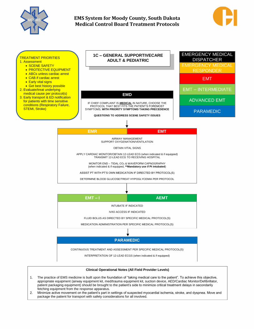
1C – GENERAL SUPPORTIVECARE
ADULT & PEDIATRIC TREATMENT PRIORITIES 1. Assessment
SCENE SAFETY
PROTECTIVE EQUIPMENT
ABCs unless cardiac arrest
CAB if cardiac arrest
Early vital signs
Get best history possible 2. Evaluate/treat underlying
medical cause per protocol(s) 3. Early transport & ED notification
for patients with time sensitive conditions (Respiratory Failure, STEMI, Stroke)

EMS System for Moody County, South Dakota Medical Control Board Treatment Protocols
EMERGENCY MEDICAL DISPATCHER
EMERGENCY MEDICAL RESPONDER
EMT
EMT – INTERMEDIATE
ADVANCED EMT
PARAMEDIC
EMD
IF CHIEF COMPLAINT IS TRAUMATIC IN NATURE, CHOOSE THE PROTOCOL THAT BEST FITS THE PATIENT’S FOREMOST
SYMPTOMS, WITH PRIORITY SYMPTOMS TAKING PRECEDENCE
QUESTIONS TO ADDRESS SCENE SAFETY ISSUES
EMR EMT
SERIOUS HEMORRHAGE CONTROL: TOURNIQUET IF INDICATED
BANDAGE/DRESSING/DIRECT PRESSURE PRESSURE DRESSING IF INDICATED (if equipped)
TOPICAL HEMOSTATIC AGENT IF INDICATED (if equipped)
AIRWAY MANAGEMENT SUPPORT OXYGENATION/VENTILATION
OBTAIN VITAL SIGNS/ASSESS FOR AND TREAT SHOCK
PREVENT HYPOTHERMIA
EMT – I AEMT
INTUBATE IF INDICATED
IV/IO ACCESS IF INDICATED
FLUID BOLUS AS DIRECTED BY SPECIFIC MEDICAL PROTOCOL(S)
PARAMEDIC
CRICOTHYROTOY IF INDICATED
NEEDLE THORACOSTOMY IF TESION PNEUMOTHORAX SUSPECTED
CONTINUOUS TREATMENT AND ASSESSMENT PER SPECIFIC MEDICAL PROTOCOL(S)
Clinical Operational Notes (All Field Provider Levels)
The practice of EMS medicine is built upon the foundation of “taking medical care to the patient”. To achieve this objective, appropriate equipment (airway equipment kit, med/trauma equipment kit, suction device, patient packaging equipment) should be brought to the patient’s side to minimize critical treatment delays in secondarily fetching equipment from the response apparatus.
1D - TRAUMA & HYPOVOLEMIC SHOCK
SUPPORTIVE CARE
ADULT & PEDIATRIC
TREATMENT PRIORITIES 1. Assessment
SCENE SAFETY
Primary Survey
“Trauma Alert” to receiving ED if indicated
2. Primary Survey Care
Control arterial bleeding
Open airway
Seal “sucking” chest wound(s)
Needle 3. Minimize scene time in critical case 4. Early transport & ED notification for
patients with time sensitive conditions (Respiratory Failure, STEMI, Stroke)

EMS System for Moody County, South Dakota Medical Control Board Treatment Protocols
In general, approach the resuscitation of the newborn or infant within the first 30 days of life focusing on basic life support interventions. Invasive, advanced procedures are rarely warranted. Best practices include focusing on basic interventions.
Warmth (Body Temperature Conservation): Due to high surface to body weight ratios, the neonate rapidly loses body heat which can lead to respiratory and circulatory distress. Keep the neonate warm and minimize skin exposures unless absolutely warranted during medical interventions.
Airway: Evaluate the patency and mechanics of the airway. Is the patient able to oxygenate and ventilate? Simple positioning intervention may be required during the assessment phase if airway patency and protection is compromised.
Breathing: Briefly expose the chest as required to accurately assess the mechanics of respiration. Note the rate, depth, and pattern of respirations and if any degree of respiratory distress or effort. Auscultate breath sounds bilaterally in the axilla to avoid confusing breath sounds from the other side of the chest. Gentle tactile stimulation (e.g. rubbing of the back, flicking the soles of the feet) may be required early in the assessment and often proves very effective in improving breathing activity.
Liberally obtain pulse oximetry readings and in patients with respiratory difficulties, waveform capnography readings (if equipped with neonatal sized equipment, **Mandatory use if the patient is intubated).
1E – NEONATAL RESUSCITATION
PEDIATRIC
TREATMENT PRIORITIES
1. Preserve patient warmth/avoid hypothermia 2. Assessment
Primary Survey Secondary Survey (when appropriate)
3. Primary Survey Care Initiate cardiopulmonary resuscitation if
indicated Open airway Support oxygenation/ventilation Support circulation
4. Minimize scene time in critical case unless working cardiac arrest
5. Enroute Care Reassess all primary care Support oxygenation/ventilation Secondary Survey (if able)
6. Most appropriate hospital destination

EMS System for Moody County, South Dakota Medical Control Board Treatment Protocols
Protocol 1E: Neonatal Resuscitation – Pediatric, cont.
Circulation: The adequacy of a neonate’s circulation is best assessed first by evaluating their level of activity and general body warmth. Next assess the rate and character of the brachial pulse. Pulse rates less than 100/minute are abnormal and a cause for concern of impending cardiovascular collapse. Pulse rates less than 60/minute indicate cardiovascular collapse and chest compressions should be initiated.
Cardiac Arrest is an exception to the above order. Aggressively initiate chest compressions, while still conserving warmth and initiating supplemental oxygenation and ventilation.
After addressing the Warmth – A-B-C order in most neonates, including evaluating and addressing any life-threatening conditions, minimize scene time and initiate timely transport to an appropriate emergency department.
Reassess patients at least every 5 minutes; consider more often if critical illness is discovered and being treated. Assess and treat per symptom or illness specific protocols.
Neonatal Assessment Comments:
1. Respiratory distress may or may not look just like adult respiratory distress, presenting with:
slowing or increasing respirations cyanosis
accessory muscle use paleness
nasal flaring lethargy/listlessness
retractions – intercostal or subcostal grunting
tachypnea mottling
2. Vital signs vary with age. In general, the younger the patient, the faster the respiratory rate, the faster the heart rate, and the lower the blood pressure. In most neonates, blood pressure is difficult to measure and often unreliable in attempts to do so in the field. Rather than focus extended time on blood pressure measurements, evaluate perfusion by overall activity level, skin temperature/color, capillary refill (normally < 3 seconds), and muscular tone.
3. Use APGAR scoring at 1 and 5 minutes post-birth, continue every 5 mins if APGAR < 7:
APGAR SCORING (SIGN) 0 1 2
APPEARANCE
BLUE OR BODY PINK, EXTREMITIES COMPLETELY PINK
PALE
BLUE
HEART RATE (BPM) ABSENT ≤100 >100
GRIMACE (REACTION TO NO GRIMACE
COUGH OR SNEEZE
CATHETER IN NARES)
RESPONSE
MUSCLE TONE LIMP SOME FLEXION ACTIVE MOTION
RESPIRATORY RATE ABSENT SLOW/IRREGULAR GOOD, CRYING

EMS System for Moody County, South Dakota Medical Control Board Treatment Protocols
The following principles should be followed to allow optimum assessment and care of the airway
without unnecessary intervention.
1. Use the least invasive method of airway management appropriate to the patient. 2. Use a method of airway management with which you are procedurally comfortable. 3. Use frequent suctioning to keep the airway clear of debris. 4. Monitor continuously to be sure that oxygenation/ventilation is as effective as intended
and as needed. 5. Understand the difference between these various aspects of airway management:
A. Patency: how open and clear is the airway, free of foreign substances, blood,
vomitus, and tongue obstruction? B. Ventilation: the amount of air the patient is able to inhale and exhale in a given time,
promoting exhalation of carbon dioxide. Use waveform capnography if equipped. C. Oxygenation: the amount of oxygen the patient is able to convey to the circulation
for tissue/organ perfusion. Use pulse oximetry when available. Although the dynamics of EMS care often dictate rapid decisions in critical skill performance,
assessment for difficult airway characteristics should precede intubation attempt(s). Several
methods of evaluating airway-related anatomy exist. One commonly used mnemonic in
emergency airway care is “LEMON,” which stands for: L Look externally (Heavy perioral facial hair? Misshaped jaw or obvious dental work?) E Evaluate 3-3-2 (Can at least three fingers be placed in the vertical axis of the mouth?
Can at least three fingers be placed in the space between the chin apex and the top of
the neck? Can at least 2 fingers fit between the top of thyroid cartilage and the top of
the neck? Three “yes” answers predict lesser anatomical difficulty in establishing
intubation.) M Mallampati Scoring – see Images A and B (View of posterior pharyngeal structures
correlated to anticipated laryngeal view.) O Obstructions (Oral or upper neck masses? Large tongue?) N Neck Mobility (Unable to assess if concerns of cervical spine injury.)
EMERGENCY MEDICAL RESPONDER
EMT
EMT – INTERMEDIATE
ADVANCED EMT
PARAMEDIC
2A – AIRWAY ASSESSMENT
ADULT & PEDIATRIC

EMS System for Moody County, South Dakota Medical Control Board Treatment Protocols
Protocol 2A - Airway Assessment - Adult & Pediatric, cont.
Mallampati Scoring: The LEMON criteria, including Mallampati scoring, is easiest to apply to compliant patients without acute respiratory distress and without need for emergent intubation. By nature, these are NOT the patients that EMS professionals are tasked with managing. However, the concepts expressed in these criteria can help in predicting more difficult invasive airway management. EMS professionals should always work in developing “Plan B” approaches in airway
management to anticipate and be capable of effective care when facing obstacles to usually successful airway management methods. The following directives guide the approach to typical medical and trauma-related airway
problems. They assume the treating EMS professional is skilled in the various procedures appropriate for their scope of practice. Advanced procedures should only be attempted if clinically indicated after less invasive measures fail or are futile to attempt. Individual cases may require modification of these protocols. Airway management decisions and actions should always be thoroughly documented in the patient care report. Medical Respiratory Arrest:
1. Open airway using head tilt-chin lift. 2. Oxygenate/ventilate with Bag-Valve-Mask (BVM) with supplemental O2 near 100% FiO2. 3. Insert nasopharyngeal airway(s) and/or oropharyngeal airway as needed for patency. 4. Suction as needed. 5. If above actions do not achieve needed oxygenation/ventilation, place supraglottic
airway if EMT-I or higher licensed EMS professional is unable to successfully intubate
or is unavailable on scene.
EMS System for Moody County, South Dakota Medical Control Board Treatment Protocols
Protocol 2A - Airway Assessment - Adult & Pediatric, cont.
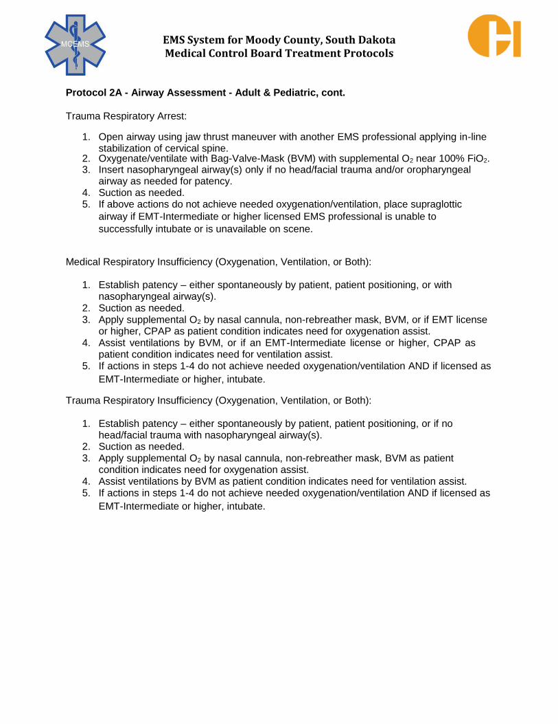
Trauma Respiratory Arrest:
1. Open airway using jaw thrust maneuver with another EMS professional applying in-line stabilization of cervical spine.
2. Oxygenate/ventilate with Bag-Valve-Mask (BVM) with supplemental O2 near 100% FiO2. 3. Insert nasopharyngeal airway(s) only if no head/facial trauma and/or oropharyngeal
airway as needed for patency. 4. Suction as needed. 5. If above actions do not achieve needed oxygenation/ventilation, place supraglottic
airway if EMT-Intermediate or higher licensed EMS professional is unable to
successfully intubate or is unavailable on scene.
Medical Respiratory Insufficiency (Oxygenation, Ventilation, or Both):
1. Establish patency – either spontaneously by patient, patient positioning, or with nasopharyngeal airway(s).
2. Suction as needed. 3. Apply supplemental O2 by nasal cannula, non-rebreather mask, BVM, or if EMT license
or higher, CPAP as patient condition indicates need for oxygenation assist. 4. Assist ventilations by BVM, or if an EMT-Intermediate license or higher, CPAP as
patient condition indicates need for ventilation assist. 5. If actions in steps 1-4 do not achieve needed oxygenation/ventilation AND if licensed as
EMT-Intermediate or higher, intubate. Trauma Respiratory Insufficiency (Oxygenation, Ventilation, or Both):
1. Establish patency – either spontaneously by patient, patient positioning, or if no head/facial trauma with nasopharyngeal airway(s).
2. Suction as needed. 3. Apply supplemental O2 by nasal cannula, non-rebreather mask, BVM as patient
condition indicates need for oxygenation assist. 4. Assist ventilations by BVM as patient condition indicates need for ventilation assist. 5. If actions in steps 1-4 do not achieve needed oxygenation/ventilation AND if licensed as
EMT-Intermediate or higher, intubate.

EMS System for Moody County, South Dakota Medical Control Board Treatment Protocols
EMERGENCY MEDICAL DISPATCHER
EMERGENCY MEDICAL RESPONDER
EMT
EMT – INTERMEDIATE
ADVANCED EMT
PARAMEDIC
EMD
VERIFY IF PATIENT IS CHOKING
AVOID BACK SLAPS ENCOURAGE COUGHING AND BREATHING EFFORTS
INSTRUCT CALLER IN HEIMLICH MANEUVER IF INDICATED
EMR EMT
GENERAL SUPPORTIVE CARE
ADULTS: HEIMLICH MANEUVER OR ABDOMINAL THRUSTS IF SUPINE (CHEST COMPRESSIONS IF PREGNANT OR MORBID OBESITY)
PEDIATRIC: HEIMLICH MANEUVER OR ABDOMINAL THRUSTS IF SUPINE (ALTERNATE 5 CHEST COMPRESSIONS WITH 5 BACKSLAPS IF <1 YR. OLD)
OBTAIN VITAL SIGNS
O2 VIA NC, NRB, OR BVM AS APPROPRIATE APPLY CARDIAC MONITOR (if equipped)
EMT OR HIGHER LICENSE:
MEASURE END – TIDAL C02 & MONITOR WAVEFORM CAPNOGRAPHY (if equipped, **Mandatory use if Pt intubated) PLACE SUPRAGLOTTIC AIRWAY IF INDICATED & ONLY IF BVM VENTILATIONS INEFFECTIVE
EMT – I AEMT
DIRECT LARYNGOSCOPY & REMOVAL OF FOREIGN BODY
ADULT: INTUBATE IF INDICATED
IV ACCESS (IF NEEDED)
PARAMEDIC
ADULT: MEDICATION ASSISTED INTUBATION IF INDICATED ADULT: CRICOTHYROTOMY FOR COMPLETE, INTRACTABLE OBSTRUCTION
PEDIATRIC: PT>6 YRS OLD, CRICOTHYROTOMY FOR COMPLETE, INTRACTABLE OBSTRUCTION
CONTINUOUS TREATMENT AND ASSESSMENT PER APPLICABLE PROTOCOL(S)
CONSULT MC IF AIRWAY OBSTRUCTION PERSISTS DESPITE ABOVE MEASURES
2B – AIRWAY ESTABLISHMENT/OBSTRUCITON
MANAGEMENT ADULT & PEDIATRIC TREATMENT PRIORITIES 1. Remove obstruction 2. Oxygenation/Ventilation support

EMS System for Moody County, South Dakota Medical Control Board Treatment Protocols
Indications:
1. Trauma to the face and/or upper airway, with potential or actual airway obstruction. 2. Vomitus, food boluses or other liquid foreign material in airway. 3. Excess secretions or pulmonary edema fluid in upper airway (or lungs with endotracheal
tube in place). 4. Amniotic fluid in naso/oropharynx of newborn with obvious obstruction to
spontaneous breathing or who require positive-pressure ventilation. 5. Meconium in naso/oropharynx of non-vigorous newborn.
Contraindications:
1. Airway patency effective without additional suctioning assistance. 2. Amniotic fluid or meconium in naso/oropharynx of vigorous, non-dyspneic newborn.
Technique:
1. Open airway and inspect for visible foreign material. 2. Turn patient on side if possible to facilitate clearance of liquid foreign material. 3. Remove large or obvious foreign particulates with gloved hands. Sweep finger ACROSS
posterior pharynx and clear material out of mouth in adults or if visible material in pediatrics.
4. Power on suction machine. 5. Suction of oropharynx:
A. Attach tonsil tip (or use open end of suction tubing for large amounts of debris). B. Oxygenate and ventilate the patient prior to the procedure as needed. C. Insert tip into oropharynx under direct vision, with sweeping motion. D. Continue intermittent suction interspersed with active oxygenation by mask. Use
positive pressure ventilation if needed. E. If suction becomes clogged, dilute by suctioning water or normal saline to clean
tubing. If suction clogs repeatedly, use connecting tubing alone, or manually remove
large debris.
EMERGENCY MEDICAL RESPONDER
EMT
EMT – INTERMEDIATE
ADVANCED EMT
PARAMEDIC
2C – AIRWAY SUCTIONING
ADULT & PEDIATRIC

EMS System for Moody County, South Dakota Medical Control Board Treatment Protocols
PROTOCOL 2C: Airway Suctioning – Adult & Pediatric, cont.
Technique, cont.:
6. Catheter suction of endotracheal tube:
A. Attach suction catheter to tubing of suction device (leaving suction end in sterile container).
B. Ventilate patient 4 - 5 times for pre-suction oxygenation. C. Detach bag from endotracheal tube and insert sterile tip of suction catheter without
suction. D. When catheter tip has been gently advanced to estimated carina depth, apply
suction and withdraw catheter slowly. E. Rinse catheter tip in sterile water or normal saline. F. Ventilate patient before each suction attempt.
Precautions:
1. Suctioning, particularly through endotracheal tubes, always risks suctioning the available oxygen as well as the fluid from the airway. In most situations, limit the suction time to a few seconds while the catheter is being withdrawn. This precaution should NOT be followed when vomitus or other material continues to well up and completely obstruct airway. Then suctioning must be continued until an airway is reestablished, with intermittent oxygenation and ventilation performed to avoid prolonged lack of oxygen.
2. Use equipment large enough for the job at hand. Large, solid matter will not be cleared
out with hard tonsil suckers. Large amounts of particulate matter require open-ended
suction using connecting tubing and physical removal with a gloved hand (using bite
precautions).
3. The catheter and tubing will require frequent rinsing with water or normal saline to permit
continued suctioning. Have a container of water or normal saline at hand before you
begin. Use gauze to remove large material from the end of the catheter.
4. Do not insert a suction catheter with the suction functioning. Suction only on withdrawal of
the catheter. Complications:
1. Hypoxia due to excessive suctioning time without adequate ventilation between attempts. 2. Persistent obstruction due to inadequate tubing size for removal of debris. 3. Lung injury from aspiration of stomach contents due to inadequate suctioning. 4. Asphyxia due to recurrent obstruction if airway is not monitored after initial suctioning. 5. Trauma to the posterior pharynx from forced use of equipment. 6. Vomiting and aspiration from stimulation of gag reflex. 7. Induction of cardio-respiratory arrest from vagal stimulation.

EMS System for Moody County, South Dakota Medical Control Board Treatment Protocols
Indications: 1. Respiratory arrest. 2. Inadequate oxygenation/ventilation not improved by non-positive pressure methods or
immediately obvious that will not improve by non-positive pressure methods. Contraindications:
1. Acute dyspnea of lesser severity able to be managed without BVM management 2. Active or suspected impending emesis
Technique:
Utilize the following mnemonic to guide correct BVM management:
C Hold mask by e-clamp (Figure 1) formed by one, preferably both hands
O Use an oropharyngeal and/or nasopharyngeal airway(s) P Place in a sniffing position to open the airway (***unless spinal injury suspected) E Elevate the jaw to additionally open the airway S Seal the mask over the mouth and nose without excessive downward force
Use 100% oxygen concentration (FiO2 = 1.0) to start and titrate down as indicated.
Squeeze the bag slowly and smoothly (over 1 second ventilation periods) delivering
adequate ventilation volume (approx. 6- 8 mL of air/kg if respiratory/cardiac arrest or shock; 8-
10 mL of air/kg up to 1000 mL if non-shock hemodynamics) and provide adequate exhalation
time.
EMERGENCY MEDICAL RESPONDER
EMT
EMT – INTERMEDIATE
ADVANCED EMT
PARAMEDIC
2D – BAG VALVE MASK (BVM) MANAGEMENT
ADULT & PEDIATRIC
Figure 1

EMS System for Moody County, South Dakota Medical Control Board Treatment Protocols
PROTOCOL 2D: Bag Valve Mask (BVM) Management – Adult & Pediatric, cont.
BVM technique that promotes optimal oxygenation/ventilation takes two, sometimes three EMS
professionals to achieve.
Utilization of the above technique will promote improved oxygenation/ventilation, while reducing
potential for gastric insufflation, vomiting, and aspiration.
Utilize the flowchart below to guide BVM management ventilation rates:
Consider the addition of a nasal canulla when utilizing a BVM in preparation of intubation to
promote oxygenation of “dead-air” space in a patient’s lungs (Figure 2.)
Figure 2

EMS System for Moody County, South Dakota Medical Control Board Treatment Protocols
Indications: 1. Hypoxia and/or hypoventilation refractory to non-invasive airway/respiratory management. 2. Airway protection to reduce aspiration in the setting of sustained altered mental status with a
Glasgow Coma Scale Score < 8. 3. Three unsuccessful oral and/or nasal intubation attempts in the above settings. An
intubation attempt has occurred when the tip of the endotracheal tube is advanced beyond
the gum line or into a nare. Attempts are counted per patient not per intubator. It is not
necessary to first attempt intubation if a difficult airway is anticipated or visualized. A
supraglottic airway may be used as the first–line airway in these cases.
Contraindications:
1. Ability to maintain oxygenation and ventilation by less invasive methods, such as Bag-Valve-
Mask ventilation. 2. Intact gag reflex 3. Known esophageal disease 4. Ingestion of caustic substance (e.g. lye, acids) or extensive airway burns 5. Tracheotomy or laryngectomy 6. Suspected Foreign Body Airway Obstruction 7. (Relative Contraindication): Patient size outside of manufacturer recommended range for
airway size used. The supraglottic airway may be utilized in such patients if the fit of the
airway allows for appropriate oxygenation and ventilation of the patient. Precaution:
Medical literature indicates concerns regarding reduction in cerebral arterial flow and
impedance of cerebral venous return due to pressure effects of supraglottic airways.
Supraglottic airways should not be utilized when other methods of airway management
are capable of achieving needed oxygenation/ventilation.
EMT
EMT – INTERMEDIATE
ADVANCED EMT
PARAMEDIC
2E – SUPRAGLOTTIC AIRWAYS
ADULT & PEDIATRIC

EMS System for Moody County, South Dakota Medical Control Board Treatment Protocols
PROTOCOL 2E: Supraglottic Airways – Adult & Pediatric, cont.
Technique (King LT-D™/LTS-D™):
Patient Size King Airway Size 15 mm Connector Color Typical Cuff Inflation
35 - 45 inches height 2 Green 25 – 35 mL
or 12-25 kg
41 - 51 inches height 2.5 Orange 30-40 mL
or 25-35 kg
4 ft – 5 ft height 3 Yellow 45 – 60 mL
5 ft – 6 ft height 4 Red 60 – 80 mL
6 ft + height 5 Purple 70 – 90 mL
The King LT-D™/LTS-D™ Airway has two cuffs that inflate from one port. The smaller, distal
cuff inflates in the esophagus and serves to isolate the laryngopharynx from the esophagus.
The larger, proximal cuff inflates at the base of the tongue and serves to isolate the
laryngopharynx from the oropharynx and nasopharynx.
To prepare the King LT-D™/LTS-D™ Airway:
• Test cuffs inflation by injecting air into the cuffs through the inflation port. • Remove all air from cuffs prior to insertion. • If lubricant is applied to the posterior aspect of the tube, take care to avoid
the introduction of lubricant in or near the ventilation portals in the airway.
Inflation Port
Proximal Cuff
Distal Cuff
Illustration of Correct Placement of King LD-DTM Airway (Size 4 Shown)

EMS System for Moody County, South Dakota Medical Control Board Treatment Protocols
PROTOCOL 2E: Supraglottic Airways – Adult & Pediatric, cont.
Hold the King LT-D™/LTS-D™ Airway at the connector with dominant hand (right hand dominant depicted)
With non – dominant hand, hold mouth open and apply chin lift, unless contraindicated by C – spine precautions or patient position
With a lateral approach from the right, introduce tip into mouth
Laryngoscope(by EMT- I85 or higher license) may allow easier oropharynx passage
Advance the tip behind the base of the tongue while rotating tube back to midline, so that the blue orientation line faces the chin of the patient
Attach bag-valve to King LT-DTM/LTS-DTM Airway
Gently ventilate the patient while withdrawing the tube until ventilation is easy (without significant resistance)
Confirm proper position by auscultation of epigastrum and chest, physiologic changes, and chest, physiologic changes, and waveform capnography if equipped; capnography is not required for King LT-DTM/LTS-DTM Airway placement
Without exerting excessive force, advance tube until base of connector is aligned with teeth or gums
Inflate cuffs with supplied syringe – use minimum mL necessary to achieve seal for appropriate oxygenation/ventilation.
Excessive cuff inflation may compromise cerebral blood flow!

EMS System for Moody County, South Dakota Medical Control Board Treatment Protocols
PROTOCOL 2E: Supraglottic Airways – Adult & Pediatric, cont.
Removal of the KING LT-D™/LTS-D™ Airway:
1. Once in correct position, the KING LT-D™/LTS-D™ Airway should be well tolerated until
return of airway reflexes.
2. Suction MUST always be available when a King LT-D™/LTS-D™ Airway is removed.
Anticipate vomiting with removal, positioning patient in lateral recumbent position unless
contraindicated. A suction catheter up to an 18 French size can be inserted through the
gastric access lumen of the King LTS-D™.
3. Completely deflate cuffs prior to removal.
Additional Information:
1. If unable to place a King LT-D™/LTS-D™ Airway in three attempts, utilize BVM ventilation. 2. Ventilation portals of the King LT-D™/LTS-D™ Airway must align with the laryngeal inlet for
adequate oxygenation and ventilation. Insertion depth should be adjusted to optimize
ventilation.
3. Ensure cuffs are not over inflated. Inflate the cuffs with the minimum volume necessary to
seal the airway. If the patient becomes more alert, it may be helpful in retaining the tube to
remove a slight amount of air from the cuffs. 4. Most unsuccessful insertion attempts relate to the failure to keep the tube in a midline
position during insertion. 5. Do not force the tube during insertion; this may result in trauma to the airway or esophagus. 6. Document any complications as well as all methods used to ensure appropriate placement of
the King LT-D™/LTS-D™ Airway including auscultation of absence of epigastric sounds and
presence of lung sounds, physiologic changes (chest rise and fall, improved oxygenation,
condensation in King LT-D™/LTS-D™ Airway with exhalations), and waveform capnography
readings (if equipped). 7. Assess and document placement verification of the King LT-D™/LTS-D™ Airway after
patient moves and periodically throughout care and transportation.

EMS System for Moody County, South Dakota Medical Control Board Treatment Protocols
Indications: 1. Hypoxia and/or hypoventilation refractory to non-invasive airway/respiratory management. 2. Airway protection to minimize aspiration in the setting of sustained altered mental status with
a Glasgow Coma Scale Score <8. 3. Impending airway edema in the setting of respiratory tract burns or anaphylaxis. Contraindications: 1. Three unsuccessful oral and/or nasal intubation attempts in the above settings. An
intubation attempt has occurred when the tip of the endotracheal tube is advanced beyond the gum line or into a nare. Attempts are counted per patient not per intubator.
2. Waveform capnography not immediately available. Technique: 1. In pulsatile (non-cardiac arrest) patients, provide supplemental oxygenation throughout the
intubation process with nasal cannula oxygen delivery at 15 lpm flow. While this flow rate is much higher than typical nasal cannula oxygen flow rates, the additional force of 15 lpm will help to reduce intra-intubation oxygen desaturation/hypoxia.
2. Walk the laryngoscope down the tongue to avoid placing the laryngoscope in the esophagus.
3. If unable to lift the mandible with the laryngoscope, place your left forearm on the pt’s head for leverage.
4. If the vocal cords are poorly visualized in any patient, manipulate the thyroid cartilage with your right hand until appropriate visualization is achieved. Have a colleague hold the thyroid cartilage in this place while you finish intubating. This technique is referred to as "bimanual laryngoscopy" and works much more reliably than cricoid pressure.
5. If the vocal cords are still poorly visualized in obese patients without suspected spinal injury, elevate their head/neck/shoulders. Place blankets or pillows under the head/neck/shoulders until the patient’s chin or nose is level with the chest.
6. If ambient light inhibits visualization of the larynx, block this light by any means possible, including a blanket stretched over your head and the patient’s head and neck.
7. In adult patients of appropriate size, strong preference is given for using the 8.0 mm endotracheal tube for orotracheal intubation. Use of this sized tube enables inpatient pulmonary care that might not be performed with smaller sized tubes.
EMT – INTERMEDIATE
ADVANCED EMT
PARAMEDIC
2F – ORAL INTUBATION
ADULT

EMS System for Moody County, South Dakota Medical Control Board Treatment Protocols
PROTOCOL 2F: Oral Intubation – Adult, cont. The Flex-Guide™ Introducer (also known as the Gum Elastic Bougie): The Flex-Guide™ Introducer (Figure 1) is a single patient use, semi-rigid plastic rod with an angled tip, promoting glottic passage when the vocal cords are completely visible during laryngoscopy. A 1 cm wide black band is located along the Flex- Guide™ to help determine correct placement depth. The Flex-Guide™ shape and elasticity allow the intubator to feel a “washboard” sensation as the anteriorly-angled tip is advanced down the tracheal rings. Failure to feel a "washboard" sensation indicates inadvertent esophageal placement and the Flex-Guide™ must be fully withdrawn before reattempting placement. The Flex-Guide™ length allows it to be advanced to the carina where resistance is met, also a means of confirming tracheal rather than esophageal placement. Avoid storing the Flex-Guide™ coiled, as it works best in these regards when it is straight. The Flex-Guide™ also comes in a pediatric size for use with those patients. Flex-Guide™ Introducer Technique: 1. Advance the angled tip facing anteriorly, with continual visualization by laryngoscopy.
Anytime resistance is met, stop advancing and reassess placement - forceful passage can result in perforation of soft tissues.
2. Stabilize the Flex-Guide™ when in place, while maintaining laryngoscopy 3. Direct a colleague to slide the endotracheal tube over the Flex-Guide™. He or she stabilizes
the proximal end of the Flex-Guide™ as it emerges from the sliding endotracheal tube. 4. Take control of the endotracheal tube, sliding it down the Flex-Guide™ length, while being
careful to avoid Flex-Guide™ migration. Once the endotracheal tube has passed to an appropriate estimated endotracheal depth, stabilize it while your colleague withdraws the Flex-Guide™ prior to laryngoscope removal.
Figure 1

EMS System for Moody County, South Dakota Medical Control Board Treatment Protocols
PROTOCOL 2F: Oral Intubation – Adult, cont. Confirmation of Oral Endotracheal Placement: The following sequence is to be used (and its use documented) to verify and maintain correct oral endotracheal placement without fail: 1. Visualization of endotracheal tube passage between the vocal cords. 2. Detection of End-tidal carbon dioxide. End-tidal carbon dioxide (EtC02) detection shall be
confirmed within 60 seconds of endotracheal tube placement. The capnography adaptor is to be placed at the bag-valve device-endotracheal tube interface for the first ventilation. The normal waveform indicating correct endotracheal placement reflects a rapid upstroke with the beginning of exhalation, the exhalation plateau ending at the point of EtC02 measurement, and a rapid downstroke with the beginning of inhalation. Any waveform that does not show rhythmic rise and fall correlating with assisted ventilations indicates incorrect tube placement and the tube must be withdrawn. To be perfectly clear, the use of an endotracheal tube for ongoing oxygenation and ventilation is dependent upon continuously measurable capnography waveforms. See Protocol 3H-Capnography for discussion of EtCO2 values.
3. Auscultation. Auscultate the epigastrum. If epigastric sounds are heard, intubation is to be reattempted. The endotracheal tube placed in the esophagus may be left in place, at the intubator’s discretion, until another endotracheal tube is correctly placed and verified. If no epigastric sounds are heard, proceed to auscultation of the thorax bilaterally. Breath sounds are best auscultated in the anterior to mid axillary lines. If breath sounds are present on the right and absent on the left, this suggests a right main stem intubation. Withdraw the endotracheal tube 1cm and repeat auscultation. If necessary, the tube may be withdrawn an additional 1-2cm.
4. Assessment of physiologic changes. These include equal rise and fall of the chest, condensation in the endotracheal tube on exhalation, improvement in the patient’s color, and improvement in the patient’s respiratory distress or failure.
5. Secure the endotracheal tube with a tube holder and place a cervical collar. When intubated patients are moved during EMS care, waveform capnography must be rechecked for any change. If the waveform continues to show a normal pattern of rapid upstroke with exhalation, exhalation plateau, and rapid downstroke with inhalation, no further repeat confirmation is required. If at any time, the capnography waveform is abnormal, steps 2-5 must be rechecked and documented. If at any time during patient care there is doubt as to correct endotracheal placement of intubation, you must either re-verify by this sequence or reattempt correct endotracheal placement. While the intubator may delegate confirmation steps to his/her colleagues, he or she is ultimately responsible to ensure that a complete confirmation sequence is performed. If the intubator accompanies the patient to the hospital, he or she remains ultimately responsible for ongoing endotracheal tube placement confirmation. If the intubator does not accompany the patient to the hospital by ambulance or helicopter ambulance transport, the primary transporting/treating paramedic or RN assumes ultimate responsibility for ongoing endotracheal tube placement confirmation.

EMS System for Moody County, South Dakota Medical Control Board Treatment Protocols
PARAMEDIC
PARAMEDIC
MEDICATION-ASSISTED INTUBTION IF INDICATED
FOLLOW PROTOCOL 2F- ORAL INTUBATION FOR TECHNIQUE & CONFIRMATION INTUBATION
FOR FACILITATING ORAL INTUBATION: ADULT: MIDAZOLAM 0.1 mg/kg TO MAX OF 5 mg IVP/IOP
MAY REPEAT ONCE IF ADULT SYS BP ≥ 100 mmHg
FOR POST-ORAL ITUBATIO SEDATION TO PREVENT EXTUBATION (IF INDICATED): ADULT: MIDAZOLAM 0.1 mg/kg TO MAX OF 5 mg IVP/IOP
MAY REPEAT OCE IF ADULT SYS BP ≥ 100 mmHg OR
DIAZEPAM 0.1 mg/kg TO MAX OF 5 mg IVP/IOP MAY REPEAT ONCE IF ADULT SYS BP ≥ 100 mmHg
OR LORAZEPAM 0.1 mg/kg TO MAX OF 2 mg IVP/IOP
MAY REPEAT ONCE IF ADULT SYS BP ≥ 100 mmHg
PEDIATRIC MEDICATION ASSISTED INTUBATION: CONSULT MC
CONTINUOUS ASSESSMENT & TREATMENT PER APPLICABLE PROTOCOL(S)
2G – MEDICATION ASSISTED INTUBATION
ADULT & PEDIATRIC TREATMENT PRIORITIES 1. Oxygenation/Ventilation Support

EMS System for Moody County, South Dakota Medical Control Board Treatment Protocols
Indications: 1. Hypoxia and/or hypoventilation refractory to non-invasive airway/respiratory management. 2. Airway protection to minimize aspiration in the setting of sustained altered mental status with
a Glasgow Coma Scale Score <8. 3. Impending airway edema in the setting of respiratory tract burns or anaphylaxis. 4. Patients more compliant with intubation attempts in a sitting position. 5. Oral anatomy, injury, or jaw clenching preventing indicated orotracheal intubation. Contraindications: 1. Apnea. 2. Pediatric patients (age ≤12 years). 3. Suspected basilar skull fracture. 4. Mid-facial injuries with bony instability. 5. Combativeness preventing patient compliance. 6. Anticoagulant use (Warfarin/Coumadin, Plavix, or Aspirin) - Relative contraindication -
orotracheal intubation preferred to minimize bleeding complications 7. Three unsuccessful oral and/or nasal intubation attempts in the above settings. An
intubation attempt has occurred when the tip of the endotracheal tube is advanced beyond the gum line or into a nare. Attempts are counted per patient not per intubator.
8. Waveform capnography not immediately available. Technique: 1. If available, apply two sprays of phenylephrine 2% in each nare to induce local
vasoconstriction. This will enlarge the nares and decrease epistaxis complications. 2. If available, apply lidocaine 2% gel to the endotracheal tube cuff. 3. Insert the well-lubricated tube along the floor of the most patent nare, bevel side facing
inward toward the septum. This positioning will prevent a turbinate from being trapped in the tube and subsequently being sheared off as the tube is advanced. Pass the tube straight back (not angulated upward) with constant, gentle pressure. Do not use an endotracheal stylet in nasotracheal intubations.
4. As the tube is advanced, there is a loss of resistance as the tube passes from the nasopharynx into the oropharynx. Continue advancing the tube.
5. As the tube nears the glottis, guide the tube by listening at the adaptor. The awake patient should be instructed to deeply inspire to help guide the tube through the vocal cords and into the trachea. Correct endotracheal placement may also be assisted by rotating the tube 90 degrees so that the bevel is up and facing the glottis.
6. Once the tube has been placed, the patient should not be capable of phonation. The ability to speak after "nasotracheal intubation" actually denotes "nasoesophageal intubation." In such cases, the tube is to be slightly withdrawn and correct placement reattempted. The Flex-Guide™ may NOT be used for difficult nasotracheal intubations.
EMT – INTERMEDIATE
ADVANCED EMT
PARAMEDIC
2H – NASAL INTUBTION
ADULT

EMS System for Moody County, South Dakota Medical Control Board Treatment Protocols
PROTOCOL 2H: Nasal Intubation – Adult, cont. Confirmation of Nasal Endotracheal Placement: The following sequence is to be used (and its use documented) to verify and maintain correct nasal endotracheal placement without fail: 1. Detection of End-tidal carbon dioxide. End-tidal carbon dioxide (EtC02) detection shall be
confirmed within 60 seconds of endotracheal tube placement. The capnography adaptor is to be placed at the bag-valve device-endotracheal tube interface for the first ventilation. The normal waveform indicating correct endotracheal placement reflects a rapid upstroke with the beginning of exhalation, the exhalation plateau ending at the point of EtC02 measurement, and a rapid down stroke with the beginning of inhalation. Any waveform that does not show rhythmic rise and fall correlating with assisted ventilations indicates incorrect tube placement and the tube must be withdrawn. To be perfectly clear, the use of an endotracheal tube for ongoing oxygenation and ventilation is dependent upon continuously measurable capnography waveforms. See Protocol 3H -Capnography for discussion of EtCO2 values.
2. Auscultation. Auscultate the epigastrum. If epigastric sounds are heard, intubation is to be reattempted. If no epigastric sounds are heard, proceed to auscultation of the thorax bilaterally. Breath sounds are best auscultated in the anterior to mid axillary lines. If breath sounds are present on the right and absent on the left, this suggests a right main stem intubation. Withdraw the endotracheal tube 1 cm and repeat breath sound auscultation. If necessary, the tube may be withdrawn an additional 1-2 cm.
3. Assessment of physiologic changes. These include equal rise and fall of the chest, condensation in the endotracheal tube on exhalation, improvement in the patient’s color, and improvement in the patient’s respiratory distress or failure.
4. Secure the endotracheal tube with tape and place a cervical collar. When intubated patients are moved during EMS care, waveform capnography must be rechecked for any change. If the waveform continues to show a normal pattern of rapid upstroke with exhalation, exhalation plateau, and rapid down stroke with inhalation, no further repeat confirmation is required. If at any time, the capnography waveform is abnormal, steps 1-4 must be rechecked and documented. If at any time during patient care there is doubt as to correct endotracheal placement of intubation, you must either reverify by this sequence or reattempt correct endotracheal placement
While the intubator may delegate confirmation steps to his/her colleagues, he or she is ultimately responsible to ensure that a complete confirmation sequence is performed. If the intubator accompanies the patient to the hospital, he or she remains ultimately responsible for ongoing endotracheal tube placement confirmation. If the intubator does not accompany the patient to the hospital by ambulance or helicopter ambulance transport, the primary transporting/treating paramedic or RN assumes ultimate responsibility for ongoing endotracheal tube placement confirmation.

EMS System for Moody County, South Dakota Medical Control Board Treatment Protocols
Indications: 1. Upper airway obstruction (e.g. facial or neck trauma occluding airway patency, foreign body
unable to be removed, angioedema) and inability to adequately oxygenate and ventilate using less invasive methods.
Contraindications: 1. Ability to oxygenate and ventilate using less invasive methods. 2. Infant and younger pediatrics – airway anatomical size not conducive to successful
cricothyrotomy in EMS care. Contact MC for direction in these ages. 3. Older pediatrics – airway anatomical size MAY not be conductive to successful
cricothyrotomy in EMS care. Contact MC for direction in these ages. 4. Suspected fractured larynx and/or cricoid cartilage. 5. Suspected tracheal transaction with retraction of the trachea into the chest. 6. Inability to find anatomical landmarks
PARAMEDIC 2I – CRICOTHYROTOMY
ADULT
Do not confuse the hyoid bone for the thyroid cartilage. Attempted placement of a cricothyrotomy airway superior to the thyroid cartilage will cause anatomical disruption and will NOT establish a secure airway capable of needed oxygenation and ventilation. The hyoid bone does NOT have the distinct notch of the thyroid cartilage.
Cricothyroid membrane

EMS System for Moody County, South Dakota Medical Control Board Treatment Protocols
PROTOCOL 2I: Emergency Cricothyrotomy – Adult, cont. Surgical Technique (6.0 endotracheal tube and tracheal hook): 1. Establish adequate space and lighting. Do not attempt cricothyrotomy in poorly visualized
conditions. 2. If rapidly available, clean anterior neck with ChloraPrep®, Betadine®, or alcohol wipes. 3. Definitively locate the following landmarks: thyroid cartilage (“Adam’s apple”) and cricoid
cartilage. The cricothyroid membrane lies between these cartilages.
4. Using the non-dominant hand, spread the overlying skin taut with the thumb and fingers, and slightly depress the skin over the cricothyroid membrane with the index finger to mark the site of cricothyrotomy. Do not release the non-dominant hand from the neck until the procedure is complete! Once the anatomy is found and defined, avoid movement of the anatomy to promote proper cricothyrotomy airway placement.
5. Stabilization of the anatomy requires assistance from a second EMS professional, preferably licensed as a paramedic as well.
6. Ask second EMS professional to aspirate all air from the endotracheal tube cuff. 7. Using a sterile scalpel, make a vertical incision in the mid-line of the neck extending from
just above the lower edge of the thyroid cartilage to the middle of the cricoid cartilage. Make the depth of this incision sufficient to extend through the skin and fatty tissue underneath.
8. Using sterile hemostats spread the incision open horizontally to expose the cricothyroid membrane. Instruct the second EMS professional to hold the hemostats in this position.
9. Using the same scalpel as in Step G, now make a short horizontal incision in the middle of the cricothyroid membrane. There is a small artery running vertically on each side of the cricothyroid membrane. Keeping the horizontal incision less than ½ inch (approx. 1 cm) will decrease bleeding that may occur.
10. Pass the 6.0 mm endotracheal tube through the horizontal incision in the cricothyroid membrane, angling the tube inferior and posterior along the tracheal anatomy. A “washboard” sensation may be felt as the tube slides along the tracheal wall. Avoid excessive pressure in placing the endotracheal tube, but a moderate degree may be required to first pass the endotracheal tube through the cricothyroid membrane. If significant resistance is encountered (without suspicion of lower respiratory tract foreign body), the hemostats used in Step H may be used to spread the cricothyroid membrane incision vertically while the endotracheal tube is passed through it and/or use of the tracheal hook may better stabilize the anatomy to overcome resistance to airway passage.
11. Inflate the endotracheal cuff and verify airway placement per Protocol 2J – Confirmation of Artificial Airway Placement.
12. Secure the airway using a cloth tie or commercial endotracheal tube restraint while continuing oxygenation and ventilation. Artificial ventilation will generally be easier if the endotracheal tube is cut to a shorter length. Be careful to cut the upper aspect of the endotracheal tube above the insertion site of the cuff inflation portal to avoid irreversible cuff deflation.

EMS System for Moody County, South Dakota Medical Control Board Treatment Protocols
PROTOCOL 2I: Emergency Cricothyrotomy – Adult, cont. Non-Surgical Technique (PerTrach® Kit and tracheal hook): 1. Establish adequate space and lighting. Do not attempt cricothyrotomy in poorly visualized
conditions. 2. If rapidly available, clean anterior neck with ChloraPrep®, Betadine®, or alcohol wipes. 3. Definitively locate the following landmarks: thyroid cartilage (“Adam’s apple”) and cricoid
cartilage. The cricothyroid membrane lies between these cartilages in the neck midline. 4. Using the non-dominant hand, spread the overlying skin taut with the thumb and fingers,
and slightly depress the skin over the cricothyroid membrane with the index finger to mark the site of cricothyrotomy. Do not release the non-dominant hand from the neck until the procedure is complete! Once the anatomy is found and defined, avoid movement of the anatomy to promote proper cricothyrotomy airway placement.
5. Stabilization of the anatomy requires assistance from a second EMS professional, preferably licensed as a paramedic as well.
6. Ask second EMS professional to aspirate all air from the tracheostomy tube cuff. 7. Using the dominant hand on the break-away needle and syringe, puncture in the lower half
of the cricothyroid membrane, mid-line, at a 45 degree angle towards the chest (following the path of the airway from superior to inferior). Once air is aspirated in the syringe, advance another few millimeters in depth and remove the syringe.
8. Using the included small scalpel, make a single vertical “stab” incision immediately to one side of the needle.
9. Place a tracheal hook in the incision and position the hook to pull anterior and superior on the inferior border of the thyroid cartilage. Exercise caution when manipulating the tracheal hook into the incision – the tip of most tracheal hooks is particularly sharp-edged.
10. The second EMS professional should now control the tracheal hook. 11. Palming the airway and dilator stylet, advance through the needle until resistance is met.
The second EMS professional should split the needle by compressing and widening the “butterfly” tips on the needle and then remove each side of the needle. Constant inward/downward pressure on the airway and stylet must be maintained to avoid inadvertent displacement of the airway and ensure the tip of the airway will remain in the trachea.
12. Continue to advance the airway/stylet until the airway is fully in the trachea (airway passed to hub contact with overlying skin) and remove the tracheal hook.
13. Inflate the airway cuff and verify airway placement per Protocol 2J – Confirmation of Artificial Airway Placement.
14. Secure the airway using the cloth tie while continuing oxygenation and ventilation. Modified Non-Surgical Technique (PerTrach® Kit):
In patients with neck edema, subcutaneous air, or fat/obesity preventing necessary tactile identification of anatomical landmarks to perform standard non-surgical cricothyrotomy, utilize the following modification: 1. Using the included small scalpel, make a single, vertical, mid-line incision in the skin
overlying the area that is estimated to contain the thyroid cartilage, cricothyroid membrane, and cricoid cartilage. When making the incision, make an incision approximately 2 inches (5 cm) in length and deep enough that the subcutaneous fat can be visualized. Using a gloved index finger palpate the structures through the incision and when identified, proceed as per standard non-surgical cricothyrotomy.

EMS System for Moody County, South Dakota Medical Control Board Treatment Protocols
The following sequence is to be used (and its use documented) to verify and maintain correct endotracheal artificial airway placement without fail: 1. Visualization of endotracheal tube passage between vocal
cords – oral intubation only (Figure 1). 2. Detection of End-tidal carbon dioxide. End-tidal carbon dioxide
(EtCO2) detection shall be confirmed within 60 seconds of endotracheal tube placement. The capnography adaptor is to be placed at the bag-valve device endotracheal tube interface for the first ventilation. The normal waveform indicating correct endotracheal placement reflects a rapid upstroke with the beginning of exhalation, the exhalation plateau ending at the point of EtCO2 measurement, and a rapid down stroke with the beginning of inhalation. Any waveform that does not show rhythmic rise and fall correlating with assisted ventilations indicates incorrect tube placement and the tube must be withdrawn. To be perfectly clear, the use of an endotracheal tube for ongoing oxygenation and ventilation is dependent upon continuously measurable capnography waveforms. See Protocol 3H – Capnography for discussion of EtCO2 values and waveforms (Figure 2).
3. Auscultation. Auscultate the epigastrium (Figure 3). If
epigastric sounds are heard, intubation is to be reattempted. The endotracheal tube placed in the esophagus may be left in place, at the intubator’s discretion, until another endotracheal tube is correctly placed and verified. If no epigastric sounds are heard, proceed to auscultation of the thorax bilaterally. Breath sounds are best auscultated in the anterior to mid-axillary lines If breath sounds are present on the right and absent on the left, this suggests a right main stem intubation. Withdraw the endotracheal tube 1 cm and repeat breath sound auscultation. If necessary, the tube may be withdrawn an additional 1-2 cm.
4. Assessment of physiologic changes. These include equal rise
and fall of the chest, condensation I the endotracheal tube on exhalation, improvement in the patient’s color, and improvement in the patient’s respiratory distress/failure.
5. Secure the endotracheal tube with a tube holder and place a
cervical collar (Figure 4).
EMT – INTERMEDIATE
ADVANCED EMT
PARAMEDIC
2J – CONFIRMATION OF ENDOTRACHEAL AIRWAY PLACEMENT
ADULT & PEDIATRIC
Epiglottis
Vocal Cord Figure 1
Figure 2
Figure 3
Figure 4

EMS System for Moody County, South Dakota Medical Control Board Treatment Protocols
PROTOCOL 2J: Confirmation of Endotracheal Artificial Airway Placement – Adult When intubated patients are moved during EMS care, waveform capnography must be rechecked for any change. If the waveform continues to show a normal pattern of rapid upstroke with exhalation, exhalation plateau, and rapid down stroke with inhalation, no further repeat confirmation is required. If at any time, the capnography waveform is abnormal, steps 2-5 must be rechecked and documented. If at any time during patient care there is doubt as to correct endotracheal placement of intubation, either re-verify by this sequence or reattempt correct endotracheal placement. While the intubator may delegate confirmation steps to his/her colleagues, he or she is ultimately responsible to ensure that a complete confirmation sequences performed. If the intubator accompanies the patient to the hospital, he or she remains ultimately responsible for ongoing endotracheal tube placement confirmation. If the intubator does not accompany the patient to the hospital by ambulance or helicopter ambulance transport, the primary transporting/treating paramedic or RN assumes ultimate responsibility for ongoing endotracheal tube placement confirmation. Upon delivery of the patient at treatment destination or at subsequent transport (e.g. helicopter transport), a capnography waveform will be obtained and documented after the patient has been physically transferred onto the destination’s/subsequent transport’s stretcher/bed/operating table to show confirmed, continued correct endotracheal tube placement at EMS transfer of patient care

EMS System for Moody County, South Dakota Medical Control Board Treatment Protocols
Emergency Management: The majority of adults and children with tracheostomies are dependent on the tube as their primary airway. Cardio-respiratory arrest most commonly results from tracheostomy obstructions. Obstruction may be due to thick secretions/mucous plug, blood clot, foreign body, or kinking or dislodgement of the tube. Work expeditiously and deliberately to re-establish airway patency and support oxygenation/ventilation. Early warning signs of obstruction include tachypnea, tachycardia, and desaturation. Cyanosis, bradycardia and apnea are late signs – do not wait for these to develop before intervening. Complications: • Airway obstruction • Aspiration • Blocked tube • Bleeding • Tracheal trauma • Pneumothorax • Subcutaneous and mediastinal emphysema • Respiratory and cardiovascular collapse • Dislodged tube • Tracheo-esophageal fistula • Infection Endotracheal Suctioning: Endotracheal suctioning is necessary to remove mucus, maintain a patent airway, and avoid tracheostomy tube blockages. Indications for suctioning include: • Audible or visual signs of secretions in the tube • Suspicion of blocked or partially blocked tube • Inability to clear the tube by coughing out the secretions • Increases in required ventilation pressures (in ventilated patients) • Request by patient for suction • Tracheal suctioning should be carried out regularly for patients with a tracheostomy. The
frequency varies between patients and is based on individual assessment. • Tracheal damage may be caused by suctioning. This can be minimized by using the
appropriate sized suction catheter and only suctioning within the tracheostomy tube.
EMT
EMT – INTERMEDIATE
ADVANCED EMT
PARAMEDIC
2K – STOMA/TRACHEOSTOMY MANAGEMENT
ADULT & PEDIATRIC

EMS System for Moody County, South Dakota Medical Control Board Treatment Protocols
PROTOCOL 2K: Stoma/Tracheostomy Management– Adult & Pediatric, cont.
Table 1: recommended suction catheter sizes
Tracheostomy tube size (in mm) 3.0 3.5 4.0 4.5 5.0 6.0 7.0 7.5 8.0 9.0-10.0
Recommended suction catheter size (Fr)
7 8 8 10 10 10-12
14 14-16
14-16
16
• The suction depth is determined by the estimated length of the tracheostomy tube. • The depth of insertion of the suction catheter needs to be determined prior to
suctioning to avoid trauma. • Using the patient’s spare tracheostomy tube of the same size (if available) to
estimate needed depth of suctioning. • The pressure setting for tracheal suctioning (suction machine pressure for small children
50-100 mmHg, for older children/adults 100-120 mmHg) to avoid tracheal damage. • In most circumstances, it is best to limit the duration of suctioning (including passing
the catheter and suctioning the tracheostomy tube) to 5-10 seconds. • Routine use of normal saline is not necessary although there is anecdotal
evidence it may thin secretions. In situations where this may be of benefit, only
1-2 mL is usually needed.
Tracheal Suctioning Procedure: 1. Inform patient of intended action. 2. Maintain appropriate PPE throughout procedure. 3. Assemble needed suction equipment and power on suction device. 4. Instill small volume of sterile normal saline into the tracheostomy tube if needed for thick or
dry secretions. Excessive use of saline is not recommended. Use saline only if the mucus is very thick, hard to cough up or difficult to suction. Recommended amount per instillation is approximately 1-2 mL.
5. Gently insert catheter into the tracheal tube without applying suction, passing to the previously estimated needed depth.
6. Put thumb over opening in catheter to create suction and use a circular motion (twirl catheter between thumb and index finger) while withdrawing the catheter so that the mucus is removed well from all areas. Avoid suctioning longer than 10 seconds because of oxygen loss. Suction normal saline from a container if needed to clear catheter.
7. For tracheostomy tubes with cuffs, it may be necessary to deflate the cuff periodically for suctioning to prevent pooling of secretions above tracheal cuff.
8. Let patient rest and breathe, then repeat suction if needed until clear (trying to allow about 30 seconds between suctioning).
9. Oxygenate/ventilate as needed.

EMS System for Moody County, South Dakota Medical Control Board Treatment Protocols
PROTOCOL 2K: Stoma/Tracheostomy Management– Adult & Pediatric, cont. Tracheostomy tube tie changes: • There is a potential risk for tracheostomy tube dislodgment when attending to tie changes,
therefore two personnel who are competent in tracheostomy care should undertake tracheostomy tie changes.
• During the tracheostomy tie change one person is to maintain the airway by securing the tracheostomy tube in place and not removing the hand until the new tracheostomy ties are applied. The other person is to change the ties and attend to stoma care.
• If the tie becomes loose, make it is a priority to re-secure the tracheostomy tube before it can become dislodged.

EMS System for Moody County, South Dakota Medical Control Board Treatment Protocols
EMERGENCY MEDICAL DISPATCHER
EMERGENCY MEDICAL RESPONDER
EMT
EMT – INTERMEDIATE
ADVANCED EMT
PARAMEDIC
EMD
CPR BY EMD INSTRUCTION
EMR EMT
ESTABLISH AIRWAY PATENCY (POSITIONING, OPA, NPA) O2 VIA BVM AS APPROPRIATE GENERAL SUPPORTIVE CARE
OBTAIN VITAL SIGNS DETERMINE BLOOD GLUCOSE
APPLY CARDIAC MOITOR/OBAIN 12-LEAD ECG (If equipped) TRANSMIT 12-LEAD ECG TO RECEIVING EMERGENCY DEPARTMENT
EMT OR HIGHER LICENSE: MEASURE END-TIDAL CO2 & MONITOR WAVEFORM CAPNOGRAPY (If equipped, **Mandatory use if Pt.
Intubated) PLACE SUPRAGLOTTIC AIRWAY IF INDICATED & ONLY IF BVM VENTILATIONS INEFFECTIVE
EMT – I AEMT
ADULT: INTUBATE IF INDICATED DO NOT INTUBATE PATIENTS WITH RAPIDLY REVERSIBLE RESP ARREST ETIOLOGY (e.g. NARCOTIC/OPIATE OERDOSE)
IV/IO ACCESS
ADULT: IV NSTKO IF SYS BP ≥ 100 mmHg WITHOUT HYPOTENSIVE SYMPTOMS IV NS 250 mL BOLUS IF SYS BP < 100 mmHg WITH HYPOTENSIVE SYMPTOMS & NO SIGNS OF PULMONARY EDEMA
REPEAT UP TO 2 LITERS NS IF SYSP REMAINS < 100 mmHg WITH HPOTENSIVE SYMPTOMS & NO SIGNS OF PULMONARY EDEMA
PEDIATRIC: IV NS TKO IF SYSBP ≥ (70 + 2x age in years) mmHg IV NS 20 mL/kg BOLUS IF SYS BP < (70 + 2x age in years) mmHg IF NO SIGNS OF PULMONARY EDEMA
ADULT & PEDIATRICWEIGHT ≥ 25 kg HYPOGLYCEMIA CARE:
IF GLUCOSE < 50 mg/dL, D50 1 mL/kg IVP UP TO 50 mL GLUCAGON 1 mg IM IF NO VASCULAR ACCESS OBTAINED
PEDITRIC WEIGHT < 25 kg HYPOGLYCEMIA CARE: IF GLUCOSE < 50 mg/dL, D25 2 mL/kg IVP UP TO 50 mL
GLUCAGON 0.5 mg IM IF NO VASCULAR ACCESS OBTAINED
ADULT & PEDIATRIC: REPEAT DETERMINATION OF BLOOD GLUCOSE POST-DEXTROSE TREATMENT
TOXINS/DRUG OVERDOSE – SUSPECTED NARCOTIC/OPIATE – APNEIC
ADULT: NALOXONE 2 mg IVP/IOP/IN, MAY REPEAT TO MAX OF 2 mg PEDIATRIC: NALOXONE 0.5 mg IVP/IOP/IN, MAY REPEAT TO MAX OF 2 mg
USE NALOXONE TO RESTORE EFFECTIVE BREATHING; AVOID EXCESSIVE DOSING TO PREVENT WITHDRAWAL
PARAMEDIC
ADULT: MEDICATON ASSISTED INTUBATION IF INDICATED
CONTINUOUS ASSESSMENT & TREATMENT OF SUSPECTED RESP ARREST ETIOLOGY PER APPLICABLE PROTOCOL(S)
3A – RESPIRATORY ARREST
ADULT & PEDIATRIC TREATMENT PRIORITIES 1. Airway Patency 2. Oxygenation/Ventilation 3. Vital signs 4. Dextrose for hypoglycemia 5. Naloxone for narcotic/opiate
overdose

EMS System for Moody County, South Dakota Medical Control Board Treatment Protocols
EMERGENCY MEDICAL DISPATCHER
EMERGENCY MEDICAL RESPONDER
EMT
EMT – INTERMEDIATE
ADVANCED EMT
PARAMEDIC
EMD
ADVISE TO AVOID PHYSICAL EXERTION OR ENVIRONMENTAL STRESS (e.g. TEMPERATURE EXTREMES.)
ADVISE PT SELF-ADMINISTRATION OF MEDICATIONS (e.g. ALBUTEROL INHALER) IF PREVIOUSLY PRESCRIBED FOR SIMILAR
SYMPTOMS
EMR EMT
GENERAL SUPPORTIVE CARE OBTAIN VITAL SIGNS
O2 VIA NC, NRB, OR BVM AS APPROPRIATE APPLY CARDIAC MONITOR (if equipped)
OBTAIN 12-LEAD ECG & TRANSMIT TO RECEIVING EMERGENCY DEPARTMENT (if equipped) ASSIST PT WITH PT’S OWN ALBUTEROL INHALER/NEBULIZER (when applicable)
EMT OR HIGHER LICENSE: MEASURE END-TIDAL CO2 & MONITOR WAVEFORM CAPNOGRAPHY (if equipped)
ADULT: APPLY CPAP IF INDICATED (if equipped)
ADULT & PEDIATRIC WEIGHT ≥15 kg: NEBULIZED ALBUTEROL 5 mg
PEDIATRIC WEIGHT <15 kg: NEBULIZED ALBUTEROL 2.5 mg
MAY REPEAT ALBUTEROL ENROUTE X 1 AS NEEDED
PLACE SUPRAGLOTTIC AIRWAY IF INDICATED & ONLY IF BVM VENTILATIONS INEFFECTIVE
EMT – I AEMT
ADULT: INTUBATE IF INDICATED
IV ACCESS
ADULT: IV NS TKO IF SYS BP ≥ 100 mmHg WITHOUT HYPOTENSIVE SYMPTOMS
IV NS 250 mL BOLUS IF SYS BP <100 mmHg WITH HYPOTENSIVE SYMPTOMS & NO SIGNS OF PULMONARY EDEMA, REPEAT UP TO 2 LITERS NS IF SYS BP REMAINS < 100 mmHg WITH HYPOTENSIVE SYMPTOMS & NO SIGNS OF PULMONARY EDEMA
PEDIATRIC: IV NS TKO IF SYS BP ≥ (70 + 2x age in years) mmHg
IV NS 20 mL/kg BOLUS IF SYS BP < (70 + 2x age in years) mmHg IF NO SIGNS OF PULMONARY EDEMA
PARAMEDIC
ADULT: MEDICATION ASSISTED INTUBATION IF INDICATED
CONTINUOUS ASSESSMENT & TREATMENT PER APPLICABLE PROTOCOL(S)
3B – DYSPENA – UNCERTAIN ETIOLOGY
ADULT & PEDIATRIC TREATMENT PRIORITIES 1. Vital Signs (including EtCO2 if
equipped) 2. Oxygenation support O2 by NC, NRB, BVM, CPAP, or
ETT if indicated 3. Ventilation support BVM, CPAP, or ETT if indicated 4. Nebulization therapy Albuterol

EMS System for Moody County, South Dakota Medical Control Board Treatment Protocols
EMERGENCY MEDICAL DISPATCHER
EMERGENCY MEDICAL RESPONDER
EMT
EMT – INTERMEDIATE
ADVANCED EMT
PARAMEDIC
EMD
ADVISE TO AVOID PHYSICAL EXERTION OR ENVIRONMENTAL STRESS (e.g. TEMPERATURE EXTREMES.)
ADVISE PT SELF-ADMINISTRATION OF MEDICATIONS (e.g. ALBUTEROL INHALER) IF PREVIOUSLY PRESCRIBED FOR ASTHMA
SYMPTOMS
EMR EMT
GENERAL SUPPORTIVE CARE
OBTAIN VITAL SIGNS O2 VIA NC, NRB, OR BVM AS APPROPRIATE
APPLY CARDIAC MONITOR (if equipped) OBTAIN 12-LEAD ECG & TRANSMIT TO RECEIVING EMERGENCY DEPARTMENT (if equipped)
ASSIST PT WITH PT’S OWN ALBUTEROL INHALER/NEBULIZER (when applicable)
EMT OR HIGHER LICENSE: MEASURE END-TIDAL CO2 & MONITOR WAVEFORM CAPNOGRAPHY (if equipped)
ADULT: APPLY CPAP IF INDICATED (if equipped)
ADULT & PEDIATRIC WEIGHT ≥15kg: NEBULIZED ALBUTEROL 5 mg & IPRATROPIUM BROMIDE 0.5 mg PEDIATRIC WEIGHT <15kg: NEBULIZED ALBUTEROL 2.5 mg & IPRATROPIUM BROMIDE 0.25 mg
MAY REPEAT ALBUTEROL ENROUTE X 2 AS NEEDED FOR SEVERE ASTHMA REFRACTORY TO NEBULIZATION:
ADULT: EPINEPHRINE 1:1000 0.3 mg (0.3 mL) AUTO-INJECTOR INTRAMUSCULAR INJECTION IN THIGH PEDIATRIC: EPINEPHRINE 1:1000 0.15 mg (0.15 mL) AUTO-INJECTOR INTRAMUSCULAR INJECTION IN THIGH
MC ORDER ONLY FOR EPHINEPHRINE IF PT ≥50 YEARS OLD, HEART ILLNESS HISTORY, OR BLOOD PRESSURE >140/90 m
PLACE SUPRAGLOTTIC AIRWAY IF INDICATED & ONLY IF BVM VENTILATIONS INEFFECTIVE
PARAMEDIC
ADULT & PEDIATRIC AGE ≥12 YEARS OLD: NEBULIZED XOPENEX 1.25 mg IN 2.5 ml NS PEDIATRIC AGE <12 YEARS: NEBULIZED XOPENEX 0.75 mg IN 2.5 ml NS
OR ADULT: METHYLPREDNISOLONE 125 mg IVP. MAY GIVE IM IF NO VASCULAR ACCESS OBTAINED.
PEDIATRIC: METHYLPREDNISOLONE 2 mg/kg NOT TO EXCEED 125 mg IVP. MAY GIVE IM IF NO VASCULAR ACCESS OBTAINED.
ADULT: MEDICATION-ASSISTED INTUBATION IF INDICATED
CONTINUOUS ASSESSMENT & TREATMENT PER APPLICABLE PROTOCOL(S)
EMT – I AEMT
INTUBATE IF INDICATED IV/IO ACCESS IF INDICATED
ADULT: IV NS TKO IF SYS BP ≥ 100 mmHg WITHOUT HYPOTENSIVE SYMPTOMS
IV NS 250 mL BOLUS IF SYS BP < 100 mmHg WITH HYPOTENSIVE SYMPTOMS & NO SIGNS OF PULMONARY EDEMA, REPEAT UP TO 2 L NS IF SYS BP REMAINS < 100 mmHg WITH HYPOTENSIVE SYMPTOMS & NO SIGNS OF PULMONARY EDEMA
PEDIATRIC: IV NS TKO IF SYS BP ≥ (70 + 2x age in years) mmHg
IV NS 20 mL/kg BOLUS IF SYS BP < (70 + 2x age in years) mmHg IF NO SIGNS OF PULMONARY EDEMA
FOR SEVERE ASTHMA REFRACTORY TO NEBULIZATION: ADULT: EPINEPHRINE 1:1000 0.3 mg (0.3 mL) IM
PEDIATRIC: EPINEPHRINE 1:1000 0.01 mg/kg (0.01 mL/kg) NOT TO EXCEED 0.3 mg (0.3 mL) IM
MC CONSULT FOR EPHINEPHRINE IF PT ≥50 YEARS OLD, HEART ILLNESS HISTORY, OR BLOOD PRESSURE >140/90 mmHg
3C – DYSPNEA – ASTHMA
ADULT & PEDIATRIC TREATMENT PRIORITIES 1. Vital Signs (including EtCO2 if
equipped) 2. Oxygenation support O2 by NC, NRB, BVM, CPAP,
or ETT if indicated 3. Ventilation support BVM, CPAP, or ETT if
indicated 4. Nebulization therapy Albuterol, Ipratropium bromide
or Xopenex

EMS System for Moody County, South Dakota Medical Control Board Treatment Protocols
EMERGENCY MEDICAL DISPATCHER
EMERGENCY MEDICAL RESPONDER
EMT
EMT – INTERMEDIATE
ADVANCED EMT
PARAMEDIC
EMD
ADVISE TO AVOID PHYSICAL EXERTION OR ENVIRONMENTAL STRESS (e.g. TEMPERATURE EXTREMES.)
ADVISE PT SELF-ADMINISTRATION OF MEDICATIONS (e.g. ALBUTEROL INHALER) IF PREVIOUSLY PRESCRIBED FOR COPD SYMPTOMS
EMR EMT
GENERAL SUPPORTIVE CARE OBTAIN VITAL SIGNS
O2 VIA NC, NRB, OR BVM AS APPROPRIATE APPLY CARDIAC MONITOR (if equipped)
OBTAIN 12-LEAD ECG & TRANSMIT TO RECEIVING EMERGENCY DEPARTMENT (if equipped) ASSIST PT WITH PT’S OWN ALBUTEROL INHALER/NEBULIZER (when applicable)
EMT OR HIGHER LICENSE:
MEASURE END-TIDAL CO2 & MONITOR WAVEFORM CAPNOGRAPHY (if equipped) ADULT: APPLY CPAP IF INDICATED (if equipped)
ADULT & PEDIATRIC WEIGHT ≥15kg: NEBULIZED ALBUTEROL 5 mg & IPRATROPIUM BROMIDE 0.5 mg
PEDIATRIC WEIGHT <15kg: NEBULIZED ALBUTEROL 2.5 mg & IPRATROPIUM BROMIDE 0.25 mg
MAY REPEAT ALBUTEROL ENROUTE X 2 AS NEEDED
PLACE SUPRAGLOTTIC AIRWAY IF INDICATED & ONLY IF BVM VENTILATIONS INEFFECTIVE
EMT – I AEMT
INTUBATE IF INDICATED
IV/IO ACCESS IF INDICATED
ADULT: IV NS TKO IF SYS BP ≥ 100 mmHg WITHOUT HYPOTENSIVE SYMPTOMS IV NS 250 mL BOLUS IF SYS BP < 100 mmHg WITH HYPOTENSIVE SYMPTOMS & NO SIGNS OF PULMONARY EDEMA,
REPEAT UP TO 2 L NS IF SYS BP REMAINS < 100 mmHg WITH HYPOTENSIVE SYMPTOMS & NO SIGNS OF PULMONARY EDEMA
PEDIATRIC: IV NS TKO IF SYS BP ≥ (70 + 2x age in years) mmHg IV NS 20 mL/kg BOLUS IF SYS BP < (70 + 2x age in years) mmHg IF NO SIGNS OF PULMONARY EDEMA
PARAMEDIC
ADULT: NITROGLYCERIN 0.4 mg SUBLINGUAL EVERY 5 MINS UNLESS SYS BP <100 mmHg
ADULT & PEDIATRIC AGE ≥12 YEARS OLD: NEBULIZED XOPENEX 1.25 mg IN 2.5 ml NS PEDIATRIC AGE <12 YEARS: NEBULIZED XOPENEX 0.75 mg IN 2.5 ml NS
OR ADULT: METHYLPREDNISOLONE 125 mg IVP. MAY GIVE IM IF NO VASCULAR ACCESS OBTAINED.
PEDIATRIC: METHYLPREDNISOLONE 2 mg/kg NOT TO EXCEED 125 mg IVP. MAY GIVE IM IF NO VASCULAR ACCESS OBTAINED.
ADULT: MEDICATION-ASSISTED INTUBATION IF INDICATED
CONTINUOUS ASSESSMENT & TREATMENT PER APPLICABLE PROTOCOL(S)
3D – DYSPNEA-CHRONIC OBSTRUCTIVE PULMONARY DISEASE (COPD)
ADULT & PEDIATRIC
TREATMENT PRIORITIES 1. Vital Signs (including EtCO2 if
equipped) 2. Oxygenation support O2 by NC, NRB, BVM, CPAP,
or ETT if indicated 3. Ventilation support BVM, CPAP, or ETT if
indicated 4. Nebulization therapy Albuterol, Ipratropium bromide
or Xopenex

EMS System for Moody County, South Dakota Medical Control Board Treatment Protocols
EMERGENCY MEDICAL DISPATCHER
EMERGENCY MEDICAL RESPONDER
EMT
EMT – INTERMEDIATE
ADVANCED EMT
PARAMEDIC
EMD
ADVISE TO AVOID PHYSICAL EXERTION OR ENVIRONMENTAL STRESS (e.g. TEMPERATURE EXTREMES.)
ADVISE Pt SELF-ADMINISTRATION OF MEDICATIONS (e.g. NITROGLYCERIN TABLETS) IF PREVIOUSLY PRESCRIBED FOR
CHF SYMPTOMS S
EMR EMT
GENERAL SUPPORTIVE CARE OBTAIN VITAL SIGNS
O2 VIA NC, NRB, OR BVM AS APPROPRIATE APPLY CARDIAC MONITOR (if equipped)
OBTAIN 12-LEAD ECG & TRANSMIT TO RECEIVING EMERGENCY DEPARTMENT (if equipped) ASSIST PT WITH PT’S OWN ALBUTEROL INHALER/NEBULIZER (when applicable)
EMT OR HIGHER LICENSE:
MEASURE END-TIDAL CO2 & MONITOR WAVEFORM CAPNOGRAPHY (if equipped)
ADULT: APPLY CPAP IF INDICATED (if equipped)
ASSIST PT WITH PT OWN NITROGLYCERIN AS PREVIOUSLY PRESCRIBED FOR CHF SYMPTOMS IF SYS BP ≥ 100 mmHg
PLACE SUPRAGLOTTIC AIRWAY IF INDICATED & ONLY IF BVM VENTILATIONS INEFFECTIVE
EMT – I AEMT
INTUBATE IF INDICATED
IV/IO ACCESS IF INDICATED
ADULT: IV NS TKO IF SYS BP ≥ 100 mmHg WITHOUT HYPOTENSIVE SYMPTOMS IV NS 250 mL BOLUS IF SYS BP < 100 mmHg WITH HYPOTENSIVE SYMPTOMS & NO SIGNS OF PULMONARY EDEMA,
REPEAT UP TO 2 L NS IF SYS BP REMAINS < 100 mmHg WITH HYPOTENSIVE SYMPTOMS & NO SIGNS OF PULMONARY EDEMA
PEDIATRIC: IV NS TKO IF SYS BP ≥ (70 + 2x age in years) mmHg
IV NS 20 mL/kg BOLUS IF SYS BP < (70 + 2x age in years) mmHg IF NO SIGNS OF PULMONARY EDEMA
PARAMEDIC
ADULT: MEDICATION-ASSISTED INTUBATION IF INDICATED
PHARMACOLOGIC TREATMENT IF SYS BP ≥ 100 mmHg: NITRO 0.4 mg SUBLINGUAL EVERY 5 MINS UNLESS SYS BP <100 mmHg OR
DOPAMINE 5-20 mcg/kg/min TITRATE TO SYS BP ≥ 100 mmHg
PEDIATRIC: MC CONSULT FOR PHARMACOLOGIC TREATMENT
TREAT PER PROTOCOL 5C - ACUTE CORONARY SYNDROME AND/OR DYSRHYTHMIA PROTOCOL(S) AS INDICATED CONTINUOUS ASSESSMENT & TREATMENT PER APPLICABLE PROTOCOL(S)
TRANSPORT ASAP TO APPROPRIATE HOSPITAL DESTINATION
3E – DYSPNEA – CONGESTIVE HEART FAILURE (CHF)
ADULT & PEDIATRIC
TREATMENT PRIORITIES 1. Vital Signs (including EtCO2 if
equipped) 2. Oxygenation support O2 by NC, NRB, BVM, CPAP,
or ETT if indicated 3. Ventilation support BVM, CPAP, or ETT if
indicated 4. Nebulization therapy Albuterol, Ipratropium
bromide

EMS System for Moody County, South Dakota Medical Control Board Treatment Protocols
Indications:
1. Medical General Assessment/General Supportive Care 2. Trauma General Assessment/Trauma & Hypovolemic Shock Supportive Care 3. Acute Dyspnea (Uncertain Etiology, Asthma, COPD, CHF, ALTE). 4. Cardiovascular Disorders (Chest Pain, Acute Coronary Syndrome, Dysrhythmias). 5. Neurologic Disorders/Altered Mental Status (Stroke, Seizure, Syncope). 6. Toxicological/Poisonings (Altered Mental Status, Dyspnea) 7. Trauma (Head, Face, Neck, Chest Injuries)
Contraindications: None
Technique:
1. Power on the pulse oximeter (may be included with monitor/defibrillator). 2. Select an appropriate site for measurement.
Best skin color on hand (or foot/ear if pediatric). Not distal to acute suspected orthopedic injuries.
3. Place the infrared sensor on the patient.
4. Read the pulse rate and oximetry reading (SpO2).
Precautions:
Pulse oximetry values may be inaccurate in hemodynamically compromised patients (shock), patients with peripheral vascular constriction, carbon monoxide poisonings/smoke inhalations, and any conditions that may cause methemoglobinemia or sulfhemoglobinemia. Always
correlate the patient's clinical condition with SpO2 readings.
Trends prove more informative than a single measurement. At least two measurements should
be performed and documented when using pulse oximetry. In the setting of advanced airway
placement, pulse oximetry should be utilized continuously.
EMERGENCY MEDICAL RESPONDER
EMT
EMT – INTERMEDIATE
ADVANCED EMT
PARAMEDIC
3F – PULSE OXIMETRY
ADULT & PEDIATRIC

EMS System for Moody County, South Dakota Medical Control Board Treatment Protocols
Indications:
1. Medical General Assessment/General Supportive Care. 2. Trauma General Assessment/Trauma & Hypovolemic Shock Supportive Care. 3. Acute Dyspnea (Uncertain Etiology, Asthma, COPD, CHF, ALTE). 4. Confirmation of Endotracheal Airway Placement – EARLY USE INDICATED; SEE
PROTOCOL 2J. 5. Neurologic Disorders/Altered Mental Status (Stroke, Sepsis, Seizure, Syncope). 6. Toxicologic/Poisonings (Altered Mental Status, Dyspnea). 7. Trauma (Head, Face, Neck, Chest Injuries).
Contraindications: None Technique: (Physio-Control LifePak® 12/15): 1. Connect the CO2 FilterLine® tubing by turning clockwise until securely fitted,
evidenced by the wings in a horizontal position and the message "CO2 Initializing" appearing.
Critical Comment: When CO2 is NOT detected, three factors must be quickly assessed:
1. Loss of airway - apnea? esophageal endotracheal tube placement/migration? obstruction? 2. Circulatory collapse - cardiac arrest? massive pulmonary embolism? exsanguination? 3. Equipment failure - disconnected or malfunctioning bag-valve or ventilator?
EMERGENCY MEDICAL RESPONDER
EMT
EMT – INTERMEDIATE
ADVANCED EMT
PARAMEDIC
3G – WAVEFORM CAPNOGRAPHY
ADULT & PEDIATRIC

EMS System for Moody County, South Dakota Medical Control Board Treatment Protocols
PROTOCOL 3G: Waveform capnography – Adult & Pediatric, cont. Interpreting Capnography:
The figure below shows a normal capnography waveform display. There are 4 phases of the waveform that require analysis. The flat A – B baseline segment (Respiratory Baseline)
represents the beginning of exhalation of CO2 – free gas that is contained in dead space from
the conduction airways (trachea, bronchi). This value normally is zero. The B – C segment (Expiratory Upstroke), a sharp rise, represents exhalation of a mixture of dead space gases and alveolar gases. The C – D segment represents the alveolar plateau, characterized by exhalation
of mostly alveolar gas. Point D is the end-tidal (EtCO2) value that is recorded and displayed by
the monitor, (peak concentration of CO2 occurring at the end of expiration). The D – E segment
(Inspiratory Downstroke), a sharp fall, reflects the inhalation of gases that are CO 2 – free (room
air or supplemental oxygen). Alterations of the normal capnograph or EtCO2 values are the
result of changes in metabolism, circulation, ventilation, or equipment function.
A normal range for EtCO2 is 35 – 45 mmHg, similar to the range of CO2 in arterial blood.
Normal Waveform:
Normal Capnography Waveform
B – C C - D
D - E
A - B

EMS System for Moody County, South Dakota Medical Control Board Treatment Protocols
PROTOCOL 3G: Waveform capnography – Adult & Pediatric, cont.
Abnormal Waveforms:
Possible Causes:
1. Endotracheal tube in esophagus. 2. Incorrect Kingtube® tube being utilized for assisted ventilations. 3. Apnea. 4. Endotracheal tube or Kingtube® not connected to capnography detector. 5. Total obstruction/mucus plugging. 6. Capnography malfunction - if abnormal waveform persists with change in capnography adaptor, the endotracheal tube or Kingtube® MUST be withdrawn and intubation or Kingtube® placement reattempted.
Possible Causes: 1. Hyperventilation (due to underlying illness/injury or excessive assisted ventilations). 2. Hypothermia (Decrease in Metabolism).
Possible causes: 1. Bronchospasm of asthma or COPD exacerbation. 2. Incomplete obstruction/mucus plugging.

EMS System for Moody County, South Dakota Medical Control Board Treatment Protocols
PROTOCOL 3G: Waveform capnography – Adult & Pediatric, cont.
Abnormal Waveforms:
Possible causes: 1. Hypoventilation (due to underlying illness/injury or inadequate assisted ventilations). 2. Hyperthermia, pain, shivering (Increase in Metabolism).
Possible causes: 1. Hypoventilation (due to underlying illness/injury or inadequate assisted ventilations). 2. Rising body temperature, increasing pain (Increasing Metabolism).
Possible causes: 1. Cardiopulmonary arrest. 2. Pulmonary embolism. 3. Sudden hypotension, massive blood loss, cardiopulmonary bypass.

EMS System for Moody County, South Dakota Medical Control Board Treatment Protocols
PROTOCOL 3G: Waveform capnography – Adult & Pediatric, cont. Abnormal Waveforms:
Cardiogenic oscillations are caused by changes in thoracic volume secondary to expansion
and contraction of the myocardium with each heartbeat. They are usually seen in patients
with small tidal volumes and slow respiratory rates, and are of little physiologic consequence.
Spontaneous breathing efforts may be evident on the CO2 waveform display. The patient on the
top demonstrates poorer quality spontaneous breathing effort than the patient on the bottom.

EMS System for Moody County, South Dakota Medical Control Board Treatment Protocols
PROTOCOL 3G: Waveform capnography – Adult & Pediatric, cont.
Troubleshooting Tips for EtCO2 monitoring:
Observation/Message Possible Cause Corrective Action
ALARM APNEA No breath has been Check the patient, then
detected for 30 seconds ventilation equipment for
since last valid breath leaks/disconnected
tubing
CO2 FILTERLINE FilterLine®, or any other Connect FilterLine®, or
OFF CO2 accessories any other CO2
disconnected or not accessories, to input
securely connected to connector or tighten
the LifePak® EtCO2 connection
Connector
CO2 FILTERLINE FilterLine® is twisted or Check the FilterLine® and
BLOCKAGE clogged. The message if necessary replace it
appears after 30 seconds
of unsuccessful purging
Airway Adapter clogged Check the Airway
Adapter and necessary,
replace it
CO2 FILTERLINE FilterLine® tube twisted Check the FilterLine® and
PURGING or clogged with water if necessary, untwist or
reconnect it
EtCO2 values erratic A leak in the tubing Check for connection
Assisted ventilated
leaks and line leaks to
patient and correct if
patient breaths necessary
Spontaneously
EtCO2 values are Physiological cause Check patient
consistently higher or Ventilator/Assisted Check ventilator &/or
lower than expected
ventilation error assisted ventilation rate
XXX appears in place CO2 module not Notify appropriate
of EtCO2 value calibrated successfully supervisor/materials
CO2 module failed
agent of critical
equipment failure

EMS System for Moody County, South Dakota Medical Control Board Treatment Protocols
Indications:
Use in chronic conditions to include:
COPD – chronic bronchitis or emphysema
Chronic lung disease – lung cancer, sarcoidosis, pulmonary fibrosis, pulmonary hypertension.
EMS administration of O2 may be at usual concentrations (e.g. nasal cannula flow at 2-3 liters
per minute) or at higher concentrations than usual during acute dyspnea episodes (e.g. non-rebreather mask flow at 12 liters per minute) if the chronic pulmonary disease patient exhibits more than typical dyspnea or more than typical hypoxemia.
Use in acute conditions to include:
Respiratory arrest Dyspnea of uncertain etiology, asthma, COPD, CHF, ALTE, acute allergic reaction Cardiac arrest Acute coronary syndrome (if associated with dyspnea or pulse oximetry<94%) Stroke (if associated with dyspnea or pulse oximetry< 94%) Multi-systems trauma
EMS administration of O2 should be goal-directed to achieve oxygen saturation levels, based
on pulse oximetry, with a target level of 94–98% in most patients, or 88–92% in COPD patients. Precautions:
Excessive oxygen levels can impair the respiratory drive in chronic pulmonary disease patients
and paradoxically contribute to as much tissue disease as hypoxemia. Multiple studies show
that hyperoxemia in certain ischemic tissue disease events (e.g. cardiac arrest, stroke) can
lead to worse outcomes than normal oxygen levels. Treat hypoxemia, but avoid excessive
oxygen levels that are unneeded in addressing patient symptoms of dyspnea or signs of
respiratory failure (e.g. low pulse oximetry readings).
EMERGENCY MEDICAL RESPONDER
EMT
EMT – INTERMEDIATE
ADVANCED EMT
PARAMEDIC
3H – OXYGEN ADMINISTRATION
ADULT & PEDIATRIC

EMS System for Moody County, South Dakota Medical Control Board Treatment Protocols
PROTOCOL 3H: Oxygen Administration – Adult & Pediatric, cont.
Supplemental oxygen concentration capabilities of different devices:
Via nasal cannula (NC) at 1 – 6 liters per minute (lpm), yields 24- 44% concentration of inhaled
oxygen (FiO2 of 0.24 – 0.40). Typically, each additional liter flow will increase the concentration of inhaled oxygen by 4%. With higher lpm flows (5-6 lpm) via NC, nasal irritation and drying of
mucosa can occur without use of humidified O2.
There are a number of face mask options, such as the simple face mask, often used between 6
and 12 lpm, resulting in a concentration of oxygen to the patient between 40% and 50%. This is
closely related to the more controlled air-entrainment masks, also known as Venturi masks,
which can accurately deliver a predetermined oxygen concentration in a range of 24 - 50%.
In some instances, a partial rebreathing mask can be used, which is based on a non-rebreather
mask, but with the valves over the exhalation ports removed. The partial rebreathing mask can
provide oxygen concentration in the 40 – 70% using up to 15 lpm flow.
Non-rebreather masks draw oxygen from an attached reservoir bags, with one-way valves that
direct exhaled air out of the mask. When properly fitted and used at flow rates of 10-15 LPM or
higher, they deliver 60 - 80% oxygen concentrations and occasionally higher, depending upon
mask/face interface and valve function. Minimum lpm flow through a non-rebreather (even
when using for presumed psychogenic hyperventilation) should be 10 lpm.

EMS System for Moody County, South Dakota Medical Control Board Treatment Protocols
EMR may only assist with patient’s own nebulizer
Indications:
1. Dyspnea – Uncertain Etiology 2. Dyspnea – Asthma 3. Dyspnea – Chronic Obstructive Pulmonary Disease (COPD) 4. Acute Allergic Reactions 5. Bronchospasm from toxic inhalations
Contraindications:
1. Non-bronchospastic respiratory distress (e.g. clear presentation of CHF)
Technique:
1. Assemble nebulization device. 2. Fill nebulization chamber with medication to be nebulized.
3. Initiate 6-10 lpm O2 flow if using hand-held nebulization device or via face mask.
4. Place nebulization chamber “in-line” with respiratory circuit if using nebulization via CPAP,
supraglottic airway or endotracheal tube. Use continuing pre-nebulization lpm flow of O2 to
deliver nebulized medication through the respiratory circuit.
Repeat steps B – D as patient condition indicates per applicable protocol(s).
EMERGENCY MEDICAL RESPONDER
EMT
EMT – INTERMEDIATE
ADVANCED EMT
PARAMEDIC
3I – NEBULIZATION THERAPY
ADULT & PEDIATRIC

EMS System for Moody County, South Dakota Medical Control Board Treatment Protocols
Indications: 1. Dyspnea – Uncertain Etiology – Adult. 2. Dyspnea – Asthma – Adult. 3. Dyspnea – Chronic Obstructive Pulmonary Disease (COPD) – Adult. 4. Dyspnea – Congestive Heart Failure (CHF) – Adult. 5. Acute Allergic Reactions – Adult (Dyspnea). 6. Water Submersion Event – Adult (Dyspnea).
Contraindications: 1. Apnea. 2. Pediatric dyspnea. 3. Adult dyspnea of lesser severity able to be managed without CPAP. 4. Adult dyspnea of greater severity requiring invasive airway management. 5. Altered mental status preventing patient cooperation with CPAP. 6. Active or suspected impending emesis. 7. High risk of aspiration/Impaired gag reflex. 8. Facial trauma/features impairing a tight CPAP mask-face seal. How CPAP Works: Continuous Positive Airway Pressure, (CPAP), is the maintenance of positive pressure throughout the complete respiratory cycle, (inspiration and expiration), when breathing spontaneously. CPAP is not the same as Positive End Expiratory Pressure or PEEP. PEEP only provides pressure on the expiratory side by offering resistance to exhalation using a spring-loaded valve or air flow. CPAP is measured in centimeters of water, written as cmH2O. Properly Applied CPAP: 1. During the inspiratory phase, patients in distress will have to create a higher flow rate of air
to meet their needs, this will present as an increase in work-of-breathing (WOB). Patients in respiratory distress may need to achieve inspiratory flow rates greater than 65 liters per min (LPM). With the inspiratory support of CPAP, the patient does not have to work as hard to inhale and overcoming the auto PEEP in the lung. Auto-PEEP represents the abnormal, and usually undetected, residual pressure above atmospheric remaining in the alveoli at end-exhalation due to air trapping. The auto PEEP has to be overcome each time a person inhales, for a healthy person the work is minimal and goes unnoticed. However, those patients with stiff lungs, (CHF and Pulmonary Fibrosis), have to work harder to overcome the increased auto PEEP on every breath.
EMT
EMT – INTERMEDIATE
ADVANCED EMT
PARAMEDIC
3J – CONTINUOUS POSITIVE AIRWAY
PRESSURE (CPAP) VENTILATION
ADULT

EMS System for Moody County, South Dakota Medical Control Board Treatment Protocols
PROTOCOL 3J: CPAP – Adult cont.
2. The increased inspiratory pressure also increases the size, therefore the surface area of the alveoli, providing a greater opportunity for gas exchange or respiration. The process increases the Functional Residual Capacity (FRC) of the lung. The FRC is the area where gas exchange takes place.
3. Since a greater oxygen percentage is able to reach the alveoli during CPAP, the partial pressure of the oxygen molecule will be greater. The higher partial pressure will allow more oxygen to diffuse into the blood stream improving oxygenation. Oxygen under pressure should be considered oxygen therapy in its most efficient form. Increased oxygenation could result in lowered cardiac work and oxygen consumption with a reduced cardiac arrhythmia risk.
4. Fluid in the alveolar space cannot only make the lung stiff, (increasing inspiratory work-of-breathing), it also creates a barrier that can reduce gas exchange. The pressure from CPAP can reduce the fluid by forcing fluid out of the alveolar space back into the interstitial space.
5. During the expiratory phase, the patient will breathe against a threshold of resistance that works as a pneumatic splint to hold the airways open. Patients with chronic lung disease have weakened airways that have a tendency to collapse on expiration, causing air trapping. Having the airways stinted open during exhalation will make inspiration on the next breath less difficult.
6. The resistance during exhalation can also open non-ventilated areas of the lung recruiting alveoli that have collapsed due to atelectasis, (a collapse of lung tissue affecting part, or all, of one lung effecting gas exchange).
7. CPAP decreases pre-load and after-load on the heart reducing the heart’s workload. However, a reduction in pre-load and after-load will have an effect on the patient’s blood pressure. Patients should have a systolic blood pressure of at least 100 mmHg before starting CPAP.
CPAP and Congestive Heart Failure (CHF): Pulmonary edema, associated with CHF, makes the lungs stiff and therefore increases the difficulty of inhalation. Since CHF is primarily a heart problem, CPAP addresses the side effects of a failing heart and its impact on the lungs. For example, pulmonary edema can cause a ventilation/perfusion (V/Q) mismatch. What this means is anytime there is a barrier that prevents the oxygen from getting to the blood, there is a mismatch, and in this case the fluid is the barrier. As the patient’s heart fails, more fluid ends up in the lungs. With more fluid in the lungs, less oxygen makes it to the heart muscle, so the heart fails more. The first step in stopping the cycle is the early use of CPAP and nitroglycerin. If the patient does not have lung disease then the airways should function normally and not collapse on exhalation. In this case, the inspiratory pressure will force fluid out of the lungs, expand the alveoli which, in turn, will increase gas exchange (respirations) improving oxygenation. In addition, with the airways being held open by the expiratory resistance, the patient does not have to overcome the auto PEEP at the beginning of each breath. With the improvement of gas exchange and reduced impact of auto PEEP, the patient’s work-of-breathing will be reduced. With the reduced work, there will be less stress on the heart. A key factor in the initiation of CPAP, as well as nitroglycerin, is the patient’s blood pressure. It is recommended that the systolic blood pressure be at least 100 mmHg before starting CPAP or nitroglycerin, due to the reduction in pre-load and after-load.

EMS System for Moody County, South Dakota Medical Control Board Treatment Protocols
PROTOCOL 3J: CPAP – Adult cont. CPAP and Chronic Obstructive Pulmonary Disease (COPD): COPD is characterized by two different disease processes, emphysema and chronic bronchitis. COPD damages the airways causing them to decrease in size or collapse on expiration. According to Poiseuille's Law, a reduction in the diameter by ½ will increase the work-of-breathing by a multiple of four, causing most COPD patients to exhibit expiratory difficulty breathing or expiratory shortness-of-breath. Chronic lung patients also trap air because of the collapsed airways making it difficult to exhale as well as inhale. In chronic bronchitis, the airways become swollen and there is an increase in mucus production, both of which can narrow the airway. CPAP can be very successful in the treatment of COPD. By stinting the airways open, the patient can exhale easier and more completely, resulting in less work. Since the airways, and not the alveoli, are the problem COPD patients are often treated with nebulizer treatments with bronchodilators. Since CPAP holds the airways open, breathing treatments are more effective at depositing the medications deeper in the lungs. CPAP and Asthma: Asthma is characterized by a triad of problems; bronchospasm, airway swelling, and increased mucus production. Due to the narrowing of the airway especially during exhalation by these problems, asthmatic patients will exhibit expiratory difficulty breathing. It has been shown that if bronchospasm and airway swelling are successfully treated, the patient can deal with the increased mucus production. Bronchodilators and steroids are still the first-line standard of care for asthma but CPAP can also be used in conjunction with bronchodilators and steroids in the treatment of asthma. Since asthmatics have trouble getting the air out, using CPAP to stint the airways open, will facilitate exhalation, (reducing air trapping), and reduce the work-of-breathing. CPAP will also make nebulizer treatments more effective by allowing deeper penetration of the medication. CPAP and Pneumonia: Pre-hospital treatment of pneumonia is supportive in nature. Historically treatment has been directed toward oxygenation and fluids to prevent dehydration. CPAP does not treat pneumonia directly; however it can reduce the work-of-breathing often seen with pneumonia. Pneumonia, in most cases, is found unilaterally, (on one side), and can interfere with gas exchange, (respiration), on the effected side. Remember, there may be additional areas of the lung that could be ventilated by using CPAP to recruit unventilated alveoli. The goal is to offset what the pneumonia is blocking with recruited alveoli or open small airways previously obstructed. CPAP and Near Drowning: Near drowning is the term for survival after suffocation caused by submersion in water or another fluid as compared to drowning where the patient is dead. CPAP is not the same as a Bag-Valve-Mask and is contraindicated for patients that are apneic or in cardiac arrest. Full face mask CPAP has been used with great success, in the hospital setting, for many years to avoid intubation of patients suffering from Acute Respiratory Distress secondary to near drowning.

EMS System for Moody County, South Dakota Medical Control Board Treatment Protocols
PROTOCOL 3J: CPAP – Adult cont.
CPAP and Pulmonary Embolism (PE):
If a patient is suspected of having a pulmonary embolism and their systolic blood pressure is above 100 mmHg, CPAP may be effective in reducing the work-of-breathing. CPAP can help correct hypoxia from a PE by recruiting alveoli that are still being perfused but may not be ventilated and increase the gas exchange happening. CPAP is contraindicated in patients with a low blood pressure since the positive pressure can reduce the pre-load reducing the blood pressure even further.
CPAP VENTIATION ALGORITHM YES NO
IS PATIENT IMPROVING?
SET CPAP AT 10 cm H2O
FiO2: TITRATE TO MAINTAIN SpO2 ≥ 94%
HOLD MASK IN PLACE AS THE PATIENT ADJUSTS TO THE CPAP
SUPPORT VENTILATION
ADJUST MASK STRAPS TO MINIMIZE AIR LEAKS
IF POSSIBLE, ALLOW FOR PATIENT COMFORT
EVALUATE PATIENT CONDITION
EASIER RESPIRATION MECHANICS
NORMALIZING RESPIRATORY RATES
IMPROVED PULSE OXIMETRY
IMPROVED EtCO2 WAVEFORM AND VALUE
MAINTAIN CPAP IF PATIENT CONTINUES TO NOT IMPROVE
(RESPIRATORY DISTRESS) CONSIDER:
BVM
SUPRAGLOTTIC AIRWAY
MEDICATION ASSISTED INTUBATION
SPECIAL CONSIDERATIONS/COMPLICATIONS
CONSIDER THE USE OF BRONCHODILATORS VIA INLINE
NEBULIZER T-PIECE WITH CPAP
CONSIDER THE USE OF NITROGLYCERIN
IT IS IMPORTANT TO ACHIEVE AND MAINTAIN A TIGHT SEAL
BETWEEN THE PATIENT FACE AND THE MASK IN ORDER TO
DELIVER ANTICIPATED LEVELS OF VENTILATION
MONITOR PATIENT FOR IMPENDING NAUSEA OR VOMITING-BE
PREPARED TO REMOVE FACEMASK TO AVOID EMESIS
ASPIRATION

EMS System for Moody County, South Dakota Medical Control Board Treatment Protocols
EMERGENCY MEDICAL DISPATCHER
EMERGENCY MEDICAL RESPONDER
EMT
EMT – INTERMEDIATE
ADVANCED EMT
PARAMEDIC
EMD
BEGIN CPR BY EMD INSTRUCTION
PARAMEDIC ASSESS FOR UNDERLYING ETIOLOGY OF CARDIAC ARREST & TREAT PER APPLICABLE PROTOCOL(S)
RHYTHM ANALYSIS AT NEXT APPROPRIATE RHYTHM CHECK
LIMIT RHYTHM ANALYSIS COMPRESSION PAUSE TO MAXIMUM OF 5 SECONDS
RHYTHM SPECIFIC MANAGEMENT PER APPLICABLE PROTOCOL(S) 4F – 4H
EMR EMT POSITION PATIENT FOR EFFECTIVE RESUSCITATION.
FOR ADULT NONTRAUMATIC CARDIAC ARREST ONLY: CHEST COMPRESSIONS AT 120 PER/MINUTE USING LUCAS® (if equipped) CHEST COMPRESSIONS AT 120 COMPRESSIONS/MINUTE USING HANDS ONLY
USE METRONOME TO GUIDE COMPRESSION RATE LIMIT PAUSES IN CHEST COMPRESSIONS; AVOID PAUSES WHENEVER PHYSICALLY POSSIBLE.
APPLY AED (OR MANUAL DEFIBRILLATION PADS IF PARAMEDIC PRESENT) PLACE PADS ANTERIOR-POSTERIOR.
IF CARDIAC ARREST DURATION ESTIMATED > 4 MINS AND WITHOUT GOOD QUALITY BYSTANDER CPR, PERFORM CPR FOR 2 MINUTES PRIOR TO AED/RHYTHM ANALYSIS FOR DEFIBRILLATION DETERMINATION.
IF CARDIAC ARREST DURATION ESTIMATED ≤ 4 MINS, IMMEDIATE AED/RHYTHM ANALYSIS FOR DEFIBRILLATION DETERMINATION.
ADULT: 120 COMPRESSIONS PER MINUTE AND 8-10 VENTILATIONS PER MINUTE WITHOUT PAUSE IN COMPRESSIONS. ATTACH ResQPOD® (if equipped) TO BVM.
PEDIATRIC: 15 COMPRESSIONS TO 2 VENTILATION CYCLES. ATTACH ResQPOD® (if equipped) TO BVM IF PT ≥ 12 YEARS OF AGE AND ESTIMATED WEIGHT ≥ 50 kg.
FOLLOW AED PROMPTS FOR RHYTHM ANALYSIS & DEFIBRILLATION IF INDICATED (USING 2015 AHA STANDARDS).
AFTER AED SHOCK (IF APPLICABLE), IMMEDIATELY RESUME CPR FOR 2 MINUTES. IF NO AED SHOCK ADVISED, IMMEDIATELY RESUME CPR FOR 2 MINUTES.
EMT OR HIGHER LICENSE ONLY:
PLACE SUPRAGLOTTIC AIRWAYIF INDICATED & ONLY IF BVM VENTILATIONS ARE INEFFECTIVE. IF RETURN OF SPONTANEOUS CIRCULATION, REFER TO PROTOCOL 4J – POST CARDIAC ARREST TREATMENT
AT 20 MINS OF RESUSCITATIVE MEASURES (IF APPLICABLE):
CONTACT MC FOR TERMINATION OF RESUSCITATION CONSULTATION (IF APPLICABLE PER PROTOCOL 4K - TERMINATION OF RESUSCITATION)
EMT – I AEMT
INTUBATE AFTER CPR INITIATED – FIRST ATTEMPT WITHOUT PAUSE IN COMPRESSIONS LIMIT INTUBATION COMPRESSION PAUSE TO MAXIMUM OF 10 SECONDS
IV / IO ACCESS
4A – RESUSCITATION (CPR)
ADULT & PEDIATRIC
TREATMENT PRIORITIES 1. Circulatory support
Chest compression rate 120/min LUCAS®
Chest compression rate 120/min hands only
Appropriate compression depth & full recoil
Limit pauses in compressions
Timely defibrillation (if indicated)
Utilize ResQPod® (if equipped) 2. Oxygenation/Ventilation support
Avoid hyperventilation in rate & volume
Use waveform EtCO2 Capnography (if equipped) **Mandatory if Pt is intubated

EMS System for Moody County, South Dakota Medical Control Board Treatment Protocols
4B – RESUSCITATION TEAM ROLES
ADULT & PEDIATRIC
Four + Rescuers/ Compression & Ventilation Leader/ Position 4 (P4)
Always outside CPR "triangle" Monitor time intervals
o Calls for compressor change every 2 minutes o Calls for rhythm analysis every 2 minutes
Monitor quality of CPR and use of metronome (if equipped) o 120 compressions per minute LUCAS®
o 120 compressions per minute hands only
Assure manual defibrillator in "paddles" mode Monitor for use of proper equipment/adjuncts (eg.LUCAS® if equipped)
Gathers concise history from family/bystanders Keeps resuscitation area quiet so team members can hear Monitors for DNR issues Avoids direct patient care to maintain supervisory duties if greater than
four rescuers throughout EMS resuscitation.
Directs "staging" of personnel beyond six rescuers away from
immediate resuscitation area to prevent crowding.
Three + Rescuers/Airway/Position 3 (P3)
Always at patient’s head
Airway management per protocol(s) For BVM ventilations, applies mask seal
with both hands while P1 and P2 alternate bag squeezing during their respective compression off cycles. Squeezes bag only when P1 AND P2 busy with other tasks.
Assist medical provider during intubation as needed (if not AEMT, I, or Paramedic)
Avoid compression interruptions for airway procedure (e.g. supraglottic airway placement or intubation)
Five + Rescuers/ Vascular and Medication/ Position 5 (P5)
Paramedic Always outside CPR "triangle" at lower 1/2 of patient • Initiates IV/IO access • Administers medications per protocol(s) in consult with P6 Delivers manual defib (when indicated) if both P1 and P2 non-Paramedic - in this situation monitor/manual defib moved from patient upper left to P5 location Paramedic - in this situation monitor/manual defib
moved from patient upper left to P5 location
Six + Rescuers/ Resuscitation Leader/ Position 6 (P6)
Paramedic Always outside CPR "triangle" at lower 1/2 of patient • Maintains overall awareness of resuscitation
dynamics • "Busiest mental activity" position on team dictates
little to no physical activity for success • Interfaces with P1-5 as situation dictates • Prioritizes communication with P1-3 through P4 • Assesses for etiologies of cardiac arrest • Determines if termination of resuscitation appropriate • Consult MC when indicated by protocol • Communicates with family/bystanders if indicated
Single Rescuer/ Circulation 1/ Position 1 (P1) Always
on patient’s right
If more than two rescuers:
continuous chest compressions 2 min Adult non-trauma 120/min LUCAS® Adult/Pediatric: 120/min hands only
alternate compressions with P2
charge manual defib (if applicable)last 15 seconds of P2 compression analyze rhythm (by AED or paramedic)
if AED is used and defib indicated, resume chest compressions while AED is charging. Clear for defib. Resume compressions immediately after P2 delivers AED defib or paramedic delivers manual defib (if paramedic present)
if BVM ventilations by P3 & when able, squeeze bag with ResQPod® light (if equipped) at 10/min rate in off compression cycle (while P2 compressing) as P3 maintains mask seal
If two rescuers:
continuous chest compressions 2 min Adult non-trauma: 120/min LUCAS®
Adult/Pediatric: 120/min hands only • Adult passive oxygenation with NRB O2 in
second minute when P2 compressing (passive oxygenation limited to 1st 6 mins of resuscitation)
• Pediatric 2 ventilations per 15 compressions by P2 o alternate compressions with P2
• charge manual defib (if applicable) last 15 seconds of P2 compressions
• if AED is used and defib indicated, resume chest compressions while AED is charging. Call “Clear” before defib. Resume compressions immediately after P2 delivers AED defib or paramedic delivers manual defib.
If alone and cardiac arrest duration estimated ≤ 4 mins:
apply AED/manual defibrillator
analyze rhythm (by AED or paramedic)
if defib if indicated (by AED or paramedic) with compressions during AED or manual defib charging. Call “Clear” before defib.
call for additional help
continuous chest compressions Adult non-trauma 120/min LUCAS® Adult/Pediatric: 120/min hands only
maintain compressions and analyze rhythm by AED or paramedic every 2 minutes (with defib if indicated as above) until additional help arrives
If alone and cardiac arrest duration estimated >4 mins:
call for additional help
continuous chest compressions 2 mins Adult non-trauma: 120/min LUCAS® Adult/Pediatric: 120/min hands only
apply AED/manual defibrillator
analyze rhythm (by AED or paramedic)
defib if indicated (by AED or paramedic) with compressions during AED or manual defib charging. Clear for defib.
maintain compressions and analyze rhythm by AED or paramedic every 2 minutes (with defib if indicated as above) until additional help arrives
P6 P5
P1
P4 P3
P2
Two + Rescuers/ Circulation 2/ Position 2 (P2)
Always on patient’s left
If more than two rescuers:
apply AED/manual defibrillator in first minute while P1 compressing
if good bystander CPR for arrest or estimated arrest ≤4 mins, charge manual defib (if applicable) last 15 seconds of P1 compressions & prepare to deliver compressions after rhythm analysis
analyze rhythm (by AED or paramedic)
start chest compressions immediately if no defib indicated or immediately after defib (if indicated)
continuous chest compressions 1 min Adult non-trauma: 120//min LUCAS® Adult/Pediatric: 120/min hands only
if no/poor bystander CPR for arrest or estimated arrest time > 4 mins, alternate compressions with P1
start compression metronome (if equipped) as soon as possible when P1 compressing (priority goes to AED/manual defibrillator attachment)
if BVM ventilations by P3 & when able, squeeze bag at 10/min rate in off compression cycle (while P1 compressing) as P3 maintains mask seal
If two rescuers:
apply AED/manual defibrillator in first minute while P1 compressing
if good bystander CPR for arrest or estimated arrest time ≤4 mins, charge manual defib (if applicable) last 15 seconds of P1 compressions & prepare to deliver compressions after rhythm analysis
analyze rhythm (by AED or paramedic)
start chest compressions immediately if no defib indicated or immediately after defib (if indicated)
continuous chest compressions 1 min Adult non-trauma: 120/min LUCAS®
Adult/Pediatric: 120/min hands only
Pediatric 2 ventilations per 15 compressions by P1
if no/poor bystander CPR for arrest or estimated arrest time > 4 mins, alternate compressions with P1
start compression metronome (if equipped) as soon as possible when P1 compressing (priority goes to AED/manual defibrillator attachment)

EMS System for Moody County, South Dakota Medical Control Board Treatment Protocols
Indications: Adults and pediatrics patients that are unresponsive, apneic or agonally breathing, and pulseless.
Contraindications:
None, although efforts futile in obvious death (decapitation, rigor mortis, dependent lividity,
and/or decomposition).
Technique (Physio-Control LifePak®1000):
1. Turn ON AED. (Figure 1) 2. Apply AED. Follow illustration for correct defibrillation pad
placement. (Figure 2) a. Avoid air spaces/incomplete skin contact under
pads. b. Avoid placing pads over suspected implanted
pacemakers and/or implanted defibrillators. c. NOTE (Pediatric): If victim is less than 8 years old
or under 25 kg (55 lbs), connect the Infant/Child Reduced Energy Defibrillation Electrodes to the AED and proceed to STEP 3. If Infant/Child Reduced Energy Defibrillation Electrodes are unavailable, place pads in anterior left chest – posterior left chest position and use a standard AED.
d. NOTE (Infant <1 year of age): Manual defibrillation
preferred. Follow STEP 2c if manual
defibrillation/paramedic unavailable.
EMERGENCY MEDICAL RESPONDER
EMT
EMT – INTERMEDIATE
ADVANCED EMT
PARAMEDIC
4C – AUTOMATED EXTERNAL DEFIBRILATION (AED)
ADULT & PEDIATRIC
Figure 1
Figure 2

EMS System for Moody County, South Dakota Medical Control Board Treatment Protocols
Protocol 4C: Automated External Defibrillation (AED) – Adult& Pediatric, cont
3. Follow AED visual and voice prompts. a. If cardiac arrest duration estimated >4 minutes and without good quality bystander CPR, perform CPR for 2 minutes prior to AED analysis for defibrillation determination. b. If cardiac arrest duration estimated ≤4 minutes, immediate AED analysis for defibrillation determination.
4. Follow all AED manufacturer recommendations for safe, effective, and accurate rhythm analysis and defibrillation.
5. Resuscitate victims of cardiac arrest per applicable protocol(s), minimizing pauses in chest compressions (see Protocol 4B – Resuscitation Team Roles).

EMS System for Moody County, South Dakota Medical Control Board Treatment Protocols
Indication:
Ventricular Fibrillation/Pulseless Ventricular Tachycardia
Contraindications:
Spontaneous pulse. All cardiac rhythms except ventricular fibrillation OR pulseless ventricular tachycardia. Technique: 1. Power ON. (Figure 1) 2. Connect the therapy electrodes (defibrillation pads) to
the therapy cable and confirm cable connection to the
monitor/defibrillator. (Figure 2)
3. Prepare the patient’s skin and apply therapy electrodes
to the patient in anterior left chest/posterior left chest
position. (Figure 3)
4. Confirm desired energy is selected, or press ENERGY
SELECT or rotate the SPEED DIAL to select the desired
energy. (Figure 4)
PARAMEDIC 4D – MANUAL DEFIBRILATION
ADULT & PEDIATRIC
Figure 1
Figure 2
Figure 3
Figure 4

EMS System for Moody County, South Dakota Medical Control Board Treatment Protocols
Protocol 4D: Manual Defibrillation, Adult & Pediatric, cont. 5. Press CHARGE. While the monitor/defibrillator is
charging, a charging bar appears and a ramping tone
sounds, indicating the charging energy level. When the
monitor/defibrillator is fully charged, the screen displays
available energy. (Figure 5). 6. Make certain all personnel, including the operator of the
monitor/defibrillator, are physically clear of the patient,
stretcher, bed and any equipment connected to the
patient. 7. Confirm ECG rhythm of ventricular fibrillation or pulseless
ventricular tachycardia. Confirm available energy. 8. Press the (shock) button on the monitor/defibrillator
to defibrillate the patient. (Figure 6)
9. NOTE: To disarm (cancel the charge), press the SPEED
DIAL. The monitor/defibrillator disarms automatically if
shock buttons are not pressed within 60 seconds, or if
the energy selection is pressed after charging begins. IF
DEFIBRILLATION NOT INDICATED UPON A RHYTHM
CHECK, DISARM (CANCELTHE CHARGE) BEFORE
RESUMING CHEST COMPRESSIONS TO PREVENT
INADVERTANT ELECTRICAL EXPOSURE TO EMS
PERSONNEL.
10. Repeat procedure starting from Step 4, when indicated.
DEFIBRILLATION CLINICAL PEARLS:
1. In an emergency resuscitation setting that requires defibrillation, if unfamiliar with monitor/
defibrillator available, look for 1-2-3 sequence (Figure 7) that all monitor/defibrillators are labeled
with by industry practice. 1 turns on the device; 2 selects energy; 3 charges the device.
Typically, immediately next to 3 is the shock or discharge button.
2. In an emergency resuscitation setting that requires defibrillation, do not interrupt or pause
chest compressions unless absolutely necessary. Continue to provide chest compressions
while a monitor/defibrillator operator is applying defibrillation pads, powering on the
monitor/defibrillator, selecting energy and charging the device.
3. DO NOT CONTINUE TO PROVIDE CHEST COMPRESSIONS WHEN THE MONITOR/ DEFIBRILLATOR IS DISCHARGING / DEFIBRILLATING.
Figure 5
Figure 6
Figure 7

EMS System for Moody County, South Dakota Medical Control Board Treatment Protocols
TREATMENT PRIORITIES:
1. Continuous chest compressions
120/min Lucas® if indicated & if equipped 120/min hands only
2. Evaluate and treat underlying cause(s) 3. Timely vasopressor administration 4. Resuscitation per Protocols 4A & 4B
PARAMEDIC
PARAMEDIC
VASOPRESSOR ADMINISTRATION:
ADULT: EPINEPHRINE 1 mg IVP/IOP. REPEAT EVERY 3 – 5 MINUTES
PEDIATRIC: EPINEPHRINE 1:10,000 0.01 mg/kg (0.1 mL/kg) IVP/IOP. REPEAT EVERY 3-5 MINUTES
4E – ASYSTOLE
ADULT & PEDIATRIC

EMS System for Moody County, South Dakota Medical Control Board Treatment Protocols
PARAMEDIC
PARAMEDIC
MANUAL DEFIBRILLATION:
PAUSE CPR FOR A SINGLE SHOCK. LIMIT DEFIBRILLATION COMPRESSION PAUSE TO MAXIMUM OF 10 SECONDS.
ADULT: FIRST 3 DEFIBS AT 360 JOULES IF PT ESTIMATED WEIGHT ≥ 100 kg; ESCALATING DEFIBS AT 200J, 300J, 360J IF PT ESTIMATED WEIGHT < 100 kg
PEDIATRIC: INITIAL DEFIB 2 JOULES/kg, SECOND & SUBSEQUENT DEFIBS 4 JOULES/kg
COUNT AED DEFIBRILLATIONS PRIOR TO DETERMINING MANUAL DEFIBRILLATION SETTING
VASOPRESSOR ADMINISTRATION: ADULT: EPINEPHRINE 1 mg IVP/IOP. REPEAT EVERY 3-5 MINUTES
PEDIATRIC: EPINEPHRINE 1:10000 0.01 mg/kg (0.1 mL/kg) IVP/IOP. REPEAT EVERY 3-5 MINUTES
ANTIARRHYTHMIC ADMINISTRATION:
ADULT: AMIODARONE 300 mg IVP/IOP. REPEAT AT 150 mg IVP/IOP IN 5 MINUTES. MAX CUMULATIVE DOSE 450 mg. EPINEPHRINE 1 mg IVP/IOP WITH EVERY AMIODARONE ADMINISTRATION
OR MAGNESIUM SULFATE 1 gram IVP/IOP IF TORSADES (POLYMORPHIC PULSELESS VENTRICULAR TACHYCARDIA)
PEDIATRIC: AMIODARONE 5 mg/kg IVP/IOP SINGLE DOSE. EPINEPHRINE 0.01 mg/kg (1:10,000. 0.1 mL/kg) WITH AMIODARONE ADMINISTRATION.
IF SUCCESSFUL CONVERSION TO SUSTAINED PULSATILE RHYTHM (RETURN OF SPONTANEOUS CIRCULATION):
ADULT: AMIODARONE 150 mg over 10 minutes (15 mg/min or 0.3 mL/min, VERY SLOW IVP/IVPB)
PEDIATRIC: MC CONSULT
4F – VENTRICULAR FIBRILLATION & VENTRICULAR TACHYCARDIA
ADULT & PEDIATRIC
TREATMENT PRIORITIES: 1. Continuous chest compressions 120/min LUCAS®
120/min hands only 2. Timely defibrillation 3. Evaluate and treat underlying cause(s) 4. Timely vasopressor administration 5. Timely antiarrhythmic administration Resuscitation per Protocols 4A & 4B

EMS System for Moody County, South Dakota Medical Control Board Treatment Protocols
PARAMEDIC
PARAMEDIC
VASOPRESSOR ADMINISTRATION:
ADULT: EPINEPHRINE 1 mg IVP/IOP. REPEAT EVERY 3 – 5 MINUTES
PEDIATRIC: EPINEPHRINE 1:10,000 0.01 mg/kg (0.1 mL/kg) IVP/IOP. REPEAT EVERY 3-5 MINUTES
4G – PULSELESS ELECTRICAL ACTIVITY
ADULT & PEDIATRIC
TREATMENT PRIORITIES: 1. Continuous chest compressions
120/min LUCAS® if indicated & if equipped 120/min hands only
2. Evaluate and treat underlying cause(s) 3. Timely vasopressor administration Resuscitation per Protocols 4A & 4B

EMS System for Moody County, South Dakota Medical Control Board Treatment Protocols
EMERGENCY MEDICAL RESPONDER
EMT
EMT – INTERMEDIATE
ADVANCED EMT
PARAMEDIC
EMR EMT
CONSIDER POSSIBLE CAUSES OF CARDIOPULMONARY ARREST & TREAT WHERE APPROPRIATE:
HYPOXIA – OXYGENATION/VENTILATION WITH 100% O2 HYPOKALEMIA – RAPID TRANSPORT
PRE-EXISTING ACIDOSIS – OXYGENATION/VENTILATION WITH 100% O2 PRE-EXISTING HYPOTHERMIA (PROLONGED COLD EXPOSURE) – REWARM PATIENT
CARDIAC TAMPONADE – RAPID TRANSPORT THROMBOSIS (AMI OR PE) – RAPID TRANSPORT
TRAUMA – SEE APPROPRIATE TRAUMA PROTOCOLS TOXINS/DRUG OVERDOSE – SUSPECTED NARCOTIC/OPIATE
ADULT: NALOXONE 2 mg IN, MAY REPEAT ONCE PEDIATRIC: NALOXONE 0.5 mg IN, MAY REPEAT ONCE
EMT – I AEMT
CONSIDER POSSIBLE CAUSES OF CARDIOPULMONARY ARREST & TREAT WHERE APPROPRIATE:
HYPOVOLEMIA
ADULT: 1 LITER NS IV/IO BOLUS IF NO SIGNS OF PULMONARY EDEMA PEDIATRIC: 20 mL/kg NS IV/IO BOLUS IF NO SIGNS OF PULMONARY EDEMA
HYPOGLYCEMIA ADULT & PEDIATRIC WEIGHT ≥25 kg: IF GLUCOSE <50 mg/dL D50 1 mL/kg IVP/IOP UP TO 50 mL
PEDIATRIC WEIGHT <25 kg: IF GLUCOSE <50 mg/dL D25 2 mL/kg IVP/IOP UP TO 50 mL CARDIAC TAMPONADE
ADULT: 500 mL NS IV/IO BOLUS IF NO SIGNS OFPULMONARY EDEMA PEDIATRIC: 10 mL/kg NS IV/IO BOLUS IF NO SIGNS OF PULMONARY EDEMA
TOXINS/DRUG OVERDOSE – SUSPECTED NARCOTIC/OPIATE ADULT: NALOXONE 2 mg IVP/IOP, MAY REPEAT ONCE
PEDIATRIC: NALOXONE 0.5 mg IVP/IOP, MAY REPEAT ONCE
PARAMEDIC
CONSIDER POSSIBLE CAUSES OF CARDIOPULMONARY ARREST & TREAT WHERE APPROPRIATE:
HYPERKALEMIA – CALCIUM CHLORIDE 10 mg/kg IVP/IOP (MAX 1 gram) & SODIUM BICARBONATE 1 mEq/kg IVP/IOP (MAX 50 mEq)
PRE-EXISTING ACIDOSIS - SODIUM BICARBONATE 1 mEq/kg IVP/IOP (MAX 50 mEq) TOXINS/DRUG OVERDOSE – SUSPECTED TRICYCLIC ANTIDEPRESSANT - SODIUM BICARBONATE 1 mEq/kg IVP/IOP (MAX 50 mEq)
TOXINS/DRUG OVERDOSE – SUSPECTED BETA BLOCKERS ADULT: GLUCAGON 1 mg IVP/IOP
PEDIATRIC: GLUCAGON 0.5 mg IVP/IOP TOXINS/DRUG OVERDOSE – SUSPECTED CALCIUM CHANNEL BLOCKERS - CALCIUM CHLORIDE 10 mg/kg IVP/IOP (MAX 1 gram)
TENSION PNEUMOTHORAX – NEEDLE THORACOSTOMY (CHEST DECOMPRESSION)
4H – SPECIFIC CAUSES OF CARDIAC ARREST
ADULT & PEDIATRIC
TREATMENT PRIORITIES: 1. Circulatory support
Chest compression rate 120/min LUCAS®
Chest compression rate 120/min hands only
Appropriate compression depth & full recoil
Limit pauses in compressions
Timely defibrillation (if indicated)
Utilize ResQPod® (if equipped)
2. Oxygenation/Ventilation support Avoid hyperventilation in rate & volume
Use waveform capnography (if equipped) **Mandatory use if patient intubated

EMS System for Moody County, South Dakota Medical Control Board Treatment Protocols
EMERGENCY MEDICAL RESPONDER
EMT
EMT – INTERMEDIATE
ADVANCED EMT
PARAMEDIC
EMR EMT
GENERAL SUPPORTIVE CARE AIRWAY MANAGEMENT
SUPPORT OXYGENATION/VENTILATION OBTAIN VITAL SIGNS
IF PATIENT MEETS CRITERIA FOR INDUCED HYPOTHERMIA: EXPOSE PATIENT AND COVER WITH SHEET PACK AXILLA AND GROIN WITH ICE/COLD PACKS
EMT OR HIGHER LICENSE ONLY: APPLY CARDIAC MONITOR/OBTAIN 12-LEAD ECG (when indicated & if equipped)
TRANSMIT 12-LEAD ECG TO RECEIVING HOSPITAL
MEASURE & MONITOR END – TIDAL CO2 & WAVEFORM CAPNOGRAPHY (when indicated & if equipped, **Mandatory use if Pt intubated)
PLACE SUPRAGLOTTIC AIRWAY ONLY IF INDICATED & BVM VENTILATIONS INEFECTIVE
EMT – I AEMT
INTUBATE IF INDICATED
IV/IO ACCESS IF INDICATED
IF PATIENT MEETS CRITERIA FOR INDUCED HYPOTHERMIA: IV/IO COLD (4◦DEGREES C) NS 30 mL/kg BOLUS UP TO 1 LITER IF NO SIGNS OF PULMONARY EDEMA)
PARAMEDIC
INTERPRET ECG/12-LEAD ECG-TREAT PER PROTOCOL(S) AS APPLICABLE
MEDICATION ASSISTED INTUBATION IF INDICATED
ACHIEVE SYSTOLIC BLOOD PRESSURE MINIMUM OF 100 mmHg IV FLUID: NS BOLUS (MAY USE COLD SALINE) UP TO 1 LITER TO ACHIEVE SYS BP ≥ 100 mmHg IF NO SIGNS OF PULMONARY EDEMA
OR DOPAMINE 10-20 mcg/kg/min IVPB/IOPB IF IV FLUID INEFFECTIVE OR CONTRAINDICATED
PEDIATRIC: ACHIEVE MINIMUM SYSTOLIC BLOOD PRESSURE OF (70 + 2 x age in years) mmHg IV FLUID: NS BOLUS OF 20 mL/kg UP TO 60 mL/kg IF NO SIGNS OF PULMONARY EDEMA
MC CONSULT FOR PHARMACOLOGIC TREATMENT IF IV FLUID INEFFECTIVE OR CONTRAINDICATED
IF PATIENT MEETS CRITERIA FOR INDUCED HYPOTHERMIA: SHIVERING CONTROL: MIDAZOLAM 0.1 mg/kg IVP/IOP MAXIMUM DOSE 5 mg
CONTINUOUS ASSESSMENT & TREATMENT PER APPLICABLE PROTOCOL(S)
INCLUSION CRITERIA FOR INDUCTION OF HYPOTHERMIA AGE ≥ 18 YEARS OF AGE
RETURN OF SPONTANEOUS CIRCULATION
NON-TRAUMATIC CARDIAC ARREST
SUPRAGLOTTIC OR INTUBATION AIRWAY IN PLACE
NO PURPOSEFUL RESPONSE TO PAIN
4I – POST CARDIAC ARREST TREATMENT
ADULT & PEDIATRIC
TREATMENT PRIORITIES 1. Support oxygenation/ventilation. Avoid hyperventilation. Avoid hyperoxemia (when possible). 2. Identify & treat underlying cause of cardiopulmonary arrest. 3. Achieve systolic blood pressure ≥ 100 mmHg using cold saline and / or vasopressor infusion. 4. Initiate therapeutic induced hypothermia (if applicable – receiving hospital must have same capability).

EMS System for Moody County, South Dakota Medical Control Board Treatment Protocols
“Do Not Resuscitate” & Advanced Directive Orders
Emergency Medical Responders, EMTs and Paramedics shall follow a physician's written Do-
Not-Resuscitate (DNR) order, a South Dakota DNR Consent Form (Care One DNR), or an
Advanced Health Care Directive accompanied by a written statement from two physicians that
the patient is a "qualified" patient.
Situations will arise at scenes wherein persons may present themselves as the patient’s family
member or friend, stating that no resuscitative measures should be taken. These requests may
only be honored if accompanied by appropriate documentation (any of the formats as noted
previously in this protocol) or upon a written or verbal order from a physician previously
established with the patient.
In any of the above confirmed situations, cease or withhold BVM ventilations, advanced airway
placement, defibrillation, CPR, and antiarrhythmic and/or vasopressor medication
administration. Provide all other appropriate care in accordance with applicable treatment
protocols and procedures if the patient is not in respiratory or cardiac arrest, specifically
addressing non-cardiopulmonary arrest conditions and maintaining appropriate comfort care for
the patient. Futility of Resuscitation Initiation
CPR should not be initiated (or continued if initiated by bystanders prior to arrival) by
Emergency Medical Responders, EMTs, and paramedics in the following clinical conditions
representing “obvious death” (regardless of cause of cardiac arrest):
No pulse AND
No spontaneous respirations AND
Pupils fixed (unreactive to light) AND
One or more of the following: Rigor Mortis Dependent Lividity Decapitation Decomposition
EMERGENCY MEDICAL DISPATCHER
EMERGENCY MEDICAL RESPONDER
EMT
EMT – INTERMEDIATE
ADVANCED EMT
PARAMEDIC
4J – “DO NOT RESUSCITATE”/SOUTH DAKOTA CARE ONE, FUTILITY OF
RESUSCITATION INITIATION, & TERMINATION OF RESUSCITATION
ADULT & PEDIATRIC

EMS System for Moody County, South Dakota Medical Control Board Treatment Protocols
PROTOCOL 4J: “Do Not Resuscitate”/South Dakota Care One, Futility of
Resuscitation, & Termination of Resuscitation – Adult & Pediatric, cont.
In blunt traumatic cardiac arrest, CPR should not be initiated (or continued if initiated by
bystanders prior to arrival) by Emergency Medical Responders, EMTs, and Paramedics in the
following clinical conditions:
No pulse AND No spontaneous respirations AND No shockable rhythm AND
No organized ECG activity, i.e., (patient is asystolic or PEA <40 beats per minute)
In penetrating traumatic cardiac arrest, CPR should not be initiated (or continued if initiated by
bystanders prior to arrival) by Emergency Medical Responders, EMTs, & paramedics in the
following clinical conditions:
No pulse AND
No spontaneous respirations AND
Pupils fixed (unreactive to light) AND
No spontaneous movement AND
No organized ECG activity (asystole or PEA <40 beats per minute)
Unless the above death criteria are clearly met, CPR and other resuscitative efforts should be
initiated and aggressively delivered to promote the best chance of patient survival. In cases
involving relative hypothermia (often involved in water submersion situations), ensure full
resuscitative efforts are delivered as outlined in Protocol 11B - Cold Illness/Injury. In cases of
lightning strike (without signs of “obvious death” as previously listed in this protocol), ensure full
resuscitative efforts.
Termination of Resuscitation
Evidence-based medicine supports the practice of field CPR termination in the following:
Adult, non-traumatic cardiac arrest patients who have not responded to full resuscitative
efforts delivered consistent with the 2017 Medical Control Board Treatment Protocols in
the following circumstance in which Paramedic (or higher level) care is available within
20 minutes of first EMS contact with the patient:
An adult patient who has a non-EMS witnessed, non-traumatic cardiac arrest and is found
in asystole or PEA upon Paramedic arrival may be considered a candidate for field
termination of resuscitation if they do not respond to full resuscitation efforts AND:
1) Location of cardiac arrest is a private residence or healthcare facility (e.g. nursing home).
2) ALS resuscitative efforts (CPR, successful placement of advanced airway, successful vascular access – IV or IO, and medication administration) have been continuously performed for 20 (twenty) minutes without return of spontaneous circulation (ROSC) or conversion of asystole or PEA to Ventricular Fibrillation/Ventricular Tachycardia at any time during the 20 minutes of advanced life support.

EMS System for Moody County, South Dakota Medical Control Board Treatment Protocols
PROTOCOL 4J: “Do Not Resuscitate”/South Dakota Care One, Futility of
Resuscitation, & Termination of Resuscitation – Adult & Pediatric, cont.
3) End-tidal carbon dioxide <15 mmHg at time of resuscitation termination. 4) The cardiac arrest did not occur in absolute or relative hypothermia. 5) The cardiac arrest did not occur due to apparent toxic agent exposure. Adult, non-traumatic cardiac arrest patients who have not responded to full resuscitative
efforts delivered consistent with the 2017 Medical Control Board Treatment Protocols
in the following circumstance in which Paramedic (or higher level) care is NOT
available within 20 minutes of first EMS contact with the patient:
An adult patient who has a non-EMS witnessed, non-traumatic cardiac arrest and is found
in a non-AED shockable rhythm upon first care arrival may be considered a candidate for
field termination of resuscitation if they do not respond to full resuscitation efforts AND: 1) Location of cardiac arrest is a private residence or healthcare facility (e.g. nursing
home). 2) BLS/ALS (non-Paramedic level) resuscitative efforts (CPR, and the possible
inclusion of successful placement of advanced airway, successful vascular access –
IV or IO, and limited medication administration) have been continuously performed
for 20 (twenty) minutes without return of spontaneous circulation (ROSC) or
conversion of a non-AED shockable rhythm to an AED-shockable rhythm at any
time during the 20 minutes of resuscitation. 3) The cardiac arrest did not occur in absolute or relative hypothermia. 4) The cardiac arrest did not occur due to apparent toxic agent exposure.
Field termination of cardiac arrest resuscitation may be based on an attending or on-line
medical control physician’s order, either by direct voice communication or in writing. The order is
based upon the physician’s decision that the patient’s condition is terminal, cardiovascular
unresponsiveness has been established despite optimal out-of-hospital ALS emergency medical
care, and biologic death has occurred. The EMS professional’s decision to stop the
resuscitation shall be based on this physician’s order.
Prior to field termination of resuscitation order requests, logistical factors should be considered
such as family expectations, safety of crew and public if resuscitation is halted on scene,
factors inhibiting safe patient movement, non-English-speaking family/cultural barriers, private
physician order to continue resuscitation and transport, possible correctable causes of cardiac
arrest yet untreated. EMS provider(s) on-scene and the family member(s) should have access
to resources including clergy, crisis workers, social workers, and other necessary personnel to
ensure field termination of resuscitation can be achieved in an efficient, humane manner.

EMS System for Moody County, South Dakota Medical Control Board Treatment Protocols
EMERGENCY MEDICAL RESPONDER
EMT
EMT – INTERMEDIATE
ADVANCED EMT
PARAMEDIC
EMR EMT ASSIST IN PHYSICAL CONTROL OF PATIENT, INCLUDING APPLYING PHYSICAL RESTRAINTS
ANY RESTRAINT(S) SHOULD MINIMIZE ANY DETRIMENT TO RESPIRATORY OR PERFUSION MECHANICS
USE ADEQUATE NUMBERS OF PUBLIC SAFETY PROFESSIONALS TO MINIMIZE RISK OF INJURY TO SELF AND OTHERS
SPEAK CALMLY TO PATIENT WITH REASSURANCE THAT HELP IS BEING PROVIDED
CONTINUE RESUCITATION CARE PER APPLICABLE PROTOCOLS
EMT – I AEMT
IV/IO ACCESS
DO NOT RISK SELF INJURY WITH NEEDLESTICK IN IV ACCESS IF PT COMBATIVE
PARAMEDIC
CHEMICAL RESTRAINT
ALL PATIENTS REQUIRING CHEMICAL RESTRAINT MUST BE PHYSICALLY RESTRAINED AS WELL ADULT: MIDAZOLAM 0.1 mg/kg IVP/IOP TO MAX OF 5 mg. MAY REPEAT ONCE.
OR DIAZEPAM 5mg IVP/IOP IF MIDAZOLAM NOT AVAILABLE. MAY REPEAT ONCE.
OR LORAZEPAM 2mg IVP/IOP IF MIDAZOLAM NOT AVAILABLE. MAY REPEAT ONCE
(MIDAZOLAM STRONGLY PREFERRED DUE TO THERAPUETIC RAPID ONSET OF BENZODIAZEPINE) .
CONTINUOUS TREATMENT AND ASSESSMENT PER SPECIFIC MEDICAL PROTOCOL(S)
4K – INTRA-ARREST WAKEFULNESS
ADULT
TREATMENT PRIORITIES 1. Safety of self 2. Safety of public safety
professionals 3. Safety of patient 4. Continuity of resuscitation

EMS System for Moody County, South Dakota Medical Control Board Treatment Protocols
Indication:
Adult non-traumatic cardiac arrest.
Contraindications:
Spontaneous circulation/pulse. Traumatic cardiac arrest (hanging/strangulation is NOT considered traumatic in this
context).
Sternotomy < an estimated 6 months’ time.
Patients < 12 years of age.
Patients who do not fit within the device, including patients who are too large and
with whom you cannot press the pressure pad down 2 inches or patients who are
too small and with whom you cannot pull the pressure pad down to touch the
sternum.
Do not use if unable to position LUCAS® safely or correctly on the patient’s chest.
Technique:
The LUCAS-2 is a device made to provide consistent automatic chest
compressions to victims of cardiac arrest. The device may only be used in
accordance with this protocol and may only be applied by those properly trained.
Proper device usage should include proper documentation as applicable in
electronic medical reporting.
EMERGENCY MEDICAL RESPONDER
EMT
EMT – INTERMEDIATE
ADVANCED EMT
PARAMEDIC
4L – LUCAS DEVICE ACTIVATION & USE DURING CPR
ADULT

EMS System for Moody County, South Dakota Medical Control Board Treatment Protocols
Protocol 4L – LUCAS Activation (Continued)
The correct compression/decompression rate is 120 cycles/minute when using the
LUCAS®.
The correct compression depth is 2 inches in adults when using the LUCAS®. Excessive depths can lead to chest wall trauma and chest wall trauma can lead to tension pneumothorax.
Any compression with the LUCAS® should be directly midline of the sternum. Avoid placing the LUCAS® laterally to the sternum. Improper placement can lead to chest wall trauma and chest wall trauma can lead to tension pneumothorax. It is suggested that a black marker be used to mark the patient’s chest to ensure the LUCAS® device remains in place while delivering compressions.
Manual chest compressions should be initiated immediately, while the LUCAS® device is being placed on the patient. 3. Limit interruptions in chest compressions to 10 seconds or less. Do not delay manual CPR for the LUCAS®. Continue manual CPR until the device can be placed. Upon arrival at the hospital, the LUCAS® device should be left in place and active until the receiving ED staff advises otherwise.
Procedure: Activate the LUCAS® by pushing the ON/OFF button for 1 second to start the self-test
and power up the LUCAS®. Pause manual CPR and carefully put the back plate under the patient, below the
armpits. Attach the LUCAS® to the back plate by pulling the release rings once; then let go of the
release rings. Listen for the click when attaching to the back plate and then pull up once to ensure attachment.
Center the suction cup over the chest. The lower edge of the suction cup should be
immediately above the end of the sternum. Push down the suction cup (with two fingers, making
sure it is in the ADJUST mode) until the pad is touching the patient’s chest. If the pad does not touch or fit properly, continue manual compressions.
Push PAUSE to lock start position, and then remove your fingers from the suction cup.
Start LUCAS® compressions by pushing ACTIVE button (play symbol) in either continuous mode or 30:2 mode. Attach the LUCAS® stabilization strap.
Defibrillation can and should be performed with the LUCAS® device in place and in operation. Apply the defibrillation pads either before or after the LUCAS® device has been put in position. The defibrillation pads and wires should not be underneath the suction cup.
If the rhythm strip cannot be assessed during compressions, one may stop the compressions for analysis by pushing the PAUSE BUTTON (The duration of interruption of compressions should be kept as short as possible and should not be > 10 seconds. There is no need to interrupt chest compressions other than to analyze the rhythm).
Once the rhythm is determined to require defibrillation, the appropriate ACTIVE BUTTON should be pushed to resume compressions while the defibrillator is charging and then the defibrillator should be discharged.

EMS System for Moody County, South Dakota Medical Control Board Treatment Protocols
Protocol 4L – LUCAS Activation (Continued)
Pulse Checks/Return of Spontaneous Circulation (ROSC)
Pulse checks should occur intermittently while compressions are occurring. If the patient
moves or is obviously responsive, the LUCAS®. Device should be paused and the patient evaluated. If there is a ROSC, then resuscitation efforts (including use of the LUCAS®) should be continued /discontinued in accordance with post-arrest procedures and/or notes.
Disruption or Malfunction of the LUCAS® device
If disruption or malfunction of the LUCAS® device occurs, immediately revert to manual CPR.
Care of the LUCAS® device after use
Remove the suction cup and the stabilization strap (if used, remove the patient straps). Clean all surfaces and straps with a cloth and warm water with an appropriate cleaning
agent. Let the device and parts air dry. Replace the used battery with a fully-charged battery. Remount (or replace) the suction cup and straps. Repack the device into the carrying bag. Store in vehicle.

EMS System for Moody County, South Dakota Medical Control Board Treatment Protocols
EMERGENCY MEDICAL DISPATCHER
EMERGENCY MEDICAL RESPONDER
EMT
EMT – INTERMEDIATE
ADVANCED EMT
PARAMEDIC
EMD
ADVISE TO AVOID PHYSICAL EXERTION AND/OR ENVIRONMENTAL STRESS (i.e. TEMP EXTREMES).
ADULT: ADVISE ASPIRIN (ASA) 324/325 mg CHEWED BY
PT (unless contraindicated). ADVISE NITROGLYCERIN (NTG)
PT SELF-ADMINISTRATION AS PREVIOUSLY PRESCRIBED FOR SIMILAR SYMPTOMS
EMR EMT GENERAL SUPPORTIVE CARE
OBTAIN VITAL SIGNS O2 VIA NC or NRB IF DYSPNEA or PULSE OX <94% AT ROOM AIR
APPLY CARDIAC MONITOR
ADULT: OBTAIN 12-LEAD ECG (if equipped)
TRANSMIT 12-LEAD ECG TO RECEIVING EMERGENCY DEPARTMENT
ASA 324/325 mg CHEWED BY PT (withhold if taken < 6 hours or contraindicated)
ASSIST NTG SELF-ADMINISTRATION 0.4 mg (hold if SYS BP ≤ 100 mmHg)
EMT – I AEMT
IV ACCESS ADULT: IV NS TKO IF SYS BP ≥ 100 mmHg WITHOUT HYPOTENSIVE SYMPTOMS
IV NS 250 mL BOLUS IF SYS BP <100 mmHg WITH SYMPTOMS & NO SIGNS OF PULMONARY EDEMA, UP TO 2 L NS IF SYS BP REMAINS < 100 mmHg WITH SYMPTOMS OR NO SIGNS OF PULMONARY EDEMA
PEDIATRIC: IV NS TKO IF SYS BP ≥ (70 + 2x age in years) mmHg
IV NS 20 mL/kg BOLUS IF SYS BP < (70 + 2x age in years) mmHg IF NO SIGNS OF PULMONARY EDEMA
PARAMEDIC
TREAT ANY CARDIAC DYSRHYTHMIAS/SHOCK BY THE RESPECTIVE PROTOCOLS ANALYZE 12-LEAD ECG – TREAT PER PROTOCOL 5C - ACUTE CORONARY SYNDROME IF INDICATED
NOTIFY RECEIVING HOSPITAL IMMEDIATELY IF SUSPECTED STEMI ADULT: NTG 0.4 mg SL
SEE CONTRAINDICATIONS TO NTG & ERECTILE DYSFUNCTION MEDICATIONS IN PROTOCOLS 5C & 16HH IF CHEST PAIN IMPROVED WITH INITIAL NTG:
IF PT STILL HAVING CHEST PAIN AFTER 3 NTG ADMINISTRATIONS & IF SYS BP > 100 mmHg: ADDITIONAL NITROGLYCERIN PER PROTOCOL 16HH
AND MORPHINE SULFATE 2 mg SLOW IVP, MAY REPEAT EVERY 5 MIN TO A TOTAL OF 10 mg.
OR FENTANYLS 0.5 mcg/kg SLOW IVP/IM/IN, MAXIMUM DOSE 50 mcg. MAY REPEAT EVERY 10 MINUTES TO MAXIMUM
CUMULATIVE DOSE OF 1.5 mcg/kg or 125 mcg WHICHEVER IS LESSER.
IF CHEST PAIN NOT IMPROVED WITH INITIAL NTG: MAY TREAT WITH OPIATE OPTION ABOVE IF INDICATED
PEDIATRIC: CONSULT FOR TREATMENT DIRECTIVE(S) IF INDICATED
5A – CHEST PAIN – UNCERTAIN ETIOLOGY
ADULT & PEDIATRIC
TREATMENT PRIORITIES
3 in 5 minutes of patient contact: 1. Vital signs 2. O2 if indicated 3. ECG rhythm (if paramedic)
5 in 10 minutes of patient contact: 1. Adult – ASA UP TO 324mg 2. IV (If EMT-I or higher) UP 3. Adult – 12-lead ECG 4. Adult – if ACS per Protocol 5C 5. Repeat vital signs

EMS System for Moody County, South Dakota Medical Control Board Treatment Protocols
Indications:
1. Respiratory Arrest 2. Dyspnea – Uncertain Etiology 3. Dyspnea – Chronic Obstructive Pulmonary Disease 4. Dyspnea – Congestive Heart Failure 5. Dyspnea – Apparent Life Threatening Event 6. Post Return of Spontaneous Circulation from Cardiac Arrest 7. Chest Pain – Uncertain Etiology 8. Acute Coronary Syndrome 9. Bradycardia 10. Tachycardia – Stable 11. Tachycardia – Unstable 12. Premature Ventricular Contractions 13. Hypertensive Emergency 14. Stroke 15. Syncope 16. Poisonings 17. Conductive Energy Weapon Related Management 18. “Less Lethal” Weapon Related Management 19. Lightning/Electrical Injury
Contraindications:
None
Technique (applicable to all 12-Lead ECG devices):
1. Prepare skin for electrode application. This may include hair removal with razor and/or
rubbing the skin with a gauze (sterile or non-sterile) to remove oil and sweat. Both actions
contribute to better electrode adhesion, leading to better quality 12-Lead ECGs.
EMERGENCY MEDICAL RESPONDER
EMT
EMT – INTERMEDIATE
ADVANCED EMT
PARAMEDIC
5B – ACQUIRING & TRANSMITTING 12 LEAD ECGS
ADULT & PEDIATRIC

EMS System for Moody County, South Dakota Medical Control Board Treatment Protocols
Protocol 5B: Acquiring & Transmitting 12–Lead ECGs – Adult & Pediatric, cont. 2. For standard 12-Lead ECG, apply leads/electrodes as follows (Figures 1 & 2):
a. RA lead on right upper extremity, preferably distal on the extremity near the wrist on the palm side.
b. LA lead (mirror image of RA) on the left upper extremity, preferably distal on the extremity near the wrist on the palm side.
c. RL lead on the right lower extremity, preferably distal on the extremity near the ankle on the outside of the leg.
d. LL lead (mirror image of RL) on the left lower extremity, preferably distal on the extremity near the ankle on the outside of the leg.
e. V1 lead to the right of the sternum in the 4th intercostal space. f. V2 lead (mirror image of V1) to the left of the sternum in the 4th intercostal space. g. V4 lead is placed next and in the mid-clavicular line in the left 5th intercostal space. h. V3 lead in the middle of the line now created between leads V2 and V4. i. V5 lead in horizontal line with V4 at anterior axillary line of the left axilla. j. V6 lead in horizontal line with V5 at mid-axillary line of the left axilla.
3. For a “right-sided” 12-lead ECG to evaluate for right ventricular myocardial infarction in the setting of suspected left ventricular inferior wall ST segment elevation myocardial infarction, simply apply four additional electrodes on the right chest, mirroring V3, V4, V5, and V6. Then move the leads off of V3-V6 and place on their right-sided mirror electrode to create V3R, V4R, V5R, and V6R (Figure 3).
Figure 1 Figure 2 Figure 3

EMS System for Moody County, South Dakota Medical Control Board Treatment Protocols
Protocol 5B: Acquiring & Transmitting 12–Lead ECGs – Adult & Pediatric, cont.
Technique (Physio-Control LifePak® 15):
To acquire and transmit a 12-lead ECG:
1. Press ON. (Figure 4)
2. Insert the lead attachments into the main cable. (Figure 5)
Figure 4
Figure 5
3. Insert the cable connector into the monitor’s green ECG
connector. (Figure 6)
4. Prepare patient’s skin as described above.
5. Apply leads/electrodes as described above.
Figure 6 Figure 7
6. Instruct patient to remain still as possible
during 12-Lead ECG acquisition to reduce
movement artifact (to improve quality of 12-
Lead ECG sent to emergency department).
7. Press 12-LEAD to acquire ECG and enter patient
Figure 8 Figure 9
demographic information of last name, first name, age, sex
(gender), incident number (if applicable) using the speed
dial.(Figures 7 & 8)
8. Once 12–Lead ECG acquired, press TRANSMIT. (Figure 9)
9. In the TRANSMIT window, select 12-Lead REPORT to be
Figure 10 Figure 11
sent. (Figure 10)
10. In the TRANSMIT window, select SITE. 11. In the SITE window, select desired transmission destination,
typically a hospital’s emergency department. (Figure 11) Figure 12
12. In the TRANSMIT window, select SEND. (Figure 12)
13. The Physio-Control LifePak®15 should
connect to the selected destination. 14. Once the transmission is completed a
transaction message is automatically printed. 15. If the transmission fails, make at least one
additional attempt at transmission.
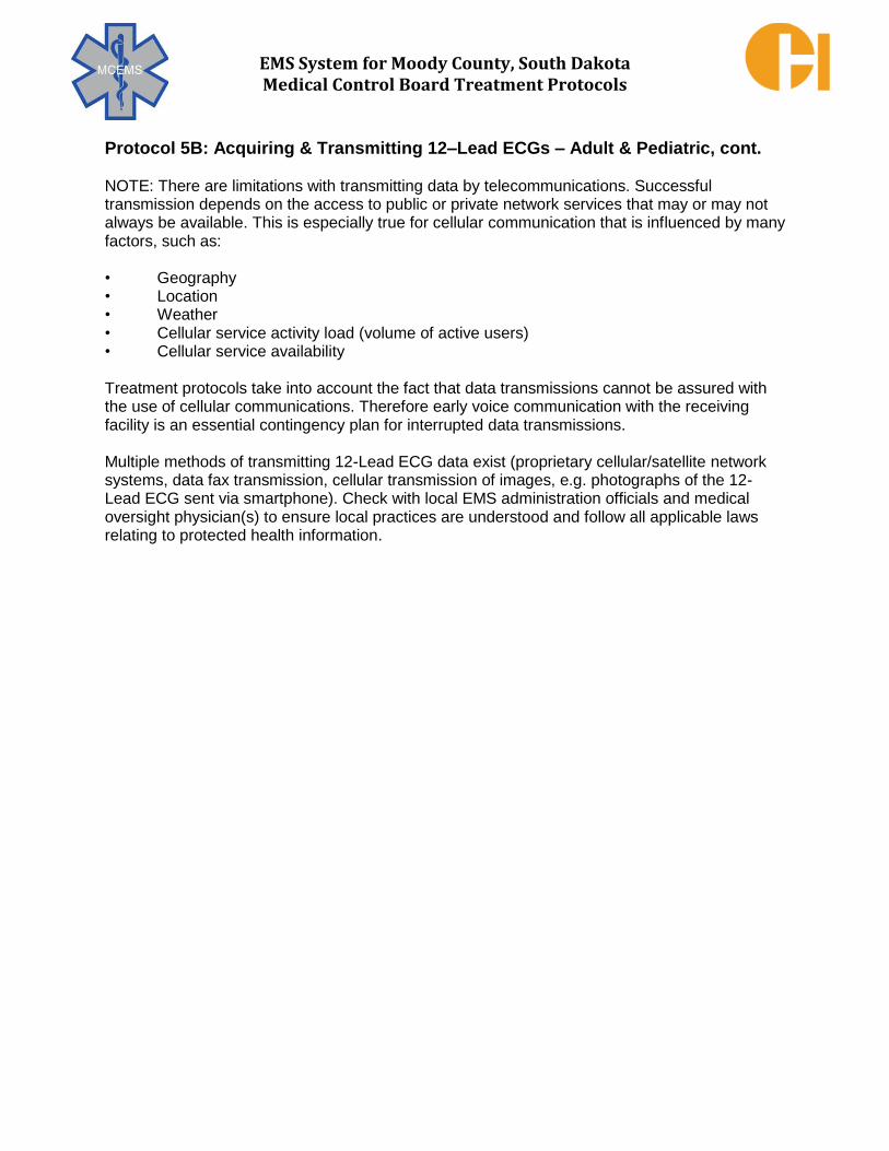
EMS System for Moody County, South Dakota Medical Control Board Treatment Protocols
Protocol 5B: Acquiring & Transmitting 12–Lead ECGs – Adult & Pediatric, cont. NOTE: There are limitations with transmitting data by telecommunications. Successful transmission depends on the access to public or private network services that may or may not always be available. This is especially true for cellular communication that is influenced by many factors, such as: • Geography • Location • Weather • Cellular service activity load (volume of active users) • Cellular service availability Treatment protocols take into account the fact that data transmissions cannot be assured with the use of cellular communications. Therefore early voice communication with the receiving facility is an essential contingency plan for interrupted data transmissions. Multiple methods of transmitting 12-Lead ECG data exist (proprietary cellular/satellite network systems, data fax transmission, cellular transmission of images, e.g. photographs of the 12-Lead ECG sent via smartphone). Check with local EMS administration officials and medical oversight physician(s) to ensure local practices are understood and follow all applicable laws relating to protected health information.

EMS System for Moody County, South Dakota Medical Control Board Treatment Protocols
EMERGENCY MEDICAL DISPATCHER
EMERGENCY MEDICAL RESPONDER
EMT
EMT – INTERMEDIATE
ADVANCED EMT
PARAMEDIC
EMD
ADVISE TO AVOID PHYSICAL EXERTION AND/OR ENVIRONMENTAL STRESS (i.e. TEMP EXTREMES).
ADULT: ADVISE ASPIRIN (ASA) 324/325 mg CHEWED BY PT (unless contraindicated).
ADVISE NITROGLYCERIN (NTG) PT SELF-ADMINISTRATION
AS PREVIOUSLY PRESCRIBED FOR SIMILAR SYMPTOMS
EMR EMT GENERAL SUPPORTIVE CARE
OBTAIN VITAL SIGNS O2 VIA NC or NRB IF DYSPNEA or PULSE OX <94% AT ROOM AIR
APPLY CARDIAC MONITOR ADULT: OBTAIN 12-LEAD ECG (if equipped)
TRANSMIT 12-LEAD ECG TO RECEIVING EMERGENCY DEPARTMENT ASA 324/325 mg CHEWED BY PT (withhold if taken < 6 hours or contraindicated)
ASSIST NTG SELF-ADMINISTRATION 0.4 mg (hold if SYS BP ≤ 100 mmHg)
EMT – I AEMT IV ACCESS
IV NS TKO if SYS BP > 100 mmHg IV NS 250 mL BOLUS if SYS BP ≤ 100 mmHg IF NO SIGNS OF PULMONARY EDEMA
PARAMEDIC
TREAT ANY CARDIAC DYSRHYTHMIAS/SHOCK BY THE RESPECTIVE PROTOCOLS ANALYZE 12-LEAD ECG – TREAT PER FOLLOWING FLOWCHART
NOTIFY RECEIVING HOSPITAL IMMEDIATELY IF SUSPECTED STEMI TRANSPORT ASAP TO APPROPRIATE HOSPITAL DESTINATION
5C – ACUTE CORONARY SYNDROME
ADULT TREATMENT PRIORITIES
3 in 5 minutes of patient contact: 1. Vital signs 2. O2 if indicated 3. ECG rhythm interpretation (if
paramedic present) 5 in 10 minutes of patient contact: 1. ASA 2. IV 3. 12 lead ECG 4. NTG or fluids (BP/Inf. MI?) 5. Repeat vital signs
OBTAIN/ANALYZE RIGHT-SIDED 12-LEAD ECG EN ROUTE *ACUTE INFERIOR INFARCT?
SIGNS OF PULMONARY EDEMA?
**ACUTE RIGHT VENTRICULAR INFARCT
SYS BP >100 mmHg
*** NTG 0.4 mg SUBLINGUAL MAY REPEAT EVERY 5 MIN
IF SYS BP >100 mmHg
IV NS 250 mL BOLUS REPEAT UNTIL SYS BP > 100 mmHg
IF NO SIGNS OF PULMONARY EDEMA
IV NS 250 mL BOLUS
REPEAT UNTIL SYS BP > 100 mmHg
IF NO SIGNS OF PULMONARY EDEMA
DOPAMINE 5-20
mcg/kg/min IVPB
TITRATE
TO SYS BP > 100 mmHg
IF PT STILL HAVING ACS SYMPTOMS AFTER 3 NTG ADMINISTRATIONS WITH PERSISTENT CHEST PAIN & IF SYS BP > 100 mmHg: ADDITIONAL NITROGLYCERIN PER PROTOCOL 16F
AND MORPHINE SULFATE 2 mg SLOW IVP, MAY REPEAT EVERY 5 MIN TO A TOTAL OF 10 mg.
OR FENTANYL 0.5 mcg/kg SLOW IVP/IM/IN, MAX DOSE 50 mcg. MAY REPEAT EVERY 10 MINS
TO MAX CUMULATIVE DOSE OF 1.5 mcg/kg or 125 mcg WHICHEVER IS LESSER.
* ACUTE INFERIOR INFARCT INDICATED BY ST SEGMENT ELEVATION IN AT LEAST
2 OF THESE 3 LEADS: II, III, aVF. **ACUTE RIGHT VENTRICULAR INFARCT INDICATED BY ST SEGMENT ELEVATION IN AT LEAST 2 OF THESE 4 LEADS: V3R,
V4R, V5R, V6R. ***DO NOT GIVE NTG TO PATIENTS TAKING VIAGRA® OR LEVITRA® WITHIN 24 HOURS OR CIALIS® WITHIN 48 HOURS WITHOUT MC CONSULT.
NO NO
NO
NO
YES YES
YES
YES

EMS System for Moody County, South Dakota Medical Control Board Treatment Protocols
EMERGENCY MEDICAL DISPATCHER
EMERGENCY MEDICAL RESPONDER
EMT
EMT – INTERMEDIATE
ADVANCED EMT
PARAMEDIC
EMD
ADVISE TO AVOID PHYSICAL EXERTION OR ENVIRONMENTAL STRESS (TEMP EXTREMES)
EMR EMT
GENERAL SUPPORTIVE CARE OBTAIN VITAL SIGNS
O2 VIA NC or NRB IF DYSPNEA OR PULSE OX < 94% AT ROOM AIR APPLY CARDIAC MONITOR/OBTAIN 12 – LEAD ECG (if equipped)
TRANSMIT 12 – LEAD ECG TO RECEIVING EMERGENCY DEPARTMENT
EMT – I AEMT
IV/IO ACCESS
PARAMEDIC
ANALYZE & TREAT PER RHYTHM FLOWCHART BELOW
5D – BRADYCARDIA
ADULT & PEDIATRIC TREATMENT PRIORITIES 1. Vital Signs 2. IV Access 3. Analyze rhythm 4. Rhythm specific treatment.
UNSTABLE (SYMPTOMATIC) BRADYCARDIA is defined by any of the following symptoms:
Dyspnea
Chest pain
Weakness
Altered mental status
Hypoxemia
Pulmonary edema
SINUS BRADYCARDIA FIRST DEGREE AV BLOCK
SECOND DEGREE AV BLOCK TYPE I (WENCKEBACH)
SECOND DEGREE AV BLOCK TYPE II (CLASSIC) THIRD DEGREE AV BLOCK (COMPLETE)
ADULT & PEDIATRIC: ASYMPTOMATIC – MONITOR & PLACE PACING PADS.
ADULT: ACS – TREAT PER PROTOCOL 5C - ACUTE CORONARY SYNDROME & PACE AT 60 bpm INSTEAD OF ATROPINE USE.
NON-ACS – PACE AT 60 bpm INSTEAD OF ATROPINE USE.
PEDIATRIC: PLACE PACER PADS. CONSULT MC
ADULT & PEDIATRIC: ASYMPTOMATIC – MONITOR ONLY. ADULT: TREAT IF SYMPTOMATIC AND SYS B/P < 100mmHg.
PEDIATRIC: TREAT IF SYMPTOMATIC AND SYS B/P < (70 + 2x age in years) mmHg. ADULT: ACS – TREAT PER PROTOCOL 5C - ACUTE CORONARY SYNDROME. AVOID ATROPINE IF HR >40 bpm.
NON-ACS – ATROPINE 0.5 mg IVP/IOP. MAY REPEAT EVERY 5 MINUTES TO CUMULATIVE OF 3 mg. PEDIATRIC: EPINEPHRINE 0.01 mg/kg IVP/IOP. (1:10,000 0.1 mL/kg IVP/IOP). MAY REPEAT ONCE.
IF UNRESPONSIVE TO EPINEPHRINE, ATROPINE 0.02 mg/kg IVP/IOP. MINIMUM DOSE 0.1 mg. MAX SINGLE DOSE 0.5 mg. MAY REPEAT ONCE

EMS System for Moody County, South Dakota Medical Control Board Treatment Protocols
Indications:
1. Symptomatic 2nd Degree AV Block-Type II (Classic) 2. Symptomatic 3rd Degree AV Block (Complete) 3. Symptomatic Bradycardia in Acute Coronary Syndrome in preference to atropine use 4. Symptomatic Bradycardia unresponsive to non-electrical interventions 5. Symptomatic Bradycardia in pediatric patients (when approved by OLMCP consultation)
Contraindications:
1. Asymptomatic Bradycardia
Technique:
(Physio-Control LifePak® 15):
1. Maintain standard ECG monitoring using electrodes/cable.
2. Apply Quik–Combo™pads at chest wall location illustrated on
the individual pad (Figure 1). Excessive diaphoresis may require
drying and/or excessive chest hair may require partial removal to
achieve appropriate pad-chest wall adhesion.
3. Connect Quik-Combo™ pad set to LifePak® monitor/defibrillator
via attached cable.
4. Advise patient of impending therapy. Administer sedation if patient condition allows, adults to receive 2-5 mg midazolam IVP
as individual patient weight and hemodynamics dictate.
5. Power on the pacing function by pressing the "PACER" button
(Figure 2).
6. Confirm ECG rhythm is sensed by Quik-Combo™ pads, looking
for triangular "sense markers" marking QRS complexes (Figure 3). If sense markers do not appear, check for correct Quik-Combo™ pad attachment to LifePak monitor/defibrillator. If sense markers are inconsistently tracking QRS complexes and/or tracking T waves, adjust ECG size or select alternate monitoring lead to achieve correct QRS complex tracking.
PARAMEDIC 5E – TRANSCUTANEOUS PACING
ADULT & PEDIATRIC
Figure 1
Figure 2
Figure 3

EMS System for Moody County, South Dakota Medical Control Board Treatment Protocols
PROTOCOL 5E: Transcutaneous Pacing, Adult & Pediatric, cont.
Technique (cont.):
7. Set pacing rate at 60 paces per minute (adults) either by
pressing the "RATE" switch up arrow to increase rate or down
arrow to decrease rate or by rotating the "SPEED DIAL" knob
(Figure 4). The "RATE" switch will allow changes in 10 paces
per minute increments; the "SPEED DIAL" knob will allow
changes in 5 paces per minute increments. 8. Set pacing current at minimum level achieving electrical AND
mechanical capture. Deliver electrical pacing current either by
pressing the "CURRENT" switch up arrow to increase
milliAmp (mA) current or down arrow to decrease mA current
or by rotating the "SPEED DIAL" knob (Figure 5). The
"CURRENT" switch will allow changes in 10 mA increments;
the "SPEED DIAL" knob will allow changes in 5 mA
increments. 9. Pressing the "PAUSE" button will cause the set pacing rate to
decrease by 25% (eg. rate of 60 paces per minute changes to
rate of 45 paces per minute) while it is being depressed. This
function should not be used without directive from MC. 10. If pacing therapy termination is required, power off the pacing
function by pressing the "PACER" button (Figure 2).
Pacing-Related Considerations: (Physio-Control LifePak® 15): 1. In the event of ventricular fibrillation or pulseless ventricular tachycardia, pressing the
yellow "CHARGE" button will automatically stop the pacing function. Proceed with defibrillation.
2. If the monitor displays "ECG LEAD OFF" during transcutaneous pacing, pacing
automatically switches to non – demand and continues at the fixed rate until the ECG lead(s) is reattached. During non – demand pacing, the pacemaker delivers pulses at the set pace rate regardless of any intrinsic beats that the patient may have. The monitor continues to display the pacing rate and the current. To reestablish demand pacing, reattach the ECG lead(s).
3. If the Quik-Combo™ electrodes detach during pacing, the monitor will display "CONNECT
ELECTRODES" and "PACING STOPPED" messages and sound an alarm. The set pacing rate is maintained, but the current resets to 0 mA. Reattaching the Quik-Combo™ electrodes silences the alarm and removes the messages. The current remains at 0 mA until manually adjusted as described above.
Figure 4
Figure 5

EMS System for Moody County, South Dakota Medical Control Board Treatment Protocols
PROTOCOL 5E: Transcutaneous Pacing, Adult & Pediatric, cont. Pacing-Related Considerations (cont.): 4. Proper electrical capture is displayed by
depolarization of the ventricles, reflected
as a wide QRS, followed by a distinct, broad T wave (Figures 6 & 7). Absence
of these findings immediately following pacing spikes generally indicates failure
of consistent electrical and mechanical
capture (Figures 8 & 9). 5. With transcutaneous pacing, it may be
difficult to see the paced QRS complex
due to washout from the pacing stimulus.
It is imperative to confirm capture by a
physiologic measure such as a pulse.
Figure 6
Figure 7
Figure 8
Figure 9

EMS System for Moody County, South Dakota Medical Control Board Treatment Protocols
EMERGENCY MEDICAL DISPATCHER
EMERGENCY MEDICAL RESPONDER
EMT
EMT – INTERMEDIATE
ADVANCED EMT
PARAMEDIC
EMD
ADVISE TO AVOID PHYSICAL EXERTION OR ENVIRONMENTAL STRESS (TEMP EXTREMES).
EMR EMT
GENERAL SUPPORTIVE CARE OBTAIN VITAL SIGNS
O2 VIA NC or NRB IF DYSPNEA OR PULSE OX < 94% AT ROOM AIR APPLY CARDIAC MONITOR/OBTAIN 12 – LEAD ECG (if equipped)
TRANSMIT 12 – LEAD ECG TO RECEIVING EMERGENCY DEPARTMENT
EMT – I AEMT
IV/IO ACCESS
PARAMEDIC
ADULT: ANALYZE & TREAT PER FLOWCHART BELOW PEDIATRIC: CONSULT MC FOR TREATMENT PLAN
5F – TACHYCARDIA – STABLE
ADULT & PEDIATRIC TREATMENT PRIORITIES
1. Vital signs 2. IV Access 3. Analyze rhythm 4. Rhythm specific treatment 5. Consult MC for pediatrics 6. If protocol interventions DO NOT
control rate of adult PSVT, or convert WIDE COMPLEX TACHYCARDIA
Adult options include: Narrow Complex Tachycardia
DILTIAZEM 0.25 mg/kg SLOW IVP over 2 mins
AMIODARONE 150 mg SLOW IVP/IVPB over 10 mins
SYNCHRONIZED CARDIOVERSION
MONITOR/TRANSPORT
Wide Complex Tachycardia
AMIODARONE 150 mg SLOW
IVP/IVPB over 10 mins
SYNCHRONIZED CARDIOVERSION
ADENOSINE 12 mg
RAPID IVP IF REGULAR & MONOMORPHIC
LIDOCAINE up to 1 mg/kg IVP at < 50 mg/min
MONITOR/TRANSPORT
UNSTABLE (SYMPTOMATIC)
tachycardia is defined by any
of the following symptoms: 1. Dyspnea 2. Chest pain 3. Weakness 4. Altered mental status 5. Hypoxemia 6. Pulmonary edema
ADULT HEART RATE ≥ 150/MIN? AFEBRILE PEDIATRIC HEART RATE ≥ 180/MIN?
UNSTABLE WITH SYMPTOMS ADULT: SYS BP <100 mmHg?
PEDIATRIC: SYS BP < (70 + 2x age in years) mmHg?
TREATMENT PER OTHER APPLICABLE PROTOCOLS
TREAT PER PROTOCOL 5G UNSTABLE TACHYCARDIA
ATRIAL FIBRILLATION
OR ATRIAL FLUTTER
PSVT WIDE-COMPLEX TACHYCARDIA
UNCERTAIN TYPE OR
MONOMORPHIC VENTRICULAR TACHYCARDIA
TORSADES
ADULT: DILTIAZEM 0.25 mg/kg
SLOW IVP OVER 2 MINS MAX DOSE 25 mg UP TO AGE 70 YEARS
MAX DOSE 10 mg AGE 70+ YEARS
VALSALVA MANUEVER
ADENOSINE 6mg RAPID IVP MAY REPEAT AT 12 MG TWICE
AMIODARONE
150 mg OVER 10 MINUTES (VERY SLOW IVP/IVPB)
MAGNESIUM SULFATE 1gram SLOW IVP/IOP
OVER 1 MINUTE MAY REPEAT x1
YES
NO
YES

EMS System for Moody County, South Dakota Medical Control Board Treatment Protocols
EMERGENCY MEDICAL DISPATCHER
EMERGENCY MEDICAL RESPONDER
EMT
EMT – INTERMEDIATE
ADVANCED EMT
PARAMEDIC
EMD
ADVISE TO AVOID PHYSICAL EXERTION OR ENVIRONMENTAL STRESS (TEMP EXTREMES).
EMR EMT
GENERAL SUPPORTIVE CARE OBTAIN VITAL SIGNS
O2 VIA NC or NRB IF DYSPNEA or PULSE OX <94% AT ROOM AIR APPLY CARDIAC MONITOR/OBTAIN 12 – LEAD ECG (if equipped)
TRANSMIT 12 – LEAD ECG TO RECEIVING EMERGENCY DEPARTMENT
EMT – I AEMT
IV/IO ACCESS IF INDICATED
PARAMEDIC
ADULT: ANALYZE & TREAT PER FLOWCHART BELOW PEDIATRIC: CONSULT MC FOR TREATMENT PLAN (MEDICATION OR ELECTRICAL THERAPY)
5G – TACHYCARDIA – UNSTABLE
ADULT & PEDIATRIC TREATMENT PRIORITIES
1. Vital signs 2. IV Access 3. Analyze rhythm 4. Rhythm specific treatment 5. Synchronized mode utilized each
synchronized cardioversion
6. Consult MC for pediatric Pts 7. If protocol measures DO NOT
convert rhythm, control
symptoms, and restore blood
pressure, CONTACT MC
UNSTABLE (SYMPTOMATIC)
TACHYCARDIA: is defined by any
of the following symptoms: 1. Dyspnea 2. Chest pain 3. Weakness 4. Altered mental status 5. Hypoxemia 6. Pulmonary edema
ADULT: HEART RATE > 150/MIN?
PEDIATRIC: AFEBRILE HEART RATE > 180/MIN TREATMENT PER
APPLICABLE PROTOCOLS
UNSTABLE WITH SYMPTOMS AND
ADULT: SYS BP < 100 mmHg?
PEDIATRIC: SYS BP < (70 + 2x age in years) mmHg
TREAT PER PROTOCOL 5F
TACHYCARDIA - STABLE
SYNCHRONIZED CARDIOVERSION DOUBLE CHECK PRIOR TO EACH AND EVERY SYNCHRONIZED CARDIOVERSION
ASSURE “SYNC” MODE IS UTILIZED PRIOR TO ENERGY DELIVERY ADULT: IF SYS BP > 80 mmHg, PRETREAT WITH MIDAZOLAM 0.1 mg/kg IVP/IOP/INP, Maximum dose 5 mg
BEGIN AT 100J SYNCHRONIZED IF RHYTHM PERSISTS & PATIENT REMAINS UNSTABLE REPEAT AT SYNCHRONIZED ENERGY SETTINGS OF 200J, 300J, 360J
TACHYCARDIA RESOLVES? CONSULT MC
ADULT: VENTRICULAR TACHYCARDIA ONLY AMIODARONE
150 mg OVER 10 MINUTES VERY SLOW IVP/IVPB
YES
YES
YES
NO
NO
NO

EMS System for Moody County, South Dakota Medical Control Board Treatment Protocols
Indication:
Unstable, symptomatic tachycardia (adult heart rate > 150 beats per minute; afebrile
pediatric heart rate > 180 beats per minute) AND hemodynamic compromise adult
systolic blood pressure <100 mmHg; pediatric systolic blood pressure < (70 + 2x age
in years) mmHg.
Contraindications:
1. Stable tachycardia (Treatment per Protocol 5F – Stable Tachycardia) 2. Normal sinus rhythm 3. Bradycardia 4. Ventricular fibrillation/pulseless ventricular tachycardia
Technique (Physio-Control LifePak® 15):
1. Power ON. (Figure 1)
2. Attach patient ECG cable and ECG electrodes.
ECG electrodes and cable must be used to
monitor the ECG when paddles are used for
synchronized cardioversion.
3. Select lead with the greatest QRS complex amplitude
Figure 1
positive or negative deflection. (Figure 2)
4. Press SYNC. The SYNC MODE message appears in the message area when SYNC is active. (Figure 3)
a. NOTE: To deactivate SYNC
MODE when not synchronized
cardioverting, press SYNC again.
PARAMEDIC 5H – SYNCHRONIZED CARDIOVERSION
ADULT & PEDIATRIC
Figure 1
Figure 2
Figure 3

EMS System for Moody County, South Dakota Medical Control Board Treatment Protocols
Protocol 5H: Synchronized Cardioversion, Adult & Pediatric, cont.
Technique (cont):
5. Observe the ECG rhythm. Confirm that a triangle sense marker ( ) appears near the MIDDLE of each QRS complex. (Figure 4) a. If the sense markers DO NOT appear or are
displayed in the wrong location (for example on
the T – wave) adjust ECG SIZE or select another
lead. It is normal for the sense marker location to
vary slightly on each QRS. 6. Connect the therapy electrodes to the therapy cable
and confirm cable connection to the
monitor/defibrillator. (Figure 5) 7. Prepare the patient’s skin and apply therapy electrodes
to the patient in the anterior-lateral position. (Figure 6) 8. Press ENERGY SELECT or rotate the SPEED DIAL to
select the desired energy. (Figure 7) Per Protocol 5G – Tachycardia – Unstable, for adult synchronized cardioversion, begin at 100 joules energy. If unstable dysrhythmia persists, repeat synchronized cardioversion at escalating energy settings of 200
joules, 300 joules, 360 joules. For pediatric synchronized cardioversion, consult on-line medical control for treatment plan and energy settings.
9. Press CHARGE. While the monitor/defibrillator is
charging a charging bar appears and a ramping tone
sounds, indicating the charging energy level. When the
monitor/defibrillator is fully charged, the screen
displays available energy. (Figure 8)
10. Make certain all personnel, including the operator; stand
clear of the patient, bed, and any equipment connected to the patient.
Figure 4
Figure 5
Figure 6
Figure 7
Figure 8

EMS System for Moody County, South Dakota Medical Control Board Treatment Protocols
Protocol 5H: Synchronized Cardioversion, Adult & Pediatric, cont.
Technique (cont.):
11. Confirm ECG rhythm. Confirm available energy. Prior to
delivering synchronized cardioversion, it is paramount to
ensure that the SYNC MODE message continues to
appear. Failure to deliver a “synchronized” cardioversion
in this setting could cause ventricular fibrillation cardiac
arrest in the patient. (Figure 9) 12. Press and hold the (shock) button on the
monitor/defibrillator until the ENERGY DELIVERED message appears on the screen. (Figure 10)
a. NOTE: To disarm (cancel the charge), press the
SPEED DIAL. The energy disarms automatically
if shock buttons are not pressed within 60
seconds, or if the energy selection after charging
begins. 13. Observe patient and ECG rhythm. Repeat procedure
starting from Step 4, if necessary.
Figure 9
Figure 10

EMS System for Moody County, South Dakota Medical Control Board Treatment Protocols
Clinical Pearls:
1. Correlate electrical pacing activity with mechanical heart activity (e.g. pulses). 2. If electrical pacer spikes seen on the ECG monitor are not consistently and immediately
followed by electrical activity of atrial, ventricular, or both atrial and ventricular depolarization,
then the pacemaker may be intermittently functioning. This may be normal if the patient’s heart
rate is above 60 beats per minute, since most pacemakers will be set to a demand mode
(pacing only when needed). Alternately, if the patient is bradycardic, the pacemaker may be
non-functional (e.g. battery failure). 3. The “sensor function” of a pacemaker attempts to anticipate increased metabolic needs and
raises heart rate. The most commonly used sensor is an accelerometer which raises pacing rate when motion is detected. Thus, physical motion of the patient (including motion created by
riding on the ambulance stretcher enroute to the hospital) can stimulate increasing rates of
pacing. If the paced rate is noticeably higher than usual set rates of 60-80 beats per minute, attempt to minimize the patient’s physical motion and observe if pacing rates decline.
4. Due to the variety of pacemaker types and settings, pacemaker manufacturers supply patients
with a card to be carried (usually in wallet or purse) that identifies the pacemaker by
manufacturer, type, and date of implantation. 5. Specific types of pacemaker malfunction include the following:
a. Failure to pace/output – no pacing spikes seen in a bradycardic patient. (example, over sensing of myopotentials, dead battery)
b. Failure to sense – pacing becomes asynchronous (example, patient’s heart voltage too low for pacer to sense)
c. Failure to capture – pacing spikes seen without capture (examples, lead becomes dislodged from myocardium or breaks)
d. Over pacing or “runaway pacing” – pacemaker pacing at fast rates without clear
reason (examples, sensor-driven pacing from motion, pacemaker-mediated
tachycardia) 6. In the setting of sustained, symptomatic rapid pacing suspected to be related to overpacing
(see Item 4 above) tachycardia may be able to be controlled by placing a doughnut-shaped medical magnet over the generator.
7. In the setting of cardiac arrest, treat per usual resuscitation, but avoid placing
defibrillation pads over the pacemaker generator. 8. Consult medical control early in the course of suspected pacemaker management
issues for further guidance.
PARAMEDIC 5I – PACEMAKER MANAGEMENT
ADULT & PEDIATRIC

EMS System for Moody County, South Dakota Medical Control Board Treatment Protocols
Clinical Pearls:
1. Correlate ICD activity with ECG rhythm - ventricular fibrillation/tachycardia? 2. Due to the variety of ICDs, manufacturers supply patients with a card to be carried (usually
in wallet or purse) that identifies the ICD by manufacturer, type, and date of implantation. 3. All ICDs have pacemaker function. Some patients have pacing indications and may have
normal or abnormal pacemaker function (see 5I – Pacemaker Management) associated with
their ICD. Many patients with ICDs, however, do not have a pacemaker indication. 4. Specific types of ICD malfunction include the following:
a. Inappropriate ICD shocks – Patient is shocked without evidence of arrhythmia (examples, lead fracture, oversensing of t-waves)
b. Failure to shock ventricular tachycardia/fibrillation - (examples, undersensing of small
fibrillatory waves, slow ventricular tachycardia below the ICD’s programmed VT zone) 5. If the patient is hemodynamically stable, acquire and transmit a 12-Lead ECG prior to
attempting any change in ICD function. 6. In the setting of oversensing, especially if multiple apparently inappropriate ICD discharges
occur, the ICD may be temporarily deactivated by placing a doughnut-shaped medical
magnet over the ICD generator. Depending upon the exact model of ICD, a beep or
sustained tone may be heard with successful magnet application. 7. While the magnet is applied to the ICD, no therapies will be delivered – even when needed
for life-threatening arrhythmias. Therefore, PRIOR TO DEACTIVATING AN ICD WITH A MAGNET, THE PATIENT MUST BE ON CONTINUOUS ECG MONITORING AND A DEFIBRILLATOR MUST BE IMMEDIATELY AVAILABLE.
8. In the setting of cardiac arrest, treat per usual resuscitation, but avoid placing defibrillation
pads over the ICD generator.
9. Consult medical control early in the course of suspected ICD management issues for
further guidance.
PARAMEDIC
5J – IMPLANTABLE CARDIOVERTER/DEFIBRILLATOR (ICD) MANAGEMENT
ADULT & PEDIATRIC

EMS System for Moody County, South Dakota Medical Control Board Treatment Protocols
PARAMEDIC
PARAMEDIC
ASYMPTOMATIC PVCs DO NOT REQUIRE ANTI-DYSRHYTHMIC MEDICATION.
TREAT IF UNSTABLE WITH SYMPTOMS AND ADULT SYS BP < 100 mmHg DO NOT PHARMACOLOGICALLY SUPPRESS PVCs IN 2nd / 3rd DEGREE HEART BLOCKS
ADULT: AMIODARONE 150mg VERY SLOW IVP/IVPB OVER 10 MINUTES.
PEDIATRIC: MC CONSULT
5K – PREMATURE VENTRICULAR CONTRACTIONS
ADULT & PEDIATRIC
TREATMENT PRIORITIES 1. O2 via NC or NRB if indicated 2. 12-Lead ECG 3. Rhythm Analysis
UNSTABLE (SYMPTOMATIC) premature ventricular contractions
are defined by any of the following
symptoms: 1. Dyspnea 2. Chest pain 3. Weakness 4. Altered mental status 5. Hypoxemia 6. Pulmonary edema

EMS System for Moody County, South Dakota Medical Control Board Treatment Protocols
EMERGENCY MEDICAL DISPATCHER
EMERGENCY MEDICAL RESPONDER
EMT
EMT – INTERMEDIATE
ADVANCED EMT
PARAMEDIC
EMD
ADVISE TO AVOID PHYSICAL EXERTION OR ENVIRONMENTAL STRESS (TEMP EXTREMES).
CONDUCT STROKE SCREENING QUERY IF AUTHORIZED BY LOCAL MEDICAL DISPATCH PROTOCOL.
EMR EMT
GENERAL SUPPORTIVE CARE
OBTAIN VITAL SIGNS
O2 VIA NC, NRB, OR BVM IF DYSPNEA OR PULSE OX <94% AT ROOM AIR
LOS ANGELES PREHOSPITAL STROKE SCREEN AGE OVER 45 YEARS?
NO PRIOR HX OF SEIZURE DISORDER? NEW ONSET OF NEUROLOGIC SYMPTOMS IN LAST 24 HRS?
PATIENT AMBULATORY AT BASELINE [PRIOR TO EVENT? BLOOD GLUCOSE 50 TO 400 mg/dL?
FACIAL DROOP; ARM DRIFT; IMPAIRED SPEECH?
EARLY “STROKE ALERT” NOTIFICATION TO RECEIVING EMERGENCY DEPARTMENT WITH DEFINITIVE STROKE SYMPTOMS IF LESS THAN 6 HOURS DURATION
ADVISE TIME OF LAST ACCURATELY KNOWN BASELINE NEUROLOGICAL STATUS (LAST “NORMAL” FOR PT) OBTAIN CELLPHONE NUMBER(S) OF FAMILY MEMBER(S) TO PROVIDE ED STAFF UPON ARRIVAL
APPLY CARDIAC MONITOR/OBTAIN 12-LEAD ECG (if equipped)
TRANSMIT 12-LEAD ECG TO RECEIVING EMERGENCY DEPARTMENT
EMT OR HIGHER LICENSE: MEASURE END-TIDAL CO2 & MONITOR WAVEFORM CAPNOGRAPHY (if equipped, **Mandatory use if Pt intubated)
PLACE SUPRAGLOTTIC AIRWAY IF INDICATED & ONLY IF BVM VENTILATIONS ARE INEFFECTIVE
EMT – I AEMT
ADULT: INTUBATE IF INDICATED IV ACCESS
IV NS TKO IF SYS BP ≥ 100 mmHg WITHOUT HYPOTENSIVE SYMPTOMS
IV NS 250 mL BOLUS IF SYS BP <100 mmHg WITH HYPOTENSIVE SYMPTOMS & NO SIGNS OF PULMONARY EDEMA, REPEAT UP TO 2 LITERS NS IF SYS BP REMAINS < 100 mmHg WITH HYPOTENSIVE SYMPTOMS & NO SIGNS OF PULMONARY EDEMA
PARAMEDIC
ADULT: MEDICATION-ASSISTED INTUBATION IF INDICATED
EVALUATE FOR OTHER ALTERED MENTAL STATUS ETIOLOGIES. TREAT PER APPROPRIATE PROTOCOL(S)
CONTINUOUS ASSESSMENT & TREATMENT PER APPLICABLE PROTOCOL(S
6A – STROKE
ADULT & PEDIATRIC TREATMENT PRIORITIES
3 in 5 minutes of patient contact:
1. Vital signs 2. O2 if indicated 3. Los Angeles Prehospital
Stroke Screen
Early transport & ED notification if symptoms <6 hours

EMS System for Moody County, South Dakota Medical Control Board Treatment Protocols
EMERGENCY MEDICAL DISPATCHER
EMERGENCY MEDICAL RESPONDER
EMT
EMT – INTERMEDIATE
ADVANCED EMT
PARAMEDIC
EMD
IF CHIEF COMPLAINT IS MEDICAL IN NATURE, CHOOSE THE PROTOCOL THAT BEST FITS THE PATIENT’S FOREMOST
SYMPTOMS, WITH PRIORITY SYMPTOMS TAKING PRECEDENCE
QUESTIONS TO ADDRESS SCENE SAFETY ISSUES
EMR EMT
GENERAL SUPPORTIVE CARE OBTAIN VITAL SIGNS
O2 VIA NC, NRB, OR BVM AS APPROPRIATE
DETERMINE BLOOD GLUCOSE FOR PATIENT ABLE TO SWALLOW
ADULT & PEDIATRIC WEIGHT ≥25 kg HYPOGLYCEMIA CARE: IF GLUCOSE <50 mg/dL, 1 tube ORAL GLUCOSE (15 grams) PO
PEDIATRIC WEIGHT <2 5kg HYPOGLYCEMIA CARE: IF GLUCOSE <50 mg/dL, ½ tube ORAL GLUCOSE (7.5 grams) PO
APPLY CARDIAC MONITOR (if equipped)
EMT OR HIGHER LICENSE: MEASURE END–TIDAL CO2 & MONITOR WAVEFORM CAPNOGRAPHY (if equipped,
**Mandatory use if Pt intubated) PLACE SUPRAGLOTTIC AIRWAY IF INDICATED &
ONLY IF BVM VENTILATIONS INEFFECTIVE
EMT – I AEMT
IV ACCESS ADULT: IV NS TKO IF SYS BP ≥ 100 mmHg WITHOUT HYPOTENSIVE SYMPTOMS
IV NS 250 mL BOLUS IF SYS BP <100 mmHg WITH HYPOTENSIVE SYMPTOMS & NO SIGNS OF PULMONARY EDEMA, REPEAT UP TO 2 LITERS NS IF SYS BP REMAINS < 100 mmHg WITH HYPOTENSIVE SYMPTOMS & NO SIGNS OF PULMONARY EDEMA
PEDIATRIC: IV NS TKO IF SYS BP ≥ (70 + 2x age in years) mmHg
IV NS 20 mL/kg BOLUS IF SYS BP < (70 + 2x age in years) mmHg IF NO SIGNS OF PULMONARY EDEMA ADULT & PEDIATRIC WEIGHT ≥25 kg HYPOGLYCEMIA CARE:
IF GLUCOSE <50 mg/dL, D50 1 mL/kg IVP UP TO 50 mL GLUCAGON 1 mg IM IF NO VASCULAR ACCESS OBTAINED
PEDIATRIC WEIGHT <25 kg HYPOGLYCEMIA CARE: IF GLUCOSE <50 mg/dL D25 2 mL/kg IVP UP TO 50 mL
GLUCAGON 0.5 mg IM IF NO VASCULAR ACCESS OBTAINED ADULT & PEDIATRIC: REPEAT DETERMINATION OF BLOOD GLUCOSE POST-DEXTROSE TREATMENT
TOXINS/DRUG OVERDOSE – SUSPECTED NARCOTIC/OPIATE – APNEIC/AGONALLY BREATHING ADULT: NALOXONE 2 mg IVP/IOP/IN MAY REPEAT ONCE
PEDIATRIC: NALOXONE 0.5 mg IVP/IOP/IN, MAY REPEAT TO MAX OF 2 mg
TOXINS/DRUG OVERDOSE – SUSPECTED NARCOTIC/OPIATE – INEFFECTIVE BREATHING ACTIVITY ADULT & PEDIATRIC: NALOXONE 0.5 mg IVP/IOP/IN, MAY REPEAT TO MAX OF 2 mg
USE NALOXONE TO RESTORE EFFECTIVE BREATHING; AVOID EXCESSIVE DOSING TO PREVENT WITHDRAWAL
PARAMEDIC
ADULT: MEDICATION-ASSISTED INTUBATION IF INDICATED CONTINUOUS ASSESSMENT & TREATMENT OF SUSPECTED AMS ETIOLOGY PER APPLICABLE PROTOCOL(S) CONSULT MC IF ABOVE TREATMENT INEFFECTIVE FOR HYPOGLYCEMIA OR NARCOTIC/OPIATE ETIOLOGY
CONSULT MC IF UNCERTAIN OF ETIOLOGY AND TREATMENT PLAN OF AMS
6B – ALTERED MENTAL STATUS
ADULT & PEDIATRIC TREATMENT PRIORITIES
1. Vital signs 2. O2
3. Dextrose for hypoglycemia
4. Benzodiazepine for sustained, active seizure (refer to 6D Seizure if applicable)
Evaluate differential diagnosis of AMS & treat per protocol(s):
Hypoxemia (Shock)
Head Injury
Stroke
Seizure
Infection (Sepsis/ Meningitis)
Medication/Alcohol
Heat or Cold Illness

EMS System for Moody County, South Dakota Medical Control Board Treatment Protocols
Indications:
1. Suspected stroke (as part of Los Angeles Prehospital Stroke Screen) 2. Altered mental status – unclear etiology 3. Seizure 4. Known or suspected diabetes
Contraindications:
None Determining Blood Glucose:
1. Assemble equipment, including performing steps as detailed above when indicated. 2. Using universal precautions, take one test strip from the container. 3. Insert a test strip into the meter in the direction of the arrows. The meter turns on. 4. Make sure the code on the display matches the code number on the test strip container. If
the code number is not seen, take the test strip out and reinsert into meter.
5. When the blood drop symbol flashes, obtain a drop of blood from the fingertip. 6. Position hand palm-side up; choose whichever finger is least calloused. 7. Apply intermittent pressure to the finger to help the blood to flow. 8. Clean the fingertip with alcohol. Start in the middle and work outward to prevent
contaminating the area. Allow area to dry. 9. Hold the finger and firmly place a new, sterile lancet off-center on the fingertip and firmly
press the lancet to puncture the fingertip. 10. Wipe away the first drop of blood with a sterile gauze pad. 11. Touch the drop of blood to the collection window of the test strip. DO NOT put blood on
top of the test strip. 12. When a flash is observed enough blood is on the test strip; if blood is applied and a flash
is not seen, more blood should be applied within 5 seconds. 13. Properly dispose of all contaminated supplies.
EMERGENCY MEDICAL RESPONDER
EMT
EMT – INTERMEDIATE
ADVANCED EMT
PARAMEDIC
6C – GLUCOMETRY (BLOOD GLUCOSE DETERMINATION)
ADULT & PEDIATRIC

EMS System for Moody County, South Dakota Medical Control Board Treatment Protocols
EMERGENCY MEDICAL DISPATCHER
EMERGENCY MEDICAL RESPONDER
EMT
EMT – INTERMEDIATE
ADVANCED EMT
PARAMEDIC
EMD
KEEP PATIENT FREE FROM INJURY HAZARDS AVOID PLACING ANYTHING IN MOUTH
PLACE IN RECOVERY POSITION POST SEIZURE
EMR EMT
GENERAL SUPPORIVE CARE OBTAIN VITAL SIGNS
O2 VIA NC or NRB AS APPROPRIATE APPLY CARDIAC MONITOR (if equipped)
EMT OR HIGHER LICENSE: MEASURE END-TIDAL CO2 & MONITOR WAVEFORM CAPNOGRAPHY (if equipped **Mandatory if Pt intubated)
EMT – I AEMT
IV ACCESS ADULT: IV NS TKO IF SYS BP ≥ 100 mmHg WITHOUT HYPOTENSIVE SYMPTOMS
IV NS 250 mL BOLUS IF SYS BP <100 mmHg WITH HYPOTENSIVE SYMPTOMS & NO SIGNS OF PULMONARY EDEMA, REPEAT UP TO 2 LITERS NS IF SYS BP REMAINS < 100 mmHg WITH HYPOTENSIVE SYMPTOMS & NO SIGNS OF PULMONARY EDEMA
PEDIATRIC: IV NS TKO IF SYS BP ≥ (70 + 2x age in years) mmHg
IV NS 20 mL/kg BOLUS IF SYS BP < (70 + 2x age in years) mmHg IF NO SIGNS OF PULMONARY EDEMA
DETERMINE BLOOD GLUCOSE ADULT & PEDIATRIC WEIGHT ≥25 kg HYPOGLYCEMIA CARE:
IF GLUCOSE <50 mg/dL, D50 1 mL/kg IVP UP TO 50 mL GLUCAGON 1 mg IM IF NO VASCULAR ACCESS OBTAINED
PEDIATRIC WEIGHT <25 kg HYPOGLYCEMIA CARE:
IF GLUCOSE <50 mg/dL, D25 2 mL/kg IVP UP TO 50 mL GLUCAGON 0.5 mg IM IF NO VASCULAR ACCESS OBTAINED
PARAMEDIC
CONTINUOUS ASSESSMENT & TREATMENT PER APPLICABLE PROTOCOL(S) EVALUATE FOR OTHER ALTERED MENTAL STATUS ETIOLOGIES. TREAT PER APPROPRIATE PROTOCOL(S)
ADULT: MIDAZOLAM 0.1 mg/kg IM/IVP/IN/IOP TO MAX OF 5 mg FOR ACTIVE SEIZURE. MAY REPEAT X 1 IN 5 MIN IF STILL SEIZING.
OR DIAZEPAM 5 mg IVP/IOP/IM FOR ACTIVE SEIZURE IF MIDAZOLAM NOT AVAILABLE.
MAY REPEAT X 1 IN 5 MIN IF STILL SEIZING. OR
LORAZEPAM 2 mg IVP/IOP/IM FOR ACTIVE SEIZURE IF MIDAZOLAM NOT AVAILABLE. MAY REPEAT X 1 IN 10 MIN IF STILL SEIZING.
PEDIATRIC: MIDAZOLAM 0.1 mg/kg IM/IVP/IN/IOP TO MAX OF 5 mg FOR ACTIVE SEIZURE. MAY REPEAT X 1 IN 5 MIN IF STILL SEIZING.
OR
DIAZEPAM 0.1 mg/kg IVP/IOP/IM FOR ACTIVE SEIZURE IF MIDAZOLAM NOT AVAILABLE. MAY REPEAT X 1 IN 5 MIN IF STILL SEIZING.
OR
LORAZEPAM 0.1 mg/kg IVP/IOP/IM FOR ACTIVE SEIZURE IF MIDAZOLAM NOT AVAILABLE. MAY REPEAT X 1 IN 10 MIN IF STILL SEIZING.
MC CONSULT IF SEIZURE CONTINUES DESPITE ABOVE TREATMENT
CONTINUOUS ASSESSMENT & TREATMENT PER APPLICABLE PROTOCOL(S)
6D – SEIZURE
ADULT & PEDIATRIC TREATMENT PRIORITIES 1. Vital signs 2. O2
3. Dextrose for hypoglycemia 4. Benzodiazepine for sustained,
active seizure

EMS System for Moody County, South Dakota Medical Control Board Treatment Protocols
EMERGENCY MEDICAL DISPATCHER
EMERGENCY MEDICAL RESPONDER
EMT
EMT – INTERMEDIATE
ADVANCED EMT
PARAMEDIC
EMD
KEEP PATIENT FREE FROM INJURY HAZARDS AVOID PLACING ANYTHING IN MOUTH
ADVISE TO AVOID PHYSICAL EXERTION OR ENVIRONMENTAL STRESS (TEMP EXTREMES)
PLACE IN RECOVERY POSITION/POSITION OF COMFORT
EMR EMT
GENERAL SUPPORTIVE CARE OBTAIN VITAL SIGNS
O2 VIA NC, NRB, OR BVM AS APPROPRIATE
DETERMINE BLOOD GLUCOSE FOR PATIENT ABLE TO SWALLOW
ADULT & PEDIATRIC WEIGHT ≥25 kg HYPOGLYCEMIA CARE: IF GLUCOSE <50 mg/dL, 1 tube ORAL GLUCOSE (15 grams) PO
PEDIATRIC WEIGHT <25 kg HYPOGLYCEMIA CARE: IF GLUCOSE <50 mg/dL, ½ tube ORAL GLUCOSE (7.5 grams) PO
APPLY CARDIAC MONITOR/OBTAIN 12-LEAD ECG (if equipped)
TRANSMIT 12-LEAD ECG TO RECEIVING EMERGENCY DEPARTMENT EMT OR HIGHER LICENSE:
MEASURE END–TIDAL CO2 & MONITOR WAVEFORM CAPNOGRAPHY (if equipped, **Mandatory use if Pt intubated)
PLACE SUPRAGLOTTIC AIRWAY IF INDICATED & ONLY IF BVM VENTILATIONS INEFFECTIVE
EMT – I AEMT
IV ACCESS ADULT: IV NS TKO IF SYS BP ≥ 100 mmHg WITHOUT HYPOTENSIVE SYMPTOMS
IV NS 250 mL BOLUS IF SYS BP <100 mmHg WITH HYPOTENSIVE SYMPTOMS & NO SIGNS OF PULMONARY EDEMA, REPEAT UP TO 2 LITERS NS IF SYS BP REMAINS < 100 mmHg WITH HYPOTENSIVE SYMPTOMS & NO SIGNS OF PULMONARY EDEMA
PEDIATRIC: IV NS TKO IF SYS BP ≥ (70 + 2x age in years) mmHg
IV NS 20 mL/kg BOLUS IF SYS BP < (70 + 2x age in years) mmHg IF NO SIGNS OF PULMONARY EDEMA ADULT & PEDIATRIC WEIGHT ≥25 kg HYPOGLYCEMIA CARE:
IF GLUCOSE <50 mg/dL, D50 1 mL/kg IVP UP TO 50 mL GLUCAGON 1 mg IM IF NO VASCULAR ACCESS OBTAINED
PEDIATRIC WEIGHT <25 kg HYPOGLYCEMIA CARE: IF GLUCOSE <50 mg/dL, D25 2 mL/kg IVP UP TO 50 mL
GLUCAGON 0.5 mg IM IF NO VASCULAR ACCESS OBTAINED ADULT & PEDIATRIC: REPEAT DETERMINATION OF BLOOD GLUCOSE POST-DEXTROSE TREATMENT
ADULT: INTUBATE IF INDICATED; DO NOT INTUBATE PATIENTS WITH RAPIDLY REVERSIBLE AMS ETIOLOGY (eg. HYPOGLYCEMIA, OPIATES) TOXINS/DRUG OVERDOSE – SUSPECTED NARCOTIC/OPIATE – APNEIC/AGONALLY BREATHING
ADULT: NALOXONE 2 mg IVP/IOP/IN MAY REPEAT ONCE PEDIATRIC: NALOXONE 0.5 mg IVP/IOP/IN, MAY REPEAT TO MAX OF 2 mg
TOXINS/DRUG OVERDOSE – SUSPECTED NARCOTIC/OPIATE – INEFFECTIVE BREATHING ACTIVITY ADULT & PEDIATRIC: NALOXONE 0.5 mg IVP/IOP/IN, MAY REPEAT TO MAX OF 2 mg
USE NALOXONE TO RESTORE EFFECTIVE BREATHING; AVOID EXCESSIVE DOSING TO PREVENT WITHDRAWAL
PARAMEDIC
ADULT: MEDICATION-ASSISTED INTUBATION IF INDICATED CONTINUOUS ASSESSMENT & TREATMENT OF SUSPECTED AMS ETIOLOGY PER APPLICABLE PROTOCOL(S) CONSULT MC IF ABOVE TREATMENT INEFFECTIVE FOR HYPOGLYCEMIA OR NARCOTIC/OPIATE ETIOLOGY
CONSULT MC IF UNCERTAIN OF ETIOLOGY AND TREATMENT PLAN OF AMS
6E – SYNCOPE
ADULT & PEDIATRIC TREATMENT PRIORITIES 1. Vital signs 2. O2
3. Dextrose for hypoglycemia 4. Benzodiazepine for sustained,
active seizure (refer to 6D Seizure if applicable)
Evaluate differential diagnosis of Syncope & treat per protocol(s):
Acute Coronary Syndrome
Cardiac Dysrhythmia Hypotension (Shock) Hypoxemia (Shock)
Head Injury
Stroke
Seizure
Infection (Sepsis/ Meningitis)
Medication/Alcohol
Heat or Cold Illness
Psychogenic/Emotional

EMS System for Moody County, South Dakota Medical Control Board Treatment Protocols
EMERGENCY MEDICAL DISPATCHER
EMERGENCY MEDICAL RESPONDER
EMT
EMT – INTERMEDIATE
ADVANCED EMT
PARAMEDIC
EMD
KEEP PATIENT FREE FROM INJURY HAZARDS AVOID PLACING ANYTHING IN MOUTH
ADVISE TO AVOID PHYSICAL EXERTION OR ENVIRONMENTAL STRESS (TEMP EXTREMES)
PLACE IN RECOVERY POSITION/POSITION OF COMFORT
EMR EMT
GENERAL SUPPORTIVE CARE OBTAIN VITAL SIGNS
O2 VIA NC, NRB, OR BVM AS APPROPRIATE DETERMINE BLOOD GLUCOSE FOR PATIENT ABLE TO SWALLOW
ADULT & PEDIATRIC WEIGHT ≥25 kg HYPOGLYCEMIA CARE: IF GLUCOSE <50 mg/dL, 1 tube ORAL GLUCOSE (15 grams) PO
PEDIATRIC WEIGHT <25 kg HYPOGLYCEMIA CARE: IF GLUCOSE <50 mg/dL, ½ tube ORAL GLUCOSE (7.5 grams) PO
APPLY CARDIAC MONITOR/OBTAIN 12-LEAD ECG (if equipped)
TRANSMIT 12-LEAD ECG TO RECEIVING EMERGENCY DEPARTMENT EMT OR HIGHER LICENSE:
MEASURE END–TIDAL CO2 & MONITOR WAVEFORM CAPNOGRAPHY
(if equipped, **Mandatory use if Pt intubated) PLACE SUPRAGLOTTIC AIRWAY IF INDICATED &
ONLY IF BVM VENTILATIONS INEFFECTIVE
EMT – I AEMT
IV ACCESS
ADULT: IV NS TKO IF SYS BP ≥ 100 mmHg WITHOUT HYPOTENSIVE SYMPTOMS IV NS 250 mL BOLUS IF SYS BP <100 mmHg WITH HYPOTENSIVE SYMPTOMS & NO SIGNS OF PULMONARY EDEMA,
REPEAT UP TO 2 LITERS NS IF SYS BP REMAINS < 100 mmHg WITH HYPOTENSIVE SYMPTOMS & NO SIGNS OF PULMONARY EDEMA PEDIATRIC: IV NS TKO IF SYS BP ≥ (70 + 2x age in years) mmHg
IV NS 20 mL/kg BOLUS IF SYS BP < (70 + 2x age in years) mmHg IF NO SIGNS OF PULMONARY EDEMA ADULT & PEDIATRIC WEIGHT ≥25 kg HYPOGLYCEMIA CARE:
IF GLUCOSE <50 mg/dL, D50 1 mL/kg IVP UP TO 50 mL GLUCAGON 1 mg IM IF NO VASCULAR ACCESS OBTAINED
PEDIATRIC WEIGHT <25 kg HYPOGLYCEMIA CARE:
IF GLUCOSE <50 mg/dL, D25 2 mL/kg IVP UP TO 50 mL GLUCAGON 0.5 mg IM IF NO VASCULAR ACCESS OBTAINED
ADULT & PEDIATRIC: REPEAT DETERMINATION OF BLOOD GLUCOSE POST-DEXTROSE TREATMENT
PARAMEDIC
ADULT: DIPHENHYDRAMINE 50 mg IM/IVP PEDIATRIC: DIPHENHYDRAMINE 1 mg/kg IM/IVP TO MAX OF 50 mg
IF NO IMPROVEMENT 15 MINUTES AFTER DEPHENHYDRAMINE ADMINISTRATION & MARKED MUSCLE SPASM/TONE: ADULT: MIDAZOLAM 2.5 mg IVP/IM/IN
OR
DIAZEPAM 5 mg IVP OR
LORAZEPAM 2 mg IVP/IM.
PEDIATRIC: MIDAZOLAM 0.1 mg/kg IM/IVP/IN TO MAX OF 2,5 mg OR
DIAZEPAM 0.1 mg/kg TO MAX OF 5 mg IVP/IM OR
LORAZEPAM 0.1 mg/kg TO MAX OF 2 mg IVP/IM
CONTINUOUS ASSESSMENT & TREATMENT OF SUSPECTED ETIOLOGY PER APPLICABLE PROTOCOL(S) CONSULT MC IF ABOVE TREATMENT INEFFECTIVE
6F – DYSTONIC REACTIONS
ADULT & PEDIATRIC TREATMENT PRIORITIES
1. Vital signs 2. O2 3. Dextrose for hypoglycemia 4. Benzodiazepine for sustained,
active seizure (refer to 6D
Seizure if applicable) Evaluate differential diagnosis of
AMS & treat per protocol(s):
Hypoxemia (Shock)
Head Injury
Stroke
Seizure
Infection (Sepsis/ Meningitis)
Medication/Alcohol Heat or Cold Illness

EMS System for Moody County, South Dakota Medical Control Board Treatment Protocols
EMERGENCY MEDICAL DISPATCHER
EMERGENCY MEDICAL RESPONDER
EMT
EMT – INTERMEDIATE
ADVANCED EMT
PARAMEDIC
EMD
KEEP VIOLENT OR SUICIDAL PATIENT ON THE LINE. IN VOLATILE/CRIMINAL SITUATIONS, FOLLOW APPLICABLE
LAW ENFORCEMENT PROTOCOL. FOR JUMPERS, NOTIFY LAW AND FIRE/RESCUE RESOURCES.
EMR EMT
GENERAL SUPPORTIVE CARE – DO NOT LEAVE PATIENT ALONE OBTAIN VITAL SIGNS
O2 VIA NC or NRB AS APPROPRIATE APPLY CARDIAC MONITOR (if equipped)
IF RESTRAINTS ARE REQUIRED USE SOFT RESTRAINTS and / or KERLIX RESTRAIN PATIENT TO LONG SPINE BOARD OR ORTHOPEDIC SCOOP
DO NOT TRANSPORT PATIENTS
“SANDWICHED” BETWEEN TWO BACKBOARDS
DURING TRANSPORT OF PATIENTS IN POLICE INSTITUTED LOCKING RESTRAINTS, A POLICE OFFICER SHOULD EITHER ACCOMPANY THE
PATIENT OR PROVIDE EMS PERSONNEL MEANS TO UNLOCK THE RESTRAINTS
DETERMINE BLOOD GLUCOSE
ADULT & PEDIATRIC WEIGHT ≥25 kg HYPOGLYCEMIA CARE: IF GLUCOSE <50 mg/dL, 1 tube ORAL GLUCOSE (15 grams) PO
PEDIATRIC WEIGHT <25 kg HYPOGLYCEMIA CARE:
IF GLUCOSE <50 mg/dL, ½ tube ORAL GLUCOSE (7.5 grams) PO
EMT OR HIGHER LICENSE: MEASURE END – TIDAL CO2 & MONITOR WAVEFORM CAPNOGRAPHY (if
equipped, **Mandatory use if Pt intubated)
EMT – I AEMT
IV ACCESS ADULT: IV NS TKO IF SYS BP ≥ 100 mmHg WITHOUT HYPOTENSIVE SYMPTOMS
IV NS 250 mL BOLUS IF SYS BP <100 mmHg WITH HYPOTENSIVE SYMPTOMS & NO SIGNS OF PULMONARY EDEMA, REPEAT UP TO 2 LITERS IF SYS BP REMAINS < 100 mmHg WITH HYPOTENSIVE SYMPTOMS & NO SIGNS OF PULMONARY EDEMA
PEDIATRIC: IV NS TKO IF SYS BP ≥ (70 + 2x age in years) mmHg
IV NS 20 mL/kg BOLUS IF SYS BP < (70 + 2x age in years) mmHg IF NO SIGNS OF PULMONARY EDEMA DETERMINE BLOOD GLUCOSE
ADULT & PEDIATRIC WEIGHT ≥25 kg HYPOGLYCEMIA CARE:
IF GLUCOSE <50 mg/dL, D50 1 mL/kg IVP UP TO 50 mL GLUCAGON 1 mg IM IF NO VASCULAR ACCESS OBTAINED
PEDIATRIC WEIGHT <25 kg HYPOGLYCEMIA CARE:
IF GLUCOSE <50 mg/dL, D25 2 mL/kg IVP UP TO 50mL GLUCAGON 0.5 mg IM IF NO VASCULAR ACCESS OBTAINED
ADULT & PEDIATRIC: REPEAT DETERMINATION OF BLOOD GLUCOSE POST-DEXTROSE TREATMENTS)
PARAMEDIC
CHEMICAL RESTRAINT: SEE PROTOCOL 7C
CONSULT MC IF UNCERTAIN OF ETIOLOGY AND TREATMENT PLAN FOR PSYCHIATRIC PROBLEM OR IF ADDITIONAL RESTRAINT MEASURES NEEDED
CONTINUOUS ASSESSMENT & TREATMENT PER APPLICABLE PROTOCOL(S)
7A – BEHAVIORAL DISORDERS
ADULT & PEDIATRIC TREATMENT PRIORITIES
1. Assess scene safety 2. Safety of self 3. Safety of public safety
professionals 4. Safety of patient 5. Observe for uncontrolled
agitation, combativeness,
AMS impeding necessary
medical care or pulling at necessary medical
interventions (IV lines, endotracheal tubes)
6. Employ alternative methods to avoid physically restraining the
patient 7. Restrain patient if alternatives fail
and/or it is necessary to maintain
necessary medical intervention or to carry out treatment
protocols 8. Treat excited delirium

EMS System for Moody County, South Dakota Medical Control Board Treatment Protocols
Indications:
1. Reducing likelihood of patient doing harm to self. 2. Reducing likelihood of patient doing harm to others (including EMS professionals). 3. Reducing likelihood of patient disrupting medically necessary interventions. 4. Patient requires/required chemical restraint per Protocol 7C.
Alternatives to physical restraint as outlined below are to be utilized so as to minimize the use of physical restraints. However, if alternatives to physical restraints are unsuccessful, then physical restraints will be applied in an effective and compassionate manner. Throughout the use of alternatives to physical restraint and physical restraint, the patient and the patient’s concerned parties (family, friends, co-workers, etc.) shall be treated with respect and informed of the need
for these procedures. This protocol is not intended to place EMS professionals at higher risk for injury. If personal safety is compromised or threatened during the course of patient care, appropriate law enforcement personnel should be summoned for assistance. If at any time questions arise as to appropriateness of using alternatives to physical restraint or physical restraint, MC should be consulted for direction.
Contraindications:
1. Patient (or patient’s legal guardian or medical power of attorney) possesses medical decision making capacity and is refusing evaluation, treatment, and/or transport (in the absence of threatened or actual harm to self or others).
2. Patient is compliant with medically necessary interventions.
Reducing likelihood of patient doing harm to self and/or others and reducing likelihood of patient disrupting medically necessary interventions can be successfully accomplished with alternatives to physical restraint in the best judgment of the EMS professional(s) treating the patient. Technique:
The following steps shall be taken and documented in determining the need for physical restraints:
1. Assessment of mental status - Observe for uncontrolled agitation, combativeness, threats of
violence to self or others, disorientation, altered mental status impeding medically necessary
interventions, or pulling at necessary medical interventions (e.g. oxygen, IV lines, and
endotracheal tubes).
EMERGENCY MEDICAL RESPONDER
EMT
EMT – INTERMEDIATE
ADVANCED EMT
PARAMEDIC
7B – PHYSICAL RESTRAINT
ADULT & PEDIATRIC

EMS System for Moody County, South Dakota Medical Control Board Treatment Protocols
PROTOCOL 7B: Physical Restraint – Adult & Pediatric, cont. 2. Alternatives to physical restraint- Reassurance, support of concerned parties (family, friends,
co-workers, etc.), reorientation, diversionary activity, explanation of illness, injury, and
medically necessary interventions. 3. Justification for physical restraint- Failure of alternatives to physical restraint, reduce
likelihood of patient harm to self, reduce likelihood of patient harm to others, enable
medically necessary interventions per EMS protocols. 4. Inform patient and concerned parties of physical restraint use. 5. Apply physical restraints - Restraints are to be soft and are not to impede airway patency,
respiratory mechanics, or circulation. Patients will not be restrained prone unless an impaled object or airway patency necessitates such positioning. Restraints will be applied in an effective, yet compassionate manner. Every effort should be made to avoid injury to the patient during application of physical restraints.
Humane restraints that reduce potential for patient injury from the restraints are those made
from roll gauze, soft leather, and those designed as single-patient use, disposable foam with
cloth ties. Restraints are to be non-locking unless applied by law enforcement officers in
appropriate circumstances and able to be released rapidly if patient condition mandates.
During treatment and transport of a patient in law enforcement-instituted restraints (including handcuffs), EMS professionals should monitor for and advocate for change in restraints that compromise airway patency, respiratory mechanics, or circulation. Patients will not be
transported with wrists cuffed to ankles either directly or indirectly (also referred to as “hog-tying”). These positions have been shown to impair respiratory mechanics and pose significant obstacles to definitive airway management if required enroute. During transport of patients in law enforcement-instituted locking restraints, a law enforcement officer should either accompany the patient in the ambulance or provide the treating EMS professionals means to unlock the restraints. This policy allows rapid restraint release should the patient deteriorate to
a condition requiring restraint release to properly treat.
Patients restrained using this protocol should generally be restrained to a backboard. This
facilitates patient transfer in the emergency department and in the case of airway secretions or
vomiting, enables rapid positioning of the patient to reduce aspiration. Patients will not be
transported “sandwiched” between two backboards; this positioning impedes patient care and
increases risk of aspiration.
Once physical restraints are applied, they will be left in place until the patient is transferred to
emergency department personnel. This policy prevents recurrent harm to self, harm to others,
and disruption of intact medical devices and treatment. Despite assurance from the patient that
they will comply with treatment, restraints are to be left in place unless a direct order from MC is
given to release the physical restraints. Such an order must be clearly documented on the
patient care form.

EMS System for Moody County, South Dakota Medical Control Board Treatment Protocols
EMERGENCY MEDICAL RESPONDER
EMT
EMT – INTERMEDIATE
ADVANCED EMT
PARAMEDIC
EMR EMT
ASSIST IN PHYSICAL CONTROL OF PATIENT FOR PARAMEDIC TO ADMINISTER CHEMICAL RESTRAINT
USE ADEQUATE NUMBERS OF PUBLIC SAFETY PROFESSIONALS TO MINIMIZE RISK OF INJURY TO SELF AND OTHERS
UNLESS UNSAFE TO DO SO, PERFORM THE FOLLOWING POST- CHEMICAL RESTRAINT:
GENERAL SUPPORTIVE CARE – DO NOT LEAVE PATIENT ALONE OBTAIN VITAL SIGNS
O2 VIA NC or NRB AS APPROPRIATE APPLY CARDIAC MONITOR (if equipped)
EMT OR HIGHER LICENSE:
MEASURE END-TIDAL CO2 & MONITOR WAVEFORM CAPNOGRAPHY (if equipped, **Mandatory use if Pt intubated)
EMT – I AEMT
IV ACCESS IF PT TEMPORARILY COOPERATIVE
DO NOT RISK SELF INJURY WITH NEEDLESTICK DURING IV ACCESS IF PT COMBATIVE
PARAMEDIC
CHEMICAL RESTRAINT:
ALL PATIENTS REQUIRING CHEMICAL RESTRAINT ARE TO BE PHYSICALLY RESSTRAINED AS WELL
ADULT: MIDAZOLAM 0.1 mg/kg IM/IVP/IN/IOP TO MAX OF 5 mg. MAY REPEAT ONCE.
OR
DIAZEPAM 5 mg IVP/IOP or 10 mg IM IF MIDAZOLAM NOT AVAILABLE. MAY REPEAT ONCE. OR
LORAZEPAM 2 mg IVP/IOP/IM IF MIDAZOLAM NOT AVAILABLE. MAY REPEAT ONCE. (MIDAZOLAM STRONGLY PREFERRED DUE TO RAPID ONSET OF ACTION OF BENZODIAZEPINE OPTIONS)
PLUS HALOPERIDOL 5 mg IM
PEDIATRIC: MIDAZOLAM 0.1 mg/kg IM/IVP/IN/IOP TO MAX OF 5 mg.
OR DIAZEPAM 0.1 mg/kg TO MAX OF 5 mg IVP/IOP/IM FOR ACTIVE SEIZURE IF MIDAZOLAM NOT AVAILABLE.
OR
LORAZEPAM 0.1 mg/kg TO MAX OF 2 mg IVP/IOP/IM FOR ACTIVE SEIZURE IF MIDAZOLAM NOT AVAILABLE.
CONSULT MC IF UNCERTAIN OF ETIOLOGY AND TREATMENT PLAN FOR PSYCHIATRIC PROBLEM OR IF ADDITIONAL CHEMICAL RESTRAINT MEASURES NEEDED
CONTINUOUS ASSESSMENT & TREATMENT PER APPLICABLE PROTOCOL(S)
7C – CHEMCAL RESTRAINT
ADULT & PEDIATRIC
TREATMENT PRIORITIES 1. Asses scene safety 2. Safety of self 3. Safety of public safety professionals 4. Safety of patient 5. Treat excited delirium

EMS System for Moody County, South Dakota Medical Control Board Treatment Protocols
EMS professionals should utilize this protocol and its principles and directives to promote and
protect the safety of mentally ill patients, drug or alcohol dependent patients, and other
involved parties who may be endangered by the patient’s disturbed or altered psychological
state to the extent of being subject to an immediate likelihood of serious harm.
Definitions:
1. “Drug Dependent Patient” for the purpose of this protocol means: A patient who is using a
controlled substance as presently defined in Section 102 of the Federal Controlled
Substances Act and who is in a state of psychic or physical dependence, or both, arising
from administration of that controlled substance on an intermittent or continuous basis. 2. “Alcohol Dependent Patient” for the purpose of this protocol means: A patient who uses
alcoholic beverages to an extent that it impairs mental or physical health, family life,
occupational life, and potentially compromises the health and safety of the community. 3. “Mentally Ill Patient” for the purpose of this protocol means: A patient afflicted with a
substantial disorder of thought, mood, perception, psychological orientation or memory
that significantly impairs judgment, behavior, capacity to recognize reality or ability to meet
the ordinary demands of life. Mental illness may be reflected in a sustained altered
mentation secondary to chronic medical condition or prior physical injury. 4. “Immediate likelihood of serious harm” posed by patients either to self or others for the
purpose of this protocol means:
a) a substantial risk of physical harm to self, manifested by active threats of, or attempts at, suicide or intentional bodily harm; OR
b) a substantial risk of physical harm to others manifested by active threats of, or attempts at, homicide or intentional bodily harm; OR
c) actively placing others in reasonable fear of imminent violent behavior or serious
physical harm; OR
d) causing a prudent EMS professional to believe with reasonable certainty that
without immediate medical treatment, severe physical impairment or injury would
be sustained by the patient or other involved party as a result of the patient’s
apparent inability to prevent suicidal activity, homicidal activity, or significant risk of
harm to self or others through distorted reality (e.g. driving while acutely psychotic
or clinically intoxicated.)
EMERGENCY MEDICAL RESPONDER
EMT
EMT – INTERMEDIATE
ADVANCED EMT
PARAMEDIC
7D – EMERGENCY MENTAL HOLD ISSUES
ADULT & PEDIATRIC

EMS System for Moody County, South Dakota Medical Control Board Treatment Protocols
PROTOCOL 7D: Emergency Mental Hold Issues – Adult & Pediatric, cont.
Emergency Mental Hold Procedures:
1. Upon dispatch to and/or subsequent assessment of a patient representing, in the EMS professional’s best judgment, “immediate likelihood of serious harm”, the EMS professional should notify dispatch to immediately request the appropriate law enforcement authority. It is the duty of the responding law enforcement officer(s) to determine if the affected person appears to be mentally unstable, alcohol dependent or drug dependent to a degree that immediate emergency action is necessary to prevent the patient from harming self or others. If the law enforcement officer determines that immediate emergency action is necessary, under South Dakota law, the law enforcement officer must take the person into protective custody. The law enforcement officer(s) can base their decision upon personal observation of the actions of the patient or upon the statement of either EMS professionals or other parties deemed credible.
2. If EMS professionals at the scene believe the patient to be actively mentally ill, alcohol
dependent, and/or drug dependent to the extent of being subject to an “immediate likelihood of serious harm” to self or others, representing a medical need to be taken into protective custody by law enforcement.
3. If EMS professionals and the law enforcement officer(s) at the scene cannot reach agreement whether the patient is actively mentally ill, alcohol dependent, and/or drug
dependent to the extent of representing an “immediate likelihood of serious harm” to self or others, representing a medical need to be taken into protective custody by law enforcement,
the appropriate EMS supervisor(s) and corresponding law enforcement supervisor(s) should
be contacted. Supervisor presence at the scene could be required to achieve consensus of actions that promote the patient’s and others’ safety.
4. If the EMS supervisor determines the patient is actively mentally ill, alcohol dependent,
and/or drug dependent to the extent of representing an “immediate likelihood of serious harm” to self or others, representing a medical need to be taken into protective custody by law enforcement and therefore is in need of immediate medical attention, this shall be conveyed to the appropriate law enforcement supervisor. If the law enforcement supervisor does not subsequently place the patient on mental hold, notify the appropriate receiving hospital’s Medical Control Physician (or the System EMS Medical Director) for formal
physician consultation, complete an agency-specified Incident Report, and submit it to the Medical Director for review.
5. If it appears that the affected person is mentally ill or suffers chronic altered mentation and
does not require emergency medical attention, EMS personnel will stay on scene only until it
can be reasonably determined that the person does not suffer from an apparent serious
physical condition, illness, or injury requiring emergency medical attention and/or until the
law enforcement officers at the scene indicate that they no longer require assistance from
EMS.

EMS System for Moody County, South Dakota Medical Control Board Treatment Protocols
PROTOCOL 7D: Emergency Mental Hold Issues – Adult & Pediatric, cont.
Emergency Detention (previously referred to as Emergency Order of Detention or EOD) Issues:
1. An affidavit that is completed by anyone (including an EMS professional) who is concerned
about the patient’s safety or who witnessed concerned behavior that could impact the safety
of others, that details the observations and impressions that serve as the basis for
involuntary detention of the patient in the safety interests of the patient and others has been
commonly referred to as an “EOD” or “Emergency Order of Detention”. This is no longer
used as a legal term. The correct terminology is a “third party statement” and copies of the
documents can be found with Law Enforcement Officers. 2. The “third party statement” must have sufficiently detailed information to justify placing the
patient, at least temporarily, into law enforcement custody. (eg. “suicidal” is not enough). A
law enforcement officer can refuse to take a patient into custody if he or she determines
there is insufficient written evidence contained within the “third party statement”. 3. The “third party statement” is the legally recognized documentation that compels a patient to
be placed into Emergency Detention (ED) in the safety interests of self and others until
emergency psychiatric assessment (and stabilization when applicable) can be conducted.
Medical facilities in South Dakota that can conduct emergency psychiatric assessment and
stabilization are referred to as “Emergency Detention (formerly EOD) designated facilities”. 4. A patient that is under Emergency Detention by use of a “third party statement” cannot
refuse transport to receive an appropriate physician evaluation. EMS professionals treating and transporting patients under Emergency Detention should not let the patient flee from
EMS care and supervision, unless the patient poses an immediate, serious physical threat to the EMS professional(s). Utilize law enforcement officers, physical restraint, and if
licensed as a paramedic, chemical restraint, as warranted to prevent patients under
Emergency Detention from fleeing. 5. Once an appropriate physician, typically a psychiatrist, has evaluated the patient under
Emergency Detention through use of a “third party statement”, he or she may validate
continued involuntary detention of the patient or may release the patient from further
involuntary medical detention. 6. EMS professionals should work with their system Medical Director, local medical
professionals, and local law enforcement officers to review applicable emergency mental
hold issues and resources.

EMS System for Moody County, South Dakota Medical Control Board Treatment Protocols
EMERGENCY MEDICAL DISPATCHER
EMERGENCY MEDICAL RESPONDER
EMT
EMT – INTERMEDIATE
ADVANCED EMT
PARAMEDIC
EMD
ADVISE TO AVOID PHYSICAL EXERTION OR ENVIRONMENTAL STRESS (TEMP EXTREMES). DO NOT MOVE THE PATIENT UNLESS IN DANGER.
OPEN AIRWAY IF NOT ALERT AND INEFFECTIVE BREATHING. DETERMINE NUMBER OF PATIENTS INVOLVED
DECIDE IF ADDITIONAL RESOURCES ARE NEEDED
EMR EMT
GENERAL SUPPORTIVE CARE OBTAIN VITAL SIGNS
O2 VIA NC, NRB, OR BVM AS APPROPRIATE APPLY CARDIAC MONITOR/OBTAIN 12-LEAD ECG (if equipped)
TRANSMIT 12-LEAD ECG TO RECEIVING EMERGENCY DEPARTMENT
EMT OR HIGHER LICENSE: MEASURE END – TIDAL CO2 & MONITOR WAVEFORM CAPNOGRAPHY (if equipped, **Mandatory use if Pt intubated)
PLACE SUPRAGLOTTIC AIRWAY IF INDICATED ONLY IF BVM VENTILATIONS INEFFECTIVE
TOXINS/DRUG OVERDOSE – SUSPECTED NARCOTIC/OPIATE – APNEIC/AGONALLY BREATHING (see opiate toxidrome in Protocol 8B) ADULT: NALOXONE 2 mg IVP/IOP/IN MAY REPEAT ONCE
PEDIATRIC: NALOXONE 0.5 mg IVP/IOP/IN, MAY REPEAT TO MAX OF 2 mg
TOXINS/DRUG OVERDOSE – SUSPECTED NARCOTIC/OPIATE – INEFFECTIVE BREATHING ACTIVITY (see opiate toxidrome in Protocol 8B)
ADULT & PEDIATRIC: NALOXONE 0.5 mg IVP/IOP/IN, MAY REPEAT TO MAX OF 2 mg
USE NALOXONE TO RESTORE EFFECTIVE BREATHING; AVOID EXCESSIVE DOSING TO PREVENT WITHDRAWAL
EMT – I AEMT
IV ACCESS
ADULT: INTUBATE IF INDICATED; DO NOT INTUBATE PATIENTS WITH RAPIDLY REVERSIBLE TOXICOLOGY ETIOLOGY (e.g. OPIATES)
PARAMEDIC
ADULT: MEDICATION ASSISTED INTUBATION IF INDICATED
TOXINS/DRUG OVERDOSE - SUSPECTED ORGANOPHOSPHATE ADULT: ATROPINE 2 mg IVP/IOP/IM, USE IVP FOR MORE SEVERE PRESENTATIONS. REPEAT EVERY 3-5 MINS IF SYMPTOMS PROGRESSIVE
PEDIATRIC: ATROPINE 0.05 mg/kg IVP/IOP/IM, USE IVP FOR MORE SEVERE PRESENTATIONS. MINIMUM DOSE 0.1 mg. CONTACT MC FOR REPEAT.
TOXINS/DRUG OVERDOSE - SUSPECTED TRICYCLIC ANTIDEPRESSANT (VENTRICULAR DYSRHYTHMIAS, SEIZURES) ADULT/PEDIATRIC: SODIUM BICARBONATE 1 mEq/kg IVP/IOP MAX DOSE 50 mEq
TOXINS/DRUG OVERDOSE - SUSPECTED STIMULANT (SEVERE AGITATION, HTN, TACHYCARDIA, DIAPHORESIS) ADULT: MIDAZOLAM 0.1 mg/kg IVP/IN/IM TO MAX 5 mg OR DIAZEPAM 2.5-5 mg IVP OR LORAZEPAM 1-2 mg IVP/IM
PEDIATRIC: CONTACT MC
TOXINS/DRUG OVERDOSE - SUSPECTED CALCIUM CHANNEL BLOCKER ADULT: CALICUM CHLORIDE 10 mg/kg IVP/IOP MAX DOSE 1 gram
TOXINS/DRUG OVERDOSE - SUSPECTED BETA-BLOCKER
ADULT: GLUCAGON 1 mg IVP/IOP
PEDIATRIC: GLUCAGON 0.5 mg IVP/IOP
CONSULT MC IF ABOVE TREATMENT INEFFECTIVE FOR TOXINS/DRUG OVERDOSE ETIOLOGY Poison Information Center Specialists are authorized to direct medical care related to the medical toxicology and/or hazardous material exposure aspects of patient
care if contacted for directives
8A – POISNINGS – GENERAL MANAGEMENT
ADULT & PEDIATRIC TREATMENT PRIORITIES Self/Others/Scene Safety 1. Vital signs 2. Oxygenation/Ventilation 3. Identify & treat toxin 4. US Poison Center/MC
consult if needed 5. Manage shock, altered mental
status, seizures, arrhythmias;
CO Poisoning per specific
protocol
6. Transport ASAP

EMS System for Moody County, South Dakota Medical Control Board Treatment Protocols
EMERGENCY MEDICAL RESPONDER
EMT
EMT – INTERMEDIATE
ADVANCED EMT
PARAMEDIC
8B – TOXIDROMES
ADULT & PEDIATRIC

EMS System for Moody County, South Dakota Medical Control Board Treatment Protocols
Indications:
A. Real-time consultation on toxic severity of exposure/inhalation/ingestion/snakebites. B. Real-time consultation regarding no/minimal toxicity exposures for patient follow-up contact
and self-treatment advice. C. Real-time consultation regarding needed hospital resources for patient toxicity. D. Identification of pills (using imprint letters/numbers on pills). E. Product ingredient identification and toxic severity potential.
Contraindications:
None.
Technique:
To contact the National Poison Control Center:
Healthcare Professional Access Number:
1-800-222-1222
When calling, identify yourself by name, EMS certification level, and agency/apparatus
identification. Have as much patient information and substance/exposure/snake information
possible readily available to share with the poison center specialist.
Calls are answered 24 hours a day by pharmacists and nurses intensively trained in clinical
toxicology and designated as specialists in poison information.
EMERGENCY MEDICAL DISPATCHER
EMERGENCY MEDICAL RESPONDER
EMT
EMT – INTERMEDIATE
ADVANCED EMT
PARAMEDIC
8C – POISON CONTROL CENTER USE
ADULT & PEDIATRIC
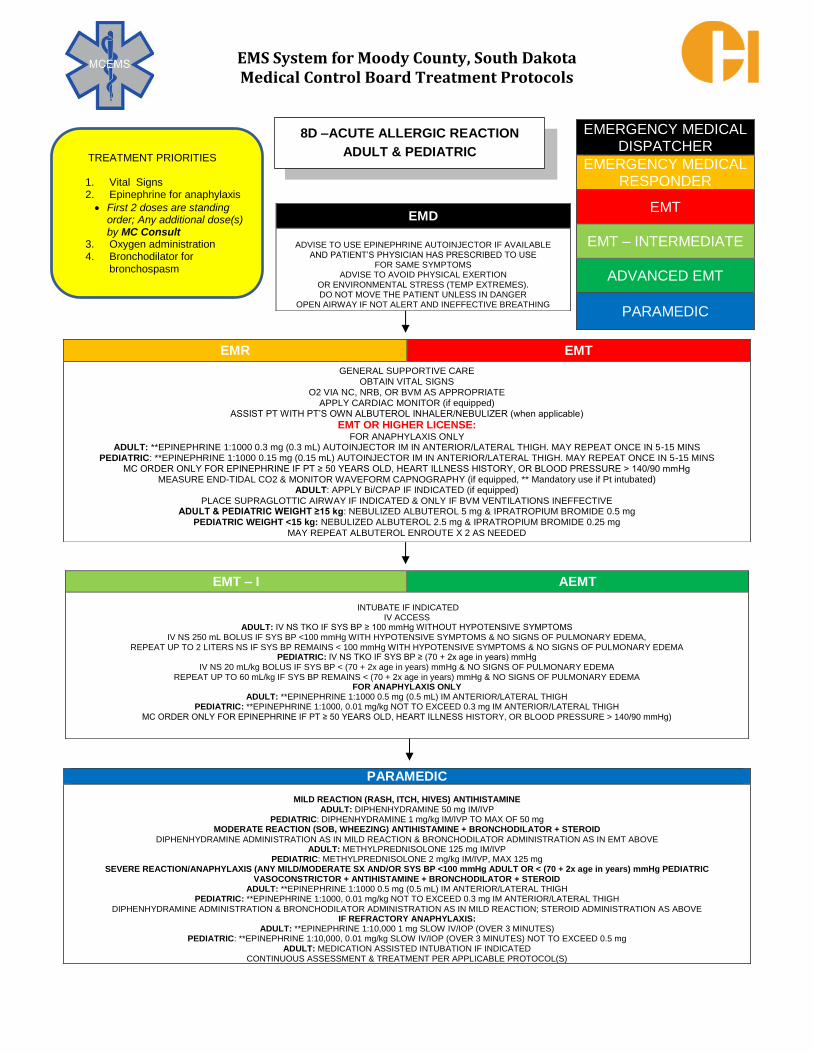
EMS System for Moody County, South Dakota Medical Control Board Treatment Protocols
EMERGENCY MEDICAL DISPATCHER
EMERGENCY MEDICAL RESPONDER
EMT
EMT – INTERMEDIATE
ADVANCED EMT
PARAMEDIC
EMD
ADVISE TO USE EPINEPHRINE AUTOINJECTOR IF AVAILABLE
AND PATIENT’S PHYSICIAN HAS PRESCRIBED TO USE FOR SAME SYMPTOMS
ADVISE TO AVOID PHYSICAL EXERTION OR ENVIRONMENTAL STRESS (TEMP EXTREMES). DO NOT MOVE THE PATIENT UNLESS IN DANGER
OPEN AIRWAY IF NOT ALERT AND INEFFECTIVE BREATHING
EMR EMT
GENERAL SUPPORTIVE CARE OBTAIN VITAL SIGNS
O2 VIA NC, NRB, OR BVM AS APPROPRIATE APPLY CARDIAC MONITOR (if equipped)
ASSIST PT WITH PT’S OWN ALBUTEROL INHALER/NEBULIZER (when applicable)
EMT OR HIGHER LICENSE: FOR ANAPHYLAXIS ONLY
ADULT: **EPINEPHRINE 1:1000 0.3 mg (0.3 mL) AUTOINJECTOR IM IN ANTERIOR/LATERAL THIGH. MAY REPEAT ONCE IN 5-15 MINS PEDIATRIC: **EPINEPHRINE 1:1000 0.15 mg (0.15 mL) AUTOINJECTOR IM IN ANTERIOR/LATERAL THIGH. MAY REPEAT ONCE IN 5-15 MINS
MC ORDER ONLY FOR EPINEPHRINE IF PT ≥ 50 YEARS OLD, HEART ILLNESS HISTORY, OR BLOOD PRESSURE > 140/90 mmHg MEASURE END-TIDAL CO2 & MONITOR WAVEFORM CAPNOGRAPHY (if equipped, ** Mandatory use if Pt intubated)
ADULT: APPLY Bi/CPAP IF INDICATED (if equipped) PLACE SUPRAGLOTTIC AIRWAY IF INDICATED & ONLY IF BVM VENTILATIONS INEFFECTIVE
ADULT & PEDIATRIC WEIGHT ≥15 kg: NEBULIZED ALBUTEROL 5 mg & IPRATROPIUM BROMIDE 0.5 mg PEDIATRIC WEIGHT <15 kg: NEBULIZED ALBUTEROL 2.5 mg & IPRATROPIUM BROMIDE 0.25 mg
MAY REPEAT ALBUTEROL ENROUTE X 2 AS NEEDED
EMT – I AEMT
INTUBATE IF INDICATED IV ACCESS
ADULT: IV NS TKO IF SYS BP ≥ 100 mmHg WITHOUT HYPOTENSIVE SYMPTOMS IV NS 250 mL BOLUS IF SYS BP <100 mmHg WITH HYPOTENSIVE SYMPTOMS & NO SIGNS OF PULMONARY EDEMA,
REPEAT UP TO 2 LITERS NS IF SYS BP REMAINS < 100 mmHg WITH HYPOTENSIVE SYMPTOMS & NO SIGNS OF PULMONARY EDEMA PEDIATRIC: IV NS TKO IF SYS BP ≥ (70 + 2x age in years) mmHg
IV NS 20 mL/kg BOLUS IF SYS BP < (70 + 2x age in years) mmHg & NO SIGNS OF PULMONARY EDEMA REPEAT UP TO 60 mL/kg IF SYS BP REMAINS < (70 + 2x age in years) mmHg & NO SIGNS OF PULMONARY EDEMA
FOR ANAPHYLAXIS ONLY ADULT: **EPINEPHRINE 1:1000 0.5 mg (0.5 mL) IM ANTERIOR/LATERAL THIGH
PEDIATRIC: **EPINEPHRINE 1:1000, 0.01 mg/kg NOT TO EXCEED 0.3 mg IM ANTERIOR/LATERAL THIGH
MC ORDER ONLY FOR EPINEPHRINE IF PT ≥ 50 YEARS OLD, HEART ILLNESS HISTORY, OR BLOOD PRESSURE > 140/90 mmHg)
PARAMEDIC
MILD REACTION (RASH, ITCH, HIVES) ANTIHISTAMINE ADULT: DIPHENHYDRAMINE 50 mg IM/IVP
PEDIATRIC: DIPHENHYDRAMINE 1 mg/kg IM/IVP TO MAX OF 50 mg MODERATE REACTION (SOB, WHEEZING) ANTIHISTAMINE + BRONCHODILATOR + STEROID
DIPHENHYDRAMINE ADMINISTRATION AS IN MILD REACTION & BRONCHODILATOR ADMINISTRATION AS IN EMT ABOVE ADULT: METHYLPREDNISOLONE 125 mg IM/IVP
PEDIATRIC: METHYLPREDNISOLONE 2 mg/kg IM/IVP, MAX 125 mg SEVERE REACTION/ANAPHYLAXIS (ANY MILD/MODERATE SX AND/OR SYS BP <100 mmHg ADULT OR < (70 + 2x age in years) mmHg PEDIATRIC
VASOCONSTRICTOR + ANTIHISTAMINE + BRONCHODILATOR + STEROID ADULT: **EPINEPHRINE 1:1000 0.5 mg (0.5 mL) IM ANTERIOR/LATERAL THIGH
PEDIATRIC: **EPINEPHRINE 1:1000, 0.01 mg/kg NOT TO EXCEED 0.3 mg IM ANTERIOR/LATERAL THIGH
DIPHENHYDRAMINE ADMINISTRATION & BRONCHODILATOR ADMINISTRATION AS IN MILD REACTION; STEROID ADMINISTRATION AS ABOVE IF REFRACTORY ANAPHYLAXIS:
ADULT: **EPINEPHRINE 1:10,000 1 mg SLOW IV/IOP (OVER 3 MINUTES) PEDIATRIC: **EPINEPHRINE 1:10,000, 0.01 mg/kg SLOW IV/IOP (OVER 3 MINUTES) NOT TO EXCEED 0.5 mg
ADULT: MEDICATION ASSISTED INTUBATION IF INDICATED CONTINUOUS ASSESSMENT & TREATMENT PER APPLICABLE PROTOCOL(S)
8D –ACUTE ALLERGIC REACTION
ADULT & PEDIATRIC TREATMENT PRIORITIES
1. Vital Signs 2. Epinephrine for anaphylaxis
First 2 doses are standing order; Any additional dose(s) by MC Consult
3. Oxygen administration 4. Bronchodilator for
bronchospasm

EMS System for Moody County, South Dakota Medical Control Board Treatment Protocols
EMERGENCY MEDICAL DISPATCHER
EMERGENCY MEDICAL RESPONDER
EMT
EMT – INTERMEDIATE
ADVANCED EMT
PARAMEDIC
EMD
ADVISE TO AVOID PHYSICAL EXERTION OR ENVIRONMENTAL STRESS (TEMP EXTREMES).
MOVE AWAY FROM SNAKE(S) IF ABLE OPEN AIRWAY IF NOT ALERT AND INEFFECTIVE BREATHING
EMR EMT
GENERAL SUPPORTIVE CARE – MARK EDGE OF SWELLING/TENDERNESS EVERY 15 MINS TO DETERMINE SYMPTOM PROGRESSION OBTAIN VITAL SIGNS & ADMINISTER O2 VIA NC, NRB, OR BVM AS APPROPRIATE
IMMOBILIZE/ELEVATE AND AVOID JOINT FLEXION IN EXTREMITY BITTEN TO MINIMIZE SWELLING OF EXTREMITY DO NOT CUT THE BITE SITE OR ATTEMPT TO “EXTRACT THE VENOM” FROM BITE SITE WITH SUCTION/VACUUM DEVICES
CONSULT US POISON CONTROL CENTER PER PROTOCOL 8C – DESCRIBE SNAKE APPEARANCE/TYPE AS BEST ABLE APPLY CARDIAC MONITOR (if equipped)
EMT OR HIGHER LICENSE: FOR ANAPHYLAXIS ONLY (ANAPHYLAXIS FROM SNAKEBITE IS RARE):
ADULT: **EPINEPHRINE 1:1000 0.3 mg (0.3 mL) AUTOINJECTOR IM IN ANTERIOR/LATERAL THIGH. MAY REPEAT ONCE IN 5-15 MINS PEDIATRIC: **EPINEPHRINE 1:1000 0.15 mg (0.15 mL) AUTOINJECTOR IM IN ANTERIOR/LATERAL THIGH. MAY REPEAT ONCE IN 5-15 MINS
MC ORDER ONLY FOR EPINEPHRINE IF PT ≥ 50 YEARS OLD, HEART ILLNESS HISTORY, OR BLOOD PRESSURE > 140/90 mmHg
MONITOR ETCO2 & WAVEFORM CAPNOGRAPHY (if equipped, ** Mandatory use if Pt intubated) ADULT: APPLY CPAP IF INDICATED (if equipped)
PLACE SUPRAGLOTTIC AIRWAY IF INDICATED & ONLY IF BVM VENTILATIONS INEFFECTIVE
EMT – I AEMT
INTUBATE IF INDICTED
IV ACCESS ADULT: IV NS TKO IF SYS BP ≥ 100 mmHg WITHOUT HYPOTENSIVE SYMPTOMS
IV NS 250 mL BOLUS IF SYS BP <100 mmHg WITH HYPOTENSIVE SYMPTOMS & NO SIGNS OF PULMONARY EDEMA REPEAT UP TO 2 LITERS NS IF SYS BP REMAINS < 100 mmHg WITH HYPOTENSIVE SYMPTOMS & NO SIGNS OF PULMONARY EDEMA
PEDIATRIC: IV NS TKO IF SYS BP ≥ (70 + 2x age in years) mmHg IV NS 20 mL/kg BOLUS IF SYS BP < (70 + 2x age in years) mmHg & NO SIGNS OF PULMONARY EDEMA REPEAT UP TO 60 mL/kg IF SYS BP REMAINS < (70 + 2x age in years) mmHg & NO SIGNS OF PULMONARY EDEMA
FOR ANAPHYLAXIS ONLY (ANAPHYLAXIS FROM SNAKEBITE IS RARE): ADULT: **EPINEPHRINE 1:1000 0.5 mg (0.5 mL) IM ANTERIOR/LATERAL THIGH
PEDIATRIC: **EPINEPHRINE 1:1000, 0.01 mg/kg IM NOT TO EXCEED 0.3 mg IM ANTERIOR/LATERAL THIGH MC ORDER ONLY FOR EPINEPHRINE IF PT ≥ 50 YEARS OLD, HEART ILLNESS HISTORY, OR BLOOD PRESSURE > 140/90 mmHg
PARAMEDIC
ANTIEMETIC (IF REQUIRED) ADULT: ONDANSETRON 4 mg IVP/ODT. MAY REPEAT ONCE IN 10 MINUTES
PEDIATRIC: ONDANSETRON 0.1 mg/kg IVP TO A MAXIMUM SINGLE DOSE OF 4 mg; IF AGE > 2 years, MAY GIVE ONDANSETRON 4 mg ODT ANALGESIA (IF REQUIRED)
ADULT MUST HAVE SYS BP ≥ 100 mmHg; PEDIATRIC MUST HAVE SYS BP ≥ (70 + 2x age in years) mmHg ADULT: FENTANYL 1 mcg/kg SLOW IVP/IM/IN, MAXIMUM DOSE 100 mcg. MAY REPEAT EVERY 10 MINUTES TO MAXIMUM CUMULATIVE DOSE OF 3
mcg/kg or 250 mcg WHICHEVER IS LESSER. OR
MORPHINE SULFATE 2 - 4 mg SLOW IVP, MAY REPEAT 2 - 4 mg EVERY 5 MINUTES TO A TOTAL OF 10 mg. PEDIATRIC: MC ORDER ONLY FOR OPIATE ANALGESIA
SEVERE REACTION/ANAPHYLAXIS (ANY MILD/MODERATE SX AND/OR SYS BP <100 mmHg ADULT OR < (70 + 2x age in years) mmHg
ADULT: **EPINEPHRINE 1:1000 0.5 mg (0.5 mL) IM ANTERIOR/LATERAL THIGH PEDIATRIC: **EPINEPHRINE 1:1000, 0.01 mg/kg IM NOT TO EXCEED 0.3 mg IM ANTERIOR/LATERAL THIGH
IF REFRACTORY ANAPHYLAXIS, ADMINISTER INTRAVASCULAR EPINEPHRINE 1:10,000 ADULT: **EPINEPHRINE 1:10,000 1 mg SLOW IV/IOP (OVER 3 MINUTES)
PEDIATRIC: **EPINEPHRINE 1:10,000, 0.01 mg/kg SLOW IV/IOP (OVER 3 MINUTES) NOT TO EXCEED 0.5 mg ADULT: MEDICATION ASSISTED INTUBATION IF INDICATED
CONTINUOUS ASSESSMENT & TREATMENT PER APPLICABLE PROTOCOL(S)
PIT 8E – SNAKEBITES (CROTALINAE ENVENOMATION) PIT VIPERS
– RATTLESNAKES, COPPERHEADS, & MOCASSINS
ADULT & PEDIATRIC TREATMENT PRIORITIES
1. Vital signs 2. Epinephrine for anaphylaxis
** First two epi doses are standing order. Any additional epi dose requires MC consult.
3. US Poison Center consult
4. Appropriate destination

EMS System for Moody County, South Dakota Medical Control Board Treatment Protocols
EMERGENCY MEDICAL DISPATCHER
EMERGENCY MEDICAL RESPONDER
EMT
EMT – INTERMEDIATE
ADVANCED EMT
PARAMEDIC
EMD
ADVISE TO USE EPINEPHRINE AUTOINJECTOR IF AVAILABLE AND PATIENT’S PHYSICIAN HAS PRESCRIBED TO USE
FOR SAME SYMPTOMS ADVISE TO AVOID PHYSICAL EXERTION
OR ENVIRONMENTAL STRESS (TEMP EXTREMES). DO NOT MOVE THE PATIENT UNLESS IN DANGER
OPEN AIRWAY IF NOT ALERT AND INEFFECTIVE BREATHING
EMR EMT
GENERAL SUPPORTIVE CARE – REMOVE STINGER(S) WITHOUT SQUEEZING IF STILL EMBEDDED IN SKIN OBTAIN VITAL SIGNS
O2 VIA NC, NRB, OR BVM AS APPROPRIATE APPLY CARDIAC MONITOR (if equipped)
ASSIST PT WITH PT’S OWN ALBUTEROL INHALER/NEBULIZER (when applicable)
EMT OR HIGHER LICENSE:
FOR ANAPHYLAXIS ONLY ADULT: **EPINEPHRINE 1:1000 0.3 mg (0.3 mL) AUTOINJECTOR IM IN ANTERIOR/LATERAL THIGH. MAY REPEAT ONCE IN 5-15 MINS PEDIATRIC:
**EPINEPHRINE 1:1000 0.15 mg (0.15 mL) AUTOINJECTOR IM IN ANTERIOR/LATERAL THIGH. MAY REPEAT ONCE IN 5-15 MINS MC ORDER ONLY FOR EPINEPHRINE IF PT ≥ 50 YEARS OLD, HEART ILLNESS HISTORY, OR BLOOD PRESSURE > 140/90 mmHg
MEASURE END-TIDAL CO2 & MONITOR WAVEFORM CAPNOGRAPHY (if equipped, ** Mandatory use if pt intubated) ADULT: APPLY Bi/CPAP IF INDICATED (if equipped)
PLACE SUPRAGLOTTIC AIRWAY IF INDICATED & ONLY IF BVM VENTILATIONS INEFFECTIVE ADULT & PEDIATRIC WEIGHT ≥15 kg: NEBULIZED ALBUTEROL 5 mg & IPRATROPIUM BROMIDE 0.5 mg
PEDIATRIC WEIGHT <15 kg: NEBULIZED ALBUTEROL 2.5 mg & IPRATROPIUM BROMIDE 0.25 mg MAY REPEAT ALBUTEROL ENROUTE X 2 AS NEEDED
EMT – I AEMT
INTUBATE IF INDICATED IV ACCESS
ADULT: IV NS TKO IF SYS BP ≥ 100 mmHg WITHOUT HYPOTENSIVE SYMPTOMS
IV NS 250 mL BOLUS IF SYS BP <100 mmHg WITH HYPOTENSIVE SYMPTOMS & NO SIGNS OF PULMONARY EDEMA, REPEAT UP TO 2 LITERS NS IF SYS BP REMAINS < 100 mmHg WITH HYPOTENSIVE SYMPTOMS & NO SIGNS OF PULMONARY EDEMA
PEDIATRIC: IV NS TKO IF SYS BP ≥ (70 + 2x age in years) mmHg
IV NS 20 mL/kg BOLUS IF SYS BP < (70 + 2x age in years) mmHg & NO SIGNS OF PULMONARY EDEMA REPEAT UP TO 60 mL/kg IF SYS BP REMAINS < (70 + 2x age in years) mmHg & NO SIGNS OF PULMONARY EDEMA
FOR ANAPHYLAXIS ONLY ADULT: **EPINEPHRINE 1:1000 0.5 mg (0.5 mL) IM ANTERIOR/LATERAL THIGH
PEDIATRIC: **EPINEPHRINE 1:1000, 0.01 mg/kg NOT TO EXCEED 0.3 mg IM ANTERIOR/LATERAL THIGH MC ORDER ONLY FOR EPINEPHRINE IF PT ≥ 50 YEARS OLD, HEART ILLNESS HISTORY, OR BLOOD PRESSURE > 140/90 mmHg
PARAMEDIC MILD REACTION (RASH, ITCH, HIVES) ANTIHISTAMINE
ADULT: DIPHENHYDRAMINE 50 mg IM/IVP PEDIATRIC: DIPHENHYDRAMINE 1 mg/kg IM/IVP TO MAX OF 50 mg
MODERATE REACTION (SOB, WHEEZING) ANTIHISTAMINE + BRONCHODILATOR + STEROID ADULT: METHYLPREDNISOLONE 125 mg IM/IVP
PEDIATRIC: METHYLPREDNISOLONE 2 mg/kg IM/IVP, MAX 125 mg SEVERE REACTION/ANAPHYLAXIS (ANY MILD/MODERATE SX AND/OR SYS BP <100 mmHg ADULT OR < (70 + 2x age in years) mmHg PEDIATRIC
VASOCONSTRICTOR + ANTIHISTAMINE + BRONCHODILATOR + STEROID ADULT: **EPINEPHRINE 1:1000 0.5 mg (0.5 mL) IM ANTERIOR/LATERAL THIGH
PEDIATRIC: **EPINEPHRINE 1:1000, 0.01 mg/kg NOT TO EXCEED 0.3 mg IM ANTERIOR/LATERAL THIGH
IF REFRACTORY ANAPHYLAXIS, ADMINISTER INTRAVASCULAR EPINEPHRINE 1:10,000 ADULT: **EPINEPHRINE 1:10,000 1 mg SLOW IV/IOP (OVER 3 MINUTES)
PEDIATRIC: **EPINEPHRINE 1:10,000, 0.01 mg/kg SLOW IV/IOP (OVER 3 MINUTES) NOT TO EXCEED 0.5 mg
ADULT: MEDICATION ASSISTED INTUBATION IF INDICATED
CONTINUOUS ASSESSMENT & TREATMENT PER APPLICABLE PROTOCOL(S)
8F – BEE/WASP STINGS
(HYMENOPERA ENVENOMATION)
ADULT & PEDIATRIC
TREATMENT PRIORITIES 1. Vital signs 2. Epinephrine for anaphylaxis ** First two epi doses are standing order. Any additional epi
dose requires MC consult. 3. Oxygen administration
4. Bronchodilator for bronchospasm

EMS System for Moody County, South Dakota Medical Control Board Treatment Protocols
This protocol contains generally accepted principles related to EMS response and activity
relating to suspected or actual hazardous materials incidents. The overriding principle is safety,
with an emphasis on minimizing, preferably preventing, further hazardous materials exposures
and related illness.
Specific practices for individual hazardous material substances are beyond the capability of a
general principle protocol and the EMS professional is directed to utilize hazardous material
specialists within local fire services as well as hazardous material information found in
resources such as:
1) Emergency Response Guidebook (ERG – 2016 edition if available), developed jointly by
the US Department of Transportation (DOT), Transport Canada, and the Secretariat of
Communications and Transportation of Mexico.
2) Wireless Information System for Emergency Responders (WISER), maintained at the US
National Library of Medicine Specialized Information Services. The webpage for WISER
is http://wiser.nlm.nih.gov/ and according to this website, “WISER is available for
download as a standalone application on Microsoft Windows PCs, Apple iPhone and iPod
Touch, Google Android devices, Windows Mobile devices, BlackBerry devices, Palm OS
PDAs, and via WebWISER.
When responding to individuals in hazardous materials environment(s) and/or contaminated
by hazardous materials, real danger exist that EMS professionals, public safety apparatus,
and hospitals may be unable to effectively function if not protected from this contamination.
Therefore, appropriate efforts must be made to protect the already apparent patient(s),
responding public safety professionals, at-risk citizenry, and the emergency healthcare system
from further contamination.
Treatment by unprotected or inappropriately protected EMS professionals should not be
attempted until appropriate protective measures can be accomplished and the patient is
decontaminated or otherwise determined non-toxic by appropriate authority (e.g. Fire
Department Hazardous Materials specialist, U.S. Poison Control Center specialist, and/or on-
line medical control physician).
EMERGENCY MEDICAL DISPATCHER
EMERGENCY MEDICAL RESPONDER
EMT
EMT – INTERMEDIATE
ADVANCED EMT
PARAMEDIC
8G – HAZARDOUS MATERIALS RESPONSE

EMS System for Moody County, South Dakota Medical Control Board Treatment Protocols
Protocol 8G: Hazardous Materials Response, cont.
Initial measures of protection for EMS professionals and equipment:
EMS professionals that are initially responding and arriving toward the incident location should
perform the following:
1. Park in an anticipated safe area (typically upwind/uphill unless otherwise directed by
Hazardous Materials specialists responding or already on-scene).
2. Determine and advise the appropriate communications center of the following (if not
previously known):
a) The exact location of the incident. b) The type of incident (transportation accident, fire, explosion, etc.). c) Identification/nature of the hazardous materials, if known. d) Environmental conditions (estimated wind direction and speed). e) Recommended routes to and from the location. f) Staging area. g) Control line (perimeter) established or recommended to be established by fire
service and/or law enforcement professionals h) Approximate number of patients (actual number preferred if known). i) Number of ambulances needed (estimated transport resources).
3. DO NOT rush into a suspected hazardous/contaminated situation until appropriate
safety measures are accomplished. If additional public safety professionals have not yet
arrived, generally accepted safe practices include: a) Do not drive any further into the area. Stay upwind and uphill. b) Establish a control line at least 300 feet from the incident and stay outside of it. c) Tell approaching persons to stop where they are. d) Designate a refuge area for victims already inside the control line and direct
those ambulatory to this refuge area.
Additional measures of protection for EMS professionals and equipment:
1. Whenever possible, use portable or disposable medical equipment for treating
hazardous materials victims. Check with local policy, but in general a safe practice is to
leave any potentially contaminated equipment with the Hazardous Materials team to
coordinate decontamination of any potentially contaminated equipment. 2. Open any windows to the patient compartment of the ambulance. Dangerous
concentrations of chemicals can develop when unintentionally contaminated victims or
rescuers are in the unventilated patient compartment of an ambulance. 3. After decontaminated patients have been treated and/or transported to the emergency
department, the EMS professionals should be formally evaluated by emergency health
care providers at an emergency department if exhibiting unusual signs or symptoms
consistent with hazardous materials exposure since participating in the incident.

EMS System for Moody County, South Dakota Medical Control Board Treatment Protocols
EMERGENCY MEDICAL DISPATCHER
EMERGENCY MEDICAL RESPONDER
EMT
EMT – INTERMEDIATE
ADVANCED EMT
PARAMEDIC
EMD
DIRECT TO MOVE AWAY FROM SUSPECTED SOURCEIF SAFE TO DO SO OCULAR EXPOSURE:
DIRECT TO IRRIGATE WITH WATER & DON’T STOP DERMAL EXPOSURE
DIRECT TO REMOVE INVOLVED CLOTHING DIRECT TO IRRIGATE/SHOWER & DON’T STOP
EMR EMT
MAINTAIN PERSONAL & PATIENT SAFETY – UTILIZE HAZMAT SPECIALISTIS IF CONDITIONS WARRANT GENERAL SUPPORTIVE CARE & OBTAIN VITAL SIGNS
O2 VIA NC, NRB, OR BVM AS APPROPRIATE APPLY CARDIAC MONITOR (if equipped) OCULAR EXPOSURE: START/CONTINUE IRRIGATION OF EYES FOR A MINIMUM OF15 MINUTES USING TAP WATER OR NS
DERMAL EXPOSURE: START/CONTINE IRRIGATION FOR SEVERAL MINUTES THEN APPLY CALCIUM GLUCONATE 2.5% GEL (if available) LIBERALLY INHALATION EXPOSURE: RESPIRATORY SUPPORT – TREAT PER PROTOCOL 3B ACUTE DYSPNEA
INGESTION EXPOSURE: IF AWAKE & AIRWAY INTACT, PATIENT TO CONSUME MINIMUM OF 8 OUNCES OF MILK OR WATER
EMT OR HIGHER LICENSE:
MEASURE END-TIDAL CO2 & MONITOR WAVEFORM CAPNOGRAPHY (if equipped) ADULT: APPLY CPAP IF INDICATED (if equipped)
PLACE SUPRAGLOTTIC AIRWAY IF INDICATED & ONLY IF BVM VENTILATIONS INEFFECTIVE
EMT – I AEMT
INTUBATE IF INDICATED
IV/IO ACCESS ADULT: IV NS TKO IF SYS BP ≥ 100 mmHg WITHOUT HYPOTENSIVE SYMPTOMS
IV NS 250 mL BOLUS IF SYS BP <100 mmHg WITH HYPOTENSIVE SYMPTOMS & NO SIGNS OF PULMONARY EDEMA, PEDIATRIC: IV NS TKO IF SYS BP ≥ (70 + 2x age in years) mmHg
IV NS 20 mL/kg BOLUS IF SYS BP < (70 + 2x age in years) mmHg IF NO SIGNS OF PULMONARY EDEMA
PARAMEDIC
ADULT: MEDICATION ASSISTED INTUBATION IF INDICATED AVOID OPIATE/NARCOTIC ANALGESIA ADMINISTRATION – MONITOR EFFECTIVENESS OF ABOVE MEASURES FOR PAIN CONTROL
CONTINUOUS ASSESSMENT & TREATMENT PER APPLICABLE PROTOCOL(S)
8H – HYDROFLUORIC ACID
ADULT & PEDIATRIC TREATMENT PRIORITIES Personal safety – avoid
exposure
Patient safety – remove exposure
Irrigate if ocular exposure
Irrigate & calcium gluconate topical application if dermal exposure
Respiratory support if inhalation exposure
Water or milk consumption if
ingestion exposure

EMS System for Moody County, South Dakota Medical Control Board Treatment Protocols
EMERGENCY MEDICAL DISPATCHER
EMERGENCY MEDICAL RESPONDER
EMT
EMT – INTERMEDIATE
ADVANCED EMT
PARAMEDIC
EMD
ADVISE TO REST IN COMFORTABLE POSITION ADVISE NO FOOD OR DRINK
ADVISE TO AVOID MOVEMENT UNLESS NECESSARY
EMR EMT GENERAL SUPPORTIVE CARE
SUPPORT OXYGENATION/VENTILATION OBTAIN VITAL SIGNS
APPLY CARDIAC MONITOR/OBTAIN 12-LEAD ECG (when indicated & if equipped) TRANSMIT 12-LEAD ECG TO RECEIVING HOSPITAL
MONITOR END – TIDAL CO2 & WAVEFORM CAPNOGRAPHY (when indicated & if equipped, **Mandatory use if Pt intubated)
EMT – I AEMT
IV ACCESS ADULT: IV NS TKO IF SYS BP ≥ 100 mmHg WITHOUT HYPOTENSIVE SYMPTOMS
IV NS 250 mL BOLUS IF SYS BP <100 mmHg WITH HYPOTENSIVE SYMPTOMS & NO SIGNS OF PULMONARY EDEMA, REPEAT UP TO 2 LITERS NS IF SYS BP REMAINS < 100 mmHg WITH HYPOTENSIVE SYMPTOMS & NO SIGNS OF PULMONARY EDEMA
PEDIATRIC: IV NS TKO IF SYS BP ≥ (70 + 2x age in years) mmHg IV NS 20 mL/kg BOLUS IF SYS BP < (70 + 2x age in years) mmHg IF NO SIGNS OF PULMONARY EDEMA
PARAMEDIC
ANTIEMETIC (IF REQUIRED)
ADULT: ONDANSETRON 4 mg IVP/ODT. MAY REPEAT ONCE IN 10 MINUTES
PEDIATRIC: ONDANSETRON 0.1 mg/kg IVP TO A MAXIMUM SINGLE DOSE OF 4 mg
IF AGE >2 years, MAY GIVE ONDANSETRON 4 mg ODT
ANALGESIA (IF REQUIRED) FOR OPIATE USE, ADULT MUST HAVE SYS BP ≥ 100 mmHg; PEDIATRIC MUST HAVE SYS BP ≥ (70 + 2x age in years) mmHg
ADULT: FENTANYL 1 mcg/kg IVP/IM/IN, MAXIMUM DOSE 100 mcg. MAY REPEAT EVERY 10 MINUTES TO MAXIMUM CUMULATIVE DOSE OF 3 mcg/kg or 250 mcg WHICHEVER IS LESSER.
OR
MORPHINE SULFATE 2 - 4 mg SLOW IVP, MAY REPEAT 2 - 4 mg EVERY 5 MINUTES TO A TOTAL OF 10 mg. PEDIATRIC: MC ORDER ONLY
MC CONSULT IF FURTHER ANALGESIA REQUIRED
CONTINUOUS ASSESSMENT & TREATMENT PER APPLICABLE PROTOCOL(S)
9A – ABDOMINAL PAIN/NAUSEA/VOMITING/DIARRHEA (NVD)
ADULT & PEDIATRIC
TREATMENT PRIORITIES
1. Supportive care 2. IVF if needed for
hypotension 3. Antiemetic for active
vomiting

EMS System for Moody County, South Dakota Medical Control Board Treatment Protocols
EMERGENCY MEDICAL DISPATCHER
EMERGENCY MEDICAL RESPONDER
EMT
EMT – INTERMEDIATE
ADVANCED EMT
PARAMEDIC
EMD
ADVISE TO REST IN COMFORTABLE POSITION ADVISE NO FOOD OR DRINK
ADVISE TO AVOID MOVEMENT UNLESS NECESSARY
EMR EMT
GENEERAL SUPPORTIVE CARE AIRWAY MANAGEMENT
SUPPORT OXYGENATION/VENTILATION OBTAIN VITAL SIGNS
APPLY CARDIAC MONITOR/OBTAIN 12-LEAD ECG (when indicated & if equipped) TRANSMIT 12-LEAD ECG TO RECEIVING HOSPITAL
MONITOR END – TIDAL CO2 & WAVEFORM CAPNOGRAPHY (when indicated & if equipped, **Mandatory use if Pt intubated)
DETERMINE BLOOD GLUCOSE/TREAT HYPOGLYCEMIA PER PROTOCOL
EMT – I AEMT
IV/IO ACCESS IF INDICATED ADULT: IV NS TKO IF SYS BP ≥ 100 mmHg WITHOUT HYPOTENSIVE SYMPTOMS
IV NS 250 mL BOLUS IF SYS BP <100 mmHg WITH HYPOTENSIVE SYMPTOMS & NO SIGNS OF PULMONARY EDEMA, REPEAT UP TO 2 LITERS NS IF SYS BP REMAINS < 100 mmHg WITH HYPOTENSIVE SYMPTOMS & NO SIGNS OF PULMONARY EDEMA
PEDIATRIC: IV NS TKO IF SYS BP ≥ (70 + 2x age in years) mmHg IV NS 20 mL/kg BOLUS IF SYS BP < (70 + 2x age in years) mmHg IF NO SIGNS OF PULMONARY EDEMA
PARAMEDIC
ANTIEMETIC (IF REQUIRED) ADULT: ONDANSETRON 4 mg IVP/ODT. MAY REPEAT ONCE IN 10 MINUTES
PEDIATRIC: ONDANSETRON 0.1 mg/kg IVP TO A MAXIMUM SINGLE DOSE OF 4 mg IF AGE >2 years, MAY GIVE ONDANSETRON 4 mg ODT
SEPTIC SHOCK UNRESPONSIVE TO IV/IO FLUIDS AS ABOVE?
ADULT: PHARMACOLOGIC TREATMENT IF SYS BP < 100 mmHg: DOPAMINE 5-20 mcg/kg/min TITRATE TO SYS BP ≥ 100 mmHg
PEDIATRIC: MC CONSULT FOR PHARMACOLOGIC TREATMENT
FEBRILE SEIZURE? IF SOLITARY AND SHORT DURATION (< 5 MINUTES) – OBSERVE
IF PROLONGED/STATUS EPILEPTICUS, TREAT PER PROTOCOL 6D – SEIZURE
MINIMIZE SCENE TIME IF SEPSIS SUSPECTED
IF CLINICAL CONCERN FOR SEPSIS, NOTIFY RECEIVING ED OF POSSIBLE SEPSIS IN RADIO REPORT CONTINUOUS ASSESSMENT & TREATMENT PER APPLICABLE PROTOCOL(S)
9B – SEPSIS
ADULT & PEDIATRIC TREATMENT PRIORITIES
1. Supportive care 2. IVF if needed for hypotension 3. Vasopressor if needed for
septic shock refractory to IVF

EMS System for Moody County, South Dakota Medical Control Board Treatment Protocols
EMERGENCY MEDICAL DISPATCHER
EMERGENCY MEDICAL RESPONDER
EMT
EMT – INTERMEDIATE
ADVANCED EMT
PARAMEDIC
EMD
NOSEBLEED CONTROL PINCH ENTIRE SOFT PART OF NOSE
(RIGHT UNDER NASAL BONE) HOLD FIRMLY UNTIL HELP ARRIVES
HAVE PATIENT SIT FORWARD AND KEEP STILL DO NOT SNIFF OR BLOW
EMR EMT
GENERAL SUPPORTIVE CARE (MEDICAL PT) OR TRAUMA AND HYPOVOLEMIC SHOCK SUPPORTIVE CARE (TRAUMA PT) HISTORY TO INCLUDE IF PT TAKING ANTIPLATELET AGENT (e.g. ASPIRIN) OR ANTICOAGULANT (e.g. WARFARIN)
NOSEBLEED CONTROL
POSITION PATIENT SO BLOOD IS NOT SWALLOWED OR ASPIRATED PINCH ENTIRE SOFT PART OF NOSE RIGHT UNDER NASAL BONE – MAY APPLY NOSE CLAMP TO ASSIST WITH PROLONGED APPLICATION
OF DIRECT PRESSURE
IF STILL BLEEDING, HAVE PATIENT BLOW THEIR NOSE TO CLEAR BLOOD CLOTS FROM NASAL PASSAGE HEMOSTATIC DRESSING IN ANTERIOR NOSTRIL(S) IF INDICATED
OBTAIN VITAL SIGNS
EMT – I AEMT
IV ACCESS ADULT: IV NS TKO IF SYS BP ≥ 100 mmHg WITHOUT HYPOTENSIVE SYMPTOMS
IV NS 250 mL BOLUS IF SYS BP <100 mmHg WITH HYPOTENSIVE SYMPTOMS & NO SIGNS OF PULMONARY EDEMA REPEAT UP TO 2 LITERS NS IF SYS BP REMAINS < 100 mmHg WITH HYPOTENSIVE SYMPTOMS & NO SIGNS OF PULMONARY EDEMA
PEDIATRIC: IV NS 20 mL/kg BOLUS IF SYS BP < (70 + 2x age in years) mmHg
REPEAT UP TO 60 mL/kg IF SYS BP REMAINS < (70 + 2x age in years) mmHg & NO SIGNS OF PULMONARY EDEMA
PARAMEDIC
CONTINUOUS TREATMENT AND ASSESSMENT PER SPECIFIC MEDICAL PROTOCOL(S)
9C – EPISTAXIS
ADULT & PEDIATRIC
TREATMENT PRIORITIES 1. Epistaxis control 2. Vital signs

EMS System for Moody County, South Dakota Medical Control Board Treatment Protocols
EMERGENCY MEDICAL DISPATCHER
EMERGENCY MEDICAL RESPONDER
EMT
EMT – INTERMEDIATE
ADVANCED EMT
PARAMEDIC
EMD
ADVISE TO AVOID PHYSICAL EXERTION OR ENVIRONMENTAL STRESS (TEMP EXTREMES). DO NOT MOVE THE PATIENT UNLESS IN DANGER
DO NOT ATTEMPT TO SPLINT INJURIES
EMR EMT
GENERAL SUPPORTIVE CARE (MEDICAL PT) OR TRAUMA AND HYPOVOLEMIC SHOCK SUPPORTIVE CARE (TRAUMA PT) OBTAIN VITAL SIGNS
O2 VIA NC, NRB, OR BVM AS APPROPRIATE APPLY CARDIAC MONITOR (when available)
EMT OR HIGHER LICENSE: MEASURE END – TIDAL CO2 & MONITOR WAVEFORM CAPNOGRAPHY (if equipped, ** Mandatory use if Pt intubated)
EMT – I AEMT
IV ACCESS ADULT: IV NS TKO IF SYS BP ≥ 100 mmHg WITHOUT HYPOTENSIVE SYMPTOMS
IV NS 250 mL BOLUS IF SYS BP <100 mmHg WITH HYPOTENSIVE SYMPTOMS & NO SIGNS OF PULMONARY EDEMA REPEAT UP TO 2 LITERS NS IF SYS BP REMAINS < 100 mmHg WITH HYPOTENSIVE SYMPTOMS & NO SIGNS OF PULMONARY EDEMA
PEDIATRIC: IV NS 20 mL/kg BOLUS IF SYS BP < (70 + 2x age in years) mmHg REPEAT UP TO 60 mL/kg IF SYS BP REMAINS < (70 + 2x age in years) mmHg & NO SIGNS OF PULMONARY EDEMA
PARAMEDIC
ANALGESIA (IF REQUIRED – PARAMEDIC DISCRETION, PARTICULARLY WITH CHRONIC PAIN SYMPTOMS)
FOR OPIATE USE, ADULT MUST HAVE SYS BP ≥ 100 mmHg; PEDIATRIC MUST HAVE SYS BP ≥ (70 + 2x age in years) mmHg
ADULT: FENTANYL 1 mcg/kg SLOW IVP/IM/IN, MAXIMUM DOSE 100 mcg. MAY REPEAT EVERY 10 MINUTES TO MAXIMUM CUMULATIVE DOSE OF 3 mcg/kg or 250 mcg WHICHEVER IS LESSER.
OR MORPHINE SULFATE 2 - 4 mg SLOW IVP, MAY REPEAT 2 - 4 mg EVERY 5 MINUTES TO A TOTAL OF 10 mg.
PEDIATRIC: MC ORDER ONLY
MC CONSULT IF FURTHER ANALGESIA REQUIRED
CONTINUOUS ASSESSMENT & TREATMENT PER APPLICABLE PROTOCOL(S)
9D – PAIN MANAGEMENT
(ACUTE ONSET & CHRONIC TYPE)
ADULT & PEDIATRIC
TREATMENT PRIORITIES
Vital signs
Opiate analgesia as clinically appropriate per Protocol

EMS System for Moody County, South Dakota Medical Control Board Treatment Protocols
EMERGENCY MEDICAL DISPATCHER
EMERGENCY MEDICAL RESPONDER
EMT
EMT – INTERMEDIATE
ADVANCED EMT
PARAMEDIC
EMD
CPR BY EMD INSTRUCTION (if applicable)
CONTROL ANY BLEEDING WITH DIRECT PRESSURE ADVISE REST
EMR EMT
GENERAL SUPPORTIVE CARE OBTAIN VITALS
DIALYSIS PORT/CATHETER/FISTULA BLEEDING
DIRECT PRESSURE HEMOSTATIC AGENT
TOURNIQUET PROXIMAL TO FISTULA IF BLEEDING SEVERE & UNCONTROLLABLE ON EXTREMITY
ADULT & PEDIATRIC WEIGHT ≥25 kg HYPOGLYCEMIA CARE:
IF GLUCOSE <50 mg/dL, 1 tube ORAL GLUCOSE (15 grams) PO PEDIATRIC WEIGHT <25 kg HYPOGLYCEMIA CARE:
IF GLUCOSE <50 mg/dL, ½ tube ORAL GLUCOSE (7.5 grams) PO
EMT – I AEMT
VASCULAR ACCESS IN MANY SITUATIONS, DIALYSIS PROFESSIONALS WILL LEAVE CATHETER IN PLACE TO USE AS IV PRN
DO NOT INITIATE IV USING EMS CATHETERS IN FISTULA/GRAFT/SHUNT – VASCULAR DAMAGE CAN OCCUR USE IO ACCESS IF IV ACCESS UNOBTAINABLE
SYMPTOMATIC HYPOTENSION
ADULT & PEDIATRIC: 10 mL/kg (MAX OF 500 mL IF ANURIC) NS IV/IO BOLUS IF NO SIGNS OF PULMONARY EDEMA
HYPOGLYCEMIA ADULT & PEDIATRIC WEIGHT ≥25 kg: IF GLUCOSE <50 mg/dL, D50 1 mL/kg IVP/IOP UP TO 50 mL
PARAMEDIC
CARDIAC ARREST OR VENTRICULAR DYSRHYTHMIA FROM KNOWN/SUSPECTED HYPERKALEMIA ADULT/PEDIATRIC:CALCIUM CHLORIDE 10 mg/kg IVP/IOP (MAX 1 gram) & SODIUM BICARBONATE 1 mEq/kg IVP/IOP (MAX 50 mEq)
CARDIAC ARREST FROM PRE-EXISTING METABOLIC ACIDOSIS
ADULT/PEDIATRIC: SODIUM BICARBONATE 1 mEq/kg IVP/IOP (MAX 50 mEq)
SYMPTOMATIC HYPOTENSION WITHOUT IMPROVEMENT AFTER 10 mL/kg IVF (MAX 500 mL IF ANURIC)
ADULT: PHARMACOLOGIC TREATMENT IF SYS BP < 100 mmHg: DOPAMINE 5-20 mcg/kg/min TITRATE TO SYS BP ≥ 100 mmHg
PEDIATRIC: MC CONSULT FOR PHARMACOLOGIC TREATMENT
CONTINUOUS ASSESSMENT & TREATMENT PER APPLICABLE PROTOCOL(S)
9E – DIALYSIS – RELATED ISSUES
ADULT & PEDIATRIC TREATMENT PRIORITIES:
1. Circulatory support
o External bleeding control
o Hypotension treatment with
fluids and/or vasopressors
o Calcium chloride & Bicarb
for hyperkalemiao Vascular access precaution:
avoid fistulas/graft/shunts
2. Hypoglycemia care

EMS System for Moody County, South Dakota Medical Control Board Treatment Protocols
In the course of EMS care of a patient (from time of emergency services request through
transport of the patient to the destination location), EMS professionals should be informed,
aware, and proactive regarding practices that reduce their exposure to infectious diseases,
with a goal of preventing transmission of those infectious diseases to and/or through EMS
professionals.
The following recommendations are general guidelines that will assist in reducing exposure to
infectious diseases in the commission of EMS treatment of patients.
1. Follow immunization recommendations from Centers for Disease Control (CDC) Advisory Committee on Immunization Practices (ACIP) (e.g. Hepatitis B and Flu vaccinations)
2. Always be prepared for isolation from body substances (blood, respiratory secretions, sputum, saliva, emesis, fecal matter).
3. Wear examination gloves (in most situations, non-sterile type) during patient care activities. 4. If patient conditions tolerate, reduce exposure of suspected infectious respiratory droplets by
covering the patient’s nose and mouth in oxygen administration (e.g. non-rebreather mask) or with a face mask (e.g. surgical type face mask) if oxygen administration is not required.
5. When providing airway assessment and management, maximize the functional distance between the patient’s nose and mouth and the EMS professional’s nose and mouth. The greater the possible distance, the lesser the risk of respiratory illness transmission.
6. Wear appropriate body substance isolation (eye splash protection, mask over nose/mouth, gloves as previously noted, gown to protect personal uniform contamination) as patient suspected illness/injury may dictate.
7. During treatment of the patient, avoid likely trajectories of bleeding, coughing, spitting, vomiting, and defecation whenever possible.
8. Exercise extremely diligent action when handling or around the handling of contaminated sharps (e.g. IV/IO needles, needle/syringe, glucometer lancets) and have appropriate sharps container readily present at patient side and on ambulance.
9. For all exposures, wash exposed area(s) as soon as possible with copious irrigation and/or antibacterial solution approved for that area(s) use.
10. In the event of exposure, follow general principles as listed in Protocol 9G – Post-Exposure
Prophylaxis Recommendations as well as agency-specific policies. Do not delay in reporting
and seeking treatment for an infectious disease/body substance exposure of concern.
EMERGENCY MEDICAL RESPONDER
EMT
EMT – INTERMEDIATE
ADVANCED EMT
PARAMEDIC
9F – INFECTIOUS DISEASE PRECAUTION RECOMMENDATIONS
EMS PROFESSIONALS

EMS System for Moody County, South Dakota Medical Control Board Treatment Protocols
Preventing an exposure is always better than the “cure” for an
exposure. Despite careful practices, EMS professionals can
experience at least one concerning infectious disease exposure in
a career. Moody County EMS has a planned course of rapid, clinically-effective
action steps (regardless of time of day, day of week) to be followed in
the event of EMS professionals sustaining concerning occupational exposures to infectious
diseases. The Medical Director has been involved in the planning of the post-exposure
evaluation and of post-exposure prophylaxis (PEP) care. The following recommendations are general guidelines that can assist in post-exposure
evaluation and PEP care: 1. Wash exposed area(s) as soon as possible with copious irrigation and/or antibacterial
solution approved for that area(s) use. 2. Gather as much information about the exposure of concern as possible – what body
substance (e.g. blood, saliva), what route of exposure, timing/amount of exposure, patient demographics, location of the exposure source (e.g. in the emergency department at “any town” hospital), and any related infectious disease medical history of the patient (e.g. known HIV or Hepatitis C.)
3. Do not delay in reporting and seeking treatment for an infectious disease/body substance
exposure of concern. Regardless of time of day or day or week, seek direction from the
appropriate EMS supervisor and/or report to the employer’s pre-designated PEP health care
facility immediately after the patient’s care can be transferred to other healthcare providers.
Time to treatment (in hours) is of the essence to reduce transmission of infectious disease.
4. A national resource exists for real-time PEP care. This clinical resource is maintained at the
University of California at San Francisco/San Francisco General Hospital. Treating
physicians can access phone advice (PEP line) at 1-888-448-4911 in the evaluation
and treatment for occupational events concerning for exposure to HIV, hepatitis, and
other blood-borne pathogens. The PEP line is answered from 0800-0100 Central
Standard Time except on holidays. Messages left during unanswered hours are
returned during the next operational morning.
5. Additional information on PEP care can be obtained at the following website: http://www.nccc.ucsf.edu/clinical-resources/prep-guidelines-and-resources/
6. The South Dakota State Department of Health has a policy and reporting form for EMS professional use in the event of an occupational exposure to infectious disease of concern. The information and form can be accessed at the following website: https://www.doh.sd.gov/
EMERGENCY MEDICAL RESPONDER
EMT
EMT – INTERMEDIATE
ADVANCED EMT
PARAMEDIC
9G – POST EXPOSURE PROPHYLAXIS (PEP) RECOMMEDATIONS
EMS PROFESSIONALS

EMS System for Moody County, South Dakota Medical Control Board Treatment Protocols
Indications: 1. Vascular access for intravenous administration of crystalloid fluids in hypotension and/or
volume insufficiency. 2. Vascular access for intravenous administration of medications for a multitude of medically
indicated effects. 3. Vascular access in a patient with an increased potential for needing either of the above
indications. Contraindications: 1. None absolute, though despite aseptic technique and using sterile angiocatheters, there is
always a risk of introducing infection when the skin integrity is violated. Do not establish IV access unless directed by applicable treatment protocol(s) or the patient meets one of the indications above.
2. Venous sites distal to a fracture. 3. Venous site underlying cellulitis/abscess. Technique: Extremity:
1) Apply IV tourniquet proximal to proposed vascular access site. 2) Clean insertion site with ChloraPrep®, Betadine®, or alcohol prep. 3) Stabilize vein in place by applying gentle traction on vein distal to point of entry. 4) Puncture the skin with the bevel of the needle upward about 0.5 - 1 cm from the vein and
enter the vein from the side or from above. 5) Note blood return and advance the catheter over the needle. 6) Remove needle and connect IV line. Note: venous blood for laboratory work may be
drawn with syringe before connecting IV line. 7) Release IV tourniquet. 8) Open IV tubing clamp full to check flow and placement, then slow rate to TKO or as
indicated by applicable treatment protocol. 9) Secure catheter and tubing with tape or commercial device in a manner that reduces
traction upon the catheter. 10) Anchor with an arm board or splint if the catheter is likely to be dislodged. 11) Recheck IV patency periodically to minimize occurrence of unrecognized
fluid/medication extravasation.
EMT – INTERMEDIATE
ADVANCED EMT
PARAMEDIC
9H – VASCULAR ACCESS – INTRAVENOUS
ADULT & PEDIATRIC

EMS System for Moody County, South Dakota Medical Control Board Treatment Protocols
PROTOCOL 9H: Vascular Access - Intravenous – Adult & Pediatric, cont.
Technique:
External Jugular Vein – for peripheral venous access in a patient in extremis only. Anatomical landmarks, including the vein, must be visible – no “blind” sticks.
Avoid multiple attempts and avoid attempts on both sides.
1) Position the patient supine, head down (this may not be necessary or desirable if congestive heart failure or respiratory distress present). Turn patient's head to opposite side from procedure. (Maintain cervical spine alignment if cervical spinal injury suspected; do not attempt external jugular vein cannulation in suspected cervical spine injury patients.)
2) Expose vein by having patient bear down if possible, and "tourniquet" vein with finger pressure just above clavicle.
3) Clean insertion site with ChloraPrep®, Betadine®, or alcohol prep. 4) Stabilize vein in place by applying gentle traction on vein distal to point of entry. 5) Align the cannula in the direction of the vein, with the point aimed toward the shoulder
on the same side. 6) Puncture skin over vein first, then puncture vein itself. Use other hand to traction vein
near clavicle to prevent rolling. 7) Proceed as with extremity vein. Do not wrap any tape/retaining device around the
circumference of neck to stabilize IV catheter/line.
Complications:
1. Local: hematoma formation, infection, thrombosis, phlebitis. 2. Systemic: bacteremia/sepsis, catheter fragment embolus.
Additional Notes:
1. Antecubital veins are useful access sites for patients in shock, but if possible, avoid areas near joints (or splint well!).
2. The point between the junction of two veins is more stable and often easier to use. 3. Start distally and, if successive attempts are necessary, make more proximal attempts. 4. The most difficult problem with IV insertion is to know when to try and when to stop trying. If
the procedure is not accomplished after two attempts or two minutes, the Intermediate or higher licensed EMS professional must consider expediting other care, including transport to the emergency department, with further attempts enroute. This does not pertain to the trauma patient where rapid transport is advised with IV’s performed enroute to the hospital.
5. Renal dialysis fistulas and surgically implanted ports should not be used for vascular access. Use IO access in critical patient situations otherwise.
6. Saline locks may be utilized in place of crystalloid infusions/IV lines in conditions less likely
to require rapid administration of IV fluid.

EMS System for Moody County, South Dakota Medical Control Board Treatment Protocols
Indications: 1. First-choice access in cardio/pulmonary arrest (unless IV access can be achieved as timely). 2. Second-choice access in dynamic, life-threatening shock or respiratory failure (if IV access
cannot be achieved in clinically needed time). Contraindications: 1. Inability to locate anatomical landmarks (blind insertion contraindicated). 2. Suspected cellulitis at insertion site. 3. Suspected acute or non-healed fracture proximal to foot in same leg (proximal tibial
insertion) or proximal to forearm in same arm (humeral head insertion). 4. Suspected total knee arthroplasty/replacement (proximal tibial insertion). 5. Suspected markedly poor circulation extremity (history of amputation, gangrene, and
bypass). Technique (EZ-IO® System): A. Assemble following materials:
1. Driver with Needle Set based on patient size and weight: 15mm 3-39 kg (PINK); 25mm 40 kg and greater (BLUE); 45mm 40 kg and greater (excessive tissue) (YELLOW).
2. EZ-Connect® 90 degree connection set. 3. Alcohol wipe (or ChloraPrep® or equivalent if available). 4. Saline flush syringe. 5. 1 mg/kg Lidocaine (up to 40mg) for intraosseous push if
patient responsive. 6. Pressure infuser. 7. EZ-IO®Stabilizer (optional if proximal tibia insertion; required if humeral head insertion).
B. Locate insertion site:
1. Proximal tibia site (Figure 1). 2. Palpate patella (1). Palpate tibial tuberosity (2)
approximately two fingers widths below patella in adults and adolescents, or one finger width below patella in smaller pediatrics. Insertion (3) at one finger width medial to tibial tuberosity in the tibial plateau.
EMT – INTERMEDIATE
ADVANCED EMT
PARAMEDIC
9I – VASCULAR ACCESS – INTRAOSSEOUS
ADULT & PEDIATRIC
Figure 1
Driver & Needle Sets
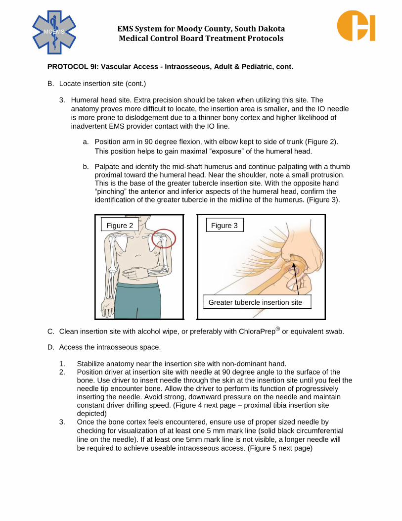
EMS System for Moody County, South Dakota Medical Control Board Treatment Protocols
PROTOCOL 9I: Vascular Access - Intraosseous, Adult & Pediatric, cont.
B. Locate insertion site (cont.)
3. Humeral head site. Extra precision should be taken when utilizing this site. The
anatomy proves more difficult to locate, the insertion area is smaller, and the IO needle
is more prone to dislodgement due to a thinner bony cortex and higher likelihood of
inadvertent EMS provider contact with the IO line.
a. Position arm in 90 degree flexion, with elbow kept to side of trunk (Figure 2).
This position helps to gain maximal “exposure” of the humeral head.
b. Palpate and identify the mid-shaft humerus and continue palpating with a thumb proximal toward the humeral head. Near the shoulder, note a small protrusion. This is the base of the greater tubercle insertion site. With the opposite hand “pinching” the anterior and inferior aspects of the humeral head, confirm the identification of the greater tubercle in the midline of the humerus. (Figure 3).
Figure 2 Figure 3
Greater tubercle insertion site
C. Clean insertion site with alcohol wipe, or preferably with ChloraPrep® or equivalent swab. D. Access the intraosseous space.
1. Stabilize anatomy near the insertion site with non-dominant hand. 2. Position driver at insertion site with needle at 90 degree angle to the surface of the
bone. Use driver to insert needle through the skin at the insertion site until you feel the needle tip encounter bone. Allow the driver to perform its function of progressively inserting the needle. Avoid strong, downward pressure on the needle and maintain constant driver drilling speed. (Figure 4 next page – proximal tibia insertion site depicted)
3. Once the bone cortex feels encountered, ensure use of proper sized needle by
checking for visualization of at least one 5 mm mark line (solid black circumferential
line on the needle). If at least one 5mm mark line is not visible, a longer needle will
be required to achieve useable intraosseous access. (Figure 5 next page)

EMS System for Moody County, South Dakota Medical Control Board Treatment Protocols
PROTOCOL 9I: Vascular Access - Intraosseous, Adult & Pediatric, cont.
Figure 4 Figure 5
4. Resume use of driver to insert a properly-sized needle through the bony cortex and
into the bony marrow (evident with a sudden decrease in resistance to needle
insertion), maintaining the 90 degree angle to the surface of the skin. Most typically,
properly-sized needles will have their hub resting on the skin surface at the time the
needle tip is correctly in the marrow space. E. While stabilizing the needle hub with a thumb and an index finger, disengage the driver
from the needle in a gentle, upward motion. F. While still stabilizing the needle hub with a thumb and an index finger, remove the stylet
by rotating it counterclockwise until disengaged. G. Do NOT attempt aspiration of blood or marrow via the catheter. Pulling marrow into the
catheter may clog the catheter and prevent its use for needed fluid and/or medication
administration. Do confirm proper EZ-IO® catheter placement using a combination of the
following signs:
1. IO catheter rests at 90 degree angle and feels firmly in bone when grasping hub. 2. Blood-tinged marrow oozes spontaneously from hub (may often be absent, yet the
catheter is still correctly placed). 3. Fluid and medication administration is possible without significant resistance and
without extravasation. H. When using the proximal tibia insertion site, use of the EZ-Stabilizer® (Figure 6 – next
page) is optional and its use is determined by the EMT-Intermediate’s or EMT-
Paramedic’s judgment. When using the humeral head insertion site, use of the EZ-
Stabilizer® is required to reduce the chances of inadvertent dislodgement (refer to earlier
discussion of humeral head insertion site). If the EZ-Stabilizer ® is used, it must be
applied prior to connecting the 90 degree connector set to the catheter hub.

EMS System for Moody County, South Dakota Medical Control Board Treatment Protocols
PROTOCOL 9I: Vascular Access - Intraosseous, Adult & Pediatric, cont. I. The EZ-Connect® 90 degree connector set (also seen
in Figure 6) is used to prevent excessive pressure on the catheter when infusing fluids or administering medications. Failure to use the 90 degree connector set can cause inadvertent dislodgement due to excessive pressure down the catheter. Flush the EZ-Connect® set with Normal Saline prior to attaching it to the catheter hub and then flush the line to flush the catheter with 10mL Normal Saline if patient unresponsive or Lidocaine 2% 1 mg/kg up to 40mg slow intraosseous push if the patient is responsive and clearly able to sense pain. If using Lidocaine as directed, follow with 10mL Normal Saline flush.
J. Medication administration is given in the same dosing as with IV administrations. K. Fluid administration will require the use of a pressure infuser on the IV fluid bag. Due to the
increased pressure of the marrow space, IV fluid will not infuse without assistance of the pressure infuser. Inflate pressure infuser until IV fluid is seen infusing with constant flow. Monitor for extravasation and monitor for need to re-inflate the pressure infuser. Fluid delivery rate may be as high as 1 liter per hour at tibial site and up to 5 liters per hour at humeral head site.
L. In determining the site for IO access, consider knowledge of the anatomy, prior training and comfort
in accessing that particular site, and how IO access at that site may or may not interfere with other care events (e.g. use of the humeral head site for medication administration in cardiac arrest could disrupt the continuity of chest compressions).
Complications of intraosseous line placement attempts: Through and through bone penetration – avoid by using correct needle and insertion technique. Extravasation – avoid by using correct needle and insertion technique. Monitor ongoing use and stop at early signs of extravasation. Fracture of bone – avoid by using correct insertion technique (avoid excessive pressure). Infection – avoid by using aseptic technique and do not insert through suspected cellulitis. Growth plate injury in pediatrics – avoid by choosing correct insertion site.
Figure 6
EMS System for Moody County, South Dakota Medical Control Board Treatment Protocols
Indications:
Use and/or monitoring of indwelling central venous devices.
Contraindications:
1. Central venous ports – easily permanently damaged with wrong needle or infected. 2. Suspected infection in the indwelling central venous devices 3. Easy peripheral venous access available or already established.
Technique:
Indwelling central venous devices may become irreversibly damaged if wrong needles and
techniques occur. In general, avoid the use of indwelling central venous devices unless already in
use (during an interfacility transfer) or in the case of a peripherally inserted central catheter (PICC)
line, peripheral venous access is not readily available or already established.
In the setting of interfacility transfer, indwelling central venous device(s) must be reviewed
with either the transferring practitioner/physician or appropriate nursing personnel prior to
conducting the interfacility transfer. Potential complications during transfer should be
discussed and contingency plans reviewed.
During packaging, patient movement, and throughout the interfacility transfer, perform
periodic inspection and assessment of indwelling central venous devices and take
precautions to avoid inadvertent dislodgement of such devices.
If the need arises to access a PICC line, clean the port with ChloraPrep®, Betadine®, or an
alcohol wipe. Aspirate 4 - 5 mL of fluid and discard (to remove any anticoagulant in the line) prior to infusing fluid or administering medication. Flush 10 mL of saline in the PICC line after administering a medication. Maintain aseptic technique throughout all handling of the PICC line.
If a closed cap on the line is required to be removed to access the indwelling central venous
device, make sure the line is clamped to avoid introduction of an air embolus.
Contact MC early for any needed advice or direction in the use of an indwelling
central venous device.
PARAMEDIC
9J – INDWELLING CENTRAL VASCULAR DEVICE MANAGEMENT
ADULT & PEDIATRIC

EMS System for Moody County, South Dakota Medical Control Board Treatment Protocols
Prior to all medication administrations, assure the “5 Rights” are reviewed:
1. Right Patient 2. Right Route 3. Right Dose 4. Right Time 5. Right Medication
If any one of these “rights” is “wrong”, stop and do not administer the medication!
Specific routes of medication administration:
9Ka: Intravenous / Intraosseous – Adult & Pediatric:
1. Assure that the IV / IO line is patent. 2. Cleanse the access port nearest the IV / IO site with alcohol prep. 3. Eject any air from syringe and insert needle or adapter into access port. 4. Pinch the IV /IO line above the medication port. This prevents the medication from traveling
toward the IV bag, forcing it instead toward the patient. 5. Inject the medication as specified per appropriate treatment protocol. 6. Remove the needle or adapter and release the tubing. 7. Open the flow regulator to allow 10 – 20 mL fluid flush.
9Kb: Intramuscular/Subcutaneous Injection – Adult & Pediatric:
1. Use a 1 inch to 1.5 inch long 21 to 25 gauge needle on a syringe. 2. Select injection site (if IM, deltoid, lateral thigh, or upper/outer quadrant of gluteus; if SubQ,
arm or lateral thigh). 3. Cleanse the injection site with alcohol prep. 4. Eject any air from syringe. 5. If IM, stretch skin over injection site and insert needle 90 degrees to skin surface, through
skin into muscle, aspirate and if no blood return, inject medication. 6. If SubQ, pinch skin in a fold over injection site and insert needle 45 degrees to skin surface,
through skin into subcutaneous fatty tissue, aspirate and if no blood return, inject medication.
7. Remove needle and put bandage over the injection site.
EMT – INTERMEDIATE
ADVANCED EMT
PARAMEDIC
PARAMEDIC
9K – MEDICATION ADMINISTRATION
ADULT & PEDIATRIC

EMS System for Moody County, South Dakota Medical Control Board Treatment Protocols
PROTOCOL 9K: Medication Administration – Adult & Pediatric, cont.
9Kc: Intranasal – Adult & Pediatric, technique: 1. Use a medication nasal atomization device on a syringe. 2. Eject any air from syringe. 3. Place the atomizer tip approximately 1.5 cm within the nostril. 4. Briskly compress the syringe plunger to spray atomized solution into the nasal cavity/onto
the nasal mucosa (gently pushing the plunger will not result in atomization).
9Kd: Sublingual/Oral – Adult & Pediatric:
1. Instruct, assist, or place the tablet or spray under the tongue (sublingual) or in the mouth (oral)/on the tongue (oral dissolving).
9Ke: Ocular – Adult & Pediatric:
1. Don’t touch the tip of the medication container to any part of the eye or face. 2. Pull the lower eyelid down while avoiding any ocular (eyeball) pressure. 3. Instill eye drop(s) in the space between the eyelid and the eyeball.
EMT – INTERMEDIATE
ADVANCED EMT
PARAMEDIC
EMERGENCY MEDICAL DISPATCHER
EMERGENCY MEDICAL RESPONDER
EMT
EMT – INTERMEDIATE
ADVANCED EMT
PARAMEDIC
PARAMEDIC

EMS System for Moody County, South Dakota Medical Control Board Treatment Protocols
PROTOCOL 9K: Medication Administration – Adult & Pediatric, cont.
9Kf: Intravascular Infusion Management – Adult & Pediatric: 1. Assure that the IV / IO line is patent. 2. Cleanse the access port nearest the IV / IO site with alcohol prep. 3. Flush any air from infusion line/set and insert needle or adapter into access port. 4. Unless simultaneously giving an IV fluid bolus, close off the IV line above the medication
port. This prevents the medication from traveling toward the IV bag, forcing it instead toward the patient.
5. Infuse the medication as specified per appropriate treatment protocol.
PARAMEDIC

EMS System for Moody County, South Dakota Medical Control Board Treatment Protocols
Indications:
Decompression of ventilated air in stomach (reduction of gastric distension), compromising
oxygenation/ventilation in the unconscious, intubated patient.
Contraindications:
1. Suspected basilar skull fracture 2. Suspected mid-facial fractures 3. Known or suspected actively bleeding esophageal varices
Technique:
1. Select correct size gastric tube. Adult patients typically require
size 16 to 18 French gastric tubes. 2. Measure length of gastric tube to pass by starting with tip just
at xiphoid process, then using distance to earlobe and over to tip of nose (Figure).
3. Mark the measured length of tube with a piece of tape. 4. Lubricate tip of tube with water soluble lubricant if inserting nasally. 5. Nasal insertion: direct gastric tube along the floor of nostril to the posterior nasopharynx,
then feed the gastric tube through the oropharynx down the esophagus and into the stomach, stopping when taped mark nears nostril.
6. Oral insertion: direct gastric tube along tongue to posterior oropharynx, then feed the gastric tube down the esophagus and into the stomach, stopping when taped mark nears lips.
7. Confirm correct gastric placement of gastric tube by injecting 10 to 20 mL of air while auscultating over the stomach for a “swoosh” or “burping/bubbling” indicating the gastric tube tip lies within the stomach. Confirm absence of similar sounds in the lungs by auscultating in the mid-axillary line bilaterally while repeating the injection of small mL volumes of air.
8. Tape the tube in place on the nose or on around the mouth. Alternatively, some commercial types of endotracheal tube holders can be used to secure gastric tubes if passed orally.
9. Attach gastric tube to low pressure suction and observe for gastric decompression.
Troubleshooting:
1. Abort gastric tube passage attempts if unsuccessful in three attempts. 2. Repetitive coughing indicates the gastric tube is erroneously passing down the trachea.
Tracheal/bronchial stimulation in gastric tube passage will typically provoke strong coughing reflex. Promptly withdraw tracheally placed gastric tubes to avoid aspiration. An endotracheal tube will not prevent inadvertent passage of a gastric tube down the trachea.
3. Avoid lavage or medications via gastric tube. Use is for gastric decompression.
PARAMEDIC 9L – NASOGASTRIC/OROGASTRIC TUBE
ADULT & PEDIATRIC

EMS System for Moody County, South Dakota Medical Control Board Treatment Protocols
Indications:
1. Concern for child abuse and/or neglect. 2. Concern for adult/elder abuse and/or neglect.
Contraindications:
None
In the course of EMS care of a patient (from time of emergency services request through transport of the patient to the destination location), EMS professionals that become concerned
regarding actual or perceived abuse and/or neglect occurring to persons of any age should
directly inform any receiving health care professionals and/or involved law enforcement officers of the concerns. If the concerns are regarding abuse and/or neglect of the patient, document
such concerns in the patient care report, including specific comments that the receiving health care professional(s) and/or law enforcement officers were directly informed of such concerns in
appropriate detail.
If EMS professionals believe an adult or child is being abused or neglected, and/or has
concerns regarding the imminent safety of an adult or child due to possible abuse or neglect,
there is a legal responsibility to report the beliefs and/or concerns.
South Dakota Department of Social Services
(Calls Answered 24 Hours a Day/7 Days a Week)
1-605-773-3165
When calling, identify yourself by name, EMS certification level, and agency/apparatus
identification. Have as much patient/person information and situational/observation information
possible readily available to share with the abuse and neglect hotline specialist.
EMERGENCY MEDICAL DISPATCHER
EMERGENCY MEDICAL RESPONDER
EMT
EMT – INTERMEDIATE
ADVANCED EMT
PARAMEDIC
9M – SUSPECTD ABUSE OR NEGLECT
ADULT & PEDIATRIC

EMS System for Moody County, South Dakota Medical Control Board Treatment Protocols
EMERGENCY MEDICAL DISPATCHER
EMERGENCY MEDICAL RESPONDER
EMT
EMT – INTERMEDIATE
ADVANCED EMT
PARAMEDIC
EMD
DO NOT MOVE THE PATIENT UNLESS IN DANGER STABILIZE HEAD AND NECK IN POSITION FOUND
OPEN AIRWAY IF NOT ALERT & INEFFECTIVE BREATHING CONTROL BLEEDING ONLY IF SERIOUS DO NOT ATTEMPT TO SPLINT INJURIES
EMR EMT
TRAUMA AND HYPOVOLEMIC SHOCK SUPPORTIVE CARE SPINAL “STABILIZATION” - DO NOT APPLY SPINAL “TRACTION” DURING MOTION RESTRICTION OR IMMOBILIZATION (if applicable)
STABILIZE IMPALED OBJECTS O2 VIA NC, NRB, OR BVM AS APPROPRIATE
APPLY CARDIAC MONITOR (if equipped)
EMT OR HIGHER LICENSE:
MEASURE END – TIDAL CO2 & MONITOR WAVEFORM CAPNOGRAPHY (if equipped, ** Mandatory use if Pt intubated) MAINTAIN EtCO2 LEVELS, 30 – 35 mmHg
DURING SIGNS SUGGESTIVE OF INCREASING INTRACRANIAL PRESSURE
(PROGRESSIVE DECLINE IN MENTAL STATUS, POSTURING, SEIZING, DILATING/NON-REACTIVE/ASYMMETRIC PUPILS) CONTROLLED HYPERVENTILATION
PLACE SUPRAGLOTTIC AIRWAY IF INDICATED & ONLY IF BVM VENTILATIONS INEFFECTIVE.
EMT – I AEMT
INTUBATE IF INDICATED IV ACCESS (IO IF INDICATED) (TWO LINES IF POSSIBLE)
ADULT: IV / IO NS 250 mL BOLUS MAINTAIN SYS BP ≥100mmHg
REPEAT UP TO 2 LITERS IF SYS BP REMAINS < 100 mmHg & NO SIGNS OF PULMONARY EDEMA PEDIATRIC: IV / IO NS 20 mL/kg BOLUS IF SYS BP < (70 + 2x age in years) mmHg & NO SIGNS OF PULMONARY EDEMA
PARAMEDIC
EVALUATE FOR OTHER ALTERED MENTAL STATUS ETIOLOGIES. TREAT PER APPROPRIATE PROTOCOL(S)
ADULT: MEDICATION ASSISTED INTUBATION IF INDICATED MIDAZOLAM 5 mg IM/IVP/IN/IOP FOR ACTIVE SEIZURE. MAY REPEAT X 1 IN 5 MINS IF STILL SEIZING
OR DIAZEPAM 5 mg IVP/IOP or 10 mg IM FOR ACTIVE SEIZURE. MAY REPEAT X 1 IN 5 MINS IF STILL SEIZING
OR
LORAZEPAM 1 mg IVP/IM/IOP FOR ACTIVE SEIZURE. MAY REPEAT X 1 IN 5 MINS IF STILL SEIZING
PEDIATRIC: MIDAZOLAM 0.1 mg/kg IM/IVP/IN/IOP TO MAX OF 5mg FOR ACTIVE SEIZURE. MAY REPEAT X 1 IN 5 MINS IF STILL SEIZING OR
DIAZEPAM 0.1 mg/kg TO MAX OF 5 mg IVP/IOP/IM FOR ACTIVE SEIZURE. MAY REPEAT X 1 IN 5 MINS IF STILL SEIZING OR
LORAZEPAM 0.1 mg/kg IVP/IM/IOP for ACTIVE SEIZURE. MAY REPEAT X 1 IN 5 MINS IF STILL SEIZING
MC CONSULT IF SEIZURE CONTINUES DESPITE ABOVE TREATMENT CONTINUOUS ASSESSMENT & TREATMENT PER APPLICABLE PROTOCOL(S)
10A – HEAD/NECK/SPINE INJURY
ADULT & PEDIATRIC TREATMENT PRIORITIES
1. Spinal stabilization;
Avoid spinal traction 2. Hemorrhage control 3. Assessment/Care for
life-threatening injuries/ shock/high intracranial pressure
4. Vital signs 5. Appropriate trauma care
destination selection

EMS System for Moody County, South Dakota Medical Control Board Treatment Protocols
EMERGENCY MEDICAL DISPATCHER
EMERGENCY MEDICAL RESPONDER
EMT
EMT – INTERMEDIATE
ADVANCED EMT
PARAMEDIC
EMD
DO NOT MOVE THE PATIENT UNLESS IN DANGER STABILIZE HEAD AND NECK IN POSITION FOUND
OPEN AIRWAY IF NOT ALERT & INEFFECTIVE BREATHING CONTROL BLEEDING ONLY IF SERIOUS DO NOT ATTEMPT TO SPLINT INJURIES
EMR EMT
TRAUMA AND HYPOVOLEMIC SHOCK SUPPORTIVE CARE OBTAIN VITAL SIGNS
BLUNT INJURY/EVISCERATION/LACERATION:
COVER INJURED EYE WITH STERILE, SALINE DAMPENED DRESSING.
PENETRATING INJURY/IMPALED OBJECT: STABILIZE IMPALED OBJECT.
COVER INJURED EYE WITH SHIELD OR OTHER DEVICE (e.g. CUP) AVOIDING DIRECT CONTACT OR PRESSURE ON THE EYEBALL.
CHEMICAL INJURY: IF EYEBALL IS INTACT, SOFT IRRIGATION WITH STERILE SALINE OR WATER
ALL EYE INJURIES:
COVER UNINJURED EYE WITH STERILE, SALINE/STERILE WATER DAMPENED DRESSING TO REDUCE STRAIN / CONSENSUAL EYE MOVEMENT
EMT – I AEMT
IV/IO ACCESS IF INDICATED
PARAMEDIC
EVALUATE FOR OTHER ALTERED MENTAL STATUS ETIOLOGIES. TREAT PER APPROPRIATE PROTOCOL(S)
ANALGESIA (IF REQUIRED)
FOR OPIATE USE, ADULT MUST HAVE SYS BP ≥ 100 mmHg; PEDIATRIC MUST HAVE SYS BP ≥ (70 + 2x age in years) mmHg
ADULT: FENTANYL 1 mcg/kg SLOW IVP/IM/IN, MAX DOSE 100 mcg. MAY REPEAT EVERY 10 MINUTES TO MAX CUMULATIVE DOSE OF 3 mcg/kg or 250 mcg WHICHEVER IS LESSER.
OR
MORPHINE SULFATE 2 - 4 mg SLOW IVP, MAY REPEAT 2 - 4 mg EVERY 5 MINUTES TO A TOTAL OF 10 mg.
PEDIATRIC: MC ORDER ONLY
MC CONSULT IF FURTHER ANALGESIA REQUIRED
CONTINUOUS ASSESSMENT & TREATMENT PER APPLICABLE PROTOCOL(S)
10B – EYE INJURY
ADULT & PEDIATRIC
TREATMENT PRIORITIES
1. Vital signs 2. Eye injury specified care 3. Appropriate trauma care
destination selection 4. Analgesia (if required)

EMS System for Moody County, South Dakota Medical Control Board Treatment Protocols
EMERGENCY MEDICAL DISPATCHER
EMERGENCY MEDICAL RESPONDER
EMT
EMT – INTERMEDIATE
ADVANCED EMT
PARAMEDIC
EMD
DO NOT MOVE THE PATIENT UNLESS IN DANGER STABILIZE HEAD AND NECK IN POSITION FOUND
OPEN AIRWAY IF NOT ALERT & INEFFECTIVE BREATHING CONTROL BLEEDING ONLY IF SERIOUS DO NOT ATTEMPT TO SPLINT INJURIES
EMR EMT
TRAUMA AND HYPOVOLEMIC SHOCK SUPPORTIVE CARE OBTAIN VITAL SIGNS
BLUNT INJURY/LACERATION:
DO NOT REMOVE LOOSENED TEETH. IF ACTIVE BLEEDING, POSITION PATIENT TO AVOID ASPIRATION.
PENETRATING INJURY/IMPALED OBJECT: STABILIZE IMPALED OBJECT. DO NOT REMOVE LOOSENED TEETH. IF ACTIVE BLEEDING, POSITION PATIENT TO AVOID ASPIRATION.
TOOTH FRACTURE:
REPLACE IN SOCKET (IF ABLE WITHOUT ASPIRATION RISK) IF UNABLE TO REPLACE IN SOCKET, PLACE IN A COMMERCIAL TOOTH CARRIER OR CONTAINER OF MILK (if available)
OTHERWISE, WRAP IN A SALINE OR STERILE WATER DAMPENED STERILE DRESSING
EMT – I AEMT
IV/IO ACCESS IF INDICATED
PARAMEDIC
ANALGESIA (IF REQUIRED)
FOR OPIATE USE, ADULT MUST HAVE SYS BP ≥ 100 mmHg; PEDIATRIC MUST HAVE SYS BP ≥ (70 + 2x age in years) mmHg
ADULT: FENTANYL 1 mcg/kg SLOW IVP/IM/IN, MAX DOSE 100 mcg. MAY REPEAT EVERY 10 MINUTES TO MAX CUMULATIVE DOSE OF 3 mcg/kg or
250 mcg WHICHEVER IS LESSER. OR
MORPHINE SULFATE 2 - 4 mg SLOW IVP, MAY REPEAT 2 - 4 mg EVERY 5 MINUTES TO A TOTAL OF 10 mg.
PEDIATRIC: MC ORDER ONLY
MC CONSULT IF FURTHER ANALGESIA REQUIRED
CONTINUOUS ASSESSMENT & TREATMENT PER APPLICABLE PROTOCOL(S)
10C – DENTAL INJURY/PAIN
ADULT & PEDIATRIC
TREATMENT PRIORITIES
1. Vital signs 2. Dental injury specified care 3. Appropriate trauma care
destination selection 4. Analgesia (if required)

EMS System for Moody County, South Dakota Medical Control Board Treatment Protocols
EMERGENCY MEDICAL DISPATCHER
EMERGENCY MEDICAL RESPONDER
EMT
EMT – INTERMEDIATE
ADVANCED EMT
PARAMEDIC
EMD
DO NOT MOVE THE PATIENT UNLESS IN DANGER STABILIZE HEAD AND NECK IN POSITION FOUND
OPEN AIRWAY IF NOT ALERT & INEFFECTIVE BREATHING CONTROL BLEEDING ONLY IF SERIOUS
DO NOT ATTEMPT TO SPLINT INJURIES
EMR EMT
TRAUMA AND HYPOVOLEMIC SHOCK SUPPORTIVE CARE SPINAL “STABILIZATION” - DO NOT APPLY SPINAL “TRACTION” DURING MOTION RESTRICTION OR IMMOBILIZATION (if applicable)
SEAL OPEN CHEST WOUNDS STABILIZE IMPALED OBJECTS
STABILIZE SUSPECTED PELVIC FRACTURE WITH SHEET/BINDER TIED LOW AROUND PELVIS IF DISTAL FRACTURES SUSPECTED STABILIZE SUSPECTED PELVIC FRACTURE WITH INTERNAL ROTATION & TAPING OF LOWER EXTREMITIES IF NO DISTAL FRACTURES EVIDENT COVER EVISCERATED ABDOMINAL/PELVIC ORGANS WITH STERILE, SALINE DAMPENED DRESSING PRIOR TO LAYERING WITH DRY DRESSINGS
OBTAIN VITAL SIGNS O2 VIA NC, NRB, OR BVM AS APPROPRIATE APPLY CARDIAC MONITOR (when available)
EMT OR HIGHER LICENSE: MEASURE END – TIDAL CO2 & MONITOR WAVEFORM CAPNOGRAPHY (if equipped, ** Mandatory use if Pt intubated)
PLACE SUPRAGLOTTIC AIRWAY IF INDICATED & ONLY IF BVM VENTILATIONS INEFFECTIVE.
EMT – I AEMT
INTUBATE IF INDICATED
IV ACCESS (IO IF INDICATED) (TWO LINES IF POSSIBLE)
ADULT: IV NS TKO IF SYS BP ≥ 100 mmHg WITHOUT HYPOTENSIVE SYMPTOMS IV NS 250 mL BOLUS IF SYS BP <100 mmHg WITH HYPOTENSIVE SYMPTOMS & NO SIGNS OF PULMONARY EDEMA,
REPEAT UP TO 2 LITERS NS IF SYS BP REMAINS < 100 mmHg WITH HYPOTENSIVE SYMPTOMS & NO SIGNS OF PULMONARY EDEMA PEDIATRIC: IV NS 20 mL/kg BOLUS IF SYS BP < (70 + 2x age in years) mmHg & NO SIGNS OF PULMONARY EDEMA REPEAT UP TO 60 mL/kg IF SYS BP REMAINS < (70 + 2x age in years) mmHg & NO SIGNS OF PULMONARY EDEMA
PARAMEDIC
NEEDLE THORACOSTOMY FOR SUSPECTED TENSION PNEUMOTHORAX ADULT: MEDICATION ASSISTED INTUBATION IF INDICATED
ANALGESIA (IF REQUIRED) FOR OPIATE USE, ADULT MUST HAVE SYS BP ≥ 100 mmHg; PEDIATRIC MUST HAVE SYS BP ≥ (70 + 2x age in years)
ADULT: FENTANYL 1 mcg/kg SLOW IVP/IM/IN, MAXIMUM DOSE 100 mcg. MAY REPEAT EVERY 10 MINUTES TO MAXIMUM CUMULATIVE DOSE OF 3 mcg/kg or 250 mcg WHICHEVER IS LESSER.
OR MORPHINE SULFATE 2 - 4 mg SLOW IVP, MAY REPEAT 2 - 4 mg EVERY 5 MINUTES TO A TOTAL OF 10 mg.
PEDIATRIC: MC ORDER ONLY
MC CONSULT IF FURTHER ANALGESIA REQUIRED
CONTINUOUS ASSESSMENT & TREATMENT PER APPLICABLE PROTOCOL(S)
10D – CHEST/ABDOMEN/PELVIS INJURY
ADULT & PEDIATRIC TREATMENT PRIORITIES
1. Hemorrhage control 2. Assessment/Care for life-
threatening injuries/shock 3. Needle thoracostomy for
tension pneumothorax 4. Vital signs 5. Appropriate trauma care
destination selection

EMS System for Moody County, South Dakota Medical Control Board Treatment Protocols
Indications:
Suspected tension pneumothorax
Clinical signs of tension pneumothorax:
Increasing respiratory insufficiency in a susceptible patient:
1. Spontaneous pneumothorax 2. CPR with appearance of PEA, increased difficulty bagging patient 3. Sucking chest wound which has been covered completely and which has not responded
to removal of the dressing 4. Chest trauma with suspected pneumothorax AND
Adult systolic blood pressure less than 100 mmHg (or pediatric systolic blood pressure less than 70 + (2 x age in years) mmHg AND
Three or more of the following:
1. "Air Hunger" 2. Cyanosis 3. Decreased breath sounds on affected side(s) 4. Jugular venous distension 5. Tracheal shift away from affected side – extremely late sign – do not wait as indication
for needle thoracostomy if other signs are developing
Etiologies of tension pneumothorax include:
1. Trauma (blunt or penetrating) - disruption of either visceral or parietal pleura; often associated
with rib fractures (rib fractures not necessary for tension pneumothorax to occur) 2. Barotrauma secondary to positive-pressure ventilation, especially when using high
amounts of positive end-expiratory pressure (PEEP) 3. Unsuccessful attempts to convert an open pneumothorax to a simple pneumothorax
in which the occlusive dressing functions as a 1-way valve 4. Chest compressions during cardiopulmonary resuscitation
Contraindications:
No absolute. Do not place a needle thoracostomy through an area of suspected cellulitis,
using instead an alternative site – 5th intercostal space mid-axillary line(s).
PARAMEDIC
10E – NEEDLE THORACOSTOMY (TENSION PNEUMOTHORAX DECOMPRESSION)
ADULT & PEDIATRIC

EMS System for Moody County, South Dakota Medical Control Board Treatment Protocols
PROTOCOL 10E: Needle Thoracostomy, Adult & Pediatric, cont. Precautions:
1. A SIMPLE pneumothorax causes some degree of respiratory distress and chest pain, and MAY be associated with decreased or absent breath sounds on the side of the collapse and with subcutaneous air if the cause is traumatic. TENSION pneumothorax is associated with progressive respiratory distress, dropping BP, "drum-like" hyper expanded chest, distended neck veins, and patient deterioration. Tracheal shift may be present, but is a late sign and needle decompression should be accomplished before waiting for the appearance of tracheal shift.
2. Pneumothorax rarely presents with tension on initial assessment. Be particularly suspicious with deterioration during transport, and with patients requiring assisted ventilation.
3. In patients who are being ventilated by bag-valve mask or ventilator, caution should be exercised when performing needle decompression. If the presumptive diagnosis of a tension pneumothorax is incorrect, the insertion of the needle may create a pneumothorax, which may be converted into a tension pneumothorax by positive-pressure ventilation.
4. If a previously covered sucking chest wound is present, remove the seal and allow chest pressures to equilibrate. No further treatment is often necessary.
Technique:
1. Expose the entire chest. 2. Locate landmark on affected side(s) second intercostal
space just superior to third rib, along the mid-clavicular line (Figure 1.)
3. Clean area of insertion with ChloraPrep®, Betadine®, or alcohol prep.
4. Attach 10 mL or larger syringe to a 15 gauge pneumothorax catheter or a 14 gauge angiocatheter. If using an angiocatheter, the length of the needle should be at least 3.25 inches to promote decompression of the pleural space. Thick chest wall musculature may prevent entry into the pleural space if using a shorter needle
5. Decisively locate the second or third intercostal space in the mid-clavicular line.
6. Insert the needle through the skin at near or at 90 degrees and advance until tip hits the top of the rib below the intercostal space. Continue to advance angling over the top of the rib margin – advance just over the lower rib avoid the neurovascular bundle running horizontally along the lower border of the upper rib.
7. Advance needle tip into the pleural space. A slight “pop” is usually felt when the needle pierces the outside pleural membrane, or parietal pleura.
Figure 1

EMS System for Moody County, South Dakota Medical Control Board Treatment Protocols
PROTOCOL 10E: Needle Thoracostomy, Adult & Pediatric, cont.
Technique, cont.:
8. When tension is present, syringe plunger will typically dislodge back out of syringe, or an immediate hiss of air escaping will be heard.
9. Remove the syringe and needle and leave the catheter in the pleural space. 10. If recurrent decompensation of the patient occurs related to suspected
redevelopment of tension pneumothorax, repeat the procedure next to the
previously successful needle thoracostomy site.
Complications:
1. Creation of pneumothorax if none existed previously. This is an unfortunate occurrence if needle thoracostomy is done too aggressively. Do not hesitate to relieve a strongly suspected tension pneumothorax, but perform an accurate assessment to validate the suspicion of tension pneumothorax.
2. Laceration of lung, which is rare, can cause significant pulmonary injury. Avoid excessive length needles.
3. Hemothorax from vascular injury. Avoid needle thoracostomy medial to the mid-clavicular line. Avoid needle thoracostomy just inferior to a rib, where the intercostal vessels run underneath the rib margin.
4. Infection. Minimize risk by clean insertion site and maintaining aseptic technique,
using sterile catheters/needles.
Note:
Studies show that needle thoracostomy in the 5th or 6th intercostal space at the mid-axillary line is effective in the release of tension pneumothorax. Utilize this location if the traditional sites of
the 2nd or 3rd intercostal space at the mid- clavicular line do not improve the respiratory or hemodynamic conditions of a patient with a strongly suspected tension pneumothorax, especially if using shorter angiocatheters. The chest wall musculature is thinner in this alternate location.

EMS System for Moody County, South Dakota Medical Control Board Treatment Protocols
Indications:
Interfacility transfer of patient with chest tube thoracostomy.
Technique:
The chest tube thoracostomy-related device(s) must be reviewed with either the transferring
practitioner/physician or appropriate nursing personnel prior to conducting the interfacility transfer. Potential complications during transfer should be discussed, such as in the possibility
of recurrent pneumothorax, and contingency plans reviewed, such as releasing the occlusive
dressing around the chest tube or performing a needle thoracostomy per Protocol 10E – Needle Thoracostomy. If the Paramedic feels unable to safely monitor and maintain the chest tube, he
or she is to request appropriate resources from the transferring hospital to accompany the patient during transfer.
Under these conditions, EMS personnel will not begin the transfer until such request is
accommodated.
During packaging, patient movement, and throughout the interfacility transfer, perform
periodic inspection and assessment of the chest tube.
The chest tube may be attached to a one-way valve (Heimlich valve) that allows for air or fluid
passage from the chest to the outside, often contained within a simple bag container. If a
Heimlich valve is present, keep it attached to the chest tube.
The chest tube may alternatively be attached to a multi- chamber container that can be attached to low suction. This container can be used for collection of blood drainage from the chest for auto-transfusion and/or to measure how much blood or other fluid is being drained from the chest tube. The chamber connecting to the chest tube is for fluid collection. The second
chamber contains a small volume of water, establishing a water seal, creating a one-way flow of air from the chest, and keeping the pressure in the chest less than atmospheric pressure. The third chamber is a suction chamber, designed to limit excessive wall suction effect on the chest. Keep the container upright to keep fluid collection measurement accurate and avoid any loss in function of the device.
Persistent bubbling in the chamber(s) indicates an air leak in the chest tube system. This may be due to a loose connection in the tube/chamber/suction circuit or due to a perforation in the airway (e.g. bronchiole). Check the visible connections of the chest tube system. If bubbling prior to transfer, discuss with transferring practitioner/physician.
PARAMEDIC 10F – CHEST TUBE MONITORING
ADULT & PEDIATRIC

EMS System for Moody County, South Dakota Medical Control Board Treatment Protocols
EMERGENCY MEDICAL DISPATCHER
EMERGENCY MEDICAL RESPONDER
EMT
EMT – INTERMEDIATE
ADVANCED EMT
PARAMEDIC
EMD
DO NOT MOVE THE PATIENT UNLESS IN DANGER STABILIZE HEAD AND NECK IN POSITION FOUND
OPEN AIRWAY IF NOT ALERT & INEFFECTIVE BREATHING CONTROL BLEEDING ONLY IF SERIOUS
DO NOT ATTEMPT TO SPLINT INJURIES
EMR EMT
TRAUMA AND HYPOVOLEMIC SHOCK SUPPORTIVE CARE SPINAL “STABILIZATION” - DO NOT APPLY SPINAL “TRACTION” DURING MOTION RESTRICTION OR IMMOBILIZATION (if applicable)
APPLY TOURNIQUET FOR HEMORRHAGE UNCONTROLLED BY DIRECT PRESSURE PER PROTOCOL 10H - TOURNIQUET OBTAIN VITAL SIGNS
O2 VIA NC, NRB AS APPROPRIATE SPLINT SUSPECTED FRACTURES INCLUDING JOINT ABOVE AND BELOW AREA OF INJURY
COVER AMPUTATED ANATOMY WITH SALINE-MOISTENED GAUZE PLACE AMPUTATED ANATOMY IN A CLEAN PLASTIC BAG
PLACE AMPUTATED ANATOMY OVER ICE, AVOIDING DIRECT CONTACT OF AMPUTATED ANATOMY WITH ICE
STABILIZE IMPALED OBJECTS APPLY CARDIAC MONITOR (if equipped)
EMT – I AEMT
IV ACCESS (IO IF INDICATED) (TWO LINES IF POSSIBLE) ADULT: IV NS TKO IF SYS BP ≥ 100 mmHg WITHOUT HYPOTENSIVE SYMPTOMS
IV NS 250 mL BOLUS IF SYS BP <100 mmHg WITH HYPOTENSIVE SYMPTOMS & NO SIGNS OF PULMONARY EDEMA, REPEAT UP TO 2 L NS IF SYS BP REMAINS < 100 mmHg WITH HYPOTENSIVE SYMPTOMS & NO SIGNS OF PULMONARY EDEMA
PEDIATRIC: IV NS 20 mL/kg BOLUS IF SYS BP < (70 + 2x age in years) mmHg & NO SIGNS OF PULMONARY EDEMA
REPEAT UP TO 60 mL/kg IF SYS BP REMAINS < (70 + 2x age in years) mmHg & NO SIGNS OF PULMONARY EDEMA
PARAMEDIC
ANALGESIA (IF REQUIRED) FOR OPIATE USE, ADULT MUST HAVE SYS BP ≥ 100 mmHg; PEDIATRIC MUST HAVE SYS BP ≥ (70 + 2x age in years) mmHg
ADULT: FENTANYL 1 mcg/kg SLOW IVP/IM/IN, MAXIMUM DOSE 100 mcg. MAY REPEAT EVERY 10 MINUTES TO MAXIMUM
CUMULATIVE DOSE OF 3 mcg/kg or 250 mcg WHICHEVER IS LESSER. OR
MORPHINE SULFATE 2 - 4 mg SLOW IVP, MAY REPEAT 2 - 4 mg EVERY 5 MINUTES TO A TOTAL OF 10 mg.
PEDIATRIC: MC ORDER ONLY
MC CONSULT IF FURTHER ANALGESIA REQUIRED
CONTINUOUS ASSESSMENT & TREATMENT PER APPLICABLE PROTOCOL(S)
10G – EXTREMITY/AMPUTATION INJURY
ADULT & PEDIATRIC TREATMENT PRIORITIES
1. Hemorrhage control 2. Assessment/Care for life-
threatening injuries/shock 3. Vital signs 4. Splint suspected fractures 5. Analgesia (if required) 6. Appropriate trauma care
destination selection

EMS System for Moody County, South Dakota Medical Control Board Treatment Protocols
Indication:
Life-threatening extremity hemorrhage unable NOT controlled by direct pressure or
immediately obvious that direct pressure alone will not provide control.
Contraindication: None
Technique (Combat-Application-Tourniquet® - C-A-T®):
The C-A-T® (Figure 1) windlass uses a free moving internal band to provide circumferential
pressure to an injured and uncontrollably bleeding extremity. Once placed, keep the tourniquet secure, but uncovered so that the bleeding site can be clearly monitored as well as the tourniquet itself. The time of tourniquet application is to be written on a piece of adhesive tape and secured to the tourniquet. Conscious patients may experience pain related to tourniquet use. In such instances, follow the pain management protocol if the patient is hemodynamically stable.
Step 1 (Figure 2):
The C-A-T® is applied over the extremity proximal to the bleeding site routing the self – adhering band around the extremity. Lower extremity wounds require feeding the strap through the inner slit and outer slit of the buckle. Upper extremity wounds typically require less pressure to control and do not require feeding the strap through both inner and outer slits of the buckle, though doing so is usually the optimal strategy. If a single slit is used for arm wounds, the inner slit (closest to windlass) is to be used.
EMERGENCY MEDICAL RESPONDER
EMT
EMT – INTERMEDIATE
ADVANCED EMT
PARAMEDIC
10H – TOURNIQUET
ADULT & PEDIATRIC
Figure 1
Figure 2

EMS System for Moody County, South Dakota Medical Control Board Treatment Protocols
PROTOCOL 10H: Tourniquet, Adult & Pediatric, cont.
Step 2 (Figure 3):
For all lower extremity wounds (and any upper
extremity wounds desired), additionally pass the band
through the outside slit of the buckle utilizing the friction
adaptor buckle which will lock the band in place.
Step 3 (Figure 4):
Pull the self - adhering band tight and secure the
band back on itself with the Velcro adhesive strap.
Step 4 (Figure 5):
Twist the windlass until the bleeding has stopped. This
will typically be at or less than 3 complete rotations of
the windlass. More could be required, but be careful not
to exert too much torque on the windlass to avoid
breakage.
Step 5 (Figure 6):
Lock the rod in place with the windlass clip.
Step 6 (Figure 7): TK 0145 Secure the rod with the strap by pulling it tight and
adhering it to the opposite hook on the windlass
hook. Indicate the time of tourniquet application on
tape.
Figure 3
Figure 4
Figure 5
Figure 6
Figure 7

EMS System for Moody County, South Dakota Medical Control Board Treatment Protocols
PROTOCOL 10H: Tourniquet, Adult & Pediatric, cont.
If one tourniquet correctly applied does not completely control hemorrhage, in addition to
direct pressure, an additional tourniquet may be applied just proximal to the first tourniquet.
Once bleeding has been controlled by a tourniquet, the usual and customary practice is to leave
the tourniquet in place throughout the remainder of scene care and transport to an emergency
department. In infrequent circumstances, if pain control becomes an issue, the tourniquet may
be loosened to see if bleeding will stay controlled. If bleeding resumes, promptly re-tighten the
tourniquet to its effective tightness.

EMS System for Moody County, South Dakota Medical Control Board Treatment Protocols
EMERGENCY MEDICAL DISPATCHER
EMERGENCY MEDICAL RESPONDER
EMT
EMT – INTERMEDIATE
ADVANCED EMT
PARAMEDIC
EMD
BLEEDING CONTROL ADVISE DIRECT PRESSURE ON BLEEDING WOUND
HOLD FIRMLY UNTIL HELP ARRIVES
EMR EMT
GENERAL SUPPORTIVE CARE (MEDICAL PT) OR TRAUMA AND HYPOVOLEMIC SHOCK SUPPORTIVE CARE (TRAUMA PT) HISTORY TO INCLUDE IF PT TAKING ANTIPLATELET AGENT (e.g. ASPIRIN) OR ANTICOAGULANT (e.g. WARFARIN)
BLEEDING CONTROL
IF EXTREMITY WOUND AND TOURNIQUET INDICATED, TREAT PER PROTOCOL 10H – TOURNIQUET
APPLY DIRECT PRESSURE AS INDICATED – MANUAL &/OR VIA PRESSURE DRESSING IF DIRECT PRESSURE INSUFFICIENT & BLEEDING IS ARTERIAL/BRISK, APPLY TOPICAL HEMOSTATIC AGENT
MAINTAIN DIRECT PRESSURE WHEN USING TOPICAL HEMOSTATIC AGENT ADVISE RECEIVING PHYSICIANS/NURSES IF HEMOSTATIC AGENT USE WAS REQUIRED & TYPE USED
EMT – I AEMT
ADULT: IV NS TKO IF SYS BP ≥ 100 mmHg WITHOUT HYPOTENSIVE SYMPTOMS
IV NS 250 mL BOLUS IF SYS BP <100 mmHg WITH HYPOTENSIVE SYMPTOMS & NO SIGNS OF PULMONARY EDEMA REPEAT UP TO 2 LITERS NS IF SYS BP REMAINS < 100 mmHg WITH HYPOTENSIVE SYMPTOMS & NO SIGNS OF PULMONARY EDEMA
PEDIATRIC: IV NS 20 mL/kg BOLUS IF SYS BP < (70 + 2x age in years) mmHg
REPEAT UP TO 60 mL/kg IF SYS BP REMAINS < (70 + 2x age in years) mmHg & NO SIGNS OF PULMONARY EDEMA
PARAMEDIC
ADULT: IN SETTING OF HEMORRHAGIC SHOCK FROM TRAUMA LESS THAN 3 HOURS OLD, WITH SUSPECTED NEED FOR MASSIVE BLOOD TRANSFUSION DUE TO MARKED INTERNAL OR EXTERNAL BLOOD LOSS
CRITERIA FOR TRANEXAMIC ACID ADMINISTRATION:
SUSTAINED TACHYCARDIA 110 BEATS PER MINUTE OR GREATER
AND SUSTAINED HYPOTENSION SYSTOLIC BP 90 mmHg OR LESS
TRANEXAMIC ACID (TXA) 1 GRAM IVPB OVER 10 MINS. ADMINISTER IN 100 mL or 250 mL NS.
CONTINUOUS ASSESSMENT & TREATMENT PER APPLICABLE PROTOCOL(S)
10I – HEMOSTATIC AGENTS
ADULT & PEDIATRIC
TREATMENT PRIORITIES
1. Hemorrhage control 2. Vital signs

EMS System for Moody County, South Dakota Medical Control Board Treatment Protocols
EMERGENCY MEDICAL DISPATCHER
EMERGENCY MEDICAL RESPONDER
EMT
EMT – INTERMEDIATE
ADVANCED EMT
PARAMEDIC
EMD
DO NOT MOVE THE PATIENT UNLESS IN DANGER CONTROL BLEEDING WITH DIRECT PRESSURE
DO NOT ATTEMPT TO SPLINT INJURIES
EMR EMT
TRAUMA AND HYPOVOLEMIC SHOCK SUPPORTIVE CARE OBTAIN VITAL SIGNS
HISTORY OF TRAUMA IN EXTREMITY IN PAST DAYS OR ACUTELY THAT COULD CAUSE PRESSURE TO BUILD IN DEEP SPACES OF EXTREMITY?
(MUSCLE OR TISSUE SWELLING? DEEP SPACE BLEEDING?)
EVALUATE EXTREMITY FOR “5 Ps” OF COMPARTMENT SYNDROME:
PAIN OUT OF PROPORTION TO ASSESSED INJURY (APPEARS MINOR INJURY, PATIENT IN EXCRUCIATING PAIN)? PARESTHESIA (NUMBNESS DUE TO NERVE COMPRESSION)?
PRESSURE (SKIN/MUSCLE FEELS TENSE DUE TO INCREASING FORCE WITHIN COMPARTMENT)? PARALYSIS (LATE SIGN DUE TO NERVE COMPRESSION - DON’T WAIT ON PARALYSIS TO REPORT CONCERN)
PULSELESSNESS IN DISTAL EXTREMITY (LATE SIGN OF COMPARTMENT SYNDROME - DON’T WAIT ON ABSENT PULSE TO REPORT CONCERN)
IN SUSPECTED COMPARTMENT SYNDROME, POSITION EXTREMITY TO PROMOTE CIRCULATION TO DISTAL EXTREMITY
EMT – I AEMT
IV/IO ACCESS
PARAMEDIC
ANALGESIA (IF REQUIRED)
FOR OPIATE USE, ADULT MUST HAVE SYS BP ≥ 100 mmHg; PEDIATRIC MUST HAVE SYS BP ≥ (70 + 2x age in years) mmHg
ADULT: FENTANYL 1 mcg/kg SLOW IVP/IM/IN, MAX DOSE 100 mcg. MAY REPEAT EVERY 10 MINUTES TO MAX CUMULATIVE DOSE OF 3 mcg/kg or 250 mcg WHICHEVER IS LESSER.
OR MORPHINE SULFATE 2 - 4 mg SLOW IVP, MAY REPEAT 2 - 4 mg EVERY 5 MINUTES TO A TOTAL OF 10 mg.
PEDIATRIC: MC ORDER ONLY
MC CONSULT IF FURTHER ANALGESIA REQUIRED
CONTINUOUS ASSESSMENT & TREATMENT PER APPLICABLE PROTOCOL(S)
1OJ – COMPARTMENT & CRUSH INJURY
SYNDROMES
ADULT & PEDIATRIC
TREATMENT PRIORITIES 1. Vital signs 2. 5 P’s exam 3. Hospital notification of
concern regarding limb circulation compromise
4. Analgesia (if required)

EMS System for Moody County, South Dakota Medical Control Board Treatment Protocols
EMERGENCY MEDICAL DISPATCHER
EMERGENCY MEDICAL RESPONDER
EMT
EMT – INTERMEDIATE
ADVANCED EMT
PARAMEDIC
EMD
IF PT CLOTHES ARE BURNING OR SMOLDERING, DOUSE THEM WITH WATER IMMEDIATELY.
IF WATER IS NOT AVAILABLE, THEN ROLL PT ON THE GROUND OR SMOTHER THE FIRE.
DO NOT TOUCH ANYTHING OR PICK UP DEBRIS
EMR EMT
TRAUMA AND HYPOVOLEMIC SHOCK SUPPORTIVE CARE STOP THE BURNING PROCESS
SPINAL “STABILIZATION” - DO NOT APPLY SPINAL “TRACTION” DURING IMMOBILIZATION (IF EXPLOSIVE MOI & if applicable) STABILIZE IMPALED OBJECTS (IF EXPLOSIVE MOI)
O2 VIA NC, NRB, OR BVM AS APPROPRIATE FOR RESPIRATORY SYMPTOMS COVER BURNED AREA WITH BURN DRESSING (if equipped) THEN APPLY DRY SHEET
APPLY CARDIAC MONITOR (if equipped)
EMT OR HIGHER LICENSE:
FOR RESPIRATORY SYMPTOMS, MEASURE END – TIDAL CO2 & MONITOR WAVEFORM CAPNOGRAPHY (if equipped, ** Mandatory use if Pt intubated)
PLACE SUPRAGLOTTIC AIRWAY IF INDICATED & ONLY IF BVM VENTILATIONS INEFFECTIVE.
EMT – I AEMT
INTUBATE IF INDICATED IV/ IO ACCESS IF INDICATED
ADULT: IV NS; FOR MAJOR THERMAL BURNS, 250 mL BOLUS IF NO SIGNS OF PULMONARY EDEMA, THEN 4 mL/kg BODY WEIGHT x % BSA BURNED
PEDIATRIC: IV NS 20 mL/kg BOLUS IF NO SIGNS OF PULMONARY EDEMA, THEN 4mL/kg BODY WEIGHT x % BSA BURNED
PARAMEDIC
ADULT: MEDICATION ASSISTED INTUBATION IF INDICATED
ANALGESIA (IF REQUIRED)
FOR OPIATE USE, ADULT MUST HAVE SYS BP ≥ 100 mmHg; PEDIATRIC MUST HAVE SYS BP ≥ (70 + 2x age in years) mmHg
ADULT: FENTANYL 1 mcg/kg SLOW IVP/IM/IN, MAXIMUM DOSE 100 mcg. MAY REPEAT EVERY 10 MINUTES TO MAXIMUM CUMULATIVE DOSE OF 3 mcg/kg or 250 mcg WHICHEVER IS LESSER.
OR
MORPHINE SULFATE 2 - 4 mg SLOW IVP, MAY REPEAT 2 - 4 mg EVERY 5 MINUTES TO A TOTAL OF 10 mg.
PEDIATRIC: MC ORDER ONLY
MC CONSULT IF FURTHER ANALGESIA REQUIRED
CONTINUOUS ASSESSMENT & TREATMENT PER APPLICABLE PROTOCOL(S)
10K – BURNS
ADULT & PEDIATRIC TREATMENT PRIORITIES
1. Thermal Burn • Stop burning process • Flood with water only if flames
not extinguished; smoldering
present; significant heat being
dissipated • Determine possibility of
smoke/toxic inhalation
2. Chemical Burn • Brush off dry chemicals • Flush with water for minimum of
15 minutes
3. Electrical Burn • Evaluate airway and cardiac
status

EMS System for Moody County, South Dakota Medical Control Board Treatment Protocols
PROTOCOL 10K: Burns, Adult &Pediatric, cont., % Body Surface Area (BSA) Estimation Chart Count only Second and Third Degree Burns when calculating estimated %BSA An alternate method of calculating %BSA involvement is to use the size of the patient’s entire hand equal to 1% of their BSA. This is a useful method when calculating smaller burn areas.

EMS System for Moody County, South Dakota Medical Control Board Treatment Protocols
EMERGENCY MEDICAL DISPATCHER
EMERGENCY MEDICAL RESPONDER
EMT
EMT – INTERMEDIATE
ADVANCED EMT
PARAMEDIC
EMD
SUPPORT LAW ENFORCEMENT OPERATION
EMR EMT
MAINTAIN PERSONAL AND PATIENT SAFETY ENSURE LAW ENFORCEMENT OFFICER DISCONNECTS EMBEDDED PROBES FROM WEAPON
TREAT PER APPLICABLE SECTION 7 PROTOCOLS – PSYCHIATRIC/BEHAVIORAL DISORDERS
REMOVE PROBES UNLESS EMBEDDED IN FACE, NECK, GENITALS, SPINE
APPLY CARDIAC MONITOR/OBTAIN 12-LEAD ECG (if equipped) TRANSMIT 12-LEAD ECG TO RECEIVING EMERGENCY DEPARTMENT
EMT OR HIGHER LICENSE:
MEASURE END – TIDAL CO2 & MONITOR WAVEFORM CAPNOGRAPHY (if equipped, ** Mandatory use if Pt intubated)
EMT – I
ADULT: INTUBATE IF INDICATED
IV ACCESS
TREAT PER APPLICABLE SECTION 7 PROTOCOLS – PSYCHIATRIC/BEHAVIORAL DISORDER
PARAMEDIC
ADULT: MEDICATION ASSISTED INTUBATION IF INDICATED
CHEMICAL RESTRAINT IF INDICATED PER PROTOCOL 7C – CHEMICAL RESTRAINT
CONSULT MC IF UNCERTAIN OF ETIOLOGY AND TREATMENT PLAN FOR PSYCHIATRIC PROBLEM
OR IF ADDITIONAL RESTRAINT MEASURES NEEDED
CONTINUOUS ASSESSMENT & TREATMENT PER APPLICABLE PROTOCOL(S)
10L – CONDUCTIVE ENERGY WEAPON RELATED MANAGEMENT
ADULT & PEDIATRIC
TREATMENT PRIORITIES 1. Personal safety 2. Patient safety 3. Physical Restraint 4. Chemical Restraint (if indicated) 5. Vital signs 6. Probe removal

EMS System for Moody County, South Dakota Medical Control Board Treatment Protocols
EMERGENCY MEDICAL DISPATCHER
EMERGENCY MEDICAL RESPONDER
EMT
EMT – INTERMEDIATE
ADVANCED EMT
PARAMEDIC
EMD
SUPPORT LAW ENFORCEMENT OPERATION
EMR EMT
MAINTAIN PERSONAL AND PATIENT SAFETY OBTAIN HISTORY FROM LAW ENFORCEMENT OFFICER(S) REGARDING WEAPON BALLISTICS
(e.g. RUBBER BULLETS, BEAN BAG PROJECTILES)
TRAUMA AND HYPOVOLEMIC SHOCK SUPPORTIVE CARE TREAT PER OTHER APPLICABLE SECTION 10 PROTOCOLS - TRAUMA
TREAT PER APPLICABLE SECTION 7 PROTOCOLS – PSYCHIATRIC/BEHAVIORAL DISORDERS
APPLY CARDIAC MONITOR/OBTAIN 12-LEAD ECG (if equipped) TRANSMIT 12-LEAD ECG TO RECEIVING EMERGENCY DEPARTMENT
EMT OR HIGHER LICENSE:
MEASURE END – TIDAL CO2 & MONITOR WAVEFORM CAPNOGRAPHY (if equipped, ** Mandatory use if Pt intubated)
EMT – I AEMT
INTUBATE IF INDICATED
IV/IO ACCESS IF INDICATED
PARAMEDIC
ADULT: MEDICATION ASSISTED INTUBATION IF INDICATED
CHEMICAL RESTRAINT IF INDICATED PER PROTOCOL 7C – CHEMICAL RESTRAINT
CONTINUOUS ASSESSMENT & TREATMENT PER APPLICABLE PROTOCOL(S)
10M – “LESS LETHAL” WEAPON RELATED MANAGEMENT
ADULT & PEDIATRIC
TREATMENT PRIORITIES
1. Personal safety 2. Patient safety 3. Physical Restraint 4. Chemical Restraint (if
indicated) 5. Vital signs 6. Appropriate trauma care
destination selection

EMS System for Moody County, South Dakota Medical Control Board Treatment Protocols
10Na: Spinal Motion Restriction – Adult & Pediatric:
Many patients evaluated by EMS professionals are placed in a cervical collar and onto a long
spine backboard based as much upon “tradition” that this practice is without risk and the benefit is without question. Like many medical practices scrutinized over time, evidence-based
medicine reveals it is with risk (pain, tissue damage leading to pressure sores, and concerns
about risks of aspiration and impaired breathing mechanics). Similarly, the benefit is not certain. Few “real” injuries are so unstable that the process of spinal motion restriction as performed in
EMS is the difference-maker between paralysis and ambulation.
This protocol does not seek to avoid spinal motion restriction when clinically indicated. This
protocol rather seeks to provide an evidence- based approach that directs the careful practice
of spinal motion “restriction” in situations where history, exam findings, and/or patient
interaction limitations make the possible benefit outweigh the risks. When the benefit does not
outweigh the risks, patients should not incur clinically unnecessary collars and boards.
When applying spinal motion restriction, include the following: 1. Avoid traction being placed on the spine in any direction. 2. Correctly size the cervical collar to additionally avoid traction being placed on the spine. 3. Maintain the spinal column alignment integrity when rolling the patient onto a long back
board, using a scoop stretcher, or placing/moving in any other spinal motion restriction device.
4. Secure the torso and extremities to the backboard first, the head/neck last. 5. Remove from the long spine backboard once on the stretcher, unless CPR is ongoing or
anticipated during transport.
Documentation of spinal motion restriction should include a neurologic assessment before and
after the process, which includes the application of a cervical collar, noting any movement
using a backboard/scoop stretcher, and indicating prompt removal from the backboard/scoop
stretcher unless CPR ongoing/anticipated. In the seated patient that is hemodynamically stable
and requiring spinal motion restriction, assist the patient in pivoting and lying supine onto the
stretcher and/or use a spinal motion restriction device to achieve the same, whichever involves
less anticipated spinal motion.
EMERGENCY MEDICAL RESPONDER
EMT
EMT – INTERMEDIATE
ADVANCED EMT
PARAMEDIC
10N – SPLINTING OF INJURIES
ADULT & PEDIATRIC

EMS System for Moody County, South Dakota Medical Control Board Treatment Protocols
PROTOCOL 10N: Splinting of Injuries, cont. 10Na - Spinal Motion Restriction – Adult & Pediatric, cont.
MECHANISM OF INJURY COMPATIBLE WITH POSSIBLE SPINAL INJURY
**SPINAL IMMOBILIZATION OR SPINAL MOTION RESTRICTION INCLUDING BACKBOARD:
ONLY IF SPINAL INJURY EVIDENT (PARALYSIS, PRIAPISM, OR NEURO SHOCK)
EMS System for Moody County, South Dakota Medical Control Board Treatment Protocols
PROTOCOL 10N: Splinting of Injuries, cont.
10Na - Spinal Motion Restriction – Adult & Pediatric, cont.
Comments regarding the Selective Spinal Motion Restriction Process:
1. The process of EMS-performed selective spinal motion restriction constitutes a formal step-wise screening of individuals suffering from mechanisms of injury compatible with possible injury to the spine. This process, now increasingly widely adopted in EMS systems across the United States, is designed from research-verified assessments, identifying individuals that may be safely transported to an emergency department, without spinal immobilization, for further appropriate physician evaluation
2. When following the decision flowchart in this protocol, there is no validated benefit to applying a cervical collar to patients who do not have any indication for spinal motion restriction and risks of pain, skin trauma, and compromise of respiratory mechanics may result if placed and left on a long spine backboard.
3. The process of EMS-performed selective spinal motion restriction, while continuing to
involve placement of a cervical collar, no longer requires continuous use of a long spine backboard. While the long spine backboard is one option to assist a patient supine onto a stretcher, in the absence of ongoing or imminent CPR, patients should be removed off the long spine backboard as soon after movement onto the stretcher as possible.
4. Patients with penetrating trauma have been shown to have worse outcomes with continuous
use of the long spine backboard, in part due to prolonged scene times relating to extensive spinal motion restriction actions. Victims of penetrating trauma (stabbings, gunshot wounds) to the head, neck, and/or torso SHOULD NOT receive spinal motion restriction unless there is one or more of the following:
• Obvious neurologic deficit to the extremities • Priapism • Neurogenic shock • Anatomic deformity to the spine secondary to injury
5. Patients with any form of trauma may experience reduction in pulmonary mechanics (ease
of oxygenation/ventilation) when placed onto a long spine backboard. Continuous use of the long spine backboard should be limited to situations involving ongoing or imminent CPR.
6. Patients at age extremes are prone to unreliable history and physical assessments.
Patients under the age of 12 years or over the age of 65 years, if they have suffered a mechanism of injury compatible with possible spinal injury, are to be placed in spinal motion restriction.
7. The designation of a Glasgow Coma Scale score of 15 includes an assessment that no
neurological deficits exist. If a patient is complaining of motor and/or sensory loss following a mechanism of injury compatible with possible spinal injury, that patient is to be placed in a cervical collar with verbal reinforcement that they limit movement of their cervical spine, keeping the spine in natural/neutral alignment.

EMS System for Moody County, South Dakota Medical Control Board Treatment Protocols
PROTOCOL 10O: Splinting of Injuries, cont.
10Na - Spinal Motion Restriction – Adult & Pediatric, cont.
Comments regarding the Selective Spinal Motion Restriction Process (cont.):
8. At any point from sustaining an acute mechanism of injury compatible with possible spinal injury through EMS care, if the patient has a reported loss of consciousness or altered mental status, regardless of normal mental status upon EMS contact and assessment, that patient is to be placed in spinal motion restriction.
9. A language barrier exists if the EMS professional and the patient cannot fluently
communicate. Fragmented communication (“broken” language) or the use of a family member or bystander to communicate with the patient does not constitute fluent communication. If the EMS professional has a language barrier with the patient following an injury involving a mechanism compatible with possible spinal injury, that patient is to be placed in spinal motion restriction.
10. Regardless of apparent “soberness” on assessment, if a patient has ingested ethanol or
mental-status altering drugs (e.g. narcotics, benzodiazepines, barbiturates, marijuana, cocaine) within six hours prior to a mechanism of injury compatible with possible spinal injury, that patient is to be placed in spinal motion restriction.
11. Distracting pain or injury is best defined as an injury in which the patient is repetitively fixated
upon to the extent the history and physical assessment is frequently interrupted to address that injury. The EMS professional must use his or her best judgment and anytime a concern exists that an injury may prove distracting to a patient with a mechanism of injury compatible with possible spinal injury, that patient is to be placed in spinal motion restriction.
12. In circumstances of acute vomiting and/or third trimester pregnancy, the patient is preferentially transported left lateral recumbent to reduce aspiration of emesis and when in advanced stages of pregnancy, to avoid compromising venous returns to the chest.
13. If the supine positioning of the patient wearing a cervical collar is compromising respiratory
mechanics and/or causing the patient to have dyspnea, the head of the stretcher may be elevated approx. 15 degrees to assist respiratory status.
14. If a patient suffering a mechanism of injury compatible with possible spinal injury complains
of pain in the spinal or paraspinal area anywhere from the base of the skull to the coccyx, that patient is to be placed in spinal motion restriction.
15. In the physical examination of a patient suffering a mechanism of injury compatible with
possible spinal injury, if the EMS professional discovers spinal or paraspinal pain or deformity upon palpation or with patient flexion, extension, or lateral rotation of the neck or back, that patient is to be placed in spinal motion restriction.

EMS System for Moody County, South Dakota Medical Control Board Treatment Protocols
PROTOCOL 10N: Splinting of Injuries, cont.
10Na - Spinal Motion Restriction – Adult & Pediatric, cont.
Comments regarding the Selective Spinal Motion Restriction Process (cont.):
16. If the EMS professional judges a complicating factor (e.g. patient stress or anxiety, the energy or nature of the mechanism of injury) to be present or significantly concerning, that patient is to be placed in spinal motion restriction. If any doubt exists in the view of the EMS professional as to whether to spinal motion restrict the patient, that patient is to be placed in spinal motion restriction.
17. An instance may occur when a patient has been deemed safe for transport without spinal
motion restriction using this protocol and the patient subsequently develops neck or back pain in the ambulance during transport to an emergency department. The EMS professional must use his or her best judgment factoring the degree of pain verbalized and the remaining transport route and time in deciding when to spinal motion restrict the patient. As a guideline, if the remaining route involves unusually rough highway or will be prolonged beyond several minutes duration, the EMS crew should temporarily stop transportation and apply spinal motion restriction to the patient in the ambulance unless the patient’s condition is otherwise unstable and requires continued emergency transport. As a guideline, if the arrival at the destination emergency department is imminent, the patient may be spinal motion restricted upon hospital arrival. In each instance, the EMS professional should inform the receiving nurse or physician of the events and timing of spinal motion restriction and appropriately reflect the events in the patient care report.
18. Any utilization of the selective spinal motion restriction protocol should be clearly
documented in the patient care report, with each requirement in this process denoted. 19. An instance may occur when a patient that is to be spinal motion restricted by this protocol
absolutely refuses a cervical collar and other such movement limitations. These are, indeed, difficult circumstances. If repeated attempts to secure the cooperation of the patient fail, guidance from MC should be sought. If such a patient is transported without spinal motion restriction by the direction of the MC, detailed documentation of the spinal motion restriction attempts, MC consultation and direction, and subsequent actions is to be contained in the patient care report.
20. For pediatric patients found in car seats and involved in motor vehicle collisions, use the
following if spinal motion restriction indicated:
• Infants restrained in a rear-facing car seat may remain in and be extricated in the car seat if secure and his/her condition allows (no signs of respiratory distress or shock)
• Children restrained in a car seat (with a high back) may remain in and be extricated in the car seat.
• Children restrained in a booster seat (without a back) need to be extricated and cared for following standard spinal motion restriction procedures.

EMS System for Moody County, South Dakota Medical Control Board Treatment Protocols
PROTOCOL 10N: Splinting of Injuries, cont.
10Na - Spinal Motion Restriction – Adult & Pediatric, cont. EVAC-U-SPLINT® Mattress Full Body Splint: Preparation Positioning and Application: 1. Lay the EVAC-U-SPLINT® Mattress out on a flat
surface near the patient. The “head and shoulder” logo indicates the head end of the mattress.
2. Remove the red vinyl leashed cap from the valve. Release any vacuum in the mattress by pushing in the red valve stem. Keep the valve stem pushed in until the mattress is pliable.
3. Disconnect the patient restraint strap from the mattress. Or, if desired, disconnect the strap only from the side of the mattress nearest the patient.
4. Manually smooth out the beads to form a level surface. 5. Connect the pump to the mattress by fastening the pump hose connector to the
MaxiValve™ on the mattress. The pump can be attached either at the foot end or the head end.
Evacuate enough air to make the mattress semi-rigid. The objective is to be able to move the mattress as a unit during positioning and have the beads stabilized enough to place the patient onto the mattress without pushing the beads to one side. With the correct amount of evacuation, the mattress surface should be smooth, but not dimpled. 1. Place the mattress next to the patient with the first buckle in-line with the patient’s axilla
(armpit). The first buckle is positioned just below the “head and shoulder” logo on the mattress.
2. Log-roll the patient using standard patient care techniques. 3. Bring the mattress in tight behind the patient, keeping the first buckle in-line with the
patient’s axilla. 4. Log-roll the patient back onto the mattress. Center the patient if necessary. You may find it easier to log-roll the patient onto the mattress with the patient’s head three to four feet down from the head end of the mattress. You can then move the patient along the long axis of their body into a position that is centered on the mattress.
4. Open the MaxiValve™ at the foot end, allowing air to enter. Keep the MaxiValve™ open until the mattress softens and begins to conform to the shape of the patient. It may be necessary to move beads either away from, or under the head of the patient, depending on the anatomy of the patient. Make sure there are sufficient beads to maintain neutral alignment of the patient. If necessary, pad appropriately to ensure neutral alignment.
5. Attach the patient restraint strap. Keep in mind that one of the primary functions of the strap is to bring the sides of the mattress up around the patient’s body. Method #1: Start at the chest with the first buckle and work toward the feet. In a zig-zag pattern, attach the black buckles and the white buckles in an alternating fashion. Finish by attaching the black buckle at the foot end. Method #2: Attach all of the black buckles on one side of the patient. Then follow by attaching all of the white buckles on the other side. NOTE: You may find it helpful to store your vacuum mattress with all the buckles attached and then unfasten only the buckles on the side of the mattress that will be slid under the patient. This should make it easier and faster to apply the mattress at the scene.

EMS System for Moody County, South Dakota Medical Control Board Treatment Protocols
PROTOCOL 10N: Splinting of Injuries, cont.
10Na - Spinal Motion Restriction – Adult & Pediatric, cont.
6. Starting at the head end of the mattress, remove slack by lifting at the center of each section of strap that spans the distance between two buckles. Excess strap is pulled through the BLACK buckle at the foot end of the mattress. VIGOROUS PULLING ON THE STRAP MAY CAUSE THE PATIENT AND/OR MATTRESS TO ROTATE. THIS CAN BE AVOIDED BY ALWAYS FEEDING THE STRAP THROUGH THE BUCKLES WHILE YOU TIGHTEN THE STRAP. Repeat the strap tightening procedure working from the head end toward the foot end of the patient. If the buckles at the foot end are very close together, it may be easier to remove any excess slack by tightening the strap in the reverse direction, moving from the foot end toward the head end of the patient. ALWAYS USE CAUTION WHEN TIGHTENING THE PATIENT RESTRAINT STRAP TO AVOID RESPIRATORY COMPROMISE OR APPLICATION OF PRESSURE TO ANY INJURED AREA.
7. Push any beads away from the area above the patient’s head, but do not alter the support under the patient’s head which is maintaining neutral alignment. Shape the mattress around the head, making sure to fill the voids by the shoulders and the base of the neck of the patient. Transfer stabilization to the outside of the mattress and continue to hold these “head blocks” that you have formed until the mattress is evacuated.
8. Evacuate the air from the mattress, using the valve located at the foot end. The head-end valve can be used, or both valves can be used simultaneously, but it is preferred to use the foot-end valve. This makes it easier to work around the patient’s head. Under normal conditions, the EVAC-U-SPLINT® manual vacuum pump may require up to 35 strokes to achieve rigid immobilization
9. To prevent accidental valve opening, whenever a patient is being moved in the mattress or when the mattress is being put away, make sure to ALWAYS place the RED LEASHED CAP over the end of the MaxiValve™.
10. Finish securing the patient’s head using medical grade adhesive tape. 11. Snug up the patient restraint strap and check the patient’s neurovascular status before
moving the patient. Cleaning, Maintenance and Repair: DO NOT lift the EVAC-U-SPLINT® mattress by itself from the ends. Use the point-of-balance lifting handles only. A minimum of two persons positioned at the sides are required. Patient size and weight will determine the number of personnel required to properly lift and move the patient for transport. The large handles on each side allow for two rescuers to grasp the mattress on each side. This provides four support points on both sides of the mattress for large or very tall patients. For extremely heavy patients, additional supportive devices may be needed in addition to the vacuum mattress. When faced with a situation that requires lifting end-to-end (i.e., going up or down stairs), use a long spine board, scoop-type stretcher, or a “flat” stretcher underneath the mattress. Vertical rescue requires the use of a basket stretcher or similar device. When using any accessory stretcher always make sure the vacuum mattress is secured to the stretcher prior to moving the patient. Always check the rigidity of the mattress before lifting the patient off the ambulance cot or other patient handling device.

EMS System for Moody County, South Dakota Medical Control Board Treatment Protocols
PROTOCOL 10N: Splinting of Injuries, cont. 10Nb – Extremity – Adult & Pediatric:
When applying extremity splinting, include the following:
1. Assess and document the assessment of distal vascular (pulse) and nerve (motor/sensation) function, before and after splinting.
2. In general, immobilize the joint on either side of the suspected fracture area.
3. Pad splints whenever possible to avoid tissue pressure from splints.
4. In the setting of that an extremity is pulseless distal to a markedly angulated fracture, make one gentle attempt to place the angulated extremity in near-normal alignment. Document the distal vascular and nerve function before and after any such maneuver.
5. Prioritize timely transport to an appropriate emergency department for extremity injuries with pulses absent distal to the suspected fracture/injury.
6. Utilize EVAC-U-SPLINT® where appropriate for extremity splinting.
Positioning: Always make sure that the basic ABC’s of airway, breathing and circulation are intact prior to any splinting activities.
Expose the injured area.
Observe skin color and symmetry.
Determine if there has been a loss of mobility and/or sensation.
Palpate distal pulse and check for prompt capillary refill.
Apply constant, “long axis” (in-line) gentle manual traction on severely deformed, long bone fractures. Return extremity to its normal anatomical position with an assistant supporting the extremity under the suspected fracture site. CAUTION: Do not return the extremity to its anatomical position when contraindicated. If the patient complains of increased pain or if there is resistance during manipulation movement, then STOP and immobilize the extremity as best possible in the position found. Joints or distal limb fractures with no neurovascular deficits should be immobilized in the position found. Maintain manual support of the injured area while coordinating additional assistance during the EVAC-U-SPLINT® immobilization process.
Preparation: Your healthcare partner or a qualified assistant should:
Apply appropriate dressings to all wounds in and around the suspected fracture site.
Open the Evac-U-Splint® carry case and select a splint that will immobilize the joints above and below the fracture site. Shoulder and hip injuries will require additional immobilization methods to accomplish this goal.
Remove the red leashed cap from the MaxiValve™ and push in on the red end of the valve to equalize the air pressure in the splint.
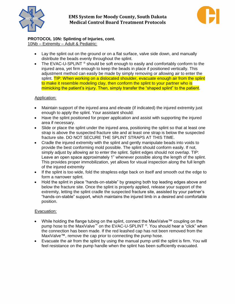
EMS System for Moody County, South Dakota Medical Control Board Treatment Protocols
PROTOCOL 10N: Splinting of Injuries, cont. 10Nb – Extremity – Adult & Pediatric:
Lay the splint out on the ground or on a flat surface, valve side down, and manually distribute the beads evenly throughout the splint.
The EVAC-U-SPLINT ® should be soft enough to easily and comfortably conform to the injured area, yet firm enough to keep the beads in place if positioned vertically. This adjustment method can easily be made by simply removing or allowing air to enter the splint. TIP: When working on a dislocated shoulder, evacuate enough air from the splint to make it resemble modeling clay, then conform the splint to your partner who is mimicking the patient’s injury. Then, simply transfer the “shaped splint” to the patient.
Application:
Maintain support of the injured area and elevate (if indicated) the injured extremity just enough to apply the splint. Your assistant should:
Have the splint positioned for proper application and assist with supporting the injured area if necessary.
Slide or place the splint under the injured area, positioning the splint so that at least one strap is above the suspected fracture site and at least one strap is below the suspected fracture site. DO NOT SECURE THE SPLINT STRAPS AT THIS TIME.
Cradle the injured extremity with the splint and gently manipulate beads into voids to provide the best conforming mold possible. The splint should conform easily. If not, simply adjust by allowing air to enter the splint. Splint edges should not overlap. TIP: Leave an open space approximately 1” whenever possible along the length of the splint. This provides proper immobilization, yet allows for visual inspection along the full length of the injured extremity
If the splint is too wide, fold the strapless edge back on itself and smooth out the edge to form a narrower splint.
Hold the splint in place “hands-on-stable” by grasping both top leading edges above and below the fracture site. Once the splint is properly applied, release your support of the extremity, letting the splint cradle the suspected fracture site, assisted by your partner’s “hands-on-stable” support, which maintains the injured limb in a desired and comfortable position.
Evacuation:
While holding the flange tubing on the splint, connect the MaxiValve™ coupling on the pump hose to the MaxiValve™ on the EVAC-U-SPLINT ®. You should hear a “click” when the connection has been made. If the red leashed cap has not been removed from the MaxiValve™, remove the cap prior to connecting the pump hose.
Evacuate the air from the splint by using the manual pump until the splint is firm. You will feel resistance on the pump handle when the splint has been sufficiently evacuated.

EMS System for Moody County, South Dakota Medical Control Board Treatment Protocols
PROTOCOL 10N: Splinting of Injuries, cont. 10Nb – Extremity – Adult & Pediatric:
Remove the pump hose from the MaxiValve™ by depressing the metal tab on the pump hose coupling. The evacuated splint has now formed a rigid supportive “cast” around the injured area. Place the red leashed cap back on the MaxiValve™.
Place the splint straps, with slight tension, around the splint. Secure the straps in place.
NOTE: The EVAC-U-SPLINT® is available with “hook and loop” or side release buckle straps. The “hook and loop” straps are shown here. The suspected fracture site is now splint-stable. Adjustments can now be made, if necessary, to add stability or to make slight positional changes for patient comfort. These adjustments can be accomplished by simply removing or allowing air to enter the splint. During aeromedical transport the splint may soften with significant altitude changes due to the decrease/increase in air pressure. Adjust as necessary. The change in rigidity will not be nearly as noticeable as with anti-shock garments because there is very little air inside the EVAC-U-SPLINT® once it has been evacuated. After evacuating the splint, re-evaluate skin color, recheck mobility and sensation, palpate distal pulse and time capillary refill. Finish treatment of the patient with the appropriate EMS equipment and supplies based on the patient’s needs and the location of injury.
Valve and Pump Operation To Evacuate Air:
Manually remove the red leashed cap from the Maxi-Valve™.
While holding the flexible flange tubing just below the MaxiValve™, attach the vacuum
pump hose to the MaxiValve™. You should hear a “click” when the connection has been
made.
Evacuate the air from the splint.
Remove the pump hose from the MaxiValve™ by depressing the metal tab on the pump
hose connector. Place the red leashed cap back on the MaxiValve™.
To Release Vacuum:
Manually remove the red leashed cap from the MaxiValve™.
While holding the MaxiValve™, depress the red plastic center section, which will allow
air to enter the splint.
Place the red leashed cap back on the MaxiValve™ for storage.
Special Applications:
The EVAC-U-SPLINT® is a versatile product that can be used in many ways for immobilizing and support of a variety of suspected injuries. The EVAC-U-SPLINT® is only limited by the creativity of the users. The medium or large size EVAC-U-SPLINT® can be effectively used as a full-body immobilizer for infants and toddlers (0-2 years old). The appropriate size splint should be selected based on the suspected injuries. The EVAC-U-SPLINT® will also act as a thermal insulator, helping to reduce temperature loss.

EMS System for Moody County, South Dakota Medical Control Board Treatment Protocols
PROTOCOL 10N: Splinting of Injuries, cont. 10Nb – Extremity – Adult & Pediatric:
The EVAC-U-SPLINT® set comes in a duffel bag style carry case that easily fits in most vehicle compartments or under a squad bench. The splints and the manual pump should always be stored in their carry case to reduce the potential for getting them soiled or damaged. They should NOT be stored in compartments exposed to extreme heat or cold and should NEVER be placed next to a muffler or exhaust system. Daily inspection should include looking for anything that would affect the integrity of performance of the EVAC-U-SPLINT® (i.e. tears, broken parts, loose hose clamps, inoperable valves, etc.) If damage is found or if maintenance is required, take the EVAC-U-SPLINT® out of service until such time that it can be properly repaired or replaced.
Keep all surfaces clean, dry and free of tears and your EVAC-U-SPLINT® should provide you with many years of reliable service. The manual vacuum pump is virtually maintenance-free. Perform general exterior cleaning as needed. Once a year, check the O-ring seal. If the “O” ring needs lubrication, use a water proof, aluminum complex formula grease. NOTE: a 1” spread spanner wrench will be needed to remove the top cap of the pump to inspect the O-ring. Clean and disinfect the EVAC-U-SPLINT® according to your medical director’s guidelines. The EVAC-U-SPLINT® and the splint straps should be cleaned using soap and water, a mild detergent, or a commercial cleaner/disinfectant. When the splint has been exposed to blood or body fluids, it should be cleaned using cold water and then allowed to soak in a detergent solution for 24 hours and then thoroughly rinsed and dried. ALWAYS keep the red leashed cap on the MaxiValve™ when soaking the EVAC-U-SPLINT®. Sodium hypochlorite (bleach) solutions may be used, but avoid prolonged exposure of the fabric to high concentrations of bleach because discoloration is possible. A 1% bleach solution can be used on the EVAC-U-SPLINT®, but it should be thoroughly rinsed off with lukewarm or cold water and then allowed to dry. Make sure the splint and splint straps are completely dry before placing them back into their storage/ carry case.

EMS System for Moody County, South Dakota Medical Control Board Treatment Protocols
EMERGENCY MEDICAL DISPATCHER
EMERGENCY MEDICAL RESPONDER
EMT
EMT – INTERMEDIATE
ADVANCED EMT
PARAMEDIC
EMD
OBTAIN AVAILABLE SCENE HAZARD INFORMATION FOR RESPONDER SAFETY
DO NOT MOVE PATIENTS UNLESS IN DANGER STABILIZE HEAD AND NECK IN POSITION FOUND
OPEN AIRWAY IF NOT ALERT & INEFFECTIVE BREATHING CONTROL BLEEDING BY DIRECT PRESSURE
EMR EMT
SCENE SAFETY CONSIDERATIONS – FLAMMABLE ENVIRONMENT? TERRORISM CAUSE – SECONDARY DEVICES? MASS CASUALTY INCIDENT MANAGEMENT/TRAUMA AND HYPOVOLEMIC SHOCK SUPPORTIVE CARE
EXPECT HEARING DIFFICULTY/DEAFNESS IN PATIENTS DUE TO PRIMARY BLAST INJURY OF RUPTURED EAR TYMPANUM APPLY TOURNIQUET FOR HEMORRHAGE UNCONTROLLED BY DIRECT PRESSURE PER PROTOCOL 10H - TOURNIQUET
OBTAIN VITAL SIGNS O2 VIA NC, NRB AS APPROPRIATE
SPLINT SUSPECTED FRACTURES INCLUDING JOINT ABOVE AND BELOW AREA OF INJURY COVER AMPUTATED ANATOMY WITH SALINE-MOISTENED GAUZE
PLACE AMPUTATED ANATOMY IN A CLEAN PLASTIC BAG PLACE AMPUTATED ANATOMY OVER ICE, AVOIDING DIRECT CONTACT OF AMPUTATED ANATOMY WITH ICE
STABILIZE IMPALED OBJECTS APPLY CARDIAC MONITOR (if equipped)
EMT – I AEMT
IV ACCESS (IO IF INDICATED) (TWO LINES IF POSSIBLE) ADULT: IV NS TKO IF SYS BP ≥ 100 mmHg WITHOUT HYPOTENSIVE SYMPTOMS
IV NS 250 mL BOLUS IF SYS BP <100 mmHg WITH HYPOTENSIVE SYMPTOMS & NO SIGNS OF PULMONARY EDEMA, REPEAT UP TO 2 LITERS NS IF SYS BP REMAINS < 100 mmHg WITH HYPOTENSIVE SYMPTOMS & NO SIGNS OF PULMONARY EDEMA
PEDIATRIC: IV NS 20 mL/kg BOLUS IF SYS BP < (70 + 2x age in years) mmHg & NO SIGNS OF PULMONARY EDEMA REPEAT UP TO 60 mL/kg IF SYS BP REMAINS < (70 + 2x age in years) mmHg & NO SIGNS OF PULMONARY EDEMA
PARAMEDIC
ANALGESIA (IF REQUIRED) FOR OPIATE USE, ADULT MUST HAVE SYS BP ≥ 100 mmHg; PEDIATRIC MUST HAVE SYS BP ≥ (70 + 2x age in years) mmHg
ADULT: FENTANYL 1 mcg/kg SLOW IVP/IM/IN, MAXIMUM DOSE 100 mcg. MAY REPEAT EVERY 10 MINUTES TO MAXIMUM
CUMULATIVE DOSE OF 3 mcg/kg or 250 mcg WHICHEVER IS LESSER. OR
MORPHINE SULFATE 2 - 4 mg SLOW IVP, MAY REPEAT 2 - 4 mg EVERY 5 MINUTES TO A TOTAL OF 10 mg.
PEDIATRIC: MC ORDER ONLY
MC CONSULT IF FURTHER ANALGESIA REQUIRED CONTINUOUS ASSESSMENT & TREATMENT PER APPLICABLE PROTOCOL(S
10O – BLAST INJURY
ADULT & PEDIATRIC TREATMENT PRIORITIES
1. SAFETY, SAFETY, SAFETY 2. Hemorrhage control 3. Assessment/Care for life-
threatening injuries/shock 4. Vital signs 5. Analgesia (if required) 6. Appropriate trauma care
destination selection

EMS System for Moody County, South Dakota Medical Control Board Treatment Protocols
EMERGENCY MEDICAL DISPATCHER
EMERGENCY MEDICAL RESPONDER
EMT
EMT – INTERMEDIATE
ADVANCED EMT
PARAMEDIC
EMD
REMOVE ANY SOURCES OF HEAT. REMOVE OUTER CLOTHING.
APPLY COOL WATER TO ENTIRE SKIN SURFACE WHILE FANNING. TURN ON AN AIR CONDITIONER OR FAN.
EMR EMT
GENERAL SUPPORTIVE CARE REMOVE FROM HOT ENVIRONMENT
COOL BODY INCLUDING HEAD WITH WATER OR SALINE APPLY ICE PACKS TO GROIN, NECK, AXILLA
DIRECT FAN ON PATIENT TO PROMOTE EVAPORATIVE COOLING/AVOID SHIVERING OBTAIN VITAL SIGNS
O2 VIA NC, NRB, OR BVM AS APPROPRIATE APPLY CARDIAC MONITOR (if equipped)
EMT OR HIGHER LICENSE:
MEASURE EtCO2 & MONITOR WAVEFORM CAPNOGRAPHY (if equipped, **Mandatory use if Pt intubated) PLACE SUPRAGLOTTIC AIRWAY IF INDICATED & ONLY IF BVM VENTILATIONS INEFFECTIVE
EMT – I AEMT
INTUBATE IF INDICATED
IV ACCESS
ADULT: IV NS 500 mL BOLUS IF NO SIGNS OF PULMONARY EDEMA. USE CHILLED NS (when available) IF HEAT STROKE SUSPECTED
REPEAT UP TO 2 LITERS NS IF FATIGUE, ALTERED MENTAL STATUS, &/OR HYPOTENSION < 100 mmHg PERSISTSIF NO SIGNS OF PULMONARY EDEMA
PEDIATRIC: IV NS 20 mL/kg BOLUS IF NO SIGNS OF PULMONARY EDEMA
REPEAT UP TO 60 mL/kg IF FATIGUE, ALTERED MENTAL STATUS, &/OR HYPOTENSION < (70 + 2x age in years) mmHg PERSISTSIF NO SIGNS OF PULMONARY EDEMA
PARAMEDIC
ADULT: MEDICATION ASSISTED INTUBATION IF INDICATED ADULT: MIDAZOLAM 0.1 mg/kg IM/IVP/IN/IOP TO MAX OF 5 mg FOR ACTIVE SEIZURE. MAY REPEAT X 1 IN 5 MINS IF STILL SEIZING.
OR DIAZEPAM 5 mg IVP/IOP or 10 mg IM FOR ACTIVE SEIZURE. MAY REPEAT X 1 IN 5 MINS IF STILL SEIZING.
OR
LORAZEPAM 2 mg IVP/IOP/IM FOR ACTIVE SEIZURE. MAY REPEAT X 1 IN 10 MINS IF STILL SEIZING
PEDIATRIC: MIDAZOLAM 0.1 mg/kg IM/IVP/IN/IOP TO MAX OF 5 mg FOR ACTIVE SEIZURE. MAY REPEAT X 1 IN 5 MINS IF STILL SEIZING. OR
DIAZEPAM 0.1 mg/kg TO MAX OF 5 mg IVP/IOP/IM FOR ACTIVE SEIZURE. MAY REPEAT X 1 IN 5 MINS IF STILL SEIZING. OR
LORAZEPAM 0.1 mg/kg TO MAX OF 2 mg IVP/IOP/IM FOR ACTIVE SEIZURE. MAY REPEAT X 1 IN 10 MINS IF STILL SEIZING.
MC CONSULT IF SEIZURE CONTINUES DESPITE ABOVE TREATMENT CONTINUOUS ASSESSMENT & TREATMENT PER APPLICABLE PROTOCOL(S)
11A – HEAT ILLNESS
ADULT & PEDIATRIC TREATMENT PRIORITIES 1. Cooling therapy 2. Vital signs 3. Fluid resuscitation

EMS System for Moody County, South Dakota Medical Control Board Treatment Protocols
EMERGENCY MEDICAL DISPATCHER
EMERGENCY MEDICAL RESPONDER
EMT
EMT – INTERMEDIATE
ADVANCED EMT
PARAMEDIC
EMD
PROTECT FROM COLD REMOVE WET CLOTHING
PASSIVE WARMING WITHOUT RUBBING AFFECTED AREAS DO NOT GIVE ALCOHOL TO DRINK
EMR EMT
GENERAL SUPPORTIVE CARE REMOVE FROM COLD ENVIRONMENT
REMOVE WET/RESTRICTIVE CLOTHING APPLY HEAT PACKS TO GROIN, NECK, AXILLA
COVER PATIENT WITH BLANKETS TO EXTENT CLINICALLY POSSIBLE OBTAIN VITAL SIGNS
O2, VIA NC, NRB, OR BVM AS APPROPRIATE (WARM & HUMIDIFIED O2 if equipped) APPLY CARDIAC MONITOR (if equipped)
UNRESPONSIVE HYPOTHERMIA PATIENT STATUS:
ASSESS BREATHING/PULSE FOR UP TO 45 SECONDS PERFORM CPR IF IN CARDIOPULMONARY ARREST
IF DEFIBRILLATION ADVISED BY AED, LIMIT TO ONE DEFIBRILLATION UNTIL WARMED
FROSTBITE: APPLY LOOSE STERILE DRESSINGS AND SPLINT INVOLVED EXTREMITIES
EMT OR HIGHER LICENSE:
MEASURE END – TIDAL CO2 & MONITOR WAVEFORM CAPNOGRAPHY (if equipped, **Mandatory use if Pt intubated) PLACE SUPRAGLOTTIC AIRWAY IF INDICATED & ONLY IF BVM VENTILATIONS INEFFECTIVE
EMT – I AEMT
INTUBATE IF INDICATED
IV/IO ACCESS
ADULT: IV NS (WARMED UP TO 43°C (109°F) if available) 500 mL BOLUS IF NO SIGNS OF PULMONARY EDEMA.
REPEAT UP TO 2 LITERS NS IF PT TEMP <30°C (<86°F), ALTERED MENTAL STATUS, &/OR HYPOTENSION < 100 mmHg PERSISTSIF NO SIGNS OF PULMONARY EDEMA
PEDIATRIC: IV NS 20 mL/kg BOLUS (WARMED AS PER ADULT if available) IF NO SIGNS OF PULMONARY EDEMA REPEAT UP TO 60 mL/kg IF PT TEMP <30°C (<86°F),
ALTERED MENTAL STATUS,&/OR HYPOTENSION < (70 + 2x age in years) mmHg PERSISTS IF NO SIGNS OF PULMONARY EDEMA
PARAMEDIC
ADULT: MEDICATION ASSISTED INTUBATION IF INDICATED
HYPOTHERMIC CARDIAC ARREST: ADULT/PEDIATRIC: WITH BODY TEMPERATURE <30°C (<86°F)
CONTINUE CPR, WITHHOLD MEDICATIONS, LIMIT TO A SINGLE DEFIBRILLATION FOR VENTRICULAR FIBRILLATION/PULSELESS VT
WITH BODY TEMPERATURE ≥30°C (≥86°F) CONTINUE CPR, ADMINISTER DYSRHYTHMIA APPLICABLE MEDICATIONS AT 10 MINUTE INTERVALS
AS CORE TEMPERATURE RISES, DEFIBRILLATE PER NORMAL BODY TEMPERATURE INTERVALS CONTINUE REWARMING MEASURES UNTIL
ROSC OR MC AUTHORIZES TERMINATION OF RESUSCITATION
CONTINUOUS ASSESSMENT & TREATMENT PER APPLICABLE PROTOCOL(S)
11B – COLD ILLNESS/INJURY
ADULT & PEDIATRIC TREATMENT PRIORITIES
1. Warming therapy 2. Vital signs 3. Modified cardiac arrest
resuscitation (when applicable)

EMS System for Moody County, South Dakota Medical Control Board Treatment Protocols
EMERGENCY MEDICAL DISPATCHER
EMERGENCY MEDICAL RESPONDER
EMT
EMT – INTERMEDIATE
ADVANCED EMT
PARAMEDIC
EMD
SEND FOR AED IF AVAILABLE FOR UNCONSCIOUS PATIENT BEWARE OF ELECTRICAL RISKS AND ELECTRIFIED WATER
TURN – OFF ELECTRICAL POWER IF SAFELY CAPABLE ATTEMPT RESCUE ONLY IF SAFELY CAPABLE
CPR BY EMD INSTRUCTION IF INDICATED
EMR EMT
MAINTAIN PERSONAL AND PATIENT SAFETY
STOP BURNING PROCESS GENERAL SUPPORTIVE CARE/CARDIOPULMONARY RESUSCITATION PER APPLICABLE PROTOCOLS
OBTAIN VITAL SIGNS O2 VIA NC, NRB, OR BVM AS APPROPRIATE
APPLY CARDIAC MONITOR/OBTAIN 12-LEAD ECG (if equipped) TRANSMIT 12-LEAD ECG TO RECEIVING EMERGENCY DEPARTMENT
COVER BURNED AREA WITH BURN DRESSING (if equipped) THEN APPLY DRY SHEET
EMT OR HIGHER LICENSE:
MEASURE END – TIDAL CO2 & MONITOR WAVEFORM CAPNOGRAPHY (if equipped, ** Mandatory use if Pt intubated) PLACE SUPRAGLOTTIC AIRWAY IF INDICATED & ONLY IF BVM VENTILATIONS INEFFECTIVE
EMT – I AEMT
INTUBATE IF INDICATED
IV/IO ACCESS
ADULT: IV NS TKO; FOR MAJOR THERMAL BURNS, 250 mL BOLUS IF NO SIGNS OF PULMONARY EDEMA, THEN 4 mL/kg BODY WEIGHT x % BSA BURNED
PEDIATRIC: IV NS TKO; FOR MAJOR THERMAL BURNS, 20 mL/kg BOLUS IF NO SIGNS OF PULMONARY EDEMA THEN 4 mL/kg BODY WEIGHT x % BSA BURNED
PARAMEDIC
TREAT CARDIAC ARREST AND SPECIFIC RHYTHM MANAGEMENT PER APPLICABLE PROTOCOL(S) ADULT: MEDICATION ASSISTED INTUBATION IF INDICATED
ANALGESIA IF REQUIRED
FOR OPIATE USE, ADULT MUST HAVE SYS BP ≥ 100 mmHg; PEDIATRIC MUST HAVE SYS BP ≥ (70 + 2x age in years) mmHg
FENTANYL 1 mcg/kg SLOW IVP/IM/IN, MAX DOSE 100 mcg. MAY REPEAT EVERY 10 MINUTES TO MAX CUMULATIVE DOSE OF 3 mcg/kg or 250 mcg WHICHEVER IS LESSER.
OR MORPHINE SULFATE 2 - 4 mg SLOW IVP, MAY REPEAT 2 - 4 mg EVERY 5 MINUTES TO A TOTAL OF 10 mg.
MC CONSULT IF FURTHER ANALGESIA REQUIRED CONTINUOUS ASSESSMENT & TREATMENT PER APPLICABLE PROTOCOL(S)
11C – ELECTRICAL/LIGHTNING INJURY
ADULT & PEDIATRIC TREATMENT PRIORITIES
1. Personal safety 2. Patient safety 3. Oxygenation/Ventilation 4. Chest compressions and
defibrillation (when applicable) 5. Vital signs 6. Red/Priority 1 patient, even in
setting of MCI

EMS System for Moody County, South Dakota Medical Control Board Treatment Protocols
EMERGENCY MEDICAL DISPATCHER
EMERGENCY MEDICAL RESPONDER
EMT
EMT – INTERMEDIATE
ADVANCED EMT
PARAMEDIC
EMD
RESCUE ONLY IF SAFE TO ATTEMPT IF DIVING ACCIDENT, AVOID ALL UNNECESSARY MOVEMENT
STABILIZE HEAD AND NECK IN POSITION FOUND OPEN AIRWAY IF NOT ALERT & INEFFECTIVE BREATHING
EMR EMT
GENERAL SUPPORTIVE CARE OBTAIN VITAL SIGNS
O2 VIA NC, NRB, OR BVM AS APPROPRIATE UTILIZE APPROPRIATE PRECAUTIONS USING POWERED DEVICE(S) ON WET PATIENT
APPLY CARDIAC MONITOR (if equipped)
EVALUATE ETIOLOGY OF WATER SUBMERSION – TREAT PER APPLICABLE PROTOCOL(S) COLD WATER SUBMERSION CARDIAC ARREST: REWARMING MEASURES/RESUSCITATION/TRANSPORT
WARM WATER SUBMERSION CARDIAC ARREST: EVALUATE RESUSCITATION/TRANSPORT UTILITY PER PROTOCOL
EMT OR HIGHER LICENSE:
MEASURE END-TIDAL CO2 & MONITOR WAVEFORM CAPNOGRAPHY (if equipped; **Mandatory use if Pt intubated) ADULT: APPLY CPAP IF INDICATED (if equipped)
EMT – I AEMT
INTUBATE IF INDICATED
IV/IO ACCESS IF INDICATED
PARAMEDIC
TREAT CARDIAC ARREST SPECIFIC RHYTHM MANAGEMENT PER APPLICABLE PROTOCOL(S)
ADULT: MEDICATION ASSISTED INTUBATION IF INDICATED
CONTINUOUS ASSESSMENT & TREATMENT PER APPLICABLE PROTOCOL(S)
11D – WATER SUBMERSION EVENT
ADULT & PEDIATRIC TREATMENT PRIORITIES
1. Rescue safety 2. Oxygenation/Ventilation 3. Assessment/Care for life-
threatening injuries and/or cardiac arrest
4. Vital signs 5. Appropriate trauma care
destination selection if injury present

EMS System for Moody County, South Dakota Medical Control Board Treatment Protocols
Indications: Active fire ground operations in which physiologic stress is exerted upon firefighters. Contraindications: None Clinical Pearls: 1. Fire ground operations place significant physiologic stress upon firefighters. Even in seemingly
"normal" weather (absence of temperature extremes, absence of precipitation) during operations on even terrain, conducted by ample numbers of firefighters, the elevated body temperatures and physical stress experienced from exertion while wearing heavy protective clothing should not be underestimated. Early and effective rehabilitation promotes desired fire fighter safety on the fire ground.
2. The "basics" of effective fire ground rehabilitation include:
a. Medical monitoring of fire fighters at rehab entry, during rehab, and at rehab release; b. Returning body temperatures to near normal (cooling in heat; warming in cold); c. Hydration and electrolyte replacement; d. Incident Command support of preventing fire fighter return to fire ground duty until medically
appropriate. 3. Fire ground rehabilitation operations conducted by EMS organizations should be performed in
close cooperation with involved fire departments and with knowledge of the rehabilitation policies of those fire departments.
4. In addition to the medical literature references for this protocol, additional fire ground
rehabilitation resources are available through the US Fire Administration, National Fire Protection Agency, International Association of Fire Chiefs, and International Association of Fire
Fighters.
EMERGENCY MEDICAL RESPONDER
EMT
EMT – INTERMEDIATE
ADVANCED EMT
PARAMEDIC
12A – FIRE GROUND REHABILITATION CONCEPTS
ADULT & PEDIATRIC

EMS System for Moody County, South Dakota Medical Control Board Treatment Protocols
EMERGENCY MEDICAL DISPATCHER
EMERGENCY MEDICAL RESPONDER
EMT
EMT – INTERMEDIATE
ADVANCED EMT
PARAMEDIC
EMD
DIRECT TO MOVE AWAY FROM SMOKE IF SAFE TO DO SO OPEN AIRWAY IF NOT ALERT OR INEFFECTIVE BREATHING
IF AWAKE, AVOID PHYSICAL EXERTION OR ENVIRONMENTAL STRESS (TEMP EXTREMES).
ADVISE PT SELF-ADMINISTRATION OF MEDICATIONS (e.g. ALBUTEROL INHALER)
IF PREVIOUSLY PRESCRIBED FOR SIMILAR SYMPTOMS
EMR EMT
MAINTAIN PERSONAL & PATIENT SAFETY GENERAL SUPPORTIVE CARE
OBTAIN VITAL SIGNS O2 VIA NC, NRB, OR BVM AS APPROPRIATE
APPLY CARDIAC MONITOR (if equipped) ASSIST PT WITH PT’S OWN ALBUTEROL INHALER/NEBULIZER (when applicable) TREAT PER 12C – CARBON MONOXIDE &/OR 12E – CYANIDE AS APPLICABLE
EMT OR HIGHER LICENSE:
MEASURE END-TIDAL CO2 & MONITOR WAVEFORM CAPNOGRAPHY (if equipped **Mandatory use if Pt intubated) ADULT: APPLY Bi/CPAP IF INDICATED (if equipped)
ADULT & PEDIATRIC WEIGHT ≥15 kg: NEBULIZED ALBUTEROL 5 mg PEDIATRIC WEIGHT <15 kg: NEBULIZED ALBUTEROL 2.5 mg
MAY REPEAT ALBUTEROL ENROUTE X 1 AS NEEDED
PLACE SUPRAGLOTTIC AIRWAY IF INDICATED & ONLY IF BVM VENTILATIONS INEFFECTIVE
EMT – I AEMT
INTUBATE IF INDICATED IV/IO ACCESS IF INDICATED
ADULT: IV NS TKO IF SYS BP ≥ 100 mmHg WITHOUT HYPOTENSIVE SYMPTOMS
IV NS 250 mL BOLUS IF SYS BP <100 mmHg WITH HYPOTENSIVE SYMPTOMS & NO SIGNS OF PULMONARY EDEMA, REPEAT UP TO 2 LITERS NS IF SYS BP REMAINS < 100 mmHg WITH HYPOTENSIVE SYMPTOMS & NO SIGNS OF PULMONARY EDEMA
PEDIATRIC: IV NS TKO IF SYS BP ≥ (70 + 2x age in years) mmHg
IV NS 20 mL/kg BOLUS IF SYS BP < (70 + 2x age in years) mmHg IF NO SIGNS OF PULMONARY EDEMA
PARAMEDIC
ADULT: MEDICATION ASSISTED INTUBATION IF INDICATED CONTINUOUS ASSESSMENT & TREATMENT PER APPLICABLE PROTOCOL(S)
INTERPRETATION OF 12-LEAD ECGS (when indicated & if equipped)
12B – SMOKE INHALATION
ADULT & PEDIATRIC TREATMENT PRIORITIES
1. Personal safety 2. Patient safety 3. Vital signs (including EtCO2, if
equipped) 4. Oxygenation support
O2 by NC, NRB BVM, CPAP, ETT if indicated
5. Ventilation support BVM, CPAP, ETT if indicated
6. Nebulization therapy Albuterol

EMS System for Moody County, South Dakota Medical Control Board Treatment Protocols
EMERGENCY MEDICAL DISPATCHER
EMERGENCY MEDICAL RESPONDER
EMT
EMT – INTERMEDIATE
ADVANCED EMT
PARAMEDIC
EMD
DIRECT TO MOVE AWAY FROM SUSPECTED SOURCE IF SAFE TO DO SO
OPEN AIRWAY IF NOT ALERT & INEFFECTIVE BREATHING IF AWAKE, AVOID PHYSICAL EXERTION
OR ENVIRONMENTAL STRESS (TEMP EXTREMES).
EMR EMT
MAINTAIN PERSONAL & PATIENT SAFETY
GENERAL SUPPORTIVE CARE OBTAIN VITAL SIGNS
O2 HIGH LITER PER MINUTE FLOW (15 LPM +) VIA NRB, OR BVM AS APPROPRIATE APPLY CARDIAC MONITOR (if equipped)
MEASURE CARBON MONOXIDE LEVEL – %SpCO (if equipped)
IF %SpCO NORMAL & NO SYMPTOMS, TREAT PER OTHER APPLICABLE PROTOCOL(S) IF %SpCO ABNORMAL, EVALUATE IF SYMPTOMS INCLUDE ALTERED MENTAL STATUS? PT PREGNANT?
MC CONSULT TO DISCUSS HYPERBARIC OXYGEN THERAPY FOR GCS ≤ 13 OR IF PREGNANT
EMT OR HIGHER LICENSE: MEASURE END-TIDAL CO2 & MONITOR WAVEFORM CAPNOGRAPHY (if equipped **Mandatory use if Pt intubated)
ADULT: APPLY CPAP IF INDICATED (if equipped)
PLACE SUPRAGLOTTIC AIRWAY IF INDICATED & ONLY IF BVM VENTILATIONS INEFFECTIVE
EMT – I AEMT
INTUBATE IF INDICATED IV/IO ACCESS IF INDICATED
ADULT: IV NS TKO IF SYS BP ≥ 100 mmHg WITHOUT HYPOTENSIVE SYMPTOMS
IV NS 250 mL BOLUS IF SYS BP <100 mmHg WITH HYPOTENSIVE SYMPTOMS & NO SIGNS OF PULMONARY EDEMA, REPEAT UP TO 2 LITERS NS IF SYS BP REMAINS < 100 mmHg WITH HYPOTENSIVE SYMPTOMS & NO SIGNS OF PULMONARY EDEMA
PEDIATRIC: IV NS TKO IF SYS BP ≥ (70 + 2x age in years) mmHg
IV NS 20 mL/kg BOLUS IF SYS BP < (70 + 2x age in years) mmHg IF NO SIGNS OF PULMONARY EDEMA
PARAMEDIC
ADULT: MEDICATION ASSISTED INTUBATION IF INDICATED CONTINUOUS TREATMENT AND ASSESSMENT PER SPECIFIC MEDICAL PROTOCOL(S)
INTERPRETATION OF 12-LEAD ECGS (when indicated & if equipped)
12C – CARBON MONOXIDE
ADULT & PEDIATRIC
TREATMENT PRIORITIES 1. Personal safety 2. Patient safety 3. Vital signs (including CO &
EtCO2, if equipped) 4. Oxygenation support
O2 by NC, NRB
BVM, CPAP, ETT if indicated
5. Ventilation support
BVM, CPAP, ETT if indicated
6. MC consult for hyperbaric oxygen use direction in serious exposures
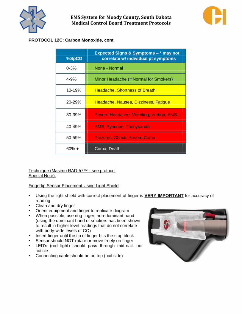
EMS System for Moody County, South Dakota Medical Control Board Treatment Protocols
PROTOCOL 12C: Carbon Monoxide, cont. Technique (Masimo RAD-57™ - see protocol Special Note):
Fingertip Sensor Placement Using Light Shield:
• Using the light shield with correct placement of finger is VERY IMPORTANT for accuracy of reading
• Clean and dry finger • Orient equipment and finger to replicate diagram • When possible, use ring finger, non-dominant hand
(using the dominant hand of smokers has been shown to result in higher level readings that do not correlate with body-wide levels of CO)
• Insert finger until the tip of finger hits the stop block
• Sensor should NOT rotate or move freely on finger • LED’s (red light) should pass through mid-nail, not
cuticle
• Connecting cable should be on top (nail side)
Expected Signs & Symptoms – * may not
%SpCO correlate w/ individual pt symptoms
0-3% None - Normal
4-9% Minor Headache (**Normal for Smokers)
10-19% Headache, Shortness of Breath
20-29% Headache, Nausea, Dizziness, Fatigue
30-39% Severe Headache, Vomiting, Vertigo, AMS
40-49% AMS, Syncope, Tachycardia
50-59% Seizures, Shock, Apnea, Coma
60% + Coma, Death

EMS System for Moody County, South Dakota Medical Control Board Treatment Protocols
PROTOCOL 12C: Carbon Monoxide, cont.
Startup Sequence:
• Place sensor on finger (clean/dry skin) • Press “POWER” button
• Verify all LED’s light up and a 1 second tone is heard
• Startup mode begins • All preset configurations are displayed
• Scrolling zeroes 0 – 0 – 0 and flashes dashed lines • May take up to 25 seconds
• Do not move sensor during startup • When complete, reading is displayed • Begin patient monitoring
• Defaults to pulse rate and oxygen saturation reading
• “PI” bar graph displays strength of arterial perfusion
Operation / Pulse Oximetry & Pulse Rate:
• Displays after startup sequence described above • Oxygen Saturation on top in red numbers
• Pulse Rate on bottom in green numbers
• Low Signal I.Q.® (SIQ) LED lit indicates poor pulse ox signal quality - evaluate finger/sensor, use alternate finger
• Press “DISPLAY” to display %SpCO
Press “Bell” to silence alarms (if needed)
Power Button: Press "ON", Hold for "OFF" Each green LED window below Power Button that illuminates indicates 25% battery power

EMS System for Moody County, South Dakota Medical Control Board Treatment Protocols
PROTOCOL 12C: Carbon Monoxide, cont.
Operation / Carbon Monoxide (Carboxyhemoglobin) Measurement:
• Press “DISPLAY” button as described above to toggle display to show %SpCO reading (to toggle back to pulse oximetry and pulse rate mode, press “DISPLAY” again)
• Carboxyhemoglobin displayed in % on top in numbers
• “CO” displayed on bottom confirming mode
• Real-time SpCO indicator continuously reads SpCO • Confirm abnormal readings by taking several
measurements on different fingers and average the readings
Operation / Troubleshooting:
Error Messages:
• “NO Cbl” = cable not seated properly into device or defective cable
• “SEN OFF” = sensor off finger or misaligned • “bAd CbL” = defective cable (cable most likely needs replaced) • “CbL” = incompatible cable (change to appropriate cable) • “bAd SEN” = defective sensor (sensor most likely needs replaced) • “SEN” = unrecognized sensor (change to appropriate sensor) • "Err" = return for service
Will not power on = check battery compartment and replace batteries Continuously in startup mode (Scrolling zeroes 0 – 0 – 0 and flashes dashed lines) = shield
sensor from flashing lights, strobes or high ambient light with Light Shield (best accurate
practice is to always use the Light Shield); try another finger
Using Physio-Control LifePak® 15 with Masimo sensing to measure %SpCO:
• Power on, connect pulse oximetry cable to monitor/defibrillator and sensor, place sensor on patient
• To display %SpCO, use the SPEED DIAL to select the pulse oximetry display area
• Select PARAMETER from menu
• Select SpCO. Selected value displays for 10 seconds. If %SpCO is elevated, an
advisory event occurs and elevated value flashes and alarm tone sounds

EMS System for Moody County, South Dakota Medical Control Board Treatment Protocols
Indications:
Carbon monoxide (CO) toxicity (as determined through Protocol 12C – Carbon Monoxide).
Contraindications:
Absence of carbon monoxide toxicity.
Clinical Pearls:
1. In the care of the suspected CO poisoned patient, exercise personal safety and avoid
becoming CO poisoned. 2. The hallmarks of effective EMS care of the suspected CO poisoned patient include removal
of the patient from the CO source and oxygenation with near 100% oxygen (via high flow
through non-rebreather mask with good seal, non-invasive positive pressure ventilation, or
through bag-valve-mask or bag-valve-artificial airway connected to an oxygen reservoir). 3. The vast majority (nearly all) of suspected CO poisoned patients may be appropriately
transported to an emergency department that does not have direct access to hyperbaric
oxygen (HBO) therapy. 4. Contact the nearest HBO capable facility’s on-line medical control for EMS to discuss the
advisability of transport for HBO therapy consideration if either of the following distinct clinical situations in which suspected/measured CO toxicity is the primary medical issue of concern: a. Glasgow Coma Scale score ≤ 13 b. Pregnancy
5. Consultation and/or transport to a HBO-capable facility does not compel use of HBO therapy
by the medical staff at that facility.
EMERGENCY MEDICAL RESPONDER
EMT
EMT – INTERMEDIATE
ADVANCED EMT
PARAMEDIC
12D – HYPERBARIC OXYGEN THERAPY CONSIDERATIONS
ADULT & PEDIATRIC
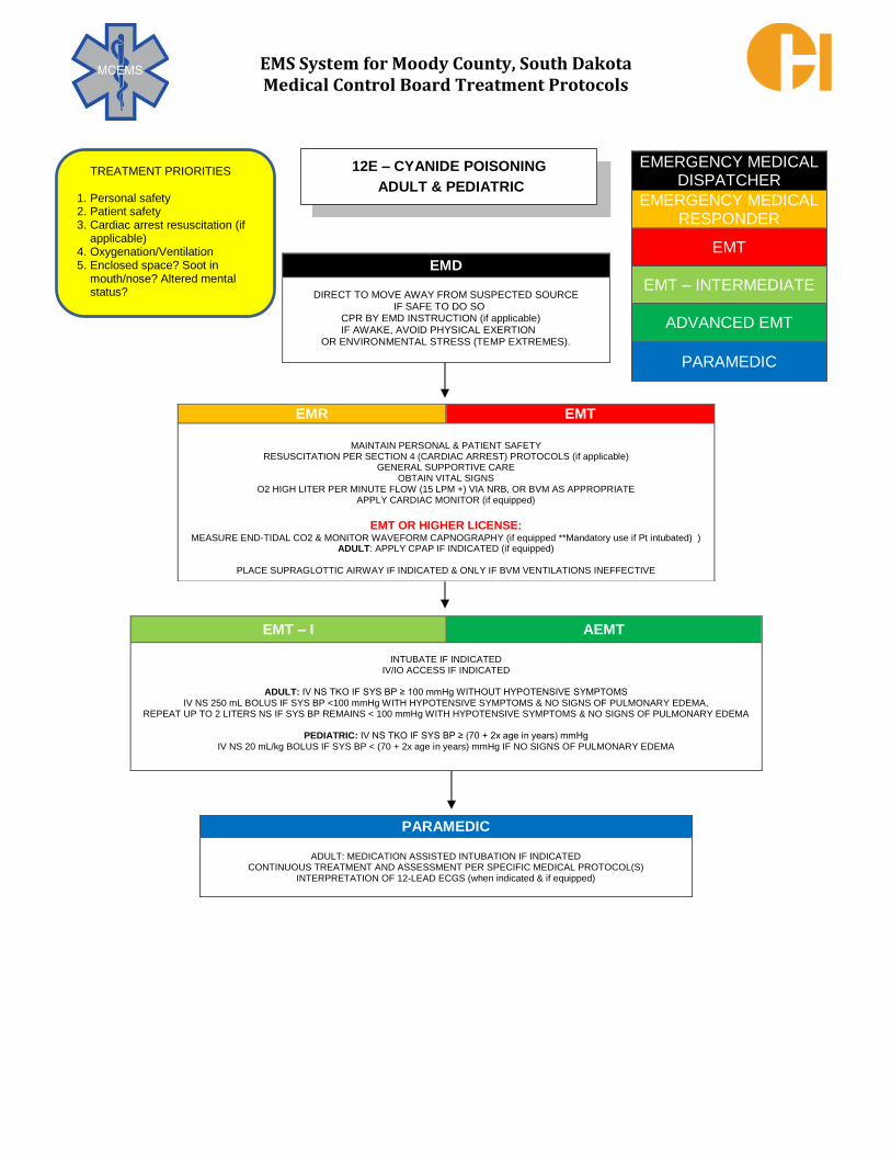
EMS System for Moody County, South Dakota Medical Control Board Treatment Protocols
EMERGENCY MEDICAL DISPATCHER
EMERGENCY MEDICAL RESPONDER
EMT
EMT – INTERMEDIATE
ADVANCED EMT
PARAMEDIC
EMD
DIRECT TO MOVE AWAY FROM SUSPECTED SOURCE IF SAFE TO DO SO
CPR BY EMD INSTRUCTION (if applicable) IF AWAKE, AVOID PHYSICAL EXERTION
OR ENVIRONMENTAL STRESS (TEMP EXTREMES).
EMR EMT
MAINTAIN PERSONAL & PATIENT SAFETY
RESUSCITATION PER SECTION 4 (CARDIAC ARREST) PROTOCOLS (if applicable) GENERAL SUPPORTIVE CARE
OBTAIN VITAL SIGNS O2 HIGH LITER PER MINUTE FLOW (15 LPM +) VIA NRB, OR BVM AS APPROPRIATE
APPLY CARDIAC MONITOR (if equipped)
EMT OR HIGHER LICENSE:
MEASURE END-TIDAL CO2 & MONITOR WAVEFORM CAPNOGRAPHY (if equipped **Mandatory use if Pt intubated) ) ADULT: APPLY CPAP IF INDICATED (if equipped)
PLACE SUPRAGLOTTIC AIRWAY IF INDICATED & ONLY IF BVM VENTILATIONS INEFFECTIVE
EMT – I AEMT
INTUBATE IF INDICATED
IV/IO ACCESS IF INDICATED
ADULT: IV NS TKO IF SYS BP ≥ 100 mmHg WITHOUT HYPOTENSIVE SYMPTOMS IV NS 250 mL BOLUS IF SYS BP <100 mmHg WITH HYPOTENSIVE SYMPTOMS & NO SIGNS OF PULMONARY EDEMA,
REPEAT UP TO 2 LITERS NS IF SYS BP REMAINS < 100 mmHg WITH HYPOTENSIVE SYMPTOMS & NO SIGNS OF PULMONARY EDEMA
PEDIATRIC: IV NS TKO IF SYS BP ≥ (70 + 2x age in years) mmHg IV NS 20 mL/kg BOLUS IF SYS BP < (70 + 2x age in years) mmHg IF NO SIGNS OF PULMONARY EDEMA
PARAMEDIC
ADULT: MEDICATION ASSISTED INTUBATION IF INDICATED CONTINUOUS TREATMENT AND ASSESSMENT PER SPECIFIC MEDICAL PROTOCOL(S)
INTERPRETATION OF 12-LEAD ECGS (when indicated & if equipped)
12E – CYANIDE POISONING
ADULT & PEDIATRIC TREATMENT PRIORITIES
1. Personal safety 2. Patient safety 3. Cardiac arrest resuscitation (if
applicable) 4. Oxygenation/Ventilation 5. Enclosed space? Soot in
mouth/nose? Altered mental status?

EMS System for Moody County, South Dakota Medical Control Board Treatment Protocols
APGAR SCORING (SIGN) 0 1 2
APPEARANCE BLUE OR PALE BODY PINK, EXTREMITIES BLUE COMPLETELY PINK
HEART RATE (BPM) ABSENT ≤100 >100
GRIMACE (REACTION TO CATHETER IN NARES) NO RESPONSE GRIMACE COUGH OR SNEEZE
MUSCLE TONE LIMP SOME FLEXION ACTIVE MOTION
RESPIRATORY RATE ABSENT SLOW/IRREGULAR GOOD, CRYING
EMERGENCY MEDICAL DISPATCHER
EMERGENCY MEDICAL RESPONDER
EMT
EMT – INTERMEDIATE
ADVANCED EMT
PARAMEDIC
EMD
ADVISE TO NOT TRY TO PREVENT THE BIRTH ADVISE NOT TO SIT ON TOILET
PATIENT TO ASSUME POSITION OF COMFORT ADVISE PATIENT TAKE DEEP BREATHS BETWEEN CONTRACTIONS
EMR EMT
GENERAL SUPPORTIVE CARE
CEPHALIC (HEAD FIRST) PRESENTATION:
POSITION MOTHER SUPINE/KNEES WIDELY SEPARATED/BUTTOCKS ELEVATED W/PILLOW/BLANKET PLACE STERILE PAD/SHEET UNDER AND AROUND VAGINA
PLACE ONE HAND OVER FETAL HEAD APPLY MINIMAL STABILIZING PRESSURE TO PREVENT EXPLOSIVE BIRTH
SUCTION MOUTH THEN EACH NOSTRIL WITH BULB SYRINGE IF NEEDED TO CLEAR AIRWAY ALLOW HEAD TO ROTATE NATURALLY
DELIVER THE REST OF INFANT – MAY REQUIRE GENTLE DOWNWARD PRESSURE ON ANTERIOR (UPPER SHOULDER & GENTLE UPWARD PRESSURE ON POSTERIOR (LOWER) SHOULDER FOR EASIER DELIVERY
CLAMP/CUT UMBILICAL CORD
ASSESS INFANT/CALCULATE APGAR SCORE AT ONE AND FIVE MINUTES POST DELIVERY USING TABLE
EMT – I AEMT
IV ACCESS IV NS TKO IF SYS BP ≥ 100 mmHg WITHOUT HYPOTENSIVE SYMPTOMS
IV NS 250 mL BOLUS IF SYS BP <100 mmHg WITH HYPOTENSIVE SYMPTOMS & NO SIGNS OF PULMONARY EDEMA, REPEAT UP TO 2 LITERS NS IF SYS BP REMAINS < 100 mmHg WITH HYPOTENSIVE SYMPTOMS & NO SIGNS OF PULMONARY EDEMA)
PARAMEDIC
CONTINUOUS TREATMENT AND ASSESSMENT PER SPECIFIC MEDICAL PROTOCOL(S)
13A – CHILDBIRTH – ROUTINE TREATMENT PRIORITIES 1. Supportive Care 2. Vital signs 3. Transport left lateral recumbent
if prior to delivery 4. MC consultation for cases of
complicated delivery:
Cord prolapse
Limb presentation
Breech delivery
Neonatal resuscitation 5. Transport directly to labor and
delivery if directed/usual hospital destination practice
6. Post Delivery Infant Care
Stimulate/dry/perform oral nasal suctioning
Prevent hypothermia
O2 administration

EMS System for Moody County, South Dakota Medical Control Board Treatment Protocols
APGAR SCORING (SIGN) 0 1 2
APPEARANCE BLUE OR PALE BODY PINK, EXTREMITIES BLUE COMPLETELY PINK
HEART RATE (BPM) ABSENT ≤100 >100
GRIMACE (REACTION TO CATHETER IN NARES) NO RESPONSE GRIMACE COUGH OR SNEEZE
MUSCLE TONE LIMP SOME FLEXION ACTIVE MOTION
RESPIRATORY RATE ABSENT SLOW/IRREGULAR GOOD, CRYING
EMERGENCY MEDICAL DISPATCHER
EMERGENCY MEDICAL RESPONDER
EMT
EMT – INTERMEDIATE
ADVANCED EMT
PARAMEDIC
EMD
ADVISE TO NOT TRY TO PREVENT THE BIRTH ADVISE NOT TO SIT ON TOILET
PATIENT TO ASSUME POSITION OF COMFORT ADVISE PATIENT TAKE DEEP BREATHS BETWEEN CONTRACTIONS
EMR EMT
GENERAL SUPPORTIVE CARE CORD PROLAPSE:
PLACE MOTHER IN TRENDELENBERG POSITION ELEVATE BUTTOCKS TO RELIEVE PRESSURE ON CORD
PLACE GLOVED HAND INTO VAGINA PUSHING UP ON HEAD TO RELIEVE COMPRESSION ON CORD MAINTAIN POSITION UNTIL RELIEVED BY ANOTHER HEALTH PROFESSIONAL OR DELIVERY OCCURS
BREECH OR LIMB PRESENTATION: PLACE MOTHER IN TRENDELENBERG POSITION
IMMEDIATE PRIORITY I TRANSPORT & EARLY HOSPITAL NOTIFICATION CEPHALIC (HEAD FIRST) PRESENTATION:
POSITION MOTHER SUPINE/KNEES WIDELY SEPARATED/BUTTOCKS ELEVATED W/PILLOW/BLANKET PLACE STERILE PAD/SHEET UNDER AND AROUND VAGINA
PLACE ONE HAND OVER FETAL HEAD, APPLY MINIMAL STABILIZING PRESSURE TO PREVENT EXPLOSIVE BIRTH AS HEAD DELIVERED CHECK FOR NUCHAL CORD WRAPPED AROUND FETAL NECK – IF PRESENT, SLIP CORD EITHER OVER HEAD OR DOWN OVER
SHOULDER OR CLAMP/CUT SUCTION MOUTH THEN EACH NOSTRIL WITH BULB SYRINGE IF NEEDED TO CLEAR AIRWAY
ALLOW HEAD TO ROTATE NATURALLY DELIVER THE REST OF INFANT – MAY REQUIRE GENTLE DOWNWARD PRESSURE ON ANTERIOR (UPPER SHOULDER & GENTLE UPWARD PRESSURE ON POSTERIOR (LOWER) SHOULDER FOR EASIER DELIVERY
CLAMP/CUT UMBILICAL CORD
ASSESS INFANT/CALCULATE APGAR SCORE AT ONE AND FIVE MINUTES POST DELIVERY USING TABLE EXCESSIVE POSTPARTUM BLEEDING – MASSAGE UTERUS
EMT – I AEMT
IV ACCESS IV NS TKO IF SYS BP ≥ 100 mmHg WITHOUT HYPOTENSIVE SYMPTOMS
IV NS 250 mL BOLUS IF SYS BP <100 mmHg WITH HYPOTENSIVE SYMPTOMS & NO SIGNS OF PULMONARY EDEMA, REPEAT UP TO 2 LITERS NS IF SYS BP REMAINS < 100 mmHg WITH HYPOTENSIVE SYMPTOMS & NO SIGNS OF PULMONARY EDEMA)
PARAMEDIC
ECLAMPSIA: MAGNESIUM SULFATE 1 gm IVP/IOP MAY REPEAT EVERY 2-3 MINUTES UNTIL SEIZURE ABATESMAXIMUM CUMULATIVE DOSE IS 4 gm
CONTINUOUS TREATMENT AND ASSESSMENT PER SPECIFIC MEDICAL PROTOCOL(S)
13B – CHILDBIRTH – COMPLICATED TREATMENT PRIORITIES
1. Supportive Care 2. Vital signs 3. Transport left lateral recumbent if
prior to delivery 4. MC consultation for cases of
complicated delivery:
Cord prolapse
Limb presentation
Breech delivery
Neonatal resuscitation 5. Transport directly to labor and
delivery if directed/usual hospital destination practice
6. Post Delivery Infant Care
Stimulate/dry/perform oral nasal suctioning
Prevent hypothermia
O2 administration

EMS System for Moody County, South Dakota Medical Control Board Treatment Protocols
EMERGENCY MEDICAL DISPATCHER
EMERGENCY MEDICAL RESPONDER
EMT
EMT – INTERMEDIATE
ADVANCED EMT
PARAMEDIC
EMD
ADVISE TO AVOID PHYSICAL EXERTION OR ENVIRONMENTAL STRESS (TEMP EXTREMES).
EMR EMT
GENERAL SUPPORTIVE CARE OBTAIN VITAL SIGNS
O2 VIA NC or NRB AS INDICATED APPLY CARDIAC MONITOR/OBTAIN 12-LEAD ECG (if equipped)
TRANSMIT 12-LEAD ECG TO RECEIVING EMERGENCY DEPARTMENT
DETERMINE BLOOD GLUCOSE HYPOGLYCEMIA CARE:
IF GLUCOSE <50 mg/dL, 1 tube ORAL GLUCOSE (15 grams) PO
EMT – I AEMT
IV ACCESS HYPOGLYCEMIA CARE:
IF GLUCOSE <50 mg/dL, D50 1 mL/kg IVP UP TO 50 mL GLUCAGON 1 mg IM IF NO VASCULAR ACCESS OBTAINED)
PARAMEDIC
ECLAMPSIA: MAGNESIUM SULFATE 1 gram IVP/IOP MAY REPEAT EVERY 2-3 MINUTES UNTIL SEIZURE ABATES MAXIMUM CUMULATIVE DOSE IS 4 grams
CONTINUOUS TREATMENT AND ASSESSMENT PER SPECIFIC MEDICAL PROTOCOL(S)
13C – COMPLICATIONS OF PREGNANCY
ADULT & PEDIATRIC TREATMENT PRIORITIES 1. Vital Signs 2. Dextrose for hypoglycemia 3. Magnesium for eclampsia

EMS System for Moody County, South Dakota Medical Control Board Treatment Protocols
EMERGENCY MEDICAL DISPATCHER
EMERGENCY MEDICAL RESPONDER
EMT
EMT – INTERMEDIATE
ADVANCED EMT
PARAMEDIC
EMD
IF CHIEF COMPLAINT IS MEDICAL IN NATURE, CHOOSE THE PROTOCOL THAT BEST FITS THE PATIENT’S FOREMOST
SYMPTOMS, WITH PRIORITY SYMPTOMS TAKING PRECEDENCE
QUESTIONS TO ADDRESS SCENE SAFETY ISSUES
EMR EMT
GENERAL SUPPORTIVE CARE OBTAIN VITAL SIGNS
O2 VIA NC OR NRB, AS APPROPRIATE
EXTERNAL GENITALIA EXAM ONLY IF ONGOING, ACTIVE BLEEDING WITHCONCERNS OF HEMODYNAMIC INSTABILITY PLACE ABD PAD/VAGINAL PAD AS NEEDED
REPORT ANY CONCERNS/SUSPICIONS OF SEXUAL ASSAULT TO RECEIVING FACILITY PERSONNEL
EMT – I AEMT
IV ACCESS ADULT: IV NS TKO IF SYS BP ≥ 100 mmHg WITHOUT HYPOTENSIVE SYMPTOMS
IV NS 250 mL BOLUS IF SYS BP <100 mmHg WITH HYPOTENSIVE SYMPTOMS IF NO SIGNS OF PULMONARY EDEMA, REPEAT UP TO 2 LITERS NS IF SYS BP REMAINS < 100 mmHg WITH HYPOTENSIVE SYMPTOMS & NO SIGNS OF PULMONARY EDEMA
PEDIATRIC: IV NS TKO IF SYS BP ≥ (70 + 2x age in years) mmHg
IV NS 20 mL/kg BOLUS IF SYS BP < (70 + 2x age in years) mmHg IF NO SIGNS OF PULMONARY EDEMA
PARAMEDIC
CONTINUOUS TREATMENT AND ASSESSMENT PER SPECIFIC MEDICAL PROTOCOL(S)
13D – VAGINAL BLEEDING/DISCHARGE
ADULT & PEDIATRIC TREATMENT PRIORITIES 1. Supportive care 2. IVF if needed for hypotension

EMS System for Moody County, South Dakota Medical Control Board Treatment Protocols
EMERGENCY MEDICAL DISPATCHER
EMERGENCY MEDICAL RESPONDER
EMT
EMT – INTERMEDIATE
ADVANCED EMT
PARAMEDIC
EMD
ADVISE TO REST IN COMFORTABLE POSITION ADVISE NO FOOD OR DRINK
ADVISE TO AVOID MOVEMENT UNLESS NECESSARY
EMR EMT
GENERAL SUPPORTIVE CARE OBTAIN VITAL SIGNS
O2 VIA NC OR NRB, AS APPROPRIATE REPORT ANY CONCERNS/SUSPICIONS OF SEXUAL ASSAULT TO RECEIVING FACILITY PERSONNEL
EMT – I AEMT
IV ACCESS ADULT: IV NS TKO IF SYS BP ≥ 100 mmHg WITHOUT HYPOTENSIVE SYMPTOMS
IV NS 250 ml BOLUS IF SYS BP <100 mmHg WITH HYPOTENSIVE SYMPTOMS & NO SIGNS OF PULMONARY EDEMA, REPEAT UP TO 2 LITERS NS IF SYS BP REMAINS < 100 mmHg WITH HYPOTENSIVE SYMPTOMS & NO SIGNS OF PULMONARY EDEMA
PEDIATRIC: IV NS TKO IF SYS BP ≥ (70 + 2x age in years) mmHg
IV NS 20 mL/kg BOLUS IF SYS BP < (70 + 2x age in years) mmHg & NO SIGNS OF PULMONARY EDEMA
PARAMEDIC
ANTIEMETIC (IF REQUIRED) ADULT: ONDANSETRON 4 mg IVP/ODT. MAY REPEAT ONCE IN 10 MINUTES
PEDIATRIC: ONDANSETRON 0.1 mg/kg IVP TO A MAXIMUM SINGLE DOSE OF 4 mg
IF AGE >2 years, MAY GIVE ONDANSETRON 4 mg ODT
ANALGESIA (IF REQUIRED)
FOR OPIATE USE, ADULT MUST HAVE SYS BP ≥ 100 mmHg; PEDIATRIC MUST HAVE SYS BP ≥ (70 + 2x age in years) mmHg
ADULT: FENTANYL 1 mcg/kg SLOW IVP/IM/IN, MAX DOSE 100 mcg. MAY REPEAT EVERY 10 MINUTES TO MAX CUMULATIVE DOSE OF 3 mcg/kg or 250 mcg WHICHEVER IS LESSER.
OR MORPHINE SULFATE 2 - 4 mg SLOW IVP, MAY REPEAT 2 - 4 mg EVERY 5 MINUTES TO A TOTAL OF 10 mg.
PEDIATRIC: MC ORDER ONLY
MC CONSULT IF FURTHER ANALGESIA REQUIRED
CONTINUOUS ASSESSMENT & TREATMENT PER APPLICABLE PROTOCOL(S)
13E – PELVIC PAIN
ADULT & PEDIATRIC
TREATMENT PRIORITIES 1. Supportive care 2. IVF if needed for hypotension

EMS System for Moody County, South Dakota Medical Control Board Treatment Protocols
EMERGENCY MEDICAL DISPATCHER
EMERGENCY MEDICAL RESPONDER
EMT
EMT – INTERMEDIATE
ADVANCED EMT
PARAMEDIC
EMD
ADVISE NO FOOD OR DRINK ADVISE TO AVOID MOVEMENT UNLESS NECESSARY
EMR EMT
GENERAL SUPPORTIVE CARE OBTAIN VITAL SIGNS
O2 VIA NC OR NRB, AS APPROPRIATE INSTRUCT PATIENT NOT TO CHANGE CLOTHING OR URINATE/DEFECATE
ADVISE PATIENT TO NOT SHOWER, WASH THEIR HANDS, OR BRUSH THEIR TEETH WHEN TRANSPORTING, PREFERENCE TO UTILIZE TREATING PROFESSIONAL SAME GENDER AS PATIENT
TRANSPORT TO FACILITY THAT PERFORMS SEXUAL ASSAULT EXAMS.
REPORT ANY CONCERNS/SUSPICIONS OF SEXUAL ASSAULT TO RECEIVING FACILITY PERSONNEL
EMT – I AEMT
IV ACCESS
ADULT: IV NS TKO IF SYS BP ≥ 100 mmHg WITHOUT HYPOTENSIVE SYMPTOMS IV NS 250 mL BOLUS IF SYS BP <100 mmHg WITH HYPOTENSIVE SYMPTOMS & NO SIGNS OF PULMONARY EDEMA,
REPEAT UP TO 2 LITERS NS IF SYS BP REMAINS < 100 mmHg WITH HYPOTENSIVE SYMPTOMS & NO SIGNS OF PULMONARY EDEMA
PEDIATRIC: IV NS TKO IF SYS BP ≥ (70 + 2x age in years) mmHg IV NS 20 mL/kg BOLUS IF SYS BP < (70 + 2x age in years) mmHg & NO SIGNS OF PULMONARY EDEMA
PARAMEDIC
CONTINUOUS TREATMENT AND ASSESSMENT PER SPECIFIC MEDICAL PROTOCOL(S)
13F – SEXUAL ASSAULT
ADULT & PEDIATRIC TREATMENT PRIORITIES
1. Supportive care 2. IVF if needed for hypotension 3. Destination to sexual assault
exam capable hospital

EMS System for Moody County, South Dakota Medical Control Board Treatment Protocols
The Medical Control Board firmly supports all appropriate operations designed to protect public safety professionals (law enforcement, fire, and EMS) from physical assault in the course of fulfilling their respective duties. While the vast majority of Medical Control Board Treatment Protocols focus upon patient care and beneficence, it is never their intent that public safety professionals purposefully compromise their personal safety in the commission of these protocols. Enroute to a scene of threatened, alleged, or actual violence, non-law enforcement/tactical fire and EMS professionals are to stage an appropriately safe distance away and not to proceed to the patient(s) until the scene is declared safe by appropriate law enforcement professionals. In the event of structural fire, non-fire EMS professionals are to stage at a perimeter assigned by appropriate fire suppression professionals and to take further access as directed by fire suppression professionals when hazards are appropriately mitigated. In all cases involving staging pending hazard mitigation, dispatch should be notified when the scene has been declared safe to ensure timely information transmission to the assigned field professionals. Further specific details related to any hazard staging communication procedure should be the responsibility of centralized communications for each responding agency. General Principles Regarding Threatened or Alleged Violent Scenes:
1. While enroute to a scene where violence might be involved, check to see whether law enforcement officer(s) are also enroute to the scene. Responding EMS professionals should be advised by dispatch to stage when a known violent incident has not been declared safe for EMS entry by appropriate law enforcement professionals.
2. While still an anticipated safe distance from the reported incident location, turn off all emergency warning devices if being used (emergency lights and sirens).
3. Avoid crossing the line of sight of the reported incident address while responding and park out of sight of the address when staging.
4. Advise dispatch of the staging location (exact address if known). If on the first arriving unit, advise dispatch of an anticipated safe approach route to the area for all other incoming emergency responders.
5. Anytime encountering a previously unidentified scene of threatened or alleged violence, rapidly promote personal safety and the safety of fellow emergency professionals. Advise dispatch for law enforcement assistance (emergency response/assistance if violence is ongoing). Withdraw to a position of safety until the scene can be appropriately secured by appropriate law enforcement. If the alleged assailant is reported to have left the scene and patient injuries are critical, utilize best judgment in whether to attempt rapid extrication and transport of casualties
EMERGENCY MEDICAL DISPATCHER
EMERGENCY MEDICAL RESPONDER
EMT
EMT – INTERMEDIATE
ADVANCED EMT
PARAMEDIC
14A – STAGING CONSIDERATIONS

EMS System for Moody County, South Dakota Medical Control Board Treatment Protocols
This protocol has been developed to promote proper emphasis on patient care while simultaneously promoting law enforcement ability to conduct effective and thorough crime scene investigation. 1. Only assigned EMS units should respond. Excess numbers of EMS professionals on scene
may lead to inadvertent evidence destruction. 2. When approaching a crime scene protected by law enforcement, EMS professionals should
request entry into the area to determine life status of the individual. The highest licensed EMS professional (e.g. Paramedic if on scene) is to enter in cases of probable irreversible death to minimize scene disturbance. Attempt scene entry and exit by same route to minimize scene disturbance.
3. If law enforcement professionals refuse EMS access into the crime scene, do not become confrontational. Follow applicable operational procedures in consulting with an appropriate Supervisor.
4. Refer to Protocol 4K -“Do Not Resuscitate”/Advanced Directive Orders, Futility of Resuscitation
5. Initiation & Termination of Resuscitation – Adult & Pediatric for information regarding when to withhold resuscitation.
6. To obtain an ECG tracing when the probable irreversibly dead patient is prone: (If using a 4- lead cable) place the left arm electrode on the left arm or upper left back. Place the right arm electrode on the right arm or upper right back. Place the left leg electrode on the lower portion of the left back or on the left leg. Place the right leg electrode on the lower portion of the right back or on the right leg. Alternatively, use monitoring/defibrillation pads. Place the sternum electrode on the upper right back and the apex electrode on the lower left back
7. If the patient has signs of life, aggressive resuscitative efforts should be initiated. During scene resuscitation:
Keep EMS professionals and medical equipment close to the victim. Keep out of any blood that has pooled. Minimize destruction of the patient's clothing. If the patient's clothing has a puncture, do not use the hole to start cutting and do not cut “through” the hole.
8. In crime victim resuscitation, work to move the victim to the ambulance expeditiously. 9. If the patient relates any information relating to the crime, report this information to the
appropriate law enforcement professionals.
EMERGENCY MEDICAL DISPATCHER
EMERGENCY MEDICAL RESPONDER
EMT
EMT – INTERMEDIATE
ADVANCED EMT
PARAMEDIC
14B – ACTIONS TO PRESERVE CRIME SCENES

EMS System for Moody County, South Dakota Medical Control Board Treatment Protocols
PROTOCOL 14B: Actions to Preserve Crime Scenes, cont. Special Notes:
DO NOT go through the victim's personal effects (if the victim has expired).
DO NOT cover the body with a sheet or other material (if the victim has expired).
DO NOT move or handle any object at the scene unless absolutely essential for life-saving medical care. Inform law enforcement professionals of any such movement or handling, preferably before doing so.
DO NOT take any object from the scene unless absolutely essential for life-saving medical care (e.g. impaled object).
DO NOT clean the body of blood, etc. (if the victim has expired)
DO NOT wander around the crime scene; return to the emergency vehicle.
DO NOT litter the crime scene with medical equipment, dressings, bandages, etc.

EMS System for Moody County, South Dakota Medical Control Board Treatment Protocols
Licensed EMS professionals must at all times act utilizing appropriate medical authority. Formats of appropriate medical authority include verbal physician medical orders, written physician medical orders, and standing orders in the form of the Medical Control Board Treatment Protocols. Licensed EMS professionals are authorized to accept medical directives from the following:
1. EMS System Medical Director. 2. Principles of accepted standard of care practice by EMS professionals, as defined by
the Medical Control Board Treatment Protocols. 3. Verbal order from an On-Line Medical Control Physician (MC). 4. Verbal or written order signed by a physician (M.D. or D.O.) present with the patient in
the medical office, clinic, or specialized treatment facility (e.g. dialysis center). 5. Bystander physician that presents a valid M.D. or D.O. South Dakota License Card. 6. U.S. Poison Control Center Specialists acting under the standing orders of the
Physician Medical Director of the U.S. Poison Control Center. Compliance with Physician's Verbal or Written Orders:
1. Verbal or written orders that are signed by the physician are acceptable. 2. If a physician (M.D. or D.O.) directs an EMS professional to provide treatment that is not
clearly defined in the Moody County Medical Control Board Treatment Protocols that EMS professional may carry out the order to the best of his or her ability as long as the ordered treatment or procedure falls within his or her authorized scope of EMS practice.
3. If an EMS professional receives a physician order for care that he or she does not feel
comfortable with, or feels the order does not represent the appropriate standard of care
for the patient’s assessed condition, he or she should advise the ordering physician of
the Moody County Medical Control Board Treatment Protocols that he or she is required to uphold these standing orders. Should the ordering physician dissent to using these
standing orders at that time, contact the appropriate MC, brief the MC on the situation, including the patient’s assessed condition and the physician orders of concern and allow
the physicians to directly discuss further treatment of the patient. At no time should
critical patient care as specified in these standing medical orders be delayed while resolution of the situation is occurring.
4. Poison Control Center Specialists are authorized to direct medical care related to the
medical toxicology and/or hazardous material exposure aspects of patient care if
contacted for directives.
EMERGENCY MEDICAL DISPATCHER
EMERGENCY MEDICAL RESPONDER
EMT
EMT – INTERMEDIATE
ADVANCED EMT
PARAMEDIC
14C – OTHER HEALTH CARE PROFESSIONALS
ON EMERGENCY SCENES

EMS System for Moody County, South Dakota Medical Control Board Treatment Protocols
PROTOCOL 14C: Other Health Care Professionals on Emergency Scenes, cont. General Principles for Working with Other Health Care Professional(s) On-Scene
1. Conduct all conversations and operations with the standards of professional
demeanor and respectful attitude.
2. Make every reasonable effort to carry out orders within appropriate standards of care
given by on-scene physician(s).
3. Orders by nurses, nurse practitioners, and physician assistants are not applicable to
EMS professionals. Proceed with managing the patient according to established
protocol.
4. If doubt exists as to whether the “physician” is indeed a validly licensed South
Dakota M.D. or D.O., ask to see the physician's registration card from the South
Dakota State Board of Medical Licensure and Supervision. If the physician cannot
verify this status, EMS professionals are to proceed with managing the patient
according to established protocol.

EMS System for Moody County, South Dakota Medical Control Board Treatment Protocols
Competent adults are entitled to make decisions about their health care.
They have the right to refuse medical care after they have been properly
informed of the benefits, risks, and alternatives to the recommended
care. This protocol defines the mechanisms by which a patient who
summoned EMS care, or for whom EMS care was summoned, may
refuse care and/or transport. A. To safely allow a patient (or their legal representatives) to exercise their rights while
protecting yourself and your organization, you need to follow the following steps – each and
every time, with each patient who is ultimately not treated or transported:
1. Perform a complete assessment, maintaining suspicion of serious illness or injury. 2. Evaluate the differential of possible medical conditions. Avoid tunnel-vision on only one
explanation for the patient’s condition. Assume worst case scenarios. These worst case scenarios must be communicated clearly to the patient and/or their legal representatives.
3. Ascertain the patient’s mental status. The patient must be alert and oriented to time, place, and events. You must determine the patient’s ability to make an informed refusal, dependent upon their ability to evaluate choices, understand risks and benefits of those choices, and have the capacity to make rational decisions. Factors that could impede or impair comprehension and decision making capacity include clinical, physical, and emotional disturbances. If a patient’s legal representative is making a refusal request, similar evaluation of that person’s mental status must be accomplished.
4. The patient (or their legal representatives) must be offered transport in a polite and
unqualified manner. Discouragement of EMS transport, intentional or not, may
represent a breach of duty.
B. For the purpose of this protocol, legal representatives of patients (by legal custody or
Durable Power of Attorney for Health Care), or parents of minor patients may refuse medical
care if they:
1. Have capacity to make medical decisions and are able to understand the nature of
the potential injury or illness and the consequences of refusing medical care and/or
transportation to an emergency department.
AND
2. At least one of the following: • Adult = 18 years of age or older. • An emancipated minor = <18 years of age, but living away from parents or guardians
and financially responsible for self.
• Married minor. • Minor in the military.
EMERGENCY MEDICAL DISPATCHER
EMERGENCY MEDICAL RESPONDER
EMT
EMT – INTERMEDIATE
ADVANCED EMT
PARAMEDIC
14D – INFORMED PATIENT
CONSENT/PATIENT REFUSAL

EMS System for Moody County, South Dakota Medical Control Board Treatment Protocols
PROTOCOL 14D: Informed Patient Consent/Refusal, cont.
Pregnant minors must still have adult consent (unless the emergency medical care being
requested or refused is directly related to the pregnancy) if they do not meet one of the
above minor exceptions.
C. At no time may a spouse or relative who is not the legal representative of the patient
make a decision to refuse evaluation, treatment, or transportation of the patient.
D. The following patients are considered NOT to have capacity to make medical decisions:
1. Altered level of consciousness, including, but not limited to alcohol/drug use or head
injury.
2. Attempted or threatened suicide (verbally or otherwise) recently and related to the call.
3. Suspected cerebral hypoxia due to, but not limited to, head injury or prolonged
seizure(s).
4. Adults with sustained severely altered vital signs (pulse >120 or <40; respirations
>30or <8 per min; pulse oximetry <85% if history of chronic respiratory illness or <90%
if previously healthy; systolic BP >220mmHg or <90mmHg).
5. Making largely irrational decisions in the presence of an obvious life or limb
threatening condition (e.g. near amputation, ST elevation acute myocardial
infarction), including persons who are emotionally unstable.
6. Under mental hold (Emergency Detention) which has been invoked by a person
authorized to invoke such a hold.
7. Known mental retardation or deficiency to the degree of being unable to care for self
without constant assistance or supervision.
E. Paperwork including an appropriate patient refusal form must accompany any call where:
1. EMS has been requested by the patient; AND 2. Patient contact has been established (occurs when EMS personnel are physically with the
patient and inquire to the patient’s well-being), the patient has evidence of acute medical condition (verbalized symptoms or physical exam findings), but further EMS assessment, treatment, and/or transport has been refused; AND
3. Any one of the following: a) Patient does NOT have medical decision making capacity to refuse (see E
1-7 immediately above); OR b) Age < 2 years or > 65 years; OR c) Minors without ability to contact parent/guardian; OR d) Communication barrier (language or handicap) to extent patient’s
understanding of condition and recommended treatment/transport cannot be verified; OR
e) Refusal of further assessment, treatment, and/or transport in the EMS
professional's judgment places the patient at significant risk.

EMS System for Moody County, South Dakota Medical Control Board Treatment Protocols
PROTOCOL 14D: Informed Patient Consent/Refusal, cont.
G. An appropriate patient care report (PCR) should contain:
1. The patient’s condition to the extent EMS assessment allows, specifically noting that EMS assessment is limited in scope and not a replacement for physician evaluation.
2. Given the apparent patient condition, the corresponding potential risks of refusal. 3. EMS will transport the patient to an appropriate emergency department for further
assessment and care regardless of the financial status of the patient. 4. Alternate forms of treatment or transport that can be offered. 5. A clear statement that the patient (or patient’s legal representative) is voluntarily
assuming all health risks that may result from the refusal for care at this time. 6. A clear statement that EMS can be recalled anytime if medical assistance is desired.
H. If you cannot successfully intervene to affect further assessment, treatment, and
transport in an obvious life or limb threatening condition (e.g. near amputation, ST
elevation acute myocardial infarction), AND on-scene personnel believe physically
restraining the patient at this juncture to be unsafe or otherwise ill-advised, you should
consult the EMS System Medical Director or his/her designee for further consultative
directives. I. If a patient is determined to NOT have medical decision making capacity, the patient should
be treated by implied consent. If this patient continues to refuse assessment, treatment,
and/or transportation, all reasonable measures, including law enforcement assistance and/or appropriate use of physical restraint should be used to assess, treat, and transport the patient. The Medical Control Board does not expect EMS professionals to place themselves in physical danger in this process. If a physical threat is imminent, withdraw to a position of safety, requesting additional appropriate resources, while attempting to leave the patient in the care of a responsible adult.
J. All patients (or their legal representatives) who are allowed to refuse further assessment,
treatment, and/or transport must have the risks, benefits, and alternatives of their decisions explained to them by EMS personnel and demonstrate an understanding of this discussion.
The reason(s) of refusal stated to EMS, benefits of recommended treatment and/or transport, alternatives to initially recommended care and/or transport, and risks of the refusal
explained to the patient (or their legal representatives) and the reactions to this explanation
must be documented in the patient care report in addition to the patient's chief complaint, vital signs and physical assessment.
K. All patients (or their legal representatives) who are allowed to refuse further assessment,
treatment, and/or transport must be advised to seek further medical examination and care
by a licensed physician (M.D. or D.O.). The limitations of EMS scope of assessment and
practice must be explained. Document this information as it was explained.

EMS System for Moody County, South Dakota Medical Control Board Treatment Protocols
PROTOCOL 14D: Informed Patient Consent/Refusal, cont.
L. All patients (or their legal representatives) who are allowed to refuse further assessment, treatment, and/or transport are to sign a refusal statement. A witness (preferably a friend or relative of the patient) is to also sign the refusal to verify its accuracy. The signature of release may or may not actually “release” an EMS professional or EMS organization from liability. If the patient (or their legal representative) refuses to sign a valid refusal form, EMS professionals should also document the details of this encounter, including reasons for refusal to sign. EMS professionals should also document on the refusal form “Patient refused to sign.” with at least one colleague signing as a witness.
M. All dispatches not resulting in the transport of a patient require completion of the appropriate
documentation.
Additional Notes:
1. DO NOT ignore clues of potentially serious injuries or illnesses, such as abnormal vital
signs, unconsciousness which may be followed by a transient lucid stage (head injury
with epidural hematoma), concern of family members or witnesses, or inconsistencies
in information obtained from different sources.
2. Refusal of assessment, treatment, and/or transport situations are often emotionally
and potentially legally charged situations. Maintain duty to act in the best interests of
all patients by avoiding any potentially discouraging tone, language, or body
positioning that conveys unwillingness to provide humane, compassionate patient
care.
3. Every patient has a right to EMS full service and attention. Take patients one at a time
and give them the best care morally, ethically, and legally possible.
4. Contact with the public does not require refusal documentation. Keep in mind that all
calls to activate EMS don’t end with a transport. Act professionally at all times and
respond accordingly.
5. If a minor aged patient presents with life or limb threatening condition, but no parent or
guardian is present, do not delay indicated care. Provide treatment and transport per
applicable protocol(s) and assign other public safety colleagues the task of notifying the
child’s parent/legal guardian of the incident, any obvious illness/injury, and hospital to
which the child was transported.
6. If a minor needs medical treatment, but no parent or guardian is present, EMS
professionals may treat per applicable protocol(s) if the parent or guardian cannot be
reached after reasonable attempts and the minor gives verbal and physical consent.
IF THE PARENT/GUARDIAN CANNOT BE REACHED AFTER REASONABLE ATTEMPTS AND THE MINOR REFUSES TREATMENT:
• Utilize Police assistance, taking the minor into their protective custody. • Utilization of family members outside the immediate parents/legal guardians. • Utilization of reliable adults with prior knowledge of the minor
• As a last resort, allowing the minor refusal of service under the same
requirements and procedures as listed above for adult patients (or their legal
representatives).

EMS System for Moody County, South Dakota Medical Control Board Treatment Protocols
The Medical Control Board supports MCEMS professionals in the field
having the availability of Medical Control Physicians (MC). The MC
represents the Medical Director in “real-time” system operations.
The MC is the resource available 24 hours per day for consultation contemporaneous with
patient care, usually by contacting Avera Flandreau ER at (605) 997-2433.
MC consultation, when desired or required, is to be made at the earliest appropriate point in the
patient's assessment and treatment in order to facilitate appropriate patient care.
Phone reports to the MC should follow Protocol 14H - Report Communications. It is the
responsibility of the consulting MCEMS professional to ensure a brief, yet pertinent report to
meet the needs of the patient and the treating EMS professional(s). If the lead EMS professional
is involved in performing critical interventions, other personnel may make the report under the
guidance of the lead EMS professional as may be beneficial for accuracy. Communications
should ideally be established via a recorded communication method.
Contact with MC will be made in the following circumstances:
• All situations in which consultation is specifically required in these treatment protocols.
• Any situation in which the treating EMS professional(s) feel it in the best interest of the patient to obtain physician consultation, ensuring the most accurate EMS care possible for the patient’s condition.
• Any situation in which the EMS professional(s) feels extensive modification is required from the standard treatment and/or procedure protocols.
• All patients in whom a refusal of care and/or transport would, in the EMS professional’s
judgment, place the patient, the EMS professional, and/or the EMS system at risk after
appropriate attempts fail to produce needed results..
Acceptable modification to the sequence of drugs or procedures must be justified as being
appropriate for field management of critically ill or injured patients in unusual circumstances and
therefore, should be an uncommon event.
EMS providers complying with orders which exceed their level of licensure and/or authorized
scope of EMS practice may be subject to disciplinary action by the South Dakota State
Department of Health (as well as locally applicable action) which may include, but is not limited
to, indefinite suspension or permanent revocation of licensure and authorization to provide
patient care.
EMERGENCY MEDICAL RESPONDER
EMT
EMT – INTERMEDIATE
ADVANCED EMT
PARAMEDIC
14E – ON-LINE MEDICAL CONTROL PHYSICIANS

EMS System for Moody County, South Dakota Medical Control Board Treatment Protocols
“No Fly” Patient Conditions Medical helicopter utilization rarely affects outcome in already moribund
patients or in the converse, stable patients without apparent serious
illness/injury. A medical helicopter should NOT be utilized for the
following patients:
1. Medical or Traumatic Cardiac Arrest without Return of Spontaneous Circulation; 2. Trauma Patients with minimal traumatic injury, without apparent risk of life/limb loss; 3. Patients with stable vital signs and without signs of serious illness/injury.
“No Fly” Zones Medical helicopter utilization is very rarely indicated within an approximate 30 minute radius of
an appropriate destination hospital unless there are extenuating circumstances. These
“extenuating circumstances” include the following:
1. Hazardous or impassible road conditions resulting in significant ground transport delays for seriously injured or ill patients;
2. Multiple casualty incidents with high numbers of red/priority 1 patients, overwhelming available ground EMS units;
3. A combination of lengthy extrication and extended ground transportation (traffic
conditions, weather conditions) of a priority 1 or priority 2 patient at the lead EMS
professional’s careful discretion.
Medical Helicopter Utilization: At incidents greater than 30 minutes from the appropriate destination hospital, the decision to
activate a medical helicopter response should be based upon an EMS professional’s assessment of the patient’s clinical condition, factoring in apparent and/or suspected illness or
injury, mechanisms of injury – if applicable, anticipated scene time, and anticipated ground
transport time to an appropriate destination hospital (e.g. cardiac catheterization capable hospital or trauma center). Medical helicopters should not be activated until an EMS
professional or medically-trained law enforcement officer has assessed the patient. Further utilization concepts include:
1. EMS professionals on scene may elect to activate a medical helicopter if flight time to
the incident, flight scene time, and return flight time would still allow a critical patient
to arrive at an appropriate destination hospital significantly faster by air.
2. If ground EMS transport capability is not on scene and a decision is being factored as to ground or air transport, the on scene EMS professionals should first request an ETA for the ground transport unit. If the on scene EMS professionals then judge transport time by ground will be detrimental to the patient clinical condition, a medical helicopter response can be activated. This decision should be communicated to ground EMS agency to keep all responding apparatus crews aware of scene and patient dynamics.
EMERGENCY MEDICAL DISPATCHER
EMERGENCY MEDICAL RESPONDER
EMT
EMT – INTERMEDIATE
ADVANCED EMT
PARAMEDIC
14F– HELICOPTER EMS (HEMS) CONSIDERATIONS

EMS System for Moody County, South Dakota Medical Control Board Treatment Protocols
PROTOCOL 14F: Helicopter EMS (HEMS) Considerations, cont.
Medical Helicopter Utilization:
3. If uncertain whether medical helicopter activation is in the best interest of the patient, contact MC at the anticipated destination hospital for consultation and determination of transport mode and destination.
4. The primary determinant of helicopter transport mode is to achieve getting the
critical patient to the most appropriate definitive care hospital in the shortest
amount of time. The medical helicopter to be utilized is the medical helicopter
appropriate for the patient’s needs and closest to the incident location.
Cancellation of Medical Helicopter Activation: An EMS professional may cancel a medical helicopter response after being activated if patient condition significantly improves or deteriorates to meet “no fly” criteria. Keep in mind, though, that once a medical helicopter is responding to the scene, it is generally unwise to cancel that response. EMS professionals should avoid requesting a medical helicopter response, canceling the response, and then having to request the helicopter again. Such a situation prolongs scene time and helicopter response time in addition to conveying indecisive patient care. Landing Zone: Appropriate fire or law enforcement personnel will be responsible for establishing and maintaining a safe landing zone. Utilization Review: All medical helicopter activations may undergo utilization review by the Medical Director and/or his/her designee and by the medical director of the aeromedical organization. This is to specifically promote optimal medical helicopter utilization and not to be interpreted as discouraging appropriate medical helicopter utilization per this protocol.
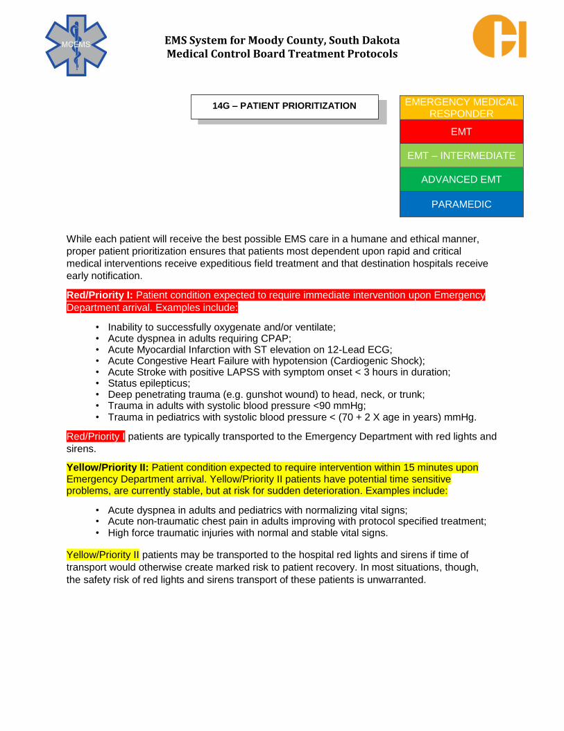
EMS System for Moody County, South Dakota Medical Control Board Treatment Protocols
While each patient will receive the best possible EMS care in a humane and ethical manner,
proper patient prioritization ensures that patients most dependent upon rapid and critical
medical interventions receive expeditious field treatment and that destination hospitals receive
early notification.
Red/Priority I: Patient condition expected to require immediate intervention upon Emergency
Department arrival. Examples include:
• Inability to successfully oxygenate and/or ventilate; • Acute dyspnea in adults requiring CPAP; • Acute Myocardial Infarction with ST elevation on 12-Lead ECG; • Acute Congestive Heart Failure with hypotension (Cardiogenic Shock); • Acute Stroke with positive LAPSS with symptom onset < 3 hours in duration; • Status epilepticus; • Deep penetrating trauma (e.g. gunshot wound) to head, neck, or trunk; • Trauma in adults with systolic blood pressure <90 mmHg; • Trauma in pediatrics with systolic blood pressure < (70 + 2 X age in years) mmHg.
Red/Priority I patients are typically transported to the Emergency Department with red lights and
sirens.
Yellow/Priority II: Patient condition expected to require intervention within 15 minutes upon Emergency Department arrival. Yellow/Priority II patients have potential time sensitive problems, are currently stable, but at risk for sudden deterioration. Examples include:
• Acute dyspnea in adults and pediatrics with normalizing vital signs; • Acute non-traumatic chest pain in adults improving with protocol specified treatment; • High force traumatic injuries with normal and stable vital signs.
Yellow/Priority II patients may be transported to the hospital red lights and sirens if time of
transport would otherwise create marked risk to patient recovery. In most situations, though,
the safety risk of red lights and sirens transport of these patients is unwarranted.
EMERGENCY MEDICAL RESPONDER
EMT
EMT – INTERMEDIATE
ADVANCED EMT
PARAMEDIC
14G – PATIENT PRIORITIZATION

EMS System for Moody County, South Dakota Medical Control Board Treatment Protocols
PROTOCOL 14G: Patient Prioritization, cont.
Green/Priority III: Patient condition expected to require routine timeliness of intervention upon Emergency Department arrival. Green/Priority III patients do not appear to require further emergent medical intervention and do not appear to have life/organ threatening conditions. Examples include:
• Asthma exacerbation dyspnea resolved with bronchodilator nebulization; • Nausea/non-bloody vomiting with normal and stable vital signs; • Isolated orthopedic injury with intact neurovascular function.
Green/Priority III patients should be transported to the hospital without red lights and without
sirens. The safety risk of red lights and sirens transport of these patients is unwarranted.
Black or Blue Obvious death or illness/injury severity incompatible with successful
resuscitation given concurrent system demands (such as in multiple casualty responses).
Adult trauma patients are determined to be Red/Priority I by either vital signs and level of
consciousness (systolic BP < 90 mmHg, sustained tachycardia, respiratory rate <10 or >29
breaths per minute, GCS ≤ 13, cool, diaphoretic skin) or any of the following anatomical injury:
• Penetrating injury of head, neck, torso, extremities proximal to elbow or knee; • Amputation proximal to the wrist or ankle; • Paralysis or suspected spinal fracture with neurological deficit; • Flail chest; • Two or more suspected proximal long - bone fractures; • Open or suspected depressed skull fracture; • Unstable pelvis or suspected pelvic fracture; • Tender and/or distended abdomen; • Burns associated with other Priority I Trauma; • Crushed, degloved, or mangled extremity, proximal to the wrist or ankle. • Pulseless extremity
Adult trauma patients are determined to be Yellow/Priority II from “high- energy” events with risk
of severe injury despite normal and stable vital signs without change in usual mentation or usual
neurologic function, or respiratory distress. Adult trauma patients may also be determined to be
Yellow/Priority II if exhibiting a single system injury as noted:
• High risk auto crash (intrusion > 12 inches in occupant site; intrusion > 18 inches any
site; partial or complete ejection from vehicle; death in same passenger compartment); • Auto v. pedestrian/bicyclist thrown, run over, or with significant ( >20 mph) impact; • Motorcycle crash > 20 mph; • Falls > 20 feet in height (one building story is 10 feet in height); • Significant force alleged assault; • Isolated closed head trauma with resolved altered mental status (Neuro System); • Isolated fractures/dislocation; lacerations/avulsions without extensive tissue damage
(Musculoskeletal System);

EMS System for Moody County, South Dakota Medical Control Board Treatment Protocols
PROTOCOL 14G: Patient Prioritization, cont.
Adult Priority II Determining Criteria, cont.
Adult trauma patients are determined to be Yellow/Priority II from “high- energy” events with
risk of severe injury despite normal and stable vital signs without change in usual mentation or
usual neurologic function, or respiratory distress. Adult trauma patients may also be
determined to be Yellow/Priority II if exhibiting a single system injury as noted:
• Facial lacerations; fractured facial bones; avulsed teeth (Maxillofacial/Dental); • Isolated abdominal pain (Transport as Level II Trauma to facility as specified by TREC
center); • Select & isolated hand injuries (“isolated” defined by the level of suspected injury
involvement being no further proximal than the carpal bones level). Only certain hand injuries require rapid treatment to avoid unfavorable outcomes. These “select” Priority II injuries include:
Vascular injuries that involve significant arterial hemorrhage; Nerve injuries that cause loss of motor function;
Amputations; High-pressure injection injuries; Flexor tendon injuries of hand.
Adult trauma patients may be determined to be Discretionary Red/Priority I or Yellow/Priority II if
clinical suspicion of significant injury and heightened by any single or particularly a combination
of the following patient attributes:
• Age > 55; • Anticoagulation and bleeding disorders; • Time sensitive extremity injury; • End – stage renal disease requiring dialysis; • Pregnancy > 20 weeks.
Discretionary Red/Priority I or Yellow/Priority II adult trauma patient radio and care reports
should clearly indicate to the receiving Trauma Center personnel the rationale for the
Discretionary Red/Priority I or Yellow/Priority II assignment.
Level III Trauma Centers are intended to receive adult patients at risk for severe injury with
normal, stable vital signs or patients with no significant anatomical injuries.
Adult trauma patients are determined to be Green/Priority III from “low energy” events with
normal and stable vital signs, without change in usual mentation or usual neurologic function,
and without new or significant organ system dysfunction. Green/Priority III adult trauma may
include:
• Single bone fractures from a same level fall; • Minor puncture wounds/lacerations/abrasions; • Isolated neck pain without new neurological deficit; • Isolated extremity pain.

EMS System for Moody County, South Dakota Medical Control Board Treatment Protocols
PROTOCOL 14G: Patient Prioritization, cont.
Level IV Trauma Centers may receive adult patients without physiologic instability, altered
mentation, neurological deficit or significant anatomical injuries and have also not been involved
in a significant mechanism of injury incident for expected care at that facility. Patients in other
categories (e.g. with physiologic instability) should be expected to be transferred to a higher
level trauma center after immediate care needs are addressed (e.g. invasive airway
management).
Pediatric (Age < 16 years of age) general medical patients are determined Red/Priority I to be
if the following organ system dysfunction is evidenced by acute symptoms or physical exam
signs:
Pulmonary System: • Respiratory arrest;
• Respiratory distress and inability to maintain O2 sat > 95% on supplemental O2; • Stridor with inability to phonate, weak cry, altered mental status, or pallor.
Cardiovascular System: • Cardiac arrest or pre-arrival CPR, bradycardia requiring chest compression;
• Multiple shock signs (pallor, cool, slow cap refill, weak pulse, altered mental status);
• Persistent tachycardia > 200/min or bradycardia< 80/min (without athletic fitness level).
Neurologic System
• Status epilepticus; • Acute sustained altered mental status without apparent etiology; • Acute focal neurological deficits.
Metabolic System/Toxicology (Overdose) • Ingestion of a tricyclic antidepressant; • Ingestion, inhalation, or contact exposure causing altered mental status, respiratory
distress, or shock.
Pediatric (Age < 16 years of age) general medical patients are determined to be Green/Priority III if
there does not appear to be an acute medical problem of life/organ threatening severity.
Pediatric trauma patients are prioritized Red/Priority I by either physiological criteria (systolic
BP < (70 + 2 x age of patient in years) mmHg, sustained tachycardia >180 bpm, respiratory rate
<10 (<20 in infant less than one year of age) or >29, pulse oximetry<95% without supplemental
oxygen, or GCS ≤ 13) or any of the adult trauma Red/Priority I anatomical injury criteria.
Fall criteria for pediatric trauma Red/Priority I is >10 feet or 2 – 3 times the height of the child.

EMS System for Moody County, South Dakota Medical Control Board Treatment Protocols
PROTOCOL 14G: Patient Prioritization, cont.
Pediatric trauma patients are prioritized Yellow/Priority II from “high-energy” events with risk of
severe injury despite normal and stable vital signs without change in usual mentation or usual
neurologic function, or respiratory distress. Pediatric trauma patients may also be determined to be
Yellow/Priority II if exhibiting any of the adult trauma priority II single system injury criteria.
Pediatric trauma patients may be determined to be Discretionary Red/Priority I or
Yellow/Priority II if clinical suspicion of significant injury warrants and is heightened by any of
the following patient attributes: • Anticoagulation and bleeding disorders; • Pregnancy > 20 weeks; • Extrication time > 20 mins, *death in same vehicle, speed >40 mph, rollover
mechanism, vehicle external intrusion >20” or compartment intrusion >12”.
Discretionary Red/Priority I or Yellow/Priority II pediatric trauma patient radio and care
reports should clearly indicate to the receiving Trauma Center personnel the rationale for the
Discretionary Red/Priority I or Yellow/Priority II assignment.
Pediatric trauma patients are determined to be Green/Priority III from “low energy” events with
normal and stable vital signs, without change in usual mentation or usual neurologic function,
and without new or significant organ system dysfunction. Green/Priority III pediatric trauma
may include any of the adult trauma Green/Priority III injury criteria as previously listed in this
protocol.
Specialized Burn Care Patients with the following burn injuries (without additional trauma center criterion injuries)
should either be transported directly to a burn care specialty center or be referred to such
after initial emergency department evaluation:
• Partial thickness(second degree) burns >10% total body surface area (TBSA); • Full thickness (third degree) burns; • Partial or full thickness burns of the face, hands, feet, genitalia/perineum, or
major joints;
• Electrical burns (includes lightning injury), inhalation burns, chemical burns; • Burn injury in patients with preexisting medical disorders compromising healing
and survival (cardiac disease, chronic respiratory illness, diabetes); • Multisystem trauma with partial or full thickness burn as the predominant injury.
If the burn patient cannot be oxygenated or ventilated, transport the patient to the
nearest appropriate emergency department for airway management.

EMS System for Moody County, South Dakota Medical Control Board Treatment Protocols
Patient Report Format:
A. Identification, Apparatus ID, & Estimated Time of Arrival B. Patient Priority – Red/Priority 1; Yellow/Priority 2; Green/Priority 3 C. Objective of communication - Notification of transport only or request for on-line medical
control. Be specific in what orders will be requested from on-line medical control so that the physician (or designate) will be oriented to the request as he/she is listening to the report
D. Patient Information – Age/Sex/Chief Complaint or Condition/Pertinent Past Medical History E. Patient Condition – LOC/Vital Signs/Exam Findings/ECG Interpretation F. Treatment and response to treatment.
Notes:
A. All communications should be brief and orderly. Patient reports should rarely take longer
than one or two minutes per patient and should be made on a recorded line. B. Describe the patient condition in enough detail to explain treatment initiated and rationale for
any request(s). Remember while the patient’s condition may be visually obvious, the nurse
or physician on the radio or phone is completely dependent on the EMS professional’s
ability to verbally “paint the picture of the patient”. C. It is critical to notify the receiving emergency department at the earliest opportunity to
describe the patient’s illness/injury and condition so that emergency department personnel can be appropriately prepared for the patient’s arrival, including preparing resources to ensure continuity of care (e.g. respiratory care, cardiac pacing, and trauma surgery notification). In many instances, the earliest opportunity for a patient report will occur even
before discovering the full extent of illness or injury in the critical patient. It is important that the “lead EMS professional” or his/her designee make the report at this time, especially to report a STEMI Alert, Stroke Alert, or Trauma Alert rather than wasting several minutes of hospital pre-arrival notification trying to make a “more complete” report.
D. Particularly critical objective findings, (e.g. cardiac arrest, critically abnormal vital signs,
gunshot wound to chest) need to take precedence in a patient report and should be
reported after the identification/ETA and patient priority statements.
EMERGENCY MEDICAL DISPATCHER
EMERGENCY MEDICAL RESPONDER
EMT
EMT – INTERMEDIATE
ADVANCED EMT
PARAMEDIC
14H – PATIENT REPORT COMMUNICATIONS

EMS System for Moody County, South Dakota Medical Control Board Treatment Protocols
A patient may require a transfer from one hospital to another hospital if:
1. Patient evaluation at the original hospital reveals care needs unavailable at that hospital
2. Another hospital is preferred by the patient, the patient’s legal representative, or the patient’s established physician(s)
A hospital must agree to facilitate a patient transfer (regardless of patient’s financial status) if the patient meets any of the above criteria.
Any interhospital transfer must be arranged as a practitioner/physician-to-physician transfer in accordance with Federal regulations.
Prior to any interhospital transfer, the EMS professional must receive appropriate transfer paperwork, including an adequate summary of the patient's condition, current treatment including nursing and practitioner/physician evaluation notes, lab results, radiology results and films, possible complications that could occur during transfer, and any further medical information deemed necessary by the EMS professional or physician(s). Any anticipated interhospital transfer treatment orders are to be written and signed by the transfer initiating practitioner/physician.
Prior to any interhospital transfer, if the EMS professional is concerned that the patient is not stabilized to the extent possible for transport, the EMS professional shall review his /her concerns with the transferring practitioner/physician with a goal to ensure appropriate clinical care is performed to further stabilize the patient. In the rare instance in which the EMS professional and transferring practitioner/physician cannot agree on the stability of the patient and/ or further care necessary prior to the interhospital transfer, the EMS professional is to consult with the accepting physician at the receiving hospital to review these concerns. If in such situation the receiving hospital has automatically accepted the patient for care to the "emergency group/doctor", the EMS professional is to discuss concerns with the on-duty emergency physician at that hospital. If the EMS professional cannot rapidly resolve the situation with the transferring practitioner/physician and receiving physician, the EMS professional is to notify the medical director for intervention, ideally via a recorded line. The overriding principle for all aspects of interhospital transfer is matching patient needs with adequate provider knowledge and skill, equipment and infrastructure that provide continued patient safety during transport.
It the professional and ethical responsibility of an EMS professional as well as of an EMS organization to not accept or perform an interfacility transfer that involves monitoring and treatment exceeding their scope of authorized practice, training, and/or ability.
EMERGENCY MEDICAL DISPATCHER
EMERGENCY MEDICAL RESPONDER
EMT
EMT – INTERMEDIATE
ADVANCED EMT
PARAMEDIC
14I – INTERFACILITY TRANSFERS

EMS System for Moody County, South Dakota Medical Control Board Treatment Protocols
PROTOCOL 14I: Interfacility Transfers, cont. An EMS professional must be licensed as an AEMT or higher to verify patency of vascular access. IV fluid type and flow rate must be specified in written practitioner/physician orders and verified prior to interhospital transfer. Any IV fluid bag supplied must contain enough solution to accommodate the expected interhospital transport time.
Any IV infusion medications must be specified in written practitioner/physician orders and verified prior to interhospital transfer. The paramedic is to verify all IV infusion medications ordered during the interhospital transfer are on the pre -approved list contained in this protocol. Medications that are not on the pre-approved list must be specifically approved by one of the receiving hospital’s on-line medical control emergency physicians or the accepting physician prior to transport. There is a limit of 4 concurrent IV infusion medications for paramedic-only accompanied interhospital transfers.
All interhospital transfer patients with IV infusion medications will be continuously cardiac monitored, including monitoring pulse oximetry during transport. Waveform capnography should be utilized as indicated by appropriate protocols. Blood pressure monitoring will be at least as frequently as every 15 minutes with a minimum of two blood pressure recordings. The interhospital transfer orders may specify more frequent measurements.
During interhospital transfer, should the patient experience signs or symptoms of intolerance (significant side effects) to the IV infusion medication(s) or the IV infusion pump indicates an error not easily addressed by the paramedic, stop the infusion and consult the transferring hospital’s on-line medical control for direction.
Mechanical ventilation settings must be confirmed with either the transferring practitioner/physician or a respiratory therapist. The paramedic must review and confirm ventilation rate, tidal volume, FiO2 (50% or 100%), and positive-end expiratory pressure (PEEP) settings. If at any time during interhospital transfer mechanical ventilation malfunctions, institute bag -valve assisted ventilations with 100% O2 while troubleshooting the mechanical ventilator and airway circuit. Ensure the patient receives appropriate oxygenation and ventilation continuously.
All indwelling devices and lines (e.g. chest tube(s), naso/orogastric tube, PEG/G/J- tube, surgical drain(s), intra-aortic balloon pump (IABP), ventricular assist device (VAD), wound vacuum) must be reviewed with either the transferring practitioner/physician or appropriate nursing personnel. Potential complications during transfer should be discussed and contingency plans reviewed. If the Paramedic feels unable to safely monitor and maintain any indwelling device, he or she is to request appropriate nursing or ancillary personnel from the transferring hospital to accompany the patient during transfer. Under these conditions, EMS personnel will not begin the transfer until such request is accommodated.
***For specialized patients not ordinarily transported by EMS professionals (e.g., high-
risk obstetrical patients, neonates) or for any patient with a condition requiring
interhospital assessment and/or treatment beyond expected EMS professional scope of
practice, an appropriately trained healthcare professional is to accompany the
transporting EMS professionals to best provide interhospital transfer care.

EMS System for Moody County, South Dakota Medical Control Board Treatment Protocols
PROTOCOL 14I: Interhospital Transfers, cont.
For acute stroke patients either having received alteplase (tPA®) just prior to interhospital
transfer or that will be continuing to receive alteplase during interhospital transfer, specific documentation and treatment should include:
1. Vital signs prior to transport and every 15 minutes enroute. Verify that systolic blood pressure
is less than 180 mmHg and diastolic blood pressure is less than 105 mmHg. If blood
pressure exceeds these limits, the transferring hospital is to lower the blood pressure via anti-
hypertensives for further vascular stabilization prior to transport.
2. Stroke neuro-exam at time of interhospital transfer, utilizing the Los Angeles
Prehospital Stroke Screen.
3. Oxygen administration via NC or NRB if dyspnea or SpO2 <94% at room air. 4. Head of cot elevated at approximately 15 degrees if tolerated and low risk of aspiration. 5. Patient NPO status, including medications, to protect against aspiration. 6. Documentation of total dose and time of IV alteplase bolus (if dose is completed prior
to transfer) and when infusion started (and completed, if applicable).
7. Infuse all alteplase from tubing by infusing saline through same tubing set following
alteplase dose.
• When bottle appears empty, there is still some alteplase left in the tubing which must be infused.
• Remove the IV tubing connector from the bottle and attach it to a newly spiked bag
of normal saline and re-start infusion, TKO. 8. Anti-hypertensive therapy adjustment enroute:
• If labetalol IV infusion started at sending facility: Increase infusion rate by 2 mg/min every 10 minutes (to maximum of 8 mg/min) until desired decrease in BP:
Systolic BP <180 mmHg and Diastolic BP <105 mmHg • Discontinue anti-hypertensive infusion for any one of the following:
Systolic BP <140 mmHg, Diastolic BP <80 mmHg, or heart rate <50 per minute
Frequently used IV Medications during Interfacility Transfer (Infusions are continuation of infusions started at the transferring facility, not initiations)
Class of Medication Significant Side Effects
Sedatives
Diazepam (Valium®) Respiratory depression, Hypotension
Diprivan (Propofol®) Lorazepam (Ativan®)
Midazolam (Versed®)

EMS System for Moody County, South Dakota Medical Control Board Treatment Protocols
PROTOCOL 14I: Interfacility Transfers, cont.
Frequently used IV Medications during Interfacility Transfer (Infusions are continuation of infusions started at the transferring facility, not initiations)
Class of Medication Significant Side Effects
Opiate Analgesics
Fentanyl (Sublimaze®) Respiratory depression, Hypotension
Hydromorphone (Dilaudid®)
Meperidine (Demerol®) Morphine
Nalbuphine (Nubain®)
Hypertension Control Agents
Labetalol (Normodyne®, Trandate®) Hypotension, Symptomatic bradycardia Nicardipine (Cardene®) Symptomatic tachycardia, Ventricular Nitroprusside(Nipride®) dysrhythmias Acute Coronary Syndrome Agents Anti-platelet (Clot Inhibitors) Abciximab (ReoPro®) Bleeding Eptifibatide (Integrilin®) Anti-coagulant (Clot Inhibitors) Heparin Bleeding Thrombolytic (“Clot Buster”) Alteplase (tPA®) Bleeding Reteplase (Retavase®) Tenectaplase (TNKase®) Anti-anginal (Coronary Vasodilator) Nitroglycerin (Tridil®) Hypotension Cardiac Anti-arrhythmics Amiodarone (Cordarone®, Pacerone®) Diltiazem (Cardizem®) Lidocaine
Procainamide
Hypotension, Symptomatic bradycardia Symptomatic tachycardia, Ventricular dysrhythmias

EMS System for Moody County, South Dakota Medical Control Board Treatment Protocols
PROTOCOL 14I: Interfacility Transfers, cont.
Frequently used IV Medications during Interfacility Transfer, cont. (Infusions are continuation of infusions started at the transferring facility, not initiations)
Class of Medication Significant Side Effects
Vasopressors (Hypotension Treatment)
Dobutamine (Dobutrex®) Hypertension, Symptomatic tachycardias Dopamine (Intropin®) Ventricular dysrhythmias Epinephrine Norepinephrine (Levophed®) Phenylephrine (Neosynephrine®) Volume Expanders (Hypovolemia Treatment)
Albumin Dextran Hetastarch (Hespan®) Plasma protein fraction (Plasmanate®) Blood Products (Anemia or Coagulopathy Treatment)
Cryoprecipitate Allergic reactions ranging from itching only to
Frozen Plasma (FFP) more serious reactions of hives, (urticaria), Packed Red Blood Cells (PRBC) respiratory distress (typically bronchospasm), Platelets tachycardia, and hypotension (evidence of Whole Blood anaphylaxis).
Gastrointestinal Bleeding Control Agents
Esomeprazole (Nexium® – acid reducer) None
Octreotide (Sandostatin®–varices constrictor)
Pantoprazole (Protonix® – acid reducer)
Acid-Base Metabolism Agents
Sodium Bicarbonate None
Hyperglycemia Control Agents
Insulin Hypoglycemia-related complications
Electrolyte Replacement
Potassium chloride (KCL) Ventricular dysrhythmias
Allergic reactions ranging from itching only to
more serious reactions of hives, (urticaria),
respiratory distress (typically bronchospasm),
tachycardia, and hypotension (evidence of
anaphylaxis).

EMS System for Moody County, South Dakota Medical Control Board Treatment Protocols
PROTOCOL 14I: Interfacility Transfers, cont.
Frequently used IV Medications during Interfacility Transfer, cont. (Infusions are continuation of infusions started at the transferring facility, not initiations)
Class of Medication Significant Side Effects
Seizure Control Agent
Fosphenytoin (Cerebyx®) Magnesium (for eclampsia)
Phenytoin (Dilantin®) Phenobarbital
Bronchospasm Control Agents
Aminophylline (Theophylline®)
Pregnancy - Related Agents
Oxytocin (Pitocin®–stimulates uterine contraction
Inducing labor and controls uterine bleeding)
Antimicrobials/Antibiotics
Aminoglycosides (e.g. gentamicin) Antifungals (e.g. fluconazole) Anti-TB (e.g. isoniazid - INH) Anti-viral (e.g. acyclovir) Carbapenams (e.g. imipenem) Cephalosporins (e.g. cetriaxone) Macrolides (e.g. azithromycin) Penicillins (e.g. ampicillin; piperacillin) Quinolones (e.g. levofloxacin)
Sulfonamides (e.g. TMP-SMX, Bactrim®) Other categories (e.g. clindamycin, vancomycin
Respiratory depression, Hypotension, Symptomatic bradycardia Symptomatic tachycardias, Hypertension Hypotension (if rapid infusion), Symptomatic tachycardias, Hypertension Allergic reactions ranging from itching only to more serious reactions of hives (urticaria), respiratory distress (typically bronchospasm), tachycardia, and hypotension (evidence of anaphylaxis). In some cases, a localized phlebitis (pain at infusion site with redness of vein) may occur due to irritation cause by the infusion itself. While the infusion is to be stopped, this usually is not a true allergy.

EMS System for Moody County, South Dakota Medical Control Board Treatment Protocols
PROTOCOL 14I: Interfacility Transfers, cont.
Priority and Timing for Interfacility Transfer Requests:
Priority 1 Clinical Condition – Immediate Life Critical Intervention at Receiving Hospital Facility Time Standard is 911 Call to Arrival = Response 10:59 or less Once ambulance is assigned cannot be re-assigned
Acute ST Elevation Myocardial Infarct Transferring Facility – No Interventional Cardiology Receiving Facility – Interventional Cardiology/Cardiac Cath Lab
Acute Thromboembolic Stroke
Transferring Facility – No Interventional Neurology
Receiving Facility – Interventional Neurology & Procedure Imminent
Acute Aortic Dissection
Transferring Facility – No Vascular Surgery Receiving Facility – Vascular Surgery & Surgery Imminent
Acute GI Bleeding with Hemodynamic Instability
Transferring Facility – No Gastrointestinal/Colorectal Capability
Receiving Facility – Gastrointestinal/Colorectal Capability and Endoscopy Imminent
Acute Amputation with Limb Salvage Attempt/Limb Ischemia/Arterial Occlusions Transferring Facility – No Vascular Surgery/Interventional Radiology
Receiving Facility – Vascular Surgery/Interventional Radiology and Intervention Imminent
Suspected or Confirmed Ectopic Pregnancy with Hemodynamic Instability
Transferring Facility – No Obstetric Surgery Receiving Facility – Obstetric Surgery & Surgery Imminent
Active Labor with Evidence for Complicated Delivery – Breech/Limb Position by Ultrasound Transferring Facility – No Obstetric Surgery
Receiving Facility – Obstetric Surgery & Surgery Imminent
Acute Angle Closure Glaucoma/Acute Retinal Artery Occlusion/Acute Vision Loss Imminent Transferring Facility – No Ophthalmology
Receiving Facility – Ophthalmology & Intervention Imminent
Level I/II Trauma with Hemodynamic Instability
Transferring Facility – No Trauma Surgery/Capability Receiving Facility – Trauma Surgery/Capability

EMS System for Moody County, South Dakota Medical Control Board Treatment Protocols
PROTOCOL 14I: Interfacility Transfers, cont.: Priority 2 Clinical Condition – No Immediate Time Critical Intervention at Receiving Hospital Facility Time Standard is 911 Call to Arrival = Response 59:59 or less Once ambulance is assigned cannot be re-assigned unless Priority 1 Call occurs
Post Cardiac Arrest with Hemodynamic Stability
Transferring Facility – No ICU and/or Cardiology Services Receiving Facility – ICU and Cardiology Services
Active Labor with Routine Delivery Planned
Transferring Facility – No Obstetric Surgery Receiving Facility – Obstetric Surgery
Dialysis Required with Hemodynamic Stability
Transferring Facility – No Dialysis Capability
Receiving Facility – Dialysis Capability with Dialysis Imminent
Ground Component of Air Ambulance Transport Assist Estimated Landing Time 25 Mins or Less
Priority 3 Clinical Condition or Priority 4 Clinical Condition – No Immediate Time Critical Intervention at Receiving Hospital/Facility
Time Standard is 911/non-911 Call to Arrival = Response 59:59 Once ambulance is assigned cannot be re-assigned unless Priority 1 Call occurs
Hospital ED or Inpatient Transfer Transferring Facility – Limit of Care Capabilities/Course of Treatment Reached
Receiving Facility – Higher Care Capabilities/Ongoing Course of Treatment
Possible
Hospital ED to Residence/Nursing Facility Post ED Evaluation and Treatment
Scheduled Outpatient Dialysis Care
Hospital Inpatient to Residence/Nursing Facility Post ED Evaluation and Treatment
Inpatient Bed Shortages and Hospital to Hospital or Facility to Facility Patient Movement
Due to Bed Shortage

EMS System for Moody County, South Dakota Medical Control Board Treatment Protocols
A Multi-Patient Scene (MPS) occurs when an incident involves more than one patient, but less
than 5 critical patients and less than 10 total patients.
A Mass Casualty Incident (MCI) occurs when an incident involves several patients, specifically
including five or greater critical patients or ten or more total patients, regardless of patient
priority composition.
Incident command at multiple patient scenes (MPS) or mass casualty incidents (MCI) will be
assigned according to the National Incident Management System (NIMS) guidelines and a
unified command team consisting of representatives of police, fire, and EMS should be rapidly
assigned and coordinated to ensure safe, efficient, and effective operations.
Multi-Patient Scene Tasks:
1. Initial Size-up Actions: (these are the same for Mass Casualty Incidents)
a. Park initial arriving apparatus in safe location at scene perimeter to avoid loss of its availability/use.
b. Advise dispatch: i. Incident location (if different from initial dispatch) ii. Incident type (transportation accident, fire, etc. if different from initial dispatch) iii. Estimated number of patients. iv. Numbers & types of additional resources needed. v. Any hazardous conditions (weather, electrical, structural, toxic chemicals, etc.)
Identify a “HOT ZONE”/”Immediate Danger Zone” if applicable. vi. Best route & access to scene (if appropriate). vii. Staging area location (if staging indicated).
c. Check in with Incident Command to determine ICS role. The medical treatment and
transport of injured patients will normally be done through the position of Medical
Branch Director and subordinate positions headed by Group Supervisors. However,
the Incident Commander will determine which positions, if any, he or she feels
needs to be filled.
EMERGENCY MEDICAL DISPATCHER
EMERGENCY MEDICAL RESPONDER
EMT
EMT – INTERMEDIATE
ADVANCED EMT
PARAMEDIC
15A – MULTIPLE PATIENT SCENES/
MASS CASUALTY CONCEPTS

EMS System for Moody County, South Dakota Medical Control Board Treatment Protocols
PROTOCOL 15A: Multi-Patient Scenes/Mass Casualty Incident Concepts, cont. Multi-Patient Scene Tasks, cont.:
2. Medical Branch Director:
a. Reports to the Operations Section Chief. b. Don identification vest for position c. Establishes Triage, Treatment, and Transport Areas (if indicated) and
assigns individuals to the role of Group Supervisors (or Officers) for each area.
d. Maintains adequate span of control within Medical Branch e. Establish appropriate EMS communications with appropriate response elements
(i.e. Annex H, MERC, staging, logistics,) f. Establish and maintains communications with assigned Group Supervisors (Officers) g. Oversees the triage, treatment, transportation and accountability of patients created
by incident. h. Monitors the potential or actual effect of the incident on the existing medical
infrastructure and communicates such with the Operations Section Chief, MERC, and/or Annex H
i. Determines resource requirements to meet the medical needs of the incident and communicates needs to Operations Section Chief or designated response element.
j. Determines the need for specialized medical resources and processes requests for such elements through appropriate channels.
k. Provides situation updates and reports to the Operations Section Chief, MERC,
and/or Annex H
3. Group Supervisor (Officer):
a. Establishes Area to perform assigned tasks b. Determine resource and staffing needs for Area of responsibility and
communicates needs to Medical Branch Director c. Follows assigned duties as outline in Agency Plan, Task Cards, or Job Action
Sheets. d. Provides situational updates and reports to the Medical Branch Director e. Establish communications with the Medical Branch Directors and other
needed response elements. f. Monitors safety and welfare of patients and assigned personnel g. Ensures patient tracking and accountability of injured patients in assigned Area.

EMS System for Moody County, South Dakota Medical Control Board Treatment Protocols
PROTOCOL 15A: Multi-Patient Scenes/Mass Casualty Incident Concepts, cont.
Mass Casualty Incident Tasks:
Triage Area Tasks:
1. Initial triage is at casualty locations unless hazards indicate that rapid extrication or casualty self-extrication should occur to a designated area safe for triage operations. In some instances, the use of priority-specific colored tape (red, yellow, green, black/blue) may be utilized to mark patients in the absence of readily usable triage tags.
2. Perform first pass (initial) triage. Do not perform any treatment in first pass triage other than very quick, simple and extremely urgent measures (i.e., open the airway by positioning). Move quickly to ensure all casualties are identified and triaged to minimize loss of life and limb.
3. Attach tag to patient using the string loop directly on their body. Over the head or on the upper arm works well. The left extremities should be utilized unless extremely injured. This will make it easier to utilize the triage tag during transport.
4. Use a reliable method to count the number of patients in each category. This information will need to be relayed to the Triage Group Supervisor (Officer) officer, and in turn, the Medical Branch Director.
5. Direct ambulatory patients to the GREEN Treatment Area when it is established. Use discretion in allowing GREEN patients to assist in caring for the YELLOW and RED patients while those more serious casualties are awaiting extrication to the treatment areas. ALL persons involved in the incident are to be triaged and tagged - those without apparent injuries should be tagged GREEN.
6. Report number of casualties in each category and in total to the Triage Group Supervisor (officer).
7. Repeat triage sequence when possible and note changes in any casualty’s condition.
Perform a more detailed assessment, provide treatment, and write-in information on the
tag while casualties are being extricated to the Treatment Area.
Treatment Area Tasks:
1. Establish treatment area in consultation with the Medical Branch Director regarding location. Think BIG to allow adequate space to treat casualties. Ensure the location promotes relative ease of ambulance loading and egress.
2. Request, assign, utilize, and oversee appropriate clinical personnel caring for patients. 3. Assemble into crews of at least 2 personnel equipped with a backboard and straps for
assignment by the treatment sector officer to perform BASIC packaging and extrication of triaged casualties into the RED, YELLOW, and GREEN treatment areas. The treatment sector officer may choose to have separate personnel perform treatment once inside the Treatment Area, depending on the logistics of the particular call. All persons involved in the incident triaged GREEN due to very minor or no apparent injury are to be kept in the GREEN Treatment Area until more fully evaluated. These individuals will be released at an appropriate time by the Transport Group Supervisor (Officer). Depending on the circumstances, the GREEN casualties may be transported early or in large groups using alternative transport means.

EMS System for Moody County, South Dakota Medical Control Board Treatment Protocols
PROTOCOL 15A: Multi-Patient Scenes/Mass Casualty Incident Concepts, cont.
Treatment Area Tasks:
4. On the clinical side of the triage tag, circle injuries on the body diagram (if present), note the BP, pulse, and respirations. Note any IM or IV medication given and the time it was given. On the administrative side of the tag, note the time, date, patient name, address, city, state, and past medical history and prescriptions. Record the primary EMS caregiver.
5. If a casualty’s condition worsens (e.g. YELLOW to RED; GREEN to YELLOW) inside the Treatment Area, apply a new triage tag indicating the more serious condition (leaving the original tag in place to indicate a change in condition occurred) and move the patient to the appropriate location in the Treatment Area. Notify the Treatment Group Supervisor (Officer) of any change in casualty condition so that this may be recorded for overall patient accountability and reported to the Medical Branch Director.
Transportation Area Tasks:
1. Establish patient loading zone. Consider proximity to treatment area and ambulance approach AND exit routes. Establish ambulance traffic routes that prevent ambulances from having to back-up to load patients. This makes for safer and more efficient transport operations. While multiple ambulances may be in staging, minimize the number of ambulances in the immediate load zone. This makes for more accurate and efficient transport operations. Work with staging to ensure at least 1 ambulance is always in the loading zone. Ensure vehicle operators stay with their ambulances to ensure as soon as patients are loaded, the ambulance leaves.
2. Assign Unit Leaders to appropriate needed subordinate roles such as tracking and communications. The Transportation Area will likely require the coordinated effort of several people and can quickly overwhelm one individual. Loss of patient accountability can be the result of an inadequately staffed Transportation Area.
3. Communicate with Treatment Group Supervisor (Officer) when ambulances are available for transport. NO MORE THAN ONE CATEGORY RED PATIENT PER AMBULANCE. May take another patient if yellow/green in category.
4. Supervise the assignment and loading of patients into available transport. 5. Communicate with response elements (Annex H, MERC, Communications Center) to
determine hospital capacity and appropriate destination of patients based upon clinical condition(s) and vehicle operator familiarity with destination.
6. Consider the use of alternate means of transportation if indicated (bus or specialty vans) 7. Before patient leaves the scene to destination, the accountability process should be
completed by whatever means being used (triage tag identifier slip, patient log). 8. Notify the Medical Branch Director when all patients have been cleared from the
scene and transported. Maintain and secure records for the Medical Branch
Director and secure the patient loading area.

EMS System for Moody County, South Dakota Medical Control Board Treatment Protocols
PROTOCOL 15A: Multi-Patient Scenes/Mass Casualty Incident Concepts, cont.
Mass Casualty Incident Tasks:
Staging Group Supervisor Tasks:
1. If not already established and/or determined by the Incident Commander (IC), establish staging area for medical transportation resources. Staging area should be an area large enough to contain numerous transportation assets, be far enough away so units don’t get caught in incident, but close enough to loading zone to allow for short drive times.
2. Select and communicate a travel route for resources assigned to the Staging Area. This route should allow for easy access, but should take units away from impacted area(s).
3. Staging Area for medical assets may be co-located with staging for other response assets, or may be a stand-alone area, depending on the desire of the IC. If co-located with other response assets, ensure medical assets are grouped together for accountability and an accurate assessment of available resources.
4. The Staging Group Supervisor (Officer) should coordinate an orderly arrangement of arriving apparatus to allow for ease of ambulance ingress to the transport loading zone. Medical equipment assets (cache, trailers) should also be organized to allow for rapid deployment upon request.
5. The Staging Group Supervisor (Officer) or the officer’s designee should maintain a log of available resources in staging and communicate with the Medical Branch Director resource levels as appropriate and as requested by the Medical Branch Director.
6. The Staging Sector Officer or the officer’s designee should assure ambulance or specialty transport crews stay with their assigned vehicles to assure rapid availability of the asset when requested at the transport loading zone.
7. Deliver equipment needed in the treatment sector that is requested from staging in an organized cache with a minimum of personnel leaving the staging area to deliver this equipment. Alternatively, all the requested equipment may be sent to the treatment area on one designated vehicle.
8. Assign and deploy transportation assets to the loading zone(s) per the request of the
Medical Branch Director or the Transportation Group Supervisor depending on the
established communication pathways.
Mass Casualty Medical Communications:
1. Medical communications during a mass casualty incident, like all other incident communications, are of critical importance and often will determine the level of effectiveness and success of the operation. Basic communication principals should be used during an incident.
a. Use of interoperable radio channels so all agencies are able to communicate. b. Following the overall incident communications plan established by the IC. c. Establishing assigned, clear, and understood lines of communications; who will
communicate with whom, for what reason, and by what method. d. Use of multiple and redundant means of communications including, but limited
to, radio, data, phone, runners, face-to-face, and even hand signals. e. Preparation for communications failure and immediately switching over to one
of the established redundant communication means. f. Ensuring communications sent receive a response of some manner to ensure
the loop has been closed.

EMS System for Moody County, South Dakota Medical Control Board Treatment Protocols
PROTOCOL 15A: Multi-Patient Scenes/Mass Casualty Incident Concepts, cont.
Mass Casualty Medical Communications:
2. Medical communications from the scene of an MCI, depending on complexity and
command structure, often involves up to three different levels of communications:
a. Communications (internal) with other Incident Command System elements i. Operations Section Chief or designee(s) ii. Unified Command medical representatives iii. Logistics, Planning, Admin/Finance if appropriate
b. Communications (internal) within the scene medical response infrastructure i. Triage, Treatment, Transportation, Staging Group Supervisors
c. Communications (external) with local, county, or regional medical coordination entities i. Local Emergency Response Coordinator (LERC) ii. County Public Health Annex H Representative iii. Medical Emergency Response center (MERC)
3. After assigning support positions, one of the first activities of the Medical Branch Director should to establish redundant communications pathways with the ICS structure, subordinate Group Supervisors (Officers), and other medical response elements by: a. Obtaining the ICS Communication Plan (incident channels etc.) b. Determine reporting lines and redundant means of communication with
Groups Supervisors (Officers). Example: Transport requesting assets through Medical Branch or directly to Staging.
c. Establishing and communicating manner for all communications acknowledgments.
d. Establish communications via radio, e-mail, or phone with outside medical coordination entities
e. Advise the ICS infrastructure of communication pathways for incorporation into updated ICS communication plan

EMS System for Moody County, South Dakota Medical Control Board Treatment Protocols
Communication Center Principles: 911 calls present the first opportunity to identify that a potential weapon of mass destruction (WMD) - chemical incident exists. Identifying the incident, relaying potential threat information, and advising precautionary measures to all of the responding public safety professionals may be a key to saving lives of responding public safety professionals. Indicators of a Possible Chemical Weapons Incident:
1. Explosion with little or no structural damage; 2. Reports of a device that dispersed a mist or vapor; 3. Multiple casualties exhibiting similar symptoms (may be without apparent reason); 4. Reports of unusual odors, liquids, spray devices, or cylinders; 5. Dead animals; 6. Discarded personal protective equipment (PPE).
Potential Notifications (actual notification needed if chemical weapon event confirmed):
1. Local Law Enforcement 2. Local Federal Bureau of Investigation (FBI) office – WMD Coordinator; 3. Local/State Office of Emergency Management (OEM); 4. Local Health Department
Initial Actions/On – Scene Arrival:
1. Approach upwind and uphill of the incident; 2. Stop at an apparent safe distance away from incident location; 3. Alert subsequent arriving responders; 4. Direct all personnel to use full PPE, including self-contained breathing apparatus (SCBA) a. At a minimum, respiratory protection; 5. Be aware of possible secondary devices; 6. Treat as a crime scene/Consider that alleged perpetrator may still be on the scene; 7. Avoid contact with liquids; 8. Request appropriate resources (HazMat specialists, law enforcement officers, etc.)
Establishing Incident Command: (Follow Specific Directives of Incident Commander)
Follow National Incident Management System (NIMS) practices as reflected in local policies. Utilize a Unified Command structure, promoting effective and efficient multi-agency
communications and operations. Further information through NIMS courses can be accessed at this website: https://www.fema.gov/national-incident-management-system
EMERGENCY MEDICAL DISPATCHER
EMERGENCY MEDICAL RESPONDER
EMT
EMT – INTERMEDIATE
ADVANCED EMT
PARAMEDIC
15B – CHEMICAL WEAPONS

EMS System for Moody County, South Dakota Medical Control Board Treatment Protocols
PROTOCOL 15B: Chemical Weapons, cont.
Casualty Rescue: (Follow Specific Directives of Incident Commander)
As many ambulatory casualties as possible should be removed from the area without rescuers
entering the incident site. It should be expected, though, that live, non-ambulatory casualties will
be present at any chemical incident.
1. Use bull horns and vehicle public address (PA) system to give directions; 2. Be alert for secondary devices; 3. Determine if there are live victims in the contaminated area; 4. Use PPE appropriate for safe rescue – PPE level most likely determined by HazMat
specialists advising the Incident Commander (IC). The IC evaluates the chemical threat, potential to save lives, risk to responders, and time constraints to achieve each level of responder protection before determining what level of PPE to use to perform rescue operations;
5. When safe and appropriate, assist/direct all victims to decontamination and triage area.
Decontamination: (Follow Specific Directives of Incident Commander)
The theories and procedures referred to by the Chemical Weapons Improved Response
Program (CWIRP) are based on decontaminating victims using large volumes of water.
Establish decontamination location(s) upwind and uphill of the incident:
1. Decontamination personnel must wear appropriate PPE, likely to include SCBA. 2. Be alert for secondary devices, weapons, and perpetrators: 3. Avoid contact with unknown liquids. 4. Decontaminate (immediately) casualties with liquid contamination on their skin or
clothing. For dry contamination, substance should be brushed off casualty immediately. 5. Clothing removal is often the most effective decontamination. Encourage victims to
remove clothing at least down to their undergarments; 6. Prioritize asymptomatic, symptomatic, and non-ambulatory casualties:
a. Coordinate decontamination with EMS triage activities. b. Establish separate technical decontamination for responders away from mass-
casualty decontamination.
The diagrams on the following page are provided to illustrate commonly recognized methods of
mass “wet” decontamination. Follow the directives of the Incident Commander and HazMat
specialists in charge of decontamination.

EMS System for Moody County, South Dakota Medical Control Board Treatment Protocols
PROTOCOL 15B: Chemical Weapons, cont. LADDER PIPE DECONTAMINATION SYSTEM (LDS)
EMERGENCY DECONTAMINATION CORRIDOR SYSTEM (EDCS)

EMS System for Moody County, South Dakota Medical Control Board Treatment Protocols
Protocol 15B: Chemical Weapons, cont.
Types of Decontamination: a. Passive (clothing removal) – often the simplest and effective method b. Dry agents
i. Dirt - Flour ii. Baking powder - Sawdust iii. Charcoal - Silica gel
c. Wet agents i. Soap and water ii. Water (only) iii. Bleach (for equipment decontamination)
d. Air decontamination (positive pressure ventilation [PPV]/portable fans)
EMS Principles: (Follow Specific Directives of Incident Commander)
One of the primary challenges facing EMS on a chemical weapons incident will be the number
of casualties (e.g. trauma and agent exposure) and segregating these casualties by severity of
illness/injury as well as attempting to segregate the “worried well” from actual exposed victims.
Work with appropriate HazMat specialists to determine proper level of PPE and respiratory
protection needed for EMS personnel and what areas are appropriate for EMS care activities.
a. Be alert for secondary devices and perpetrators; b. Avoid contact with liquids other than non-contaminated water; c. Rapid prioritization of number of patients; d. Triage victims based on medical necessity, using MCI protocols; e. Establish patient identification and tracking. f. Establish:
i. Communications with command post and hospitals; ii. Staging for EMS personnel, ambulances, supplies, and resources; iii. Transportation area – avoid transporting any contaminated patient(s).

EMS System for Moody County, South Dakota Medical Control Board Treatment Protocols
NERVE AGENT EXPOSURES
Comments:
1. Nerve agent exposure should be considered at multiple causality incidents in which patients are exhibiting the DUMBELS constellation of symptoms and signs. In particular, nerve agent exposure should be considered while responding to any reports of multiple casualties at a location of high occupancy (should malls, stadiums, etc.), high visibility (crowds gathered for public speeches, protests, etc.), or high political symbolism (places of worship, governmental offices, etc.).
2. Immediate countermeasures to nerve agent exposure with developing DUMBELS
symptoms and signs are administration of the DuoDote® auto-injectors, auto-injector-as indicated and evacuation from the exposure are for decontamination.
3. Any personnel exposed to a nerve agent and requiring treatment with the DuoDote® auto-injectors is restricted from providing patient care and should be promptly transported for emergency physician evaluation.
4. Atropine is utilized in nerve agent exposure treatment to dry secretions, reduce bronchospasm, and decrease gastrointestinal motility. If significant bronchorrhea continues
after three DuoDote® auto-injector have been administered in the adult patient, further
atropine may be given by paramedic as follows until the bronchorrhea subsides:
Adult – 1 mg atropine IVP every 3-5 minutes
Adult – 2 mg atropine IM every 5 minutes
5. In the case of nerve agent exposure with bronchorhea, there is no maximum atropine dosing
in the adult patient, though atropine should be withheld in the case of developing ventricular
tachydysrhythmias. In this case, treat the ventricular tachydysrhythmia according to 5G
Tachycardia – Unstable – Adult & Pediatric or 4G Ventricular Fibrillation/Pulseless
Ventricular Tachycardia – Adult & Pediatric as applicable.
6. DuoDote® is utilized in nerve agent exposure to reverse the nerve agent effect on
acetylcholinesterase, the enzyme responsible for neurotransmitter regulation. Refer also to
Protocol 16M for self/buddy care using DuoDote®.
7. Patients contaminated by vapor-only nerve agent exposures should be decontaminated by
clothing removal (dry decon). Patients contaminated by liquid nerve agent exposures should
be decontaminated by clothing removal and thoroughly washed with soap and water (wet
decon).
8. In the absence of DUMBELS symptoms and signs, nerve agent exposure has not occurred.
The DuoDote® auto-injectors are not authorized for patients not exhibiting DUMBELS symptoms and signs.
EMERGENCY MEDICAL DISPATCHER
EMERGENCY MEDICAL RESPONDER
EMT
EMT – INTERMEDIATE
ADVANCED EMT
PARAMEDIC
15C – NERVE AGENTS

EMS System for Moody County, South Dakota Medical Control Board Treatment Protocols
PROTOCOL 15C: Nerve Agents, cont. 9. Pediatric patients (<25 kg) with DUMBELS symptoms and signs in the setting of suspected
nerve agent exposures should be treated with one DuoDote® auto-injector kit and MC
should be contacted for further direction in relation to any further atropine and/or 2-PAM usage.
10. Patients treated with DuoDote® auto-injector kits should either have the auto-injector hooked to their clothing or a prominent vertical mark on their forehead for each kit
administered to indicate to further healthcare providers the number of DuoDote® auto-injector kits the patient has received.
LOOK FOR “DUMBELS” SIGNS AND SYMPTOMS D: DIARRHEA U: URINATION M: MIOSIS (PINPOINT PUPILS) B: BRONCHOSPASM, BRONCHORRHEA (COPIOUS RESPIRATORY SECRETIONS) E: EMESIS (NAUSEA/VOMITING) L: LACRIMATION (TEARING) S: SALIVATION
Additional resources regarding nerve agents can primarily be accessed through the Centers for Disease Control at www.bt.cdc.gov/agent/nerve National Disaster Life Support training also includes nerve agent education in: Basic Disaster Life Support (one day classroom course)
Advanced Disaster Life Support (two day classroom/practical exercise course)

EMS System for Moody County, South Dakota Medical Control Board Treatment Protocols
Currently, a multitude of biological agents exist that have the possibility in weaponized use, the Centers for Disease Control categorizes these agents into priority of concern and of probable use. Category A agents include organisms that pose a risk to national security due to easy dissemination or person to person transmission, high mortality rates, high impact upon public health, ability to cause public panic and social disruption, and require special action for public health preparedness. Category A agents and their diseases include:
Bacillus anthracis – Anthrax Clostridium botulinum – Toxin Botulism Yersinia pestis – Plague
Variola major – Smallpox Francisella tularensis – Tularemia
Filoviruses (e.g. Ebola, Marburg) & arenaviruses (e.g. Lassa, Machupo) – Viral hemorrhagic fevers
Of particular concern among these agents is anthrax. While anthrax is a naturally occurring disease among animal skin handlers, the bacteria has already been successfully used in domestic terrorism in the United States. Concern about anthrax prompts responses by public safety agencies, including EMS in some locales, to investigate these concerns and in some cases, acute onset of symptoms, in response to exposure to “suspicious white powder”. The following information serves as one resource in preplanning responses to such substances in efforts to protect EMS and other safety professionals.
Additional resources regarding biologic weapons can primarily be accessed through the Centers for Disease Control at www.bt.cdc.gov/bioterrorism. Suspicious Powder Response Model Procedure – Follow Local HazMat Directives
Definitions:
Isolation Perimeter – The designated crowd control line surrounding the Hazard Control Zones.
The isolation perimeter is always the line between the general public and the Cold Zone. Field Test – A procedure that will be determined by on-scene FD Hazmat and PD personnel to
check the presence of radiological, biological, chemical, and volatility (flammability) in or around
a package. ID Test – FD procedure to obtain information to identify a specific substance (i.e., salt, sugar,
flour, etc.) or chemical compound. Testing to identify a substance is more geared toward dry
product or liquids without water content. The test signature of water overrides/masks the graph
spikes preventing identification.
EMERGENCY MEDICAL DISPATCHER
EMERGENCY MEDICAL RESPONDER
EMT
EMT – INTERMEDIATE
ADVANCED EMT
PARAMEDIC
15D – BIOLOGICAL WEAPONS

EMS System for Moody County, South Dakota Medical Control Board Treatment Protocols
PROTOCOL 15D: Biological Weapons, cont. Procedures:
The following actions should be taken at incidents involving a package suspicious for anthrax:
1. Once law enforcement (LE) arrives on scene and decides that FD is needed, HazMat
resource mobilization should be considered.
2. If the first arriving FD company is not a Hazmat Unit and receives information that the
incident may be a potential chemical or biological threat, the OIC should:
Not make entry or attempt to mitigate the incident
Establish an isolation perimeter of at least 100 feet
Should keep all citizens on the outside of the isolation perimeter
Call for the Hazmat Team
Wait upwind until arrival of Hazmat Team
Operate in a support role for the Hazmat Team upon their arrival
3. The Hazardous Materials Team OIC will be either Command or Hazmat Branch as
appropriate.

EMS System for Moody County, South Dakota Medical Control Board Treatment Protocols
1. Potential radiologic weapon devices in the United States include:
a. Simple radiation emitting devices (example would be dumping radioactive waste in a water supply – typically NOT a significant threat due to dilution effect of large amount of water)
b. Conventional explosive device containing radiation (“dirty bomb”)
– typically NOT a significant threat due to the fact that
explosions are very inefficient in producing radioactive particles
of a size that are easily inhaled
2. Either of the above devices may be utilizing radioactive isotopes initially manufactured for
medical use (e.g. nuclear imaging).
3. Radiation types include the following: a. Irradiation = gamma radiation passing through a body b. External contamination = radioactive “dust” particles falling on a body c. Internal contamination = radioactive “dust” particles being ingested or inhaled
4. Protection takes the simple format of:
a. Reducing time of exposure b. Increasing distance from exposure source – biggest factor in protection. Radiation does
not travel far, but contamination can. c. Shielding device use to minimize exposure uptake. Airborne illness PPE protection is
excellent for radiation protection as well). Think of radioactive particles as “dirt” that
shouldn’t be inhaled (wear N95 masks) and shouldn’t be in contact with skin.
5. Three myths that can paralyze medical response: a. “Radioactive contamination is highly dangerous & requires extraordinary protective
measures.” (see above) b. “Decon is highest medical priority.” Decon is actual very simple = remove clothing and
shower. Most of radiation goes away with removal of clothing. “Special skills are needed to handle radioactive patients” (see above.)
6. Multiple resources exist to aid in the understanding and response planning for radiological
weapons. The following are suggested resources: Radiation Emergency Medical Management: www.remm.nlm.gov.
National Alliance for Radiation Readiness (NARR): www.radiationready.org.
Society of Nuclear Medicine and Molecular Imaging: www.snmmi.org.
Health Physics Society: www.hps.org.
National Disaster Life Support training:
Basic Disaster Life Support (one day classroom course) Advanced Disaster Life Support (two day classroom/practical exercise course)
EMERGENCY MEDICAL DISPATCHER
EMERGENCY MEDICAL RESPONDER
EMT
EMT – INTERMEDIATE
ADVANCED EMT
PARAMEDIC
15E – RADIOLOGICAL WEAPONS

EMS System for Moody County, South Dakota Medical Control Board Treatment Protocols
1. Potential nuclear weapon devices impacting the United States
include: a. Improvised nuclear device. b. 1 kiloTon “suitcase nuke.” c. Tactical weapons of 5-50 kiloTons. d. Electromagnetic pulse detonation = nuclear weapon
detonation in atmosphere wherein gamma waves hit radio waves, causing phones, pagers, radios, etc go down.
e. Ballistic missile attack. f. 250 kiloTon nuclear bomb = “city killer.”
2. Nuclear detonation = 50% blast effect; 35% thermal effect; 10% fallout; 5% ionizing radiation
effect. 3. Mass blindness is a concern due to retinal burns (non-thermal) from viewing detonation. 4. Radiation types include the following:
a. Irradiation = gamma radiation passing through a body. b. External contamination = radioactive “dust” particles falling on a body. c. Internal contamination = radioactive “dust” particles being ingested or inhaled.
5. Protection takes the simple format of:
a. Reducing time of exposure. b. Increasing distance from exposure source – biggest factor in protection. Radiation
does not travel far, but contamination can. c. Shielding device use to minimize exposure uptake. Airborne illness PPE protection is
excellent for radiation protection as well). Think of radioactive particles as “dirt” that shouldn’t be inhaled (wear N95 masks) and shouldn’t be in contact with skin.
6. Three myths that can paralyze medical response: a. “Radioactive contamination is highly dangerous & requires extraordinary protective
measures.” (see above) b. “Decon is highest medical priority.” Decon is actual very simple = remove clothing
and shower. Most of radiation goes away with removal of clothing. c. “Special skills are needed to handle radioactive patients.” (see above)
7. Multiple resources exist to aid in the understanding and response planning for nuclear
weapons. The following are suggested resources:
Radiation Emergency Medical Management: www.remm.nlm.gov.
National Alliance for Radiation Readiness (NARR): www.radiationready.org.
Nuclear Regulatory Commission: www.nrc.gov.
Society of Nuclear Medicine and Molecular Imaging: www.snmmi.org.
Health Physics Society: www.hps.org.
EMERGENCY MEDICAL DISPATCHER
EMERGENCY MEDICAL RESPONDER
EMT
EMT – INTERMEDIATE
ADVANCED EMT
PARAMEDIC
15F – NUCLEAR WEAPONS

EMS System for Moody County, South Dakota Medical Control Board Treatment Protocols
PROTOCOL 15F: Nuclear Weapons, cont.
Planning Guidance for Response to a Nuclear Detonation June 2010 – Second Edition www.remm.nlm.gov/PlanningGuidanceNuclearDetonation.pdf. National Disaster Life Support training:
Basic Disaster Life Support (one day classroom course) Advanced Disaster Life Support (two day classroom/practical exercise course)

EMS System for Moody County, South Dakota Medical Control Board Treatment Protocols
Motorcycle-style helmet comments:
While helmets offer protection for the head, they have not proven to reduce spine injuries. While their use is encouraged, helmets prevent airway access and complicate spinal immobilization.
Because most full helmets do not hold the head firmly and also prevent cervical collar
application, spinally immobilizing a helmeted patient does not result in effective cervical immobilization. Helmets must be removed prior to spinal immobilization on a long board to
assure airway access and adequate spinal immobilization. In motocross events, look for neck braces and chest protectors that are attached by clasps and hinges that will also need to be
removed.
EMT
EMT – INTERMEDIATE
ADVANCED EMT
PARAMEDIC
16A – HELMET REMOVAL

EMS System for Moody County, South Dakota Medical Control Board Treatment Protocols
PROTOCOL 16A: Helmet Removal, cont.

EMS System for Moody County, South Dakota Medical Control Board Treatment Protocols
PROTOCOL 16A: Helmet Removal, cont.

EMS System for Moody County, South Dakota Medical Control Board Treatment Protocols
PROTOCOL 16A: Helmet Removal, cont.

EMS System for Moody County, South Dakota Medical Control Board Treatment Protocols
PROTOCOL 16A: Helmet Removal, cont.

EMS System for Moody County, South Dakota Medical Control Board Treatment Protocols
Indication:
Federal, State of South Dakota, and Medical Control Board (MCB) regulations and requirements for appropriate use of controlled substances. These procedures apply to all scheduled controlled substances: Class II - Fentanyl (Sublimaze) and Morphine Sulfate; Class IV - Diazepam (Valium), Lorazepam (Ativan) and Midazolam (Versed).
Authorized Handling, Inventory, & Custody:
1. While in field use inventory, only system-certified ALS Providers may access and handle
controlled substances.
2. At the start and end of every shift and at any time of resupply of controlled substance,
direct inspection of each controlled substance container (e.g. vial, ampule, pre-filled
syringe or cartridge) will be conducted for any signs of damage to the individually
numbered and/or letter tamper-evident seals and overall container, recording of
controlled substance containers present and/or missing, and such inspection shall be
signed by the oncoming paramedic with an appropriate witness signature as well (e.g.
off going paramedic if at shift change, or supervising officer). Expiration dates are to be
noted at these inspections. All such inspection/inventory shall be recorded in the service
specific controlled substance log book, itself having secured access.
3. In the event of tampered/damaged and/or unaccounted controlled substances at any
inspection, all involved personnel will remain on-duty and the last authorized personnel
will retain custody of the controlled substances until all discrepancies are immediately
reported to the EMS Supervisor with sufficient resolution acceptable to both the EMS
Supervisor and reporting party.
4. In the event of expired controlled substances, the expired controlled substance will be
removed from immediate patient use stock, reflected in the apparatus specific controlled
substance log book and be secured, using a clear chain of custody per specific agency
policy, until the expired controlled substance is in the secured custody of the agency's
EMS Supervisor.
5. All completed pages in the controlled substance log book will be retained by the agency's EMS Supervisor.
Storage for Immediate Patient Use:
1. All MCB-approved controlled substances will be maintained in locked, temperature-
controlled locations on paramedic-staffed apparatus. 2. Securing of controlled substances will be primarily by mechanical lock.
EMT – INTERMEDIATE
ADVANCED EMT
PARAMEDIC
16B – CONTROLLED SUBSTANCE
HANDLING & DOCUMENTATION

EMS System for Moody County, South Dakota Medical Control Board Treatment Protocols
PROTOCOL 16B: Controlled Substance Handling & Documentation cont.
3. Securing of controlled substances will be secondarily by consistent personal control of devices for accessing the controlled substance location on the apparatus. ALS Providers are not to share access codes, keys, or other devices for access with anyone other than agency clinical leadership, Office of the Medical Director Personnel, and/or law enforcement personnel conducting a formal inspection of controlled substances assigned to the individual paramedic/apparatus.
4. Securing of controlled substances in the supply closet will be by both lock and
individually numbered and/or lettered tamper-evident seals (as approved and assigned
by the Office of the Medical Director) that are uniquely assigned to the controlled
substance container.
Patient Administration
1. ALS Providers may only administer a controlled substance in accordance with MCB
treatment protocol(s) and/or a direct order from an on-line medical control physician.
2. When a controlled substance is administered in patient care, the patient care record will contain at a minimum in relation to the controlled substance: date, time, Patient Care Report number, patient name, physician ordering (if applicable), drug name and dose of the controlled substance administered.
3. Any partially unused amount of opened controlled substance will require the log book
entry to bear the signature of two persons each attesting to the fact that the drug was
properly disposed. One of two persons should be a physician, nurse, or the paramedic’s
partner (if unable to obtain nurse or physician’s signature.) 4. Any unused amount of an opened controlled substance (e.g. vial seal opened but not
administered to patient) will be denoted in both the service specific controlled substance
log book and an incident report that details the specifics of why the controlled substance
was accessed but not administered to the patient (e.g. seizure abated prior to
medication administration.)

EMS System for Moody County, South Dakota Medical Control Board Treatment Protocols
Class: Anti-Tachydysrhythmic (Purine Nucleoside)
Actions/Pharmacodynamics: Slows electrical conduction through the cardiac atrioventricular
(AV) node, with ability to interrupt reentry pathways through the AV and sinoatrial (SA) nodes.
Adenosine is administered to convert paroxysmal supraventricular tachycardia (PSVT) to
normal sinus rhythm.
Indications: Tachycardia - Stable (5F) PSVT (sustained regular, narrow-complex tachycardia >150 bpm in
adults) & systolic BP ≥ 100mmHg, failed Valsalva maneuver.
Contraindications: 2nd/3rd degree AV Blocks (may induce asystole) Known Wolff-Parkinson-White Syndrome (may increase heart rate) Known Sick Sinus Syndrome (may induce asystole) Bradycardia (may induce symptomatic hypotension)
Pharmacokinetics: Onset of action within 10-20 seconds after IV administration. Medication has a very rapid metabolism (and duration of effect) within 10-20 seconds after IV administration.
Side Effects: Common, though transient, symptoms include chest pain, palpitations of irregular
bradycardia, dyspnea, lightheadedness, numbness, and sweating. A constellation of these side
effects may produce significant patient apprehension and/or sense of impending doom. The
patient should be advised of these possibilities prior to adenosine administration and given
reassurance such symptoms will be short-lived in duration of seconds. Transient asystolic or
profound, irregular bradycardic rhythms may be observed on ECG monitoring.
Dosage: Tachycardia - Stable - Adult (5F) (PSVT as described above) 12 mg rapid IVP (1 – 2 seconds) followed rapidly by 10 mL saline flush. May attempt to treat symptoms by repeating once at 12 mg.
**MC Order Only for use in pediatric patients.
MC may direct use of adenosine in evaluating etiology of regular, monomorphic wide
complex tachycardia.
How Supplied: 6 mg/2 mL in vials. (Always check concentration and dose per container at time of patient medication administration).
PARAMEDIC 17A – ADENOSINE (ADENOCARD®)

EMS System for Moody County, South Dakota Medical Control Board Treatment Protocols
Class: Sympathomimetic Bronchodilator
Actions/Pharmacodynamics: Albuterol is a relatively selective beta2 adrenergic
stimulant. Albuterol causes relaxation of the smooth muscles of the bronchial tree thus decreasing airway resistance, facilitating mucus drainage, and increasing vital
capacity. It exerts mild effects on beta1 (heart) or alpha (peripheral vasculature)
receptors. In therapeutic doses, albuterol, by inhibiting histamine release from mast cells, also reduces the mucus secretion, capillary leaking, and mucosal edema caused by an allergic response in the lungs.
Indications: Dyspnea - Uncertain Etiology (3B) Dyspnea - Asthma (3C) Dyspnea - Chronic Obstructive Pulmonary Disease (3D) Acute Allergic Reactions (8D) Bee/Wasp Stings (8F) Smoke Inhalation (12B)
Contraindications: Known hypersensitivity to albuterol. Albuterol should not be
used if the sole etiology of dyspnea is strongly suspected to be CHF, as albuterol-
induced tachycardia may worsen the compromised cardiac output in CHF.
Pharmacokinetics: Onset within 5 – 15 minutes; peak effect in 1 – 1.5 hours;
duration of effect is up to 3 – 6 hours; half – life is less than 3 hours. Distribution:
When inhaled, albuterol is distributed to muscle cells along the bronchial tree. Very
little is systemically absorbed and distributed.
Side Effects: Tremors, anxiety, dizziness, headache, cough, reflex bronchospasm, palpitations, tachycardia, and hypertension.
EMERGENCY MEDICAL DISPATCHER
EMERGENCY MEDICAL RESPONDER
EMT
EMT – INTERMEDIATE
ADVANCED EMT
PARAMEDIC
17B – ALBUTEROL (PROVENTIL®, VENTOLIN®)
SELF-ADMINISTRATION PHONE DIRECTION – 3B, 3C, 3D, 12B
ASSIST Pt with SELF-ADMINISTRATION – 3B, 3C, 3D, 12B

EMS System for Moody County, South Dakota Medical Control Board Treatment Protocols
PROTOCOL 17B: Albuterol (Proventil®, Ventolin®)
Dosage: Dyspnea - Uncertain Etiology - Adult & Pediatric Weight ≥ 15kg (3B) Smoke Inhalation - Adult & Pediatric Weight ≥ 15kg (12B) 5 mg nebulized, may repeat once
Dyspnea - Uncertain Etiology - Pediatric Weight < 15kg (3B) Smoke Inhalation - Pediatric Weight < 15kg (12B) 2.5 mg nebulized, may repeat once
Dyspnea - Asthma - Adult & Pediatric Weight ≥ 15kg (3C) Dyspnea - Chronic Obstructive Pulmonary Disease - Adult (3D) Acute Allergic Reactions - Adult & Pediatric Weight ≥ 15kg (8D) Bee/Wasp Stings - Adult & Pediatric Weight ≥ 15kg (8F) 5 mg nebulized (with ipratropium bromide 0.5 mg), may repeat twice
Dyspnea - Asthma - Pediatric Weight < 15kg (3C) Acute Allergic Reactions - Pediatric Weight < 15kg (8D) Bee/Wasp Stings - Pediatric Weight < 15kg (8F) 2.5 mg nebulized (with ipratropium bromide 0.25 mg), may repeat twice
How Supplied: 2.5 mg/3 mL (0.083%) in nebulizer vials. (Always check concentration and dose per container at time of patient
medication administration)
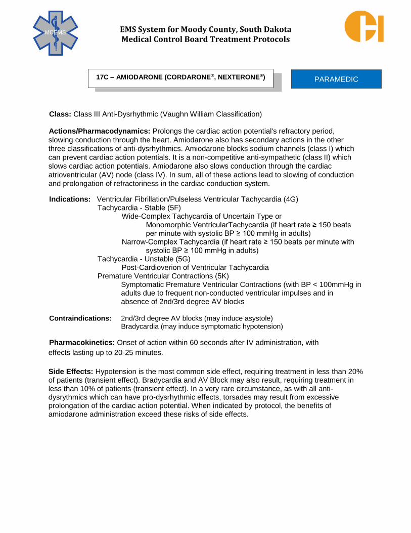
EMS System for Moody County, South Dakota Medical Control Board Treatment Protocols
Class: Class III Anti-Dysrhythmic (Vaughn William Classification)
Actions/Pharmacodynamics: Prolongs the cardiac action potential's refractory period,
slowing conduction through the heart. Amiodarone also has secondary actions in the other
three classifications of anti-dysrhythmics. Amiodarone blocks sodium channels (class I) which
can prevent cardiac action potentials. It is a non-competitive anti-sympathetic (class II) which
slows cardiac action potentials. Amiodarone also slows conduction through the cardiac
atrioventricular (AV) node (class IV). In sum, all of these actions lead to slowing of conduction
and prolongation of refractoriness in the cardiac conduction system.
Indications: Ventricular Fibrillation/Pulseless Ventricular Tachycardia (4G) Tachycardia - Stable (5F)
Wide-Complex Tachycardia of Uncertain Type or Monomorphic VentricularTachycardia (if heart rate ≥ 150 beats per minute with systolic BP ≥ 100 mmHg in adults)
Narrow-Complex Tachycardia (if heart rate ≥ 150 beats per minute with systolic BP ≥ 100 mmHg in adults)
Tachycardia - Unstable (5G) Post-Cardioverion of Ventricular Tachycardia
Premature Ventricular Contractions (5K) Symptomatic Premature Ventricular Contractions (with BP < 100mmHg in adults due to frequent non-conducted ventricular impulses and in absence of 2nd/3rd degree AV blocks
Contraindications: 2nd/3rd degree AV blocks (may induce asystole) Bradycardia (may induce symptomatic hypotension)
Pharmacokinetics: Onset of action within 60 seconds after IV administration, with
effects lasting up to 20-25 minutes.
Side Effects: Hypotension is the most common side effect, requiring treatment in less than 20% of patients (transient effect). Bradycardia and AV Block may also result, requiring treatment in less than 10% of patients (transient effect). In a very rare circumstance, as with all anti-dysrythmics which can have pro-dysrhythmic effects, torsades may result from excessive prolongation of the cardiac action potential. When indicated by protocol, the benefits of amiodarone administration exceed these risks of side effects.
PARAMEDIC 17C – AMIODARONE (CORDARONE®, NEXTERONE®)

EMS System for Moody County, South Dakota Medical Control Board Treatment Protocols
PROTOCOL 17C: Amiodarone (Cordarone®, Nexterone®), cont.
Dosage: Ventricular Fibrillation/Pulseless Ventricular Tachycardia - Adult (4G) (refractory to initial defibrillation attempt)
300 mg IVP/IOP. Repeat at 150 mg IVP/IOP in 5 minutes to maximum cumulative dose of 450 mg. Epinephrine 1 mg (1:10,000) IVP/IOP is to be given with every amiodarone administration.
Ventricular Fibrillation/Pulseless Ventricular Tachycardia - Pediatric (4G) (refractory to initial defibrillation attempts)
5 mg/kg IVP/IOP in single dose. Epinephrine 0.01 mg/kg (1:10,000, 0.1
mL/kg) IVP/IOP is to be given with every amiodarone administration.
Ventricular Fibrillation/Pulseless Ventricular Tachycardia - Adult (4G) (post return of sustained spontaneous circulation)
150 mg over 10 minutes (15 mg/minute or 0.3 mL/minute very slow
IVP/IOP/IVPB) IF maximum cumulative dose of 450 mg has not been achieved.
Ventricular Fibrillation/Pulseless Ventricular Tachycardia - Pediatric (4G) (post return of sustained spontaneous circulation)
Tachycardia - Stable - Pediatric (5F) (wide-complex tachycardia of uncertain type or monomorphic ventricular tachycardia; narrow-complex tachycardia)
Tachycardia - Unstable - Pediatric (5G) Premature Ventricular Contractions - Pediatric (5K) **MC Consult
Tachycardia - Stable - Adult (5F) (wide–complex tachycardia of uncertain type - standing order; monomorphic ventricular tachycardia - standing order; narrow complex - **OLMC order only)
Tachycardia - Unstable - Adult (5G) (post cardioversion of ventricular
tachycardia) Premature Ventricular Contractions - Adult (5K) 150 mg over 10 minutes (15 mg/minute or 0.3 mL/minute very
slow IVP/IOP/IVPB).
How Supplied: 150 mg/3 mL in vial, ampule, or pre-filled syringe.150 mg/100 mL
pre-mixed infusion.(Always check concentration and dose per
container at time of patient medication administration)
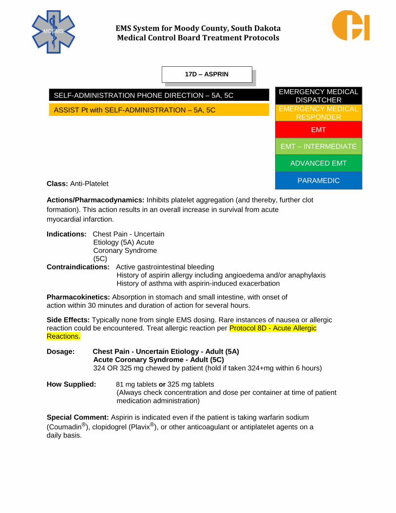
EMS System for Moody County, South Dakota Medical Control Board Treatment Protocols
Class: Anti-Platelet
Actions/Pharmacodynamics: Inhibits platelet aggregation (and thereby, further clot
formation). This action results in an overall increase in survival from acute
myocardial infarction.
Indications: Chest Pain - Uncertain Etiology (5A) Acute Coronary Syndrome (5C)
Contraindications: Active gastrointestinal bleeding
History of aspirin allergy including angioedema and/or anaphylaxis History of asthma with aspirin-induced exacerbation
Pharmacokinetics: Absorption in stomach and small intestine, with onset of action within 30 minutes and duration of action for several hours.
Side Effects: Typically none from single EMS dosing. Rare instances of nausea or allergic reaction could be encountered. Treat allergic reaction per Protocol 8D - Acute Allergic Reactions.
Dosage: Chest Pain - Uncertain Etiology - Adult (5A)
Acute Coronary Syndrome - Adult (5C) 324 OR 325 mg chewed by patient (hold if taken 324+mg within 6 hours)
How Supplied: 81 mg tablets or 325 mg tablets
(Always check concentration and dose per container at time of patient medication administration)
Special Comment: Aspirin is indicated even if the patient is taking warfarin sodium
(Coumadin®), clopidogrel (Plavix®), or other anticoagulant or antiplatelet agents on a daily basis.
EMERGENCY MEDICAL DISPATCHER
EMERGENCY MEDICAL RESPONDER
EMT
EMT – INTERMEDIATE
ADVANCED EMT
PARAMEDIC
17D – ASPRIN
SELF-ADMINISTRATION PHONE DIRECTION – 5A, 5C
ASSIST Pt with SELF-ADMINISTRATION – 5A, 5C

EMS System for Moody County, South Dakota Medical Control Board Treatment Protocols
Class: Parasympatholytic
Actions/Pharmacodynamics: Blocks parasympathetic impulses to the heart via the vagus nerve. Atropine increases the rate of cardiac sinoatrial (SA) node discharges, enhances conduction through the atrioventricular (AV) node, and by increasing heart rate, increases the cardiac output and blood pressure. Additionally, in the treatment of indicated poisonings (organophosphates) atropine reverses muscarinic effects of acetylcholine, including diaphoresis, diarrhea, urination, bronchorrhea (secretions from the lower respiratory tract), emesis, lacrimation (tearing), and salivation. Atropine produces dilation of pupils by blocking stimulation of the ciliary muscle surrounding the pupils.
Indications: Bradycardia (5D) Poisonings – General Management (Organophosphate) (8A)
Contraindications: None absolute in indicated situations.
Pharmacokinetics: Typical onset within 60 seconds given IV. Effects can persist for over 1 hour or more.
Side Effects: Tachycardia (either supraventricular or ventricular), hypertension, palpitations, blurred vision due to pupillary dilation, photophobia, dry mouth.
Adult organophosphate poisoning: 2 mg IVP/IOP/IM. Use IVP for more severe presentations. May repeat as often as every 3-5 minutes if symptoms progressive or persistent.
Dosage: Bradycardia – Symptomatic & Systolic BP < 100 mmHg
(Sinus, First Degree, 2nd Degree Type I) - Adult (5D) In Non-Acute Coronary Syndrome: 0.5 mg IVP/IOP. May repeat every 5 minutes to cumulative maximum dose of 3 mg
Bradycardia - Symptomatic & Systolic BP < 70 + (2 x age in years) mmHg
(Sinus, First Degree, 2nd Degree Type I) - Pediatric (5D) Unresponsive to Epinephrine, 0.02 mg/kg IVP/IOP; minimum dose 0.1 mg
Max single dose 0.5 mg. May repeat once.
Poisonings – General Management (Organophosphate) – Adult (8A)
2 mg IVP/IOP/IM. Use IVP for more severe presentation. Repeat every 3-5 minutes if symptoms progressive. Poisonings – General Management (Organophosphate) – Pediatric (8A)
0.05 mg/kg IVP/IOP/IM. Use IVP for more severe presentation. Minimum dose 0.1 mg. Consult with MC for repeat dosing needs.
How Supplied: 1 mg/10 mL prefilled syringe 1 mg/1 mL vial 0.25 mg/5 mL prefilled syringe for pediatric use (Always check concentration and dose per container at time of patient medication administration.)
PARAMEDIC 17E – ATROPINE SULFATE

EMS System for Moody County, South Dakota Medical Control Board Treatment Protocols
Class: Electrolyte
Actions/Pharmacodynamics: Calcium causes a significant increase in myocardial
contractility and in ventricular automaticity. It is used as an antidote for some electrolyte
imbalances (e.g. stabilizing cardiac rhythm in the setting of hyperkalemia) and to minimize the
side effects from calcium channel blocker overdose.
Indications: Specific Causes of Cardiac Arrest (Hyperkalemia) (4I) Poisonings - General Management (Calcium Channel Blocker Overdose) (8A) Dialysis-Related Issues (Hyperkalemia) (9E) Crush Injury Syndrome (Hyperkalemia Prophylaxis) (10K)
Contraindications: Calcium gluconate is contraindicated in ventricular fibrillation unless
known hyperkalemia, in known hypercalcemia, and in suspected digitalis toxicity. It should be
used with caution in patients taking digoxin as it may precipitate toxicity. Safe use in
pregnancy and in children has not been established, though in indicated conditions, benefits
outweigh risks.
Pharmacokinetics: Onset nearly immediate when given IVP/IOP. The peak effect time
frame and duration of effect is not well established.
Side Effects: Paresthesia (tingling), syncope, sensations of heat waves (peripheral
vasodilation), pain and burning at IV site, skin necrosis and sloughing (with
extravasation), hypotension, bradycardia, cardiac dysrhythmias, cardiac arrest.
Dosage: Specific Causes of Cardiac Arrest (Hyperkalemia)-Adult & Pediatric (4I)
Poisonings - Management (Calcium Channel Blocker Overdose) - Adult & Pediatric (8A)
Dialysis-Related Issues (Hyperkalemia) - Adult & Pediatric (9E)
Crush Injury Syndrome (Hyperkalemia Prophylaxis) - Adult & Pediatric (10K) 10 mg/kg (10% solution) IVP/IOP, maximum dose of 1 gram
How Supplied: 1 gram in a 10 mL prefilled syringe (100 mg/mL) (Always check concentration and dose per container at time of
patient medication administration)
Special Comments: Calcium gluconate will interact with sodium bicarbonate and form a precipitate. Do not give both medications via the same vascular access line unless giving a copious flush of NS - approximately 50+ mL - between medications. In general, use an 18-20 gauge angiocatheter in a proximal IV site or use an IO line and test line patency before administration. In non-cardiac arrest or non-impending cardiac arrest settings, administer at 0.5 -1.0 mL per minute to reduce chances of venous irritation and extravasation.
PARAMEDIC 17F – CALCIUM GLUCONATE

EMS System for Moody County, South Dakota Medical Control Board Treatment Protocols
Class: Carbohydrate
Actions/Pharmacodynamics: Dextrose is the principal form of glucose (sugar) used by the body to create energy and support critical metabolic processes. Since serious brain injury can occur in prolonged hypoglycemia, the timely administration of glucose is essential in treating hypoglycemia (blood glucose < 50 mg/dL). Dextrose 50% IV is the treatment of choice for hypoglycemic patients of adult age or of pediatric age with weight at or exceeding 25 kg. Dextrose 25% IV is the treatment of choice for hypoglycemic patients of pediatric age with weight less than 25 kg. Dextrose 10% IVPB is the treatment of choice for hypoglycemic patients in which vascular access is limited to small gauge angiocatheters (smaller than 20 GA) or in any other situations in which there is a higher risk for extravasation. The lower concentration of D10 results in less extravasation tissue damage than D50. Indications: Respiratory Arrest (3A)
Altered Mental Status (6B) Seizure (6D) Syncope (6E) Dystonic Reaction (6F) Behavioral Disorder (7A) Poisonings - General Management (8A) Dialysis -Related Issues (9E) Complications of Pregnancy (13D) For all listed situations, indication is hypoglycemia (blood glucose < 50 mg/dL).
Contraindications: Hyperglycemia (blood glucose > 100 mg/dL)
Normoglycemia in the setting of suspected cerebral ischemia.
Pharmacokinetics: Onset with 60 seconds after IVP with peak effect and duration of action dependent upon degree and cause of hypoglycemia. Effective duration is usually in the excess of 30 minutes. Medical literature shows speed of hypoglycemia reversal to be near clinically equivalent when comparing D10 infusion wide open with D50 IVP. Side Effects: Warmth, pain, or burning at the injection site. D50 extravasation can cause tissue necrosis (requiring skin graft surgery), phlebitis, sclerosis, or thrombosis at the injection site.
EMT – INTERMEDIATE
ADVANCED EMT
PARAMEDIC
17G – DEXTROSE (50% as D50; 25% as D25; 10% as D10)

EMS System for Moody County, South Dakota Medical Control Board Treatment Protocols
PROTOCOL 17G: Dextrose (50% as D50; 25% as D25; 10% as D10)
Dosage: Respiratory Arrest - Adult & Pediatric weight ≥ 25 kg (3A)
Altered Mental Status - Adult & Pediatric weight ≥ 25 kg (6B) Seizure - Adult & Pediatric weight ≥ 25 kg (6D) Syncope - Adult & Pediatric weight ≥ 25 kg (6E) Dystonic
Reaction - Adult & Pediatric weight ≥ 25 kg (6F) Behavioral
Disorder - Adult & Pediatric weight ≥ 25 kg (7A) Dialysis -Related Issues - Adult & Pediatric weight ≥ 25 kg (9E) Complications of Pregnancy - Adult & Pediatric weight ≥ 25 kg (13D) For hypoglycemia (blood glucose < 50 mg/dL): Dextrose 50% (D50) 1 mL/kg IVP up to 50 mL Dextrose 10% (D10) 25 grams in 250mL of NS IVPB wide open up to 250mL
Respiratory Arrest - Pediatric weight < 25 kg (3A) Altered Mental Status - Pediatric weight < 25 kg (6B) Seizure - Pediatric weight < 25 kg (6D) Syncope - Pediatric weight < 25 kg (6E) Dystonic Reaction - Pediatric weight < 25 kg (6F) Behavioral Disorder - Pediatric weight < 25 kg (7A) Dialysis -Related Issues - Pediatric weight < 25 kg (9E) For hypoglycemia (blood glucose < 50 mg/dL) Dextrose 25% (D25) 2 mL/kg IVP up to 50 mL Dextrose 10% (D10) 25 grams in 250mL of NS IVPB wide open up to 125mL
Specific Cause of Cardiac Arrest - Adult & Pediatric weight ≥ 25 kg (4I) Dextrose 50% (D50) 1 mL/kg IVP up to 50 mL
Specific Cause of Cardiac Arrest - Pediatric weight < 25 kg (4I) Dextrose 25% (D25) 2 mL/kg IVP up to 50 mL
How Supplied: Prefilled syringes of D50 - 25 grams dextrose in 50 mL of water (0.5 gram/mL) Prefilled syringes of D25 - 2.5 grams dextrose in 10 mL of water (0.25 gram/mL) Prefilled syringe of D50 – 25 grams dextrose in 50 mL of water added to 250 mL bag of normal saline (0.1 gram/mL)
Special Comments: D50 should be administered using an infusing IV, NOT a saline lock. The tissue caustic nature of D50 can be decreased by performing a slow and non-forceful IV push through the side port of an IV line that is flowing with normal saline into the patient’s vein. Because of the risk of extravasation and the consequences of local tissue damage from extravasation, neither D50 nor D25 should be administered through an external jugular IV. High concentrations of dextrose can lead to cerebral edema in younger/smaller pediatric patients, requiring 1:1 dilution of D50 with normal saline to make D25 or using prefilled D25. A repeat determination of blood glucose level is to be performed post D50, D25, or D10 administration.

EMS System for Moody County, South Dakota Medical Control Board Treatment Protocols
Class: Sedative; Anticonvulsant; Muscle Relaxant; Anxiolytic (Benzodiazepine)
Actions/Pharmacodynamics: Intermediate - acting benzodiazepine with central
nervous system depressant, anticonvulsant, muscle relaxant, and anxiolytic effects. Like
the other benzodiazepines, it has no effect on pain. Diazepam has considerably more
muscle relaxant properties than midazolam, though no substantial amnestic effects as
with midazolam.
Indications: Medication Assisted Intubation (2G)
Post-intubation sedation - onset delay does not favor pre-intubation use Seizure (6D) (Midazolam preferred benzodiazepine due to faster onset of action) Dystonic Reactions (6F) Chemical Restraint (7C) (Midazolam preferred benzodiazepine due to faster onset of action) Poisonings - General Management (8A) (Suspected stimulant toxicity = severe agitation, HTN, tachycardia, diaphoresis) Head/Neck/Spine Injury (10A) Heat Illness (11A)
Contraindications: Patients with intolerance to benzodiazepines, acute narrow - angle glaucoma, shock, or coma. Caution with use in patients with COPD, chronic hepatic or renal failure, CHF, acute alcohol intoxication, and the elderly due to increased risk of respiratory depression.
Pharmacokinetics: Onset is 3-5 minutes, IVP/IOP; 15-30 minutes IM with erratic absorption, mandating IM dosing only utilized as a last option in adults; peak effects in 15-45 minutes. Duration is 2+ hours IVP/IOP/IM; half – life can reach 20 – 50 hours.
Side Effects: Headache, euphoria, drowsiness, excessive sedation, confusion, dizziness, blurred vision, diplopia, nystagmus, respiratory arrest, hypotension, nausea, vomiting. Dosage: Medication Assisted Intubation (Post Intubation Sedation) - Adult (2G)
0.1 mg/kg to max 5 mg IVP/IOP, may repeat once if systolic BP > 100 mmHg
Seizure - Adult (6D) Head/Neck/Spine Injury - Adult (10A) Heat Illness - Adult (11A)
5 mg IVP/IOP or 10 mg IM for active seizure. May repeat once in 5 minutes if still seizing.
PARAMEDIC 17H – DIAZEPAM (VALIUM®)
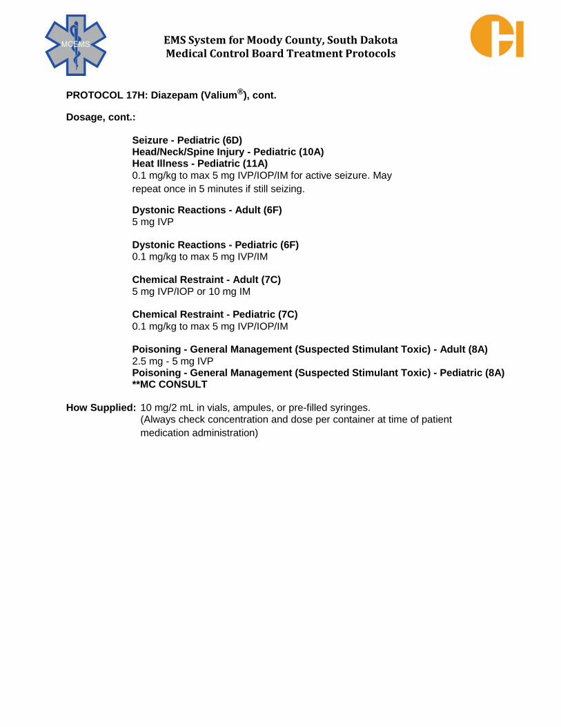
EMS System for Moody County, South Dakota Medical Control Board Treatment Protocols
PROTOCOL 17H: Diazepam (Valium®), cont.
Dosage, cont.:
Seizure - Pediatric (6D) Head/Neck/Spine Injury - Pediatric (10A) Heat Illness - Pediatric (11A) 0.1 mg/kg to max 5 mg IVP/IOP/IM for active seizure. May
repeat once in 5 minutes if still seizing.
Dystonic Reactions - Adult (6F) 5 mg IVP
Dystonic Reactions - Pediatric (6F) 0.1 mg/kg to max 5 mg IVP/IM
Chemical Restraint - Adult (7C) 5 mg IVP/IOP or 10 mg IM
Chemical Restraint - Pediatric (7C) 0.1 mg/kg to max 5 mg IVP/IOP/IM
Poisoning - General Management (Suspected Stimulant Toxic) - Adult (8A) 2.5 mg - 5 mg IVP
Poisoning - General Management (Suspected Stimulant Toxic) - Pediatric (8A) **MC CONSULT
How Supplied: 10 mg/2 mL in vials, ampules, or pre-filled syringes. (Always check concentration and dose per container at time of patient
medication administration)
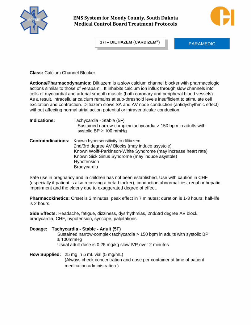
EMS System for Moody County, South Dakota Medical Control Board Treatment Protocols
Class: Calcium Channel Blocker
Actions/Pharmacodynamics: Diltiazem is a slow calcium channel blocker with pharmacologic
actions similar to those of verapamil. It inhabits calcium ion influx through slow channels into
cells of myocardial and arterial smooth muscle (both coronary and peripheral blood vessels) .
As a result, intracellular calcium remains at sub-threshold levels insufficient to stimulate cell
excitation and contraction. Diltiazem slows SA and AV node conduction (antidyshythmic effect)
without affecting normal atrial action potential or intraventricular conduction.
Indications: Tachycardia - Stable (5F)
Sustained narrow-complex tachycardia > 150 bpm in adults with systolic BP ≥ 100 mmHg
Contraindications: Known hypersensitivity to diltiazem 2nd/3rd degree AV Blocks (may induce asystole) Known Wolff-Parkinson-White Syndrome (may increase heart rate) Known Sick Sinus Syndrome (may induce asystole) Hypotension
Bradycardia
Safe use in pregnancy and in children has not been established. Use with caution in CHF (especially if patient is also receiving a beta-blocker), conduction abnormalities, renal or hepatic impairment and the elderly due to exaggerated degree of effect. Pharmacokinetics: Onset is 3 minutes; peak effect in 7 minutes; duration is 1-3 hours; half-life is 2 hours.
Side Effects: Headache, fatigue, dizziness, dysrhythmias, 2nd/3rd degree AV block, bradycardia, CHF, hypotension, syncope, palpitations. Dosage: Tachycardia - Stable - Adult (5F)
Sustained narrow-complex tachycardia > 150 bpm in adults with systolic BP ≥ 100mmHg Usual adult dose is 0.25 mg/kg slow IVP over 2 minutes
How Supplied: 25 mg in 5 mL vial (5 mg/mL) (Always check concentration and dose per container at time of patient
medication administration.)
PARAMEDIC 17I – DILTIAZEM (CARDIZEM®)

EMS System for Moody County, South Dakota Medical Control Board Treatment Protocols
Class: Antihistamine, Anticholinergic
Actions/Pharmacodynamics: Diphenhydramine competes for H1 – histamine receptor sites
on effector cells, thus blocking histamine release. Histamine release creates some of the
common signs and symptoms of an allergic response: pruritis (itching), mucus secretion, and capillary leaking, which contributes to the formation of urticaria (hives), erythematous skin, and
mucosal edema. In the setting of a dystonic reaction, the balance of dopamine and choline must be changed within the brain. The most clinically feasible method of reversing a dystonic
reaction, though inhibiting the enzyme acetylcholinesterase, is through the anti-cholinergic
effect of a medication like diphenhydramine.
Indications: Dystonic Reactions (6F)
Mild Sedation (7C) Acute Allergic Reactions (8D) Bee/Wasp Stings (8F)
Contraindications: Known hypersensitivity to diphenhydramine. While rare, allergic reaction to
diphenhydramine is possible and should be considered valid if stated or documented in a
patient’s medical history.
Pharmacokinetics: Onset within 15 – 30 minutes; duration is approximately 6 hours.
Side Effects: Drowsiness, dizziness, disturbed coordination.
Dosage: Dystonic Reactions - Adult (6F) Mild Sedation (7C) Acute Allergic Reactions- Adult (8D) Bee/Wasp Stings - Adult (8F) 50 mg IM/IVP
Dystonic Reactions - Pediatric (6F) Mild Sedation (7C) Acute Allergic Reactions- Pediatric (8D) Bee/Wasp Stings - Pediatric (8F) 1 mg/kg IM/IVP to maximum of 50 mg
How Supplied: 50 mg/1 mL in vial, ampule, or pre-filled syringe. (Always check concentration and dose per container at time of patient
medication administration.)
PARAMEDIC 17J – DIPHENHYDRAMINE (BENADRYL®)

EMS System for Moody County, South Dakota Medical Control Board Treatment Protocols
Class: Vasoconstrictor
Actions/Pharmacodynamics: Dose dependent. Higher doses (5+ mcg/kg/min)
increasingly stimulate alpha receptors in the peripheral vasculature, producing
vasoconstriction-related increases in system blood pressure. Concurrent beta receptor
stimulation may produce increases in heart rate and mild bronchodilation. Lower doses (<5
mcg/kg/min), as may be encountered infrequently in interhospital transfers, produce
mesenteric (intestinal) and renal vascular dilation to ensure continued perfusion to these
organ systems in complicated medical illness that would otherwise sacrifice such
circulation.
Indications: Dyspnea - Congestive Heart Failure (Cardiogenic Shock) (3E) Post Cardiac Arrest Treatment (Cardiogenic Shock) (4J) Acute Coronary Syndrome (Cardiogenic Shock) (5C) Fever (Septic Shock) (9B) Dialysis-Related Issues (9E) For all listed situations, indication is hypotension (adult = systolic <
100 mmHg) due to cardiogenic, septic, or neurogenic shock either
refractory to intravascular fluid boluses or in which intravascular fluid
bolusing is contraindicated (e.g. pulmonary edema).
Contraindications: Hypertension
Pharmacokinetics: Onset of action within 5 minutes after IV/IO infusion initiated. Rapid metabolism of medication requires ongoing IV/IO infusion to maintain clinical effects.
Side Effects: Palpitations, tachycardia, chest pain, and hypertension if not titrated.
Dosage: Dyspnea - Congestive Heart Failure (Cardiogenic Shock) - Adult (3E) Post Cardiac Arrest Treatment (Cardiogenic Shock) - Adult (4J) Acute Coronary Syndrome (Cardiogenic Shock) - Adult (5C) Fever (Septic Shock) - Adult (9B) Dialysis-Related Issues - Adult (9E) For hypotension (shock) refractory to fluids or fluids contraindicated 5 – 20 mcg/kg/minute - titrate to a sys B/P ≥ 100 mmHg (see following dosage chart.)
Dyspnea - Congestive Heart Failure (Cardiogenic Shock) - Pediatric (3E) Post Cardiac Arrest Treatment (Cardiogenic Shock) - Pediatric (4J) Fever (Septic Shock) - Pediatric (9B) Dialysis-Related Issues - Pediatric (9E) For hypotension (shock) refractory to fluids or fluids contraindicated
**MC CONSULT
PARAMEDIC 17K – DOPAMINE (INTROPIN®)

EMS System for Moody County, South Dakota Medical Control Board Treatment Protocols
PROTOCOL 17K: Dopamine (Intropin®), cont.
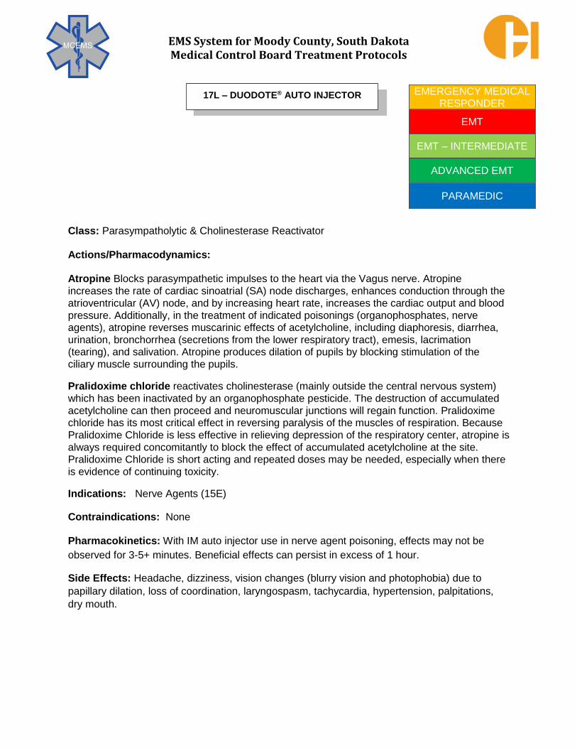
EMS System for Moody County, South Dakota Medical Control Board Treatment Protocols
Class: Parasympatholytic & Cholinesterase Reactivator
Actions/Pharmacodynamics:
Atropine Blocks parasympathetic impulses to the heart via the Vagus nerve. Atropine
increases the rate of cardiac sinoatrial (SA) node discharges, enhances conduction through the
atrioventricular (AV) node, and by increasing heart rate, increases the cardiac output and blood
pressure. Additionally, in the treatment of indicated poisonings (organophosphates, nerve agents), atropine reverses muscarinic effects of acetylcholine, including diaphoresis, diarrhea,
urination, bronchorrhea (secretions from the lower respiratory tract), emesis, lacrimation (tearing), and salivation. Atropine produces dilation of pupils by blocking stimulation of the
ciliary muscle surrounding the pupils.
Pralidoxime chloride reactivates cholinesterase (mainly outside the central nervous system)
which has been inactivated by an organophosphate pesticide. The destruction of accumulated
acetylcholine can then proceed and neuromuscular junctions will regain function. Pralidoxime chloride has its most critical effect in reversing paralysis of the muscles of respiration. Because
Pralidoxime Chloride is less effective in relieving depression of the respiratory center, atropine is
always required concomitantly to block the effect of accumulated acetylcholine at the site. Pralidoxime Chloride is short acting and repeated doses may be needed, especially when there
is evidence of continuing toxicity.
Indications: Nerve Agents (15E)
Contraindications: None
Pharmacokinetics: With IM auto injector use in nerve agent poisoning, effects may not be
observed for 3-5+ minutes. Beneficial effects can persist in excess of 1 hour.
Side Effects: Headache, dizziness, vision changes (blurry vision and photophobia) due to
papillary dilation, loss of coordination, laryngospasm, tachycardia, hypertension, palpitations,
dry mouth.
EMERGENCY MEDICAL RESPONDER
EMT
EMT – INTERMEDIATE
ADVANCED EMT
PARAMEDIC
17L – DUODOTE® AUTO INJECTOR

EMS System for Moody County, South Dakota Medical Control Board Treatment Protocols
PROTOCOL 17L: DuoDote®
Auto injector, cont.
Dosage: Nerve Agents - Adult & Pediatric > 12 years of age (15E) 2.1 mg atropine/ 600 mg pralidoxime IM May repeat every 5-15 minutes to cumulative maximum dose of 6.3 mg/1800
mg. In the setting of serious symptoms (cardiopulmonary distress), repeat
doses in rapid succession.
Nerve Agents - Pediatric ≤ 12 years of age (15E) Typical pediatric dose is 0.05 mg/kg atropine & 15 mg/kg pralidoxime IM
per dose, max single dose of 2.1 mg atropine/600 mg pralidoxime
How Supplied: DuoDote® auto injector
(Always check concentration and dose per container at time of patient medication administration)
Special Comments: Ideally, every public safety professional should have ready access to three
DuoDote® auto injectors for self/buddy use should emergent conditions warrant. In the setting of
suspected/actual nerve agent exposure, administration of the DuoDote® auto injector(s) must
occur within minutes of exposure for clinically effective results.

EMS System for Moody County, South Dakota Medical Control Board Treatment Protocols
Class: Vasoconstrictor, Bronchodilator (Catecholamine) Actions/Pharmacodynamics: Stimulates alpha receptors in the peripheral vasculature,
producing vasoconstriction-related increases in systemic blood pressure. Stimulates beta-1
receptors in the myocardium, producing increases in heart rate, myocardial contraction, and as
a result, cardiac output. Stimulates beta-2 receptors in the lower respiratory tract smooth
musculature, producing bronchodilation. Indications: Dyspnea - Asthma (Severe & Refractory to Nebulization)
(3C) Asystole (4F) Ventricular Fibrillation/Pulseless Ventricular Tachycardia (4G) Pulseless Electrical Activity (4H) Bradycardia (Pediatric) (5D) Acute Allergic Reactions (Anaphylaxis) (8D) Snakebites (Anaphylaxis) (8E) Bee/Wasp Stings (Anaphylaxis) (8F)
Contraindications: None absolute in indications above.
Safety in pregnancy not firmly established, though when clinically indicated the benefits outweigh risks. Pharmacokinetics: Onset of action within 2 minutes after IVP/IOP; within 5-10 minutes after IM. Duration of effect ranges from 3-5 minutes after IVP/IOP to upwards of 30 minutes after IM.
Side Effects: Restlessness, anxiety, generalized tremors, headache, dizziness, chest pain, palpitations, hypertension, premature ventricular contractions, tachycardia.
Dosage: Dyspnea - Asthma (Severe & Refractory to Nebulization) - Adult (3C) 1:1000 0.3 mg IM
Dyspnea - Asthma (Severe & Refractory to Nebulization) - Pediatric (3C) 1:1000 0.01 mg/kg (0.01 mL/kg) not to exceed 0.3 mg (0.3 mL) IM Asystole - Adult (4F) Ventricular Fibrillation/Pulseless Ventricular Tachycardia - Adult (4G) Pulseless Electrical Activity - Adult (4H) 1:10,000 1 mg IVP/IOP Repeat every 3 - 5 minutes while resuscitating cardiac arrest
Asystole - Pediatric (4F) Ventricular Fibrillation/Pulseless Ventricular Tachycardia - Pediatric (4G) Pulseless Electrical Activity - Pediatric (4H) 1:10,000 0.01 mg/kg (0.1 mL/kg) IVP/IOP Repeat every 3 - 5 minutes while resuscitating cardiac arrest
PARAMEDIC 17M – EPINEPHRINE 1:1000 & 1:10,000

EMS System for Moody County, South Dakota Medical Control Board Treatment Protocols
PROTOCOL 17M: Epinephrine 1:1000 & 1:10,000, cont.
Dosage, cont.:
Bradycardia - Symptomatic & Systolic BP < 70 + (2 x age in years) mmHg (Sinus, First Degree, 2nd Degree Type I) - Pediatric (5D) 1:10,000 0.01 mg/kg (0.1 mL/kg) IVP/IOP May repeat once. Acute Allergic Reactions (Anaphylaxis) - Adult (8D) Snakebites (Anaphylaxis) - Adult (8E) Bee/Wasp Stings (Anaphylaxis) - Adult (8F) 1:1000 0.5 mg IM
If anaphylaxis refractory to above IM dose: 1:10,000 1 mg slow IVP/IOP over 3 minutes
Acute Allergic Reactions (Anaphylaxis) - Pediatric (8D) Snakebites (Anaphylaxis) - Pediatric (8E) Bee/Wasp Stings (Anaphylaxis) - Pediatric (8F) 1:1000 0.01 mg/kg (0.01 mL/kg) not to exceed 0.3 mg (0.3 mL) IM If anaphylaxis refractory to above IM dose: 1:10,000 0.01 mg/kg slow IVP/IOP over 3 minutes
How Supplied: Epinephrine 1:1000 in 1 mg/1mL ampules or 30 mg/30 mL vial
(Always check concentration and dose per container at time of patient medication administration)
Epinephrine 1:10,000 in 1 mg/10 mL prefilled syringes (Always check concentration and dose per container at time of patient medication administration)
Special Comments: Be sure to administer correct concentration. Pulsatile patients ages 35 years or greater, particularly those with known coronary artery disease, receiving epinephrine should have ECG monitoring initiated and continued as soon as an ECG monitor is available.

EMS System for Moody County, South Dakota Medical Control Board Treatment Protocols
Class: Vasoconstrictor, Bronchodilator (Catecholamine)
Actions/Pharmacodynamics: Stimulates alpha receptors in the peripheral vasculature,
producing vasoconstriction-related increases in systemic blood pressure. Stimulates beta-1
receptors in the myocardium, producing increases in heart rate, myocardial contraction, and as
a result, cardiac output. Stimulates beta- 2 receptors in the lower respiratory tract smooth
musculature, producing bronchodilation.
Indications: Dyspnea - Asthma (Severe - Refractory to Inhaler/Nebulization) (3C) Acute Allergic Reactions (Anaphylaxis) (8D) Snakebites (Anaphylaxis) (8E)
Bee/Wasp Stings (Anaphylaxis) (8F)
Contraindications: None in indications above.
Pharmacokinetics: Onset of action within 5-10 minutes after IM administration. Duration of
effect may range upwards of 30 minutes intramuscularly.
Adverse/Side Effects: Restlessness, anxiety, generalized tremors, headache, dizziness, chest
pain, palpitations, hypertension, premature ventricular contractions, tachycardia. Pulsatile
patients ages 35 years or greater, particularly those with known coronary artery disease,
receiving epinephrine should have ECG monitoring initiated and continued as soon as an ECG
monitor is available. Safety in pregnancy not firmly established, though when clinically indicated
the benefits outweigh risks and should not deter clinically necessary usage.
Dosage: Dyspnea - Asthma (Severe - Refractory to Inhaler/Nebulization) - Adult (3C) Acute Allergic Reactions (Anaphylaxis) - Adult (8D) Snakebites (Anaphylaxis) - Adult (8E) Bee/Wasp Stings (Anaphylaxis) - Adult (8F) Adult Epinephrine Auto injector (0.3 mg of Epinephrine 1:1000) IM lateral thigh
Dyspnea- Asthma (Severe-Refractory to Inhaler/Nebulization)-Pediatric (3C) Acute Allergic Reactions (Anaphylaxis) - Pediatric (8D) Snakebites (Anaphylaxis) - Pediatric (8E) Bee/Wasp Stings (Anaphylaxis) - Pediatric (8F) Pediatric Epinephrine Auto injector (0.15 mg of Epinephrine 1:1000) IM lateral thigh.
EMT
EMT – INTERMEDIATE
ADVANCED EMT
PARAMEDIC
17N – EPINEPHRINE AUTO INJECTOR (EPIPEN®, Auvi-Q®)
ADULT & PEDIATRIC

EMS System for Moody County, South Dakota Medical Control Board Treatment Protocols
Class: Narcotic analgesic
Actions/Pharmacodynamics: Stimulates central nervous system opiate receptors, producing
systemic analgesia. On a milligram weight basis, fentanyl is 50-100 times more potent than
morphine. Its duration of action is shorter than morphine or hydromorphone. An IV dose of 100
mcg of fentanyl is roughly equivalent to an IV dose of 10 mg of morphine. Fentanyl has less
emetic effects than other narcotic analgesics.
Indications: Chest Pain – Uncertain Etiology (5A) Acute Coronary Syndrome (5C) Snakebites (8E) Abdominal Pain/Nausea/Vomiting/Diarrhea (9A) Pain Management (Acute Onset &Chronic Type) (9D) Eye Injury (10B) Dental Injury/Pain (10C) Chest/Abdomen/Pelvis Injury (10D) Extremity/Amputation Injury (10G) Compartment Syndrome (10J) Crush Injury Syndrome (10K) Burns (10L) Lightning/Electrical Injury (11C) Pelvic Pain (13E)
For all listed situations, indication is acute pain control in alert, hemodynamically stable patient.
Contraindications: Hypotension Respiratory Depression Minor Degrees of Pain Pain Assessed as Factitious
Side Effects: Hypotension, respiratory depression, euphoria, dizziness. Nausea and/or vomiting are rarely seen if administration is slow IVP.
Pharmacokinetics: Onset of action nearly immediate after IV administration. Peak effects occur within 3 – 5 minutes. Duration of effect is 30 - 60 minutes, with a half-life of 6 – 8 hours.
PARAMEDIC 17O – FENTANYL (SUBLIMAZE®)

EMS System for Moody County, South Dakota Medical Control Board Treatment Protocols
PROTOCOL 17O: Fentanyl (Sublimaze®), cont.
Dosage: Chest Pain – Uncertain Etiology – Adult (5A) Acute Coronary Syndrome – Adult (5C) 0.5 mcg/kg slow IVP/IM/IN, maximum single dose of 50 mcg
May repeat every 10 minutes to a maximum cumulative dose of 1.5 mcg/kg or 125 mcg, whichever is lesser
Snakebites – Adult (8E) Abdominal Pain/Nausea/Vomiting/Diarrhea – Adult (9A) Pain Management (Acute Onset &Chronic Type) – Adult (9D) Eye Injury – Adult (10B) Dental Injury/Pain – Adult (10C) Chest/Abdomen/Pelvis Injury – Adult (10D) Extremity/Amputation Injury – Adult (10G) Compartment Syndrome – Adult (10J) Crush Injury Syndrome – Adult (10K) Burns – Adult (10L) Lightning/Electrical Injury – Adult (11C) Pelvic Pain – Adult (13E)
For all listed situations, indication is acute pain control in an alert, hemodynamically stable patient. 1 mcg/kg slow IVP/IM/IN, maximum single dose of 100 mcg
May repeat every 10 minutes to a maximum cumulative dose of 3 mcg/kg or 250 mcg, whichever is lesser
Chest Pain – Uncertain Etiology – Pediatric (5A)
Snakebites – Pediatric (8E) Abdominal Pain/Nausea/Vomiting/Diarrhea – Pediatric (9A) Pain Management (Acute Onset &Chronic Type) – Pediatric (9D) Eye Injury – Pediatric (10B) Dental Injury/Pain – Pediatric (10C) Chest/Abdomen/Pelvis Injury – Pediatric (10D) Extremity/Amputation Injury – Pediatric (10G) Compartment Syndrome – Pediatric (10J) Crush Injury Syndrome – Pediatric (10K) Burns – Pediatric (10L) Lightning/Electrical Injury – Pediatric (11C) Pelvic Pain – Pediatric (13E)
For all listed situations, indication is acute pain control in an alert, hemodynamically stable patient **MC Order Only – Typical dose is 1 mcg/kg up to 50 mcg per dose.
How Supplied: 100 mcg/2 mL (50 mcg/mL) ampule, vial, or pre-filled syringe 250 mcg/5 mL (50 mcg/mL) ampule or vial 500 mcg/10 mL (50 mcg/mL) vial
(Always check concentration and dose per container at time of patient medication administration.)

EMS System for Moody County, South Dakota Medical Control Board Treatment Protocols
Class: Hormone
Actions/Pharmacodynamics: Glucagon is a hormone produced in the pancreas. When released in times of hypoglycemia, it causes a breakdown of glycogen (stored in the liver) to glucose and inhibits the subsequent synthesis of glycogen from circulating glucose. Both actions increase the blood levels of glucose. Given via the IM route, it is a useful drug in hypoglycemia when IV access is unsuccessful. Glucagon also increases heart rate, myocardial contractility and improves AV conduction in a manner similar to that produced by a catecholamine. Its actions are independent of beta blockade and therefore may be useful via IV/IO administration by paramedics for reversing cardiovascular collapse effects of suspected beta blocker toxicity.
Indications: Respiratory Arrest (3A) Specific Causes of Cardiac Arrest (4I) Altered Mental Status (6B) Seizure (6D) Syncope (6E) Dystonic Reactions (6F) Behavioral Disorder (7A) Poisonings – General Management (8A) Complications of Pregnancy (13D)
For all listed situations, indication is hypoglycemia (blood glucose <50 mg/dL) without ability to safely administer oral glucose (due to aspiration concern) and without ability to establish IV access in EMT-I, AEMT, and Paramedic Scopes of Practice.
There is an additional indication for beta blocker toxicity with hypotension and bradycardia within the Paramedic Scope of Practice.
Contraindications: None
Pharmacokinetics: Onset 5 – 20 minutes; peak effects in 30 minutes; duration is 1 – 1.5 hours.
Side Effects: Dizziness, headache, nausea/vomiting, hyperglycemia.
EMT – INTERMEDIATE
ADVANCED EMT
PARAMEDIC
17P – GLUCAGON
INTRAMUSCULAR USE ONLY – 3A, 4I, 6B, 6D, 6E, 6F, 7A, 8A, 13D
INTRAMUSCULAR USE ONLY – 3A, 4I, 6B, 6D, 6E, 6F, 7A, 8A, 13D

EMS System for Moody County, South Dakota Medical Control Board Treatment Protocols
PROTOCOL 17P: Glucagon, cont.
Dosage: Respiratory Arrest – Adult & Pediatric weight ≥ 25 kg (3A) Specific Causes of Cardiac Arrest - Adult& Pediatric weight ≥ 25 kg (4I) Altered Mental Status – Adult & Pediatric weight ≥ 25 kg (6B) Seizure – Adult & Pediatric weight ≥ 25 kg (6D) Syncope – Adult & Pediatric weight ≥ 25 kg (6E) Dystonic Reactions – Adult & Pediatric weight ≥ 25 kg (6F) Behavioral Disorder – Adult & Pediatric weight ≥ 25 kg (7A)
Poisonings – General Management – Adult & Pediatric weight ≥ 25 kg (8A) Complications of Pregnancy – Adult & Pediatric weight ≥ 25 kg(13D) All indicate hypoglycemia without safe PO access and without IV access 1 mg IM
Respiratory Arrest – Pediatric weight < 25 kg (3A)
Specific Causes of Cardiac Arrest– Pediatric weight < 25 kg (4I) Altered Mental Status – Pediatric weight < 25 kg (6B) Seizure – Pediatric weight < 25 kg (6D) Syncope – Pediatric weight < 25 kg (6E) Dystonic Reactions – Pediatric weight < 25 kg (6F) Behavioral Disorder – Pediatric weight < 25 kg (7A)
Poisonings – General Management – Pediatric weight < 25 kg (8A) Complications of Pregnancy – Pediatric weight < 25 kg (13D) All indicate hypoglycemia without safe PO access and without IV access 0.5 mg IM
Specific Causes of Cardiac Arrest - Adult (4I) Poisonings – General Management - Adult (8A)
Suspected beta blocker toxicity with arrest or hypotension/bradycardia (Paramedic only) 1 mg IVP/IOP; May be given IM if no IV access obtainable
Specific Causes of Cardiac Arrest - Pediatric (4I) Poisonings – General Management –Pediatric (8A) Suspected beta blocker toxicity with arrest or hypotension/bradycardia
(Paramedic only) 0.5 mg IVP/IOP; May be given IM if no IV access obtainable
How Supplied: 1 mg dry powder in vial with 1 mL of diluting solute for reconstitution
(Always check concentration and dose per container at time of patient
medication administration)

EMS System for Moody County, South Dakota Medical Control Board Treatment Protocols
Class: Carbohydrate
Actions/Pharmacodynamics: Increases blood sugar level.
Indications: Altered Mental Status (Hypoglycemia) (6B) Syncope (Hypoglycemia) (6E) Dystonic Reaction (Hypoglycemia) (6F) Behavioral Disorder (Hypoglycemia) (7A) Dialysis-Related Issues (Hypoglycemia) (9E) Complications of Pregnancy (Hypoglycemia) (13D)
Contraindications: Unconscious or semi–conscious and unable to follow simple commands. Care should be taken to prevent choking or aspiration of medication in semi–conscious patient.
Pharmacokinetics: Rapid oral absorption uptake to increase circulating blood sugar levels. Onset of effect normally occurs within several minutes of oral dosing. Duration of effect up to 30+ minutes, but patient should be advised to consume complex carbohydrates within minutes of restoration of normal blood sugar, unless otherwise contraindicated. Side Effects: None
Dosage: Altered Mental Status (Hypoglycemia) - Adult & Pediatric Weight ≥ 25 kg (6B)
Syncope (Hypoglycemia) - Adult & Pediatric Weight ≥ 25 kg (6E) Dystonic Reaction (Hypoglycemia) - Adult & Pediatric Weight ≥ 25 kg (6F) Behavioral Disorder (Hypoglycemia) - Adult & Pediatric Weight ≥ 25 kg (7A) Dialysis-Related Issues (Hypoglycemia) - Adult & Pediatric Weight ≥ 25 kg (9E) Complications of Pregnancy (Hypoglycemia) - Adult (13D) 15 grams (1 tube) PO or SL for blood glucose < 50 mg/dL Altered Mental Status (Hypoglycemia) - Pediatric Weight < 25 kg (6B) Syncope (Hypoglycemia) - Pediatric Weight < 25 kg (6E) Dystonic Reaction (Hypoglycemia) - Pediatric Weight < 25 kg (6F) Behavioral Disorder (Hypoglycemia) - Pediatric Weight < 25 kg (7A) Dialysis-Related Issues (Hypoglycemia) - Pediatric Weight < 25 kg (9E) 7.5 grams (1/2 tube) PO or SL for blood glucose < 50 mg/dL
How Supplied: 15 grams of glucose for oral administration in a squeeze tube container. (Always check concentration and dose per container at time of patient medication administration)
Special Comment: Medical grade glucose should be utilized in place of sodas, candy, and other carbohydrate-heavy solid food. In many cases, the carbohydrate grams cannot be measured.
EMERGENCY MEDICAL RESPONDER
EMT
EMT – INTERMEDIATE
ADVANCED EMT
PARAMEDIC
17Q – GLUCOSE (ORAL)

EMS System for Moody County, South Dakota Medical Control Board Treatment Protocols
Class: Antipsychotic
Therapeutic Action/Pharmacodynamics: Haloperidol is a potent, long – acting antipsychotic agent. While its exact mechanism is unclear, it appears to block the dopamine receptors in the brain associated with mood and behavior. It exerts strong antiemetic effects and impairs central thermoregulation. It also produces weak central anticholinergic effects and transient orthostatic hypotension
Indications: Chemical Restraint (7C)
Contraindications: Known hypersensitivity
Behavioral disorder etiology easily reversed (eg. hypoglycemia) Minor degrees of agitation
Parkinson's disease
Known seizure disorders (lowers seizure threshold)
CNS depressants, opiates, and alcohol may increase the CNS depression effect of haloperidol. Use with caution in elderly or debilitated patients due to exaggerated effect. Safe use in pregnancy has not been established, though in the indicated setting, benefit outweighs risks.
Pharmacokinetics: Onset is within 10-20 minutes IM; peak effect in 30-45 minutes; duration is 3+ hours, reported up to 35 hours.
Side Effects: CNS depression, seizure, dystonic reactions, dry mouth, blurry vision, bronchospasm, tachycardia, hypertension, hypotension, dysrhythmias, hyperpyrexia, diaphoresis, urinary retention.
Dosage: Chemical Restraint - Adult (7C) 5 mg IM (use deep IM injection in large muscle - lateral thigh if possible)
Chemical Restraint - Pediatric (7C)
** MC Order Only
How Supplied: 5 mg/1 mL vial. (Always check concentration and dose per container at time of patient medication administration)
Special Comments: In emergency situations where the patient’s behavior poses an immediate risk to rescuers and bystanders, the IM injection may be given through the patient’s clothing to minimize risk of needle stick injuries to rescuers. Dystonic reactions are common with haloperidol; diphenhydramine should be readily available - see Protocol 6F - Dystonic Reactions.
PARAMEDIC 17R – HALOPERIDOL (HALDOL®)

EMS System for Moody County, South Dakota Medical Control Board Treatment Protocols
Class: Parasympatholytic Bronchodilator
Actions/Pharmacodynamics: Atrovent® is an anticholinergic agent, chemically related to
atropine. Given in a nebulized form, it acts directly on the smooth muscle of the bronchial tree
by inhibiting acetylcholine at receptor sites. By blocking parasympathetic action, it dilates the
bronchial smooth muscle and decreases secretions. It also abolishes the Vagally mediated
reflex bronchospasm caused by inhaled irritants such as smoke, dust, and cold air and by a
range of inflammatory mediators such as histamine.
Indications: Dyspnea - Asthma (3C) Dyspnea - Chronic Obstructive Pulmonary Disease (3D) Acute Allergic Reactions (8D) Bee/Wasp Stings (8F)
Contraindications and Precautions: Atrovent® is contraindicated in patients with hypersensitivity to atropine. It should not be used as the sole pharmacologic treatment for acute bronchospasm. By protocol, Atrovent® is always administered in conjunction with albuterol. Pharmacokinetics: Absorption: 10% of inhaled dose reaches lower airway; approximately 0.5% of dose is systemically absorbed; onset within 5-15 minutes; peak effect in 1.5 – 2 hours; duration of effect is up to 4 – 6 hours; half – life is 1.5 – 2 hours.
Side Effects: Cough, reflex bronchospasm, hoarseness, nasal/oral dryness, bitter taste.
Dosage: Dyspnea - Asthma - Adult & Pediatric weight ≥ 15 kg (3C)
Acute Allergic Reactions - Adult & Pediatric weight ≥ 15 kg (8D) Bee/Wasp Stings - Adult & Pediatric weight ≥ 15 kg (8F) 0.5 mg nebulized (with albuterol 5 mg)
Dyspnea - Chronic Obstructive Pulmonary Disease - Adult (3D) 0.5 mg nebulized (with albuterol 5 mg), may repeat twice
Dyspnea - Asthma - Pediatric weight < 15 kg (3C) Acute Allergic Reactions - Pediatric weight < 15 kg (8D) Bee/Wasp Stings - Pediatric weight < 15 kg (8F) 0.25 mg nebulized (with albuterol 2.5 mg)
How Supplied: 0.5 mg/2.5 mL nebulizer solution vials. (Always check concentration and dose per container at time of patient
medication administration)
EMT
EMT – INTERMEDIATE
ADVANCED EMT
PARAMEDIC
17S – IPRATROPIUM BROMIDE (ATROVENT®)

EMS System for Moody County, South Dakota Medical Control Board Treatment Protocols
Class: Intraosseous local Anesthetic & Antidysrhythmic Therapeutic Actions/Pharmacodynamics: As a local anesthetic, reduces nerve activation that carries painful stimulus from intraosseous fluid and/or medication administration. As an antidysrhythmic, suppresses ventricular automaticity, chemically converting ventricular tachycardia. Indications: Tachycardia - Stable (5F)-Wide complex tachycardia, refractory to amiodarone Vascular Access - Intraosseous (9I) Contraindications: Narrow complex tachycardia
Second degree AV Block-Type II (Classic Type) Third degree AV Block (Complete Heart Block) Premature ventricular contractions with underlying bradycardias No indication for IO anesthetic (unresponsive patients)
Pharmacokinetics: Onset of action within 3 minutes IVP/IOP. Duration for 10-20 minutes. Side Effects: None expected in indicated dosing. Erroneous use in high degree heart blocks can lead to complete ventricular suppression/cardiac arrest. Dosage: Tachycardia - Stable - Wide Complex Tachycardia - Adult (5F) - Refactory to
Amiodarone Up to 1 mg/kg, slow IVP/IOP at < 50 mg/minute Tachycardia - Stable - Pediatric (5F) Consult with MC for use and dosing. Vascular Access - Intraosseous (Local Anesthetic) - Adult & Pediatric (9I) 1 mg/kg up to 40 mg IOP
How Supplied: 100 mg/5 mL (20 mg/mL of 2% lidocaine) prefilled syringe. (Always check concentration and dose per container at time of patient medication administration)
EMT – INTERMEDIATE
ADVANCED EMT
PARAMEDIC
17T – LIDOCAINE 2% INTRAVASCULAR (XYLOCAINE®)

EMS System for Moody County, South Dakota Medical Control Board Treatment Protocols
Class: Sedative; Anticonvulsant; Muscle Relaxant; Anxiolytic (Benzodiazepine)
Actions/Pharmacodynamics: Long - acting benzodiazepine with central nervous system depressant, anticonvulsant, muscle relaxant, and anxiolytic effects. Like the other benzodiazepines, it has no effect on pain. Ativan® has less muscle relaxant properties than diazepam, though no substantial amnestic effects as with midazolam. Midazolam preferred benzodiazepine due to faster onset of action.
Indications: Medication Assisted Intubation (2G)
Post-intubation sedation - onset delay does not favor pre-intubation use Seizure (6D) Dystonic Reactions (6F)
Chemical Restraint (7C) Poisonings - General Management (8A)
Suspected stimulant toxicity = severe agitation, HTN, tachycardia, diaphoresis Head/Neck/Spine Injury (10A) Heat Illness (11A)
Contraindications: Patients with intolerance to benzodiazepines, acute narrow - angle glaucoma, shock, or coma. Caution with use in patients with COPD, chronic hepatic or renal failure, CHF, acute alcohol intoxication, and the elderly due to increased risk of respiratory depression.
Pharmacokinetics: Onset is 5-10 minutes, IVP/IOP; up to 30 minutes IM; peak effects in 2-3 hours. Duration is 3-6+ hours IVP/IOP/IM; half – life can reach 20 – 50 hours.
Side Effects: Headache, euphoria, drowsiness, excessive sedation, confusion, dizziness, blurred vision, diplopia, nystagmus, respiratory arrest, hypotension, nausea, vomiting.
Dosage: Medication Assisted Intubation (Post Intubation Sedation) - Adult (2G) 0.1 mg/kg to max 2 mg IVP/IOP, may repeat once if systolic BP > 100 mmHg
Seizure - Adult (6D) Heat Illness - Adult (11A)
2 mg IVP/IOP/IM for active seizure. May repeat once in 10 minutes if still seizing. Seizure - Pediatric (6D) Heat Illness - Pediatric (11A)
0.1 mg/kg to max 2 mg IVP/IOP/IM for active seizure May repeat once in 5 minutes if still seizing. Dystonic Reactions - Adult (6F) 2 mg IVP/IM
Dystonic Reactions - Pediatric (6F) 0.1 mg/kg to max 2 mg IVP/IM Chemical Restraint - Adult (7C) 2 mg IVP/IOP/IM May repeat once.
PARAMEDIC 17U – LORAZEPAM (ATIVAN®)

EMS System for Moody County, South Dakota Medical Control Board Treatment Protocols
PROTOCOL 17U: Lorazepam (Ativan®), cont.
Dosage, cont.:
Chemical Restraint - Pediatric (7C) 0.1 mg/kg to max 2 mg IVP/IOP/IM
Poisoning - General Management (Suspected Stimulant Toxic) - Adult (8A) 1 -2 mg IVP/IM
Poisoning - General Management (Suspected Stimulant Toxic) - Pediatric (8A) **MC CONSULT
Head/Neck/Spine Injury - Adult (10A) 1 mg IVP/IM/IOP for active seizure. May repeat once in 5 minutes if still seizing. Head/Neck/Spine Injury - Pediatric (10A) 0.1 mg/kg IVP/IM/IOP for active seizure. May repeat once in 5 minutes if still seizing.
How Supplied: 2 mg/1 mL or 4 mg/1 mL in vials, ampules, or pre-filled syringes (Always
check concentration and dose per container at time of patient medication
administration.)
Special Comment: Lorazepam must be kept refrigerated.

EMS System for Moody County, South Dakota Medical Control Board Treatment Protocols
Class: Electrolyte
Therapeutic Actions/Pharmacodynamics: As an antidysrhythmic, reverses low circulating magnesium levels associated with ventricular arrhythmias, particularly polymorphic ventricular tachycardia, commonly called torsades des pointes. It is the anticonvulsant of greatest benefit for eclampsia.
Indications: Ventricular Fibrillation/Pulseless Ventricular Tachycardia (Torsades) (4G) Tachycardia - Stable (Torsades) (5F) Childbirth - Complicated (Eclampsia) (13B) Complications of Pregnancy (Eclampsia) (13D)
Contraindications: Hypotension or Known Renal Failure (when treating asthma)
Pharmacokinetics: Onset of action typically within 1-2 minutes after IVP/IOP. Effects
persist for up to 30 minutes.
Side Effects: None expected in indicated dosing. High doses (exceeding 4-6 grams) may cause sedation, muscle weakness, depressed reflexes, hypotension, bradycardia, and respiratory depression.
Dosage: Ventricular Fibrillation/Pulseless Ventricular Tachycardia (Torsades) - Adult (4G) 1 gram IVP/IOP
Tachycardia - Stable (Torsades) - Adult (5F) 1 gram slow IVP/IOP over 1 minute. May repeat once. Tachycardia - Stable (Torsades) - Pediatric (5F) Consult with MC for use and dosing.
Childbirth - Complicated (Eclampsia) (13B) Complications of Pregnancy (Eclampsia) (13D)
1 gram IVP/IOP. May repeat every 2-3 mins until seizure abates. Maximum cumulative dose is 4 grams.
How Supplied: 1 gram/2 mL (500 mg/mL in 50% solution) vials 5 grams/10 mL (500 mg/mL in a 50% solution) vials
5 grams/10 mL (50% mg/mL in a 50% solution) pre-filled syringes (Always
check concentration and dose per container at time of patient medication
administration.)
PARAMEDIC 17V – MAGNESIUM SULFATE

EMS System for Moody County, South Dakota Medical Control Board Treatment Protocols
Class: Steroid
Actions/Pharmacodynamics: Methylprednisolone is an intermediate-acting synthetic adrenal corticosteroid with glucocorticoid activity. It exerts anti-inflammatory effects in the setting of inflammatory-mediated illness.
Indications: Dyspnea - Asthma (3C) Dyspnea - Chronic Obstructive Pulmonary Disease (3D) Acute Allergic Reactions (8A) Bee/Wasp Stings (8F)
Contraindications and Precautions: Known hypersensitivity to methylprednisolone. In the setting of anaphylaxis, the only true contraindication is prior severe allergy (anaphylaxis) caused by methylprednisolone.
Pharmacokinetics: Onset of action within 4 – 6 hours; may have effect in excess of 24 hours.
Side Effects: None expected immediately. May occasionally see any of the following effects with onset of action: euphoria, insomnia, confusion, psychosis, edema, hypertension, nausea/ vomiting, hyperglycemia.
Dosage: Dyspnea - Asthma - Adult (3C) Dyspnea - Chronic Obstructive Pulmonary Disease - Adult (3D) Acute Allergic Reactions - Adult (8A) Bee/Wasp Stings - Adult (8F) 125 mg IVP. Give IM if no IV access obtainable.
Dyspnea - Asthma - Pediatric (3C) Acute Allergic Reactions - Pediatric (8A) Bee/Wasp Stings - Pediatric (8F) 2 mg/kg not to exceed 125 mg IVP. Give IM if no IV access obtainable.
How Supplied: 125 mg Act-O-Vial™ System (Single Dose Vial) (Always check concentration and dose per container at time of patient
medication administration.)
PARAMEDIC 17W – METHYLPREDNISOLONE (SOLU-MEDROL®)

EMS System for Moody County, South Dakota Medical Control Board Treatment Protocols
Class: Sedative; Anticonvulsant; Amnestic; Muscle Relaxant, Anxiolytic (Benzodiazepine)
Actions/Pharmacodynamics: Short - acting benzodiazepine with central nervous system depressant, anticonvulsant, anterograde amnestic, muscle relaxant, and anxiolytic effects. Like the other benzodiazepines, it has no effect on pain.
Indications: Medication Assisted Intubation (Pre & Post Intubation Sedation) (2G)
Post Cardiac Arrest Treatment (Hypothermia Induced Shivering Control) (4J) Transcutaneous Pacing (Sedation) (5E) Synchronized Cardioversion (Sedation) (5G) Seizure (6D) Dystonic Reactions (6F) Chemical Restraint (7C) Poisonings - General Management (8A) Suspected stimulant toxicity = severe agitation, HTN, tachycardia, diaphoresis
Head/Neck/Spine Injury (10A) Heat Illness (11A)
Contraindications: Patients with intolerance to benzodiazepines, acute narrow - angle glaucoma, shock, or coma. Caution with use in patients with COPD, chronic hepatic or renal failure, CHF, acute alcohol intoxication, and the elderly due to increased risk of respiratory depression.
Pharmacokinetics: Onset is 3-5 minutes, IVP/IOP; 6-14 minutes IN; up to 15 minutes IM (though clinically evident much faster); peak effects in 20-60 minutes. Duration is 2 hours IVP/IOP/IN; 1-6 hours IM; half – life is 1-4 hours.
Side Effects: Retrograde amnesia, headache, euphoria, drowsiness, weakness, excessive sedation, confusion, dizziness, blurred vision, diplopia, nystagmus, respiratory arrest, tachypnea, hypotension, nausea, vomiting.
Dosage: Medication Assisted Intubation (Pre & Post Intubation Sedation) - Adult (2G) 0.1 mg/kg to max 5 mg IVP/IOP, may repeat once if systolic BP > 100 mmHg
Post Cardiac Arrest Treatment (Hypothermia Induced Shivering Control) - Adult & Pediatric (4J) 0.1 mg/kg to max 5 mg IVP/IOP
Transcutaneous Pacing (Sedation) - Adult (5E) 2 - 5 mg IVP based upon weight and hemodynamics (0.1 mg/kg to max 5 mg) Synchronized Cardioversion (Sedation) - Adult (5G) 0.1 mg/kg to max 5 mg IVP/IOP/INP
Seizure - Adult (6D) Heat Illness - Adult (11A)
0.1 mg/kg to max 5 mg IM/IVP/IN/IOP for active seizure. May repeat once in 5 minutes if still seizing.
PARAMEDIC 17X – MIDAZOLAM (VERSED®)

EMS System for Moody County, South Dakota Medical Control Board Treatment Protocols
PROTOCOL 17X: Midazolam (Versed®), cont.
Dosage, cont.:
Seizure - Pediatric (6D) Head/Neck/Spine Injury - Pediatric (10A) Heat Illness - Pediatric (11A)
0.1 mg/kg to max 5 mg IM/IVP/IN/IOP for active seizure May repeat once in 5 minutes if still seizing.
Dystonic Reactions - Adult (6F) 2.5 mg IVP/IM/IN
Dystonic Reactions - Pediatric (6F) 0.1 mg/kg to max 2.5 mg IM/IVP/IN
Chemical Restraint - Adult (7C) 0.1 mg/kg to max 5 mg IM/IVP/IN/IOP. May repeat once. Chemical Restraint - Pediatric (7C) 0.1 mg/kg to max 5 mg IM/IVP/IN/IOP
Poisoning - General Management (Suspected Stimulant Toxic) - Adult (8A) 0.1 mg/kg to max 5 mg IVP/IN/IM
Poisoning - General Management (Suspected Stimulant Toxic) - Pediatric (8A) **MC CONSULT
Head/Neck/Spine Injury - Adult (10A) 5 mg IM/IVP/IN/IOP for active seizure. May repeat once in 5 minutes if still seizing.
How Supplied: 5 mg/1 mL in vials, ampules, or pre-filled syringes. (Always check concentration and dose per container at time of patient medication administration.)

EMS System for Moody County, South Dakota Medical Control Board Treatment Protocols
Class: Narcotic analgesic
Actions/Pharmacodynamics: Stimulates central nervous system opiate receptors, producing systemic analgesia. Modest vasodilation effects increase peripheral venous capacitance, and reduce venous return, myocardial workload, and myocardial oxygen demand.
Indications: Chest Pain – Uncertain Etiology (5A) Acute Coronary Syndrome (5C) Snakebites (8E) Abdominal Pain/Nausea/Vomiting/Diarrhea (9A) Pain Management (Acute Onset &Chronic Type) (9D) Eye Injury (10B) Dental Injury/Pain (10C) Chest/Abdomen/Pelvis Injury (10D) Extremity/Amputation Injury (10G) Compartment Syndrome (10J) Crush Injury Syndrome (10K) Burns (10L) Lightning/Electrical Injury (11C) Pelvic Pain (13E)
For all listed situations, indication is acute pain control in alert, hemodynamically stable patient.
Contraindications: Hypotension
Respiratory Depression
Minor Degrees of Pain
Pain Assessed as Factitious
Side Effects: Hypotension, respiratory depression, euphoria, dizziness. Nausea and/or vomiting are rarely seen if administration is slow IVP. Rapid IVP will lead to an accompanying histamine release, producing the nausea and/or vomiting often erroneously attributed to morphine itself.
Pharmacokinetics: Onset of action within 3-5 minutes after IV administration. Duration of effect can reach 4 hours depending upon end-organ function. Dosage: Chest Pain – Uncertain Etiology – Adult (5A)
Acute Coronary Syndrome – Adult (5C) 2 mg slow IVP May repeat every 5 minutes to a max cumulative dose of 10 mg.
PARAMEDIC 17Y – MORPHINE SULFATE

EMS System for Moody County, South Dakota Medical Control Board Treatment Protocols
PROTOCOL 17Y: Morphine Sulfate, cont. Dosage, cont.:
Snakebites – Adult (8E) Abdominal Pain/Nausea/Vomiting/Diarrhea – Adult (9A)
Pain Management (Acute Onset &Chronic Type) – Adult (9D) Eye Injury – Adult (10B) Dental Injury/Pain – Adult (10C) Chest/Abdomen/Pelvis Injury – Adult (10D) Extremity/Amputation Injury – Adult (10G) Compartment Syndrome – Adult (10J) Crush Injury Syndrome – Adult (10K) Burns – Adult (10L) Lightning/Electrical Injury – Adult (11C) Pelvic Pain – Adult (13E) For all listed situations, indication is acute pain control in alert, hemodynamically stable patient. 2 – 4 mg slow IVP May repeat every 5 minutes to a maximum cumulative dose of 10 mg
Chest Pain – Uncertain Etiology – Pediatric (5A)
Snakebites – Pediatric (8E) Abdominal Pain/Nausea/Vomiting/Diarrhea – Pediatric (9A) Pain Management (Acute Onset &Chronic Type) – Pediatric (9D) Eye Injury – Pediatric (10B) Dental Injury/Pain – Pediatric (10C) Chest/Abdomen/Pelvis Injury – Pediatric (10D) Extremity/Amputation Injury – Pediatric (10G) Compartment Syndrome – Pediatric (10J) Crush Injury Syndrome – Pediatric (10K) Burns – Pediatric (10L) Lightning/Electrical Injury – Pediatric (11C) Pelvic Pain – Pediatric (13E) For all listed situations, indication is acute pain control in alert, hemodynamically stable patient
How Supplied: 2 mg/1 mL pre-filled syringe
4 mg/1 mL vial, ampule, or pre-filled syringe 8 mg/1 mL pre-filled syringe
10 mg/1 mL vial 10 mg/10 mL vial (Always check concentration and dose per container at time of patient medication administration.)

EMS System for Moody County, South Dakota Medical Control Board Treatment Protocols
Class: Narcotic antagonist
Actions/Pharmacodynamics: The primary action of interest is reversal of respiratory depression associated with narcotic agents. Naloxone competes with and displaces narcotic substances from opiate receptors.
Indications: Respiratory Arrest (3A) Specific Causes of Cardiac Arrest (4I) Altered Mental Status (6B) Syncope (6E) Poisonings – General Management (8A)
Contraindications: Known or suspected narcotic substance use or abuse without cardiopulmonary compromise. Post-intubation in known or suspected narcotic substance use or substance abuse situations. Avoid whenever possible in known or suspected narcotic addicts. In these patients, use the smallest clinically effective dose possible (titrating administration slowly) to avoid acute narcotic withdrawal.
Pharmacokinetics: Onset of action within 2 minutes after IVP/IOP/IN administration with duration of effect up to 2 hours.
Side Effects: Agitation, anxiety, diaphoresis, tachycardia, nausea, vomiting, headache, hypertension, hypotension, seizures. Dosage: Respiratory Arrest - Adult (3A)
Specific Causes of Cardiac Arrest - Adult (4I) Altered Mental Status – Adult (6B) Syncope – Adult (6E) Poisonings – General Management – Adult (8A)
In Apnea/Agonal Breathing, 2 mg IVP/IOP/IN. May repeat once to maximum cumulative dose of 4 mg.
In Ineffective Breathing Activity, 0.5 mg IVP/IOP/IN. May repeat to a maximum cumulative dose of 4 mg.
EMERGENCY MEDICAL RESPONDER
EMT
EMT – INTERMEDIATE
ADVANCED EMT
PARAMEDIC
17Z – NALOXONE (NARCAN®)
INTRANASAL ADMINISTRATION ONLY – 3A, 4I, 6B, 6E, 8A
INTRANASAL ADMINISTRATION ONLY – 3A, 4I, 6B, 6E, 8A

EMS System for Moody County, South Dakota Medical Control Board Treatment Protocols
PROTOCOL 17Z: Naloxone (Narcan®), cont.
Dosage cont.:
Respiratory Arrest - Pediatric (3A) Specific Causes of Cardiac Arrest - Pediatric (4I) Altered Mental Status – Pediatric (6B) Syncope – Pediatric (6E) Poisonings – General Management – Pediatric (8A)
In Apnea/Agonal Breathing, 0.5 mg IVP/IOP/IN. May repeat to a maximum cumulative dose of 2 mg.
In Ineffective Breathing Activity, 0.5 mg IVP/IOP/IN. May repeat to a maximum cumulative dose of 2 mg.
How Supplied: 0.4 mg/1 mL vial 0.4 mg/1 mL prefilled syringe
2 mg/2 mL prefilled syringe
4 mg/10 mL vial (Always check concentration and dose per container at time of patient medication administration)
Special Comment: In non-respiratory arrest or non-cardiac arrest situations, always titrate administration slowly, using the lowest clinically effective amount of naloxone possible to avoid inadvertent acute narcotic withdrawal and/or other side effects.

EMS System for Moody County, South Dakota Medical Control Board Treatment Protocols
Class: Anti-Anginal, Vasodilator, Anti-Hypertensive (Nitrate)
Actions/Pharmacodynamics: Arterial and venous vasodilator through relaxing vascular smooth muscle. Reduces cardiac afterload resistance and cardiac preload volume respectively. Myocardial oxygen consumption/demand is decreased. Systemic blood pressure is decreased.
Indications: Dyspnea - Congestive Heart Failure (3E) Chest Pain - Uncertain Etiology (5A) Acute Coronary Syndrome (5C)
Contraindications: Hypotension
Asymptomatic Hypertension
Erectile Dysfunction Medications-Sildenafil (Viagra®) or
Vardenafil (Levitra®) use within 24 hours
Tadafil (Cialis®) use within 48 hours
Pharmacokinetics: Rapid vascular uptake within 3 minutes of sublingual dosing, with duration of effect up to 30 minutes. Rapid vascular effect within 1-3 minutes of intravenous dosing, with ongoing effect while continuous infusion. Vascular effect within 15-30 minutes of transdermal dosing, with ongoing effect while continued transdermal absorption.
Side Effects: The most serious side effect is hypotension, usually transient and responsive to supine positioning and intravenous fluid bolusing. Common, though non-serious, symptoms include: headache due to vasodilation, blurred vision, and dizziness. Paramedics should exercise caution when applying transdermal nitroglycerin ointment, avoiding contact with bare hands to avoid experiencing personal side effects, typically headache and dizziness.
EMERGENCY MEDICAL DISPATCHER
EMERGENCY MEDICAL RESPONDER
EMT
EMT – INTERMEDIATE
ADVANCED EMT
PARAMEDIC
17AA – NITROGLYCERIN (NITROLINGUAL®, NITROMIST®, NITROSTAT®,
NITROQUICK®, TRIDIL (IV INFUSION), NITRO-BID® - DERMAL)
SELF-ADMIN OF OWN DOSE BY PHONE DIRECTIVE – 3E, 5A, 5C
ASSIST Pt WITH OWN SELF-ADMINISTRATION – 3E, 5A, 5C
ASSIST Pt WITH OWN SELF-ADMINISTRATION – 3E, 5A, 5C
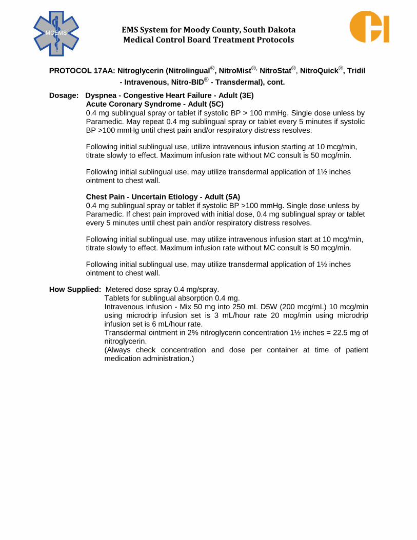
EMS System for Moody County, South Dakota Medical Control Board Treatment Protocols
PROTOCOL 17AA: Nitroglycerin (Nitrolingual®, NitroMist®, NitroStat®, NitroQuick®, Tridil
- Intravenous, Nitro-BID® - Transdermal), cont. Dosage: Dyspnea - Congestive Heart Failure - Adult (3E)
Acute Coronary Syndrome - Adult (5C) 0.4 mg sublingual spray or tablet if systolic BP > 100 mmHg. Single dose unless by Paramedic. May repeat 0.4 mg sublingual spray or tablet every 5 minutes if systolic BP >100 mmHg until chest pain and/or respiratory distress resolves.
Following initial sublingual use, utilize intravenous infusion starting at 10 mcg/min, titrate slowly to effect. Maximum infusion rate without MC consult is 50 mcg/min.
Following initial sublingual use, may utilize transdermal application of 1½ inches ointment to chest wall.
Chest Pain - Uncertain Etiology - Adult (5A)
0.4 mg sublingual spray or tablet if systolic BP >100 mmHg. Single dose unless by Paramedic. If chest pain improved with initial dose, 0.4 mg sublingual spray or tablet every 5 minutes until chest pain and/or respiratory distress resolves.
Following initial sublingual use, may utilize intravenous infusion start at 10 mcg/min, titrate slowly to effect. Maximum infusion rate without MC consult is 50 mcg/min.
Following initial sublingual use, may utilize transdermal application of 1½ inches ointment to chest wall.
How Supplied: Metered dose spray 0.4 mg/spray. Tablets for sublingual absorption 0.4 mg. Intravenous infusion - Mix 50 mg into 250 mL D5W (200 mcg/mL) 10 mcg/min using microdrip infusion set is 3 mL/hour rate 20 mcg/min using microdrip infusion set is 6 mL/hour rate. Transdermal ointment in 2% nitroglycerin concentration 1½ inches = 22.5 mg of nitroglycerin. (Always check concentration and dose per container at time of patient medication administration.)
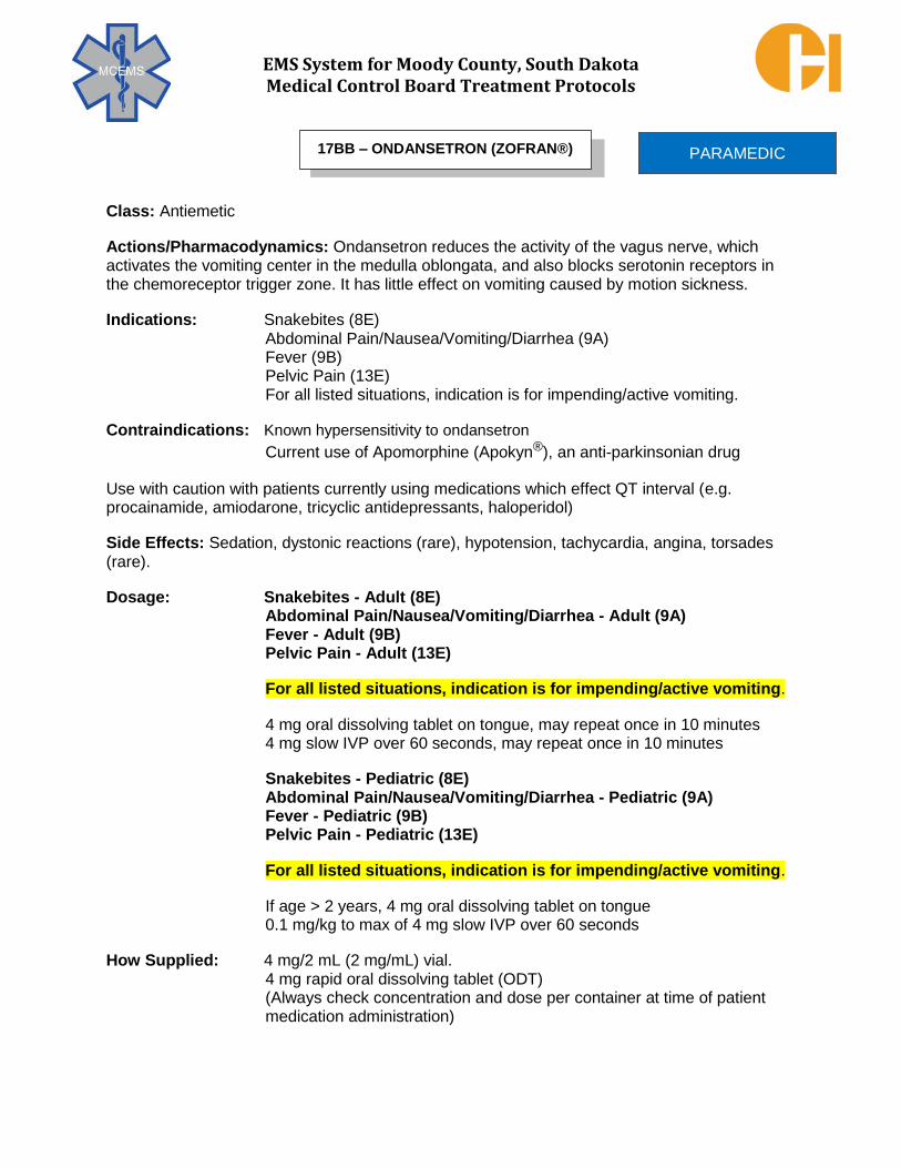
EMS System for Moody County, South Dakota Medical Control Board Treatment Protocols
Class: Antiemetic
Actions/Pharmacodynamics: Ondansetron reduces the activity of the vagus nerve, which activates the vomiting center in the medulla oblongata, and also blocks serotonin receptors in the chemoreceptor trigger zone. It has little effect on vomiting caused by motion sickness.
Indications: Snakebites (8E) Abdominal Pain/Nausea/Vomiting/Diarrhea (9A) Fever (9B) Pelvic Pain (13E) For all listed situations, indication is for impending/active vomiting.
Contraindications: Known hypersensitivity to ondansetron
Current use of Apomorphine (Apokyn®), an anti-parkinsonian drug
Use with caution with patients currently using medications which effect QT interval (e.g. procainamide, amiodarone, tricyclic antidepressants, haloperidol)
Side Effects: Sedation, dystonic reactions (rare), hypotension, tachycardia, angina, torsades (rare).
Dosage: Snakebites - Adult (8E) Abdominal Pain/Nausea/Vomiting/Diarrhea - Adult (9A)
Fever - Adult (9B) Pelvic Pain - Adult (13E)
For all listed situations, indication is for impending/active vomiting.
4 mg oral dissolving tablet on tongue, may repeat once in 10 minutes
4 mg slow IVP over 60 seconds, may repeat once in 10 minutes
Snakebites - Pediatric (8E) Abdominal Pain/Nausea/Vomiting/Diarrhea - Pediatric (9A)
Fever - Pediatric (9B) Pelvic Pain - Pediatric (13E)
For all listed situations, indication is for impending/active vomiting.
If age > 2 years, 4 mg oral dissolving tablet on tongue
0.1 mg/kg to max of 4 mg slow IVP over 60 seconds
How Supplied: 4 mg/2 mL (2 mg/mL) vial. 4 mg rapid oral dissolving tablet (ODT) (Always check concentration and dose per container at time of patient medication administration)
PARAMEDIC 17BB – ONDANSETRON (ZOFRAN®)

EMS System for Moody County, South Dakota Medical Control Board Treatment Protocols
Class: Cholinesterase Reactivator
Actions/Pharmacodynamics: Pralidoxime chloride reactivates cholinesterase (mainly
outside the central nervous system) which has been inactivated by an organophosphate pesticide. The destruction of accumulated acetylcholine can then proceed and neuromuscular junctions will regain function. Pralidoxime chloride has its most critical effect in reversing paralysis of the muscles of respiration. Because Pralidoxime Chloride is less effective in relieving depression of the respiratory center, atropine is always required
concomitantly to block the effect of accumulated acetylcholine at the site. Pralidoxime Chloride is short acting and repeated doses may be needed, especially when there is evidence of continuing toxicity.
Indications: Poisonings – General Management (8A)
Contraindications: None
Pharmacokinetics: With IM autoinjector use, effects may not be observed for up to 15
minutes. Beneficial effects can persist in excess of 1 hour.
Side Effects: Headache, dizziness, vision changes, loss of coordination, laryngospam,
tachycardia, palpitations.
Dosage: Poisonings – General Management - Adult & Pediatric > 12 years of age (8A) 600 mg IM
May repeat every 15 minutes to cumulative maximum dose of 1800 mg. In the setting of serious symptoms (cardiopulmonary distress), repeat doses in
rapid succession.
Poisonings – General Management - Pediatric ≤ 12 years of age(8A) Typical pediatric dose is 15 mg/kg IM per dose, max single dose 600 mg
How Supplied: 600 mg/2 mL auto injector (Always check concentration and dose per container at time of patient medication administration)
PARAMEDIC 17CC – PRALIDOXIME CHLORIDE (2PAM)

EMS System for Moody County, South Dakota Medical Control Board Treatment Protocols
Class: Alkalinizing agent
Actions/Pharmacodynamics: Raises the pH of blood by buffering excess hydrogen ions that are present in acidotic states. The role of sodium bicarbonate is limited in cardiac arrest. Because ventilation is an effective tool in managing respiratory acidosis, sodium bicarbonate should rarely be administered for cardiac arrest, unless the arrest is suspected to be secondary to hyperkalemia, a preexisting metabolic acidosis, or a tricyclic antidepressant over ingestion.
Indications: Specific Causes of Cardiac Arrest (Hyperkalemia) (4I) Poisonings – General Management (Tricyclic Antidepressant) (8A) Dialysis-Related Issues (Hyperkalemia) (9E) Crush Injury Syndrome (Hyperkalemia Prophylaxis) (10K)
Contraindications: Known metabolic alkalosis.
Pharmacokinetics: Onset of effect is observed within 3-5 minutes after IVP/IOP administration.
Side Effects: Sodium bicarbonate may inhibit oxygen release secondary to a shift in oxyhemoglobin saturation. It also may produce a paradoxical acidosis that can depress cerebral and cardiac function. Severe soft tissue damage can occur in extravasated administrations.
Dosage: Specific Causes of Cardiac Arrest – Hyperkalemia - Adult & Pediatric (4I) Poisonings – Management of Tricyclic Antidepressants - Adult & Pediatric (8A)
Dialysis-Related Issues – Hyperkalemia - Adult & Pediatric (9E) Crush Injury Syndrome – Hyperkalemia Prophylaxis - Adult & Pediatric (10K)
1 mEq/kg IVP/IOP with maximum dose of 50mEq
How Supplied: 50 mEq/50 mL (1 mEq/mL) prefilled syringe. (Always check concentration and dose per container at time of patient
medication administration)
Special Comment: Do not administer with calcium gluconate. A precipitate will form and obstruct the vascular access being utilized.
PARAMEDIC 17DD – SODIUM BICARBONATE

EMS System for Moody County, South Dakota Medical Control Board Treatment Protocols
Class: Anti-Fibrinolytic
Actions/Pharmacodynamics: Promotes clot formation in the setting of massive hemorrhage.
Indications: Hemostatic Agents (10I) Traumatic hemorrhagic shock less than 3 hours from injury with suspected
need for massive blood transfusion (clinical evidence of marked blood loss –
internal or external, sustained tachycardia and hypotension, see Protocol
10I for exact VS parameters by age group)
Contraindications: Non-hemorrhagic shock Non-traumatic hemorrhagic shock Hemorrhagic shock stabilized with other hemostatic agents/measures
Pharmacokinetics: Onset of action within 4 hours after IV administration, exact time of onset unclear and variable. Delayed effects up to 48 hours consistent with anti-inflammatory actions.
Side Effects: While a theoretical concern, TXA has not been shown to cause significant increase in deep venous thrombosis, pulmonary embolism, myocardial infarction, or stroke in published trials to date.
Dosage: Hemostatic Agents – Adult (10I) (Hemorrhagic shock as described above) 1 gram IVPB over 10 minutes. Administer in 100 mL or 250 mL NS.
Hemostatic Agents – Pediatric Ages 10 and Above (10I) (Hemorrhagic shock as described above) 15 mg/kg up to 1 gram IVPB over 10 minutes.
Administer in 100 mL or 250 mL NS.
How Supplied: 1 gram/10 mL vial or ampule (100 mg/mL) (Always check concentration and dose per container at time of patient
medication administration)
PARAMEDIC 17EE – TRANEXAMIC ACID (TXA, CYCLOKAPRON®)

EMS System for Moody County, South Dakota Medical Control Board Treatment Protocols
Class: Sympathomimetic Bronchodilator
Actions/Pharmacodynamics: Xopenex is a nebulized medication used for the treatment of Asthma and COPD. Xopenex is a smooth muscle relaxant for all airway muscles from the trachea to the terminal bronchioles. Xopenex is indicated for the treatment or prevention of broncho-spasm in adults and adolescents 12 years of age or older with reversible obstructive airway disease. Xopenex can be used successfully with patients with the history of supraventricular tachycardia or other arrhythmias because of the lowered effects on beta1- adrenergic receptors in cardiac muscle.
Indications: Dyspnea - Asthma (3C) Dyspnea - Chronic Obstructive Pulmonary Disease (3D)
Contraindications and Precautions: Xopenex is contraindicated in patients with
hypersensitivity to Xopenex. It should not be used as the sole pharmacologic treatment for
acute bronchospasm, Xopenex can be administered in conjunction with albuterol.
Pharmacokinetics: Absorption: 20% of inhaled dose reaches lower airway; approximately
0.5% of dose is systemically absorbed; onset within 10 - 17 minutes; peak effect in 2 – 3
hours; duration of effect is up to 3 – 4 hours; half – life is 3.5 – 4 hours.
Side Effects: Cough, reflex bronchospasm, hoarseness, nasal/oral dryness, bitter taste.
Dosage: Dyspnea - Asthma - Adult & Pediatric Age ≥ 12 years (3C) Dyspnea - Chronic Obstructive Pulmonary Disease - Adult (3D) 1.25 mg in 2.5 mL NS using Nebulizer. May repeat once. Dyspnea - Asthma - Pediatric weight < 15 kg (3C) 0.75-1.25 mg in 2.5 mL using Nebulizer.
How Supplied: 1.25 mg/0.5 mL nebulizer solution vials. Dilute with NS prior to administration. (Always check concentration and dose per container at time of patient
medication administration.)
PARAMEDIC 17FF – XOPENEX (LEVABUTEROL® HCL)

EMS System for Moody County, South Dakota Medical Control Board Treatment Protocols
The purpose of the abbreviation list is to define recommended abbreviations that may be used in the documentation of patient care and in documented protocols with in the MCEMS system. Abbreviations are often used to facilitate and shorten written narratives. This is an acceptable practice as long as it is universally recognized and understood abbreviations are used. The following are medical abbreviations that are acceptable for use by EMS personnel in the MCEMS system. In situations where an abbreviation is not listed and the provider intends to use the abbreviation more than once in the patient care report (PCR), the provider may spell out the complete term to be abbreviated with the abbreviation immediately following the term enclosed in brackets (i.e.,”()”). Unlisted abbreviations that are acceptable to use are the following:
1. Hospitals, clinics, or other medical facilities can be abbreviated by using the first letter of each word of the proper title and only abbreviate the facility if it is already written in entirely on the PCR as the response location or destination.
2. When abbreviating a Fire Department, “FD” must follow any letters used to abbreviate the city or municipality fire department.
3. When abbreviating a Police Department or Police Officer response, “PD” must follow any letters used to abbreviate the city or municipality police department.
4. When abbreviating a Sheriff Department or Sheriff Officer, “SO” must follow any letters used to abbreviate the county or municipality sheriff department.
Abbreviations acceptable for use on PCR’s in the MCEMS system are as follows;
ABBREVIATIONS A ABCs AIRWAY, BREATHING, AND CIRCULATION Abd ABDOMEN ACS ACUTE CORONARY SYNDROME AED AUTOMATED EXTERNAL DEFIBRILLATOR AEMT ADVANCED EMERGENCY MEDICAL TECHNICIAN AKA/BKA ABOVE-BELOW-KNEE AMPUTATION ALTE APPARENT LIFE THREATENING EVENT AMI ACUTE MYOCARDIAL INFARCTION AMS ALTERED MENTAL STATUS ASA ASPIRIN ASAP AS SOON AS POSSIBLE AV ATRIOVENTRICULAR
EMERGENCY MEDICAL RESPONDER
EMT
EMT – INTERMEDIATE
ADVANCED EMT
PARAMEDIC
18A – ACCEPTED ABBREVIATIONS LIST
EMS PROFESSIONALS

EMS System for Moody County, South Dakota Medical Control Board Treatment Protocols
PROTOCOL 18A: Accepted abbreviations list, cont.
B BBS BILATERAL BREATH SOUNDS BiPAP Bi-LEVEL POSTIVE AIRWAY PRESSURE BP BLOOD PRESSURE BSA BODY SURFACE AREA BVM BAG VALVE MASK C C CELSIUS DEGREES CABs CIRCULATION, AIRWAY, AND BREATHING CAOx4 CLEAR, ALERT, ORIENTED TIMES FOUR CHF CONGESTIVE HEART FAILURE CM CENTIMETER CNS CENTRAL NERVOUS SYSTEM CO CARBON MONOXIDE CO2 CARBON DIOXIDE COPD CHRONIC OBSTRUCTIVE PULMONARY DISEASE CPAP CONTINUOUS POSITIVE AIRWAY PRESSURE CPR CARDIOPULMONARY RESUSCITATION D D10 DEXTROSE 10% D25 DEXTROSE 25% D50 DEXTROSE 50% DCAPP-BLS DEFORMITIES, CONTUSIONS, ABRASIONS, PENETRATIONS, PARADOXICAL MOVEMENTS, BURNS, LACERATIONS, SWELLING DBP DIASYSTOLIC BLOOD PRESSURE dL DECILITER DNI DO NOT INTUBATE DNR DO NOT RESUSCITATE
D.O. DOCTOR OF OSTEOPATHY
E ECG ELECTROCARDIOGRAM ED EMERGENCY DEPARTMENT EMD EMERGENCY MEDICAL DISPATCHER EMR EMERGENCY MEDICAL RESPONDER EMS EMERGENCY MEDICAL SERVICES EMT EMERGENCY MEDICAL TECHNICIAN EMT-I EMERGENCY MEDICAL TECHNICIAN-INTERMEDIATE 1985 ER EMERGENCY ROOM ETA ESTIMATED TIME OF ARRIVAL EtCO2 END-TIDAL CARBON DIOXIDE ETOH ETHANOL

EMS System for Moody County, South Dakota Medical Control Board Treatment Protocols
PROTOCOL 18A: Accepted abbreviations list, cont. E
ETT ENDOTRACHEAL TUBE
F F FAHRENHEIT DEGREES FiO2 FRACTION OF INSPIRED OXYGEN FT FEET (in measurement) G GBS GLUCOSE BLODD SUGAR GCS GLASGOW COMA SCALE H HEENT HEAD, EARS, EYES. NOSE, THROAT HEMS HELICOPTER EMERGENCY MEDICAL SERVICE HIV HUMAN IMMUNODEFICIENCY VIRUS
HR HOUR HTN HYPERTENSION HX HISTORY I ICD IMPLANTABLE CARDIOVERTER DEFIBRILLATOR ICS INCIDENT COMMAND STRUCTURE ID IDENTIFICATION IM INTRAMUSCULAR IN INTRANASAL IO INTRAOSSEOUS IOP INTRAOSSEOUS PUSH IV INTRAVENOUS IVP INTRAVENOUS PUSH J J JOULES K KCL POTASSIUM CHLORIDE kg KILOGRAM L L LITER
LA LEFT ARM
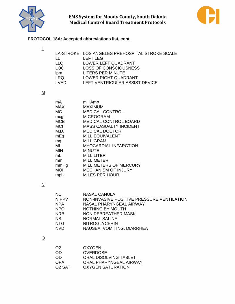
EMS System for Moody County, South Dakota Medical Control Board Treatment Protocols
PROTOCOL 18A: Accepted abbreviations list, cont. L LA-STROKE LOS ANGELES PREHOSPITAL STROKE SCALE LL LEFT LEG LLQ LOWER LEFT QUADRANT LOC LOSS OF CONSCIOUSNESS lpm LITERS PER MINUTE LRQ LOWER RIGHT QUADRANT LVAD LEFT VENTRICULAR ASSIST DEVICE M mA milliAmp MAX MAXIMUM MC MEDICAL CONTROL mcg MICROGRAM MCB MEDICAL CONTROL BOARD MCI MASS CASUALTY INCIDENT M.D. MEDICAL DOCTOR mEq MILLIEQUIVALENT mg MILLIGRAM MI MYOCARDIAL INFARCTION MIN MINUTE mL MILLILITER mm MILLIMETER mmHg MILLIMETERS OF MERCURY MOI MECHANISM OF INJURY mph MILES PER HOUR N NC NASAL CANULA
NIPPV NON-INVASIVE POSITIVE PRESSURE VENTILATION NPA NASAL PHARYNGEAL AIRWAY NPO NOTHING BY MOUTH NRB NON REBREATHER MASK NS NORMAL SALINE NTG NITROGLYCERIN NVD NAUSEA, VOMITING, DIARRHEA
O
O2 OXYGEN OD OVERDOSE ODT ORAL DISOLVING TABLET OPA ORAL PHARYNGEAL AIRWAY O2 SAT OXYGEN SATURATION

EMS System for Moody County, South Dakota Medical Control Board Treatment Protocols
PROTOCOL 18A: Accepted abbreviations list, cont. P PEA PULSELESS ELECTRICAL ACTIVITY PEEP POSITIVE END-EXPIRATORY PRESSURE PEP POST-EXPOSURE PROPHYLAXIS PICC PERIPHERLLY INSERTED CENTRAL CATHETER PO BY MOUTH
PRN AS NEEDED Psi POUNDS PER SQUARE INCH PSVT PAROXYSMAL SUPRAVENTRICULAR TACHYCARDIA Pt PATIENT PVC PREMATURE VENTRICULAR CONTRACTION
PERRL PUPILS EQUAL ROUND REACTIVE TO LIGHT Q QRS THE DEFLECTIONS IN AN ELECTROCARDIOGRAM THAT REPRESENTS ELECTRICAL ACTIVITY GENERATED BY VENTRICULAR DEPOLARIZATION R RA RIGHT ARM RESP RESPIRATIONS RL RIGHT LEG RN REGISTERED NURSE ROSC RETURN OF SPONTANEOUS CIRCULATION RVAD RIGHT VENTRICULAR ASSIST DEVICE S SA SINOATRIAL SBP SYSTOLIC BLOOD PRESSURE SDDOH SOUTH DAKOTA DEPARTMENT OF HEALTH SL SUBLINGUAL SOB SHORTNESS OF BREATH SpCO CARBON MONOXIDE SATURATION OF ARTERIAL BLOOD SpO2 OXYGEN SATURATION OF ARTERIAL BLOOD
ST SINUS TACHYCARDIA STEMI ST SEGMENT ELEVATION MYOCARDIAL INFARCTION SubQ SUBCUTANEOUS SYS BP SYSTOLIC BLOOD PRESSURE

EMS System for Moody County, South Dakota Medical Control Board Treatment Protocols
PROTOCOL 18A: Accepted abbreviations list, cont.
T TB TUBERCULOSIS TBSA TOTAL BODY SURFACE AREA TCA TRICYCLIC ANTIDEPRESSANT TEMP TEMPERATURE TKO TO KEEP OPEN TXA TRANEXAMIC ACID U ULQ UPPER LEFT QUADRANT URQ UPPER RIGHT QUARDANT U.S. UNITED STATES V VAD VENTRICULAR ASSIST DEVICE VF VENTRICULAR FIBRILLATION VS VITAL SIGNS VT VENTRICULAR TACHYCARDIA W WISER WIRELESS INFORMATION SYSTEM FOR EMERGENCY RESPONDERS WPD WARM, PINK, DRY X Y
Z