emphysematous pyelonephritis: our experience...
TRANSCRIPT

Urology Annals | Jul - Sep 2013 | Vol 5 | Issue 3 157
Emphysematous pyelonephritis: Our experience with conservative management in 14 cases
Pramod Kumar Sharma, Ritu Sharma1, Mukesh K. Vijay, Punit Tiwari, Amit Goel, Anup K. KunduDepartment of Urology, Institute of Post Graduate Medical Education and Research, SSKM Hospital, 1Department of Obstetrics and
Gynecology, NRS Medical College, Kolkata, India
Original Article
INTRODUCTION
Emphysematous pyelonephritis (EPN) is a rare, severe, acute
necrotizing infection of the kidney characterized by the presence of gas within the renal parenchyma, collecting system and peri‑renal tissue.[1] EPN is common in diabetics, especially in females. Non‑diabetic patients can also develop EPN, albeit rarely, with a less severe clinical course as compared to diabetics.[2] The term Emphysematous Pyelonephritis was first used by Schultz and Klorfein.[3] In the yesterdays, this condition was associated with high morbidity and mortality but now, with the availability of better modalities, the natural history seems to have changed. We, in this study, present the clinical details and outcome of 14 patients of
Context: Emphysematous pyelonephritis (EPN) is a rare, severe, acute, necrotizing infection of the kidney. In this study, we present the clinical details, the management strategies, and the outcome of fourteen patients of EPN managed at our center.Materials and Methods: A retrospective analysis of the hospital records was done. A total of fourteen patients with EPN were admitted in our hospital from August 2007 to February 2011. All the patients were managed conservatively. Follow-up ranged from six months to one year.Results: Of the fourteen patients, four belonged to class I, five to class II, four to class IIIA and one to class IIIB. All the patients had history of fever, 43% had localized flank pain while 36% had vague abdominal discomfort. Renal angle tenderness was the most common sign, seen in 86% of the patients. E. coli was the most common bacteria, which was isolated from urine in 57% of the patients. On the risk factor stratification, three patients had simultaneous presence of 2 or more risk factors (thrombocytopenia-2 patients; renal function impairment-7 patients; shock-1 patient). All the patients were initially managed with aggressive fluid and electrolyte resuscitation, control of blood sugar levels, and broad spectrum antibiotics. Intervention, in the form of percutaneous drainage or DJ stenting, was done in six patients. One patient failed to respond to this minimally invasive modality of treatment and had to undergo an open drainage. Thus, the acute episode was managed with conservative management strategies in all the patients; however, three patients underwent nephrectomy due to poorly-functioning kidney during follow-up.Conclusions: EPN is now being more readily diagnosed, at an early stage, making conservative management of EPN a safe, effective, and feasible option.
Key Words: Emphysematous pyelonephritis, nephrectomy, renal infection
Abstract
Access this article onlineQuick Response Code:
Website:
www.urologyannals.com
DOI:
10.4103/0974-7796.115734
Address for correspondence: Dr. Pramod Kumar Sharma, Room No. 508, Junior Doctors Hostel, IPGMER and SSKM Hospital, 242, AJC Bose Road, Kolkata ‑ 700 020, India. E‑mail: [email protected]: 04.02.2012, Accepted: 07.04.2012
[Downloaded free from http://www.urologyannals.com on Friday, April 25, 2014, IP: 197.35.251.139] || Click here to download free Android application for this journal

Sharma, et al.: Conservative management of emphysematous pyelonephritis
158 Urology Annals | Jul - Sep 2013 | Vol 5 | Issue 3
EPN managed at our center and discuss their management and outcomes.
MATERIALS AND METHODS
Between August 2007 and February 2011, a total of 14 patients were admitted in our hospital with emphysematous pyelonephrits. All the patients were studied with respect to the clinical features at presentation. Computerized tomography was done in 13 cases to confirm the diagnosis and for classification while 1 patient was referred to us with an MR Urogram. All the patients were thoroughly investigated, and the risk factors (as proposed by Huang and Tseng) were evaluated. All the patients were initially managed by aggressive diabetic control, correction, and maintenance of fluid and hemodynamic status, and antibiotics. The patients were followed up for 6‑12 months.
RESULTS
Out of the fourteen patients with EPN, eleven were females while three were males (Male:female; 1:3.66). Age range was 35‑58 years with a mean age of 51 years. Twelve patients (86%) were diabetic, out of which, nine were known diabetics while three were diagnosed following admission. seven of the known diabetics were on regular treatment while two were non‑compliant; however, all the diabetic patients had raised blood sugar levels at the time of admission. Both the non‑diabetic patients had ureteral obstruction due to stone disease. One of the diabetic patients too had hydronephrosis due to stone in the ureter. The clinical presentation of the patients is given in Table 1. All the fourteen patients (100%) had fever at the time of presentation while localized flank pain was present in six (43%) of the patients. Five (36%) patients had vague abdominal discomfort while nausea with or without vomiting was present in six (43%) patients. Dysuria and increased urinary frequency was seen in four
(29%) patients. None of the patient had altered sensorium or pneumaturia.
On examination, renal angle tenderness was present in twelve (86%) patients while abdominal mass was found in four (29%) patients. Two (14%) patients had diffuse abdominal tenderness while only one had hypotension at the time of presentation. None of our patient had crepitations in the flank.
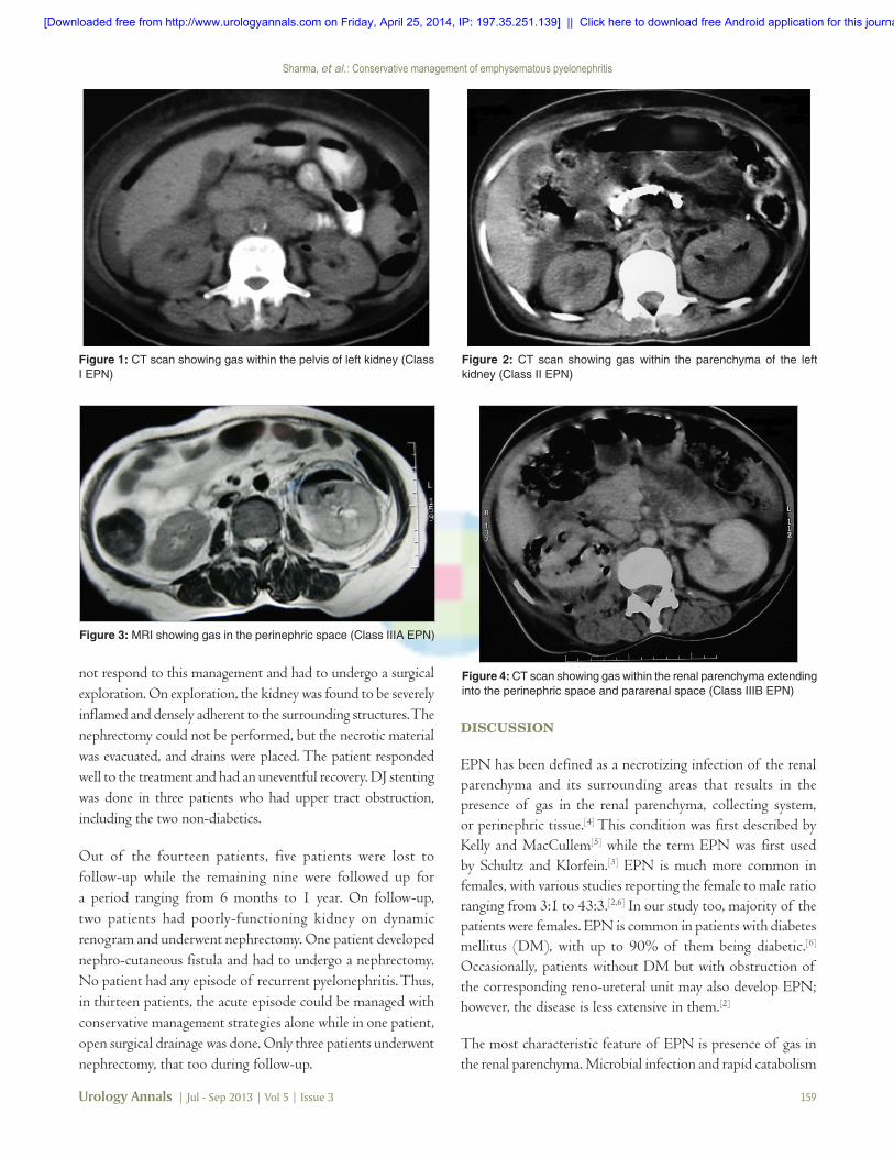
Pyuria was found in all the patients while leucocytosis was found in twelve (86%) patients. Two (14%) had thrombocytopenia while seven had deranged renal parameters at the time of admission. The urine of all the patients was submitted for culture and sensitivity testing. Out of the fourteen patients, E. coli was isolated from eight (57%) patients, Klebsiella from two (14%) and Proteus from one (7%) patient. In three (21%) patients, no bacteria could be isolated from urine. CT scan was performed for confirmation of the diagnosis as well as for classification while one patient was referred to us with an MRI. The distribution of the patients into various classes based on radiological investigation is given in Table 2. The representative images of patients with EPN are shown in Figures 1‑4.
On risk factor stratification, as per the criteria proposed by Huang and Tseng,[4] three patients had simultaneous presence of two or more risk factors (Thrombocytopenia‑2 patients; Renal Function Impairment‑7 patients; Shock‑1 patient).
Management strategies adoptedAfter admission, all the patients were initially managed by aggressive diabetic control, correction, and maintenance of fluid and hemodynamic status, and antibiotics. Initially, broad spectrum antibiotics were used. A combination of piperacillin and tazobactum was used as the first line antibiotic. Aminoglycosides were added in patients who had normal renal parameters while a quinolone was added in patients with deranged renal parameters (with dose adjustment). Antibiotics were changed in accordance with the sensitivity report when it was available. Percutaneous drainage under CT guidance was done in three patients who had more than two risk factors. One patient of class IIIB EPN did
Table 2: Radiological classification of patients (n=14)No. of patients Percentage
According to the classification by Wan et al.
Type I 4 29Type II 10 71
According to the classification by Huang and Tseng
Class I 4 29Class II 5 36Class IIIA 4 29Class IIIB 1 6Class IV 0 0
Table 1: Clinical Features at presentationNo. of patients Percentage
SexMale 3 21Female 11 79
Clinical presentationFever 14 100Flank pain 6 43Vague abdominal discomfort 5 36Nausea and vomitting 6 43Dysuria and frequency 4 29Depressed level of consciousness NonePneumaturia None
SignsAbdominal mass 4 29Renal angle tenderness 12 86Abdominal tenderness 2 14Hypotension 1 7Crepitus in flank region None
[Downloaded free from http://www.urologyannals.com on Friday, April 25, 2014, IP: 197.35.251.139] || Click here to download free Android application for this journal
Sharma, et al.: Conservative management of emphysematous pyelonephritis
Urology Annals | Jul - Sep 2013 | Vol 5 | Issue 3 159
not respond to this management and had to undergo a surgical exploration. On exploration, the kidney was found to be severely inflamed and densely adherent to the surrounding structures. The nephrectomy could not be performed, but the necrotic material was evacuated, and drains were placed. The patient responded well to the treatment and had an uneventful recovery. DJ stenting was done in three patients who had upper tract obstruction, including the two non‑diabetics.
Out of the fourteen patients, five patients were lost to follow‑up while the remaining nine were followed up for a period ranging from 6 months to 1 year. On follow‑up, two patients had poorly‑functioning kidney on dynamic renogram and underwent nephrectomy. One patient developed nephro‑cutaneous fistula and had to undergo a nephrectomy. No patient had any episode of recurrent pyelonephritis. Thus, in thirteen patients, the acute episode could be managed with conservative management strategies alone while in one patient, open surgical drainage was done. Only three patients underwent nephrectomy, that too during follow‑up.
DISCUSSION
EPN has been defined as a necrotizing infection of the renal parenchyma and its surrounding areas that results in the presence of gas in the renal parenchyma, collecting system, or perinephric tissue.[4] This condition was first described by Kelly and MacCullem[5] while the term EPN was first used by Schultz and Klorfein.[3] EPN is much more common in females, with various studies reporting the female to male ratio ranging from 3:1 to 43:3.[2,6] In our study too, majority of the patients were females. EPN is common in patients with diabetes mellitus (DM), with up to 90% of them being diabetic.[6] Occasionally, patients without DM but with obstruction of the corresponding reno‑ureteral unit may also develop EPN; however, the disease is less extensive in them.[2]
The most characteristic feature of EPN is presence of gas in the renal parenchyma. Microbial infection and rapid catabolism
Figure 1: CT scan showing gas within the pelvis of left kidney (Class I EPN)
Figure 2: CT scan showing gas within the parenchyma of the left kidney (Class II EPN)
Figure 4: CT scan showing gas within the renal parenchyma extending into the perinephric space and pararenal space (Class IIIB EPN)
Figure 3: MRI showing gas in the perinephric space (Class IIIA EPN)
[Downloaded free from http://www.urologyannals.com on Friday, April 25, 2014, IP: 197.35.251.139] || Click here to download free Android application for this journal

Sharma, et al.: Conservative management of emphysematous pyelonephritis
160 Urology Annals | Jul - Sep 2013 | Vol 5 | Issue 3
have been proposed as the cause for increased gas formation, which is trapped in the tissues due to impaired transport of gas as there is vascular compromise in the pyelonephritic kidney. Huang and Tseng have postulated that 4 factors are involved in the pathogenesis of EPN, which were gas‑forming bacteria, high tissue glucose level, impaired tissue perfusion, and a defective immune response.[4] Leukocyte dysfunction seen in diabetics may contribute to the pathogenesis of EPN. Gas production in the renal parenchyma in the absence of infection has also been described following traumatic renal infarction.[7] The infecting organisms are usually glucose‑fermenting bacteria. E. coli is the most common bacteria implicated in EPN, others are Klebsiella and Proteus. EPN caused by Streptococcus and Candida have also been reported.[8]
The clinical presentation is often suggestive of severe acute pyelonephritis, with fever, flank pain, and pyuria being the most common clinical manifestations. However, these are non‑specific and may be seen in other forms of upper urinary tract infections. Other clinical features of EPN include non‑specific abdominal pain, nausea, vomiting, depressed levels of consciousness, shock, renal angle tenderness, dysuria, crepitations in the flanks, and pneumaturia.[2‑4,6,9] In our study too, fever and flank pain were the most common symptom. Pyuria was also seen in all the cases. Huang and Tseng[4] in their study had found that thrombocytopenia (46%), acute renal function impairment (35%), disturbance of consciousness (19%), and shock (29%) can be the initial presentations. Shokier et al.[6] in their study found renal functional impairment in 80% of their patients and shock and coma in 15% of patients. In our study, only 14% of the patients had thrombocytopenia, 50% had deranged renal functions, and only 1 patient had hypotension.
Diagnosis of EPN is made radiologically, with CT being the most definitive modality. EPN can also be diagnosed by abdominal X‑ray and ultrasonography. CT images are useful to define the presence, extent, and position of gas within the renal parenchyma, beside any other associated renal pathology, like calculi, and/or presence of obstruction may also be evident on CT scan. Contrast‑enhanced CT scan is better as it give an idea about the function status of the renal units as well as it facilitates the description of the intraparenchymal gas (streaky, mottled, bubbly, rimlike, crescent shaped, locular, and so on). However, in patients with deranged renal parameters, a non‑contrast CT scan may suffice. In addition to diagnosis and staging of EPN, CT scan is also helpful in monitoring the response to treatment. It might show resolution of the gas and abscesses or the development of new lesions.
Abdominal X‑ray may reveal mottled gas shadow in the renal
region followed by development of a crescent of gas surrounding parenchyma. In the absence of CT facilities, Intravenous Urography (IVU) can be used; however, quite a few EPN patients are in shock or have elevated serum creatinine levels; therefore, IVU cannot be used in such patients. The sensitivity of IVU has been reported as 85% in a study by Paivansalos et al.[10] and 100% by Ahlering and colleagues.[11] Ultrasound reveals strong focal echoes, typically described as ‘dirty shadows’. The sensitivity and specificity of ultrasonography in EPN is low. Obesity and bowel gas interfere with the interpretation of plain X‑Ray, IVU as well as ultrasonography.
Staging of EPN is done radiologically based on the extent of gas in the renal parenchyma and surrounding tissues. It might be useful for decision making and prognostication. Langston and Pfister[12] suggested a classification on the basis of abdominal X‑ray and an intravenous pyelography, which was later modified by Michaeli et al.[13] They classified EPN into 3 classes:Class I ‑ Gas in renal parenchyma or perinephric tissueClass II ‑ Gas in the kidney and its surroundingsClass III ‑ Extension of gas through fascia, or bilateral disease.
Wan et al.[1] classified the gas collection as type I or type II, on the basis of CT scans.Type I: Renal necrosis with presence of gas but no fluidType II: Parenchymal gas associated with fluid in renal
parenchyma, perinephric space, or collecting system.
Mortality was 69% in patients with type I EPN and only 18% in patients with type II EPN. Similar mortality rates for type I and type II EPN were observed by Chen et al.[14]
Huang and Tseng[4] also used CT to classify patients with EPN as follows:Class I: Gas in collecting system onlyClass II: Parenchymal gas onlyClass IIIA: Extension of gas into perinephric spaceClass IIIB: Extension of gas into pararenal spaceClass IV: EPN in solitary kidney, or bilateral disease.
The classification by Huang and Tseng is a superior due to the better prognostic value and is also helpful in selecting a management protocol. In their study, class I and II patients, all survived following treatment with percutaneous procedures and medical therapy. While in patients belonging to class III or IV, those with fewer than two risk factors (i.e. thrombocytopenia, acute renal function impairment, disturbance of consciousness and shock) had an 85% survival rate with percutaneous drainage and medical therapy, whereas patients of class III or IV EPN and two or more risk factors had a 92% failure rate with percutaneous drainageand medical therapy. In their study,
[Downloaded free from http://www.urologyannals.com on Friday, April 25, 2014, IP: 197.35.251.139] || Click here to download free Android application for this journal

Sharma, et al.: Conservative management of emphysematous pyelonephritis
Urology Annals | Jul - Sep 2013 | Vol 5 | Issue 3 161
33.33% of the patients belonged to class I or II while 66.66% of the patients belonged to class III or IV; while in our study, majority of the patients (64%) belonged to either class I or II while 36% of the patients belonged to class III or IV.
Despite the morbidity and mortality associated with EPN, there is still controversy regarding its proper management. Shokier et al.[6] and Ahlering et al.[11] proposed immediate nephrectomy following resuscitation of the patient. Huang and Tseng[4] proposed the management protocol based on the radiological classification and presence of risk factors. They managed class I and II EPN with antibiotics along with percutaneous drainage or relief of obstruction while those with class III or IV EPN were given a trial of conservative management, and nephrectomy was done in patients who had more than 2 risk factors for poor prognosis or in patients in whom conservative management failed. There has been a recent trend towards conservative treatment strategies for EPN. A number of reports of successful conservative management of EPN have come up.[14‑22]
Initial treatment of patients with EPN should start with vigorous resuscitation. Fluid and electrolyte imbalances should be corrected, Diabetes should be controlled, and antibiotics should be started. Broad spectrum antibiotics targeting gram‑negative bacteria should be star ted. Aminoglycosides should be used with caution as quite a few patients may have deranged renal functions. Patients who have obstruction of the renal drainage system should have their obstruction relieved. DJ stenting may be tried in such patients. Percutaneous drainage under CT/USG guidance is done in patients who have well‑formed collections within the renal parenchyma or the surrounding tissue. Nephrectomy should be considered in a select group of patients who have gross destruction of renal parenchyma, have class IIIA or class IIIB gas distribution when there is simultaneous presence of 2 or more risk factors or when the involved kidney is non‑functioning.[2] Jayesh V Dhabalia et al.[23] in their study similarly found that EPN can be successfully managed with conservative treatment modalities, and the such treatment strategies are associated with lower mortalities than emergency nephrectomies.
In our study, we started with conservative management, and reserved nephrectomy for patients who did not respond to conservative management. One patient in our study failed conservative management and was planned for nephrectomy; however, only open drainage could be done due marked inflammation and adhesion. The patient responded well to open drainage. Thus, minimally invasive modalities were successful for emergency management of most of the patients.
CONCLUSION
EPN is a rare and severe infection of the renal parenchyma. However, with increasing availability and decrease in the threshold for using imaging modalities like ultrasonography and CT scan in patients with severe urinary tract infections and sepsis, more number of patients are now diagnosed with emphysematous pyelonephritis and at an early stage. With early diagnosis, availability of more potent antibiotics and advances in critical care support systems, an increasing number of patients can be managed with conservative approaches, and nephrectomy should be reserved for patients who fail conservative management, especially those belonging to higher radiological class with simultaneous presence of two or more risk factors.
REFERENCES
1. Wan YL, Lee TU, Bullard MJ, Tsai CC. Acute gas‑producing bacterial renal infection: correlation between imaging findings and clinical outcome. Radiology 1996;198:433‑8.
2. Pontin AR, Barnes RD. Current management of emphysematous pyelonephritis. Nat Rev Urol 2009;6:272‑9.
3. Schultz EH Jr, Klorfein EH. Emphysematous Pyelonephritis. J Urol 1962;87:762‑6.
4. Huang JJ, Tseng CC. Emphysematous pyelonephritis: clinicoradiological classification, management, prognosis, and pathogenesis. Arch Intern Med 2000;160:797‑805.
5. Kelly HA, MacCullem WG. Pneumaturia. JAMA 1898;31:375.6. Shokeir AA, EL‑Azab M, Mohsen T, El‑Diasty T. Emphysematous
pyelonephrit is: A 15‑year experience with 20 cases. Urology 1997;49:343‑6.
7. Subramanyam BR, Lefleur RS, Van Natta FC. Renal emphysema secondary to traumatic renal infarction. Urol Radiol 1980;2:53‑4.
8. Hildebrand TS, Nibbe L, Frei U, Schindler R. Bilateral emphysematous pyelonephritis caused by candida infection. Am J Kidney Dis 1999;33:E10.
9. Kumar VS, Lakshmi AY. Emphysematous pyelonephritis. Indian J Nephro 2004;14:192‑4.
10. Päivänsalo M, Hellström P, Sinihroto T, Leinonen A. Emphysematous pyelonephritis. Radiologic and clinical findings in six cases. Acta Radiol 1989;30:311‑5.
11. Ahlering TE, Boyd SD, Hamilton CL, Bragin SD, Chandramma PT, Lieskovsky G, et al. Emphysematous pyelonephritis. A 5‑year experience with 13 patients. J Urol 1985;134:1086‑8.
12. Langston CS, Pfister RC. Renal emphysema. A case report and review of the literature. Am J Roentgenol Radium Ther Nucl Med 1970;110:778‑86.
13. Michaeli J, Mogle P, Perlberg S, Herman S, Caine M. Emphysematous pyelonephritis. J Urol 1984;131:203‑8.
14. Chen MT, Huang CN, Chou YH, Huang CH, Chiang CP, Liu GC. Percutaneous drainage in the treatment of emphysematous pyelonephritis: 10‑year experience. J Urol 1997;157:1569‑73.
15. Godec CJ, Cass AS, Berkseth R. Emphysematous pyelonephritis in a solitary kidney. J Urol 1980;124:119‑21.
16. DePauw AP, Ross G Jr. Emphysematous pyelonephritis in a solitary kidney. J Urol 1981;125:734‑6.
17. Hudson MA, Weyman PJ, Van der Vliet AH, Catalona WJ. Emphysematous pyelonephritis: successful management by percutaneous drainage. J Urol 1986;136:884‑6.
18. Zagoria RJ, Dyer RB, Harrison LH, Adams PL. Percutaneous management of localized emphysematous pyelonephritis. J Vasc Interv Radiol 1991;2:156‑8.
19. Hall JR, Choa RG, Wells IP. Percutaneous drainage in emphysematous pyelonephritis–an alternative to major surgery. Clin Radiol 1988;39:622‑4.
[Downloaded free from http://www.urologyannals.com on Friday, April 25, 2014, IP: 197.35.251.139] || Click here to download free Android application for this journal
Sharma, et al.: Conservative management of emphysematous pyelonephritis
162 Urology Annals | Jul - Sep 2013 | Vol 5 | Issue 3
20. Corr J, Glesson M, Wilson G, Grainger R. Percutaneous management of emphysematous pyelonephritis. Br J Urol 1993;71:487‑8.
21. Koh KB, Lam HS, Lee SH. Emphysematous pyelonephritis: drainage or nephrectomy? Br J Urol 1993;71:609‑11.
22. Cardinael AS, De Blay V, Gilbeau JP. Emphysematous pyelonephritis: successful treatment with percutaneous drainage. AJR Am J Roentgenol 1995;164:1554‑5.
23. Dhabalia JV, Nelivigi GG, Kumar V, Gokhale A, Punia MS, Pujari N.
How to cite this article: Sharma PK, Sharma R, Vijay MK, Tiwari P, Goel A, Kundu AK. Emphysematous pyelonephritis: Our experience with conservative management in 14 cases. Urol Ann 2013;5:157-62.
Source of Support: Nil, Conflict of Interest: None.
Emphysematous pyelonephritis: Tertiary care center experience in management and review of the literature. Urol Int 2010;85:304‑8.
Author Help: Reference checking facility
The manuscript system (www.journalonweb.com) allows the authors to check and verify the accuracy and style of references. The tool checks the references with PubMed as per a predefined style. Authors are encouraged to use this facility, before submitting articles to the journal.
• The style as well as bibliographic elements should be 100% accurate, to help get the references verified from the system. Even a single spelling error or addition of issue number/month of publication will lead to an error when verifying the reference.
• Example of a correct style Sheahan P, O’leary G, Lee G, Fitzgibbon J. Cystic cervical metastases: Incidence and diagnosis using fine needle aspiration biopsy.
Otolaryngol Head Neck Surg 2002;127:294-8. • Only the references from journals indexed in PubMed will be checked. • Enter each reference in new line, without a serial number.• Add up to a maximum of 15 references at a time.• If the reference is correct for its bibliographic elements and punctuations, it will be shown as CORRECT and a link to the correct
article in PubMed will be given.• If any of the bibliographic elements are missing, incorrect or extra (such as issue number), it will be shown as INCORRECT and link to
possible articles in PubMed will be given.
[Downloaded free from http://www.urologyannals.com on Friday, April 25, 2014, IP: 197.35.251.139] || Click here to download free Android application for this journal