emergency physicianperformed ultrasound to diagnose cholelithiasis: a … · progressive clinical...
TRANSCRIPT

PROGRESSIVE CLINICAL PRACTICE
Emergency Physician–performed Ultrasoundto Diagnose Cholelithiasis: A SystematicReviewMarshall Ross, Michael Brown, MD, MSc, Kyle McLaughlin, MD, CCFP-EM,Paul Atkinson, MB, BCh, BAO, MA, MRCP(UK), FCEM, Jenny Thompson,Susan Powelson, MLS, Steve Clark, MD, CCFP-EM, and Eddy Lang, MD
AbstractObjectives: The authors sought to determine the diagnostic test characteristics of bedside emergencyphysician (EP)-performed ultrasound (US) for cholelithiasis in symptomatic emergency department (ED)patients.
Methods: A search was conducted of MEDLINE, EMBASE, the Cochrane Library, bibliographies of pre-vious systematic reviews, and abstracts from major emergency medicine conference proceedings. Weincluded studies that prospectively assessed the diagnostic accuracy of emergency US (EUS) for choleli-thiasis, compared to a criterion reference standard of radiology-performed ultrasound (RADUS), com-puted tomography (CT), magnetic resonance imaging (MRI), or surgical findings. Two authorsindependently performed relevance screening of titles and abstracts, extracted data, and performed thequality analysis. Disagreements were resolved by conference between the two reviewers. EUS perfor-mance was assessed with summary receiver operator characteristics curve (SROC) analysis, with inde-pendently pooled sensitivity and specificity values across included studies.
Results: The electronic search yielded 917 titles; eight studies met the inclusion criteria, yielding a sam-ple of 710 subjects. All included studies used appropriate selection criteria and reference standards, butonly one study reported uninterpretable or indeterminate results. The pooled estimates for sensitivityand specificity were 89.8% (95% confidence interval [CI] = 86.4% to 92.5%) and 88.0% (95% CI = 83.7%to 91.4%), respectively.
Conclusions: This study suggests that in patients presenting to the ED with pain consistent with biliarycolic, a positive EUS scan may be used to arrange for appropriate outpatient follow-up if symptoms haveresolved. In patients with a low pretest probability, a negative EUS scan should prompt the clinician toconsider an alternative diagnosis.
ACADEMIC EMERGENCY MEDICINE 2011; 18:227–235 ª 2011 by the Society for Academic EmergencyMedicine
I t is estimated that over 20 million people in the Uni-ted States have gallbladder disease.1 Approximatelyone-third of patients with gallstones develop biliary
colic,2–4 and each year about 1% of these symptomaticpatients develop potentially life-threatening complica-tions including acute cholecystitis, pancreatitis, and
acute cholangitis.5 Unfortunately, it is not possible todiagnose gallbladder pathology with a reasonabledegree of certainty using history and physical examalone.6 The unreliability of clinical findings commonlyleads to patients with suspected gallbladder diseaseundergoing radiology-performed ultrasound (RADUS).
ª 2011 by the Society for Academic Emergency Medicine ISSN 1069-6563doi: 10.1111/j.1553-2712.2011.01012.x PII ISSN 1069-6563583 227
From the School of Medicine (MR, JT) and the Department of Emergency Medicine (KM, SC, EL), University of Calgary, Calgary,Alberta, Canada; the Division of Emergency Medicine, College of Human Medicine, Michigan State University (MB),Grand Rapids, MI; the Department of Emergency Medicine, Dalhousie University (PA), Halifax, Nova Scotia, Canada; Saint JohnRegional Hospital (PA), Saint John, New Brunswick, Canada; the Health Sciences Library, University of Calgary (SP), Calgary,Alberta, Canada; and Alberta Health Services (SC, EL), Calgary, Alberta, Canada.Received July 9, 2010; revision received August 30, 2010; accepted August 31, 2010.Presented at the American College of Emergency Physicians scientific assembly, Las Vegas, NV, September 2010.The authors have no relevant financial information or potential conflicts of interest to disclose.Supervising Editor: Shahriar Zehtabchi, MD.Address for correspondence and reprints: Marshall Ross; e-mail: [email protected].

These RADUS are typically performed by a technicianoutside of the emergency department (ED) and are inter-preted by a radiologist. However, inconsistent availabil-ity of technicians and high volumes of ED patients oftencause delays to RADUS, extending ED wait times, com-promising quality of care, and contributing to through-put delays and crowding.7,8
These limitations have led to emergency physicians(EPs) exploring the prospect of performing bedsideemergency ultrasound (EUS) in the ED to assess forgallbladder pathology.9–11 EUS enables the operator tovisualize the gallbladder. Stones are typically identifiedas bright, mobile, and dependent structures that cast ashadow. False negatives are most commonly due tostones that are either less than 4 mm in diameter andthus do not consistently cast a shadow or impacted inthe gallbladder neck as they are no longer dependent.12
Whereas the use of EUS for certain core ED applica-tions, such as screening for abdominal aortic aneurysm,or detecting large pericardial effusions, is supported bystrong evidence,13,14 the use of EUS for many otherindications remains controversial due to concernsabout the diagnostic accuracy. The goal of this system-atic review was to assess the diagnostic accuracy ofEUS in detecting gallstones among ED patients whopresent with symptoms consistent with biliary colic.
METHODS
The clinical question addressed in this systematic reviewwas the following: Among patients presenting to the EDwith abdominal pain of suspected biliary origin, what isthe diagnostic accuracy of EP-performed ultrasound(US) for the diagnosis of acute cholelithiasis? A system-atic review protocol was created to specifically addressthis question and was reviewed and agreed upon by allco-investigators a priori (available online15). The onlyamendment made to the protocol was to expand ourpossible criterion reference standard beyond RADUSalone. The PRISMA statement16 was followed for thepurpose of reporting this systematic review.
Search TechniquesWe developed a comprehensive search strategy thatincluded foreign language publications. The MEDLINEsearch used a combination of MeSH headings and freetext terms (Data Supplement S1, available as supportinginformation in the online version of this paper). Similarsearch strategies were adapted for EMBASE, OpenSI-GLE, and the Cochrane Library. Our gray literaturesearch included manually reviewing abstracts from thefollowing conferences: Canadian Association of Emer-gency Physicians (2000–2009), American College ofEmergency Physicians (ACEP) (2000–2009), Society forAcademic Emergency Medicine (2000–2009), Mediterra-nean Congress of Emergency Medicine (2001–2009),Annual Scientific Meeting of the Australasian Collegefor Emergency Medicine (2001–2009), and all availableabstracts from the College of Emergency Medicine, for-merly known as the British Association and Faculty ofEmergency Medicine (2000–2009). In addition, the refer-ences for the ACEP Policy Statement on EmergencyUltrasound Guidelines9 were screened. We contacted
all major US manufacturers, the Society for AcademicEmergency Medicine Ultrasound Interest Group, sev-eral independent experts in the field, and the CanadianEmergency Ultrasound Society and requested addi-tional unpublished data sources. The bibliographies ofall included studies17–24 as well as those of previousreviews25,26 were hand-searched.
Article SelectionSpecific inclusion and exclusion criteria were applied tominimize bias in the selection process and ensure thatany inferences made based upon the results of themeta-analysis were appropriate and applicable to theED setting.27 We included studies with patients whopresented to the ED with signs and symptoms of sus-pected biliary colic including right upper quadrant(RUQ) pain, epigastric pain, or flank pain. We includedstudies if they used an acceptable criterion referencestandard defined as at least one of the following: RA-DUS, computed tomography (CT), magnetic resonanceimaging (MRI) interpreted by a radiologist, or surgicalfindings. If other reference standards were used, stud-ies were included only if data were obtained comparingresults of EUS to at least one of the criterion standards.We included studies assessing the use of EUS for multi-ple purposes only if data comparing EUS to an appro-priate reference standard to detect cholelithiasis werepresented or obtained through author contact. We onlyincluded studies deemed original research using pro-spective data collection. We excluded studies if perti-nent data could not be obtained through authorcontact.
Data Collection and ProcessingTwo reviewers (MR, JT) independently performed a rel-evance search; each reviewer examined the titles andabstracts of all references identified in the electronicsearch to determine whether an article was relevant tothe research question. The two reviewers then com-pared their inclusion and exclusion logs, and the levelof agreement was calculated using the kappa statistic.The same two reviewers (MR, JT) then performed a fullreview of all potentially relevant articles applying spe-cific inclusion and exclusion criteria to determine whichpapers to include in the final analysis.
Data were extracted and collected from includedstudies using a standardized data collection form. Anattempt was made to contact the authors of studies thatrequired data clarification or supplementation. Tworeviewers (MR, EL) independently abstracted the data.Disagreements were resolved by conference betweenthe reviewers.
Quality AssessmentTwo reviewers with experience in critical appraisal(KM, EL) independently assessed the quality of thestudies according to the most relevant items from theQUADAS tool.28 QUADAS was developed as an evi-dence-based quality assessment tool to be used in sys-tematic reviews to assess the quality of primary studiesof diagnostic accuracy.28 Although all 14 points of theQUADAS instrument were assessed, seven were con-sidered to be of particular relevance to our clinical
228 Ross et al. • BEDSIDE US TO DIAGNOSE CHOLELITHIASIS

question: 1) was the spectrum of patients representativeof the patients who will receive the test in practice,2) were selection criteria clearly described, 3) is thereference standard likely to correctly classify the targetcondition, 4) were the reference standard results inter-preted without knowledge of the results of the indextest, 5) were the index test results interpreted withoutknowledge of the results of the reference standard,6) did patients receive the same reference standardregardless of the index test result, and 7) were uninter-pretable or intermediate test results reported?
No attempt was made to combine the results into anoverall quality score; instead, the results of the assess-ment were reported in a summary figure. Ultrasoundtechnology used in each study was assessed indepen-dently from other methods and results.
Data AnalysisEmergency US test performance was assessed with atraditional summary receiver operator curve (SROC)with a regression model based on unweighted leastsquares estimation.29,30 We independently pooled thesensitivity and specificity with 95% confidence intervals(CIs) using the random effects model of DerSimonianand Laird.31 All calculations were done with the Meta-Disc software.32 We estimated likelihood ratios (LRs)based on our summary estimates of sensitivity andspecificity.27
One of the principal causes of heterogeneity betweenstudies of diagnostic test accuracy is a threshold effect.A threshold effect is present when differences in sensi-tivities and specificities among studies occur due to dif-ferent cutoffs or test thresholds used to define apositive or negative result.27,33 EUS exams may have animplicit variation in threshold as a positive or negativeresult depends on operator interpretation.27 Weassessed for a threshold effect using a validated regres-sion model (see Data Supplement S2 for technicaldetails, available as supporting information in theonline version of this paper).
The random effects model of DerSimonian andLaird31 accounts for sampling variability and unex-plained heterogeneity providing suitably conservativeestimates of sensitivity and specificity.27 Formal statisti-cal tests for assessing heterogeneity and publicationbias in diagnostic meta-analysis have not been vali-dated.27,33,34 Therefore, funnel plots and statistical testssuch as I2 were not reported.
RESULTS
Figure 1 summarizes the results of the search and arti-cle selection. The relevance screen of 917 titles identi-fied in the MEDLINE, Cochrane, OpenSIGLE, andEMBASE databases showed good agreement betweenthe two reviewers (j = 0.67). Expert contact and rele-vance screening of gray literature and conference pro-ceedings yielded seven potentially relevant articles.After the relevance search was adjudicated, a completearticle review was performed on the remaining 33 arti-cles. Upon full-text review, 25 studies did not meet ourinclusion criteria (j = 1) for various reasons asdescribed in Figure 1. One article retrieved through the
manual search,17 one article retrieved through expertcontact,19 and six articles18,20–24 from the electronicdatabase search were included in the final analysis.Seven articles17,18,20–24 were written in English and onearticle was available only in Korean.19 This article wastranslated into English for full review. These eight stud-ies yielded a sample of 710 subjects who underwentboth EUS and a criterion reference standard. Thesensitivity and specificity of each study are presented inFigure 2 and Table 1.
Study DescriptionTable 2 outlines the characteristics of the eight includedstudies. The prevalence of gallstones ranged from 46%to 80%, with a median of 60%. In five of the eight stud-ies selected, assessment of the accuracy of EUS todetect gallstones was a primary outcome measure.
The primary focus of the article by Davis et al.18 wasto determine the association between operator confi-dence and the accuracy of EUS using different criterionstandards for each of six different US examinations.The primary goal of the study by Summers et al.24 wasto determine the test characteristics of EUS as well asRADUS for detection of acute cholecystitis, not choleli-thiasis.24 This study compared the outcomes of bothEUS and RADUS to clinical follow-up or surgicalpathology for 189 patients. The primary focus of thestudy by Rowland et al.23 was to determine the accu-racy of 9 different EUS exams including a RUQ scan todetect for the presence of gallstones.23
There was significant variability in the level of USeducation and previous experience with RUQ US acrossstudies. Davis et al.,18 Rowland et al.,23 and Ha et al.19
used admittedly naïve operators; Alexander et al.17 didnot report operator experience; and the other fourstudies included operators with a wide range of experi-ence. In all studies, EUS was performed with a USmachine of acceptable quality that is regularly used inemergency practice (Table 2).
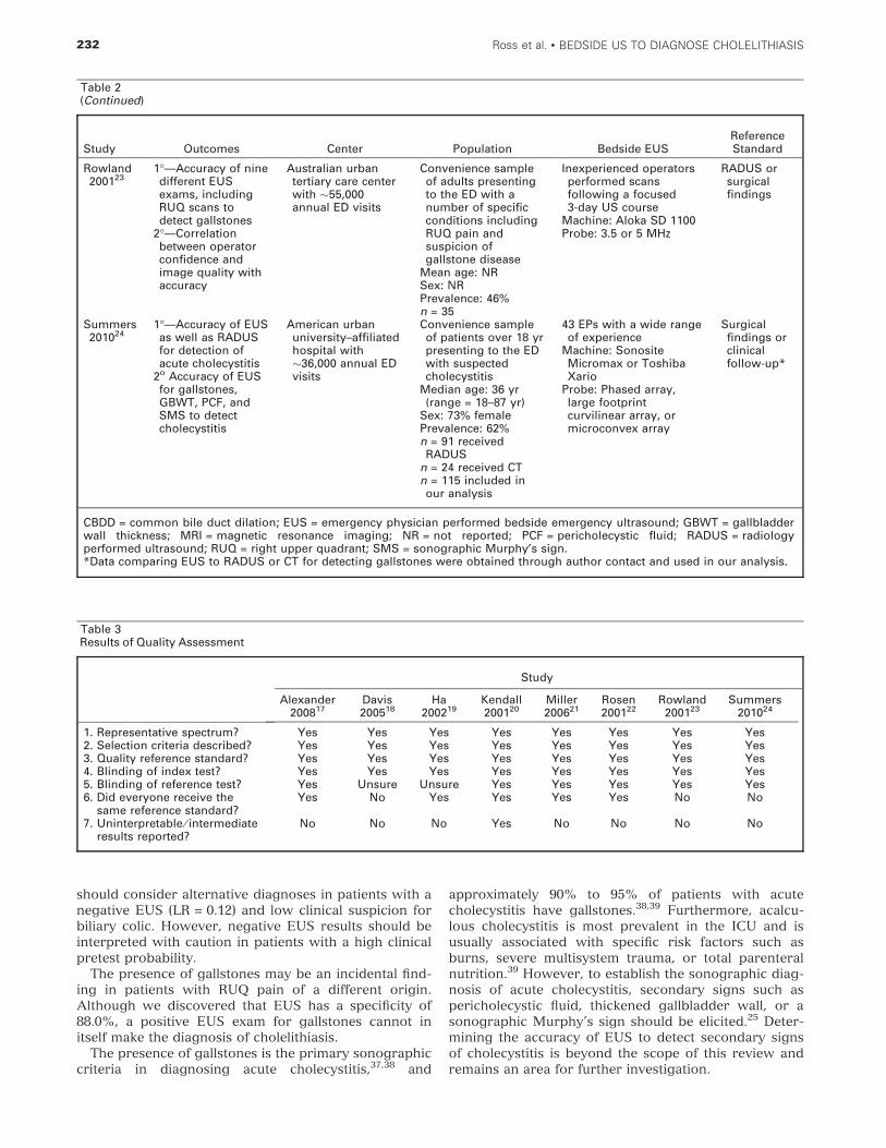
Quality AssessmentTable 3 summarizes the seven key quality items. Theinclusion criteria required that all eight studies be rated
Figure 1. Results of the search strategy.
ACAD EMERG MED • March 2011, Vol. 18, No. 3 • www.aemj.org 229

‘‘yes’’ for having an acceptable reference standard andselection criteria. Seven studies17,18,20–24 adequatelydescribed a representative patient spectrum. Ha et al.19
did not describe their patient population in sufficientdetail. In five studies,17,19–22 all patients underwent RA-DUS as the only reference standard. Davis et al.18 usedUS, CT, MRI, or surgical findings as a criterion refer-ence. Rowland et al.23 used RADUS or laparotomy as acriterion reference. Summers et al.24 provided data for115 of 189 patients who underwent formal radiologytesting; 91 were tested with RADUS and 24 with CT.Only one study reported indeterminate results.20 In sixstudies,17,20–24 the RADUS was interpreted without theknowledge of EUS findings. In two studies,18,19 this wasnot adequately reported. In all studies, the EUS wasperformed and interpreted without knowledge of theresults of the RADUS.
Data AnalysisAccording to the traditional SROC curve analysis,30
there was no evidence of a significant threshold effect(b = )0.072, 95% CI = )0.42 to 0.56); in other words,there was no implicit variation in sensitivity and speci-ficity across studies due to operator-dependent differ-ences in what defined a positive or negative test result.The random effects pooled results for sensitivity andspecificity were 89.8% (95% CI = 86.4% to 92.5%)and 88.0% (95% CI = 83.7% to 91.4%), respectively.
According to these summary estimates, the positive LRwas 7.5 and the negative LR was 0.12. The SROC curveis presented in Figure 3. The small size and number ofstudies precluded any meaningful subgroup analysis.
DISCUSSION
It is estimated that over 20 million people in the UnitedStates have gallbladder disease,1 and nearly 1 in 10asymptomatic individuals with gallstones will requiretreatment within 5 years.35 Despite the high prevalenceof the disease, the clinical diagnosis is still problematicand often relies on a RADUS performed in the radiol-ogy department. The use of RADUS is limited by incon-sistent availability in smaller community settings.Benefits of using EUS to assess the RUQ include itsportability, decreased costs,36 and ability to decreasethe ED length of stay.8
Based on the results of this meta-analysis, a positiveEUS (LR = 7.5) in patients presenting with a clinical pic-ture consistent with a high probability of biliary colicmay be sufficient to make the provisional diagnosis ofacutely symptomatic cholelithiasis and arrange forappropriate follow-up if the symptoms resolve in theED. For example, if we were to assume a pretest proba-bility of 60% (the median prevalence across studies inthis review), a positive EUS provides a posttest proba-bility of 92%. At the other end of the spectrum, the EP
Figure 2. Forest plot of individual study and pooled sensitivity and specificity.
Table 1Individual Study and Pooled Sensitivity and Specificity
Study TP FP TN FN n Sensitivity (95% CI) Specificity (95% CI)
Alexander 200817 24 1 21 4 50 0.86 (0.67–0.96) 0.95 (0.77–1.00)Davis 200518 50 6 37 12 105 0.81 (0.69–0.90) 0.86 (0.72–0.95)Ha 200219 16 2 40 1 59 0.94 (0.71–1.00) 0.95 (0.84–0.99)Kendall 200120 49 7 51 2 109 0.96 (0.87–1.00) 0.88 (0.77–0.95)Miller 200621 95 1 25 6 127 0.94 (0.88–0.97) 0.96 (0.80–1.00)Rosen 200122 60 10 35 5 110 0.92 (0.83–0.97) 0.78 (0.63–0.89)Rowland 200123 12 3 16 4 35 0.75 (0.48–0.93) 0.84 (0.60–0.97)Summers 201024 63 6 38 8 115 0.89 (0.79–0.95) 0.86 (0.73–0.95)Pooled 710 0.90 (0.86–0.93) 0.88 (0.84–0.91)
FN = false negative; FP = false positive; n = number of subjects; TN = true negative; TP = true positive.
230 Ross et al. • BEDSIDE US TO DIAGNOSE CHOLELITHIASIS
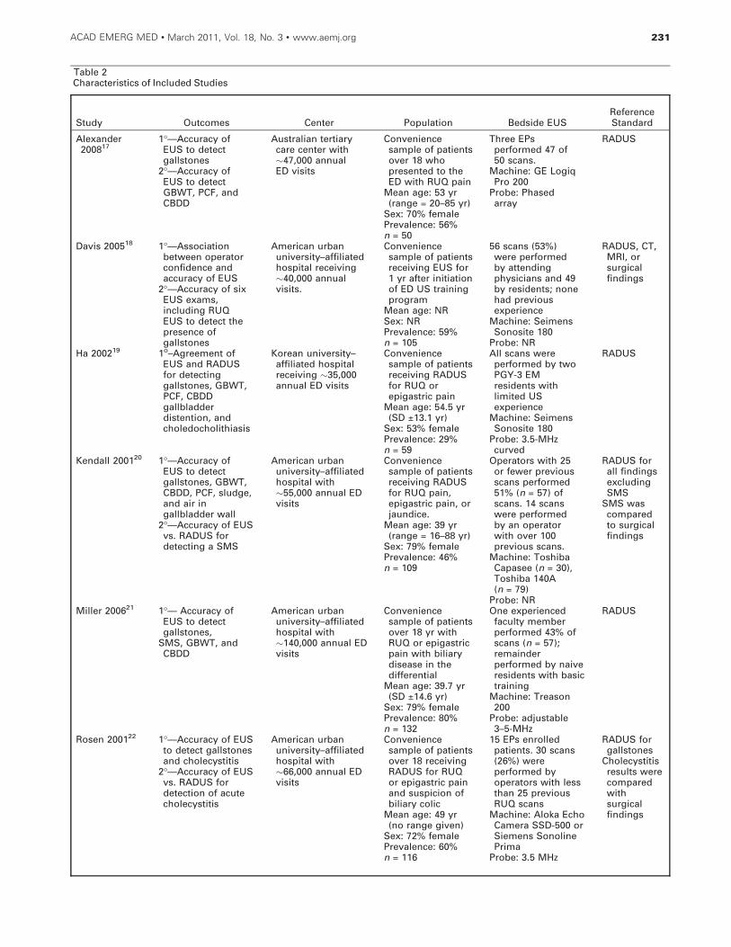
Table 2Characteristics of Included Studies
Study Outcomes Center Population Bedside EUSReferenceStandard
Alexander200817
1!—Accuracy ofEUS to detectgallstones
2!—Accuracy ofEUS to detectGBWT, PCF, andCBDD
Australian tertiarycare center with!47,000 annualED visits
Conveniencesample of patientsover 18 whopresented to theED with RUQ pain
Mean age: 53 yr(range = 20–85 yr)
Sex: 70% femalePrevalence: 56%n = 50
Three EPsperformed 47 of50 scans.
Machine: GE LogiqPro 200
Probe: Phasedarray
RADUS
Davis 200518 1!—Associationbetween operatorconfidence andaccuracy of EUS
2!—Accuracy of sixEUS exams,including RUQEUS to detect thepresence ofgallstones
American urbanuniversity–affiliatedhospital receiving!40,000 annualvisits.
Conveniencesample of patientsreceiving EUS for1 yr after initiationof ED US trainingprogram
Mean age: NRSex: NRPrevalence: 59%n = 105
56 scans (53%)were performedby attendingphysicians and 49by residents; nonehad previousexperience
Machine: SeimensSonosite 180
Probe: NR
RADUS, CT,MRI, orsurgicalfindings
Ha 200219 1o–Agreement ofEUS and RADUSfor detectinggallstones, GBWT,PCF, CBDDgallbladderdistention, andcholedocholithiasis
Korean university–affiliated hospitalreceiving !35,000annual ED visits
Conveniencesample of patientsreceiving RADUSfor RUQ orepigastric pain
Mean age: 54.5 yr(SD ±13.1 yr)
Sex: 53% femalePrevalence: 29%n = 59
All scans wereperformed by twoPGY-3 EMresidents withlimited USexperience
Machine: SeimensSonosite 180
Probe: 3.5-MHzcurved
RADUS
Kendall 200120 1!—Accuracy ofEUS to detectgallstones, GBWT,CBDD, PCF, sludge,and air ingallbladder wall
2!—Accuracy of EUSvs. RADUS fordetecting a SMS
American urbanuniversity–affiliatedhospital with!55,000 annual EDvisits
Conveniencesample of patientsreceiving RADUSfor RUQ pain,epigastric pain, orjaundice.
Mean age: 39 yr(range = 16–88 yr)
Sex: 79% femalePrevalence: 46%n = 109
Operators with 25or fewer previousscans performed51% (n = 57) ofscans. 14 scanswere performedby an operatorwith over 100previous scans.
Machine: ToshibaCapasee (n = 30),Toshiba 140A(n = 79)
Probe: NR
RADUS forall findingsexcludingSMS
SMS wascomparedto surgicalfindings
Miller 200621 1!— Accuracy ofEUS to detectgallstones,
SMS, GBWT, andCBDD
American urbanuniversity–affiliatedhospital with!140,000 annual EDvisits
Conveniencesample of patientsover 18 yr withRUQ or epigastricpain with biliarydisease in thedifferential
Mean age: 39.7 yr(SD ±14.6 yr)
Sex: 79% femalePrevalence: 80%n = 132
One experiencedfaculty memberperformed 43% ofscans (n = 57);remainderperformed by naiveresidents with basictraining
Machine: Treason200
Probe: adjustable3–5-MHz
RADUS
Rosen 200122 1!—Accuracy of EUSto detect gallstonesand cholecystitis
2!—Accuracy of EUSvs. RADUS fordetection of acutecholecystitis
American urbanuniversity–affiliatedhospital with!66,000 annual EDvisits
Conveniencesample of patientsover 18 receivingRADUS for RUQor epigastric painand suspicion ofbiliary colic
Mean age: 49 yr(no range given)
Sex: 72% femalePrevalence: 60%n = 116
15 EPs enrolledpatients. 30 scans(26%) wereperformed byoperators with lessthan 25 previousRUQ scans
Machine: Aloka EchoCamera SSD-500 orSiemens SonolinePrima
Probe: 3.5 MHz
RADUS forgallstones
Cholecystitisresults werecomparedwithsurgicalfindings
ACAD EMERG MED • March 2011, Vol. 18, No. 3 • www.aemj.org 231
should consider alternative diagnoses in patients with anegative EUS (LR = 0.12) and low clinical suspicion forbiliary colic. However, negative EUS results should beinterpreted with caution in patients with a high clinicalpretest probability.
The presence of gallstones may be an incidental find-ing in patients with RUQ pain of a different origin.Although we discovered that EUS has a specificity of88.0%, a positive EUS exam for gallstones cannot initself make the diagnosis of cholelithiasis.
The presence of gallstones is the primary sonographiccriteria in diagnosing acute cholecystitis,37,38 and
approximately 90% to 95% of patients with acutecholecystitis have gallstones.38,39 Furthermore, acalcu-lous cholecystitis is most prevalent in the ICU and isusually associated with specific risk factors such asburns, severe multisystem trauma, or total parenteralnutrition.39 However, to establish the sonographic diag-nosis of acute cholecystitis, secondary signs such aspericholecystic fluid, thickened gallbladder wall, or asonographic Murphy’s sign should be elicited.25 Deter-mining the accuracy of EUS to detect secondary signsof cholecystitis is beyond the scope of this review andremains an area for further investigation.
Table 2(Continued)
Study Outcomes Center Population Bedside EUSReferenceStandard
Rowland200123
1!—Accuracy of ninedifferent EUSexams, includingRUQ scans todetect gallstones
2!—Correlationbetween operatorconfidence andimage quality withaccuracy
Australian urbantertiary care centerwith !55,000annual ED visits
Convenience sampleof adults presentingto the ED with anumber of specificconditions includingRUQ pain andsuspicion ofgallstone disease
Mean age: NRSex: NRPrevalence: 46%n = 35
Inexperienced operatorsperformed scansfollowing a focused3-day US course
Machine: Aloka SD 1100Probe: 3.5 or 5 MHz
RADUS orsurgicalfindings
Summers201024
1!—Accuracy of EUSas well as RADUSfor detection ofacute cholecystitis
2o Accuracy of EUSfor gallstones,GBWT, PCF, andSMS to detectcholecystitis
American urbanuniversity–affiliatedhospital with!36,000 annual EDvisits
Convenience sampleof patients over 18 yrpresenting to the EDwith suspectedcholecystitis
Median age: 36 yr(range = 18–87 yr)
Sex: 73% femalePrevalence: 62%n = 91 receivedRADUS
n = 24 received CTn = 115 included inour analysis
43 EPs with a wide rangeof experience
Machine: SonositeMicromax or ToshibaXario
Probe: Phased array,large footprintcurvilinear array, ormicroconvex array
Surgicalfindings orclinicalfollow-up*
CBDD = common bile duct dilation; EUS = emergency physician performed bedside emergency ultrasound; GBWT = gallbladderwall thickness; MRI = magnetic resonance imaging; NR = not reported; PCF = pericholecystic fluid; RADUS = radiologyperformed ultrasound; RUQ = right upper quadrant; SMS = sonographic Murphy’s sign.*Data comparing EUS to RADUS or CT for detecting gallstones were obtained through author contact and used in our analysis.
Table 3Results of Quality Assessment
Study
Alexander200817
Davis200518
Ha200219
Kendall200120
Miller200621
Rosen200122
Rowland200123
Summers201024
1. Representative spectrum? Yes Yes Yes Yes Yes Yes Yes Yes2. Selection criteria described? Yes Yes Yes Yes Yes Yes Yes Yes3. Quality reference standard? Yes Yes Yes Yes Yes Yes Yes Yes4. Blinding of index test? Yes Yes Yes Yes Yes Yes Yes Yes5. Blinding of reference test? Yes Unsure Unsure Yes Yes Yes Yes Yes6. Did everyone receive the
same reference standard?Yes No Yes Yes Yes Yes No No
7. Uninterpretable ⁄ intermediateresults reported?
No No No Yes No No No No
232 Ross et al. • BEDSIDE US TO DIAGNOSE CHOLELITHIASIS

LIMITATIONS
Over 80% of patients included in our analysis under-went RADUS as a criterion reference standard. Tosome extent, the SROC curve represents agreementdata as opposed to true diagnostic test performance. Asystematic review found the sensitivity and specificityof RADUS to be 97 and 95%, respectively.40 However,after accounting for a partial verification bias, these val-ues are reported as 84 and 99%. The authors admit thatthe best estimate probably lies between the adjustedand unadjusted values.
The reference standards used in each study could bechallenged, as it is unknown how many patients withcholelithiasis were missed by both EUS and the crite-rion reference. It is possible that the number of false-negative studies was therefore underestimated.
A differential verification bias may be present, asdata from three articles used more than one referencestandard. If the results of the index test in any wayinfluenced the selection of reference standard, wewould expect an overestimation of accuracy.
That all eight studies used convenience sampling sug-gests a potential for selection bias. For example, corpu-lent subjects may have been selectively excluded fromparticipation. If publication bias was present, our esti-mates for test sensitivity and specificity may be inflated.
Ultrasound is an operator-dependent test. The perfor-mance of RUQ US is difficult to standardize and is notnecessarily uniform. The level of operator experienceand training is considered an important determinant oftest accuracy and was highly variable within theincluded studies (Table 2). Although we planned a sub-group analysis based on operator experience, we wereunable to address this question due to the limited num-ber and size of the included studies. None of the eight
included studies sufficiently described their scanningtechnique. The variation of technical ability betweenoperators may be a significant source of clinical hetero-geneity and may have distorted our pooled estimates ofsensitivity and specificity.
CONCLUSIONS
This systematic review suggests that emergency bed-side ultrasound is a useful adjunct for the diagnosis orexclusion of cholelithiasis in the ED. Based on theresults of eight studies (n = 710) of variable quality, theestimates for emergency bedside ultrasound sensitivityand specificity for cholelithiasis are 89.8 and 88.0%,respectively. In patients presenting to the ED with ahigh pretest probability of symptomatic cholelithiasis, apositive emergency bedside ultrasound scan (likelihoodratio = 7.5) may be used to confirm the diagnosis andarrange for appropriate outpatient follow-up if symp-toms have resolved. In patients with a low pretest prob-ability, a negative emergency bedside ultrasound scan(likelihood ratio = 0.12) should prompt the clinician toconsider an alternative diagnosis or further diagnostictesting.
We thank Shaina Lee for her assistance with translation.
References
1. Everhart JE, Khare M, Hill M, Maurer KR. Preva-lence and ethnic differences in gallbladder diseasein the United States. Gastroenterology. 1999;117:632–9.
2. Attili AF, De Santis A, Capri R. The natural historyof gallstones: the GREPCO experience. The GRE-PCO Group. Hepatology. 1995; 21:655–60.
Figure 3. Summary receiver operator characteristics (SROC) curve. Black circles represent individual studies.
ACAD EMERG MED • March 2011, Vol. 18, No. 3 • www.aemj.org 233

3. Wenckert A, Robertson B. The natural courseof gallstone disease: eleven year review of 781nonoperated cases. Gastroenterology. 1966; 50:376–81.
4. Barbara L, Sama C, Morselli Labate AM. A popula-tion study on the prevalence of gallstone disease:the Sirmione Study. Hepatology. 1987; 7:913–7.
5. Friedman GS, Raviola CA, Fireman B. Prognosis ofgallstones with mild or no symptoms: 25 years offollow-up in a health maintenance organization.J Clin Epidemiol. 1989; 42:127–36.
6. Trowbridge RL, Rutkowski NK, Shojania KG. Doesthis patient have acute cholecystitis? JAMA. 2003;289:80–6.
7. Kell MR, Aherne NJ, Coffey C, Power CP, KirwanWO, Redmond HP. Emergency surgeon- performedhepatobiliary ultrasonography. Br J Surg. 2002;89:1402–4.
8. Blaivas M, Harwood RA, Lambert MJ. Decreasinglength of stay with emergency ultrasound examina-tion of the gallbladder. Acad Emerg Med. 1999;6:1020–3.
9. American College of Emergency Physicians. Emer-gency ultrasound guidelines. Ann Emerg Med.2009; 53:550–70.
10. Mateer J, Plummer D, Heller M, et al. Model curric-ulum for physician training in emergency ultra-sound. Ann Emerg Med. 1994; 23:95–102.
11. Bektas F, Eken C, Soyuncu S, Kusoglu L, Cete Y.Contribution of goal-directed ultrasonography toclinical decision-making for emergency physicians.Emerg Med J. 2009; 26:169–72.
12. Roy S. Hepatobiliary. In: Ma OJ, Mateer JR (eds).Emergency Ultrasound. New York, NY: McGraw-Hill Professionals, 2002.
13. Dent B, Kendall RJ, Boyle AA, Atkinson PR. Emer-gency ultrasound of the abdominal aorta by UKemergency physicians: a prospective cohort study.Emerg Med J. 2007; 24:547–9.
14. Mandavia DP, Hoffner RJ, Mahaney K, HendersonSO. Bedside echocardiography by emergency phy-sicians. Ann Emerg Med. 2001; 38:377–82.
15. Ross M, McLaughlin K, Lang E, Thompson J, BrownM, Atkinson P, Clark S. Protocol for EmergencyPhysician Performed Ultrasonography to DiagnoseCholelithiasis: A Systematic Review. Available from:http://docs.google.com/Doc?docid=0AZyT1p1lltlDZGdzZDl2c3RfMWZ3cDN3cWc2&hl=en. Accessed April 10,2010.
16. Moher D, Liberati A, Tetzlaff J, Altman DG.Preferred reporting items for systematic reviewsand meta-analyses: the PRISMA statement. BMJ.2009; 339:b2535.
17. Alexander DN, Ragg M, Stella J. Emergency depart-ment ultrasound for the investigation of right upperquadrant abdominal pain. Emerg Med Australasia.2008; 20(Suppl 1):A21.
18. Davis DP, Campbell CJ, Poste JC, Ma G. The associ-ation between operator confidence and accuracy ofultrasonography performed by novice emergencyphysicians. J Emerg Med. 2005; 23:259–64.
19. Ha YR, Kim H, Yoo S, Chung SP, Kim SH, Yoo IS.Accuracy of emergency ultrasonography for biliary
parameters by physicians with limited training.J Korean Soc Emerg Med. 2002; 13:407–10.
20. Kendall JL, Shrimp RJ. Performance and interpreta-tion of focused right upper quadrant ultrasound byemergency physicians. J Emerg Med. 2001; 21:7–13.
21. Miller AH, Pepe PE, Brockman CR, Delaney KA. EDultrasound in hepatobiliary disease. J Emerg Med.2006; 30:69–74.
22. Rosen CL, Brown DF, Chang Y, et al. Ultrasono-graphy by emergency physicians in patients withsuspected cholecystitis. Am J Emerg Med. 2001;19:32–6.
23. Rowland JL, Kuhn M, Bonnin RL, Davey MJ, Lang-lois SL. Accuracy of emergency department bedsideultrasonography. Emerg Med. 2001; 13:305–13.
24. Summers SM, Scruggs W, Menchine MD, et al. Aprospective evaluation of emergency departmentbedside ultrasonography for the detection of acutecholecystitis. Ann Emerg Med. 2010; 56:114–22.
25. Shah K, Wolf RE. Hepatobiliary ultrasound. EmergMed Clin North Am. 2004; 22:661–73.
26. Hintz KK, Jones JS. BET 3: emergency physicianbedside ultrasound for the diagnosis of cholelithia-sis. Emerg Med J. 2009; 26:667–9.
27. Leeflang MM, Deeks JJ, Gatsonis C, Bossuyt PM;Cochrane Diagnostic Test Accuracy WorkingGroup. Systematic reviews of diagnostic test accu-racy. Ann Intern Med. 2008; 149:889–97.
28. Whiting P, Rutjes AW, Reitsma JB, Bossuyt PM,Kleijnen J. The development of QUADAS: a tool forthe quality assessment of studies of diagnostic accu-racy included in systematic reviews. BMC Med ResMethodol. 2003; 3:25.
29. Moses LE, Shapiro D, Littenberg B. Combiningindependent studies of a diagnostic test into a sum-mary ROC curve: data-analytic approaches andsome additional considerations. Stat Med. 1993;12:1293–316.
30. Irwig L, Macaskill P, Glasziou P, Fahey M. Meta-analytic methods for diagnostic test accuracy. J ClinEpidemiol. 1995; 48:119–30.
31. DerSimonian R, Laird N. Meta-analysis in clinicaltrials. Control Clin Trials. 1986; 7:177–88.
32. Zamora J, Abraira V, Muriel A, Khan K, Coomaras-amy A. Meta-DiSc: a software for meta-analysis oftest accuracy data. BMC Med Res Method. 2006;6:31–43.
33. Tatsioni A, Zarin DA, Aronson N, et al. Challengesin systematic reviews of diagnostic technologies.Ann Intern Med. 2005; 142(12 pt 2):1048–55.
34. Terrin N, Schmid CH, Lau J. In an empirical evalua-tion of the funnel plot, researchers could not visu-ally identify publication bias. J Clin Epidemiol. 2005;58:894–901.
35. Halldestam I, Enell EL, Kullman E, Borch K. Devel-opment of symptoms and complications in individu-als with asymptomatic gallstones. Br J Surg. 2004;91:734–8.
36. Durston W, Carl ML, Guerra W, et al. Comparisonof quality and cost-effectiveness in the evaluationof symptomatic cholelithiasis with different appro-aches to ultrasound availability in the ED. Am JEmerg Med. 2001; 19:260–9.
234 Ross et al. • BEDSIDE US TO DIAGNOSE CHOLELITHIASIS

37. Cooperberg PL, Gibney RG. Imaging of the gall-bladder 1997. Radiology. 1987; 163:605–13.
38. Ralls PW, Colletti PM, Lapin SA, et al. Real-timesonography in suspected acute cholecystitis: a pro-spective evaluation of primary and secondary signs.Radiology. 1985; 155:767–71.
39. Hanbidge AE, Buckler PM, O’Malley ME, WilsonSR. Imaging evaluation for acute pain in the rightupper quadrant. Radiographics. 2004; 24:1117–35.
40. Shea JA, Berlin JA, Escarce JJ, et al. Revised esti-mates of diagnostic test sensitivity and specificity insuspected biliary tract disease. Arch Intern Med.1994; 154:2573–81.
Supporting Information
The following supporting information is available in theonline version of this paper:
Data Supplement S1. Medline search strategyData Supplement S2. Assessment of a threshold
effect.The documents are in DOC format.Please note: Wiley Periodicals Inc. is not responsible
for the content or functionality of any supporting infor-mation supplied by the authors. Any queries (other thanmissing material) should be directed to the correspond-ing author for the article.
ACAD EMERG MED • March 2011, Vol. 18, No. 3 • www.aemj.org 235