emergency medicine practice - california · thomas e. terndrup, md, professor and chair,...
TRANSCRIPT
May 2003Volume 5, Number 5
Author
Kip Benko, MD, FACEPClinical Instructor in Emergency Medicine,University of Pittsburgh School of Medicine;Attending Physician, Mercy Hospital of Pittsburgh,Pittsburgh, PA.
Peer Reviewers
Marianne C. Burke, MDEmergency Medicine Consultants, Los Angeles, CA.
Charles Stewart, MD, FACEPColorado Springs, CO.
CME Objectives
Upon completing this article, you should beable to:1. describe the structure, classification,
and identification of primary andpermanent teeth;
2. discuss appropriate historical questionsand physical examination techniques forboth traumatic and non-traumaticdental emergencies;
3. describe appropriate anesthesia for patientswith dental emergencies, including dentalblock techniques most frequently required inthe ED;
4. describe the ED treatment of traumatic andnon-traumatic dental emergencies; and
5. describe appropriate disposition of patientswith different types of dental emergencies,including indications for referral.
Date of original release: May 1, 2003.Date of most recent review: April 9, 2003.
See “Physician CME Information” on back page.
EMERGENCY MEDICINE PRACTICEA N E V I D E N C E - B A S E D A P P R O A C H T O E M E R G E N C Y M E D I C I N E
EMPRACTICE.NET
Editor-in-Chief
Stephen A. Colucciello, MD, FACEP,Assistant Chair, Department ofEmergency Medicine, CarolinasMedical Center, Charlotte, NC;Associate Clinical Professor,Department of EmergencyMedicine, University of NorthCarolina at Chapel Hill, ChapelHill, NC.
Associate Editor
Andy Jagoda, MD, FACEP, Vice-Chair of Academic Affairs,Department of EmergencyMedicine; Residency ProgramDirector; Director, InternationalStudies Program, Mount SinaiSchool of Medicine, New York, NY.
Editorial Board
Judith C. Brillman, MD, ResidencyDirector, Associate Professor,Department of EmergencyMedicine, The University of
New Mexico Health SciencesCenter School of Medicine,Albuquerque, NM.
W. Richard Bukata, MD, ClinicalProfessor, Emergency Medicine,Los Angeles County/USC MedicalCenter, Los Angeles, CA; MedicalDirector, Emergency Department,San Gabriel Valley MedicalCenter, San Gabriel, CA.
Francis M. Fesmire, MD, FACEP,Director, Heart-Stroke Center,Erlanger Medical Center;Assistant Professor of Medicine,UT College of Medicine,Chattanooga, TN.
Valerio Gai, MD, Professor and Chair,Department of EmergencyMedicine, University of Turin, Italy.
Michael J. Gerardi, MD, FACEP,Clinical Assistant Professor,Medicine, University of Medicineand Dentistry of New Jersey;Director, Pediatric EmergencyMedicine, Children’s MedicalCenter, Atlantic Health System;
Vice-Chairman, Department ofEmergency Medicine, MorristownMemorial Hospital.
Michael A. Gibbs, MD, FACEP, Chief,Department of EmergencyMedicine, Maine Medical Center,Portland, ME.
Gregory L. Henry, MD, FACEP,CEO, Medical Practice RiskAssessment, Inc., Ann Arbor,MI; Clinical Professor, Departmentof Emergency Medicine,University of Michigan MedicalSchool, Ann Arbor, MI; President,American Physicians AssuranceSociety, Ltd., Bridgetown,Barbados, West Indies; PastPresident, ACEP.
Jerome R. Hoffman, MA, MD, FACEP,Professor of Medicine/EmergencyMedicine, UCLA School ofMedicine; Attending Physician,UCLA Emergency Medicine Center;Co-Director, The DoctoringProgram, UCLA School of Medicine,Los Angeles, CA.
Francis P. Kohrs, MD, MSPH, AssociateProfessor and Chief of the Divisionof Family Medicine, Mount SinaiSchool of Medicine, New York, NY.
John A. Marx, MD, Chair and Chief,Department of EmergencyMedicine, Carolinas MedicalCenter, Charlotte, NC; ClinicalProfessor, Department ofEmergency Medicine, Universityof North Carolina at Chapel Hill,Chapel Hill, NC.
Michael S. Radeos, MD, MPH,Attending Physician, Departmentof Emergency Medicine, LincolnMedical and Mental Health Center,Bronx, NY; Assistant Professor inEmergency Medicine, Weill Collegeof Medicine, Cornell University,New York, NY.
Steven G. Rothrock, MD, FACEP, FAAP,Associate Professorof Emergency Medicine, Universityof Florida; Orlando RegionalMedical Center; Medical Director ofOrange County Emergency
Medical Service, Orlando, FL.
Alfred Sacchetti, MD, FACEP,Research Director, Our Lady ofLourdes Medical Center, Camden,NJ; Assistant Clinical Professorof Emergency Medicine,Thomas Jefferson University,Philadelphia, PA.
Corey M. Slovis, MD, FACP, FACEP,Professor of Emergency Medicineand Chairman, Department ofEmergency Medicine, VanderbiltUniversity Medical Center;Medical Director, Metro NashvilleEMS, Nashville, TN.
Mark Smith, MD, Chairman,Department of EmergencyMedicine, Washington HospitalCenter, Washington, DC.
Charles Stewart, MD, FACEP,Colorado Springs, CO.
Thomas E. Terndrup, MD, Professorand Chair, Department ofEmergency Medicine, Universityof Alabama at Birmingham,Birmingham, AL.
Acute Dental EmergenciesIn Emergency Medicine
“What do you mean, you don’t have a dentist in the emergency department?”
“Go ahead, Doc—just pull it.”
“I called my dentist’s office but got no answer. He pulled my tooth this morningand now it’s bleeding like crazy. Oh, yeah—I’m on Coumadin!”
SOUND familiar? Complaints pertaining to teeth are common, andpatients frequently present to the ED for initial care. Many patients
realize that definitive care must be provided by a dentist or oral surgeon,but either pain, trauma, inability to contact their dentist, or the lack offinancial resources leads patients to our EDs.1 While treating dentalemergencies in the ED can be challenging and frustrating, it can also beimmensely satisfying. There is no more appreciative patient than onerelieved of severe dental pain. Many emergency physicians are unable torecognize and treat acute dental problems because of a lack of specifictraining, yet proper initial care will limit morbidity such as tooth loss, pain,infection, and, potentially, craniofacial abnormality. Moreoever, while“dental patients” are often triaged as non-emergent, some of these patientsdeserve ICU-level care from the moment that they come into the ED.
Critical Appraisal Of The Literature
The literature regarding the treatment of dental emergencies in the ED ismostly extrapolated from other specialties. There are no ED-based outcomestudies or randomized, controlled studies addressing specific treatmentmodalities for fractured teeth, avulsed teeth, infected teeth, odontalgia andnerve block anesthesia, or alveolar osteitis (dry sockets). Interestingly,prospective outcome data with regard to traumatized teeth are also scarcein the dental literature. There are no universally accepted guidelinesregarding the ED treatment of acutely injured or infected teeth. Severalstudies are currently under way that address the treatment of dental painwith dental blocks in the ED, but none have yet been published.
Emergency Medicine Practice 2 www.empractice.net • May 2003
Although adequate treatment guidelines can bederived from other specialty literature, it must beremembered that because dentists and maxillofacial/oralsurgeons often provide definitive care, their recom-mended modalities are not necessarily appropriate forthe ED. Several textbooks make treatment recommenda-tions based on dental literature that are impractical or notappropriate for emergency physicians unskilled in thosetechniques. There are several reviews in the emergencymedicine literature that present guidelines for emergencyphysicians, but these are not based on ED studies.2-4 Theapplication of principles based on the dental literature toan ED population requires a working knowledge of basicdental anatomy and physiology as well as an under-standing of the medications, medicaments, anesthetics,glues, pastes, and bonding/splinting materials used inthe dental professional’s office.
Epidemiology, Etiology, And Pathophysiology
The incidence of dental complaints presenting to the EDappears to be rising, ranging from 0.4% to 10.5%, whichmay reflect the increasing use of EDs as primary carefacilities.1,5 Injuries in the younger population are oftenrelated to falls or accidents, whereas those in the olderage group are often secondary to falls, assaults, or motorvehicle accidents.1,3 Dental trauma presenting to EDsusually involves the permanent dentition and mostcommonly the anterior teeth. Dentoalveolar injuries inadults are also found in association with fractures of themandible and face. Patients with combined mandibularbody and condyle fractures are more likely to haverelated tooth injury than either isolated body or condylefractures alone.3
A thorough understanding of the dental anatomicsubunits allows not only proper ED treatment, but also amore concise and directed discussion with dental
consultants. Simply saying “There’s a broken middleupper tooth that’s bleeding” is not very informative toyour dental colleague.
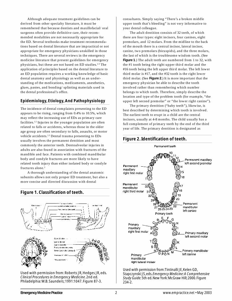
The adult dentition consists of 32 teeth, of whichthere are four types: eight incisors, four canines, eightpremolars, and 12 molars. From the midline to the backof the mouth there is a central incisor, lateral incisor,canine, two premolars (bicuspids), and the three molars,the last of which is the troublesome wisdom tooth. (SeeFigure 1.) The adult teeth are numbered from 1 to 32, withthe #1 tooth being the right upper third molar and the#16 tooth being the left upper third molar. The left lowerthird molar is #17, and the #32 tooth is the right lowerthird molar. (See Figure 2.) It is more important that theemergency physician be able to describe the toothinvolved rather than remembering which numberbelongs to which tooth. Therefore, simply describe thelocation and type of the problem tooth (for example, “theupper left second premolar” or “the lower right canine”).
The primary dentition (“baby teeth”), likewise, isbest described by determining which tooth is involved.The earliest teeth to erupt in a child are the centralincisors, usually at 4-8 months. The child usually has afull complement of primary teeth by the end of the thirdyear of life. The primary dentition is designated as
Figure 1. Classification of teeth.
Used with permission from: Roberts JR, Hedges JR, eds.Clinical Procedures in Emergency Medicine. 2nd ed.Philadelphia: W.B. Saunders; 1991:1047. Figure 87-3.
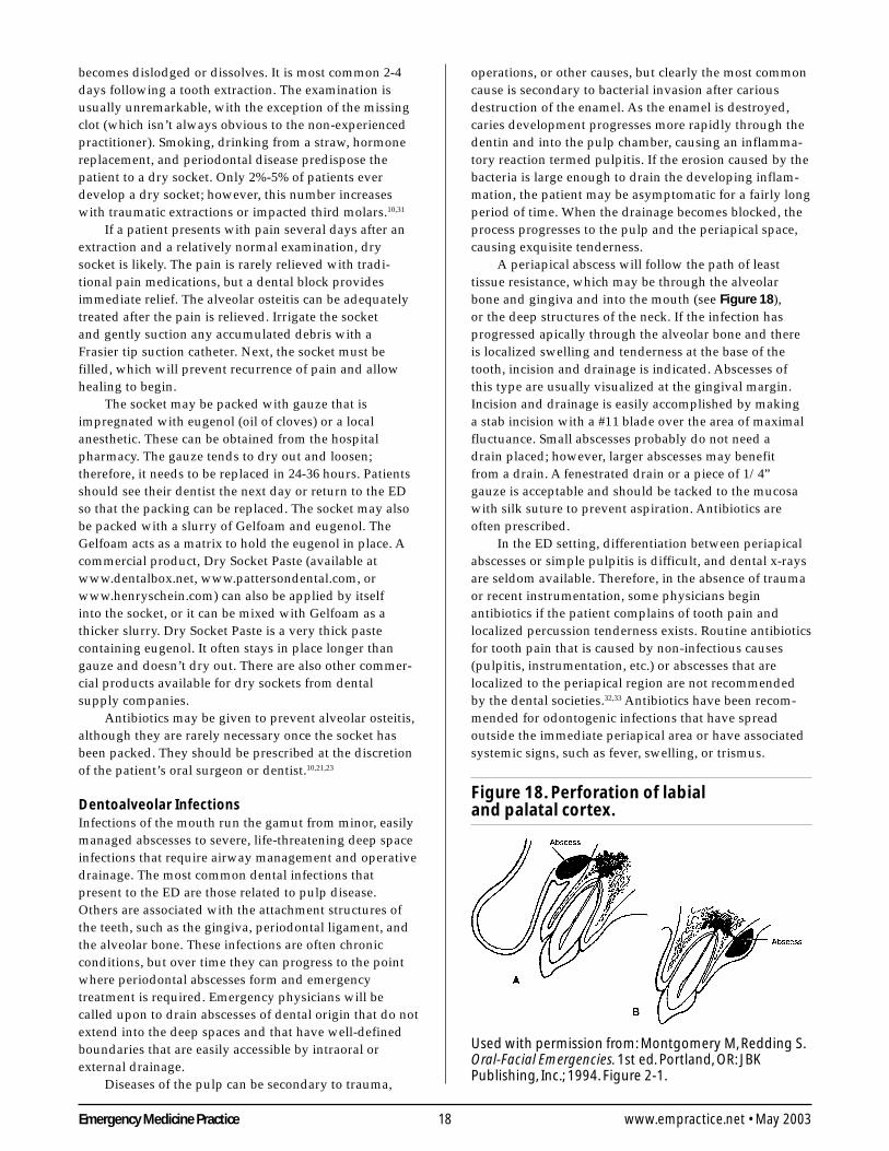
Figure 2. Identification of teeth.
Used with permission from: Tintinalli JE, Kelen GD,Stapczynski JS, eds. Emergency Medicine: A ComprehensiveStudy Guide. 5th ed. New York: McGraw Hill; 2000. Figure234-2.

3 Emergency Medicine PracticeMay 2003 • www.empractice.net
follows: The right maxillary second molar is designatedas A, and the left maxillary second molar is designated asJ. The left mandibular second molar is designated as K,and the right mandibular second molar is designated asT. The teeth in between are lettered accordingly(See Table 1.) Children often have teeth that are missingor incompletely erupted; therefore, it is best to use thename of the tooth instead of letters when communicatingwith a specialist. The permanent teeth usually startreplacing the primary teeth at approximately 5 years ofage, and this process begins with the incisors.
A tooth consists of the central pulp, the dentin, andthe enamel. (See Figure 3.) The pulp contains the neu-rovascular supply of the tooth that carries nutrients to thedentin, a microporous substance that consists of a systemof microtubules. The dentin makes up the majority of thetooth and also serves to cushion the tooth duringmastication. The enamel is the white visible portion ofthe tooth and is the hardest part of the body. The toothmay also be described in terms of the crown (coronalportion) or the root. The crown is that portion covered inenamel, and the root is the part that serves to anchor thetooth in the alveolar bone.
The following descriptive terminology is used forthe different anatomic surfaces of the tooth and is helpfulwhen describing a specific tooth injury to a consultantor colleague.
• Facial: that part of the tooth that you see whensomebody smiles. This is a general term and isapplicable to all teeth. It is sufficient to use whendescribing an injury, but the following is morespecific and precise:• Labial: refers to the facial surface of the incisors
and canines.• Buccal: refers to the facial surface of the premolars
and molars.
• Oral : that part of the tooth that faces the tongue or
the palate. This is also a general term and is appli-cable to all teeth, but the following is more precise:• Lingual: toward the tongue, the oral surface of the
mandibular teeth.• Palatal: toward the palate, the oral surface of the
maxillary teeth.
• Approximal/Interproximal: the contacting surfacesbetween two adjacent teeth.• Mesial: the interproximal surface facing anteriorly
or closest to the midline.• Distal: the interproximal surface facing posteriorly
or away from the midline.
• Occlusal: the biting or chewing surface of thepremolars or molars.
• Incisal: the biting or chewing surface of the incisorsand canines.
• Apical: toward the root of the tooth.• Coronal: toward the crown or the biting surface of
the tooth.
The attachment apparatus, also known as theperiodontium, consists of two major subunits and isnecessary for maintaining the integrity of the normaldentoalveolar unit.
• The gingival subunit consists of the junctionalepithelium and the gingival tissue.
• The periodontal subunit includes the periodontalligament, alveolar bone, and the cementum of theroot of the tooth.
Infections and certain disease states of the gingivaweaken the attachment apparatus and can result in tooth
Table 1. Dentition and age at eruption.
Primary (baby) teeth
Name of tooth Appearance in the mouthCentral incisor 4-14 monthsLateral incisor 8-18 monthsCanine tooth 14-24 monthsFirst molar 10-20 monthsSecond molar 20-36 months
Permanent (adult) teeth
Name of tooth Appearance in the mouthCentral incisor 5-9 yearsLateral incisors 6-10 yearsCanine tooth 8.5-14 yearsFirst premolar (bicuspid) 9-14 yearsSecond premolar (bicuspid) 10-15 yearsFirst molar (6-year molar) 5-9 yearsSecond molar (12-year molar) 10-15 yearsThird molar (wisdom tooth) 17-25 years
Figure 3. The dental anatomic unit andattachment apparatus.
Used with permission from: Tintinalli JE, Kelen GD,Stapczynski JS, eds. Emergency Medicine: A ComprehensiveStudy Guide. 5th ed. New York: McGraw Hill; 2000. Figure234-3.

Emergency Medicine Practice 4 www.empractice.net • May 2003
loss. Likewise, avulsed teeth, even if replaced in a timelymanner, often will not reattach if the gingival subunit isweakened by poor hygiene or disease.
Differential Diagnosis
The differential diagnosis is fairly straightforward whendealing with dental trauma. The primary considerationsto take into account are whether or not the existenttrauma could potentially cause airway compromise.Associated injuries include mandibular, maxillary, oralveolar ridge fractures that may leave the tongueunsupported or the midface unstable. Each scenario maylead to difficulty when performing intubation or ventilat-ing the patient with a bag-valve mask. Mucosal or tonguelacerations may cause bleeding severe enough to compro-mise the airway. It is also important to evaluate forassociated injuries, such as closed head or cervicalspine injuries.
Trauma to the teeth usually consists of fracture,subluxation (loose, nondisplaced teeth), luxation (dis-placed teeth), or complete avulsion. The diagnosis isprimarily determined by a meticulous physical examina-tion, accounting for each tooth and portion of tooth.Radiography usually serves a confirmatory role.
Non-traumatic dental emergencies usually resultfrom poor oral hygiene, recent instrumentation, orinfection. Uncomplicated tooth pain (odontalgia) usuallyreflects pulpitis, and further diagnostic studies are notrequired in the ED. The other consideration is periodon-tal or pulpal infection or abscess. Referred pain from thesinuses or the temporomandibular joint also must beconsidered, especially when non-localizable tooth painexists. (See Table 2.) Dry sockets (alveolar osteitis),hematomas, or hemorrhage may present to the ED afterinstrumentation or extraction.
While pulpitis represents one end of the spectrum,deep space infections represent the other. These rapidlyspreading infections can be life-threatening, especially ifthey dissect into the chest, causing mediastinitis.6 Theexact location of the deep space infection is not criticallyimportant, but rapid initiation of treatment is crucial inpreventing airway compromise. The urgency of theintervention, of course, depends on the severity ofpresentation; a patient with severe trismus, stridor, anddrooling may require an emergent surgical airway.
Prehospital Care
Prehospital care should focus primarily on protection ofthe airway and secondarily on preservation of dentition.EMS providers as well as patients and bystanders cansignificantly alter outcomes with regard to the preserva-tion of avulsed teeth. Loose or displaced teeth should notbe manipulated unless airway intervention is required.Hemorrhage control can be initiated with gauze anddirect pressure if the site of hemorrhage is visible. Medicsshould evaluate the risk of gauze aspiration if the patientis immobilized on a backboard or is intoxicated.
Significant airway hemorrhage or swelling maycause complete airway obstruction if the patient is placedsupine. The position of choice in these patients is theupright sitting position. If cervical spine injury is a majorconcern and the patient must be placed in a supineposition, airway equipment including suction must bereadily available, and personnel must be prepared toplace a definitive airway. In most cases of isolated dentaltrauma, expeditious transport is all that is required. Ifthere are avulsed teeth, the patient and/or EMS provid-ers should be instructed that:
• The tooth should be handled by the crown only.Handling the tooth by the root can damage theperiodontal ligament.
• The tooth should not be replaced if the root is
Table 2. The differential diagnosisof orofacial pain.
Odontogenic originDental cariesReversible pulpitisIrreversible pulpitisPulpal necrosis and
abscessTooth eruptionPericoronitisPostrestorative painPostextraction discomfortPostextraction alveolar
osteitisBruxismCervical erosionDeep space odontogenic
infectionDeep space hematomaAlveolar osteitisPeriapical abscessDentoalveolar abscessOral hemorrhage
Periodontal pathologyGingivitisPeriodontal diseasePeriodontal abscessAcute necrotizing
gingivostomatitis
Orofacial traumaDental fractures: Subtle
enamel cracks, Ellisfractures
Dental subluxation,luxation, intrusion, andavulsion
Facial fracturesAlveolar ridge fracturesSoft-tissue lacerationsTraumatic ulcers
Mandible/maxilla fractureMucosa/tongue lacera-
tions
InfectionOral candidiasisHerpes simplex, types 1
and 2Varicella-zoster, primary
and secondaryHerpanginaHand, foot, and mouth
diseaseSexually transmitted
diseasesMycobacterial infectionsMumps
MalignanciesSquamous cell carcinomaKaposi’s sarcomaLymphomaLeukemiaGraft-versus-host diseaseMelanoma
Other etiologiesCranial neuralgiasStomatitis and mucositis:
uremia, vitamindeficiency, other
Erythema migransPyogenic granulomaUlcerative disease: Lichen
planus, cicatricialpemphigoid, pemphi-gus vulgaris, erythemamultiforme
Crohn’s diseaseBehçet’s syndrome
Adapted from: Tintinalli JE, Kelen GD, Stapczynski JS, eds. EmergencyMedicine: A Comprehensive Study Guide. 5th ed. New York: McGraw-Hill; 2000.

5 Emergency Medicine PracticeMay 2003 • www.empractice.net
fractured or if there is significant maxillofacialtrauma such as an alveolar ridge fracture.
• If the tooth can be replaced in the prehospital setting,the root should be gently rinsed off first to removeany debris (preferably with saline). The root should notbe wiped off as this removes the periodontal ligament.7,8
• If the tooth cannot be successfully reimplanted in thefield, it should be placed in a transport medium asoutlined in the “Treatment” section later in thisarticle. Transporting the tooth in the oral cavity suchas in the cheek may risk aspiration. This location isalso not ideal for keeping the periodontal ligamentalive because of the bacterial flora and low osmolal-ity of the saliva.4,9
ED Evaluation
HistoryThe evaluation of dental complaints in the ED consists ofa focused history and physical examination. Importanthistorical information with regard to traumatic injuriesincludes the following:
1. When did the incident occur? The timing of theincident is critically important when evaluatingavulsed permanent teeth, as the decision to reimplantthe tooth is largely based on the duration that thetooth was avulsed.
2. Were any teeth found at the scene?3. Did the patient experience any coughing after the
injury? This may suggest aspiration of a tooth.4. Has the patient been drinking alcohol or using other
sedatives (including recreational drugs)? This mayincrease the possibility of aspiration.
5. Was a loss of consciousness associated withthe injury?
6. Does the patient complain of pain? Do the teeth feelas if they are meeting normally? Is the pain associ-ated with occlusion? Mandibular fractures are oftenworse with moving the jaw, and patients will oftencomplain that their teeth are not meeting normally.Pain from temporomandibular joint injuries is oftenreferred to the ear. Fractured teeth hurt worse withthe inspiration of air or contact with cold substances.
7. Did the patient apply any substance to the teeth todecrease the pain? Over-the-counter anesthetics andtopical analgesics can cause sterile abscesses ifapplied onto the pulp or dentin.10
8. Does the patient have a history of dental workinvolving the traumatized tooth?
9. Is the tooth a primary or secondary tooth? Trauma-tized primary teeth are managed differently thanpermanent teeth.
10. Does the patient have a history of bleeding disor-ders or allergies to medicines?
The following additional information should beobtained in cases of nontraumatic dental complaints:
1. Has there been any recent dental work or instrumen-tation performed? Dry sockets, for example, occur
after a tooth has been extracted.2. Has the patient had a history of poor dentition or
multiple caries? Has he or she had routine preventa-tive dental care, and when was the most recent visitto the dentist?
3. Is the patient having difficulty opening his or hermouth or swallowing? Has there been a change inhis or her voice? Is there any shortness of breath?Such complaints would raise the concern for deepspace infections.
4. Is the patient immunocompromised in any way?Deep space infections can spread rapidly andprogress to the mediastinum or the cavernous sinusin immunocompromised patients.
5. Is the patient taking aspirin, warfarin, or otheranticoagulants? Is there a history of bleedingdisorders, heavy menstrual periods, bleeding intojoints, or heavy bleeding after dental procedures inthe past?
6. What over-the-counter preparations has the patientbeen using?
7. What has the time course of the symptoms been?Has it been rapid or insidious? Have there beensymptoms consistent with severe infection, such asfevers, chills, or vomiting?
8. Does the patient have drug allergies?9. Does the patient have a history of rheumatic fever or
valvular disease such as mitral valve prolapse?11
Does the patient have implanted devices such asartificial joints, valves, or shunts? These maypredispose to endocarditis or infection of an implant.
Physical ExaminationThe physical examination needs to be meticulous.Injuries to the dentition are easily missed because ofmore impressive traumatic findings or a casual examina-tion. Likewise, the nooks and crannies of the mouth canhide fairly significant-sized abscesses and injuries.
The examination should begin with observation andsimply talking to the patient. Look for airway involve-ment, voice change, muffling, drooling, etc. Externalinspection is important as many injuries such as man-dibular dislocations and fractures are diagnosed bynoting asymmetry, deformity, or swelling of the face.Abscesses or deep space infections will often result inswelling over the involved space. Visualize the face frommultiple angles to note subtle asymmetries. The openingand closing of the mouth should be smooth and com-plete, with no limitations or hesitation. Erythema,warmth, or drainage is indicative of abscess, cellulitis, orhematoma formation. Muffling of the voice providesclues to impending airway compromise. Palpate the facefor tenderness, crepitus, or step-offs. The entire mid-faceand mandible should be palpated, with particularattention paid to the maxilla, zygomas, and the mandibu-lar condyles and coronoid processes. The area of thetemporomandibular joints should be palpated carefullythroughout the range of motion. There should not be anypops, clicks, or pain.
Emergency Medicine Practice 6 www.empractice.net • May 2003
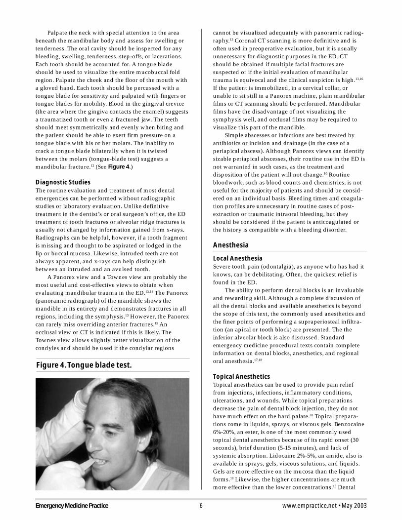
Palpate the neck with special attention to the areabeneath the mandibular body and assess for swelling ortenderness. The oral cavity should be inspected for anybleeding, swelling, tenderness, step-offs, or lacerations.Each tooth should be accounted for. A tongue bladeshould be used to visualize the entire mucobuccal foldregion. Palpate the cheek and the floor of the mouth witha gloved hand. Each tooth should be percussed with atongue blade for sensitivity and palpated with fingers ortongue blades for mobility. Blood in the gingival crevice(the area where the gingiva contacts the enamel) suggestsa traumatized tooth or even a fractured jaw. The teethshould meet symmetrically and evenly when biting andthe patient should be able to exert firm pressure on atongue blade with his or her molars. The inability tocrack a tongue blade bilaterally when it is twistedbetween the molars (tongue-blade test) suggests amandibular fracture.12 (See Figure 4.)
Diagnostic StudiesThe routine evaluation and treatment of most dentalemergencies can be performed without radiographicstudies or laboratory evaluation. Unlike definitivetreatment in the dentist’s or oral surgeon’s office, the EDtreatment of tooth fractures or alveolar ridge fractures isusually not changed by information gained from x-rays.Radiographs can be helpful, however, if a tooth fragmentis missing and thought to be aspirated or lodged in thelip or buccal mucosa. Likewise, intruded teeth are notalways apparent, and x-rays can help distinguishbetween an intruded and an avulsed tooth.
A Panorex view and a Townes view are probably themost useful and cost-effective views to obtain whenevaluating mandibular trauma in the ED.13,14 The Panorex(panoramic radiograph) of the mandible shows themandible in its entirety and demonstrates fractures in allregions, including the symphysis.13 However, the Panorexcan rarely miss overriding anterior fractures.15 Anocclusal view or CT is indicated if this is likely. TheTownes view allows slightly better visualization of thecondyles and should be used if the condylar regions
cannot be visualized adequately with panoramic radiog-raphy.13 Coronal CT scanning is more definitive and isoften used in preoperative evaluation, but it is usuallyunnecessary for diagnostic purposes in the ED. CTshould be obtained if multiple facial fractures aresuspected or if the initial evaluation of mandibulartrauma is equivocal and the clinical suspicion is high.13,16
If the patient is immobilized, in a cervical collar, orunable to sit still in a Panorex machine, plain mandibularfilms or CT scanning should be performed. Mandibularfilms have the disadvantage of not visualizing thesymphysis well, and occlusal films may be required tovisualize this part of the mandible.
Simple abscesses or infections are best treated byantibiotics or incision and drainage (in the case of aperiapical abscess). Although Panorex views can identifysizable periapical abscesses, their routine use in the ED isnot warranted in such cases, as the treatment anddisposition of the patient will not change.10 Routinebloodwork, such as blood counts and chemistries, is notuseful for the majority of patients and should be consid-ered on an individual basis. Bleeding times and coagula-tion profiles are unnecessary in routine cases of post-extraction or traumatic intraoral bleeding, but theyshould be considered if the patient is anticoagulated orthe history is compatible with a bleeding disorder.
Anesthesia
Local AnesthesiaSevere tooth pain (odontalgia), as anyone who has had itknows, can be debilitating. Often, the quickest relief isfound in the ED.
The ability to perform dental blocks is an invaluableand rewarding skill. Although a complete discussion ofall the dental blocks and available anesthetics is beyondthe scope of this text, the commonly used anesthetics andthe finer points of performing a supraperiosteal infiltra-tion (an apical or tooth block) are presented. The theinferior alveolar block is also discussed. Standardemergency medicine procedural texts contain completeinformation on dental blocks, anesthetics, and regionaloral anesthesia.17,18
Topical AnestheticsTopical anesthetics can be used to provide pain relieffrom injections, infections, inflammatory conditions,ulcerations, and wounds. While topical preparationsdecrease the pain of dental block injection, they do nothave much effect on the hard palate.18 Topical prepara-tions come in liquids, sprays, or viscous gels. Benzocaine6%-20%, an ester, is one of the most commonly usedtopical dental anesthetics because of its rapid onset (30seconds), brief duration (5-15 minutes), and lack ofsystemic absorption. Lidocaine 2%-5%, an amide, also isavailable in sprays, gels, viscous solutions, and liquids.Gels are more effective on the mucosa than the liquidforms.18 Likewise, the higher concentrations are muchmore effective than the lower concentrations.18 Dental
Figure 4. Tongue blade test.

7 Emergency Medicine PracticeMay 2003 • www.empractice.net
offices usually stock the higher concentrations oflidocaine (5%) and benzocaine (20%).
The gel-form topical anesthetics are usually appliedwith a cotton-tipped applicator onto the mucosa over thepoint of injection. Benzocaine requires a few minutes formaximal effect. Lidocaine requires several more minutesthan benzocaine until maximal anesthesia is obtained.
Injectable AnestheticsThe choice of injectable anesthetic depends on a numberof factors, including duration of action, side effects,patient allergies, and whether a vasoconstrictor isneeded. Table 3 shows the average durations of action ofcommon local anesthetic preparations. The use of alonger-lasting anesthetic, such as bupivacaine, is recom-mended for painful conditions in which definitive carewill most likely be provided in the dentist’s office, suchas pulpitis, periapical abscess, or a fractured tooth.Bupivacaine with epinephrine provides 6-8 hours ofcomplete anesthesia and several more hours of partialanesthesia.18 A shorter-acting anesthetic is more appropri-ate when a procedure such as laceration repair needs tobe undertaken, such as a buccal mucosa or tonguelaceration. Both bupivacaine and lidocaine (with andwithout a vasoconstrictor) are available as carpules,which are used in ringed syringe injectors. These injec-tors have a ring in which the thumb is placed to providegreater control over the procedure.
The use of ringed syringe aspirators is recommendedwhen performing any intraoral injections for a number ofreasons—the foremost being that they afford more one-handed maneuverability than a standard medicalplunger as well as an increased ability to aspirate. Themost common complications and side effects of intraoralinjections can be minimized by avoiding intravascularinjection.18 Ringed syringe aspirators allow aspirationbefore injection. Both reusable and disposable ringedsyringes are available.
Supraperiosteal InfiltrationThe supraperiosteal infiltration is ideally suited foranesthesia of a single tooth or circumscribed portionof the mandible or maxilla. A slightly larger dose ofanesthetic should be used when performing asupraperiosteal infiltration on mandibular teeth second-ary to the density and increased thickness of the man-dible. The discussion that follows applies to all indi-vidual teeth. To obtain maximal effect, the needle tipshould overlie the apex of the tooth being anesthetized.The amount of anesthetic will vary with the experience ofthe clinician and the location of the tooth (mandibularteeth require more, as do posterior teeth), but generallywithin 1.0-2.5 cc is adequate.
The topical anesthetic is applied to the mucobuccalfold in the general area where the injection will takeplace. After the topical sets up, the area of injectionshould be wiped dry. The patient’s lip should be retractedby the clinician’s non-injecting hand. The needle isinserted near the greatest concavity of the mucobuccalfold and is directed toward the apex of the tooth, usuallyat a depth of 3-4 mm. If the bone is contacted, the needleshould be slightly withdrawn in order to prevent theanesthetic from being inadvertently injected beneath theperiosteum. The local anesthetic is injected after anegative aspirate is obtained.
Inferior Alveolar BlockThe inferior alveolar nerve block provides pain controlfrom the retromolar region of the mandible to themidline. It is ideal for all of the mandibular teeth, lowerlip, and chin on one side. The landmarks that are impor-tant are the retromolar fossa and the anterior border ofthe ramus of the mandible (coronoid notch) as well as thecontralateral premolars.
The block is performed by placing the non-injectingthumb into the mouth and retracting the mucosa. Thethumb itself should be placed up against the anterior
Table 3. Dental anesthesia: Duration of action in minutes.
Maxillary infiltration Inferior alveolar block
Preparation Pulpal tissue Soft tissue Pulpal tissue Soft tissue
0.4% Propoxycaine HCl, 2% prilocaine HCl; 40 145 60 1751:20,000 levonordefrin or 1:30,000 norepinephrine
2% Lidocaine HCl; 1:100,000 or 1:50,000 epinephrine 60 170 85 190
2% Mepivacaine HCl; 1:20,000 levonordefrin 50 130 75 185
3% Mepivacaine HCl 25 90 40 165
4% Prilocaine HCl 20 105 55 190
4% Prilocaine HCl; 1:200,000 epinephrine 40 140 60 220
0.5% Bupivacaine HCl; 1:200,000 epinephrine 40 340 240 440
1.5% Etidocaine HCl; 1:200,000 epinephrine 30 280 240 470
4% Articaine HCl; 1:200,000 or 1:100,000 epinephrine 60 170 90 220

Emergency Medicine Practice 8 www.empractice.net • May 2003
border of the ramus of the mandible in the coronoidnotch. (See Figure 5.) The injection point should beapproached from the opposite premolars, and the needleis placed approximately 1.0-1.5 cm posteriorly to themidline of the thumbnail. The needle tip is advanceduntil the mandibular bone is contacted, usually 1.5-2.0cm. Aspiration is performed to rule out intravascularinjection, and the anesthetic is delivered. Usually, 1.0-3.0cc of anesthetic is adequate.
Treatment
Dental FracturesInjury to maxillary central incisors accounts for between70% and 80% of all fractured teeth.19 Although not life-threatening, the morbidity associated with dentalfractures can be significant, including failure to completeeruption, abscess, loss of space in the dental arch, colorchange of the tooth, ankylosis, abnormal exfoliation, androot resorption.
Some general principles apply to the ED evaluationand management of dental trauma.
1. Identify all fracture fragments and mobile teeth and note ifa mandible fracture is open or closed. Radiographsshould be taken if there is intrusion of fragments intothe mucosa or alveolar bone. Obtain a chest x-ray if apatient with a missing tooth has pulmonary com-plaints after the injury, such as cough or shortness ofbreath. It must be remembered, however, thatpatients who may present with avulsion of a toothmay not recall coughing because of intoxication orother trauma.
2. The dentition is much more easily manipulated if thepatient is not in significant discomfort. Tooth infiltrationand dental block anesthesia should be part of theemergency physician’s armamentarium. Narcoticand nonnarcotic alternatives, while helpful aftertreatment is completed, do not usually offer thepatient the comfort required to perform most dentalmanipulations. If the procedure to be performed issimple, such as applying some glue to a lost cap orfilling, a dental block is unnecessary and oralalternatives offer a more reasonable choice.
3. Avoid topical tooth remedies and analgesics, bothover-the-counter and prescribed, as their use canlead to the development of sterile abscesses andsoft-tissue irritation.10
4. Administer tetanus vaccination if indicated. EDmanagement of fractured teeth depends on theextent of fracture with regard to the pulp, the degreeof development of the apex of the tooth, and the ageof the patient. There are many ways to classifydentoalveolar injuries and, in particular, toothfractures.20 The Ellis classification system is one oftencited in the emergency medicine literature;21 how-ever, many dentists and maxillofacial surgeons donot use this nomenclature. The most easily under-
Figure 5. Inferior alveolar block.
1. Radiography for routine ED evaluation of dental pain
should be discouraged, as the most useful x-rays (bite-wing
x-rays) usually cannot be obtained, and rarely would they
ever change treatment. Likewise, isolated mandibular trauma
with normal occlusion, no suspicion of open fracture, and a
negative tongue blade test is very unlikely to be associated
with a mandibular fracture that needs immediate attention.
Although the tongue blade test may occasionally miss a
coronoid process fracture, these rarely require any definitive
treatment, and a delayed diagnosis usually is not associated
with morbidity.
2. Become familiar with dental anesthetic techniques, as
ED treatment often cannot be successfully performed
without removing the patient’s pain first. An individual with
a fracture through the pulp will allow manipulation if the
tooth is numb. They most certainly will not if each contact
with the tooth is exquisitely painful. Dental anesthesia will
not only speed up the delivery of care, but also will increase
patient satisfaction.
3. Learn the proper dental terminology so that discussions
with consultants are concise and mutually understandable.
4. Storing a proper dental tray or kit in the ED will prove
invaluable and time-saving. It is often the case that the
emergency physician has to search for CaOH paste or other
suitable materials from the pharmacy. This is often frustrating,
time-consuming, and, unfortunately, fruitless. Being prepared
prior to the emergency is what we are trained for, and we
should not approach dental emergencies any differently than
other emergencies we encounter.
5. Routine laboratory testing should not be performed
on patients who present for dental bleeding except
when anticoagulation, intractable vomiting, or liver disease
are present. ▲
Cost- and Time-Saving Strategies For Dental Emergencies
9 Emergency Medicine PracticeMay 2003 • www.empractice.net
stood method of classification is based on a descrip-tion of the injury.3
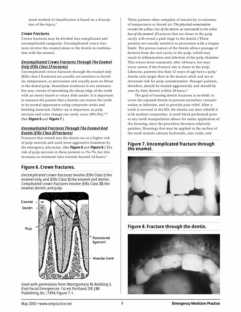
Crown FracturesCrown fractures may be divided into complicated anduncomplicated categories. Uncomplicated crown frac-tures involve the enamel alone or the dentin in combina-tion with the enamel.
Uncomplicated Crown Fractures Through The EnamelOnly (Ellis Class I Fractures)Uncomplicated crown fractures through the enamel only(Ellis class I fractures) are usually not sensitive to forcedair, temperature, or percussion and usually pose no threatto the dental pulp. Immediate treatment is not necessarybut may consist of smoothing the sharp edge of the toothwith an emery board or rotary disk sander. It is importantto reassure the patient that a dentist can restore the toothto its normal appearance using composite resins andbonding materials. Follow-up is important, as pulpnecrosis and color change can rarely occur (0%-3%).3,22
(See Figure 6 and Figure 7.)
Uncomplicated Fractures Through The Enamel AndDentin (Ellis Class II Fractures)Fractures that extend into the dentin are at a higher riskof pulp necrosis and need more aggressive treatment bythe emergency physician. (See Figure 6 and Figure 8.) Therisk of pulp necrosis in these patients is 1%-7%, but thisincreases as treatment time extends beyond 24 hours.3
These patients often complain of sensitivity to extremesof temperature or forced air. The physical examinationreveals the yellow tint of the dentin as contrasted to the whitehue of the enamel. (Fractures that are closer to the pulpcavity will reveal a pink tinge to the dentin.) Thesepatients are usually sensitive to percussion with a tongueblade. The porous nature of the dentin allows passage ofbacteria from the oral cavity to the pulp, which mayresult in inflammation and infection of the pulp chamber.This occurs most commonly after 24 hours, but mayoccur sooner if the fracture site is closer to the pulp.Likewise, patients less than 12 years of age have a pulp/dentin ratio larger than in the mature adult and are atincreased risk for pulp contamination. Younger patients,therefore, should be treated aggressively and should beseen by their dentist within 24 hours.4
The goal of treating dentin fractures is twofold: tocover the exposed dentin to prevent secondary contami-nation or infection, and to provide pain relief. After atooth is covered in the ED, the dentist can later rebuild itwith modern composites. A tooth block performed priorto any tooth manipulation allows for easier application ofthe dressing, since the procedure becomes relativelypainless. Dressings that may be applied to the surface ofthe tooth include calcium hydroxide, zinc oxide, and
Figure 6. Crown fractures.
Uncomplicated crown fractures involve (Ellis Class I) theenamel only, and (Ellis Class II) the enamel and dentin.Complicated crown fractures involve (Ellis Class III) theenamel, dentin, and pulp.
Used with permission from: Montgomery M, Redding S.Oral-Facial Emergencies. 1st ed. Portland, OR: JBKPublishing, Inc.; 1994. Figure 7-1.
Figure 7. Uncomplicated fracture throughthe enamel.
Figure 8. Fracture through the dentin.

Emergency Medicine Practice 10 www.empractice.net • May 2003
glass ionomer composites.10,19,23,24 The literature suggeststhat glass ionomer dressings may be slightly superior toother dressings when applied by dentists; however, thisis debated in the dental community. The ease of applica-bility of calcium hydroxide paste, however, and the factthat it can be used easily by itself make it attractive foruse in ED patients.19,21 Composites that are cured with abonding light are beyond the scope of most emergencypractice. Skin adhesives and bone wax are sometimesused in the ED. They are not recommended, however, asbone wax is relatively porous, and skin adhesives are notapproved for intraoral use. Most dressings are availableas a catalyst and a base and are easily mixed with adental spatula and mixing pad.
A commonly used ED dressing is calcium hydroxide(CaOH) paste, which is mixed and applied to the frac-tured surface of the tooth. CaOH preparations includeboth catalyst and base and pre-mixed formulations. (Twobrands are Dycal and Pulpdent, which are available atwww.dentalbox.net, www.smartpractice.com,www.pattersondental.com, and www.henryschein.com.)The tooth surface should be as dry as possible beforeapplication to ensure adherence. This can be accom-plished by having the patient bite into gauze pads priorto application. The calcium hydroxide will dry withinminutes after being exposed to the moist environment ofthe mouth. Although placing dental foil over the CaOHpaste has been recommended, it is not necessary if thepatient follows up within 24-48 hours. The patient shouldbe instructed to eat soft foods until seen by the dentist toprevent dislodging of the dressing. Some practitionersbegin antibiotic treatment if the period of exposure issignificantly long.4,10 Penicillin or clindamycin offers goodoral flora coverage.25 Many patients sustaining a fracturethrough the dentin will eventually require a root canal orother definitive endodontic treatment. The timelyapplication of an appropriate dressing in the ED, how-ever, may prevent contamination of the pulp and makeroot canal unnecessary. As with any trauma to theanterior teeth, it is advisable to explain to the patient thatdisruption of the neurovascular supply is possible andthat long-term complications such as pulp necrosis, colorchange, and root resorption may occur.
Complicated Fractures Of The Crown Involving ThePulp (Ellis Class III Fractures)Complicated fractures of the crown that extend into thepulp of the tooth are true dental emergencies. (See Figure9 and Figure 10.) These fractures result in pulp necrosis in10%-30% of cases, even with appropriate treatment.3 Theyare distinguished from fractures of the dentin by the pink colorof the pulp. The fracture surface of the tooth should bewiped off with gauze and observed for frank bleeding ora pink blush, which indicates exposure of the pulp.Fractures through the pulp are often excruciatinglypainful, but occasionally there is a lack of sensitivitysecondary to a disruption of the neurovascular supply ofthe tooth.23
Immediate management includes referral to a
dentist, oral surgeon, or endodontist. The patient oftenrequires a pulpectomy (complete removal of the pulp) or,in the case of primary teeth, a pulpotomy (partialremoval of the pulp) as definitive treatment.22,24 Thelonger the pulp is exposed, the greater the chance ofcontamination and abscess formation. If a dentist cannotsee the patient immediately, the emergency physicianshould attempt to relieve the pain and cover the exposedpulp. Supraperiosteal infiltration (dental block) should beperformed if significant pain is present. Subsequently, thetooth should be covered with one of the dressingsdescribed in the preceding section. Sometimes bleeding isbrisk and needs to be controlled before the application ofa dressing. This can usually be accomplished by havingthe patient bite into a gauze pad that has been soakedwith a topical anesthetic containing a vasoconstrictorsuch as epinephrine. Alternatively, a small amount of theanesthetic/vasoconstrictor may be injected into the pulpto control bleeding. After the covering is applied, instructthe patient to stay on a liquid diet and see the dentist assoon as possible. No more than 24 hours should lapsebefore definitive treatment is initiated. Antibioticcoverage should be considered as described the preced-ing section if exposure was prolonged.4,10
There are currently no randomized, controlled trials
Figure 9. Fracture through the pulp.
Figure 10. Complicated fracture throughthe pulp.
11 Emergency Medicine PracticeMay 2003 • www.empractice.net
that address the question as to whether antibiotics shouldbe prescribed for fractured teeth seen in the ED. Patientswho present to the dentist and then undergo definitivetreatment do not routinely receive antibiotic prophy-laxis.19,24 However, in the ED, we must treat dental
fractures with the following assumptions: first, it isuncertain as to whether the patient will be able to securerapid dental follow-up, and second, it is usually un-known what the patient’s underlying dentoalveolarhealth is. Delayed fracture care and poor gingival healthincrease the risk of pulp necrosis and, potentially,periapical abscess. These factors suggest to some thattooth fractures that involve the dentin or pulp shouldreceive antibiotic prophylaxis.
Removal of the pulp with specialized instruments bythe emergency physician is not recommended, althoughsome authors have recommended this in the past. Thisprocedure is in the realm of the dental professional andcan result in complications if not done properly.
Luxation, Subluxation, Intrusion, And AvulsionSubluxation refers to teeth that are mobile but notdisplaced, and luxation refers to teeth that are displaced,either partially or completely, from their sockets. Lux-ation injuries are divided into four types:
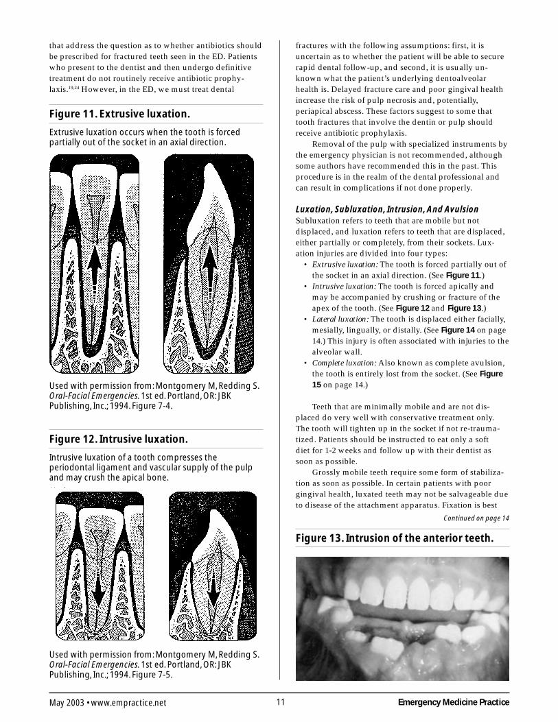
• Extrusive luxation: The tooth is forced partially out ofthe socket in an axial direction. (See Figure 11.)
• Intrusive luxation: The tooth is forced apically andmay be accompanied by crushing or fracture of theapex of the tooth. (See Figure 12 and Figure 13.)
• Lateral luxation: The tooth is displaced either facially,mesially, lingually, or distally. (See Figure 14 on page14.) This injury is often associated with injuries to thealveolar wall.
• Complete luxation: Also known as complete avulsion,the tooth is entirely lost from the socket. (See Figure15 on page 14.)
Teeth that are minimally mobile and are not dis-placed do very well with conservative treatment only.The tooth will tighten up in the socket if not re-trauma-tized. Patients should be instructed to eat only a softdiet for 1-2 weeks and follow up with their dentist assoon as possible.
Grossly mobile teeth require some form of stabiliza-tion as soon as possible. In certain patients with poorgingival health, luxated teeth may not be salvageable dueto disease of the attachment apparatus. Fixation is best
Figure 13. Intrusion of the anterior teeth.
Figure 11. Extrusive luxation.
Extrusive luxation occurs when the tooth is forcedpartially out of the socket in an axial direction.
Used with permission from: Montgomery M, Redding S.Oral-Facial Emergencies. 1st ed. Portland, OR: JBKPublishing, Inc.; 1994. Figure 7-4.
Figure 12. Intrusive luxation.
Intrusive luxation of a tooth compresses theperiodontal ligament and vascular supply of the pulpand may crush the apical bone.
Used with permission from: Montgomery M, Redding S.Oral-Facial Emergencies. 1st ed. Portland, OR: JBKPublishing, Inc.; 1994. Figure 7-5.
Continued on page 14
Emergency Medicine Practice 12 www.empractice.net • May 2003
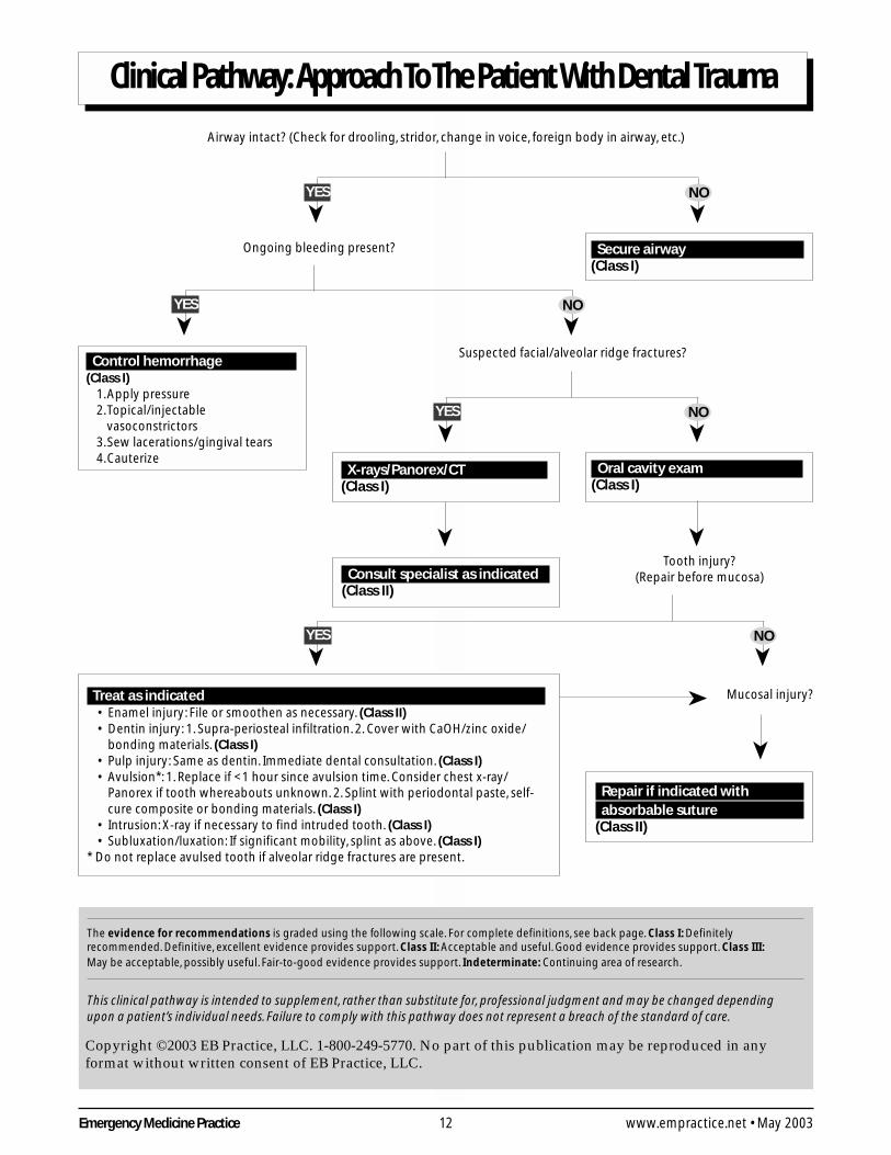
The evidence for recommendations is graded using the following scale. For complete definitions, see back page. Class I: Definitelyrecommended. Definitive, excellent evidence provides support. Class II: Acceptable and useful. Good evidence provides support. Class III:May be acceptable, possibly useful. Fair-to-good evidence provides support. Indeterminate: Continuing area of research.
This clinical pathway is intended to supplement, rather than substitute for, professional judgment and may be changed dependingupon a patient’s individual needs. Failure to comply with this pathway does not represent a breach of the standard of care.
Copyright ©2003 EB Practice, LLC. 1-800-249-5770. No part of this publication may be reproduced in anyformat without written consent of EB Practice, LLC.
Clinical Pathway: Approach To The Patient With Dental Trauma
Airway intact? (Check for drooling, stridor, change in voice, foreign body in airway, etc.)
Ongoing bleeding present?
Suspected facial/alveolar ridge fractures?
Tooth injury?(Repair before mucosa)
Mucosal injury?
➤NO➤YES
Secure airway(Class I)
Control hemorrhage(Class I)
1.Apply pressure2.Topical/injectable
vasoconstrictors3.Sew lacerations/gingival tears4.Cauterize
➤NO➤YES
➤NO➤YES
X-rays/Panorex/CT(Class I)
➤
Consult specialist as indicated(Class II)
Oral cavity exam(Class I)
➤
➤NO➤YES
Treat as indicated• Enamel injury: File or smoothen as necessary. (Class II)• Dentin injury: 1. Supra-periosteal infiltration. 2. Cover with CaOH/zinc oxide/
bonding materials. (Class I)• Pulp injury: Same as dentin. Immediate dental consultation. (Class I)• Avulsion*: 1. Replace if <1 hour since avulsion time. Consider chest x-ray/
Panorex if tooth whereabouts unknown. 2. Splint with periodontal paste, self-cure composite or bonding materials. (Class I)
• Intrusion: X-ray if necessary to find intruded tooth. (Class I)• Subluxation/luxation: If significant mobility, splint as above. (Class I)
* Do not replace avulsed tooth if alveolar ridge fractures are present.
Repair if indicated with absorbable suture(Class II)
➤
➤

13 Emergency Medicine PracticeMay 2003 • www.empractice.net
Ten Pitfalls To Avoid
1. “We didn’t have anything to put the tooth in, so we just
left it out. They can just get an implant anyway.”
Milk suffices just fine for temporary avulsed tooth storage
until you are ready to reimplant it. Every attempt should be
made to reimplant and, therefore, preserve an avulsed
tooth. Every person clearly is not a candidate for implants,
and the practitioner should not make this supposition.
2. “I was always taught that you replace a tooth as soon
as possible. What difference does it make if it’s a kid
or not?”
Primary teeth are best not reimplanted. They can certainly
cause problems such as infections and disruptions of the
secondary teeth. If there is a question as to whether a tooth
is a primary tooth, first replace the tooth and then obtain
an x-ray. If you see the secondary teeth have not yet
erupted, you can always remove the primary tooth.
Likewise, as long as the patient is referred expeditiously
(within 48 hours) to a dentist, the dentist can remove the
replaced tooth.
3. “My kid looks as if he was in a horror movie. That ER
doctor never told me he could have discoloration in his
tooth from a simple bump.”
Any trauma to the teeth can result in pulp necrosis and
permanent change in the coloration of the tooth. Although
the dentist can usually repair this with bleaching or
restoration, it is important that you warn patients who
sustain tooth trauma that they may have discoloration.
4. “It’s not really important that you aspirate before
you inject when doing a dental block. That’s just for
beginners. How was I supposed to know he would
develop that huge hematoma?”
Aspiration is extremely important when performing any
dental blocks. It is a simple and important means to
diminish side effects and complications. Intravascular
injection can lead to cardiovascular effects, syncope,
hematomas, and palpitations.
5. “He didn’t have a cough or shortness of breath. There was
no way to know he aspirated his tooth during that fight.”
When a tooth has been avulsed and its location is
unknown, obtain a chest x-ray if there is a question that it
may have been aspirated. Removal of a tooth is easier prior
to the development of pneumonia/empyema or effusion.
6. “I didn’t see the tooth and, therefore, I assumed that it
was avulsed. There was no way to tell it was pushed up into
the gingiva and that it would cause an abscess.”
The clinician must have a high index of suspicion in
patients with intruded teeth. They often have the
appearance of avulsed teeth. Intruded teeth can lead to
abscesses and growth abnormalities if not diagnosed. X-ray
if there is any question.
7. “I just assumed that the change in his voice was
laryngitis. I figured the antibiotics would be good for his
dental abscess and his pharyngitis.”
Deep space infections of the neck can be very serious and
occasionally life-threatening. Bilateral extension, change in
voice, drooling, or difficulty swallowing or breathing are
indications of a serious deep space infection. These patients
require admission, IV antibiotics, CT scanning to delineate
the extent of tissue spread, and oromaxillofacial/oral
surgery consultation.
8. “There was no way I could have sewn that kid’s
tongue. He was thrashing all over the place. They
usually heal fine anyway.”
Tongue lacerations that are gaping or involve the
tongue margin should be repaired. Epithelialization of
a significant non-repaired tongue laceration can result
in a bifid tongue or a grooved tongue. Both of these
conditions can be difficult for plastic surgeons to repair.
Repair of difficult tongue lacerations may require sedation
or referral to a specialist.
9. “We don’t have anything to cover fractured teeth in our
ED and the patient didn’t have any insurance. I told him to
follow up in the university dental clinic.”
Teeth that have been fractured through the pulp have a
high incidence of pulp necrosis and tooth loss if not
treated. Immediate covering in the ED can, occasionally,
prevent root canal. It will also help with pain by keeping
the nerve covered and decrease the chance of abscess or
pulpitis. Immediate referral usually will result in root canal
for fractures through the pulp, but the patient will keep a
functional tooth.
10. “I always thought that penicillin was fine for deep space
infections of the neck.”
Penicillin or clindamycin alone are adequate for suspected
periapical abscesses, but broader coverage should be
instituted for deep space infections of the neck. Penicillin
should be used in conjunction with metronidazole.
Alternatives include the expanded-spectrum penicillins
such as ticarcillin/clavulanic acid, piperacillin/tazobactam,
or clindamycin plus cefoxitin. Far more important than
antibiotics is surgical consultation. ▲

Emergency Medicine Practice 14 www.empractice.net • May 2003
performed by the dental specialist with enamel bondingmaterials or wire ligation. Although many different homeremedies exist for splinting loose teeth in the ED, onemust be aware of the concern for aspiration of teeth orthe splint if the splint should fail. The physician shouldalso avoid the use of non-approved medications in themouth. An example would be the use of skin adhesives,which, to date, have not been approved for intraoral use.
Temporizing splinting techniques suitable for use byemergency physicians include periodontal paste and self-cure composite. A commercially available form ofperiodontal paste known as Coe-Pak consists of a baseand a catalyst that, when mixed together, form a moder-ately sticky clay-like dressing that becomes firm afterapplication. The splint performs best if applied to thefacial and oral surfaces of the teeth; however, it is usuallysufficient to apply it only to the facial surface of the
affected teeth. Coe-Pak is most easily applied when thephysician’s gloves are moistened with water or lubricat-ing jelly and the gingiva and enamel are completely dry.It is important to apply the dressing into the groovesbetween the teeth as well as to the adjacent teeth, and thepatient should be reminded to eat a soft diet until seenfor follow-up in 24 hours.
Self-cure composite is another splinting option in theED. Although many composites used in the dental officerequire a curing “light” and etching acids to affix thebonding material, self-cure composite requires neitherand is easy to use. It is applied only to the enamel of theinvolved tooth, not the gingiva, and to the adjacent non-mobile teeth. Both periodontal paste and self-curecomposites are easy to remove during formal restorationby the dentist.
Teeth that are luxated in either the horizontal or axialplanes or are slightly extruded can also be splinted usingthe preceding techniques. The teeth do not need to be inperfect alignment prior to discharge from the ED. Finaladjustments can be made in the specialist’s office.
Intrusion And AvulsionIntruded teeth have been forced apically into the alveolarbone and often result in disruption of the attachmentapparatus or fracture of the supporting alveolar bone.This is especially common in permanent teeth withmature roots.7 Intruded teeth are often immobile and,therefore, do not require stabilization in the ED, but oftendo require later endodontic treatment because of pulpnecrosis. It is very important to consider the possibility ofan intruded tooth if there is an abnormal space in thedentition, as these can cause infection and craniofacialabnormalities if undiagnosed. X-rays should be obtainedanytime there is uncertainty as to whether a tooth isintruded or avulsed. The dental specialist should manageintruded teeth, and referral should take place within 24hours. Permanent teeth often require repositioning andimmobilization, but primary teeth are usually given atrial period to erupt on their own before any interventionis taken.
Avulsed teeth are true dental emergencies. The firstquestion to ask is, “Where is the tooth?” Missing teethmay be intruded, aspirated, fractured, swallowed, orembedded in the oral mucosa somewhere. Radiographyincluding Panorex, facial films, or a chest x-ray may needto be considered to find fragments of fractured teeth oran avulsed tooth. ED management is based on a numberof factors, including patient age, time elapsed sinceavulsion, presence of other maxillofacial trauma such asalveolar ridge fractures, and the overall health of theperiodontium.7 Primary teeth are not replaced because theycan fuse to the alveolar bone and potentially cause craniofacialabnormalities or infection, and they may prevent normaleruption of the permanent teeth.26 There are small reports ofsuccessful reimplantation of primary teeth by dentists;however, most resources do not recommend it beperformed by dentists, family members, or emergencyphysicians.25 Reimplanted primary teeth may also
Figure 15. An avulsed tooth.
Continued from page 11
Figure 14. Lateral luxation.
Lateral luxation occurs when the tooth is displaced ineither a lingual, mesial, distal, or facial direction.Fractures of the alveolus frequently accompany lateralluxation injuries.
Used with permission from: Montgomery M, Redding S.Oral-Facial Emergencies. 1st ed. Portland, OR: JBKPublishing, Inc.; 1994. Figure 7-6.
15 Emergency Medicine PracticeMay 2003 • www.empractice.net
interfere with the eruption of the secondary teeth. Parentscan be reassured that prosthetic replacements can beworn until the permanent teeth erupt, if desired.
Time is the essential consideration when decidingwhether to replace an avulsed tooth. In general, thelonger the tooth is out of the socket, the higher theincidence of periodontal ligament necrosis and subse-quent failure of reimplantation.7 Periodontal ligamentcells generally die within 60 minutes outside of the oralcavity if they are not placed in an appropriate transportmedium.4 Significant research has been conducted ondifferent media used to keep the cells of the periodontalligament alive. Various transport media, including milk,Hank’s balanced salt solution, Save-A-Tooth, saliva,water, and Gatorade, have all been studied. Certain cellculture media have been developed that cause periodon-tal ligament cells to proliferate and remain viable, butmilk and the commercially available Save-A-Tooth arebest for both prehospital care and ED storage.27 Both milkand Save-A-Tooth (a commercial version of Hank’sbalanced salt solution) preserve the periodontal ligamentfor at least 8-12 hours; however, reimplantation shouldtake place at the earliest opportunity. The critical factor isto get the tooth into some sort of transport medium,because even 5-10 minutes outside some kind of storagemedium can cause desiccation and death of the periodon-tal ligament cells. Saline should be used at the scene ifnothing else is available, and the patient or prehospitalproviders should reimplant the tooth if possible. Salivaand water are less desirable alternatives, although salivais preferable to water.28 If the medics or the patient isreluctant to do this or there are conditions preventingreimplantation in the field, it will need to be done onarrival using the following guidelines:
1. Store the tooth in an appropriate medium ifreimplantation is delayed for any reason.
2. It is very helpful to perform a supraperiostealinfiltration prior to the manipulation or replacementof teeth. This makes the procedure more comfortablefor the patient and easier for the physician toperform. Regional blocks also are acceptable and areespecially useful if more than one tooth is involved.
3. Check the oral cavity for trauma. If an alveolar ridgefracture is present or the socket is severely damaged,the tooth should not be reimplanted.
4. If available, suction the socket first with a Frasier tipsuction catheter to remove any accumulated clot.Overly aggressive suctioning can damage theperiodontal ligament fibers lining the socket. Next,gently irrigate to remove any remaining clot.Reimplantation and realignment is difficult if the clotis not entirely removed. Debris on the tooth shouldbe gently rinsed, not scrubbed, with saline. It is betterto reimplant the tooth with a small amount of debrispresent than to wipe off the periodontal ligament.Implant the tooth using firm, but gentle pressure.
5. The tooth will require splinting (see above) afterreimplantation if it is still loose, although teeththat are still very mobile after reimplantation
are less likely to develop firm attachment of theperiodontal ligament.
6. Update the patient’s tetanus prophylaxis as neces-sary and send him or her home on a soft diet.
Antibiotics are controversial in the management offractured and avulsed teeth. Although the AmericanAssociation of Endodontics does not recommend theroutine use of antibiotics for fractures or avulsions, otherauthors recommend the use of antibiotics that covermouth flora (such as penicillin or clindamycin) todecrease the inflammatory resorption of the root.3,8 It isprobably prudent to use antibiotics if the root is heavilysoiled; otherwise, treatment should be tailored to theindividual patient and discussed with the consultant.
The prognosis is dependent on many things, themost critical being time to reimplantation. Likewise, theage of the patient, the stage of development of the root(younger is better), and the overall health of the gingivaare also very important.
The goal of the emergency physician in any toothavulsion or fracture is to keep the native tooth if possible.A tooth that has been reimplanted usually loses themajority of its neurovascular supply and undergoes pulpnecrosis. If the periodontal ligament stays intact, how-ever, the likelihood is greater that the tooth will remainfunctional. It is important to remind the patient that afterreimplantation, some root resorption is inevitable andthere is always the chance that tooth loss may occur.
Alveolar Bone FracturesTrauma to the anterior teeth may result in fractures of thealveolus, which is the tooth-bearing portion of themaxilla or mandible. Alveolar ridge fractures often occurin multi-tooth segments and will vary in the number ofteeth involved, the amount of displacement, and theamount of the mobility of the affected segment. Thediagnosis is often obvious as the examination is notablefor a section of teeth that are misaligned and mobile.Dental bitewing x-rays confirm the diagnosis andPanorex or facial films may show the fracture line apicalto the roots of the involved teeth. (However, these filmsare often inconclusive or normal.) In addition, most EDsdon’t have the capability of doing dental bitewing films.
Treatment of these fractures involves rigid splintingof the affected segment, which should be done urgentlyby an oral surgeon or dentist. This should ideally be done assoon as possible within 24 hours. The urgency is dependentupon the mobility, the extent, and the displacement of theinvolved segment. For example, a fragment that is largeand very mobile would present an aspiration risk andshould be fixed immediately. Likewise, an open fracturewould need immediate attention. A stable, small segmentcould be repaired in 48-72 hours. The role of the emer-gency physician is to identify the injury as well as anyavulsed or fractured teeth and preserve as much of thealveolar bone and surrounding mucosa as possible.Alveolar bone that is lost, debrided, or missing is difficultfor the specialist to restore properly.21

Emergency Medicine Practice 16 www.empractice.net • May 2003
Lacerations And Dentoalveolar Soft-Tissue TraumaIt is imperative for the emergency physician to carefullyinspect all wounds and lacerations of the perioral regionand determine whether any foreign bodies are present.Through-and-through lacerations are easily overlooked,as are small foreign bodies and tooth fragments.
Generally, the repair of injured teeth should takeplace prior to soft-tissue repair, as soft-tissue manipula-tion (as would occur during repair of tooth injuries) canresult in damage to sutures already placed in the softtissue. The repair of oral lacerations follows standardwound repair principles.
The rates of infection of intraoral lacerations are extremelysmall, and most heal uneventfully without antibiotic coverage.One prospective study showed a trend toward decreasedinfections in intraoral lacerations treated with prophylac-tic penicillin; however, the results were not statisticallysignificant.29 Some practitioners use antibiotics if asignificant amount of devitalized or crushed tissue ispresent or if the wound is through-and-through. Cover-age of oral flora (penicillin or clindamycin) is adequatefor mouth lacerations, whereas additional skin coverage(clindamycin or dicloxacillin) is sometimes employed forthrough-and-through lacerations.
The emergency physician will inevitably be facedwith the following challenging injuries.
Buccal Mucosa InjuriesMost lacerations and abrasions of the buccal mucosa healquickly and rapidly and don’t require repair. Largelacerations (> 1-2 cm) should generally be repaired using4-0 or 5-0 chromic sutures. Absorbable sutures should beplaced so that the knots are buried. Silk has been recom-mended as an alternative and is acceptable, but it has ahigher reactivity and will need to be removed afterhealing. Avoid nylon sutures, as they are sharp andirritating to tissue.
Through-and-through lacerations can present asticky situation. The integrity of the Wharton’s andStenson’s ducts as well as the facial nerve needs to beestablished prior to repair. Clues to ductal injury includesaliva leaking from the wound or blood at the orifice ofStenson’s duct (which exits in the buccal mucosa at thelevel of the upper second molar). The Wharton’s ductexits the buccal mucosa under the tongue in the midlineand also should be examined for bloody drainage.
Test all five branches of the facial nerve for integrity:• To test the temporal branch, ask the patient to
contract his or her forehead and elevate his orher brow.
• Test the zygomatic branch by having the patientopen and shut his or her eyes.
• Evaluate the buccal and mandibular branches byhaving the patient smile and frown.
• Check the cervical branch by having the patientcontract the platysma muscle.
Guidelines for closure are controversial, but gener-ally, larger lacerations (> 1 cm) should be closed.30 The
mucosa is repaired as described, and the skin is closedwith 6-0 nylon, prolene, or rapidly absorbable suture.Close the mucosa first so as not to disturb the skin layer.If the intraoral wound is very small or is a puncturewound, it is reasonable to close only the skin layer.
Large lacerations, through-and-through lacerations,or lacerations with large amounts of devitalized tissueshould be rechecked in a couple of days. Facial suturesare removed in five days. Saline rinses and a soft dietshould be prescribed.
Gingiva InjuriesSmall lacerations over the maxillary or mandibulargingiva usually heal uneventfully without intervention. Ifthe laceration is large, gaping, there is bone exposed, or ifa flap is present, you should approximate the edges with4-0 or 5-0 absorbable suture. As mentioned, silk is ararely used option. If difficulty arises because there islittle soft tissue underneath the gingiva to anchor yoursuture, then superficially wrap the suture around theteeth and use them as anchors for your suture. (SeeFigure 16 and Figure 17.)
Frenulum InjuriesThe maxillary frenulum rarely requires sutures for simplelacerations, but more complex lacerations extending into
Figure 16 and Figure 17. Wrapping thesuture around the teeth to assist inrepairing the gingiva.
17 Emergency Medicine PracticeMay 2003 • www.empractice.net
the surrounding mucosa or gingiva should be approxi-mated with absorbable suture. These wounds are painful!Even if suturing is not required, analgesia should beprescribed in many cases. The lingual frenulum is veryvascular in nature and may need a suture or two tocontrol hemostasis. A local anesthetic with a vasoconstric-tor aids in hemostasis while the wound is repaired.
Tongue InjuriesMost tongue lacerations that are less than a centimeterand whose wound edges are not gaping do not requirerepair.30 Lacerations that gape widely need sutures, as thecleft left by the non-repaired wound will epithelialize,leaving a grooved or bifid appearance. Likewise, woundsthat are bleeding profusely, are flap-shaped, or are on theedge of the tongue should be sutured. Small avulsionsheal without intervention.
The securing of the tongue is the challenging part ofthis repair. Explaining the procedure to the patient indetail before proceeding goes a long way in determiningsuccess. Have an assistant hold the tongue with gauze or,if necessary, use a towel clip on the end of the anesthe-tized tongue to hold the tongue in place. An assistant canalso pull the tongue out of the mouth using a sutureplaced through the tip of the tongue. Children withtongue lacerations that need to be repaired may requireprocedural sedation or repair by a specialist in thesurgical suite, but fortunately most pediatric tonguelacerations are small and heal uneventfully withoutsuture repair.30
Repair should be initiated with anesthesia, eitherlocally or via a lingual block. For the repair, 4-0absorbables are ideal to use, but silk can be used as well.Silk has the advantages of being smooth and nonirritat-ing to tissues as well as secure, but it must be removed in5-7 days. Deep lacerations extending into the muscle canbe closed with one deep stitch penetrating both themucosa and the muscle. Bury the knots of absorbablesutures as they often work their way loose with theconstant movement of the tongue. Full-thickness lacera-tions can be closed in a number of ways. Suturing allthree layers together is acceptable, as is the technique ofclosing the top mucosa and muscle with one layer and,subsequently, doing the same thing on the underside ofthe tongue. Bleeding is almost always controlled withprimary repair, as described in the following section.
HemorrhageBleeding from the oral cavity is commonly associatedwith dental procedures. Often, when that bleeding isdelayed, it is the emergency physician who must controlthe problem. First, ascertain whether it was dental workthat caused the problem. Spontaneous bleeding of theoral cavity or gingiva not associated with dental manipu-lation or trauma suggests advanced periodontal diseaseor a systemic process.
Gingival bleeding after scaling or other minorroutine dental procedures is usually controllable withdirect pressure and saline/hydrogen peroxide rinses.
Bleeding that persists from the gingival areas despitepressure and rinses should raise suspicion for bleedingabnormalities, the majority of which are medication-induced. Much more common is hemorrhage arising aftera molar has been extracted. These patients usuallypresent when the dentist cannot be contacted and aftermany futile attempts to stop the bleeding at home. Thereare a number of options available in the ED to control thistype of bleeding:
• Apply direct pressure. Patients have probably beendoing this at home, but a couple of simple tech-niques make it more effective. Any excessive clot thathas built up around the oozing site should beremoved with a suction catheter after giving a localanesthetic. Gently irrigate the socket. Adherent clotcan be left intact. Next, insert a dental roll gauze(dental tampon) over the bleeding site, covered by 2x 2s. Dental roll gauze has the advantage of fittingmore precisely between the teeth and, therefore,affords more pressure. It helps to moisten the gauzewith a topical vasoconstrictor such as epinephrine.Instruct the patient to bite down and hold for 15minutes or so.
• If bleeding persists after 15 minutes, infiltration withan anesthetic containing a vasoconstrictor should beperformed. Infiltrate the bleeding area and thegingiva surrounding the socket with lidocaine andepinephrine (1:100,000) until blanching occurs.Reapply the gauze over the site and instruct thepatient to bite down for another 15 minutes. Theinjection serves two purposes: it causes vasoconstric-tion, and it anesthetizes the area so that adequatepressure can be generated during biting.
• If bleeding persists, insert coagulating agents (suchas Gelfoam, Surgicel, Avitene, or Instat) into thesocket and then loosely close the gingiva surround-ing the socket with silk suture. Instruct the patient tobite down on gauze placed over the sutures.
• Electrocautery works extremely well. Thermalcautery units that use battery power and do notrequire the patient to be grounded are available.Anesthetize the site prior to cauterization.
• If the preceding measures are not effective incontrolling bleeding, consult a specialist. It isalso reasonable to consider the use of fresh frozenplasma or platelets if a coagulopathy is determinedto be present.
Patients whose bleeding can be controlled can bedischarged and instructed not to take anything by mouthfor four hours and then only cold liquids and soft foods.Silk sutures require removal in seven days.
Alveolar OsteitisAlveolar osteitis (dry socket) pain can be severe and oftenrequires definitive treatment. Alveolar osteitis is alocalized osteomyelitis that occurs when the alveolarbone becomes inflamed. This occurs when the clotnormally present in the tooth after a tooth extraction
Emergency Medicine Practice 18 www.empractice.net • May 2003
becomes dislodged or dissolves. It is most common 2-4days following a tooth extraction. The examination isusually unremarkable, with the exception of the missingclot (which isn’t always obvious to the non-experiencedpractitioner). Smoking, drinking from a straw, hormonereplacement, and periodontal disease predispose thepatient to a dry socket. Only 2%-5% of patients everdevelop a dry socket; however, this number increaseswith traumatic extractions or impacted third molars.10,31
If a patient presents with pain several days after anextraction and a relatively normal examination, drysocket is likely. The pain is rarely relieved with tradi-tional pain medications, but a dental block providesimmediate relief. The alveolar osteitis can be adequatelytreated after the pain is relieved. Irrigate the socketand gently suction any accumulated debris with aFrasier tip suction catheter. Next, the socket must befilled, which will prevent recurrence of pain and allowhealing to begin.
The socket may be packed with gauze that isimpregnated with eugenol (oil of cloves) or a localanesthetic. These can be obtained from the hospitalpharmacy. The gauze tends to dry out and loosen;therefore, it needs to be replaced in 24-36 hours. Patientsshould see their dentist the next day or return to the EDso that the packing can be replaced. The socket may alsobe packed with a slurry of Gelfoam and eugenol. TheGelfoam acts as a matrix to hold the eugenol in place. Acommercial product, Dry Socket Paste (available atwww.dentalbox.net, www.pattersondental.com, orwww.henryschein.com) can also be applied by itselfinto the socket, or it can be mixed with Gelfoam as athicker slurry. Dry Socket Paste is a very thick pastecontaining eugenol. It often stays in place longer thangauze and doesn’t dry out. There are also other commer-cial products available for dry sockets from dentalsupply companies.
Antibiotics may be given to prevent alveolar osteitis,although they are rarely necessary once the socket hasbeen packed. They should be prescribed at the discretionof the patient’s oral surgeon or dentist.10,21,23
Dentoalveolar InfectionsInfections of the mouth run the gamut from minor, easilymanaged abscesses to severe, life-threatening deep spaceinfections that require airway management and operativedrainage. The most common dental infections thatpresent to the ED are those related to pulp disease.Others are associated with the attachment structures ofthe teeth, such as the gingiva, periodontal ligament, andthe alveolar bone. These infections are often chronicconditions, but over time they can progress to the pointwhere periodontal abscesses form and emergencytreatment is required. Emergency physicians will becalled upon to drain abscesses of dental origin that do notextend into the deep spaces and that have well-definedboundaries that are easily accessible by intraoral orexternal drainage.
Diseases of the pulp can be secondary to trauma,
operations, or other causes, but clearly the most commoncause is secondary to bacterial invasion after cariousdestruction of the enamel. As the enamel is destroyed,caries development progresses more rapidly through thedentin and into the pulp chamber, causing an inflamma-tory reaction termed pulpitis. If the erosion caused by thebacteria is large enough to drain the developing inflam-mation, the patient may be asymptomatic for a fairly longperiod of time. When the drainage becomes blocked, theprocess progresses to the pulp and the periapical space,causing exquisite tenderness.
A periapical abscess will follow the path of leasttissue resistance, which may be through the alveolarbone and gingiva and into the mouth (see Figure 18),or the deep structures of the neck. If the infection hasprogressed apically through the alveolar bone and thereis localized swelling and tenderness at the base of thetooth, incision and drainage is indicated. Abscesses ofthis type are usually visualized at the gingival margin.Incision and drainage is easily accomplished by makinga stab incision with a #11 blade over the area of maximalfluctuance. Small abscesses probably do not need adrain placed; however, larger abscesses may benefitfrom a drain. A fenestrated drain or a piece of 1/4”gauze is acceptable and should be tacked to the mucosawith silk suture to prevent aspiration. Antibiotics areoften prescribed.
In the ED setting, differentiation between periapicalabscesses or simple pulpitis is difficult, and dental x-raysare seldom available. Therefore, in the absence of traumaor recent instrumentation, some physicians beginantibiotics if the patient complains of tooth pain andlocalized percussion tenderness exists. Routine antibioticsfor tooth pain that is caused by non-infectious causes(pulpitis, instrumentation, etc.) or abscesses that arelocalized to the periapical region are not recommendedby the dental societies.32,33 Antibiotics have been recom-mended for odontogenic infections that have spreadoutside the immediate periapical area or have associatedsystemic signs, such as fever, swelling, or trismus.
Figure 18. Perforation of labialand palatal cortex.
Used with permission from: Montgomery M, Redding S.Oral-Facial Emergencies. 1st ed. Portland, OR: JBKPublishing, Inc.; 1994. Figure 2-1.
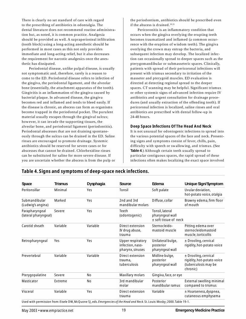
19 Emergency Medicine PracticeMay 2003 • www.empractice.net
There is clearly no set standard of care with regardto the prescribing of antibiotics in odontalgia. Thedental literature does not recommend routine administra-tion but, as noted, it is common practice. Analgesiashould be provided as well. A supraperiosteal infiltration(tooth block) using a long-acting anesthetic should beperformed in most cases as this not only providesimmediate and long-acting relief, but it also decreasesthe requirement for narcotic analgesics once the anes-thetic has dissipated.
Periodontal disease, unlike pulpal disease, is usuallynot symptomatic and, therefore, rarely is a reason tocome to the ED. Periodontal disease refers to infection ofthe gingiva, the periodontal ligament, and the alveolarbone (essentially, the attachment apparatus of the tooth).Gingivitis is an inflammation of the gingiva caused bybacterial plaque. In advanced disease, the gingivabecomes red and inflamed and tends to bleed easily. Ifthe disease is chronic, an abscess can form as organismsbecome trapped in the periodontal pocket. The purulentmaterial usually escapes through the gingival sulcus;however, it can invade the supporting tissues, thealveolar bone, and periodontal ligament (periodontitis).Periodontal abscesses that are not draining spontane-ously through the sulcus can be drained in the ED. Salinerinses are encouraged to promote drainage. Systemicantibiotics should be reserved for severe cases or forabscesses that cannot be drained. Chlorhexidine rinsescan be substituted for saline for more severe disease. Ifyou are uncertain whether the abscess is from the pulp or
the periodontium, antibiotics should be prescribed evenif the abscess is drained.10,31
Pericoronitis is an inflammatory condition thatoccurs when the gingiva overlying the erupting teethbecomes traumatized and inflamed (a common occur-rence with the eruption of wisdom teeth). The gingivaoverlying the crown may entrap the bacteria, andsubsequent infection may develop. The localized infec-tion can occasionally spread to deeper spaces such as thepterygomandibular or submasseteric spaces. Clinically,patients with spread of their pericoronal infections willpresent with trismus secondary to irritation of themasseter and pterygoid muscles. ED evaluation isdirected at detecting regional spread to the deeperspaces. CT scanning may be helpful. Significant trismusor other systemic signs of advanced infection require IVantibiotics and urgent consultation for drainage proce-dures (and usually extraction of the offending tooth). Ifpericoronal infection is localized, saline rinses and oralantibiotics are prescribed with dental follow-up in24-48 hours.
Deep Space Infections Of The Head And NeckIt is not unusual for odontogenic infections to spread intothe various potential spaces of the face and neck. Present-ing signs and symptoms consist of fever, chills, pain,difficulty with speech or swallowing, and trismus. (SeeTable 4.) Although certain teeth usually spread toparticular contiguous spaces, the rapid spread of theseinfections often makes localizing the exact space involved
Table 4. Signs and symptoms of deep-space neck infections.
Space Trismus Dysphagia Source Edema Unique Sign/Symptom
Peritonsillar Minimal Yes Tonsil Soft palate Uvular deviation,hot-potato voice, otalgia
Submandibular Marked Yes 2nd and 3rd Diffuse, collar Brawny edema, firm floor(Ludwig’s angina) mandibular molars of mouth
Parapharyngeal Severe Yes Teeth Tonsil, lateral(lateral pharyngeal) (odontogenic) pharyngeal wall
± soft tissue of neck
Carotid sheath Variable Variable Direct extension Sternocleido- Pitting edema overIV drug abuse, mastoid muscle sternocleidomastoidtrauma muscle, torticollis
Retropharyngeal Yes Yes Upper respiratory Unilateral bulge, ± Drooling, cervicalinfection, naso- posterior rigidity, hot-potato voicepharynx, sinuses pharyngeal wall
Prevertebral Variable Variable Direct extension Midline bulge, ± Drooling, cervicaltrauma, posterior rigidity, hot-potato voicetuberculosis pharyngeal wall (tuberculosis may be
chronic)
Pterygopalatine Severe No Maxillary molars Gingiva, face, or eye
Masticator Extreme No 3rd mandibular Posterior External swelling minimalmolar mandibular ramus compared to trismus
Visceral Variable Yes Direct extension Variable ± Hoarseness, dyspnea,trauma cutaneous emphysema
Used with permission from: Eisele DW, McQuone SJ, eds. Emergencies of the Head and Neck. St. Louis: Mosby; 2000. Table 19-1.

Emergency Medicine Practice 20 www.empractice.net • May 2003
difficult. Infections may involve the buccal, temporal,submasseteric, sublingual, submandibular,parapharyngeal, and other regions. (See Figure 19 andFigure 20.) Maxillary extension of periapical abscessescan spread into the infraorbital space and, subsequently,to the cavernous sinus through the ophthalmic veins,resulting in cavernous sinus thrombosis. Cavernous sinusinvolvement is usually associated with periorbitalcellulitis as well as meningeal signs or a decreased levelof consciousness.
Periapical infections of the anterior mandibular teethoften spread to the buccinator space or the sublingualspace, while those of the mandibular molars spread intothe submandibular space. The submandibular spaceconnects with the sublingual space. Bilateral involvementof both of these spaces is known as Ludwig’s angina andcan be life-threatening. Most cases of Ludwig’s anginaare caused by dental infections. Specific attention shouldbe paid to the neck and the floor of the mouth during thephysical examination. Woody induration of the tissues inthe mouth floor or elevation of the tongue is indicative ofspread of the infection across the midline. Airwayprecautions are of paramount importance.34 As infectionprogresses, the submandibular, submental, and sublin-gual spaces all become edematous, and there may beelevation of the tongue and the soft tissues of the mouth.
The soft tissues of the posterior pharynx can also becomeinvolved. The suprahyoid region of the neck appearstense and indurated, and landmarks may be obscured.
The management of complicated odontogenic headand neck infections centers on airway management,surgical drainage, and antibiotics. If there is uncertaintyas to whether the deep spaces are involved, CT scanningcan delineate the extension of the infectious process.31 CTscanning has become the imaging modality of choice for deepspace infections of the head and neck as it can localize collec-tions of cellulits or abscesses that cannot otherwise be exam-ined.35,36 Airway intervention should be performed early ifthere is any question of compromise, and tracheostomymay be indicated.37,38 Surgical consultation with theappropriate specialist is mandatory. Intravenous antibiot-ics should be administered in the ED.
The bacteria involved are typically a mixed Strepto-coccus/Staphylococcus combination, but a mixed aerobic/anaerobic infection is also possible. More than 40% ofisolates from odontogenic neck abscesses are resistant tobeta-lactam antibiotics.31 Antimicrobials of choice includepenicillin G plus metronidazole or expanded-spectrumpenicillins such as ampicillin/sulbactam, ticarcillin/clavulanate, and piperacillin/tazobactam. These combi-nation antibiotics are effective against beta-lactamase-producing bacteria and also cover anaerobes such asBacteroides fragilis. Clindamycin is an effective choicewhen patients are allergic to penicillin and should beused in combination with a cephalosporin such ascefotetan or cefoxitin in order to cover resistant organ-isms. In deep space infections, antibiotics are adjunctivetherapy and not a substitute for surgical intervention.
Figure 19. Temporal spaces of the neck.
Used with permission from: Eisele DW, McQuone JS, eds.Emergencies of the Head and Neck. St. Louis: Mosby; 2000.Figure 16-2 (a).
Figure 20. Location of temporalspace abscesses.
Used with permission from: Eisele DW, McQuone JS, eds.Emergencies of the Head and Neck. St. Louis: Mosby; 2000.Figure 16-2 (b).

21 Emergency Medicine PracticeMay 2003 • www.empractice.net
Special Considerations
ChildrenIt is important to remember that primary dentition isvery different than the permanent dentition, especiallywith respect to intrusion and avulsion. Intruded babyteeth are usually allowed to erupt, whereas permanentteeth that are intruded usually require repositioning andfixation. Avulsed primary teeth should not be replaced inthe ED. If there is a question of whether a tooth is apermanent tooth or not, replace the tooth and consult thepatient’s dentist. The tooth can be removed in the officein a day or two if necessary. The other way to determinewhether a tooth is permanent or not is to obtain aPanorex or facial films and look for the permanent toothin the alveolar bone.
The ElderlyThe elderly are much more likely to have prostheticteeth, implants, or other appliances. This must be takeninto consideration when determining what steps arenecessary to best preserve the function of a traumatizedor infected tooth. It is best to consult your dental col-leagues when intervening in the patient with significantdental hardware.
The ImmunosuppressedThe population least likely to have preventative dentalcare is perhaps also the population that is at risk forpoorly treated chronic disease or infectious diseases thatmay alter immune function. Those patients withimmunocompromised conditions are not only moresusceptible to chronic dental disease such as periodontaldisease, but they are also at higher risk for the develop-ment of deep space infections. Any patient who presentsto the ED with severe dentoalveolar disease should bequestioned about risk factors for HIV, diabetes, tubercu-losis, malignancy, etc., and the appropriate testing andfollow-up should be arranged.
Cutting Edge
Today’s EDs are awash in technological advancement.Ultrasound, electronic charting, clot-busting medications,bedside testing, and other technologies have trulyadvanced medical care. Unfortunately, many EDs don’thave the proper materials to take care of patients withsimple dental problems. This is most likely secondary tolack of dental-specific training among emergencyphysicians, but also to an over-dependence on our dentalcolleagues and a perceived lack of importance of dentalemergencies. Many medications, glues, pastes, andanesthetics are available to EDs, and our patients wouldbenefit immensely from them. Table 5 lists several thatEDs should have to provide basic dental care untildefinitive care from a dental professional can be pro-vided. The Dental Box (www.dentalbox.net, 412-364-8712) is one commercially available dental kit designedfor the emergency department. Also available is Dr.
Stahl’s Emergency Dental Kit (www.apohealth.com, 1-800-365-2839), which is designed for home use. Otherdental supply companies that supply these medicationsand materials include www.pattersondental.com,www.henryschein.com, and www.smartpractice.com.Many of these items can also be obtained from localdental suppliers.
Disposition
The vast majority of patients presenting to the ED withdental complaints can be treated as outpatients. Frac-tured, luxated, and carious teeth as well as simpleabscesses can all be followed up as outpatients. Themanagement of fractured teeth should be discussed witha dentist or oral surgeon prior to discharge if possible,especially if there is pulpal or deep dentinal involvement.Likewise, intruded teeth and alveolar ridge fracturesrequire repair within 24 hours, and early consultation ishelpful. Remember that most dental visits are precipi-tated by pain. Treat pain aggressively with oral analgesicsand, when necessary, dental blocks.
Deep space infections can be life-threatening andmay appear deceptively innocuous early in their course.Generally, if the abscess is limited to the buccal spaceand cheek area and there is no airway involvement,the patient can be treated as an outpatient. Likewise, ifthere is only slight swelling unilaterally below the bodyof the mandible, but the airway and voice are normal,the patient can usually be seen the next day. CT scanningshould be obtained, however, if the diagnosis is uncertainor the patient has significant swelling or trismus orif the infection appears to cross the midline. In mildinfections, it is prudent to begin antibiotic therapy inthe ED and arrange for definitive follow-up the nextday. Any patient with airway difficulty, bilateral involve-ment of the neck, toxic appearance, or a high likelihoodof noncompliance should be admitted to the hospital.Borderline cases should be treated with IV antibioticsand asked to return in 24 hours for a recheck if they
Table 5. Dental Equipment NeededIn The ED.
• Packing gauze• Dental roll gauze• Calcium hydroxide paste or glass ionomer cement or zinc
oxide cement• Dry Socket Paste or eugenol• Topical anesthetic gel (20% benzocaine or 5% lidocaine)• Topical bactericidal intraoral solution (Ora-5)• Periodontal paste (Coe-Pak) or self-cure composite• Bupivacaine cartridges with epinephrine• EMT ToothsaverTM Preservation System or fresh milk• Zinc oxide/eugenol temporary cement (Temrex)• Ringed injection syringe• Stainless steel spatula and mixing pads• Oral surgery tray with arch bars and ligature wires• Tongue blades and cotton-tipped applicators• Disposable electrocautery (optional)

Emergency Medicine Practice 22 www.empractice.net • May 2003
cannot follow up with the dentist.
Conclusion
Dental emergencies presenting to the ED are onlyoccasionally life-threatening. However, they are oftenvery painful and frequently of great cosmetic concern tothe patient. While many of these patients require defini-tive care by dentists or oral surgeons, the emergencyphysician plays an important role in their care. Theemergency physician must have a basic working knowl-edge of the dental anatomy and be prepared to save atooth, repair soft tissue, treat severe infection, and relievepain. The medications, glues, pastes, and medicamentswill improve over time. ED personnel should feel free todiscuss these advances with their dental colleagues.Likewise, the treatment of any dental or maxillofacialemergency should involve specialty consultation when itis beyond the scope of emergency practice. ▲
References
Evidence-based medicine requires a critical appraisal ofthe literature based upon study methodology andnumber of subjects. Not all references are equally robust.The findings of a large, prospective, randomized, andblinded trial should carry more weight than a case report.
To help the reader judge the strength of eachreference, pertinent information about the study, such asthe type of study and the number of patients in the study,will be included in bold type following the reference,where available. In addition, the most informativereferences cited in the paper, as determined by theauthors, will be noted by an asterisk (*) next to thenumber of the reference.
1. Waldrop RD, Ho B, Reed S. Increasing frequency of dentalpatients in the urban ED. Am J Emerg Med 2000 Oct;18(6):687-689. (Retrospective chart review; 3943 patients)
2.* Bringhurst C, Herr RD, Aldous JA. Oral trauma in theemergency department. Am J Emerg Med 1993 Sep;11(5):486-490.(Review)
3. Dale RA. Dentoalveolar trauma. Emerg Med Clin North Am 2000Aug;18(3):521-538. (Review)
4.* Klokkevold P. Common dental emergencies. Evaluation andmanagement for emergency physicians. Emerg Med Clin NorthAm 1989 Feb;7(1):29-63. (Review)
5. Holmes C, Sutcliffe P. Changes in the characteristics of patientsattending an out-of-hours emergency dental service inEdinburgh. Community Dent Health 1993 Mar;10(1):65-71.(Questionnaire)
6.* Haraden BM, Zwemer FL Jr. Descending necrotizing medias-tinitis: complication of a simple dental infection. Ann EmergMed 1997 May;29(5):683-686. (Case report)
7. Barrett EJ, Kenny DJ. Avulsed permanent teeth: a review of theliterature and treatment guidelines. Endod Dent Traumatol 1997Aug;13(4):153-163. (Review)
8.* Krasner P. Modern treatment of avulsed teeth by emergencyphysicians. Am J Emerg Med 1994 Mar;12(2):241-246. (Review)
9. Marino TG, West LA, Liewehr FR, et al. Determination ofperiodontal ligament cell viability in long shelf-life milk. JEndod 2000 Dec;26(12):699-702. (Prospective, basic science)
10. King R. Orofacial. In: Montgomery M, Redding S, eds. Oral-Facial Emergencies—Diagnosis and Management. 1st ed. Portland:JBK Publishing; 1994. (Textbook)
11. Dajani AS, Taubert KA, Wilson W, et al. Prevention of bacterial
endocarditis. Recommendations by the American HeartAssociation. JAMA 1997 Jun 11;277(22):1794-1801. (Consensusdevelopment conference, practice guideline, review)
12. Alonso LL, Purcell TB. Accuracy of the tongue blade test inpatients with suspected mandibular fracture. J Emerg Med 1995May-Jun;13(3):297-304. (Prospective; 110 patients)
13. Druelinger L, Guenther M, Marchand EG. Radiographicevaluation of the facial complex. Emerg Med Clin North Am 2000Aug;18(3):393-410. (Review)
14. Humphreys BF. Otolaryngologic emergencies. Emerg Med ClinNorth Am 1986 Aug;4(3):605-615. (Review)
15. Noyek AM, Kassel EE, Wortzman G, et al. Contemporaryradiologic evaluation in maxillofacial trauma. Otolaryngol ClinNorth Am 1983 Aug;16(3):473-508. (Review)
16. Markowitz BL, Sinow JD, Kawamoto HK Jr, et al. Prospectivecomparison of axial computed tomography and standard andpanoramic radiographs in the diagnosis of mandibularfractures. Ann Plast Surg 1999 Feb;42(2):163-169. (Prospective;33 mandibular fractures in 21 consecutive patients)
17. Roberts J, Hedges J, eds. Clinical Procedures in EmergencyMedicine. 2nd ed. Philadelphia: W.B. Saunders & Co.; 1991.(Textbook)
18. Jastak T, Yagiela J, Donaldson D, eds. Local Anesthesia of the OralCavity. Philadelphia: W.B. Saunders & Co.; 1995. (Textbook)
19. Rauschenberger CR, Hovland EJ. Clinical management ofcrown fractures. Dent Clin North Am 1995 Jan;39(1):25-51.(Review)
20.* Ellis SG. Incomplete tooth fracture—proposal for a newdefinition. Br Dent J 2001 Apr 28;190(8):424-428. (Review)
21. Beaudreau R. Oral and dental emergencies. In: Tintinalli JE,Kelen GD, Stapczynski JS, eds. Emergency Medicine—AComprehensive Study Guide. 5th ed. New York: McGraw-Hill;2000. (Textbook)
22. Bakland LK, Milledge T, Nation W. Treatment of crownfractures. J Calif Dent Assoc 1996 Feb;24(2):45-50. (Review)
23. Amsterdam J. Dental disorders. In: Rosen P, Barkin R, eds.Emergency Medicine: Concepts and Clinical Practice. 4th ed. St.Louis: Mosby; 1998. (Textbook)
24. Blatz MB. Comprehensive treatment of traumatic fracture andluxation injuries in the anterior permanent dentition. PractProced Aesthet Dent 2001 May;13(4):273-279. (Review)
25. Gilbert DN, et al, eds. The Sanford Guide to AntimicrobialTherapy. Hyde Park, VT: Antimicrobial Therapy, Inc.; 2002.(Textbook)
26. Dumsha TC. Luxation injuries. Dent Clin North Am 1995Jan;39(1):79-91. (Review)
27. Olson BD, Mailhot JM, Anderson RW, et al. Comparison ofvarious transport media on human periodontal ligament cellviability. J Endod 1997 Nov;23(11):676-679. (Comparative, basicscience)
28. Lee JY, Vann WF Jr, Sigurdsson A. Management of avulsedpermanent incisors: a decision analysis based on changingconcepts. Pediatr Dent 2001 Jul-Aug;23(4):357-360. (Review)
29. Steele MT, Sainsbury CR, Robinson WA, et al. Prophylacticpenicillin for intraoral wounds. Ann Emerg Med 1989Aug;18(8):847-852. (Randomized, controlled trial; 62 patients)
30. Trott A. Wounds and Lacerations. 2nd ed. New York: Mosby;1997. (Textbook)
31. McQuone S. Neck emergencies. In: Eisele D, McQuone S.Emergencies of the Head and Neck. 1st ed. St. Louis: Mosby; 2000.(Textbook)
32. Loesche WJ. Dental infections. In: Gorbach SL, Bartlett JG,Blacklow NR, eds. Infectious Diseases. 2nd ed. Philadelphia:W.B. Saunders; 1998:499-508. (Textbook)
33. Herrera D, Roldan S, Sanz M. The periodontal abscess: areview. J Clin Periodontol 2000 Jun;27(6):377-386. (Review)
34. Ashman S. Oral cavity and dental emergencies. In: Eisele DW,McQuone SJ, eds. Emergencies of the Head and Neck. St. Louis:Mosby; 2000. (Textbook)
35. Flynn TR. The swollen face. Severe odontogenic infections.Emerg Med Clin North Am 2000 Aug;18(3):481-519. (Review)
36. Lazor JB, Cunningham MJ, Eavey RD, et al. Comparison ofcomputed tomography and surgical findings in deep neckinfections. Otolaryngol Head Neck Surg 1994 Dec;111(6):746-750.
23 Emergency Medicine PracticeMay 2003 • www.empractice.net
(Retrospective; 38 patients)37. Neff SP, Merry AF, Anderson B. Airway management in
Ludwig’s angina. Anaesth Intensive Care 1999 Dec;27(6):659-661.(Case report)
38.* Shockley WW. Ludwig angina: a review of current airwaymanagement. Arch Otolaryngol Head Neck Surg 1999May;125(5):600. (Review)
Physician CME Questions
65. Which of the following about adult dentition iscorrect?a. There are eight incisors, four canines, eight
premolars, and 12 molars.b. There are four incisors, four canines, 12
premolars, and 12 molars.c. There are six incisors, four canines, 10
premolars, and 12 molars.d. None of the above.
66. Of the following dental emergencies, whichrequires the least urgent intervention?a. Trauma that could potentially cause airway
compromiseb. Mucosal or tongue lacerationsc. Deep space infectionsd. Uncomplicated tooth pain
67. Avulsed teeth should be rinsed and wiped off priorto reimplantation.a. Trueb. False
68. In obtaining a patient history in the case of dentaltrauma, which of the following suggest(s) that anavulsed tooth could have been aspirated?a. Coughing after the injuryb. Use of alcohol, sedatives, or recreational drugsc. Loss of consciousnessd. All of the above
69. Which of the following systemic conditions maycause oral pain or symptoms?a. Cranial neuralgiasb. Stomatitis and mucositisc. Erythema migransd. Crohn’s diseasee. All of the above
70. In cases of dental trauma, prehospital careproviders should avoid all of the following except:a. placing the patient in a supine position (unless
cervical spine injury is a concern).b. placing over-the-counter topical anesthetics on
the wound.c. controlling hemorrhages with gauze and direct
pressure.d. transporting an avulsed tooth in the patient’s
mouth to preserve the periodontal ligament.
71. The use of ringed syringe aspirators is recom-mended for intraoral injections.a. Trueb. False
72. The inability to crack a tongue blade bilaterallywhen it is twisted between the molars suggests:a. a mandibular fracture.b. a deep space infection.c. gingivitis.d. temporomandibular joint disorder.
73. The quickest way to relieve severe odontalgia inthe ED is with:a. NSAIDs.b. narcotic pain medication.c. dental block.d. none of the above.
74. Which of the following affect successfulreimplantation after a tooth has been completelyknocked out of the socket?a. Duration of time the tooth has been outb. Type of solution or medium that the tooth has
been kept inc. The health of the surrounding periodontiumd. All of the above
75. Which of the following is the best choice for adental block for a condition for which the patientwill be treated by a dentist the next day?a. Mepivacaine with levonordefrinb. Bupivacaine with epinephrinec. Mepivacained. Prilocaine with epinephrine
76. The most commonly fractured teeth are:a. the molars.b. the canines.c. the maxillary central incisors.d. the premolars.
77. All of the following are true about complicatedfractures of the crown that extend into the pulp ofthe tooth except:a. They are also known as Ellis class III fractures.b. They do not need to be handled urgently.c. They result in pulp necrosis in 10%-30% of cases,
even with appropriate treatment.d. They are distinguished from fractures of the
dentin by the pink color of the pulp.
78. All of the following are true about intraorallacerations except:a. They almost always become infected, so
prophylactic antibiotics are recommended bymost specialty societies.
b. If there is a significant amount of devitalized orcrushed tissue present, coverage of oral flora(penicillin or clindamycin) may be considered.
c. If the wound is through-and-through, coverageof oral flora (penicillin or clindamycin) andadditional skin coverage (clindamycin ordicloxacillin) may be considered.
d. Most tongue lacerations that are less than acentimeter and whose wound edges are notgaping do not require repair.

Emergency Medicine Practice 24 www.empractice.net • May 2003
Emergency Medicine Practice is not affiliated with any pharmaceutical firm or medical device manufacturer.
Direct all editorial or subscription-related questions to EB Practice, LLC: 1-800-249-5770 • Fax: 1-770-500-1316 • Non-U.S. subscribers, call: 1-678-366-7933
EB Practice, LLC • 305 Windlake Court • Alpharetta, GA 30022E-mail: [email protected] • Web Site: http://www.empractice.net
Emergency Medicine Practice (ISSN 1524-1971) is published monthly (12 times per year) by EB Practice, LLC, 305 Windlake Court, Alpharetta, GA 30022. Opinions expressed are not necessarilythose of this publication. Mention of products or services does not constitute endorsement. This publication is intended as a general guide and is intended to supplement, rather thansubstitute, professional judgment. It covers a highly technical and complex subject and should not be used for making specific medical decisions. The materials contained herein are notintended to establish policy, procedure, or standard of care. Emergency Medicine Practice is a trademark of EB Practice, LLC. Copyright 2003 EB Practice, LLC. All rights reserved. No part of thispublication may be reproduced in any format without written consent of EB Practice, LLC. Subscription price: $299, U.S. funds. (Call for international shipping prices.)
Publisher: Robert Williford. Executive Editor: Heidi Frost.
Physician CME InformationThis CME enduring material is sponsored by Mount Sinai School of Medicine andhas been planned and implemented in accordance with the Essentials andStandards of the Accreditation Council for Continuing Medical Education. Creditmay be obtained by reading each issue and completing the printed post-testsadministered in December and June or online single-issue post-testsadministered at www.empractice.net.
Target Audience: This enduring material is designed for emergency medicinephysicians.
Needs Assessment: The need for this educational activity was determined by asurvey of medical staff, including the editorial board of this publication; reviewof morbidity and mortality data from the CDC, AHA, NCHS, and ACEP; andevaluation of prior activities for emergency physicians.
Date of Original Release: This issue of Emergency Medicine Practice was publishedMay 1, 2003. This activity is eligible for CME credit through May 1, 2006. Thelatest review of this material was April 9, 2003.
Discussion of Investigational Information: As part of the newsletter, faculty maybe presenting investigational information about pharmaceutical products thatis outside Food and Drug Administration approved labeling. Informationpresented as part of this activity is intended solely as continuing medicaleducation and is not intended to promote off-label use of any pharmaceuticalproduct. Disclosure of Off-Label Usage: This issue of Emergency Medicine Practicediscusses no off-label use of any pharmaceutical product.
Faculty Disclosure: In compliance with all ACCME Essentials, Standards, andGuidelines, all faculty for this CME activity were asked to complete a fulldisclosure statement. The information received is as follows: Dr. Benko is theproprietor of Dental Box Company. Dr. Burke and Dr. Stewart report nosignificant financial interest or other relationship with the manufacturer(s) ofany commercial product(s) discussed in this educational presentation.
Accreditation: Mount Sinai School of Medicine is accredited by the AccreditationCouncil for Continuing Medical Education to sponsor continuing medicaleducation for physicians.
Credit Designation: Mount Sinai School of Medicine designates this educationalactivity for up to 4 hours of Category 1 credit toward the AMA Physician’sRecognition Award. Each physician should claim only those hours of creditactually spent in the educational activity. Emergency Medicine Practice is approvedby the American College of Emergency Physicians for 48 hours of ACEP Category1 credit (per annual subscription). Emergency Medicine Practice has been reviewedand is acceptable for up to 48 Prescribed credit hours by the American Academyof Family Physicians. Emergency Medicine Practice has been approved for 48Category 2-B credit hours by the American Osteopathic Association.
Earning Credit: Two Convenient Methods
• Print Subscription Semester Program: Paid subscribers with current andvalid licenses in the United States who read all CME articles during eachEmergency Medicine Practice six-month testing period, complete the post-test and the CME Evaluation Form distributed with the December and Juneissues, and return it according to the published instructions are eligible forup to 4 hours of Category 1 credit toward the AMA Physician’s RecognitionAward (PRA) for each issue. You must complete both the post-test and CMEEvaluation Form to receive credit. Results will be kept confidential. CMEcertificates will be delivered to each participant scoring higher than 70%.
• Online Single-Issue Program: Paid subscribers with current and validlicenses in the United States who read this Emergency Medicine Practice CMEarticle and complete the online post-test and CME Evaluation Form atwww.empractice.net are eligible for up to 4 hours of Category 1 credittoward the AMA Physician’s Recognition Award (PRA). You must completeboth the post-test and CME Evaluation Form to receive credit. Results willbe kept confidential. CME certificates may be printed directly from the Website to each participant scoring higher than 70%.
Class I• Always acceptable, safe• Definitely useful• Proven in both efficacy and
effectiveness
Level of Evidence:• One or more large prospective
studies are present (withrare exceptions)
• High-quality meta-analyses• Study results consistently
positive and compelling
Class II• Safe, acceptable• Probably useful
Level of Evidence:• Generally higher levels
of evidence• Non-randomized or retrospec-
tive studies: historic, cohort, orcase-control studies
• Less robust RCTs• Results consistently positive
Class III• May be acceptable• Possibly useful• Considered optional or
alternative treatments
Level of Evidence:• Generally lower or intermediate
levels of evidence
• Case series, animal studies,consensus panels
• Occasionally positive results
Indeterminate• Continuing area of research• No recommendations until
further research
Level of Evidence:• Evidence not available• Higher studies in progress• Results inconsistent,
contradictory• Results not compelling
Significantly modified from: TheEmergency Cardiovascular CareCommittees of the American HeartAssociation and representativesfrom the resuscitation councils ofILCOR: How to Develop Evidence-Based Guidelines for EmergencyCardiac Care: Quality of Evidenceand Classes of Recommendations;also: Anonymous. Guidelines forcardiopulmonary resuscitation andemergency cardiac care. EmergencyCardiac Care Committee andSubcommittees, American HeartAssociation. Part IX. Ensuringeffectiveness of community-wideemergency cardiac care. JAMA1992;268(16):2289-2295.
Class Of Evidence Definitions
Each action in the clinical pathways section of Emergency Medicine Practicereceives an alpha-numerical score based on the following definitions.
79. Avulsed primary teeth:a. should be replaced only if it can be done within
30 minutes.b. should be replaced only if the child is younger
than 6 years.c. should be replaced only if the root has been
preserved adequately.d. should not be replaced, because they could
cause craniofacial abnormalities, infection,or interfere with normal eruption of thepermanent teeth.
80. Treatment of alveolar bone fractures requires rigidsplinting of the affected segment by an oralsurgeon or dentist as soon as possible.a. Trueb. False