emergency evaluation for pulmonary embolism, part 2 ... · clinical reviews emergency evaluation...
TRANSCRIPT

The Journal of Emergency Medicine, Vol. -, No. -, pp. 1–14, 2015Copyright � 2015 Elsevier Inc.
Printed in the USA. All rights reserved0736-4679/$ - see front matter
http://dx.doi.org/10.1016/j.jemermed.2014.12.041
RECEIVED: 30 SeACCEPTED: 21 D
ClinicalReviews
EMERGENCY EVALUATION FOR PULMONARY EMBOLISM, PART 2:DIAGNOSTIC APPROACH
Jeffrey A. Kline, MD* and Christopher Kabrhel, MD, MPH†
*Department of Emergency Medicine and Department of Cellular and Integrative Physiology, Indiana University School of Medicine,Indianapolis, Indiana and †Department of Emergency Medicine, Center for Vascular Emergencies, Massachusetts General Hospital and
Department of Emergency Medicine, Harvard Medical School, Boston, Massachusetts
Reprint Address: Jeffrey A. Kline, MD, Department of Emergency Medicine and Department of Cellular and Integrative Physiology, IndianaUniversity School of Medicine, 720 Eskanazi Avenue, Indianapolis, IN 46202
, Abstract—Background: In part 1 of this two-part re-view, we discussed which risk factors, historical features,and physical findings increase risk for pulmonary embolism(PE) in symptomatic emergency department (ED) patients.Objectives: Use published evidence to describe criteriathat a reasonable and prudent clinician can use to initiateand guide the process of excluding and diagnosing PE. Dis-cussion: Thecareful anddiligent emergencyphysician canuseclinical criteria to safely obviate a formal evaluation of PE,including the use of gestalt reasoning and the pulmonary em-bolism rule-out criteria (PERC rule, Table 2, part 1). We pre-sent published clinical and radiographic features of patientswith PE who eluded diagnosis in the ED. D-dimer can beused to exclude PE in many patients, and employing age-based adjustments to the threshold to define an abnormalvalue can further reduce patient exposure to pulmonaryvascular imaging. Moreover, we discuss benefits, limitations,and potential harms of computed tomographic pulmonaryvascular imaging relevant topatients and thepractice of emer-gency care. We present algorithms to guide exclusion anddiagnosis of PE in patients with suspected PE, including thosewho are pregnant. Conclusions: Reasonable and prudentemergency clinicians can exclude PE in symptomatic ED pa-tients on clinical grounds alone in many patients, and manymore can have PE ruled out by use of the D-dimer. � 2015Elsevier Inc.
, Keywords—pulmonary embolism; medicolegal; defen-sive medicine; decision making; venous thromboembolism;pregnancy; diagnosis; pregnancy
ptember 2014; FINAL SUBMISSION RECEIVED: 17ecember 2014
1
INTRODUCTION
This second part of a two-part review provides anin-depth analysis of issues critical to deciding when toinitiate a formal diagnostic evaluation for pulmonaryembolism (PE) in emergency department (ED) patients,and what diagnostic tests, if any, need to be ordered. Weexplore evidence-based options for excluding PE to areasonable degree of diagnostic certainty but withminimal exposure to radiation and iodinated contrastmaterial.
DISCUSSION
Decision to Initiate the Work-up and Empiric Treatment
Figure 1 presents an algorithm for the diagnostic evalua-tion of patients with possible PE. For PE to enter theactive differential diagnosis list for any patient, he orshe must have at least one possible physiologic manifes-tation of PE. The physiologic manifestation may be asymptom (e.g., dyspnea, pleuritic chest pain, or newfatigue) or a sign (e.g., heart rate > 100 beats/min or pulseoximetry < 95% near sea level) that is not explained byanother cause. Other bedside physiological signs of PEinclude a low (<30 mm Hg) end-tidal CO2, measuredby capnography, or signs of pulmonary hypertension on12-lead electrocardiography, including T-wave inversion
December 2014;
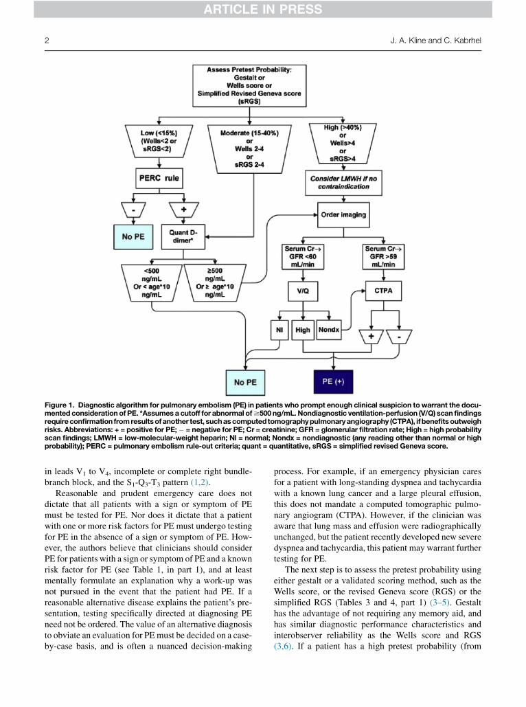
Figure 1. Diagnostic algorithm for pulmonary embolism (PE) in patients who prompt enough clinical suspicion towarrant the docu-mented considerationof PE. *Assumesa cutoff for abnormal of$500 ng/mL.Nondiagnostic ventilation-perfusion (V/Q) scanfindingsrequireconfirmation fromresultsofanother test, suchascomputedtomographypulmonaryangiography (CTPA), ifbenefitsoutweighrisks. Abbreviations: + = positive for PE;� = negative for PE; Cr = creatinine; GFR = glomerular filtration rate; High = high probabilityscan findings; LMWH = low-molecular-weight heparin; Nl = normal; Nondx = nondiagnostic (any reading other than normal or highprobability); PERC = pulmonary embolism rule-out criteria; quant = quantitative, sRGS = simplified revised Geneva score.
2 J. A. Kline and C. Kabrhel
in leads V1 to V4, incomplete or complete right bundle-branch block, and the S1-Q3-T3 pattern (1,2).
Reasonable and prudent emergency care does notdictate that all patients with a sign or symptom of PEmust be tested for PE. Nor does it dictate that a patientwith one or more risk factors for PE must undergo testingfor PE in the absence of a sign or symptom of PE. How-ever, the authors believe that clinicians should considerPE for patients with a sign or symptom of PE and a knownrisk factor for PE (see Table 1, in part 1), and at leastmentally formulate an explanation why a work-up wasnot pursued in the event that the patient had PE. If areasonable alternative disease explains the patient’s pre-sentation, testing specifically directed at diagnosing PEneed not be ordered. The value of an alternative diagnosisto obviate an evaluation for PEmust be decided on a case-by-case basis, and is often a nuanced decision-making
process. For example, if an emergency physician caresfor a patient with long-standing dyspnea and tachycardiawith a known lung cancer and a large pleural effusion,this does not mandate a computed tomographic pulmo-nary angiogram (CTPA). However, if the clinician wasaware that lung mass and effusion were radiographicallyunchanged, but the patient recently developed new severedyspnea and tachycardia, this patient may warrant furthertesting for PE.
The next step is to assess the pretest probability usingeither gestalt or a validated scoring method, such as theWells score, or the revised Geneva score (RGS) or thesimplified RGS (Tables 3 and 4, part 1) (3–5). Gestalthas the advantage of not requiring any memory aid, andhas similar diagnostic performance characteristics andinterobserver reliability as the Wells score and RGS(3,6). If a patient has a high pretest probability (from
Emergency Evaluation for Pulmonary Embolism 3
any method), the clinician should consider immediatelyadministering heparin or low-molecular-weight heparinfor patients with low bleeding risk. However, the benefitsof ‘‘empiric’’ anticoagulation remain unproven. One re-view suggested that the benefit of empiric systemic anti-coagulation for 24 h exceeds the risks (bleeding andheparin-induced thrombocytopenia) for any patient witha pretest probability of PE of >20% (7). Several studieshave suggested that delay in administration of heparinto patients with PE can increase mortality, but no studyhas found that heparin administered prior to imaging im-proves morbidity or mortality (8–10).
Three studies have provided data on patients whopassed through the ED and were soon after diagnosedwith PE (8,11–13). These patients can be categorized asthose admitted to the hospital and those dischargedhome. Compared with patients who were promptlydiagnosed and treated for PE, patients admitted to thehospital who went on to have delayed recognition of PEtended to have a higher frequency of altered mentalstatus (either new or at baseline dementia) andpreexisting heart and lung disease (8,11–13). Only onestudy provided data on patients apparently dischargedwith PE, and those patients were more likely to not havedyspnea, have isolated pleuritic chest pain andhemoptysis together with a pulmonary infiltrate onimaging, and a lower D-dimer concentration with a smalldistal clot seen on pulmonary vascular imaging (12). Coin-cidentally, in an analysis of PE(+) but pulmonary embo-lism rule-out criteria (PERC)(�) patients (see Table 2 inpart 1) in a large database, the presence of pleuritic chestpain emerged as a common feature (14). Thus, it seemsthat highly competent emergency physicians may missdistal lung clots that produce pulmonary infarction and aclinical picture of pneumonia. More evidence is neededto determine if patients with these small distal clots, inthe absence of deep venous thrombosis (DVT), actuallybenefit from systemic anticoagulation.
Exclusion of PE at the Bedside
About two-thirds of patients who are considered for testingfor PE in the United States have a low pretest probability,regardless of the method used, and the prevalence of PE inthis group is <5% (15). Patients with a low gestalt pretestprobability (defined as a global estimate that the patienthas <15% probability of PE) are eligible to have PE ruledout with the PERC rule (see Table 2 in part 1).
The authors suggest that ruling out PE requires a com-bination of pretest probability and diagnostic test resultsthat predict an outcome rate, or false negative rate<2.0% for any one patient. This false negative rate,synonymous with posttest probability, equals the productof the likelihood ratio (LR) for a negative diagnostic test
result (LR� = [1 � sensitivity]/specificity) times the pre-test odds (odds = probability/[1� probability]) (note thatodds are always higher than probability), which is thenconverted from an odds value back to probability. Thus,for a low-risk population—for example, one with an un-derlying prevalence of venous thromboembolism (VTE)of 4–5% defined by gestalt low clinical probability—the PERC rule, functioning as a diagnostic test, has anLR� of about 0.2 or less, and therefore clearly can ruleout VTE, based upon a predefined posttest threshold of2.0% (3,16,17):
Pretest probability = 4%.Pretest odds = 0.04 (1�0.04) = 0.04/0.96 = 0.042.Post-test odds = LR� * pretest odds = 0.2 *
0.042 = 0.0084.Post-test probability = odds/(1 + odds) = 0.0084/
1.0084 = 0.0083 or 0.8%.Here we refer to ‘‘posttest’’ under the assumption that
clinical criteria, namely the PERC rule, can function asthe diagnostic test. Importantly, a population of patients,each with a pretest probability <2%, collectively has alower false negative rate. The combination of a low clin-ical gestalt impression plus a negative PERC rule reliablypredicts a probability of PE below 1% even in Europeanpopulations (16–20). Use of the PERC rule after a lowpretest probability using other methods of pretestprobability assessment besides gestalt assessment hasnot been validated (21). At a pretest probability <2%,the risk of further testing outweighs the low probabilityof failing to diagnose PE (22,23). Therefore, if allcriteria of the PERC rule are met in the setting of agestalt-based low pretest probability, not only is furthertesting unnecessary, but it should be avoided if possible.
The PERC rule (Table 2, part 1) does not have 100%sensitivity, and will be negative in the presence of smallPE at a rate of about 1 in 100 patients considered, andeven more rarely in the presence of larger PE (14,24).In most cases, in a patient suspected of having PE ifany one of the eight criteria is not met, or the doctorsimply thinks a test is indicated, the patient shouldundergo a diagnostic test for PE. Not all patients who‘‘fail’’ the PERC rule need an objective test for PEordered; the PERC rule provides only one set of criteriato rule out PE, and other sets likely exist.
D-dimer Testing
Assuming PE cannot be ruled out with the PERC rule, thenext step is to determine which specific diagnostic testmakes sense in view of the patient’s pretest probability.If the ED clinician has access to a quantitative D-dimerassay, it should be strongly considered as a first diagnostictest in patients for whom clinical suspicion is low or mod-erate based on either gestalt estimation, a Wells score of
4 J. A. Kline and C. Kabrhel
#4, or an RGS #4 (see Tables 3 and 4 in part 1) (25,26).Available data from the United States suggest that aquantitative D-dimer at standard threshold produces afalse negative rate <1% even with a Wells score up to6 (15). Most commercially available, automated,quantitative D-dimer assays that employ either immuno-turbidimetric latex agglutination or enzyme-linked immu-noabsorbance colorimetry as the detection method have anLR� of <0.15 (27,28). Different D-dimer assays havevariable thresholds for normal due to different captureantibodies and optical methods of detection. Somelaboratories report results in D-dimer mass concentration(e.g., nanograms per milliliter or micrograms permilliliter) and others report fibrinogen equivalent units,which are twice the mass concentration. The D-dimerhas a half-life in plasma of approximately 8 h, and extrap-olating from humans and animal models of autologous PE,the D-dimer level probably remains abnormally high for atleast 3 days after symptomatic PE (29–32). However, as D-dimer may be continuously shed by unstable clot, it isdifficult to know exactly how long after an acute PE a D-dimer assay will remain positive.
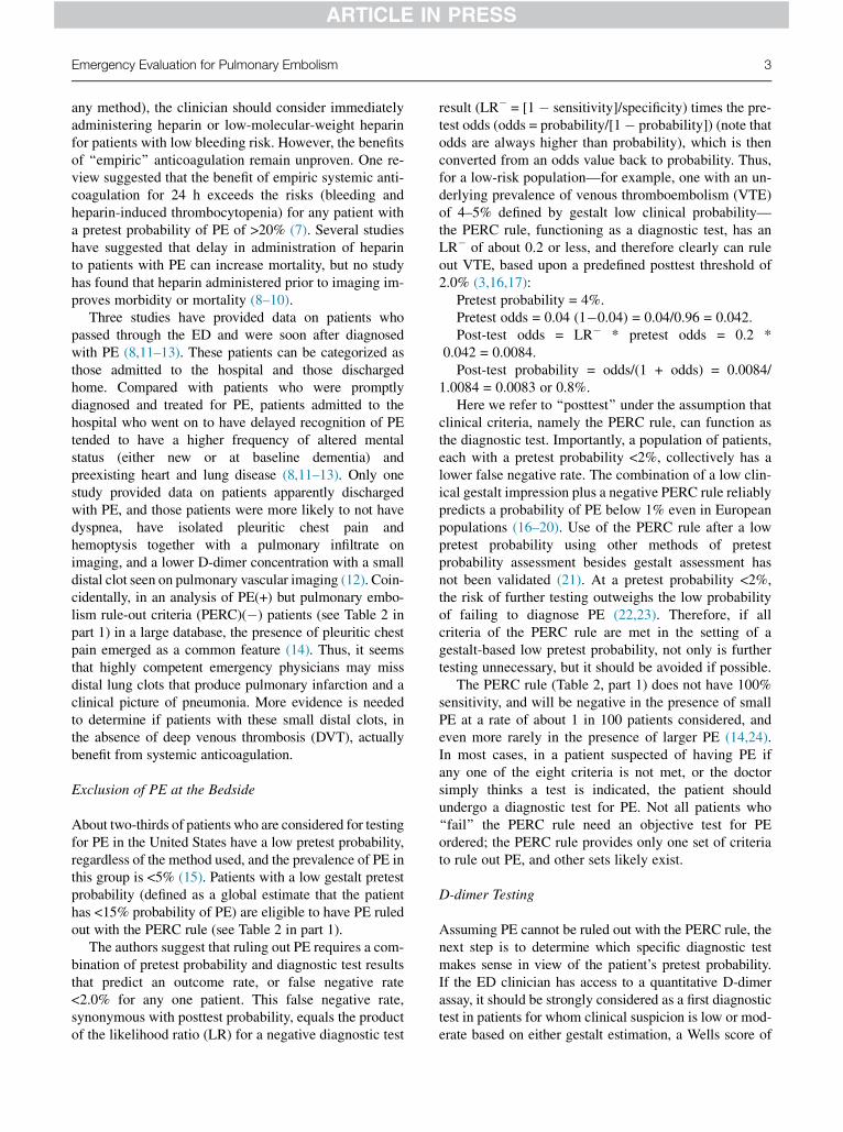
The most common causes of false positive and falsenegative D-dimer results are listed in Table 1 (29–31,33,34). Almost all risk factors for PE also elevate theD-dimer concentration. The fact that D-dimer increaseswith age has prompted numerous researchers to test if theD-dimer can be adjusted upward for age and maintainadequate exclusionary ability, mainly for suspected PE.The most common formula studied is age * 10 ng/mL,where a patient aged 80 years would have an age-adjusted threshold for abnormal at 800 ng/mL (35–37). Ina large multicenter management study, this approach,
Table 1. Factors that Cause Errors in D-dimer Measurements (33–
False Positives
Patient factors:� Increasing age: (60–69 years [OR 2.6], 70–79 years [OR 4.5],
$80 years [OR 10.5])� Cocaine use (OR 2.0)� Immobility: general (OR 2.3), limb (OR 2.8), or neurologic
(OR 3.0)� Hemoptysis (OR 2.0)� Hemodialysis (OR 2.2)� Malignancy, active (OR 2.6)� Rheumatoid arthritis (OR 2.8)� Systemic lupus erythematosus (OR 2.1)� Sickle cell disease (OR 24.2)� Pregnancy and postpartum state: (2nd trimester [OR 7.3],
3rd trimester [OR 51.3], postpartum [OR 4.2])� Surgery (<4 weeks prior): abdominal (OR 3.5), chest (OR
2.7), orthopedic (OR 2.2), other surgery (OR 3.2)
OR = odds ratio; PE = pulmonary embolism.* Derived from case reports, experience and manufacturer’s informatio† Theoretically, risk is greatest with vitamin K antagonists and dabigatrafactor XIII generation, which could allow for non-cross-linked but insolugulation are simply chronic and thus liberate small amounts of D-dime
when used in conjunction with a Wells score #4 or asimplified revised Geneva score #4, was associated witha very low rate (0.3%) of PE diagnosis on 3-monthfollow-up (see Tables 3 and 4, part 1) (37). Age adjustmentof D-dimer is also supported by previous meta-analyses ofother studies, as well as recent studies not yet aggregatedinto a systematic review. It is our opinion that it is reason-able to use age-adjusted D-dimer values to rule out PE inpatients with low or moderate pretest probability (36).
All patients with a positive D-dimer result that cannotbe explained by another finding must undergo imagingdirected at discovering clots, and the choice of thenext test must be determined by a mix of patient and fa-cility factors. As the physician becomes aware of newinformation, PE may move up, down, or off the differen-tial diagnosis list even after a D-dimer test result isfound to be positive. Removing VTE from the differen-tial must be justified by the presence of a condition thatobviously explains the elevated D-dimer (e.g., one of thecauses listed in Table 1) together with a plausible expla-nation for the patient’s symptoms that is unlikely to co-exist with PE (e.g., pneumothorax) (34). In particular,clinicians should not assume that an elevated troponindecreases the probability of PE (e.g., in favor of cardiacischemia), as troponin elevation occurs in about 20% ofpatients with PE and is associated with worse outcomes(38–40). Similarly, emergency physicians must beaware that about 45% of patients with PE have anelevated brain natriuretic peptide concentration(39,40). Clinicians should be aware that normalizationof initially abnormal vital signs has not been found toreduce the probability of PE and should not be used tojustify cancelling a previously ordered test for PE (41).
37))
False Negatives*
Patient factors:� Concomitant anticoagulation†� Symptoms lasting more than 5 days� Subsegmental PE� Isolated pulmonary infarction� Chronic PE
System and machine issues:� Wrong sample� Severe lipemia or hemolysis� Protein degradation by proteolysis that can occur withprolonged time from sample draw to analysis
n.n, as both inhibit active thrombin generation and therefore reduceble clots. More likely, most PE diagnosed in patients on anticoa-r.

Emergency Evaluation for Pulmonary Embolism 5
IMAGING
CT Pulmonary Angiography (CTPA)
A good quality computed tomography (CT) scan, whichrequires about 200 Hounsfield units of contrast opacifica-tion in the main pulmonary artery, rules out PE at all pre-test probabilities on the day of examination (42,43).Chest CT does not rule out the possibility of future PEfrom undiagnosed DVT. Chest CT angiography onlyidentifies filling defects in contrast-enhanced pulmonaryarteries. Most scanning protocols require the patient tolie supine and hold his or her breath for a few seconds.CT scanning requires injection of approximately120 mL of contrast by a computer-controlled injectiondevice. The patient must have a peripheral intravenous(i.v.) catheter (20 gauge or larger) or an approvedindwelling line to allow injection of the contrast. Equip-ment with multiple detector heads (e.g., 64-head scan-ners) allows better resolution so that filling defects canbe observed even in subsegmental pulmonary arteries(44). The diagnostic sensitivity and specificity of a tech-nically adequate CT scan, performed on a multidetectorCT scanner in an ED population independently of pretestprobability, are both about 90% (43). Interobserver agree-ment in identifying segmental or larger filling defects hasbeen consistently demonstrated to be very good, but inter-observer agreement for subsegmental clots is poor (45).Benefits of CT scanning include a binary positive or nega-tive result and the ability to detect evidence supporting aclinically significant alternative diagnosis (where pneu-monia is most common, found in 8–22% of cases)(33,46–50).
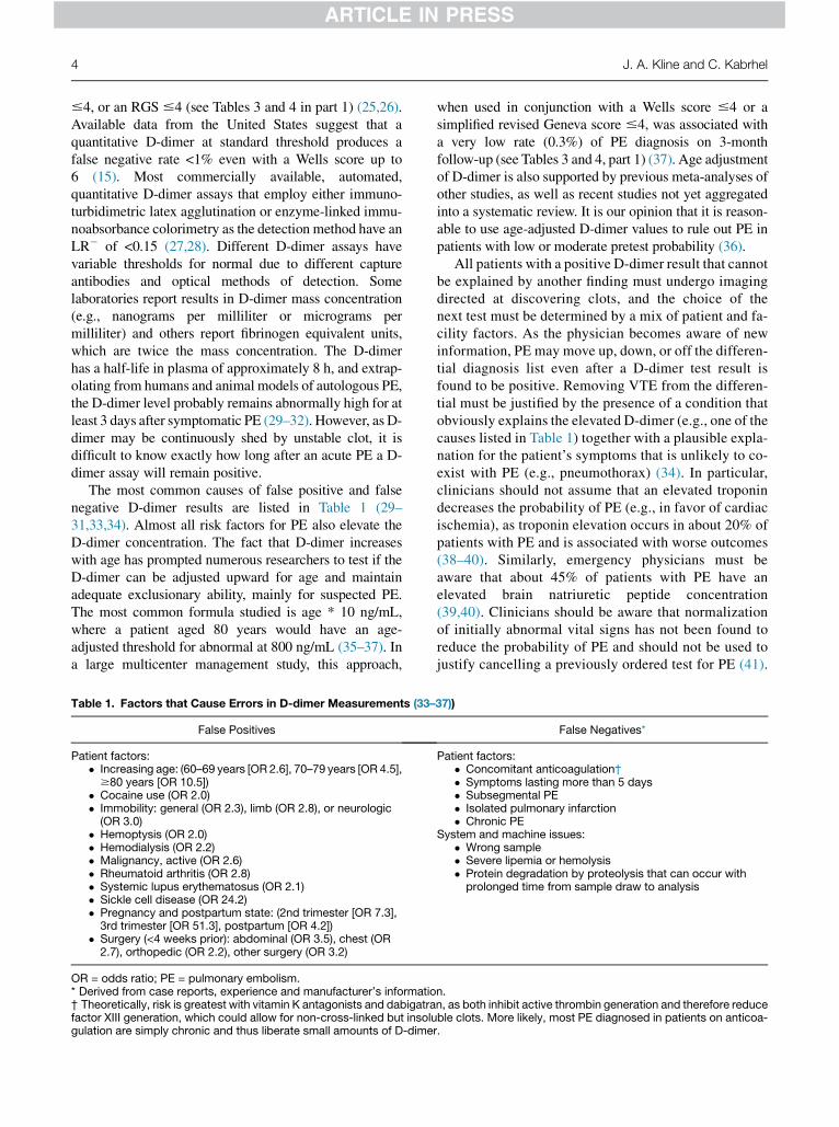
Radiologists indicate presence of suboptimal imagequality in about 10% of their formal interpretations ofCTPA scans (45,51). Figure 2 (A and B) shows examplesof high-quality scan images and a degraded image. Im-age quality is most commonly degraded by low arterialopacification, or motion artifact (e.g., from severe ta-chypnea) (52). Obesity increases risk of inadequateCTPA imaging (53,54). Radiologists probably cannotdetect filling defects with <150 Hounsfield units ofopacification (42,52). In real practice, approximately10% of CT scans yield technically inadequate imagessecondary to motion artifact or poor pulmonary arteryopacification, most commonly in obese or verytachypneic patients (53,54). Many diagnostic studiesof CTPA scanning exclude these scans from analysis,but in the PIOPED II study, 11/51 patients withindeterminate CTPA scans had PE on reference testing(51). Patients with indeterminate CTPA results andmoderate or high pretest probability may need bridginganticoagulation until a PE can be ruled out. This can bedone with a follow-up Ventilation Perfusion (V/Q) scan
that has homogeneous perfusion. Because the outcomerate of PE after a negative quantitative D-dimer is<1%, even in high pretest probability patients, the quan-titative D-dimer, if negative (age adjustment allowed),provides strong evidence to rule out PE in the settingof a degraded image CTPA. However, as about 80–90% of high pretest probability patients will have a pos-itive D-dimer, the usefulness of this approach is limited(15,34). Alternatively, standard care also includesbilateral lower-extremity venous ultrasonography, per-formed in the ED and again within 3–7 days (55–58).If results of this repeat examination are negative, VTEcan be ruled out in high-risk patients after indeterminateCTPA scanning (27,37,51,59–61).
The CT scan, despite its remarkable value as a diag-nostic tool, poses risks to patients that emergency physi-cians must know. First among these is a 6–10% falsepositive rate in low-risk populations, possibly leading toover-diagnosis and unnecessary anticoagulation(44,45,51,62). CTPA imparts approximately 10 to20 mSv of radiation, with an estimated increasedlifetime risk of fatal cancer of at least 1 in 500 per chestCT (63,64). This risk may be higher in young womendue to radiation to the breast (65). Furthermore, within5 years afterward, more than one-third of patients whohave one CT angiography to rule out PE can be expectedto undergo subsequent CT pulmonary angiography,incurring a second dose of radiation (66). Acute life-threatening complications from CT scanning includeanaphylactoid reaction to contrast and pulmonary edema.Other complications from CT scanning include contrastextravasation into a limb that causes pain, or in severecases, compartment syndrome. Fortunately, extravasationis rare, occurring in <1 in 500 patients (67).
For patients who report a history of previous immedi-ate reaction to iodinated contrast (itching, urticarial,wheezing, or full anaphylactoid reaction), their recur-rence rate is approximately 6–15% with re-exposure,compared to 1% for patients with no prior contrast reac-tion (67,68). The risk of breakthrough hypersensitivityseems to be reduced by one-half with pretreatment withparenteral corticosteroids (e.g., hydrocortisone, 200 mg,i.v.) and antihistamines (e.g., chlorpheniramine 4 mgi.v. or diphenhydramine 25 mg i.v.) (69–73). In general,patients with prior allergic diathesis (e.g., any allergy,asthma or general atopy) have a 3–10-fold increasedrisk of immediate contrast reaction, but data are mixedas to whether a shellfish allergy increases a patient’srisk of immediate contrast media reaction (74,75).
About 15% of patients undergoing contrast-enhancedchest CT scanning go on to develop contrast nephropathy,which, according to its minimal definition, comprises a25% increase in the serum creatinine concentration,measured within 2–7 days of the examination (76).
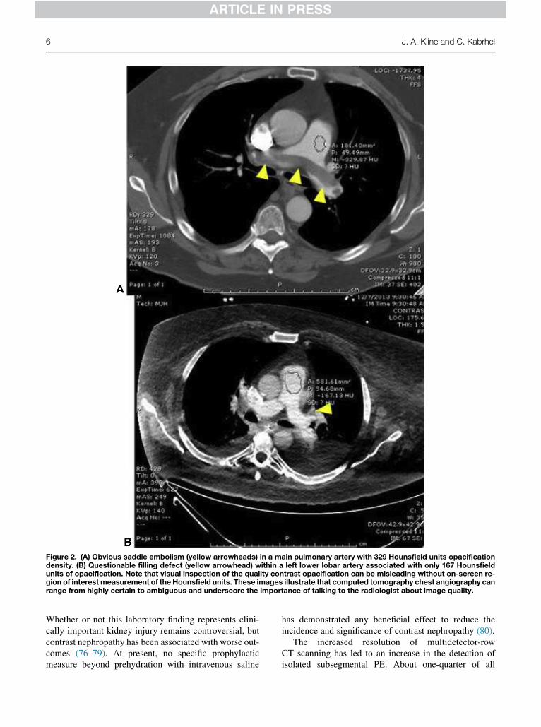
Figure 2. (A) Obvious saddle embolism (yellow arrowheads) in a main pulmonary artery with 329 Hounsfield units opacificationdensity. (B) Questionable filling defect (yellow arrowhead) within a left lower lobar artery associated with only 167 Hounsfieldunits of opacification. Note that visual inspection of the quality contrast opacification can be misleading without on-screen re-gion of interest measurement of the Hounsfield units. These images illustrate that computed tomography chest angiography canrange from highly certain to ambiguous and underscore the importance of talking to the radiologist about image quality.
6 J. A. Kline and C. Kabrhel
Whether or not this laboratory finding represents clini-cally important kidney injury remains controversial, butcontrast nephropathy has been associated with worse out-comes (76–79). At present, no specific prophylacticmeasure beyond prehydration with intravenous saline
has demonstrated any beneficial effect to reduce theincidence and significance of contrast nephropathy (80).
The increased resolution of multidetector-rowCT scanning has led to an increase in the detection ofisolated subsegmental PE. About one-quarter of all

Emergency Evaluation for Pulmonary Embolism 7
contrast-enhanced chest CT scans read as positive for PEhave isolated subsegmental PE (81–83). Isolatedsubsegmental PE refers to a filling defect seen in onesmall pulmonary artery, usually <3 mm in diameter, inthe absence of DVT. The problem with this diagnosis isthat when the same images are shown to a secondblinded radiologist, in about half of all cases, thesecond radiologist finds no PE (45). This raises concernthat subsegmental PE may be a radiographic artifactrather than a true disease. One survey found that most cli-nicians in Canada would opt not to treat these patientswithout further testing (84). However, another studyfound that the prognosis of patients with subsegmentalPE was not different than patients with segmental ormore proximal PE (85). No randomized trial has exam-ined the safety of withholding anticoagulation for iso-lated subsegmental PE and perhaps as a result, oneclinical guideline recommends standard course anticoa-gulation (86,87). In general, most experts agree thatisolated subsegmental PE in patients with active cancershould be treated, even if discovered incidentally(84,88–92). For patients without cancer, a secondinternational survey found that a majority thrombosisexperts recommend anticoagulation for most isolatedsubsegmental PE (93). The authors recommend treatmentof isolated subsegmental PE if the patient has PE symp-toms, a prior history of PE, an ongoing risk of PE (e.g.,indwelling catheter or immobility), or an elevatedDdimer (93). Patients with isolated subsegmental PEand a high risk of bleeding (e.g., Registro Informatizadode pacientes con Enfermedad TromboEmbolica [RIETE]bleeding score > 1) probably should not be treated (94). Inthese situations, and in any case where the risks and ben-efits of treatment are unclear, the patient should beinformed of the situation and help make the decisionabout anticoagulation.
Ventilation Perfusion Scintillation (V/Q)
Ventilation-perfusion scintillation (V/Q) lung scanningrequires peripheral intravenous access, and for the patientto sit upright during injection of a radioisotopic nuclide,usually 99Tc macroaggregate, followed by positioningthe patient in front of a gamma camera to capture thegamma emission from the radionuclide as it traversesthe pulmonary vasculature. Use of a central line to injectisotope will often lead to inadequate images. If the perfu-sion lung scan shows a homogenous perfusion pattern(i.e., a ‘‘normal’’ perfusion scan; Figure 3A), this is asso-ciated with a likelihood ratio negative of 0.05, and essen-tially rules out PE (95,96). Patients without PE and withnormal chest radiographs are far more likely to havenormal V/Q scan than patients with intrinsic lungdisease seen on chest radiograph. In patients with
nonnormal perfusion, most U.S. radiology departmentsalso perform the ventilation phase of the V/Q scan,which requires the patient to inhale an aerosolcontaining 99mTc diethylenetriamine-pentaacetic acidor 133Xe. Although starting with the perfusion scan mayobviate the need for the ventilation scan, and therebyreduce radiation exposure, image quality is best if theventilation phase is performed first because the back-ground emission from the perfusion scan persists forhours. To diagnose PE, the perfusion scan must showtwo or more apex-central, wedge-shaped defects in perfu-sion pattern in a segmental or larger vascular distribution,together with evidence of normal ventilation in the samelung segments (Figure 3B). The primary technical limita-tions of V/Q scanning include the availability ofpersonnel to perform and interpret them and the availabil-ity of isotope. Some emergency clinicians may not beaware that the availability of the 99Tc isotope dependsupon a cyclotron particle accelerator to manufactureeach day. The primary clinical limitation of V/Q scanningis the fact that approximately two-thirds of scans areneither normal nor diagnostic of PE, which requires pa-tients to undergo further testing.
Bilateral Ultrasonography
Performing lower-extremity venous ultrasonography issensible due to its lack of ionizing radiation and the factthat diagnosing DVT is tantamount to diagnosing PEfrom the standpoint of the decision to administer heparinanticoagulation in the ED. In fact, studies suggest that acombination of D-dimer testing and lower-extremity ul-trasound may be the most cost-effective approach to theinitial evaluation of PE (22,97). However, in theabsence of physical findings that suggest DVT, bilateralultrasonography is of limited usefulness for excludingPE in the ED. Data from the largest study thatsimultaneously performed bilateral leg ultrasonographyand performed pulmonary vascular imaging indicatethat a negative bilateral proximal lower-extremity venousultrasound has a sensitivity of 30% and a specificity of57% (LR for a negative test = 1.22) for PE (98). Otherstudies have yielded similar results, so all patients sus-pected of having PE for whom ultrasound findings arenegative require pulmonary vascular imaging (51,99).Nonetheless, following the logic that discovery of DVTis tantamount to diagnosis of PE in terms of treatmentaction in the ED, pursuing DVT first makes sense forpatients with a positive D-dimer and for patients whoobject to pulmonary vascular imaging.
Follow-up bilateral venous ultrasound is also the bestoption to rule out PE in patients with high pretest proba-bility, a positive D-dimer, and a negative CTPA scan withany radiologist comment about degraded image quality.

Figure 3. (A) Normal ventilation-perfusion (V/Q) scan showing homogenous ventilation (top) and perfusion (bottom) images.These images rule out pulmonary embolism. (B) V/Q lung scan series consistent with a high probability of acute pulmonary em-bolismusing standard criteria. The top rowof each panel shows the ventilation phase, conductedwith 133Xe,which producesonlyone planar image. The images labeled ‘‘equilibrium’’ or ‘‘w/o’’ represent washout images taken later. The second row of eachpanel demonstrates perfusion phases of the examination, obtained with 99Tc macroaggregate. The black arrowheads point towedge-shaped defects in the perfusion images. Scans are read by looking for defects in the perfusion phase where the corre-sponding ventilation view shows relatively homogeneous scintillation activity in the anatomic segments that lack perfusion.
8 J. A. Kline and C. Kabrhel
Management protocols using this approach find that 5%of these high-risk patients will have a DVT at 3–7 daysfollow-up (55–58).
Pregnant Women
Figure 4 proposes an algorithm to rule out and diagnose PEin pregnant patients. The algorithm starts with bilateral
lower-extremity venous ultrasound. If the bilateral ultra-sound is positive, then treatment can be started. Otherwise,the next step is determined by pretest probability assess-ment. To our knowledge, no pretest probability ruleshave been validated in pregnant patients. It is clear thatover one-half of all VTEs diagnosed in pregnancy occurin the third trimester (100). In the authors’ experience(JAK and CK) and based on available patient-level data

Figure 4. Proposed algorithm for the exclusion and diagnosis of pulmonary embolism in pregnant patients with suspected pulmo-nary embolism (PE) in the emergency department setting. This algorithm has not been formally tested. Shared decision-making re-fers to discussion of the diagnostic options with the patient, including uncertain, but probably <5% risk of undiagnosed PE and thepotential risks of computed tomographic pulmonary angiogram (CTPA) or V/Q scanning to the fetus. Nonhigh pretest probability(PTP) refers to absence of high PTP by gestalt, Wells or sRGS. See text for references. *D-dimer concentrations per trimester givenin ng/mL assuming a standard D-dimer threshold for abnormal of 500 ng/mL. Abbreviations: CXR = chest radiograph; Q = perfusionlung scan; + = positive for PE; � = negative for PE; Cr = creatinine; High = high-probability scan findings; LMWH = low-molecular-weight heparin; Nl = normal; Nondx = nondiagnostic (any reading other than normal or high probability); PERC = pulmonary embo-lism rule-out criteria; quant = quantitative, sRGS = simplified revised Geneva score; V/Q = ventilation perfusion.
Emergency Evaluation for Pulmonary Embolism 9
from pregnant ED patients, highWells and Geneva scores,the third trimester, or unexplained hypoxemia(SaO2 < 95% breathing room air at sea level) predict a rela-tively higher pretest probability for PE (101).
The evaluation of possible PE in pregnancy challengesclinicians, who must consider the epidemiological datashowing increased risk of PE, the potential catastropheof failing to protect the life of mother and child, and thepotential for increased lifetime risks from unnecessary ra-diation and contrast exposure to the mother and the fetus.
It is worth noting that most patients with pregnancyselected by emergency physicians for PE work-up havea low clinical probability (101). No firm guidelines existto guide the work-up of pregnant patients with suspectedPE (102,103). Efforts should be made to avoid fetalexposure to radiation and iodinated contrast (104,105).The proposed algorithm in Figure 4 draws from availableliterature and expert opinion, and has been used infor-mally by one of the authors for over 8 years, but hasnot been formally tested (102). If the patient has a high

10 J. A. Kline and C. Kabrhel
pretest probability by Wells or the revised Geneva score,or is in the third trimester, or has unexplained hypoxemia,then she should probably proceed to testing. Unfortu-nately, the D-dimer at standard threshold is not usuallyhelpful because almost all women in the third trimesterhave a positive D-dimer when the standard positivitythreshold is used, and pulmonary vascular imaging isfrequently required (106). Because imaging with ionizingradiation may be required, the authors recommend dis-cussing the diagnostic approach with the patient prior toinitiating the work-up using a shared decision-makingapproach, similar to that described by Hess et al. forlow-risk chest pain (107). When the risks and benefitsof testing are explained, some mothers will opt to pro-ceed, whereas others will choose to avoid imaging at allcosts. To minimize radiation exposure, the authors pro-pose a combined approach, where negative bilaterallower-extremity venous ultrasonography is supportedby a negative PERC rule and a threshold-adjusted D-dimer assay. The exclusionary power of a single negativebilateral leg ultrasound for PE per se has not been testedin pregnancy, but seems to be similar to that of nonpreg-nant patients for excluding DVT (108,109). The D-dimerthreshold can be adjusted according to the trimester ofpregnancy, as follows: first trimester, 750 ng/mL;second trimester, 1000 ng/mL; third trimester, 1250 ng/mL (assuming a standard cutoff of 500 ng/mL)(106,110,111). If the patient has a non-high-pretest prob-ability, has no high-risk features, is PERC negative, andthe bilateral ultrasound is negative, and the D-dimer isbelow the trimester-adjusted values, PE can be ruledout to a reasonable degree of medical certainty. Notethat this recommendation does not state that the PERCcriteria can be used alone in pregnancy.
If the D-dimer is abnormal or the patient fails thePERC criteria, then a pulmonary vascular imaging studyis warranted. The best choice of pulmonary vascular im-aging is controversial and uncertain (102). Some evi-dence has suggested up to a 35% rate of inadequatepulmonary vascular opacification with CPTA, especiallyin the third trimester, resulting in a higher rate of nondiag-nostic studies with CTPA than V/Q or Q-alone scanning(112,113). Other data indicate that either CT pulmonaryangiography or V/Q scanning will produce adequateimages to rule out and diagnose PE in a pregnantpatient (103,114,115). The data used to estimate therisk of fetal exposure to radiation for CT scanning vs.V/Q scanning are both highly speculative. Shielding theabdomen with a lead or bismuth-antimony apron duringCT scanning may reduce radiation based upon phantommodeling (116). When available, tube voltage modu-lating technology may also serve to lower fetal radiationexposure more than shielding (116). Magnetic resonanceimaging has not been adequately tested in pregnancy to
provide any basis for recommendation, but had too lowa sensitivity (78%) to rule out PE in nonpregnant patients(117). As Figure 4 demonstrates, both CTPA and V/Qscanning are equally justifiable when imaging a pregnantpatient is necessary. If a V/Q scan is chosen, the authorssuggest first performing a plain film chest radiograph, andperforming the V/Q scan only if the radiograph is normal.Then, we suggest performing a perfusion-only (Q) scanwith half-dose 99Tc macroaggregate. Because 99Tc isexcreted in the urine, prehydration with 1 L of intrave-nous saline and insertion of a Foley catheter seems alogical, but unproven, step to reduce fetal exposure to ra-diation. The risk of this approach is that if the perfusionlung scan is nonnormal, and CT scanning is ultimatelyrequired, the mother and fetus will be exposed to more ra-diation than if CTPA had been performed first. In patientswith an abnormal chest radiograph, we suggest perform-ing a CTPA rather than a V/Q scan.
CONCLUSION
Acute pulmonary embolism can be ruled out on clinicalgrounds without laboratory or radiographic imaging bythe combined use of gestalt pretest probability estimationplus negative PERC rule. In the presence of non-high-pretest probability by any method, including gestaltassessment, a negative quantitative D-dimer rules outPE at standard or age-adjusted threshold. However,threshold adjustment is complicated by differing,manufacturer-specific thresholds to define the cutoff foran abnormal D-dimer. A good-quality CTPA scan rulesout PE. Patients with high pretest probability and a nega-tive CTPA but with degraded image quality, can have PEruled out with a normal V/Q scan, a negative quantitativeD-dimer, or negative bilateral lower-extremity venous ul-trasound performed in the ED and again 3–7 days later.Exclusion of PE in pregnancy remains a controversialsubject, but a shared decision-making model that priori-tizes testing without fetal radiation exposure may offerthe most effective and safe approach.
REFERENCES
1. Manara A, D’hoore W, Thys F. Capnography as a diagnostic toolfor pulmonary embolism: a meta-analysis. Ann Emerg Med2013;62:584–91.
2. Marchick MR, Courtney DM, Kabrhel C, et al. 12-Lead ECGfindings of pulmonary hypertension occur more frequently inemergency department patients with pulmonary embolism thanin patients without pulmonary embolism. Ann Emerg Med2010;55:331–5.
3. Lucassen W, Geersing GJ, Erkens PM, et al. Clinical decisionrules for excluding pulmonary embolism: a meta-analysis. AnnIntern Med 2011;155:448–60.
4. Ceriani E, Combescure C, Le Gal G, et al. Clinical predictionrules for pulmonary embolism: a systematic review and meta-analysis. J Thromb Haemost 2010;8:957–70.

Emergency Evaluation for Pulmonary Embolism 11
5. Klok FA,Mos IC, Nijkeuter M, et al. Simplification of the revisedGeneva score for assessing clinical probability of pulmonary em-bolism. Arch Intern Med 2008;168:2131–6.
6. Runyon MS, Webb WB, Jones AE, et al. Comparison of the un-structured clinician estimate of low clinical probability for pul-monary embolism to the Canadian score or the Charlotte rule.Acad Emerg Med 2005;12:587–93.
7. Hogg KE, Brown MD, Kline JA. Estimating the pretest probabil-ity to justify the empiric admininistration of heparin prior to pul-monary vascular imaging for pulmonary embolism. Thromb Res2006;118:547–53.
8. Smith SB, Geske JB, Maguire JM, et al. Early anticoagulation isassociated with reduced mortality for acute pulmonary embo-lism. Chest 2010;137:1382–9.
9. Jelinek GA, Ingarfield SL, Mountain D, et al. Emergency depart-ment diagnosis of pulmonary embolism is associated with signif-icantly reduced mortality: a linked data population study. EmergMed Australas 2009;21:269–76.
10. Kline JA, Marchick MR, Kabrhel C, et al. Prospective study of thefrequency and outcomes of patients with suspected pulmonary em-bolism administered heparin prior to confirmatory imaging.Thromb Res 2012;129:e25–8.
11. Kline JA, Hernandez J, Jones AE, et al. Prospective study of theclinical features and outcomes of emergency department patientswith delayed diagnosis of pulmonary embolism. Acad EmergMed 2007;14:592–8.
12. Torres-Macho J, Mancebo-Plaza AB, Crespo-Gimenez A, et al.Clinical features of patients inappropriately undiagnosed of pul-monary embolism. Am J Emerg Med 2013;31:1646–50.
13. den Exter PL, van den Hoven P, van der Hulle T, et al. Performanceof the revised Geneva score in patients with a delayed suspicion ofpulmonary embolism. Eur Respir J 2014;43:1801–4.
14. Kline JA, Slattery D, O’Neil BJ, et al. Clinical features of patientswith pulmonary embolism and a negative PERC rule result. AnnEmerg Med 2013;61:122–4.
15. Kabrhel C, Courtney DM, Camargo CA Jr, et al. Potential impactof adjusting the threshold of the quantitative D-dimer based uponpretest probability of acute pulmonary embolism. Acad EmergMed 2009;16:325–42.
16. Singh B, Mommer SK, Erwin PJ, et al. Pulmonary embolism rule-out criteria (PERC) in pulmonary embolism–revisited: a system-atic review and meta-analysis. Emerg Med J 2013;30:701–6.
17. Singh B, Parsaik AK, Agarwal D, Surana A, Mascarenhas SS,Chandra S. Diagnostic accuracy of pulmonary embolism rule-out criteria: a systematic review and meta-analysis. Ann EmergMed 2012;59:517–20. e1–e4.
18. Fesmire FM, Brown MD, Espinosa JA, et al. Critical issues in theevaluation and management of adult patients presenting to theemergency department with suspected pulmonary embolism.Ann Emerg Med 2011;57:628–52.
19. Penaloza A, Verschuren F, Dambrine S, et al. Performance of thepulmonary embolism rule-out criteria (the PERC rule) combinedwith low clinical probability in high prevalence population.Thromb Res 2012;129:e189–93.
20. Bokobza J, Aubry A, Nakle N, et al. Pulmonary embolism rule-outcriteria vs D-dimer testing in low-risk patients for pulmonary em-bolism: a retrospective study. Am J Emerg Med 2014;32:609–13.
21. Hugli O, Righini M, Le Gal G, et al. The pulmonary embolismrule-out criteria (PERC) rule does not safely exclude pulmonaryembolism. J Thromb Haemost 2011;9:300–4.
22. Lessler AL, Isserman JA, Agarwal R, et al. Testing low-risk pa-tients for suspected pulmonary embolism: a decision analysis.Ann Emerg Med 2010;55:316–26.
23. Pauker SG, Kassirer JP. The threshold approach to clinical deci-sion making. N Engl J Med 1980;302:1109–17.
24. Hennessey A, Setyono DA, Lau WB, et al. A patient with a largepulmonary saddle embolus eluding both clinical gestalt and vali-dated decision rules. Ann Emerg Med 2012;59:521–3.
25. van Belle A, Buller HR, HuismanMV, et al. Effectiveness of man-aging suspected pulmonary embolism using an algorithmcombining clinical probability, D-dimer testing, and computed to-mography. JAMA 2006;295:172–9.
26. Bertoletti L, Le Gal G, Aujesky D, et al. Prognostic value of theGeneva prediction rule in patients with pulmonary embolism.Thromb Res 2013;132:32–6.
27. Stein PD, Hull RD, Patel KC, et al. D-dimer for the exclusion ofacute venous thrombosis and pulmonary embolism: a systematicreview. Ann Intern Med 2004;140:589–602.
28. BrownMD, Lau J, Nelson RD, et al. Turbidimetric D-Dimer test inthe diagnosis of pulmonary embolism: a meta-analysis. Clin Chem2003;49:1846–53.
29. Couturaud F, Kearon C, Bates SM, Ginsberg JS. Decrease insensitivity of D-dimer for acute venous thromboembolism afterstarting anticoagulant therapy. Blood Coagul Fibrinolysis 2002;13:241–6.
30. Taira T, Taira BR, Carmen M, et al. Risk of venous thromboembo-lism in patients with borderline quantitative D-dimer levels. Am JEmerg Med 2010;28:450–3.
31. Kutinsky I, Blakley S, Roche V. Normal D-dimer levels in patientswith pulmonary embolism. Arch Intern Med 1999;159:1569–72.
32. Runyon MS, Gellar MA, Sanapareddy N, et al. Development andcomparison of a minimally-invasive model of autologous clot pul-monary embolism in Sprague-Dawley and Copenhagen rats.Thromb J 2010;8:3.
33. Kline JA, Hogg MM, Courtney DM, et al. D-dimer threshold in-crease with pretest probability unlikely for pulmonary embolismto decrease unnecessary computerized tomographic pulmonaryangiography. J Thromb Haemost 2012;10:572–81.
34. Kabrhel C, Mark Courtney D, Camargo CA Jr, et al. Factors asso-ciated with positive D-dimer results in patients evaluated for pul-monary embolism. Acad Emerg Med 2010;17:589–97.
35. Penaloza A, Roy PM, Kline J, et al. Performance of age-adjustedD-dimer cut-off to rule out pulmonary embolism. J Thromb Hae-most 2012;10:1291–6.
36. Adams D, Welch JL, Kline JA. Clinical utility of an age-adjustedD-dimer in the diagnosis of venous thromboembolism. Ann EmergMed 2014;64:232–4.
37. Righini M, van Es J, den Exter PL, et al. Age-adjusted D-dimercutoff levels to rule out pulmonary embolism: the ADJUST-PEstudy. JAMA 2014;311:1117–24.
38. Becattini C, Vedovati MC, Agnelli G. Prognostic value of tropo-nins in acute pulmonary embolism: a meta-analysis. Circulation2007;116:427–33.
39. Kline JA, Hernandez J, Rose G, et al. Surrogate markers foradverse outcomes in normotensive patients with pulmonary embo-lism. Crit Care Med 2006;34:2773–80.
40. Jimenez D, Kopecna D, Tapson V, et al. Derivation and validationof multimarker prognostication for normotensive patients withacute symptomatic pulmonary embolism. Am J Respir Crit CareMed 2014;189:718–26.
41. Kline JA, Corredor DM, Hogg MM, et al. Normalization of vitalsigns does not reduce the probability of acute pulmonary embo-lism in symptomatic emergency department patients. Acad EmergMed 2012;19:11–7.
42. Burnside P, Kline JA. Indirect computed tomography venography:quality of vascular opacification and diagnostic implications.Emerg Radiol 2010;17:195–202.
43. Mos IC, Klok FA, Kroft LJ, et al. Safety of ruling out acute pulmo-nary embolism by normal computed tomography pulmonary angi-ography in patients with an indication for computed tomography:systematic review and meta-analysis. J Thromb Haemost 2009;7:1491–8.
44. Schissler AJ, Rozenshtein A, KulonME, et al. CT pulmonary angi-ography: increasingly diagnosing less severe pulmonary emboli.PLoS One 2013;8:e65669.
45. Courtney DM, Miller CD, Smithline HA, et al. Prospective multi-center assessment of interobserver agreement for radiologist inter-pretation of multidetector CT angiography for pulmonary embo-lism. J Thromb Haemost 2010;8:533–40.
46. Richman PB, CourtneyDM, Kline JA. Prevalence and significanceof non-thromboembolic findings on chest computerized tomogra-phy angiography performed to rule-out pulmonary embolism-Amulti-center study of 1025 Emergency Department patients.Acad Emerg Med 2004;11:642–7.

12 J. A. Kline and C. Kabrhel
47. Hall WB, Truitt SG, Scheunemann LP, et al. The prevalence ofclinically relevant incidental findings on chest computed tomo-graphic angiograms ordered to diagnose pulmonary embolism.Arch Intern Med 2009;169:1961–5.
48. van Es J, Douma RA, Schreuder SM, et al. Clinical impact of find-ings supporting an alternative diagnosis on CT pulmonary angiog-raphy in patients with suspected pulmonary embolism. Chest2013;144:1893–9.
49. van Strijen MJ, Bloem JL, de Monye W, et al. Helical computedtomography and alternative diagnosis in patients with excludedpulmonary embolism. J Thromb Haemost 2005;3:2449–56.
50. Shujaat A, Shapiro JM, Eden E. Utilization of CT pulmonary angi-ography in suspected pulmonary embolism in a major urban emer-gency department. Pulm Med 2013;2013:915213.
51. Stein PD, Fowler SE, Goodman LR, et al. Multidetector computedtomography for acute pulmonary embolism. N Engl J Med 2006;354:2317–27.
52. Kay FU, Macedo AC, Chate RC, et al. Reduction of poor contrastenhancement of the pulmonary artery in computed tomographyangiography using an alternative respiratory maneuver. J ThoracImaging 2014;29:107–12.
53. Hawley PC, HawleyMP. Difficulties in diagnosing pulmonary em-bolism in the obese patient: a literature review. Vasc Med 2011;16:444–51.
54. Bae KT, Tao C, Gurel S, et al. Effect of patient weight and scan-ning duration on contrast enhancement during pulmonary multide-tector CT angiography. Radiology 2007;242:582–9.
55. Wells PS, Anderson D, Bormanis J. Value of assessment of pretestprobability of deep-vein thrombosis in clinical management. Lan-cet 1997;350:1795–8.
56. Heijboer H, Buller HR, Lensing AW, et al. A comparison of real-time compression ultrasonography with impedance plethysmog-raphy for the diagnosis of deep-vein thrombosis in symptomaticoutpatients. N Engl J Med 1993;329:1365–9.
57. Birdwell BG, Raskob GE, Whitsett TL, et al. The clinical val-idity of normal compression ultrasonography in outpatients sus-pected of having deep vein thrombosis. Ann Intern Med 1998;128:1–7.
58. Wells P, Ginsberg J, Anderson D, et al. Use of a clinical model forsafe management of patients with suspected pulmonary embolism.Ann Intern Med 1998;129:997–1005.
59. Kline JA, Runyon MS, Webb WB, Jones AE, Mitchell AM. Pro-spective study of the diagnostic accuracy of the simplify D-dimerassay for pulmonary embolism in emergency department patients.Chest 2006;129:1417–23.
60. Runyon MS, Beam DM, King MC, et al. Comparison of thesimplify D-dimer assay performed at the bedside with alaboratory-based quantitative D-dimer assay for the diagnosis ofpulmonary embolism in a low prevalence emergency departmentpopulation. Emerg Med J 2008;25:70–5.
61. Gottschalk A, Sostman HD, Coleman RE, et al. Ventilation-perfu-sion scintigraphy in the PIOPED study. Part II. Evaluation of thescintigraphic criteria and interpretations. J Nucl Med 1993;34:1119–26.
62. Hoffman JR, Cooper RJ. Overdiagnosis of disease: a modernepidemic. Arch Intern Med 2012;172:1123–4.
63. Einstein AJ, Henzlova MJ, Rajagopalan S. Estimating risk ofcancer associated with radiation exposure from 64-slicecomputed tomography coronary angiography. JAMA 2007;298:317–23.
64. Health Physics Society. Radiation exposure from medical diag-nostic imaging procedures Health Physics Society fact sheet.McLean, VA: Health Physics Society; 2008.
65. Parker MS, Hui FK, Camacho MA, et al. Female breast radiationexposure during CT pulmonary angiography. AJR Am J Roent-genol 2005;185:1228–33.
66. Kline JA, Courtney DM, BeamDM, et al. Incidence and predictorsof repeated computed tomographic pulmonary angiography inemergency department patients. Ann Emerg Med 2009;54:41–8.
67. Mitchell AM, Jones AE, Tumlin JA, et al. Immediate complica-tions of intravenous contrast for computed tomography imaging
in the outpatient setting are rare. Acad Emerg Med 2011;18:1005–9.
68. Kopp AF, Mortele KJ, Cho YD, et al. Prevalence of acute reactionsto iopromide: postmarketing surveillance study of 74,717 patients.Acta Radiol 2008;49:902–11.
69. Williams AN, Kelso JM. Radiocontrast-induced anaphylaxisdespite pretreatment and use of iso-osmolar contrast. Ann AllergyAsthma Immunol 2007;99:467–8.
70. Marshall GD Jr, Lieberman PL. Comparison of three pretreatmentprotocols to prevent anaphylactoid reactions to radiocontrast me-dia. Ann Allergy 1991;67:70–4.
71. Kim SH, Lee SH, Lee SM, et al. Outcomes of premedication fornon-ionic radio-contrast media hypersensitivity reactions in Ko-rea. Eur J Radiol 2011;80:363–7.
72. Bae YJ, Hwang YW, Yoon SY, et al. The effectiveness of auto-matic recommending system for premedication in reducing recur-rent radiocontrast media hypersensitivity reactions. PLoS One2013;8:e66014.
73. Delaney A, Carter A, Fisher M. The prevention of anaphylactoidreactions to iodinated radiological contrast media: a systematic re-view. BMC Med Imaging 2006;6:2.
74. Wolf GL, MishkinMM, Roux SG, et al. Comparison of the rates ofadverse drug reactions. Ionic contrast agents, ionic agents com-bined with steroids, and nonionic agents. Invest Radiol 1991;26:404–10.
75. Baig M, Farag A, Sajid J, et al. Shellfish allergy and relation toiodinated contrast media: United Kingdom survey.World J Cardiol2014;6:107–11.
76. Sinert R, Brandler E, Subramanian RA, et al. Does the current defi-nition of contrast-induced acute kidney injury reflect a true clinicalentity? Acad Emerg Med 2012;19:1261–7.
77. Mitchell AM, Jones AE, Tumlin JA, et al. One year outcomesfollowing contrast induced nephropathy. Am J Intern Med 2013;1:1–6.
78. Mitchell AM, Jones AE, Tumlin JA, et al. Prospective study of theincidence of contrast-induced nephropathy among patients evalu-ated for pulmonary embolism by contrast-enhanced computed to-mography. Acad Emerg Med 2012;19:618–25.
79. Mitchell AM, Kline JA. Contrast-induced nephropathy: doubtsand certainties. Acad Emerg Med 2012;19:1294–6.
80. Au TH, Bruckner A, Mohiuddin SM, Hilleman DE. The preven-tion of contrast-induced nephropathy. Ann Pharmacother 2014;48:1332–42.
81. Stein PD, Goodman LR, Hull RD, et al. Diagnosis and managementof isolated subsegmental pulmonary embolism: review and assess-ment of the options. Clin Appl Thromb Hemost 2012;18:20–6.
82. Carrier M, Righini M, Le Gal G. Symptomatic subsegmental pul-monary embolism: what is the next step? J Thromb Haemost 2012;10:1486–90.
83. Carrier M, Righini M, Wells PS, et al. Subsegmental pulmonaryembolism diagnosed by computed tomography: incidence andclinical implications. A systematic review and meta-analysis ofthe management outcome studies. J Thromb Haemost 2010;8:1716–22.
84. Carrier M, KimptonM, LE Gal G, et al. The management of a sub-segmental pulmonary embolism: a cross-sectional survey of Cana-dian thrombosis physicians. J Thromb Haemost 2011;9:1412–5.
85. den Exter PL, van Es J, Klok FA, et al. Risk profile and clinicaloutcome of symptomatic subsegmental acute pulmonary embo-lism. Blood 2013;122:1144–9.
86. Kearon C, Akl EA, Comerota AJ, et al. Antithrombotic therapy forVTE disease: Antithrombotic Therapy and Prevention of Throm-bosis, 9th ed: American College of Chest Physicians Evidence-Based Clinical Practice Guidelines. Chest 2012;141(2 Suppl):e419S–94.
87. Yoo HH, Queluz TH, El Dib R. Anticoagulant treatment for sub-segmental pulmonary embolism. Cochrane Database Syst Rev2014;(4):CD010222.
88. den Exter PL, Jimenez D, Kroft LJ, Huisman MV. Outcome ofincidentally diagnosed pulmonary embolism in patients with ma-lignancy. Curr Opin Pulm Med 2012;18:399–405.

Emergency Evaluation for Pulmonary Embolism 13
89. Abdel-Razeq HN, Mansour AH, Ismael YM. Incidental pulmo-nary embolism in cancer patients: clinical characteristics andoutcome–a comprehensive cancer center experience. Vasc HealthRisk Manag 2011;7:153–8.
90. Douma RA, Kok MG, Verberne LM, et al. Incidental venousthromboembolism in cancer patients: prevalence and conse-quence. Thromb Res 2010;125:e306–9.
91. Dentali F, AgenoW, Becattini C, et al. Prevalence and clinical his-tory of incidental, asymptomatic pulmonary embolism: a meta-analysis. Thromb Res 2010;125:518–22.
92. LimWY, Bozas G, Noble S, Hart S,Maraveyas A. Anticoagulatingthe subsegmental pulmonary embolism in cancer patients: a surveyamongst different medical specialties. J Thromb Thrombolysis2014 Oct 18. [Epub ahead of print].
93. Pesavento R, Casazza F, Filippi L, et al. An international survey onisolated subsegmental pulmonary embolism. Thromb Res 2013;131:183–4.
94. Nieto JA, Solano R, Trapero IN, et al. Validation of a score for pre-dicting fatal bleeding in patients receiving anticoagulation forvenous thromboembolism. Thromb Res 2013;132:175–9.
95. The PIOPED Investigators. Value of the ventilation/perfusion scanin acute pulmonary embolism. JAMA 1990;263:2753–9.
96. Worsley DF, Alavi A. Comprehensive analysis of the results of thePIOPED study. J Nucl Med 1995;36:2380–7.
97. Duriseti RS, Brandeau ML. Cost-effectiveness of strategies fordiagnosing pulmonary embolism among emergency departmentpatients presenting with undifferentiated symptoms. Ann EmergMed 2010;56:321–32.
98. Righini M, Le Gal G, Aujesky D, et al. Diagnosis of pulmonaryembolism by multidetector CT alone or combined with venous ul-trasonography of the leg: a randomised non-inferiority trial. Lan-cet 2008;371:1343–52.
99. Daniel KR, Jackson RE, Kline JA. Utility of the lower ex-tremity venous ultrasound in the diagnosis and exclusion ofpulmonary embolism in outpatients. Ann Emerg Med 2000;35:547–54.
100. Meng K, Hu X, Peng X, et al. Incidence of venous thromboembo-lism during pregnancy and the puerperium: a systematic reviewand meta-analysis. J Matern Fetal Neonatal Med 2014 May 7;1–9. [Epub ahead of print].
101. Kline JA, Richardson DM, Than MP, et al. Systematic review andmeta-analysis of pregnant patients investigated for suspected pul-monary embolism in the emergency department. Acad EmergMed2014;21:949–59.
102. Leung AN, Bull TM, Jaeschke R, et al. An official AmericanThoracic Society/Society of Thoracic Radiology clinical practiceguideline: evaluation of suspected pulmonary embolism in preg-nancy. Am J Respir Crit Care Med 2011;184:1200–8.
103. TanM, HuismanMV. The diagnostic management of acute venousthromboembolism during pregnancy: recent advancements andunresolved issues. Thromb Res 2011;127(Suppl 3):S13–6.
104. Moon AJ, Katzberg RW, Sherman MP. Transplacental passage ofiohexol. J Pediatr 2000;136:548–9.
105. MatthewsS. Imaging pulmonary embolism inpregnancy:what is themost appropriate imaging protocol? Br J Radiol 2006;79:441–4.
106. Kline JA, Hambleton GW, Hernandez J. D-dimer concentrations innormal pregnancy: new diagnostic thresholds are needed. ClinChem 2005;51:825–9.
107. Hess EP, Knoedler MA, Shah ND, et al. The chest pain choice de-cision aid: a randomized trial. Circ Cardiovasc Qual Outcomes2012;5:251–9.
108. Ratiu A, Navolan D, Spatariu I, et al. Diagnostic value of a negativesingle color duplex ultrasound in deep vein thrombosis suspicionduring pregnancy. RevMedChir SocMedNat Iasi 2010;114:454–6.
109. Le Gal G, Kercret G, Ben YahmedK, et al. Diagnostic value of sin-gle complete compression ultrasonography in pregnant and post-partum women with suspected deep vein thrombosis:prospective study. BMJ 2012;344:e2635.
110. Chan WS, Lee A, Spencer FA, et al. D-dimer testing in pregnantpatients: towards determining the next ‘‘level’’ in the diagnosisof deep vein thrombosis. J Thromb Haemost 2010;8:1004–11.
111. ChanWS, Ginsberg JS. Management of venous thromboembolismin pregnancy. In: van Beek EJR, Buller HR, OudkerkM, eds. Deepvein thrombosis and pulmonary embolism. 1st edn. Chichester,West Sussex: John Wiley & Sons; 2009:353–71.
112. Ridge CA,McDermott S, Freyne BJ, et al. Pulmonary embolism inpregnancy: comparison of pulmonary CT angiography and lungscintigraphy. AJR Am J Roentgenol 2009;193:1223–7.
113. Cahill AG, Stout MJ, Macones GA, et al. Diagnosing pulmonaryembolism in pregnancy using computed-tomographic angiographyor ventilation-perfusion. Obstet Gynecol 2009;114:124–9.
114. Nijkeuter M, Tan M, Middeldorp S, Kroft LJM, Huisman MV.Safety of ruling out pulmonary embolism (PE) in pregnancy bycomputed tomography pulmonary angiography (CTPA). J ThrombHaemost 2013;11:130.
115. Shahir K, Goodman LR, Tali A, et al. Pulmonary embolism inpregnancy: CT pulmonary angiography versus perfusion scanning.AJR Am J Roentgenol 2010;195:W214–20.
116. Chatterson LC, Leswick DA, Fladeland DA, et al. Lead versusbismuth-antimony shield for fetal dose reduction at different gesta-tional ages at CT pulmonary angiography. Radiology 2011;260:560–7.
117. Stein PD, Chenevert TL, Fowler SE, et al. Gadolinium-enhancedmagnetic resonance angiography for pulmonary embolism: amulticenter prospective study (PIOPED III). Ann Intern Med2010;152:434–43.

14 J. A. Kline and C. Kabrhel
ARTICLE SUMMARY
1. Why is this topic important?Acute pulmonary embolism (PE) can cause sudden
death, and failure to diagnose PE can lead to devastatingpatient outcomes and medicolegal allegations of negli-gence. However, overtesting and overdiagnosis for PEpose a major threat to public health.2. What does this review attempt to show?
With awareness of medicolegal implications, and a richliterature base, this in-depth review considers current ev-idence to define a rational approach to the exclusion anddiagnosis of PE in the emergency department (ED) settingthat maximizes patient safety.3. What are the key findings?
Not all patients with a sign, symptom, or risk factor forPE require a formal evaluation for PE. In gestalt low-riskpatients, the pulmonary embolism rule-out criteria rule ora D-dimer can be used to rule out PE. Emergency physi-cians should know the difference between a high-qualityand a low-quality computed tomographic pulmonaryangiogram scan. Algorithms are presented to guide theprocess of evaluating possible PE in both nonpregnantand pregnant patients.4. How is patient care impacted?
Specific test modalities can rule out PE without patientexposure to radiation and iodinated contrast material.Although the exclusion and diagnosis of PE in symptom-atic pregnant women remains controversial, a protocolthat includes a shared decision-making approach may bea rational approach.