embryonic and adult stem cells surface applications in feeder-independent culture of human
TRANSCRIPT

Surface Applications in Feeder-Independent Culture of Human Embryonic Stem Cells – Deepa Saxena
BD, BD logo, and all other trademarks are the property of Becton, Dickinson and Company. ©2008 BD
Derivation of Human Embryonic Stem (hES) Cells
http://stemcells.nih.gov/staticresources/info/scireport/PDFs/C.%20Chapter%201.pdf
Embryonic and Adult Stem Cells
Schematic adapted from Wobus, A. M. and Boheler, K. R. Physiol. Rev. 85:635-678 (2005).
Embryonic(ESC)
Adult
ICM: Inner Cell Mass, PGC: Primordial Germ Cell, EGC: Embryonic Germ Cell
Slide 1 Embryonic stem cells are most fascinating area of cell biology. Potential application of these cells in regenerative medicine offers hope to several people suffering with many disabling disorders. Research is needed to determine ideal culture conditions and reliable ways to promote differentiation of these cells into the desired cell types. Today I will talk about surface application in feeder-independent culture of human embryonic stem cells. Slide 2 Human embryonic stems cells were first isolated in lab in 1998 by group of Thomson and paper was published in Science. Mouse embryonic stem cells have been cultured by various groups for so many years starting early 1980. Embryonic stem cells can be derived post-fertilization at the blastocyst stage where inner cell mass of the cells can be cultured in the petri dish and they are pluripotent stem cells. Slide 3 Stem cells can fall into two categories – embryonic and adult stem cells. Adult and embryonic stems cells differ in number and types of differentiated cells they can become. Embryonic stems cells have capability to give rise to three germ layers – endoderm, ectoderm, and mesoderm and also primordial germ cells. Those are the founder of male and female gametes. In adult tissue multipotent stem cells exist in the tissue and organs to replace injured cells and adult stem cells typically generate cell types of the tissue in which they reside. For example, a blood-forming hematopoietic stem cell can give rise to RBC, WBC platelet and so forth. Large number of embryonic stem cells are relatively easy to culture and adult stem cells are rare and it’s difficult to isolate them so embryonic stem cells are a good choice for purpose of regenerative medicine.
Surface Applications in Feeder-Independent Culture of Human Embryonic Stem Cells
Deepa Saxena, Ph.D.Research ScientistBD Biosciences – Discovery Labware

Surface Applications in Feeder-Independent Culture of Human Embryonic Stem Cells – Deepa Saxena
BD, BD logo, and all other trademarks are the property of Becton, Dickinson and Company. ©2008 BD
Characteristics of Embryonic Stem Cells
http://stemcells.nih.gov/staticresources/info/scireport/PDFs/C.%20Chapter%201.pdf
http://stemcells.nih.gov/staticresources/info/scireport/PDFs/C.%20Chapter%201.pdf
The Promise of Stem Cell Research
While ES cells may ultimately be used for therapies in humans, considerable basic research is required to achieve this goal.
Slide 4 Embryonic stem cells have some characteristic properties and they can be isolated from blastocyst stage. They have property of self-renewal and that is what, when a cell divides, they can make copies of themselves without differentiation and this process can go indefinitely. These cells can be cultured for a long, long period of time in the culture without differentiation. These cells are pluripotent, that’s when a cell can differentiate into different germ types. As shown in this figure, the cells are differentiated into neurons from ectoderm, blood cells from mesoderm and liver cells from endoderm and that is the property that makes them really useful for regenerative medicine. Slide 5 There is a great promise for research in cell therapy but there are several other uses of these cells. For example embryonic stem cells can be used for understanding and prevention of birth defect. Understanding growth and development will allow treatment of birth defects. These cells could be a useful tool in screening of new drugs and also testing organ toxicity. Other than that, differentiation potential of these cells into different organs or different cell types definitely offers hope for treatment of several diseases. For example Parkinson’s, heart disease, diabetes, leukemia and so forth. Slide 6 While ES cells may ultimately be used for therapies in humans, considerable basic research is required to achieve this goal and certainly this requires growing large number of cells in the lab.
Surface Applications in Feeder-Independent Culture of Human Embryonic Stem Cells – Deepa Saxena
BD, BD logo, and all other trademarks are the property of Becton, Dickinson and Company. ©2008 BD
ES Cells are Typically Cultured on Mouse Embryonic Fibroblasts Feeder Layer
or irradiate
http://stemcells.nih.gov/index.asp
MEF: Mouse Embryonic Fibroblasts
Limitations of Mouse Embryonic Fibroblast (MEF) Feeder System
ES Cells are typically cultured on MEF Feeder Layer• Labor intensive isolation and maintenance• Variation of different MEF lots • Undefined media composition• Low transfection efficiency, also some “quenching” from
the feeder layers• Potential contamination with animal pathogens • Difficult to isolate pure DNA, RNA (cross-contamination
from feeder layers)
Key Challenges with Stem Cell Culture
• Lack of complete culturing environment with a standard protocol
• Spontaneous differentiation• Scaling up cultures• Simulating physiological conditions in vitro• Time required for full characterization of new
culturing condition: in vitro and in vivo• Difficulty in comparing data from different
laboratories
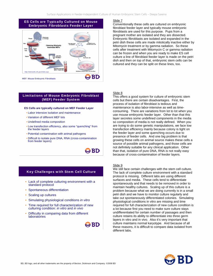
Slide 7 Conventionally these cells are cultured on embryonic fibroblast feeder layer and typically mouse embryonic fibroblasts are used for this purpose. Pups from a pregnant mother are isolated and they are dissected. Embryonic fibroblasts are isolated and expanded in the petri dish these cells are made mitotically inactive either by Mitomycin treatment or by gamma radiation. So these cells after treatment with Mitomycin C or gamma radiation can be frozen and when you are ready to make ES cell culture a line of fibroblast feeder layer is made on the petri dish and then on top of that, embryonic stem cells can be cultured and they can be split on these lines, too. Slide 8 This offers a good system for culture of embryonic stem cells but there are certain disadvantages. First, the process of isolation of fibroblast is tedious and maintenance is also labor-intensive as well as time-consuming. There are variations from lot to lot when you use mouse embryonic feeder layer. Other than that this layer secretes some undefined components in the media so composition of media is not really defined. When you are trying to do some genetic manipulations, we face low transfection efficiency mainly because colony is tight on the feeder layer and some quenching occurs due to presence of feeder cells. And one big problem is that growing these cells on animal source makes these cells a source of possible animal pathogens, and those cells are not definitely suitable for any clinical application. Other than that, isolation of pure DNA, RNA is not really easy because of cross-contamination of feeder layers. Slide 9 We still face certain challenges with the stem cell culture. The lack of complete culture environment with a standard protocol is missing. Different labs are using different surfaces and media. These cells tend to differentiate spontaneously and that needs to be removed in order to maintain healthy cultures. Scaling up of this culture is a problem because what we are doing currently is in a small petri dish and we have to monitor culture regularly and take out spontaneously differentiated colonies. Simulating physiological conditions in vitro are missing and time required for full characterization of new culture condition is a lot because first you need to make sure culture stays undifferentiated for certain number of passages and then culture retains its ability to differentiate into three germ layers in vitro and in vivo. Also it’s very important that culture maintains normal karyotype. And because of all these reasons, it is difficult to compare data isolated from different labs.

Surface Applications in Feeder-Independent Culture of Human Embryonic Stem Cells – Deepa Saxena
BD, BD logo, and all other trademarks are the property of Becton, Dickinson and Company. ©2008 BD
Feeder-free hES Cell Culture
• First documented in 2001 (Nature Biotech. Xu, et al.)
• BD Matrigel™ Matrix-coated surface used with Mouse Embryonic Fibroblast feeder layer conditioned media (MEF-CM)
• Multiple media conditions and defined media have been used successfully in combination with BD Matrigel Matrix-coated surface for culturing hES cells
BD Matrigel™ Matrix Substrate for hES Culture
hES cells cultured on BD Matrigel™ Matrix:
• Maintain normal karyotype
• Demonstrate a stable proliferation rate and high telomerase activity
• Express characteristic undifferentiated hES cell markers
• Form teratomas in severe combined immunodeficient (SCID) mice and differentiate into cells from all three germ layers
Advantages of BD BioCoat™ Matrigel™ Matrix Plates for ES Cell Culture
• Ready-to-Use Convenience• Optimized BD Matrigel™ Matrix coating process
• Patent pending• Stable, when stored at -20°C• Lot-to-lot consistency
• Eliminates tedious maintenance of MEF feeder layers• Surface compatible with multiple hES cell culturing
media and hES cell lines• Quality assurance testing to ensure functional
performance reproducibility of experiments• Easier DNA, RNA, and protein isolation• Optimized protocol enables standardization of cultures
Slide 10 So feeder-free culture of human embryonic stem cells was first documented in 2001 when a Nature Biotech paper was published and BD Matrigel-coated surfaces with mouse embryonic fibroblast feeder layer conditioned media was used to culture these cells. And since then, multiple media conditions, defined and undefined, have been successfully used in combination with BD Matrigel-coated surface for culture of human ES cells. Slide 11 Human ES cells cultured on BD Matrigel Matrix maintain normal karyotype, demonstrate a stable proliferation rate and high telomerase activity. They express characteristic undifferentiated markers of human embryonic stem cells and also form teratomas in SCID mice and differentiate into cells from all three germ layers. Slide 12 Matrigel is offered by BD Biosciences and it’s ready to use. It’s really convenient. You don’t have to make embryonic feeder layers, fibroblast feeder layers, and coating process is optimized. Product is stable. It can be aliquoted and stored and BD offers lot-to-lot consistency. This eliminates need for maintenance of feeder layer and surface is compatible with multiple culture media and multiple human ES cell lines and quality assurance testing is done to ensure functional performance and reproducibility of the experiments. It’s easy to isolate DNA, RNA, and protein for further studies and optimized protocol enables standardization of the cultures.

Surface Applications in Feeder-Independent Culture of Human Embryonic Stem Cells – Deepa Saxena
BD, BD logo, and all other trademarks are the property of Becton, Dickinson and Company. ©2008 BD
Serum-free Defined Media: mTeSR™1 Maintenance Medium
• A defined media for feeder-independent and serum-free culture of hES cells
• Developed at the WiCell Research Institute in 2006
References:1. Derivation of human embryonic stem cells in defined conditions, Ludwig T.E., et al., Nat. Biotechnol.
24(2):185-7, 2006
2. Feeder-independent culture of human embryonic stem cells, Ludwig T.E., et al., Nat. Methods. 3(8):637-46, 2006
Complete Culture Environment Feeder-free Surface + Defined Media
+
BD Matrigel™ hESC-qualified Matrixfrom BD Biosciences mTeSR™1 Maintenance Medium
from StemCell Technologies
Strategic collaboration by BD Biosciences, StemCell Technologies, and WiCell™Research Institute to develop optimized, feeder-independent cell culture environments for hES cell research
BD Matrigel™ hESC-qualified Matrix
• Optimized surface for hES cell culture• Qualified as mTeSR™1-compatible
Available at bdbiosciences.com/stemcellsource
Xu, C., et al., Feeder-free growth of undifferentiated human embryonic stem cells, Nature Biotechnology, 19:971-4 (2001).Xu, C., et al., Immortalized fibroblast-like cells derived from human embryonic stem cells support undifferentiated cell growth, Stem Cells 22:972-80 (2004).Ludwig, T.E., et al., Derivation of human embryonic stem cells in defined conditions, Nature Biotechnology, 24:185-7 (2006).
Slide 13 Recently scientists at WiCell Research Institute have demonstrated culture of human ES cells in defined animal-free media. It supports long-term undifferentiated expansion of human ES cell and mTeSR is the modified version of this media. Slide 14 And here we offer a complete environment where human ES cell-qualified Matrigel by BD Biosciences can be used with multiple media along with mTeSR that is marketed by StemCell Technologies. Slide 15 So BD Matrigel Matrix, the human ES cell-qualified matrix is optimized surface for human ES cell culture and it is compatible with mTeSR along with other defined and conditioned media.

Surface Applications in Feeder-Independent Culture of Human Embryonic Stem Cells – Deepa Saxena
BD, BD logo, and all other trademarks are the property of Becton, Dickinson and Company. ©2008 BD
BD Matrigel™ hESC-qualified Matrix
Modified from Mallon, et al., (2006) International Journal of Biochemistry and Cell Biology, 38:1063-1075
HES-df-CM: human ES cell-derived fibroblast conditioned media, EB: Embryoid body, TER: Teratoma formation, DF: in vitro differentiation
Summary of hES Culture on BD Matrigel™ Matrix Surface
Induced Human Pluripotent Stem (iPS) Cells Successfully Cultured on Matrigel
• Induced pluripotent stem cell Lines derived from human somatic cell. (Junying, Y. et al Science 2007)OCT-4, SOX2, NANOG, LIN28 reprogrammed human somatic cells to pluripotent stem cells exhibiting characteristics of embryonic stem cells
• Induction of pluripotent stem cells from adult human fibroblasts by defined factors. (Takahashi, K. et al Cell 2007)Oct-3/4, Sox2, Klf4, c-Myc in adult human fibroblast could generate pluripotent stem cells similar to embryonic stem cells
Undifferentiated hES Cell Colony
• Compact and dense H9 colonies on MEF feeders• Spread-out and monolayer-like colonies on BD Matrigel™ Plates
MEF-conditioned mediahES mediaMEF feeder layer BD Matrigel™ hESC-qualified Matrix BD Matrigel™ hESC-qualified Matrix
mTeSR™1
Slide 16 Enough literature is out there that demonstrates successful culture of human ES cells on Matrigel-coated surface and here is a table summarized from Mallon et al. where they demonstrated several human ES cell lines were successfully cultured on Matrigel in presence of different growth factors or other components and they were cultured for long-term. For example, six months, seven months or multiple passages – and they all maintained characteristic stem cell markers. They retained their pluripotency, differentiated into embryoid bodies, also formed teratomas demonstrating their potential for differentiation. Slide 17 Recently two papers were published where pluripotency was induced in somatic cells and a paper in Science where somatic cells were induced to express OCT4, SOX2, NANOG and LIN28. This reprogramming made induced pluripotent stem cells and these cells were also successfully cultured on Matrigel. Similarly, another paper from Cell where pluripotency was induced in adult human fibroblast also grew well on Matrigel. Slide 18 In our lab, we tested conventional ES cell culture and used feeder-free culture to compare defined media and conditioned media. As you see here, the colony is compact and dense when grown on feeder layer. Well when it is grown on Matrigel either in conditioned media or in presence of defined media, the mTeSR, they are nicely spread out.
Surface Applications in Feeder-Independent Culture of Human Embryonic Stem Cells – Deepa Saxena
BD, BD logo, and all other trademarks are the property of Becton, Dickinson and Company. ©2008 BD
Spontaneous Differentiation
Nature Biotechnology18, 399 - 404 (2000)
Differentiated colonies
1
Markers of Undifferentiated hES Cells on MEF-CM & mTeSR™1
mTeSR™1
H9 on BD Matrigel™ hESC-qualified Matrix
OCT-4 + Hoechst33342
MEF-CM
Markers of Undifferentiated hES Cells
OCT-4 SSEA-4
H9 in mTeSR™1 on BD Matrigel™ hESC-qualified Matrix
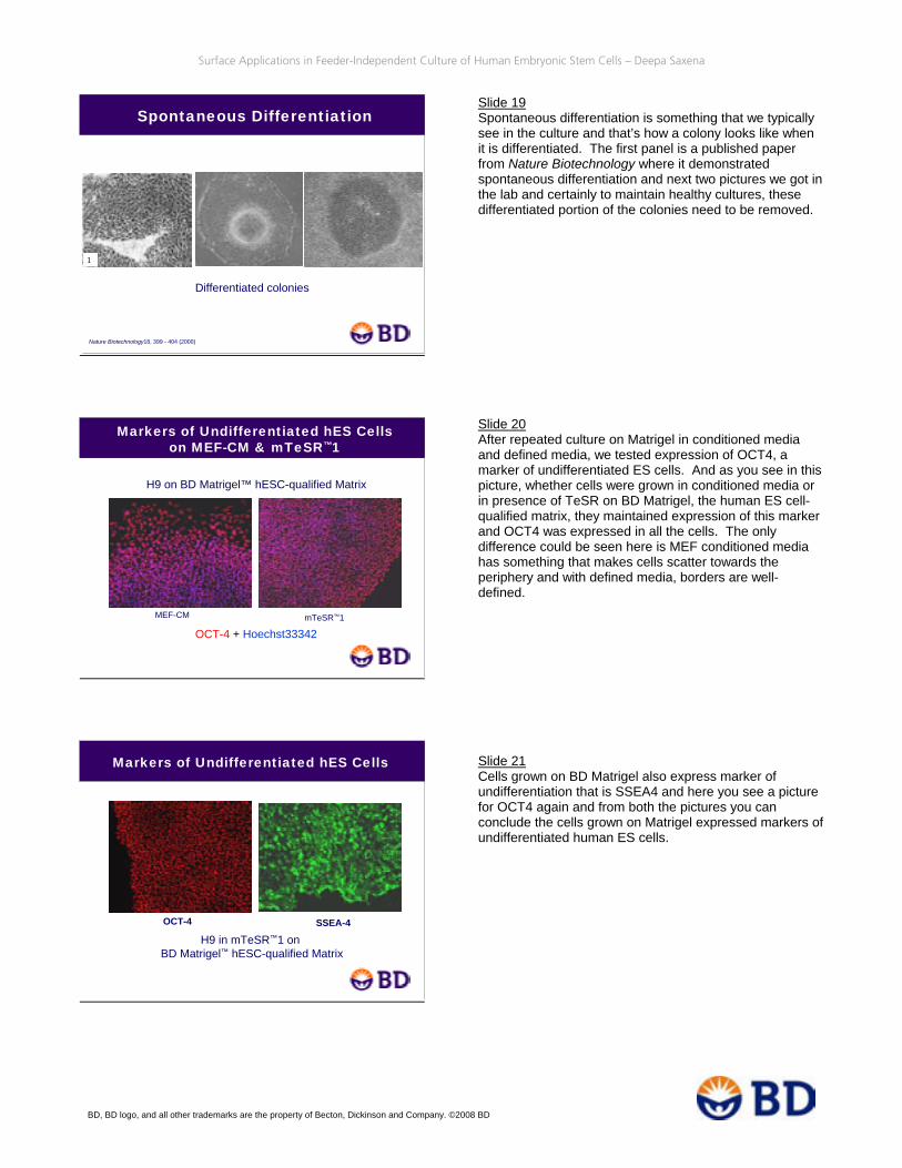
Slide 19 Spontaneous differentiation is something that we typically see in the culture and that’s how a colony looks like when it is differentiated. The first panel is a published paper from Nature Biotechnology where it demonstrated spontaneous differentiation and next two pictures we got in the lab and certainly to maintain healthy cultures, these differentiated portion of the colonies need to be removed. Slide 20 After repeated culture on Matrigel in conditioned media and defined media, we tested expression of OCT4, a marker of undifferentiated ES cells. And as you see in this picture, whether cells were grown in conditioned media or in presence of TeSR on BD Matrigel, the human ES cell-qualified matrix, they maintained expression of this marker and OCT4 was expressed in all the cells. The only difference could be seen here is MEF conditioned media has something that makes cells scatter towards the periphery and with defined media, borders are well-defined. Slide 21 Cells grown on BD Matrigel also express marker of undifferentiation that is SSEA4 and here you see a picture for OCT4 again and from both the pictures you can conclude the cells grown on Matrigel expressed markers of undifferentiated human ES cells.

Surface Applications in Feeder-Independent Culture of Human Embryonic Stem Cells – Deepa Saxena
BD, BD logo, and all other trademarks are the property of Becton, Dickinson and Company. ©2008 BD
Spontaneous Differentiation
DAPIOCT-4
Merge
Expression of OCT4 is diminished upon differentiation
Undifferentiated Marker Expression qRT-PCR Analysis
SOX2 gene expression in hESC
0.000
0.500
1.000
1.500
2.000
2.500
Control Lot1, p2 Lot2, p5 Lot3, p5Raw
RQ
(Rel
ativ
e Q
uant
ifica
tion)
OCT4 gene expression in hESC
0.000
0.500
1.0001.500
2.000
2.500
3.000
Control Lot1, p2 Lot2, p5 Lot3, p5
Raw
RQ
(Rel
ativ
e Q
uant
ifica
tion)
hESC media onMEF feeder layer
mTeSR™1 onBD Matrigel™ hESC-qualified Matrix
Undifferentiated Marker Expression FACS Analysis
98% OCT4 positive cells
H9 cells grown on BD Matrigel™ hESC-qualified Matrix in mTeSR™1 for 5 passages
Isotype Oct-3/4
Isotype SSEA4
94% SSEA4 positive cells
Slide 22 Spontaneous differentiation, is something we routinely see and OCT4 is a good marker to judge that. Here in this picture the batch of differentiated cells was seen where OCT4 expression is completely diminished and on an overlay you see DAPI but OCT4 is still missing so expression of OCT4 is diminished upon differentiation. Slide 23 We also compared cells grown on conventional embryonic fibroblast layer and Matrigel and we tested three different lots of Matrigel for this purpose. Cells were grown for multiple passages and RNA was isolated and we analyzed expression of SOX2 and OCT4 gene. Those are again markers for undifferentiated cells by quantitative real-time PCR and in both the graphs, expression of SOX2, as well as expression of OCT4 is comparable to control, that is human ES cells grown on feeder layer. Slide 24 We also quantified expression of undifferentiated markers OCT4 and SSEA4 by flow cytometry analysis and 98% cells stayed positive for OCT4 expression and 94% of the cells showed SSEA4 expression by flow cytometry analysis and this experiment was conducted at passage five when cells were grown in defined media on BD human ES cell-qualified Matrigel Matrix.
Surface Applications in Feeder-Independent Culture of Human Embryonic Stem Cells – Deepa Saxena
BD, BD logo, and all other trademarks are the property of Becton, Dickinson and Company. ©2008 BD
Isotype SSEA4
Isotype Tra-1-60 Tra-1-81
H9 cells grown on BD Matrigel™ hESC-qualified Matrix in mTeSR™1
SSEA4 (PE) BD Pharmingen 560128
IgG3 isotype (PE) BD Pharmingen 559926
Tra-1-60 (PE) BD Pharmingen 560193
Tra-1-81 (PE) BD Pharmingen 560161
IgM isotype (PE) BD Pharmingen 555584
OCT-3/4 BD Pharmingen 611202
IgG1 isotypeBD Pharmingen 557273
Isotype OCT-3/4
Undifferentiated Marker Expression FACS Analysis
Embryoid Body (EB) Formation
Phase contrast image of EBs formed by H9 cells grown on BD BioCoat™ Matrigel™ Plates for ES Cell Culture
Beating Cardiomyocytes from EBs
H9 Cells grown on BD Matrigel™ hESC-qualified Matrix in mTeSR™1 for >10 passages formed EBs and differentiated to beating cardiomyocytes
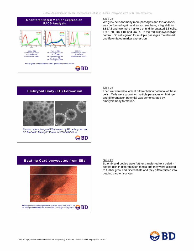
Slide 25 We grew cells for many more passages and this analysis was performed again and as you see here, a big shift for SSEA4 and two more markers of undifferentiated ES cells, Tra-1-60, Tra-1-81 and OCT4. In the red is shown isotype control. So cells grown for multiple passages maintained undifferentiated marker expression. Slide 26 Then we wanted to look at differentiation potential of these cells. Cells were grown for multiple passages on Matrigel and differentiation potential was demonstrated by embryoid body formation. Slide 27 So embryoid bodies were further transferred to a gelatin-coated dish in differentiation media and they were allowed to further grow and differentiate and they differentiated into beating cardiomyocytes.

Surface Applications in Feeder-Independent Culture of Human Embryonic Stem Cells – Deepa Saxena
BD, BD logo, and all other trademarks are the property of Becton, Dickinson and Company. ©2008 BD
Neurons from EBs
H9 Cells grown on BD Matrigel™ hESC-qualified Matrix in mTeSR™1 for >10 passages formed EBs and differentiated to Neurons
Expression of 3-Germ Layer Markers in Embryoid Bodies by QRT-PCR
AFP (alpha fetoprotein) - Endoderm (CT 21.28 vs. Not Detected in H9 cells) HAND1 (heart and neural crest derivatives expressed 1) - Mesoderm TUBB3 (tubulin beta 3) - Ectoderm
0.1
1
10
100
1000
10000
H9 Cells EB Day9
Rel
ativ
e ge
ne E
xpre
ssio
n
HAND1TUBB3
• Single defined ECM protein• Defined ECM combinations• Chemically modified surfaces
• Critical considerations• Functionality• Cost• Complexity
Potential Alternative Surfaces to BD Matrigel™ Matrix
Slide 28 These embryoid bodies could also be differentiated into neurons and there are the images captured from the petri dish. Slide 29 We also demonstrated expression of three-germ layer markers in embryoid bodies by quantitative RT-PCR and as seen here, HAND1 heart and neural crest derivative expressed and TUBB3 tubulin 3 markers of mesoderm and ectoderm were highly expressed as compared to H9 cells and endoderm marker alpha fetoprotein was not detected in H9 cells but it was detected in embryoid bodies at Day 9 and the CT value was 21.28. So here we demonstrate differentiation potential of these cells after multiple passage on BD Matrigel Matrix and they differentiate into all three germ layers. Slide 30 Matrigel is relatively undefined because it’s a complex mixture of certain extracellular matrix protein but what could be potential alternative surfaces to BD Matrigel Matrix. It could be single defined ECM protein or maybe a combination of ECM proteins or a chemically modified surface but definitely critical considerations for a surface would be functionality, cost, as well as complexity.

Surface Applications in Feeder-Independent Culture of Human Embryonic Stem Cells – Deepa Saxena
BD, BD logo, and all other trademarks are the property of Becton, Dickinson and Company. ©2008 BD
Comparison of BD Matrigel™ Matrixvs. other ECM Proteins
• BD Matrigel™ Matrix is equivalent or better than most single extracellular matrices (ECMs) tested:
• Laminin equivalent to BD Matrigel Matrix (Xu, et al. 2001)• Laminin, collagen, fibronectin, and vitronectin were combined for
optimal ECM complex for culturing hES cells (Ludwig, et al. 2006)• Fibronectin and collagen combination is required to match the
performance of BD Matrigel Matrix (Lu, et al. 2006)
• Pure ECM often much more expensive to use
• ECM combination requires more steps and time to coat
New Surface for ES Cell Culture
BD™ Laminin/Entactin complex
• Major component of basement membrane in Engelbreth-Holm-Swarm (EHS) mouse tumors
• Provides a 3D environment that more closely models the cellular microenvironment in vivo
Comparison of MEF Conditioned Media and mTeSR™1
MEF-Conditioned media mTeSR™1
A & B: BD Matrigel™hESC-qualified Matrix
C & D: Laminin/Entactin Complex
A B
C D
Slide 31 BD Matrigel Matrix is equivalent or better than most of the single extracellular matrices tested so far. Laminin was found to be equivalent to Matrigel. Laminin, collagen, fibronectin and vitronectin combination was required to match performance of Matrigel by another group and fibronectin and collagen combination is required to match performance of BD Matrigel Matrix. That was reported by another group. But pure ECMs are often much more expensive to use and a combination of ECM’s definitely requires more steps to code and it is time-consuming. Slide 32 In an attempt to test alternative surfaces, we tested BD Laminin/Entactin complex and that is a major component of basement membrane in EHS mouse tumor. It provides a 3D environment that more closely models the cellular microenvironment in vivo and this is a step further from Matrigel and that’s a further purified complex. Slide 33 Cells maintained undifferentiated morphology as they did on human ES cell-qualified BD Matrigel Matrix and again we tested this Laminin/Entactin complex for conditioned media as well as defined media. And as you see here, undifferentiated colonies were maintained in presence of either conditioned or defined media, both on Matrigel and Laminin/Entactin complex.

Surface Applications in Feeder-Independent Culture of Human Embryonic Stem Cells – Deepa Saxena
BD, BD logo, and all other trademarks are the property of Becton, Dickinson and Company. ©2008 BD
BD™ Laminin/Entactin complex
Comparison of Different Surfaces and Media by IHC
BD Matrigel™ hESC-qualified Matrix
mTeSRTM1
MEF-CM
OCT-3/4 expression in H9 cells
BD™ Laminin/Entactin complex
Comparison of Different Surfaces and Media by IHC
BD Matrigel™ hESC-qualified Matrix
mTeSRTM1
MEF-CM
SSEA4 expression in H9 cells
H9 Cells Expressing Markers Specific for Undifferentiated Cells
SSEA-4
OCT-4
BD™ Laminin/Entactin complex BD Matrigel™ hESC-qualified Matrix
H9 colony grown in mTeSR™1 medium
Slide 34 We also tested expression of OCT4 in these cells and again, when cells were grown on Matrigel or BD Laminin/Entactin complex in presence of either conditioned media or TeSR media, they expressed plenty of OCT4 and as seen earlier in presence of conditioned media, cells are scattered at the periphery. Slide 35 Cells grown on Laminin/Entactin complex expressed SSEA4 as well and expression was similar when compared to Matrigel. Slide 36 Here is a comparison again showing cells cultured on Laminin/Entactin complex or BD Matrigel Matrix and expression of SSEA4 and OCT4 and cells were grown in defined media here and as you see, both the surfaces, Laminin/Entactin as well as Matrigel, performed really well for culture of human embryonic stem cells.

Surface Applications in Feeder-Independent Culture of Human Embryonic Stem Cells – Deepa Saxena
BD, BD logo, and all other trademarks are the property of Becton, Dickinson and Company. ©2008 BD
Comparison of BD Matrigel™ Matrix and Laminin/Entactin by BD FACS™ Analysis
SSEA-4
SSEA-4 83% on BD Matrigel™ hESC-qualified Matrix91% on BD™ Laminin/Entactin ComplexIsotype control
OCT-4 98% on BD Matrigel™ hESC-qualified Matrix95% on BD™ Laminin/Entactin ComplexIsotype control
OCT-4
Embryoid Body (EB) Formationfrom hES Cells
Phase contrast image of Embryoid bodies formed by H9 cells grown on different surfaces.
BD Matrigel™ hESC-qualified MatrixBD™ Laminin/Entactin complex
Expression of 3-Germ Layer Markers in Embryoid Bodies by QRT-PCR
Differentiation Markers in Embryoid Bodies
0.1
1
10
100
1000
10000
100000
1000000
H9 Cells Embryoid BodiesRel
ativ
e G
ene
Expr
essi
on
HAND1
TUBB3
FOX2A
H9 cells maintained on BD Laminin/Entactin complex in mTeSR1 media expressed 3-germ layer marker upon differentiation
Slide 37 We also quantified expression of those two markers by flow cytometry and 83% of the cells stayed SSEA4-positive on human ES cell-qualified matrix, BD Matrigel, and 91% of the cells expressed this marker when grown on Laminin/Entactin complex and in black is the isotype control. Similarly for OCT4, 95% of the cells stayed positive for OCT4 expression when grown on Laminin/Entactin complex so again, these two BD surfaces are good feeder-free culture alternative for culture of human embryonic stem cells. Slide 38 We also tested differentiation potential of H9 cells when they were cultured on Laminin/Entactin complex for multiple passages and they were differentiated into embryoid bodies. Slide 39 We also monitored expression of three-germ layer markers in embryoid body and on the Y axis it is represented as a log scale and when you compare expression of three-germ layer markers between H9 cells and embryoid body, you see all these genes are highly upregulated upon differentiation.

Surface Applications in Feeder-Independent Culture of Human Embryonic Stem Cells – Deepa Saxena
BD, BD logo, and all other trademarks are the property of Becton, Dickinson and Company. ©2008 BD
Beating Cardiomyocytes
H9 –Derived Cardiomyocytes and Neuronal Cells
GATA-4 + DAPI Nestin + DAPINestin (BD Pharmingen 611658)(BD Pharmingen coming soon)
Differentiated to Cardiomyocytes and Neuronal cells after culture on BD™ Laminin/Entactin complex in mTeSR1 media for 32 passages
Human ES Cells Cultured on Laminin/Entactin Complex Demonstrated Karyotype Stability
G banding chromosome analysis of H9 grown on Laminin/Entactin in mTeSR1 for P26
Slide 40 So after demonstrating expression of all three-germ layers in the embryoid bodies, embryoid bodies were transferred to gelatin-coated dishes and allowed to differentiate further and again we saw beating cardiomyocytes as seen in this movie. Slide 41 And later the cells were fixed and stained for marker GATA-4. Cardiomyocytes expressed this marker and differentiation into neural cells was demonstrated by Nestin and as you see here in the green, the Nestin-positive cells. Slide 42 After multiple passages, another concern was if Laminin/Entactin also maintains karyotypic stability of the cells and after 26 passages in defined media we send these cultures for karyotype analysis and they turned out to be normal so Laminin/Entactin also maintained karyotype stability.

Surface Applications in Feeder-Independent Culture of Human Embryonic Stem Cells – Deepa Saxena
BD, BD logo, and all other trademarks are the property of Becton, Dickinson and Company. ©2008 BD
Future Directions
• Next Step:• Cost-effective human ECM surface to be
used with the animal-free TeSR1 media• Ultimate goal:
• Chemically-defined surface
References
1. J. A. Thomson et al., Science, 282:1145 (1998).2. A. G. Smith et al., Nature. 336:688 (1988).3. R. L., Williams et al., Nature, 336:684 (1988).4. C. Xu et al., Nat. Biotech.,9:971 (2001).5. C. Xu, J et al., Stem Cell, 22:972 (2004).6. P. Stojkovic et al., Stem Cells, 23:306 (2005).7. E. Sjögren-Jansson et al., Dev.Dynamics, 233:1304 (2005). 8. M. Amit et al., Dev. Biology, 227:271 (2000).9. C. Xu et al., Stem Cells, 23:315 (2005a).10. L. Wang, L. Li, P. Menendez, C. Cerdan, M. Bhatia, Blood, 105:4598 (2005a). 11. M. E. Levenstein et al., Stem Cells, 24:568 (2006).12. R. H. Xu et al., Nat. Biotech., 20:1261 (2002).13. R. H. Xu, R. M. Peck, D. S. Li, T. Ludwig, J. A. Thomson, Nat. Methods, 2:185 (2005b).14. G. Wang et al., Biochem. Biophys. Communications, 330:934 (2005b).15. N. Sato et al., Nat. Med., 10:55 (2004).16. T. E. Ludwig et al., Nat. Biotech,.Brief Communications, January (2006).17. J. Lu, R. Hou, C. J. Booth, S.-H.Yang, M. Snyder, PNAS, 103:5688 (2006).18. S. Yao et al., PNAS, 103:6907 (2006).19. B. S. Mallon, Intl. J. Biochem. & Cell Biol., 38:1063 (2006).20. U. Lakshmipathy et al., Stem Cells, 22:531 (2004).21. L. Gerrard, D. Zhao, A. J. Clark, W. Cui, Stem Cells, 23:124 (2005).22. C. P. Ren et al., Acta Biochim Biophys Sin (Shanghai), 37:68 (2005).23. H. Zaehres et al., Stem Cells, 23:299 (2005).24. A. R. Greenlee et al., Toxicol. In Vitro, 19:389 (2005).
Contact Information
BD Biosciences has a variety of products for stem cell research.For a complete listing of products, please visit our website at:
bdbiosciences.com/stemcellsource
Technical SupportTel: 877.232.8995 (US)
978.901.7300 (outside US)Email: [email protected]
bdbiosciences.com/webinars
Slide 43 Next step could be cost-effective human ECM surface that is to be used with animal-free version of TeSR media but it will be ideal to have chemically defined surfaces. While research continues to search for animal-free surface and animal-free media, there are many basic questions that can be answered using BD Laminin/Entactin and BD Matrigel surfaces with defined and conditioned media. Slide 43 Here are some of the references that are used for this talk. Slide 44 If you have any question, you can contact our technical support department at [email protected]. Thank you for your attention.

Surface Applications in Feeder-Independent Culture of Human Embryonic Stem Cells – Deepa Saxena
BD, BD logo, and all other trademarks are the property of Becton, Dickinson and Company. ©2008 BD