elsevier review unstable proteins: how to subject them to...
TRANSCRIPT

ELSEVIER Journal of Chromatography B, 699 (1997) 347-369
JOURNAL OF CHROMATOGRAPHY B
R e v i e w
Unstable proteins: how to subject them to chromatographic separations for purification procedures 1
Michael Kaufmann
Department of Biochemistry., Universi~' of Wittenl Herdecke, Stockumerstrasse 10, D-58448 Witten, Germany
Abstract
The chromatographic separation of an unstable protein is often a challenge to the scientist working in the field of life sciences. Especially for the purification of sensitive enzymes, making use of conventional chromatographic techniques is difficult and frequently results in a complete loss of biological activity of the target protein. This report summarizes some general strategies that may help to keep unstable proteins in their native conformation during the rather harsh conditions of a purification procedure. In this context, a recently developed hollow fiber membrane module, suitable for performing on-line dialysis, is introduced and examples of its application to liquid column chromatography are given. Many innovative separation techniques, characterized by dramatic improvements in both performance and separation time, have recently been developed. Since the chromatographic separation of unstable proteins requires the use of modern state-of-the-art equipment and technology, emphasis is given to newly developed separation techniques such as expanded bed adsorption, perfusion chromatography, protein free flow electrophoresis and the use of tentacle gels. In addition, examples of recently published purifications of unstable proteins are discussed with respect to strategies ensuring the preservation of the native protein structure during chromatographic separation. © 1997 Elsevier Science B.V.
Keywords: Reviews; Proteins
Contents
1. Introduction ............................................................................................................................................................................ 348 2. Protein stability ....................................................................................................................................................................... 348
2.1. Time ............................................................................................................................................................................... 349 2.2. Temperature .................................................................................................................................................................... 350 2.3. Pressure .......................................................................................................................................................................... 351 2.4. Proteases ......................................................................................................................................................................... 351 2.5. Buffer composition .......................................................................................................................................................... 353
3. Separation techniques .............................................................................................................................................................. 355 3.1. On-line dialysis ................................................................................................................................................................ 355 3.2. Expanded bed adsorption .................................................................................................................................................. 358 3.3. Tentacle supports ............................................................................................................................................................. 359 3.4. Perfusion chromatography ................................................................................................................................................ 362 3.5. Free flow electrophoresis .................................................................................................................................................. 364
Dedicated to Professor Peter Bartholmes.
0378-4347/97/$17.00 © 1997 Elsevier Science B.V. All rights reserved. PII S0378-4347 (96)005 12-9
348 M. Kaufmann I J. Chromatogr. B 699 (1997) 347-369
4. Conclusions ............................................................................................................................................................................ 366 5. List of abbriviations ................................................................................................................................................................ 366 Acknowledgements ...................................................................................................................................................................... 367 References .................................................................................................................................................................................. 367
1. Introduction
In the context of the chromatographic separation of unstable proteins, it is important to consider in what respect such instabilities actually are crucial to serve the desired purpose. For example, the mainte- nance of the functional properties of a certain protein, if at all, only plays a minor role during chromatographic separations for analytical purposes. For most of such applications the proteins are intentionally denatured, derivatized or even hydro- lytically cleaved. The term "analytical purpose" implies the use of only small sample amounts designated to become waste. Examples for such analytical separation procedures are SDS-PAGE, reversed-phase HPLC, CE or MALDI-MS. On the other hand, the maintenance of protein structure and its function and especially the art of keeping unstable proteins in their native conformation during chro- matographic separations is one of the most important demands in the broad field of protein purification. For that reason, protein purification as a whole will play a central role in this report.
The recent development of improved and new chromatographic and electrophoretic separation tech- niques undoubtedly opens new doors to design smooth protein purification strategies. Many old obstacles can be surmounted by using sophisticated separation methods and modem state-of-the-art equipment. Nevertheless, protein purification still requires special knowledge about the individual properties of the protein of interest as well as skill and experience in performing chromatographic sepa- rations. If the purification of a relatively unstable protein is planned, all parameters influencing protein stability must be carefully taken into consideration. Therefore, it is inevitable to start the discussion with a brief summary about general important aspects on protein stability. Then, the logical consequences for
sample preparation, sample conditioning and new technical solutions for that purpose are described. The final chapter explains new chromatographic techniques which were developed to take special care of proteins during the separation, and examples of their application in protein purification, especially of unstable proteins, are given.
2. Protein stability
The physiological environment, e.g., the intact cell or living tissue delivers ideal conditions with respect to the stability of a certain protein. As a conse- quence, those conditions should be kept up as authentic as possible during the chromatographic separations for protein purification. Unfortunately, the separation processes themselves are crucial inter- ventions that change the physiological environment to a more artificial one which often has a negative effect on the stability of the target protein. Already at the very beginning of a typical protein purification protocol, cell disruption, tissue destruction or other extraction procedures replace the natural environ- ment of the protein of interest by a composite buffer solution. It is important, that the composition of the buffer system meets both the individual and general requirements on the stability of the target protein. As a rule of thumb, the values of all parameters of the natural environment of the target protein are most probably also the most suitable to be taken into consideration for the design of the respective buffer system. Besides chemical buffer parameters such as pH, ionic strength, reducing properties, protein con- centration or the presence of cofactors, general physical aspects such as time, temperature, surface effects or pressure also play an important role in protein stability. They therefore will now be dis- cussed in more detail.

M. Kaufmann / J. Chromatogr. B 699 (1997) 347-369 349
2.1. Time
What is an unstable protein? In most of cases, instability of proteins means the loss of biological activity of an enzyme. Obviously, it is difficult to define where a "stable protein" ends and an "un- stable protein" begins. The deactivation of a certain enzyme depends exponentially on time according to
Cnative ~ C 0 X e - t / k
Thus, the relative stability of a protein is best represented by its half life time [ t so=k×ln (2)] under defined conditions. Increasing the stability of a protein means increasing its half life time (and thus increasing k). The half life time of a protein depends on many parameters which change during the pro- cess of purification. Consequently, the half life time of a protein during purification changes too. For example, directly after preparing crude extracts, proteases can be present which, despite of using protease inhibitors, may decrease the half life time of the target protein dramatically. At a more progressed state during downstream processing, those proteases eventually are eliminated and the stability of the considered protein increases. On the other hand, the background proteins present at high concentrations during the early steps of purification act as competi- tive protease inhibitors. In fact, the intentional addition of background protein such as BSA is a well known strategy to stabilize a purified protein. Com- pared to the protein stability at almost all steps during a purification procedure, the final purified protein preparation stored under suitable conditions, normally exhibits the highest stability. As a conse- quence, the whole purification procedure should be as quick as possible.
All time-consuming steps, especially sample con- ditioning and assays between the separations, should be optimized with respect to their duration. For example, by shortening the whole time required to purify the highly unstable protein component P of the Euphausia pacifica luciferase system from 3-4 days to 5 h by introducing an affinity technique, a 50-100 fold increase in the specific activity could be observed [1]. In general, the purification of an unstable protein is always a fight against the process time. Time is one of the most important parameters
responsible for a successful purification procedure of an unstable protein and many of the chromatographic methods described in this report are superior because of their comparably short duration.
Extensive dialysis steps should be avoided or, if essential, performed on-line. If liquid column chro- matography using adsorbing beds are part of the purification protocol, the elution conditions should carefully be determined already during method de- velopment. If they finally are known, all elutions during the chromatographic separations should be performed by using buffer steps rather than continu- ous gradients. This saves process time. In addition, time-consuming assay procedures after each sepa- ration step can be omitted. An important approach to shorten the time required for a purification procedure is the combination of two or more separation or conditioning steps so that they are performed at the same time. A good example is expanded bed ad- sorption (EBA), a method that combines the removal of whole cells or debris with a chromatographic separation step at the very beginning of a protein purification procedure.
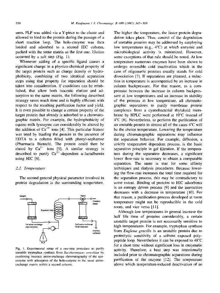
If liquid column chromatography is performed by isocratic elution, i.e., background material is ad- sorbed whereas the target protein passes the column without being restrained, a subsequent chromato- graphic column can be loaded at the same time. If sample conditioning for adsorption to the second column requires additives, such as salts, a Y-piece and a second pump can be used. On the other hand, on-line dialysis allows to remove eventually disturb- ing low-molecular-mass material prior to adsorption to the second column. For example, tryptophan synthase from Saccharomyces cerevisiae, an enzyme known to be very unstable [2], was successfully purified by combining two IEC steps [3]. The pyridoxal 5'-phosphate (PLP) dependent enzyme undergoes a conformational change upon PLP bind- ing accompanied by a dramatic change in its surface charge density" whereas the holt-enzyme strongly binds to Q-Sepharose fast flow (Pharmacia Biotech, Uppsala, Sweden) under standard buffer conditions, no interaction of the apt-enzyme with this anion- exchange matrix takes place. Consequently, the purification procedure included a first isocratically eluted anion-exchange step of the enzyme in the apt-form. Directly after elution from the first col-
350 M. Kaufmann / J. Chromatogr. B 699 (1997) 347-369
umn, PLP was added via a Y-piece to the eluate and allowed to bind to the protein during the passage of a short reaction loop. The holo-enzyme was then loaded and adsorbed to a second IEC column, packed with the same matrix as the first one. Elution occurred by a salt step (Fig. 1).
Whenever adding of a specific ligand causes a significant change in a physico-chemical property of the target protein such as charge density or hydro- phobicity, combining of two identical separation steps using that property for separation should be taken into consideration. If conditions can be estab- lished, that allow both isocratic elution and ad- sorption to the same matrix, the following described strategy saves much time and is highly efficient with respect to the resulting purification factor and yield. It is even possible to change a certain property of the target protein that already is adsorbed to a chromato- graphic matrix. For example, the hydrophobicity of equine milk lysozyme can considerably be altered by the addition of Ca 2+ ions [4]. This particular feature was used by binding the protein in the presence of EDTA to a column filled with phenyl-sepharose (Pharmacia Biotech). The protein could then be eluted by Ca 2+ ions [5]. A similar strategy is described to purify CaZ+-dependent a-lactalbumin using HIC [6].
2.2. Temperature
The second general physical parameter involved in protein degradation is the surrounding temperature.
Sm'nple Valve
Reaction loop
Gradient mixer
I st anion ©xchmag .,,,~x [ 2rid anion ¢xchmage (apo-enzym¢) (holo-enzym¢) +
Fraction collector
PLP
Fig. 1. Experimental setup of a one-step procedure to purify unstable tryptophan synthase from Saccharomyces cerevisiae by combining isocratic anion-exchange chromatography of the apo- enzyme with adsorption of the holo-enzyme to the same anion- exchange matrix within a second column.
The higher the temperature, the faster protein degra- dation takes place. Thus, control of the degradation of unstable proteins may be addressed by employing low temperatures (e.g., 4°C) at which enzymic and microbiological activity is minimized. However, some exceptions of that rule should be noted. At low temperature numerous enzymes have been shown to undergo reversible cold inactivation which in the case of oligomeric proteins usually stands for cold dissociation [7]. If separations are planned, a reduc- tion in temperature is accompanied by an increase in column backpressure. For that reason, as a com- promise between the increase in column backpres- sure at low temperatures and an increase in stability of the proteins at low temperature, all chromato- graphic separations to purify membrane protein complexes from a cyanobacterial thylakoid mem- brane by HPLC were performed at 10°C instead of 4°C [8]. Nevertheless, to perform the purification of an unstable protein in almost all of the cases 4°C will be the choice temperature. Lowering the temperature during chromatographic separations may influence the separation behavior. For example, diffusion, a strictly temperature dependent process, is the basic separation principle in gel filtration. If the tempera- ture during the separation decreases, a significant lower flow-rate is necessary to obtain a comparable separation. The same is true for some affinity techniques and dialysis procedures. Because lower- ing the flow-rate increases the total time required for the separation process, this may be contradictory to some extent. Binding of proteins to HIC adsorbents is an entropy driven process [9] and the interaction decreases with a decrease in temperature [10]. For that reason, a purification process developed at room temperature might not be reproducible in the cold room, and vice versa [11].
Although low temperatures in general increase the half life time of proteins considerably, a certain unstable target protein is not necessarily sensitive to high temperatures. For example, tryptophan synthase from Euglena gracilis is an unstable protein due to proteolytic sensitivity of a solvent exposed poly- peptide loop. Nevertheless it can be exposed to 48°C for a short time without significant loss in enzymatic activity. Therefore, a heat step was intentionally included prior to chromatographic separations during purification of the enzyme [12]. The temperature above which temperature-induced deactivation of an
M. Kaufmann I J. Chromatogr. B 699 (1997) 347-369 351
enzyme begins can be determined by displaying graphically log (k) against 1/T according to Ar- rhenius [13]. The linear part of the resulting curve represents the temperature range within no thermal denaturation of the enzyme occurs.
Especially if proteins from thermophillic organ- isms are cloned in E. coli, a heat step at the very beginning of the purification procedure is suitable to remove almost all background proteins. For instance, indole-3-glycerol phosphate synthase from Sul-
folobus solfataricus, a microorganism growing in hot, acidic and sulfurous mud pools in the Solfatara volcanic crater near Naples, Italy [14] was cloned in E. coli. The purification procedure of the enzyme included a highly effective heat step exposing the crude cell extract to 75°C for 10 rain prior to the chromatographic separation [ 15].
2.3. Pressure
It is well known that high pressure may cause dissociation of tightly associated protein subunits [16]. Furthermore, pressure-induced denaturation may result in a complete loss of biological activity of many proteins. For example, complete deactivation of the 132-subunit in the 0t213 z bienzyme complex tryptophan synthase from E. coli occurs at 1300 bar [17] although even in the absence of stabilizing c~-subunits the 132-dimer is known to be remarkably stable [18]. Despite the fact that those pressure values seem to be very high and the deactivation process in most of the investigated cases is revers- ible, the situation with regard to unstable proteins may be more critical. During conventional liquid chromatography and FPLC-applications the pressures within the chromatographic systems certainly have no negative effect even on an unstable protein. In contrast, if HPLC is part of a certain purification procedure of an unstable protein, it could be advan- tageous to replace this step despite the fact that HPLC in general is known to achieve high res- olutions.
Many protein purification protocols include den- sity gradient centrifugation as a powerful separation technique. Unfortunately, considerable pressures may be present within the centrifugation tube. The pres- sure gradient along the tube depends on the given dimensions according to
p(r) = P(ro) + 0.5ro)Z(r 2 - r2o)
Ignoring a density gradient, pressures up to 1840 bar (bottom of the tube) are present if a standard swing-out rotor (Hitachi RPS 40 T) is spinning at 40 000 rpm. Indeed, density gradient centrifugation was intentionally used to characterize the pressure- induced unfolding and deactivation of tryptophan synthase from E. coli [19]. Consequently, if the target protein is sensitive against pressure, both density gradient centrifugation and conventional ultracentrifugation steps should be avoided.
2.4. Proteases
Unwanted proteolysis is a widespread problem encountered in the analysis of proteins from every type of organism. Any study involving proteins should be conducted with the assumption that proteolytic problems could arise [20]. Especially the instability of the target protein during a certain purification procedure is often caused by proteolytic degradation. Although it is not possible to give a magic antiprotease formula guaranteed to eliminate all artifacts in every case in which it is applied [21], some strategies given here have proved successful in a broad range of studies and thus, especially should be taken into consideration when separating unstable proteins by chromatography. As already stated, both time and temperature in general have an influence on the degree of protein degradation. Consequently, shortening the process time and lowering the tem- perature will also minimize proteolytic fragmentation of the target. Specific strategies to prevent unwanted proteolysis can be subdivided in stabilizing the target protein and inhibiting or removing potential hydro- lytic activities.
The smaller a protein, the more resistant it is to proteolysis. In most of the cases proteins are most resistant in their native state. Within tightly folded structures of small globular proteins, no peptide bond normally is sufficiently exposed to allow attack by proteinases. If such proteins are degraded their conformation is affected first. Thus, measures to keep the target in its tight native fold should be considered. The binding of molecules such as sub- strates, cofactors or metal ions tends to maintain a protein in a more stable conformation. As an exam- ple, the holo form of cytochrome P450ca m is more

352 M. Kaufmann / J. Chromatogr. B 699 (1997) 347-369
resistant to proteolytic digestion by carboxypeptidase A, chymotrypsin, clostripain, papain, pepsin, pron- ase, Endoproteinase Glu-C and trypsin than the apo form [22]. Individual components of multimeric proteins may also have vulnerable sites which are hidden until the components are dissociated. For instance, the tetrameric (a[313o0 bienzyme complex tryptophan synthase from E. coli is resistant to proteolytic digestion using endoproteinase GIu-C, whereas isolated 132-subunits are nicked rapidly within a solvent exposed loop that is shielded by the peripheral cx-subunits in the native complex [23].
An unstable target protein should be carefully investigated with respect to the conditions that help minimizing its susceptibility to proteolytic attack. In addition, the reduction of proteinase activities by suitable inhibitors or, even better, the complete removal of contaminating proteolytic activities dur- ing the very early steps of a purification procedure is of great importance with respect to chromatographic separations of unstable proteins. There are many cocktails of protease inhibitors available that are especially designed to cover the inhibition of a broad spectrum of proteinases during protein purification. We will first discuss the most widely used protease inhibitors and then make some suggestions for tissue specific inhibitor cocktails.
Aspartic proteases normally are inhibited by pep- statin A that binds very tightly and specifically, but which is a reversible inhibitor with limited solubility [24]. The inhibition of serine proteases is often achieved by PMSF [25], TLCK [26] or TPCK [27]. The latter two inhibitors are specific to inhibit trypsin- and chymotrypsin-like proteases, respective- ly. It should be noted that all those inhibitors are highly unstable in aqueous solutions around neutral pH and consequently should be stored in alcoholic stock solutions to be added to the aqueous buffer solution directly prior to use. Due to the instability of those inhibitors, repeated addition of the respective stocks is useful during sample preparation. Although DFP is the most efficient inhibitor of serine proteases [28] it is not recommended for routine use since it is a highly toxic nerve gas which can be absorbed by the skin. The reversible inhibition of trypsin-like proteases can be achieved by benzamidine or p- aminobenzamidine. Trypsin-like serine- and some cysteineproteases are reversibly inhibited by leupep-
tin [29]. As high-molecular-mass proteinaceous in- hibitors, SBTI [30] and aprotinin [31] are effective inhibitors of serine proteinases. In addition to TPCK, chymotrypsin-like proteases can be inhibited by chymostatin [29]. Metalloproteases usually are in- hibited by removing essential metal ions via chelat- ing agents such as 1,10-phenanthroline or EDTA [311.
The composition of different crude cell extracts with respect to the presence of unwanted proteases depends on the respective source. Consequently, different protease inhibitor cocktails are suitable for different sources of the target protein. Table 1 summarizes some suggestions for inhibitor cocktails suitable for protease sensitive protein purification.
The most noteworthy example of an inhibitor that binds and inhibits most endoproteinases is %-macro- globulin. As a high-molecular-mass protein (M r 750 000) it inhibits proteinases irreversibly by a trapping mechanism after being cleaved by a pro- tease within a special bait region [32]. However, since inhibition by binding occurs at an 1:1 ratio, the inhibitor itself will be a contaminant which due to its very high molecular mass possibly cannot be neg- lected. In addition, az-macroglobulin is expensive and thus not recommended for routine protein purifi- cation. Not only the specific inhibition by protease inhibitors but also shifting the equilibrium of the proteolytic reaction towards peptide bond synthesis may be suitable to prevent unwanted proteolysis. Some organic cosolvents such as glycerol or ethyl- eneglycol are described to cause such an effect if added to aqueous buffer solutions [33].
Although inhibition of proteolytic contaminations as well as the stabilization of the target protein are effective, the most important measure to prevent proteolytic degradation is the design of a purification strategy that allows to get rid of proteases as early as possible. The choice of the starting material plays an important role with respect to the endogenous pro- teinase activities. For the purification of proteinase- sensitive proteins, a suitable cell disruption pro- cedure keeps proteinase-rich vacuoles or lysosomes intact. It may sometimes be better to sacrifice yield in order to avoid excessive rupture of such organel- les. Various exemplary protocols avoiding unwanted proteolysis by gentle and selective homogenization are described in the literature [34,35].

M. Kaufmann / J. Chromatogr. B 699 (1997) 347-369
Table 1 Suggestions for inhibitor cocktails for protein isolation and purification
353
Source of crude extract Inhibitor Stock
Animal tissues 1 mM PMSF 0.2 M in methanol 1 mM EDTA 0.1 M in aqueous solution 1 mM Benzamidine 0.1 M in aqueous solution 10 ~g ml t Leupeptin 1 mg ml-~ in aqueous solution 10 Izg ml -~ Pepstatin 5 mg ml -~ in methanol
1 Izg ml ~ SBTI or aprotinin 0.1 mg ml ~ in aqueous solution
Plant tissues 1 mM PMSF 0.2 M in methanol 20 Izg ml t Chymostat in 1 mg rnl ~ in DMSO
1 mM EDTA 0.1 M in aqueous solution
Protozoa 1 mM PMSF 0.2 M in methanol
10 Izg ml ~ Leupeptin 1 mg ml ~ in aqueous solution
Slime moulds 1 mM PMSF 0.2 M in methanol 10 I-Lg ml L Leupeptin 1 mg nal -~ in aqueous solution
1 mM Benzamidine 0.1 M in aqueous solution 0.1 mM TLCK 10 mM in 1 mM HCI
Yeast and fungi 1 mM PMSF 0.2 M in methanol 5 mM Phenanthroline 1 M in ethanol 15 izg ml ~ Pepstatin 5 mg ml -~ in methanol
Bacteria 1 mM PMSF 0.2 M in methanol 1 mM EDTA 0.1 M in aqueous solution
The purification of unstable yeast proteins is known to be difficult due to high amounts of endogenous yeast proteases. A good idea to avoid those problems is to clone the target protein in E. coli. There are also yeast mutants lacking several hydrolytic activities and which therefore can be taken into consideration as a suited source to purify sensitive yeast enzymes [36]. A special strategy to successfully purify highly unstable tryptophan synth- ase from yeast cells is described by Bartholmes et al. [37]. The whole preparation - including all proteolytic enzymes - was completely denatured in 8 M urea/pH 2.3 at an early step of purification, followed by conventional chromatographic separa- tion steps under denaturing conditions and a final refolding procedure.
If proteolytic problems persist, the selective re- movai of proteinases during purification can help, Such a step is recommended even if only minor overall purification efficiencies are obtained. There are specially designed chromatographic media that specifically remove proteinases by affinity adsorp- tion. Examples are agarose-soybean trypsin inhib- itor, benzamidine-sepharose, agarose-hemoglobin,
lysin-sepharose, arginin-sepharose, aprotinin-aga- rose or pepstatin A-agarose. Ethylamino-sepharose empirically was found to be suitable for the removal of a major portion of yeast protease activities during the early phase of the purification procedure of indole-3-glycerolphosphate synthase -anthranilate synthase complex from Saccharomyces cerevisiae [38] and thus can also be recommended as the first separation step during the purification of other protease sensitive yeast proteins.
2.5. Buffer composition
There is a number of molecules that have a general stabilizing effect on protein structure and therefore may be useful to protect unstable proteins during chromatographic separations against refolding or denaturation. Reducing agents such as DTr , DTE or [3-mercaptoethanol prevent the oxidation of thiol groups and thereby help to maintain native protein structures. Nevertheless, the concentration of reduc- ing compounds should not exceed a range sufficient to reduce disulphide bridges within the target pro- tein.
354 M. Kaufmann I J. Chromatogr. B 699 (1997) 347-369
A further important prerequisite for the mainte- nance of protein stability is the correct solvent pH. If the pH of the buffer system is identical to the isoelectric point of the target protein, the solubility of the protein decreases which may cause aggrega- tion. Therefore, if the pI of the target protein is known, the pH-value of the chosen buffer system should by no means be the same. If nothing is known about the pI of the target, an experiment in order to determine the pH-optimum with respect to the biological activity may be useful to define the solvent pH. If such an experiment reveals a narrow pH-optimum, the elution during IEC should be performed by salt rather than pH-gradients.
An unsuitable ionic strength of the buffer system may also lead to protein precipitation. In the ionic strength range from zero to physiological values (the latter are generally around 0.15-0.2 M) some pro- teins tend to form precipitates because the repulsive forces are insufficient. The solubility of globulin- type proteins decreases as the salt concentration decreases. An impressive example for the inactiva- tion induced by low salt concentrations are proteins derived from halophilic microorganisms which may be completely inactivated even at physiological ionic strengths. A certain minimum salt concentration (in most cases about 0.1 M is sufficient) should be present to prevent ionic strength induced inactiva- tions. On the other hand, if the salt concentration is too high, salting out of the protein may occur. In this process, the nature of the salt is all-important. Whereas the cation only plays a minor role, salting- out abilities of anions follow the Hofmeister series [39,40], which for some common anions is SCN , I - , C104, NO 3, B r , CL- , CH3COO-, SO24 , PO43-. Although from this data phosphate would appear to be more effective than sulfate, in practice phosphate at neutral pH consists of a mixture of HPO]- and H2PO 4 ions, which are in fact less effective in salting out than pO34 - . Indeed, potassium phosphate is a commonly used buffer system in enzyme preparations, whereas ammonium sulfate is the choice salt to intentionally perform precipita- tions.
The addition of exogenous protein such as BSA is often described in order to stabilize proteins [41,42]. For example, purified c~-acetolactate decarboxylase from Lactococcus lactis NCDO 2118 totally looses
its activity within 24 h at 4°C. Addition of 5 mg/ml BSA to the buffer system allowed the preservation of the enzymatic activity for at least two weeks under the same storage conditions [43].
A group of molecules synthesized by halophillic microorganisms suited to maintain an osmotic equilibrium, the so-called compatible solutes [44], were recently discussed with respect to their ability to stabilize proteins (for review see Ref. [45]). Those organic molecules belong to the following major classes: polyols, sugars, some amino acids, betaines and, the most predominant group of solutes, the ectoines. The compounds are highly water-soluble, they have no (net) charge and they do not interact with proteins. A model proposed by Wiggins ex- plains this behavior on the basis that water near interfaces is structurally different (more dense), allowing preference of compatible solutes towards the less dense water fraction [46]. Others describe this predominant mechanism of non-specific exclu- sion in terms of increased surface tension of water, the presence of solutes effecting forces of cohesion between water molecules [47] and/or in terms of enhancement of water structure by solutes fitting into the water lattice and enforcing the formation of large hydration clusters [48]. As a thermodynamic conse- quence, compatible solutes display a general stabiliz- ing effect opposing the unfolding and denaturation of proteins [49]. In a recent study it has been shown that betaine, sucrose, trehalose, maltose, ectoine and hydroxyectoine are able to protect lactic dehydro- genase and phosphofructokinase against stress such as freezing, heating and drying [50]. Recombinant dimethylallyl pyrophosphate:5'-AMP transferase normally accumulates as inclusion complexes in E. coli. In contrast, inducing the uptake of the compat- ible solute glycyl betaine by osmotic stress results in a high overexpression of the enzyme in an active and soluble form [51 ].
The use of polyols or sugars in order to stabilize protein or especially enzyme preparations is known empirically for a long time and can be encountered in many textbooks. The most common compounds are organic reagents such as glycerol, poly(ethylene glycol), trehalose, sucrose or DMSO. Nevertheless, the use of such protecting compounds within the buffer system throughout chromatographic separa- tions during protein purification is only rarely de-
M. Kaufmann / J. Chromatogr. B 699 (1997) 347-369 355
scribed. Since compatible solutes do not have a net charge and are highly water soluble, their use, could be useful especially in IEC to stabilize sensitive proteins. It should be noted, however, that compat- ible solutes have an influence on the separation behavior in HIC and some affinity techniques. Espe- cially ectoine and hydroxyectoine seem to be very effective compatible solutes with respect to protein stabilization [49,50]. Very recently, a biotechnologi- cal procedure was developed (bacterial milking) to obtain large amounts of ectoine [52,53] and it is likely that those compounds will soon become commercially available.
If a protein successfully has been purified, a suitable storage method is important to maintain its biological activity. For that reason, experiments to determine the suitable storage method should be performed. There are four major storage strategies: storage as a precipitated suspension e.g., in am- monium sulfate, storage in the frozen state at - 20°C or lower, storage in the lyophilized state, and storage in the liquid state at 4°C with suitable preservatives being added. The highest stability usually can be achieved by storage as a suspension or in the lyophilized state. However, if a high salt concen- tration disturbs the planned application of the target, centrifugation followed by resuspension and in some cases even dialysis may be necessary prior to use if the target is stored as a suspension. In such a case, storage in the frozen state may be more appropriate. Due to the comparably accelerated enzymatic and microbiological activities at 4°C in aqueous solution, storage under these conditions should always be the exception in case that other storage methods fail.
3. Separation techniques
There are a lot of newly developed and con- siderably improved separation techniques with re- spect to resolution, time of performance and specific properties to keep unstable proteins in their native conformation. This chapter deals with the description of such separation procedures and the equipment required for an adequate performance. The specific advantages, particularly with respect to unstable proteins are pointed out. In addition, examples of typical applications are given. This chapter is not
intended to be a comprehensive theoretical descrip- tion of chromatographic separation principles in general. Basic chromatographic principles and meth- ods are summarized in a number of excellent text- books to which the unexperienced reader is referred to [54-61 ].
3.1. On-line dialysis
Interaction chromatography, in principle, requires the alteration of the buffer composition during the elution phase in order to desorb matrix-bound macro- molecules. For that purpose, the concentration of low-molecular-mass substances (protons, salts or competitors) is changed in the course of chromatog- raphy in most of the applications. Examples are affinity chromatography, IEC, hydroxyapatite chro- matography or HIC. On the other hand, the stability of several proteins strictly depends on defined salt concentrations [62]. Furthermore, low-molecular- mass compounds such as salts may disturb both subsequent chromatographic separations and en- zymatic assay procedures. Thus, buffer exchange is a frequently necessary but time-consuming condition- ing procedure which has to be performed between two separation steps during protein purification. For instance, if IEC is planned after HIC, a desalting step is necessary to remove salts that are essential to desorb matrix-bound macromolecules from the HIC column but, on the other hand, prevent their binding to the IEC matrix. For that purpose, the pool of HIC is often dialyzed extensively against a low ionic strength dialysis buffer, diluted with distilled water or even is applied to molecular sieve chromatog- raphy, for example a PD 10 column (Pharmacia Biotech). All those batch procedures are time-con- suming and, thus, should be avoided if unstable proteins are purified.
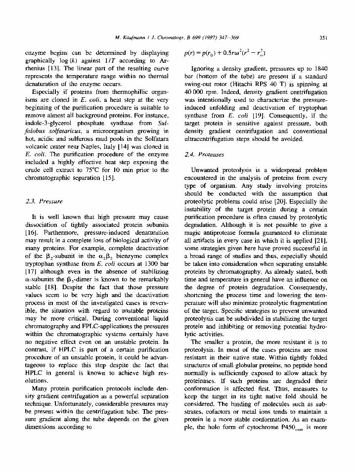
An alternative to buffer exchange making use of such batch procedures is the application of a recently developed hollow-fiber membrane module (HFMM) that was especially designed to perform dialysis on-line i.e., without increasing process time [63,64]. The hollow-fiber technology was originally de- veloped for kidney dialysis [65]. Fig. 2 shows the principle of countercurrent ultra-dialysis using such a device. The HFMM contains capillaries consisting of Cuprophan C1, a cellulose based dialysis membrane

356 M. Kaufmann / J. Chromatogr. B 699 (1997) 347-369
Smnpl¢ inl~,/g
II +
W~te ~-aeapiU~y space
• ~Sampl¢ outlet
III
1" Dialysis buffer
Extraeapilla~ s p ~
Fig. 2. Schematic representation of a HFMM and the principle of counter current on-line dialysis.
having a molecular mass cutoff at 5000. The sample flow containing the macromolecules passes the mod- ule through the interior of the capillaries (intracapil- lary space) whereas the dialysis buffer is pumped in the opposite direction outside of the capillaries (extracapillary space). The special ratio of capillary number to capillary length allows highly efficient buffer exchange at a minimum of total intracapillary void volume. For that reason, the passage of a sample flow which contains sequentially appearing different macromolecules occurs without major peak broadening of the macromolecular pattern. Thus, such a module can be connected directly to the outlet of a chromatographic column and buffer exchange can be performed via on-line dialysis already during the separation.
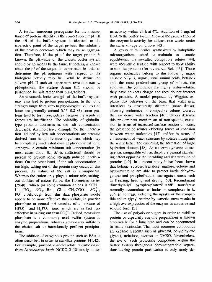
The efficiency of dialysis depends on the tempera- ture, the osmotic pressure, the size of the molecules to be exchanged, the viscosity of the solutions and the intracapillary as well as the extracapillary flow- rates. The higher the extracapillary and the lower the intracapillary flow-rates, the higher is the total efficiency of dialysis. Fig. 3 shows the influence of both the intracapillary and the extracapillary flow- rates on the resulting efficiency in desalting a 1 M NaCI solution using the HFMM VariPerm M (bitop GmbH, Witten, Germany). VariPerm M is an HFMM especially developed for buffer exchange during liquid column chromatography at a scale where flow- rates of 1 ml/min or less are commonly used [63]. As can be seen, a desalting efficiency of 95% at an intracapillary flow-rate of 1 ml/min and an ex- tracapillary flow-rate of 30 ml/min can be obtained at room temperature if a NaC1 solution is dialyzed on-line against water [63]. This intracapillary flow- rate is in the range frequently used in FPLC-applica- tions. However, if the flow-rate during a certain chromatographic separation is significantly higher (for example after scaling up of the respective
concentration NaCI (M) 1.0
0.8
0.6
0.4
0.2
0 0 0.5 1.0 1.5 2.0 2.5 3.0 3.5 4.0 4.5 5.0
IC f low (rnl/min)
Fig. 3. Efficiency of desalting 1 M NaCI by VariPerm M at extracapillary flow-rates of ( 0 ) 1, ( I ) 5, ($) 15 and (A) 30 ml ! rain.
procedure), two or even more HFMM can be con- nected to each other in series to maintain the desired efficiency of dialysis. The main advantage of on-line dialysis during chromatographic separations is the considerably reduced process time. Particularly dur- ing separation of unstable proteins, the process time plays an important role with respect to the functional maintenance of the target protein.
Buffer exchange of a certain eluate directly at the outlet of a chromatographic column is not the only possible application for an HFMM. Saving process time by on-line dialysis of an unstable protein prior to the application to a chromatographic column may be a further advantage. For example, halophillic proteins in general are known to be enormously sensitive to low ionic strengths [66]. However, the deactivation of such proteins by exposure to low salt concentrations is a time-dependent process [67]. On the other hand, on-line desalting directly prior to a chromatographic separation considerably shortens the time of low ionic strength exposure and conse- quently of protein deactivation. Thus, one would expect that even powerful separation procedures such as 1EC, though they require low salt concentrations for matrix adsorption, can be successfully used to separate halophillic proteins by those means. Indeed, alkaline p-nitrophenylphosphate phosphatase from Halobacterium salinarium was partially purified using a combined cell disruption and desalting
M. Kaufmann / J. Chrornatogr. B 699 (1997) 347-369 357
procedure by applying an HFMM prior to anion- exchange chromatography [68]. In contrast, control experiments performed with the same enzyme mak- ing use of conventional sample preparation protocols prior to anion-exchange chromatography resulted in a complete loss of enzymatic activity. Since IEC requires low salt containing buffers for protein adsorption to the matrix, this technique had not been recommended for purification protocols of halophil- lic proteins in the past. The quick on-line desalting procedure via an HFMM prior to chromatographic separation will certainly change this point of view in the near future.
A further helpful application of an HFMM during liquid column chromatography is on-line concen- tration of macromolecules within the intracapillary sample flow via transmembrane osmotic pressure gradients. The addition of hygroscopic, macro- molecular compounds to the dialysis buffer generates an osmotic pressure that pulls water and small buffer molecules out of the intracapillary sample flow into the extracapillary space. Thereby, the macromole- cules e.g., proteins or nucleic acids within the sample flow are considerably concentrated. For instance, in an experiment using VariPerm M (bitop GmbH), the addition of 20% poly(ethylene glycol) 20 000 to the dialysis buffer resulted in a fivefold concentration of a 0.715 mg/ml BSA solution at an extracapillary flow-rate of 5 ml/min and an intracapillary flow-rate of 0.9 ml/min [63]. It should be noted that the efficiency to concentrate proteins by osmotic pres- sure gradients is not limited by properties of the HFMM itself. Limitations are rather caused by other effects such as intracapillary protein precipitation, membrane adsorption or inaccuracy of the pump velocity for the intracapillary flow-rate. The advan- tages with respect to the chromatographic separation of unstable proteins are virtually the same like those described for on-line desalting. Since concentrating by an HFMM is an on-line procedure, it can be combined with liquid column chromatography and thus, saves precious process time. In addition, com- pared to ammonium sulfate precipitation, lyophiliza- tion, freeze drying or ultrafiltration, the continuous, gentle removal of buffer is certainly the mildest way of concentrating a protein solution.
There are applications of an HFMM in liquid column chromatography for analytical purposes as
well. On-line removal of a low molecular compound necessary for the desorption from a chromatographic stationary phase that has a high optical density at the detection wavelength may solve the detection prob- lem. For instance, the detection of proteins at 280 nm during covalent chromatography using thiol-Sepha- rose [69] is a problem due to 2-thio-pyridyl groups replaced by both protein binding to the column and thiol-reducing agents during elution. On-line dialysis of the column eluate by VariPerm M (bitop GmbH) completely took remedial measures in that case [63].
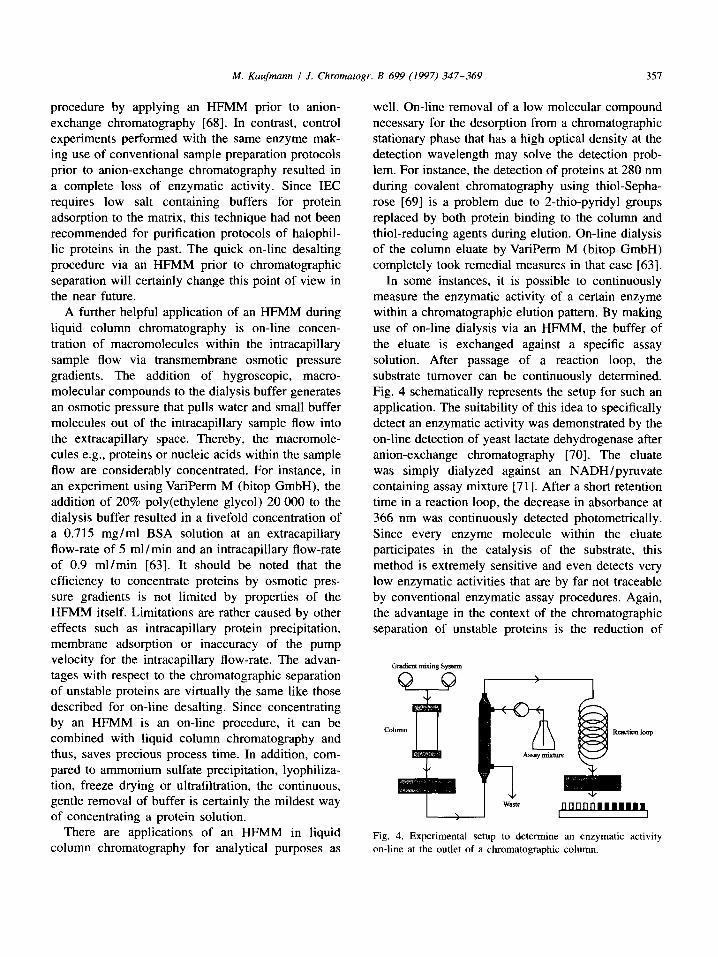
In some instances, it is possible to continuously measure the enzymatic activity of a certain enzyme within a chromatographic elution pattern. By making use of on-line dialysis via an HFMM, the buffer of the eluate is exchanged against a specific assay solution. After passage of a reaction loop, the substrate turnover can be continuously determined. Fig. 4 schematically represents the setup for such an application. The suitability of this idea to specifically detect an enzymatic activity was demonstrated by the on-line detection of yeast lactate dehydrogenase after anion-exchange chromatography [70]. The eluate was simply dialyzed against an NADH/pyruvate containing assay mixture [71 ]. After a short retention time in a reaction loop, the decrease in absorbance at 366 nm was continuously detected photometrically. Since every enzyme molecule within the eluate participates in the catalysis of the substrate, this method is extremely sensitive and even detects very low enzymatic activities that are by far not traceable by conventional enzymatic assay procedures. Again, the advantage in the context of the chromatographic separation of unstable proteins is the reduction of
Gradient mixing System
Coh xrc••'on loop
O B O [ l r l l I I l l l l l l l [ I
Fig. 4. Experimental setup to determine an enzymatic activity on-line at the outlet of a chromatographic column.

358 M. Kaufmann / J. Chromatogr. B 699 (1997) 347-369
process time because on-line detection of the target enzyme enables automated peak pooling during the separation.
On-line dialysis, as described above, is not limited to liquid column chromatography. It can be used during separation processes of any kind. For exam- ple, free flow electrophoresis (FFE) is a suitable on-line separation technique that requires low ionic strengths within the sample buffer [72]. Consequent- ly, on-line dialysis prior to the separation is an ideal application if the advantages of on-line FFE are required, particularly if the protein of interest is unstable if exposed to a low ionic strength [73].
Since on-line dialysis is a very recent develop- ment, the described examples of HFMM applications are probably only a short selection of its possibilities. Nevertheless, one should keep in mind, that when- ever buffer exchange is required as part of a certain purification protocol, on-line dialysis using a HFMM will be the choice method. Furthermore, the applica- tion of on-line dialysis is not limited to chromato- graphic separations but can be performed in any situation where the buffer of continuous flow con- taining sequentially appearing macromolecules should be exchanged.
3.2. Expanded bed adsorption
The combination of two or more separation or conditioning steps of a certain purification protocol enables performing them at the same time which reduces the total process time and for that reason saves biological activity of the target protein. Col- umn chromatography combined with on-line dialysis as described above is one example. A further elegant approach to combine several purification steps is liquid column chromatography including expanded bed adsorption (EBA). EBA joins the clarification of a particulate containing turbid solution such as a crude fermentor broth with a chromatographic sepa- ration step. The concept of EBA is "to combine chromatographys specificity and volume reduction with centrifugation throughput and ability to remove gross cellular debris - with some of filtrations advantages thrown in" [74]. Time-consuming cen- trifugation steps to obtain a supernatant, free of particles after cell disruption can be avoided. If the target protein is secreted into the culture supernatant,
both adsorption of protein and the separation of intact cells can be performed at the same time. EBA makes use of a fluidized expanded chromatographic bed during sample application. It is based on stable fluidization and uses adsorbent particles with well- defined size and density distributions, together with columns designed to give even liquid flow distribu- tion. The bed expands as the adsorbent particles are lifted by an upward liquid flow through the column. When the feedstock is applied, the target protein is captured by the adsorbent while cells and debris pass the column without being restrained. Loading of the expanded matrix is performed from bottom to top, while the product is eluted with a reversed flow after sedimenting the bed to a more tightly packed con- figuration (Fig. 5).
As an application of EBA, ZZ-M5, a recombinant protein that is expressed in E. coli and is secreted into the culture supernatant was purified [75]. This fusion protein consists of two synthetic IgG-binding domains ZZ [76] and the repeat structure M5 [77] from the central repeat region of the malaria antigen Pf155/RESA [78]. For purification via EBA, 8 1 of a crude E. coli fermentor broth were applied onto a Streamline 50 borosilicate glass column (Pharmacia Biotech) filled with a DEAE anion-exchange ad- sorbent consisting of spherical macroporous, cross- linked agarose-particles, containing crystalline quartz to increase the bead density. The ZZ-M5 fusion protein could be recovered with a yield of 90% within 4 h. The preparation almost exclusively contained ZZ-M5 [75].
A more recent application of EBA describes the purification of lysozyme from skimmed equine milk
Sample loading Elufion
Fig. 5. Principle of sample loading and elution during EBA.

M. Kaufmann / J. Chromatogr. B 699 (1997) 347-369 359
[5]. Conventional purification schemes that include various time-consuming manipulations such as pre- cipitation, dialysis or lyophilization were accom- panied by a significant inactivation of the enzyme [79]. In contrast, a two-step procedure including cation-exchange chromatography using EBA fol- lowed by a HIC step resulted in 20-fold purified electrophoretically homogenous milk lysozyme at a 80% yield [5].
Even if a turbid cell homogenate is the feedstock, EBA can be performed successfully. For example, recombinant human annexin V, an anticoagulant 34 kD protein that interacts with membranes in a calcium-dependent manner and irreversibly loses its activity below pH 5.0 was partially purified by anion-exchange EBA. For that purpose, a 26.5-1 pressure homogenate derived from a 50-1 fermen- tation was applied to a Streamline 200 DEAE column. Annexin V could be purified with a yield of more than 95% within less than 2.5 h. The purity of the protein after EBA as determined by SDS-PAGE was 20% [80].
EBA originally was developed to save process time and to reduce the purification steps during large scale downstream bioprocessing. The longer a target protein is exposed to the fermentation broth or homogenate, the more the product may be degraded. Furthermore, if the product is removed from the bulk feed and adsorbed to a chromatography adsorbent, it is less susceptible to proteases [80]. Thus, the technique of EBA in particular should be taken into consideration at the beginning of a purification procedure of an unstable protein.
3.3. Ten tac le s u p p o r t s
examples of commercially available affinity ma- trices, that allow coupling of specific ligands to suitable spacer arms are AH-Sepharose 4B [83], CH-Sepharose 4B [84], or epoxy-activated Sepha- rose 6B [85,86].
More recently, the idea of using spacer arms in liquid column chromatography resulted in a further innovative invention which applies a similar concept to less specific separation principles such as ion- exchange chromatography [87]. In a standard ion- exchange matrix the ionic groups are fixed via short arms on the support surface, thus forming a rigid array of binding sites for the poly-counter-ionic analyte (Fig. 6A). This implies that the target to be adsorbed, particularly if it is an unstable protein, may be affected with respect to its native conformation during the process of maximizing the number of ion pairs formed between the rigid ionic groups of the exchange material and the charged groups of the flexible protein. It is obvious that this process may be of special relevance at low ionic strength at which the electrostatic effects are sufficiently large, i.e., under conditions under which the protein is bound. To reduce those unwanted forces that may cause considerable tensions within the overall protein structure, the charged groups of the stationary phase should be rather flexible.
The introduction of linear polymer chains (molec- ular tentacles) to anchor the exchanger groups to the surface of the chromatographic matrix exactly serves that purpose (Fig. 6B). Such linear molecular tenta- cles consist of up to 50 monomers, each carrying a charged exchanger group, corresponding to a length of about 10 nm in the fully extended state. This emphasizes the importance of using supports with
The active center or a certain ligand binding site of an enzyme is often located deeply inside the protein molecule. For that reason, affinity ligands, if coupled directly to a chromatographic support, are often sterically hindered to reach their specific binding site. This may cause a decrease in capacity, a deterioration in specificity or even the complete loss in binding affinity of the immobilized ligand to the target protein. To solve these problems, the use of inert and flexible spacer arms interposed between the matrix and the ligand is often described in the context of affinity chromatography [81,82]. Common
Protein
Exehnnger
A B
Fig. 6. Proteins adsorbed to the surface of a conventional (A) and the surface of a tentacle matrix (B).

360 M. Kaufmann / J. Chromatogr. B 699 (1997) 347-369
sufficiently wide pores varying within a range of 100 to 500 nm in diameter. The process of binding of a protein to such a tentacle-containing support sig- nificantly differs from binding to a standard-type ion-exchanger. The flexible molecular tentacles are adapted to the individual protein surface and its charge distribution rather than the other way round. The charges located on the uncrosslinked polyelec- trolyte chains can easily adopt a configuration that is optimal for their electrostatic interaction with the protein, exhibiting a tentacle-like function. This causes considerably reduced intramolecular tensions of matrix bound proteins. Consequently, unfolding and deactivation of an unstable protein bound to a tentacle support is less likely compared with the damage that may occur upon binding to conventional supports. Indeed, anion-exchange chromatography to purify tyrocidine synthetase I from Bacillus brevis ATCC 8185 using a mono-Q HR 5/5 column (Pharmacia Biotech) resulted in a considerable deg- radation of the enzyme complex. In contrast, anion- exchange chromatography of the same enzyme using a column packed with the tentacle-carrying support Fractogel EMD TMAE-650 (Merck, Darmstadt, Germany) resulted in unaltered enzymatic activities after separation [88].
Chromatographic supports carrying linear tentacles that contain the exchanger groups are superior in several additional aspects. The capacity of a tentacle ion-exchanger no longer depends exclusively on the surface area of the support. Whereas conventional supports, although porous, only present a two-dimen- sional surface for analyte binding, tentacle gels provide additional binding sites in the third dimen- sion. It is obvious, that the number of charged groups per bed volume in such a configuration is increased considerably, and therefore, the total capacity of tentacle gels can be enhanced. In fact, the binding capacities of tentacle ion-exchangers (Fractogel EMD, Merck) determined with BSA, lysozyme and hemoglobin as model proteins revealed to be sub- stantially higher (2-4-fold) compared with capacities measured in identical runs using conventional sup- ports (Fractogel TSK, Merck). The experiments were performed with COO-, SO 3 and DEAE as the ion exchanging groups, and a higher capacity of the tentacle containing support was found in all three cases [89]. These results were recently confirmed by
capacity measurements of Fractogel EMD 650(s) SO 3 (Merck) and SP-Sepharose HP (Pharmacia). Compared with the conventional support, the tenta- cle-type cation-exchanger showed almost four-fold capacity if determined by binding of lysozyme as model protein [90].
Not only the total binding capacity but also the dynamic binding capacity of a tentacle-based chro- matographic support is very high. This means that the total binding capacity is nearly independent of the linear flow-rate during sample application. Dur- ing the process of protein binding the tentacles partially behave like polymers in solution and the binding to the matrix is favored by a number of cooperative effects that come into play. Consequent- ly, high flow-rates can be used without accepting a loss in total binding capacity. A high flow-rate corresponds to a short process time which, as already emphasized, is very important for the separation of unstable proteins.
The net-charge concept [91 ] has been widely used as a basis to predict the retention properties of proteins in IEC. According to this model, for strong anion-exchange systems, a protein will be retained only when the solvent pH is greater than the pl of the protein. However, this concept is not universally true. Retention experiments at pH 9.6 using ribonu- clease (pi=9.6) and lysozyme (pI= 11) as model proteins resulted in significant retention of both proteins to a mono-Q resin (Pharmacia Biotech) [92].
More recently, a refined model characterized the ion-exchange mechanism for proteins by an inter- action of the analyte with a conventional ion-ex- change matrix with only a distinct surface area [93]. In other words, some of the charged groups located on a certain protein surface are not involved in adsorption to the chromatographic support. Due to the non-uniform distribution of charged amino acids on the protein surface, there are patches of uniquely charged residues through which binding of the protein to the stationary phase surface may occur. Furthermore, during adsorption of identical proteins to a conventional chromatographic matrix, the charged sites that interact electrostatically with the stationary charged groups may differ from molecule to molecule. Consequently, the intensity of the individual electrostatic interaction that depends on the surface charge density of the particular region

M. Kaufmann / J. Chromatogr. B 699 (1997) 347-369 361
involved in matrix binding differs too. This may cause peak broadening during elution. In contrast, in tentacle-type ion-exchangers the flexibility of the charge arrangement allows additional or other electrostatic interactions that should improve the selectivity. Mfiller states that the action of the tentacle-type ion-exchanger involves an entire new separation parameter, the overall steric distribution of the charges on the analyte [89]. He demonstrated a clear band sharpening of lysozyme and a con- siderably improved separation of chymotrypsinogen A and cytochrome c after introduction of a strong tentacle-type cation-exchanger [Fractogel EMD 650(s) SO3-, Merck]. A comparable result was obtained using the same proteins but a weak cation- exchanger [Fractogel EMD 650(s) COO- , Merck] or acidic proteins (conalbumin, ovalbumin, and human serum albumin) and an anion-exchanger [Fractogel EMD 650(s) DEAE, Merck]. A further impressive example for the high selectivity of tentacle ion- exchangers is the separation of recombinant human deoxyribonuclease I [94]. The enzyme contains asparagine followed by serine (residues 74 and 75). Asparagine is particularly prone to deamidate under alkaline conditions through a cyclic imide inter- mediate yielding two products, aspartate or iso-as- partate [95,96]. Consequently, in addition to the charge heterogeneity caused by differences in the carbohydrate structures of the protein there are two glycoprotein subpopulations, the unaltered and the deamidated ones. Using tentacle cation-exchange chromatography, it was possible to resolve those subpopulations. The elution profile showed two distinct peaks, and isoelectric focusing clearly dem- onstrated that the first peak exclusively contained the unaltered species whereas the deamidated variants were detected in the second peak. Such a separation on the basis of a specific single characteristic is termed "protein sorting". The authors explain this sorting effect by biomimetic interaction of the poly- anionic tentacles with the target protein. They appar- ently mimic the interaction of recombinant human desoxyribonuclease I with its natural DNA substrate. This interaction was facilitated by the involvement of residue Asn74 in DNA binding.
The general improvement in selectivity and the band sharpening effect observed with tentacle based ion-exchangers may also be attributed to a reduction
of non-specific interactions between the analyte and the support, which eliminates unnecessary kinetic barriers. Due to the tentacle arrangement, the protein barely contacts the support surface and thus is prevented from unwanted non-specific interactions with the support material. This property of tentacle- based chromatographic supports was used to success- fully purify factor VIII and von Willebrand factor from human blood. Isolated von Willebrand factor is known to have a particular tendency towards non- specific interaction, especially with slightly hydro- phobic, organic supports. Isolated factor VIII is highly unstable if not stabilized by human serum albumin [41,42]. However, the association between factor VIII and von Willebrand factor in blood is very tight but dissociation can be brought about in vitro by a high concentration of calcium ions [97]. DEAE and TMAE tentacle gels were the only hydrophillic polymer supports with which elution of von Willebrand factor could be carried out in the presence of calcium ions, after dissociation of the complex [98]. The authors observed plugging of the column in the case of all other investigated supports, such as Toyopearl DEAE (Tosohaas, Stuttgart, Ger- many), Mono-Q, Mono-P (Pharmacia Biotech) and TSK-DEAE 5PW (Merck). They argue that this effect was probably due to the increased concen- tration of isolated von Willebrand factor and its interaction with the matrix. The extended tentacle ends prevented this interaction by catching the protein before it reaches the support, thus allowing its subsequent elution. Since isolated factor VIII and von Willebrand factor are known to be very sensi- tive, easily losing their biological activity, the de- scribed ion-exchange step using tentacle-type sup- ports is a convincing example for the suitability of such gels to separate unstable proteins. Even the purification of highly sensitive membrane bound protein complexes using tentacle-type ion-exchange HPLC is described [8]. The photosynthetic reaction centers photosystem I and photosystem II from a prepurified membrane extract derived from the cyanobacterium Synechocystis PCC 6803 were sepa- rated. For that purpose, three ion-exchange supports were compared with respect to their resolution. A TSK DEAE-5PW column (Tosohaas) showed a poorer separation of photosystem I and II than a mono-Q HR 5/5 column (Pharmacia) but, although

362 M. Kaufmann / J. Chrornatogr. B 699 (1997) 347-369
run at a higher flow-rate (due to a larger bed volume) and with a larger amount of sample, a LiChrospher TMAE-tentacle-type-column (Merck) showed the best resolution. The photosystem II reaction center is known to be labile under both acidic and alkaline conditions and the loss of subunits at high salt concentrations and at elevated temperatures is de- scribed. Furthermore, during all steps of purification, detergent at 3-5 times the micellar concentration and an appreciable amount of sugar (0.5-1.0 M) must be present to prevent irreversible aggregation of these hydrophobic complexes [8].
In addition to ion-exchange supports, further tenta- cle-type chromatographic media based on other separation principles were recently developed and are commercially available. These are at the moment hydrophobic interaction media (Fractogel EMD Pro- pyl and Fractogel EMD Phenyl, Merck), a thiophillic adsorption support (Fractogel EMD TA, Merck) and a metalchelate affinity matrix (Fractogel EMD Chelat, Merck). Even a tentacle based molecular sieve (Fractogel EMD BioSEC, Merck) and activated tentacle phases for affinity chromatography (Frac- togel EMD Azlactone and Fractogel EMD Epoxy, Merck) have been developed. The tentacle modi- fication of the size exclusion support results in dynamic pores that specifically influence the migra- tion speed of molecules that differ in their size.
For two reasons, tentacle-based chromatographic supports may be helpful to separate unstable pro- teins. On the one hand the protein is not forced to undergo an unfavorable conformational change upon binding that may lead to irreversible deformations, on the other hand resolution and capacity are im- proved. Better resolution of a certain separation step may reduce the total number of separation steps that are necessary during a purification protocol and are thereby potentially time-saving.
3.4. Perfusion chromatography
A further technical improvement with respect to the fluid dynamics in liquid column chromatography is the development of perfusion chromatography [99] (for review on theoretical aspects see Refs. [100,101]). Every conventional chromatographic separation making use of adsorption-desorption processes in principle can also be performed in the
perfusion mode. Perfusion chromatography is a technique based on fluid dynamics for reducing stagnant mobile phase mass transfer in liquid chro- matography and thereby reducing peak broadening [102]. This is achieved by using supports with large pores that allow the mobile phase to both passing by and flow through the particles. Such separations are characterized by high dynamic capacities, high res- olutions and separation speeds typically 10-100-fold faster than obtained with conventional media. Al- though this technical improvement has no direct effect on the protein stability (as it is the case with tentacle-based chromatographic supports), the in- direct advantages given by the higher resolution and speed result in a considerable reduction of separation time. As mentioned, once again saving process time always means saving biological activity, particular in the case of separations necessary for the purification of an unstable protein. For that reason a presentation of perfusion chromatography seems to be appro- priate.
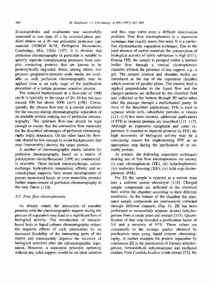
To understand the advantageous principle underly- ing perfusion chromatography, a brief description of the effects that influence peak broadening in conven- tional liquid column chromatography is necessary. Theory indicates that band spreading is predominate- ly caused by intraparticle diffusion. Longitudinal diffusion, solute diffusion in the mobile phase, Eddy diffusion and adsorption-desorption kinetics play a less significant role [ 103-105]. Within a convention- al liquid chromatographic matrix there are four mechanisms that contribute to peak broadening. Longitudinal diffusion, Eddy diffusion, mobile phase mass transfer and stagnant mobile mass transfer (Fig. 7).
Longitudinal diffusion, the diffusion in the direc- tion of the fluid flow is usually small and rather neglectable for peak broadening at flow-rates likely to be used in practice. Eddy diffusion causes peak broadening due to the numerous different paths and different path lengths that a molecule can take when passing through a packed bed. Consequently, Eddy diffusion is determined by column geometry and packing. As is the case for longitudinal diffusion, Eddy diffusion only plays a minor role in peak broadening.
Under normal operating conditions, mass transfer effects are what mainly cause peak broadening. To
M. Kaufmann / J. Chromatogr. B 699 (1997) 347-369 363
C
B
Fig. 7. Mechanisms contributing to peak broadening in liquid chromatography: longitudinal diffusion (A), Eddy diffusion (B), mobile phase mass transfer (C) and stagnant mobile phase mass transfer (D).
minimize those effects, the particle size and thus, diffusion path lengths can be reduced as it is the case in HPLC. Indeed, HPLC provides both higher speed and higher resolution than conventional chromatog- raphy. On the other hand, reducing the particle size of the chromatographic supports leads to high back- pressures and limited capacities. High backpressures may further destabilize unstable proteins and low capacities made HPLC more suited to analytical than to preparative purposes.
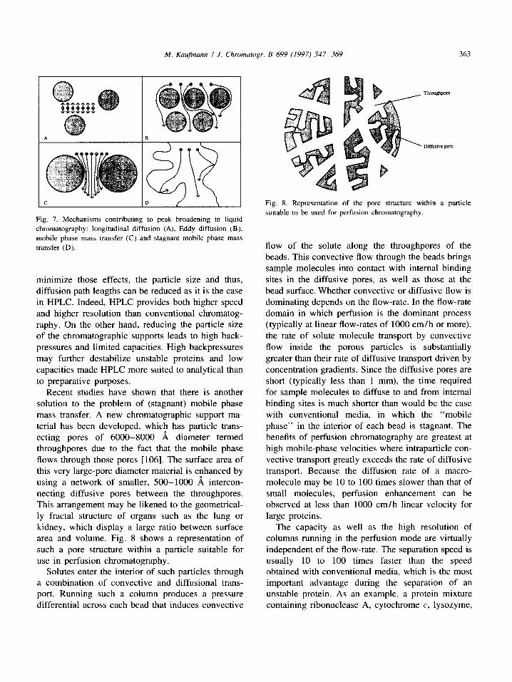
Recent studies have shown that there is another solution to the problem of (stagnant) mobile phase mass transfer. A new chromatographic support ma- terial has been developed, which has particle trans- ecting pores of 6000-8000 A diameter termed throughpores due to the fact that the mobile phase flows through those pores [106]. The surface area of this very large-pore diameter material is enhanced by using a network of smaller, 500-1000 ,~ intercon- necting diffusive pores between the throughpores. This arrangement may be likened to the geometrical- ly fractal structure of organs such as the lung or kidney, which display a large ratio between surface area and volume. Fig. 8 shows a representation of such a pore structure within a particle suitable for use in perfusion chromatography.
Solutes enter the interior of such particles through a combination of convective and diffusional trans- port. Running such a column produces a pressure differential across each bead that induces convective
m ~
Fig. 8. Representation of the pore structure within a particle suitable to be used for perfusion chromatography.
flow of the solute along the throughpores of the beads. This convective flow through the beads brings sample molecules into contact with internal binding sites in the diffusive pores, as well as those at the bead surface. Whether convective or diffusive flow is dominating depends on the flow-rate. In the flow-rate domain in which perfusion is the dominant process (typically at linear flow-rates of 1000 cm/h or more), the rate of solute molecule transport by convective flow inside the porous particles is substantially greater than their rate of diffusive transport driven by concentration gradients. Since the diffusive pores are short (typically less than 1 mm), the time required for sample molecules to diffuse to and from internal binding sites is much shorter than would be the case with conventional media, in which the "mobile phase" in the interior of each bead is stagnant. The benefits of perfusion chromatography are greatest at high mobile-phase velocities where intraparticle con- vective transport greatly exceeds the rate of diffusive transport. Because the diffusion rate of a macro- molecule may be 10 to 100 times slower than that of small molecules, perfusion enhancement can be observed at less than 1000 cm/h linear velocity for large proteins.
The capacity as well as the high resolution of columns running in the perfusion mode are virtually independent of the flow-rate. The separation speed is usually 10 to 100 times faster than the speed obtained with conventional media, which is the most important advantage during the separation of an unstable protein. As an example, a protein mixture containing ribonuclease A, cytochrome c, lysozyme,
364 M. Kaufmann / J. Chromatogr. B 699 (1997) 347-369
13-1actoglobulin and ovalbumin was successfully separated in less than 15 s by reversed-phase gra- dient elution on a 20 mm polymeric perfusion type material (POROS R/M, PerSeptive Biosystems, Cambridge, MA, USA) [107]. It is obvious that perfusion chromatography in particular is suitable to quickly separate contaminating proteases from sam- ples containing proteins that are known to be proteolytically degraded. Since, among others, low pressure preparative/process scale media are avail- able as well, perfusion chromatography may be applied even at an early stage of the purification procedure of a certain protease sensitive protein.
The induced backpressure at a flow-rate of 1000 cm/h is typically in the range of 20-30 bar but may exceed 100 bar above 4000 cm/h [108]. Conse- quently, the chosen flow-rate is a crucial parameter for the success during chromatographic separation of an unstable protein making use of perfusion chroma- tography. The optimum flow-rate should be high enough to ensure that the convective flow necessary for the described advantages of perfusion chromatog- raphy really dominates. On the other hand the flow- rate should be low enough to avoid backpressure that may (irreversibly) destroy the target protein.
A number of chromatographic media suitable for perfusion chromatography based on a matrix of poly(styrene-divinylbenzene) [109] are commercial- ly available. These include anion-exchange, cation- exchange, hydrophobic interaction, affinity and re- versed-phase supports. Very recent developments of porous monosized beads or even monoliths promise further improvement of perfusion chromatography in the near future [ l 10].
3.5. Free flow electrophoresis
As already stated, the interaction of unstable proteins with the chromatographic support during the process of separation may lead to a significant loss of biological activity. The introduction of tentacle- based beds to liquid column chromatography reduce the negative effects of such interactions by an increased flexibility of the interacting parts of the matrix and consequently improve the recovery of biological activities after the chromatographic sepa- ration. However, a separation principle operating without any solid support would be an ideal solution
and thus may solve even a difficult deactivation problem. Free flow electrophoresis is a separation technique that exactly meets that need. It is a carrier- free electrophoretic separation technique. Due to the total absence of carrier materials the conservation of biological activity of labile substances is high [111]. During FFE, the sample is pumped within a laminar buffer flow through a vertical electrophoresis chamber without the presence of any support, e.g., a gel. The sample solution and chamber buffer are introduced at the top of the separation chamber which consists of parallel plates. The electric field is applied perpendicular to the liquid flow and the charged proteins are deflected by the electrical field and collected at the bottom through 90 outlet tubes after the passage through a multichannel pump. In most of the described applications, FFE is used to separate whole cells, subcellular particles or viruses [ 112-114] but more recently, additional applications of FFE to separate proteins are described [115-117]. Although an expensive equipment and some ex- perience is required to separate proteins by FFE, the high recoveries of biological activity may be a convincing reason for implementing FFE as an appropriate step during the purification of an un- stable protein.
At present, the following separation techniques making use of free flow electrophoresis are known: (i) zone electrophoresis (ZE), (ii) isotachophoresis, (iii) isoelectric focusing (IEF), (iv) field step electro- phoresis (FSE).
For ZE the sample is injected as a narrow zone into a uniform cartier electrolyte [118]. Charged sample compounds are deflected in the electrical field within the chamber according to their different mobilities. At the bottom of the chamber the sepa- rated sample compounds are continuously collected through different channels (Fig. 9). ZE has been performed to successfully separate alcohol dehydro- genase from a crude yeast cell extract [115]. Quanti- fication of that step revealed a purification factor of 5.4 and a recovery of 97%. Those values are comparable to the average quality obtained by purification steps using liquid column chromatog- raphy. A further example for protein separation by continuous ZE is the purification of formate dehydro- genase, formaldehyde dehydrogenase and methanol oxidase from Candida boidinii crude extract [72]. By

M. Kaufmann / J. Chromatogr. B 699 (1997) 347-369 365
O-- -
UUUIIUUUUUIIUUUUU Fig. 9. Schematic diagram of an FFE-apparatus.
using a larger electrophoresis chamber, the authors were able to scale up the separation procedure by a factor of 10. Despite band broadening and dilution of the sample compounds, ZE is characterized by a high separation efficiency - even for the purification of a protein from a complex protein extract - and the recovery of nearly 100% of the biological activity [119]. Consequently, ZE may recover biological activity of an unstable protein which is deactivated if applied to a cartier-based separation technique.
ITP is the electrophoretic counterpart of displace- ment chromatography. According to their different mobilities, either cations or anions are separated in ITP. The sample is introduced into the separation compartment as a parallel zone between a leading and a terminating electrolyte. The mobilities of the sample compounds must be intermediate between those of the leading ion with the highest mobility and the terminating ion with the lowest mobility. The counter ions are chosen to have a high buffer capacity. If the steady state is achieved, all in- dividually separated ions move with the same ve- locity in a sequence of decreasing mobility towards the counter electrode. According to their number, all sample compounds are separated into as many zones without any space at the interfaces. Therefore, by the addition of suitable spacer compounds (normally amino acids or dipeptides) to the sample, a sub- fractionation of complex protein mixtures is possible. ITP has been described as an elegant technique for the continuous purification of immunoglobulins from human serum in one step using methionine and glycine as spacer substances [115]. However, experi-
ments using a-amylase lead to a 10% loss in biological activity. According to the authors this loss may have been the result from partial denaturation of the enzyme due to desalting and concentration during electrophoresis [115]. Investigations about the per- formance of ITP for the preparative purification of proteins are still at the beginning and the small differences in mobility of proteins in crude extracts render the separation quite difficult.
There are two different basic modes of performing continuous IEF in free solution. The first is to set up a pH gradient during electrophoresis by admitting a mixture of selected buffer substances with low electrophoretic mobilities, e.g., amino acids or Good buffers, into the separation chamber [120]. These simple buffers then migrate electrophoretically to produce a stepwise pH gradient according to their individual pK or p! values in a manner analogous to the formation of a continuous pH gradient with carrier ampholytes. The number of pH steps is determined by the number of different buffer com- pounds. In the second mode, an artificial stepwise pH gradient is established by admitting the individual buffer solutions into the separation chamber as parallel zones [121]. When the voltage is applied, the compound to be purified migrates under the influence of its charge until it reaches the pH step at which it loses its charge and is focused. This experimental design is best suited to the purification of one or more components of a complex protein mixture. Stepwise IEF has been described to separate a- amylase from E. col i cell proteins [116]. However, since proteins are known to aggregate and precipitate at pH values near their isoelectric points, IEF is not recommended for the separation of unstable proteins.
The low solubility and instability of proteins near their isoelectric points inhibits high concentration factors and therefore high throughputs in all continu- ous IEF methods. This drawback can be overcome by using a discontinuous conductivity profile instead of a discontinuous pH profile. The fact that the migration velocity of charged compounds can be drastically reduced by increasing the conductivity of the medium means that a concentration of migrating compounds can occur in a conductivity step just as it is possible in a pH step. FFE takes advantage of this principle. The sample is dissolved in a low con- ductivity buffer and introduced as a more or less

366 M. Kaufrnann / J. Chromatogr. B 699 (1997) 347-369
broad zone between the anolyte and catholyte, which act as flanking zones of high conductivity. The field strength in the sample zone is much higher than in the flanking zones since it is inversely proportional to the conductivities. Hence charged sample com- pounds migrate with high velocity towards the oppositely charged electrode. On reaching the anolyte or catholyte boundary they decelerate and are concentrated. FSE was shown to be suitable to separate e~-amylase from E. coli cell proteins as well as alcohol dehydrogenase from cytochrome c [116].
In contrast with IEC, where local clusters of charged residues on the protein surface are involved in protein binding to the matrix, the separation parameter in FFE is the net charge of the protein. Consequently, the separation behavior of a certain sample in IEC is not necessarily comparable to the separation behavior in FFE. FFE uses a different separation principle. Whether FFE is a suitable method to separate a certain unstable protein with high recovery of its biological activity depends on the individual properties of the protein itself. If surface or matrix interaction of the protein is the major cause of deactivation, FFE certainly may be a helpful separation method. Using a suitable equip- ment, the separation of proteins by FFE easily can be scaled up as described for the purification of alcohol dehydrogenase from a crude yeast cell extract [122,123].
4. Conclusions
Three major parameters are responsible for a successful chromatographic separation or for a com- plete purification of an unstable protein: (i) the total time required for the respective separation step and for the whole purification procedure, (ii) the nature and intensity of interaction of the protein with chromatographic matrices, columns, tubing other proteins such as proteases etc., and (iii) the res- olution of the chosen procedure. All chromatograph- ic separation methods described in this review are designed to optimize one or more of those parame- ters. On-line dialysis and expanded bed adsorption are mainly suited to save process time, whereas the advantage of using tentacle supports or performing
free flow electrophoresis is the reduction of interac- tions that may cause a loss in biological activity. Any newly developed separation methods providing an improvement in resolution (as perfusion chroma- tography) may be suitable for the purification of an unstable protein since this may reduce the total number of steps necessary for a successful purifica- tion and consequently saves process time. However, there is no universal rule how to subject an unstable protein to chromatographic separation. Keeping in mind the three major parameters as summarized above will always be helpful to develop a suitable procedure for the chromatographic separation or purification of a certain unstable target protein.
5. List of abbriviations
BSA CE DEAE DFP DMSO DTE DTT EBA EDTA FFE FPLC
FSE HFMM HIC
IEC IEF ITP MALDI-MS
NADH PLP PMSF SBTI SDS-PAGE
TLCK
Bovine serum albumin Capillary electrophoresis Diethylaminoethyl Di-isopropylfluorophosphate Dimethylsulphoxide Dithioerythritol Dithiothreitol Expanded bed adsorption Ethylenedinitriloacetic acid Free flow electrophoresis Fast performance liquid chromatog- raphy Field step electrophoresis Hollow fiber membrane module Hydrophobic interaction chromatog- raphy Ion-exchange chromatography Isoelectric focusing Isotachophoresis Matrix assisted laser desorption- ionisation mass spectrometry Nicotinamide adenine dinucleotide Pyridoxal 5'-phosphate Phenylmethylsulphonyl fluoride Soybean trypsin inhibitor Sodium dodecylsulfate-poly- acrylamide gel electrophoresis L - 1 - Chloro - 3 - [4 - tosylamido] - 7 - amino - 2 - heptanone
M. Kaufmann I J. Chromamgr. B 699 (1997) 347-369 367
T M A E
T P C K
Z E
T f i m e t h y l a m m o n i u m e t h y l
L - 1 - C h l o r o - 3 - [ 4 - t o s y l a m i d o ] -
4 - p h e n y l - 2 - b u t a n o n e
Z o n e e l e c ~ o p h o r e s i s
Acknowledgements
I would like to thank Drs. Katharina Uthoff and Thomas Schwarz for their helpful comments on the manuscript.
References
[1] O. Shimomura, J. Biolumin. Chemilumin., 10 (1995) 91. [2] M. Dettwiler and K. Kirschner, Eur. J. Biochem., 102 (1979)
159. [3] B. Reckmann, Thesis, University of Witten/Herdecke, Ger-
many, 1995. [4] P. Haezebrouck, W. Noppe, H. Van Dael and I. Hanssens,
Biochim. Biophys. Acta, 1122 (1992) 305. [5] W. Noppe, I. Hanssens and M. De Cuyper, J. Chromatogr. A,
719 (1996) 327. [6] L. Lindahl and H.J. Vogel, Anal. Biochem., 140 (1984) 394. [7] R. Jaenicke, Annu. Rev. Biophys. Bioeng., 10 (1981) l. [8] M. R6gner, J. Chromatogr., 512 (1990) 219. [9] S. Hjert6n, in N. Catsimpoolas (Editor), Methods of Protein
Separation, Plenum, New York, 1976. [10] H.P. Jennissen, J. Chromatogr., 159 (1978) 71. [ l 1 ] Pharmacia, Hydrophobic Interaction Chromatography,
Principles and Methods. [12] T. Schwarz, P. Baxtholmes and M. Kaufrnann, Biotechnol.
Appt. Biochem., 22 (1995) 179. [13] S. Arrhenius, Z. Physik. Chem., 4 (1889) 226. [14] M. de Rosa, A. Gambacorta and J.D. Bu'lock, J. Gen.
Microbiol., 86 (1975) 156. [15] M. Henning, B. Darimont, R. Sterner, K. Kirschner and J.N.
Jansonius, Structure, 3 (1995) 1295. [16] R. Jaenicke, Prog. Biophys. Molec. Biol., 49 (1987) 117. [17] S. Sindern, R. van Eldik and P. Bartholmes, Biochemistry, 34
(1995) 1959. [18] E.W. Miles, Adv. Enzymol. Relat. Areas Mol. Biol., 49
(1979) 127. [19] T. Seifert, P. Bartholmes and R. Jaenicke, FEBS Lett., 173
(1984) 381. [20] M.J. North, in R.J. Beynon and J.S. Bond (Editors),
Proteolytic Enzymes: A Practical Approach, IRL Press, UK, 1989, p. 105.
[21] J.R. Pringle, in D.M. Presscott (Editor), Methods in Cell Biology, Vol. 12, Academic Press, London and New York, 1975, p. 149.
[22] W. Pfeil, B.O. N61ting and C. Jung, Biochemistry, 32 (1993) 8856.
[23] M. Kaufmann, T. Schwarz, R. Jaenicke, K.D. Schnackerz, H.E. Meyer and P. Bartholmes, Biochemistry, 30 (1991) 4173.
[24] D.H. Rich, M.S. Bematowicz, N.S. Agarwal, M. Kawai, F.G. Salituro and P.G. Schmidt, Biochemistry, 24 (1985) 3165.
[25] G.T. James, Anal. Biochem., 86 (1978) 574. [26] E. Shaw, Biochemistry, 4 (1965) 2219. [27] G, Schoellman and E. Shaw, Biochemistry, 2 (1963) 252. [28] J.A. Cohen, R.A. Oosterbaan and F Berends, Methods
Enzymol., 11 (1967)686. [29] H. Umezawa, Methods Enzymol., 45 (1976) 678. [30] Y. Birk, Methods Enzymol., 45 (1976) 700. [31] J.C. Powers and J.W. Harper, in A.J. Barrett and G. Salvesen
(Editors), Proteinase Inhibitors, Elsevier, Amsterdam, 1986. [32] A.J. Barrett and P.M. Starkey, Biochem. J., 133 (1973) 709. [33] G.A. Homandberg, J.A. Mattis and M. Laskowski, Biochem-
istry, 17 (1978) 5220. [34] R,S. Hock and J.S. Condeelis, J. Biol. Chem., 262 (1987)
394. [35] J. Schwenke, H. Canut and A. Flores, FEBS Lett., 156
(1983) 274. [36] P. Suarez Rendueles and D.H. Wolf, FEMS Microbiol. Rev.,
54 (1988) 17. [37] P. Bartholmes, H. B6ker and R. Jaenicke, Eur. J. Biochem.,
102 (1979) 167. [38] F. Prantl, A. Strasser, M. Aebi, R. Furter, P. Niederberger, K.
Kirschner and R. Huetter, Eur. J. Biochem., 146 (1985) 95. [39] F, Hofmeister, Arch. Exp. Pathol. Pharmakol., 24 (1888)
247. [40] K.D. Collins and M.W. Washabaugh, Q. Rev. Biophys., 18
(1984) 323. [41] J.M. Lusher, S, Arkin, Ch.F. Abildgaard and R.S. Schwartz,
New Engl. J. Med., 328 (1993) 453. [42] M.J. Griffith, in H.R. Roberts (Editor), Proc. Symp. of
Biotechnol. and Promise of Pure FVIII, Baxter Healthcare Corp., Duarte, CA, 1988, p. 69.
[43] V. Phalip, C. Monnet, P. Schmitt, P. Renault, J.-J. Godon and C. Divi/:s, FEBS LeU., 351 (1994) 95.
[44] A.D. Brown, Bacteriol. Rev., 40 (1976) 803. [45] E.A. Galinski, in R.K. Poole (Editor), Advances in Microbial
Physiology, Vol. 37, Academic Press, London, 1995, p. 273. [46] P.M. Wiggins, Microbiol. Rev., 54 (1990) 432. [47] H.B. Bull and K. Breese, Arch. Biochem. Biophys., 161
(1974) 665. [48] K. Gekko and S.N. Timasheff, Biochemistry, 20 (1981)
4667. [49] E.A. Galinski, Experimentia, 49 (1993) 487. [50] K. Lippert and E.A. Galinski, Appl. Microbiot. Biotechnol.,
37 (1992) 61. [51] J.R. Blackwell and R. Horgan, FEBS Lett., 295 (1991) 10. [521 E.A. Galinski, H.G. Triiper and T. Saner, Eur. Pat. Appl., EP
93/03687 (1993) CI. C12PI/00. [53] T. Sauer, T. Schwarz and E.A. Galinski, GIT Fachz. Lab.,
(1995) 892. [54] R.K. Scopes, Protein Purification: Principles and Practice,
Springer, New York, 1993.

368 M. Kaufmann / J. Chromatogr. B 699 (1997) 347-369
[55] J.-C. Janson and L. Ryden (Editors), Protein Purification: Principles, High Resolution Methods and Applications, VCH, New York, NY, 1989.
[56] E.L.V. Harris and S. Angal (Editors), Protein Purification Methods: A Practical Approach, IRL Press, UK, 1989.
[57] E.L.V. Hams and S. Angal (Editors), Protein Purification Applications: A Practical Approach, IRL Press, UK, 1990.
[58] M.P. Deutscher (Editor), Guide to Protein Purification, Academic Press, San Diego, CA, 1990.
[59] J.A. Asenjo (Editor), Separation Processes in Biotechnology, Marcel Dekker, New York, Basel, 1990.
[60] M.S. Verall and M.J. Hudson (Editors), Separations for Biotechnology, Ellis Horwood, Chichester, UK, 1987.
[61] D. Freifelder, Physical Biochemistry, Freeman, New York, 1982.
[62] M. Mevarech and E. Neumann, Biochemistry, 16 (1977) 3786.
[63] M. Kaufmann, T. Schwarz and P. Bartholmes, J. Chroma- togr., 639 (1993) 33.
[64] P. Bartholmes, M. Kaufmann and T. Schwarz, Eur. Pat,, EP 0664724 (1993).
[65] K. Ota, T. Shimoji, A. Saito, K. Maeda and K. Kobayashi, Trans. Am. Soc. Artif. Intern. Organs, 19 (1973) 98.
[66] R. Mengele and M. Sumper, J. Biol. Chem., 267 (1992) 8182.
[67] J.K. Lanyi, Bacteriol. Rev., 38 (1974) 272. [68] K. Heesche-Wagner, T. Schwarz, P. Bartholmes and M.
Kaufmann, J. Chromatogr. A, 711 (1995) 175. [69] A.C. de Graaf-Hess and A. de Kok, FEBS Lett., 143 (1982)
261. [70] M. Kaufmann, T. Schwarz and P. Bartholmes, GIT Chro-
matographie, 14 (1994) 13. [71] A. Denicola-Seoane and B.M. Anderson, J. Biol. Chem., 265
(1990) 3691. [72] S. Nath, H. Schutte, G. Weber, H. Hustedt and W.D.
Deckwer, Electrophoresis, 11 (1990) 937. [73] T. Schwarz, S. Sindern, P. Bartholmes and M. Kaufmann,
Electrophoresis, 15 (1994) 1118. [74] D.K. McCormick, Bio/Technology, 11 (1993) 1059. [75] M. Hansson, S. St~hl, R. Hjorth, M. Uhl6n and T. Moks,
Bio/Technology, 12 (1994) 285. [76] B. Nilsson, T. Moks, B. Jansson, L. Abrahams6n, A.
Elmblad, E. Holmgren, C. Henrichson, T.A. Jones and M. Uhl6n, Protein Engineering, 1 (1987) 107.
[77] A. Sj61ander, M. Hansson, K. L6vgren, B. W~hlin, K. Berzins and P. Perlmann, Parasite Immunol., 15 (1993) 355.
[78] J.M. Favaloro, R.L. Coppel, L.M. Corcoran, S.J. Foote, G.V. Brown, R.F. Anders and D.J. Kemp, Nucleic Acids Res., 14 (1986) 8265.
[79] F.H. White, Jr., H.A. McKenzie and D.C. Shaw, Biochem. Internat., 16 (1988) 521.
[801 A.-K. Barnfield Frej, R. Hjorth and ]k. Hammarstr6m, Biotechnol. Bioeng. 44 (1994) 922.
[81] C.R. Lowe, Eur. J. Biochem., 73 (1977) 265. [82] D.H. Watson and J.C. Wootton, Biochem. J., 167 (1977) 95. [83] H. Anttinen and K.I. Kivirikko, Biochim. Biophys. Acta, 429
(1976) 750.
[84] D. Robinson, N.C. Phillips and B. Winchester, FEBS Lett., 53 (1975) 110.
[85] M. Landt, S.C. Boltz and L.G. Butler, Biochemistry, 17 (1978) 915.
[86] P.C. Simons and D.L. Van der Jagt, Anal. Biochem., 82 (1977) 334.
[87] W. Miiller, Eur. Pat., EP 0337144 (1988). [88] E. Pfeifer, Thesis, University of Berlin, Germany, 1994. [89] W. Miiller, J. Chromatogr., 510 (1990) 133. [90] F.B. Anspach, H. Spille and U. Rinas, J. Chromatogr. A, 711
(1995) 129. [91] N.K. Boardman and S.M. Partridge, Biochem. J., 59 (1955)
543. [92] M.T.W. Hearn, A.N. Hodder, F.W. Fang and M.I. Aguilar, J.
Chromatogr., 548 (1991) 117. 193] W. Kopaciewicz, M.A. Rounds, J. Fausnaugh and F.E.
Regnier, J. Chromatogr., 266 (1983) 3. [94] J. Cacia, C.P. Quan, M. Vasser, M.B. Sliwkowski and J.
Frenz, J. Chromatogr., 634 (1993) 229. [95] H.T. Wright, Crit. Rev. Biochem. Molec. Biol., 26 (1991) 1. [96] Y.C. Meinwald, E.R. Stimson and H.A. Scheraga, Int. J.
Pept. Protein Res., 28 (1986) 79. [97] T.S. Zimmermann and D. Meyer, in A.L. Bloom and D.P.
Thomas (Editors), Haemostasis and Thrombosis, Churchill Livingstone, Edinburgh, UK, 1987, p. 131.
[98] Dj. Josic, H. Schwinn, M. Stadler and A. Strancar, J. Chromatogr. B, 662 (1994) 181.
[99] N.B. Afeyan, F.E. Regnier and R.C. Dean, U.S. Pat., 5 019 270 (1991).
[100] A.I. Liapis and M.A. McCoy, J. Chromatogr., 599 (1992) 87.
[101] M.A. McCoy, A.I. Liapis and K.K. Unger, J. Chromatogr., 644 (1993) 1.
[102] F.E. Regnier, Nature, 350 (1991) 634. [103] J.C. Giddings, Dynamics of Chromatography, Dekker, New
York, 1965. [104] J.N. Done and J.H. Knox, J. Chromatogr., 10 (1972) 606. [105] C. Horvath and H.J. Lin, J. Chromatogr., 126 (1976) 401. [106] N.B. Afeyan, N.F. Gordon, I. Mazsaroff, L. Varady, S.P.
Fulton, Y.B. Yang and F.E. Regnier, J. Chromatogr., 519 (1990) 1.
[107] S.P. Fulton, N.B. Afeyan, N.F. Gordon and F.E. Regnier, J. Chromatogr., 547 (1991) 452.
[108] N.B. Afeyan, S.P. Fulton and F.E. Regnier, J. Chromatogr., 544 (1991) 267.
[109] L. Varady, N. Mu, Y.-B. Yang, S.E. Cook, N. Afeyan and F.E. Regnier, J. Chromatogr., 631 (1993) 107.
[110] F. Svec and J.M.J. Fr6cbet, Science, 273 (1996) 205. [111] S. Nath, H. Schiitte, H. Hustedt and W.-D. Deckwer,
Electrophoresis, 11 (1990) 612. [112] K. Hannig and W.F. Kriissmann, Hoppe-Seylers Z. Physiol.
Chem., 349 (1968) 161. [113] K. Hannig and K. Zeiller, Hoppe-Seylers Z. Physiol.
Chem., 350 (1969) 467. [114] G.V. Sherbet, The Biophysical Characterization of the Cell
Surface, Academic Press, New York, 1978, p. 36. [115] S. Hoffstetter-Kuhn, R. Kuhn and H. Wagner, Electro-
phoresis, 11 (1990) 304.

M. Kaufmann / J. Chromatogr. B 699 (1997) 347-369 369
[116] R. Kuhn, S. Hoffstetter-Kuhn and H. Wagner, Electro- phoresis, 11 (1990) 942.
[117] M.J. Clifton, N. Jouve, H. de Balmann and V. Sanchez, Electrophoresis, 11 (1990)913.
[118] K. Hanning, Electrophoresis, 3 (1982) 235. [119] S. Hoffstetter-Kuhn, Thesis, (1989), Universitat of Saar-
land, Saarbriicken, Germany. [120] R.A. Mosher, W. Thormann, R. Kuhn and H. Wagner, J.
Chromatogr., 469 (1989) 444.
[121] R. Kuhn, H. Wagner, R.A. Mosher and W. Thormann, Electrophoresis, 8 (1987) 503.
[122] S. Hoffstetter-Kuhn and H. Wagner, Electrophoresis, 11 (1990) 451.
[123] S. Hoffstetter-Kuhn and H. Wagner, Electrophoresis, 11 (1990) 457.