elevated hepatic 11β-hydroxysteroid dehydrogenase …elevated hepatic 11β-hydroxysteroid...
TRANSCRIPT

Elevated hepatic 11β-hydroxysteroid dehydrogenasetype 1 induces insulin resistance in uremiaAnanda Chapagaina,1, Paul W. Catonb,1, Julius Kieswicha, Petros Andrikopoulosa, Nanda Nayunia, Jamie H. Longa,Steven M. Harwooda, Scott P. Websterc, Martin J. Rafterya, Christoph Thiemermanna, Brian R. Walkerc,Jonathan R. Secklc, Roger Cordera, and Muhammad Magdi Yaqooba,2
aCentre of Translational Medicine and Therapeutics, William Harvey Research Institute, Barts and the London, Queen Mary University of London, LondonEC1M 6BQ, United Kingdom; bCentre for Diabetes, Blizard Institute, Barts and the London, Queen Mary University of London, London E1 2AT, UnitedKingdom; and cEndocrinology Unit/British Heart Foundation Centre for Cardiovascular Science, University of Edinburgh, Queen’s Medical Research Institute,Edinburgh EH16 4TJ, United Kingdom
Edited by David W. Russell, University of Texas Southwestern Medical Center, Dallas, TX, and approved January 29, 2014 (received for review July 2, 2013)
Insulin resistance and associated metabolic sequelae are commonin chronic kidney disease (CKD) and are positively and indepen-dently associated with increased cardiovascular mortality. However,the pathogenesis has yet to be fully elucidated. 11β-Hydroxysteroiddehydrogenase type 1 (11βHSD1) catalyzes intracellular regenera-tion of active glucocorticoids, promoting insulin resistance in liverand other metabolic tissues. Using two experimental rat models ofCKD (subtotal nephrectomy and adenine diet) which show early in-sulin resistance, we found that 11βHSD1 mRNA and protein increasein hepatic and adipose tissue, together with increased hepatic11βHSD1 activity. This was associated with intrahepatic but not cir-culating glucocorticoid excess, and increased hepatic gluconeogene-sis and lipogenesis. Oral administration of the 11βHSD inhibitorcarbenoxolone to uremic rats for 2 wk improved glucose toleranceand insulin sensitivity, improved insulin signaling, and reduced he-patic expression of gluconeogenic and lipogenic genes. Furthermore,11βHSD1−/− mice and rats treated with a specific 11βHSD1 inhibitor(UE2316) were protected from metabolic disturbances despitesimilar renal dysfunction following adenine experimental uremia.Therefore, we demonstrate that elevated hepatic 11βHSD1 is animportant contributor to early insulin resistance and dyslipidemiain uremia. Specific 11βHSD1 inhibitors potentially represent a noveltherapeutic approach for management of insulin resistance inpatients with CKD.
The prevalence of chronic kidney disease (CKD) has increaseddramatically in recent years causing substantial morbidity and
mortality (1). Although diabetic patients with CKD sometimesdevelop recurrent hypoglycemia, possibly due to reduced renalcatabolism of insulin, it is increasingly recognized that insulinresistance and associated hyperinsulinemia are common com-plications in patients with CKD (2, 3) with an insulin resistance-like syndrome occurring even at the earliest stage of renal dys-function (4). CKD-induced insulin resistance is positively andindependently associated with increased cardiovascular mortality(5, 6). Furthermore, mortality among patients treated with he-modialysis is higher in those with more severe insulin resistance(7). Despite this, the mechanisms responsible for the onset ofinsulin resistance in CKD are unclear.Increased hepatic gluconeogenesis can cause hyperinsulinemia
and hyperglycemia (8, 9). Expression of genes encoding key glu-coneogenic enzymes such as phosphoenolpyruvate carboxykinase 1(PCK1) and glucose-6-phosphatase (G-6pase) are transcriptionallyinduced in response to stimuli such as glucagon and glucocorti-coids, and suppressed by insulin. This process is tightly regulatedby transcription factors and cofactors, in particular peroxisomeproliferator-activated receptor gamma coactivator 1alpha(PGC1α) (10). Hepatic gluconeogenesis is inappropriately ele-vated in rodent models and human patients with insulin re-sistance and type 2 diabetes mellitus (T2DM). Abnormal elevationof gluconeogenesis leading to insulin resistance can occur as a resultof circulating glucocorticoid excess, as observed in Cushingsyndrome (11, 12). However, the role of glucocorticoids in the
pathophysiology of CKD-induced insulin resistance has not beendescribed.11β-Hydroxysteroid dehydrogenase (11βHSD) enzymes func-
tion to regulate intracellular glucocorticoid levels. 11βHSD type 1(11βHSD1) catalyzes the conversion of intrinsically inactive cor-tisone to active cortisol (11-dehydrocorticosterone to corticoste-rone in rats), thus amplifying local glucocorticoid levels, whereas11βHSD2 catalyzes the opposite reaction (11, 13) but is largelyconfined to the distal nephron. 11βHSD1 is expressed at highlevels in the major organs underpinning metabolism such as liverand adipose tissue. Hepatic overexpression of 11βHSD1 leads toinsulin resistance in mice with increased lipogenesis (14), consis-tent with increased intrahepatic glucocorticoid action, whereas11βHSD1 inhibition or deficiency leads to decreased hepatic glu-coneogenesis (and decreased PCK1), improved insulin sensitivity,and correction of hyperglycemia in rodent models of insulin re-sistance and patients with T2DM (15–18).We investigated the hypothesis that 11βHSD1-induced gluco-
corticoid excess mediates abnormal elevation of gluconeogenesisand lipogenesis in uremia, using two experimental rodent modelswith entirely distinct mechanisms of development of renal failure;subtotal nephrectomy (SNx) and adenine feeding. To investigate
Significance
Prevalence of chronic kidney disease (CKD) has reached epidemicproportions in the Western world in recent decades. Abnormallyelevated blood insulin and impaired insulin action (insulin re-sistance) are common complications of CKD, and are associatedwith increased cardiovascular-related deaths in CKD patients.Therefore, novel therapies are required to treat insulin resistancein CKD. Abnormally elevated levels of glucocorticoids can causeinsulin resistance. Here we demonstrate a crucial role for theenzyme 11β-hydroxysteroid dehydrogenase type 1 (11βHSD1)predominantly in liver, which is essential for glucocorticoidproduction, in causing insulin resistance in CKD. Additionally,11βHSD1 inhibition corrected insulin resistance in CKD rodentmodels. Taken together, this is strong evidence that selectiveinhibition of 11βHSD1 is a promising therapeutic target fortreatment of insulin resistance in CKD.
Author contributions: A.C., P.W.C., J.K., P.A., N.N., S.M.H., S.P.W., M.J.R., B.R.W., R.C., andM.M.Y. designed research; A.C., P.W.C., J.K., N.N., J.H.L., and R.C. performed research; S.P.W.,B.R.W., and J.R.S. contributed new reagents/analytic tools; A.C., P.W.C., J.K., P.A., N.N., J.H.L.,S.M.H., M.J.R., C.T., J.R.S., R.C., and M.M.Y. analyzed data; and A.C., P.W.C., and M.M.Y.wrote the paper.
Conflict of interest statement: S.P.W., B.R.W., and J.R.S. are inventors on patentsowned by the University of Edinburgh, which claim therapeutic use of compounds,including UE2316.
This article is a PNAS Direct Submission.1A.C. and P.W.C. contributed equally to this work.2To whom correspondence should be addressed. E-mail: [email protected].
This article contains supporting information online at www.pnas.org/lookup/suppl/doi:10.1073/pnas.1312436111/-/DCSupplemental.
www.pnas.org/cgi/doi/10.1073/pnas.1312436111 PNAS | March 11, 2014 | vol. 111 | no. 10 | 3817–3822
MED
ICALSC
IENCE
S
Dow
nloa
ded
by g
uest
on
Apr
il 8,
202
0

a potential causal role for 11βHSD1 in uremia-induced insulinresistance, we used the 11βHSD1 inhibitors carbenoxolone (CBX)(16, 19) and UE2316 and investigated 11βHSD1−/− mice.
ResultsMarkers of Renal Failure in Models of Experimental Uremia. Serumcreatinine was elevated 3.6-fold in SNx and 8.1-fold in adenine-fedrats, and 3.5-fold in adenine-fed 11β-HSD1−/− mice, whereas se-rum urea was elevated 5.5-, 11.8-, and 4.5-fold, respectively. Fur-ther markers of chronic renal injury are shown in Tables S1–S3.Body weights, mean food intake and average heart rate were notsignificantly different between the uremic and sham groups. Meanblood pressure, although tending to be higher in CBX treatedgroups, was not significantly different because of high variability(Tables S4 and S5).
Hepatic 11βHSD1 Is Elevated in CKD.Hepatic 11βHSD1 mRNA andprotein levels, together with 11βHSD1 reductase activity, weresignificantly elevated in SNx (Fig. 1 A and B) and adenine-fedrats (Fig. 1 E and F). Intrahepatic corticosterone levels were alsomarkedly elevated in SNx and adenine-fed rats (Fig. 1 C and G)however, consistent with the notion that 11βHSD1 determinestissue-specific intracrine glucocorticoid levels, this occurred inthe absence of elevated systemic serum corticosterone levels(Fig. 1 D and H). Administration of CBX (50 mg·kg−1·d−1; 2 wk)normalized hepatic 11βHSD1 mRNA and protein levels, sup-pressed 11βHSD1 reductase activity and lowered hepatic corti-costerone levels in SNx and adenine-fed rats. In contrast, CBXhad no effect on serum corticosterone levels, demonstrating thatCBX selectively influences intrahepatic glucocorticoid levels.Similar to observations in liver, white adipose tissue levels of11βHSD1 mRNA and protein were increased in uremia, an ef-fect that was reversed by CBX (Fig. S1). In contrast, 11βHSD1mRNA and protein levels in skeletal muscle were unchangedacross all four experimental groups (Fig. S1).Consistent with recent evidence demonstrating induction of
11βHSD1 transcription by proinflammatory cytokines (20–23) se-rum levels of IL-1β, TNFα, and IL-6 were elevated in the chronicphase of SNx and adenine-induced uremia (Fig. S2), but were un-affected by CBX, suggesting a possible uremia-dependent mecha-nism upstream of 11βHSD1 induction and hepatic glucocorticoidmetabolism, involving elevated proinflammatory cytokine levels.
Uremic Rats Develop Impaired Glucose Tolerance and Reduced InsulinSensitivity. Serum insulin levels were markedly elevated in SNx(Fig. 2A) and adenine-fed rats (Fig. 3A). However, fasting serumglucose levels were unchanged (Figs. 2B and 3B), suggestive ofuremia-induced insulin resistance. To further analyze potentialuremia-induced changes in systemic glucose tolerance and in-sulin sensitivity, we conducted i.p. glucose and insulin tolerancetests (GTT and ITT). During the GTT, blood glucose levels weresignificantly higher in SNx and adenine-fed rats compared withsham up to 60 min post-glucose administration (Figs. 2C and3C). In addition, serum insulin levels were elevated duringa GTT, remaining elevated 45 min post-glucose administration(Figs. 2D and 3D) in SNx and adenine-fed rats compared withsham. Moreover, impairment in insulin’s ability to lower bloodglucose levels in an ITT was observed in SNx and adenine-fedrats, with blood glucose levels remaining significantly elevated inboth models 30 min post-insulin administration (Figs. 2E and3E). Taken together, these data demonstrate the presence ofinsulin resistance in both models of uremia. To determinewhether uremia-induced insulin resistance may be linked to el-evated hepatic glucose production, we conducted a pyruvatetolerance test (PTT). Consistent with abnormally elevated he-patic glucose production, SNx and adenine-fed rats displayedsignificantly increased blood glucose levels up to 90 min post-pyruvate administration (Figs. 2F and 3F).Collectively, these data demonstrate significant insulin resistance
associated with abnormally elevated hepatic glucose production inboth SNx and adenine-feeding models of CKD.
11βHSD1 Inhibition Improves Systemic Glucose Tolerance and InsulinSensitivity in CKD Rats. To assess the potential role of increases in11βHSD1 levels and activity in mediating insulin resistance inuremia, we analyzed rats treated with CBX. Administration ofCBX did not improve markers of renal failure in any group(Tables S1 and S2). Despite this, CBX completely preventeduremia-induced increases in serum insulin levels in both models(Figs. 2A and 3A). Moreover, 11βHSD1 inhibition with CBXresulted in a significant improvement in insulin sensitivity and
Fig. 1. Hepatic 11βHSD1 levels and acitivity are elevated in SNx and Ad rats.(A and E) 11βHSD1 mRNA and protein, (B and F) hepatic corticosteroneproduction (pg·min−1·mg−1 liver protein), (C and G) hepatic corticosteronelevels, and (D and H) plasma corticosterone levels. Data are expressed asmean ± SEM (n = 8 per group). Statistically significant differences betweensham/control and Ad or SNx are indicated by *P < 0.05, **P < 0.01, and***P < 0.001. Statistically significant effects of CBX treatment are indicatedby #P < 0.05, ##P < 0.01, and ###P < 0.001.
Fig. 2. 11βHSD1 inhibition ameliorates insulin resistance in SNx rats. (A)Fasting plasma insulin, (B) fasting plasma glucose, (C) plasma glucose re-sponse to GTT, (D) plasma insulin response to GTT, (E) plasma glucose re-sponse to ITT, and (F) plasma glucose response to PTT. Data are expressed asmean ± SEM (n = 8 per group). Statistically significant differences betweensham and SNx are indicated by *P < 0.05. Statistically significant effects ofCBX treatment are indicated by #P < 0.05.
3818 | www.pnas.org/cgi/doi/10.1073/pnas.1312436111 Chapagain et al.
Dow
nloa
ded
by g
uest
on
Apr
il 8,
202
0

glucose tolerance (Figs. 2 A–F and 3 A–F), as well as reducedhepatic glucose production following a PTT. Taken together,these data demonstrate that abnormally elevated 11βHSD1 playsa crucial role in mediating insulin resistance in uremia.
11βHSD1 Inhibition Suppresses Hepatic Gluconeogenic Gene Expressionand Markers of Impaired Insulin Signaling in Uremia. Because uremia-induced insulin resistance was associated with elevated bloodglucose levels following a PTT, we examined alterations in hepaticgluconeogenic enzymes. Levels of PCK1 mRNA and protein andG6Pase protein were elevated in SNx (Fig. 4 A–C) and adenine-fed rats (Fig. 4 F–H). PGC1α mRNA and protein levels were alsoincreased in SNx (Fig. 4 D and E) and adenine-fed rats (Fig. 4 Iand J). Similar to effects observed on systemic insulin resistance,CBX administration reversed these effects on gluconeogenicenzymes in both models of uremia. To assess whether increasedhepatic gluconeogenesis occurs in association with impaired in-sulin signaling, we measured phosphorylation of AKT at serine473. Protein levels of phospho-(Ser473)-AKT were reduced inliver and also in the skeletal muscle and white adipose tissue ofuremic rats. In all three tissues, this effect was reversed by CBXadministration (Fig. S1). Taken together, these data suggest that11βHSD1 mediates insulin resistance in uremia through abnor-mal elevation of hepatic gluconeogenesis and dysregulationinsulin signaling.
Uremia-Induced Dyslipidemia Is Corrected by 11βHSD1 Inhibition.Dyslipidemia manifested by raised serum levels of triglycerides,free fatty acids, and cholesterol is observed in CKD patients androdent models of uremia (5, 6, 24). Dyslipidemia can cause in-sulin resistance (25, 26). The de novo synthesis of fatty acid and
cholesterol is controlled through transcriptional regulation ofseveral key genes, in particular acetyl CoA carboxylase (ACC),fatty acid synthase (FASN), and 3-hydroxy-3-methylglutaryl CoAreductase (HMGCR) (27). Transcriptional regulation of thesegenes is controlled by sterol regulatory element-binding protein1 (SREBP1) (28), which is induced by insulin (29).Consistent with previous reports in experimental models of
uremia (30, 31), SNx and adenine-fed rats showed elevated se-rum levels of cholesterol, triglycerides, and nonesterified fattyacids (NEFAs), (Figs. 5 A–C and 6 A–C). Hepatic mRNA ex-pression of HMGCR was elevated in both uremic models (Figs.5D and 6D) suggesting increased de novo hepatic cholesterolbiosynthesis. Moreover, mRNA and protein levels of ACC (Figs.5E and 6E) and FASN (Figs. 5F and 6F) and mRNA levels ofSREBP1c (Figs. 5G and 6G) were markedly increased in SNxand adenine-fed rats, indicative of increased hepatic de novolipogenesis. Importantly, in both CKD models, abnormally ele-vated serum levels of cholesterol, triglycerides, and NEFAs wereameliorated following administration of CBX, demonstratingthat uremia-induced dyslipidemia, like gluconeogenesis, mayoccur in part through an 11βHSD1-mediated mechanism (Figs. 5A–C and 6 A–C). Consistent with this, mRNA and protein levelsof ACC1, FASN, SREBP1c, and HMGCR were also partiallynormalized by CBX in SNx and adenine-fed rats (Figs. 5 D–Gand 6 D–G).We also assessed liver and skeletal muscle lipid content by
measuring triglyceride levels in SNx rats. Despite observed changesin lipogenic gene and protein levels, hepatic triglyceride contentwas unchanged between sham and uremic rats (Fig. S3). In con-trast, uremia increased skeletal muscle triglyceride levels, butthese increases were not reversed by CBX.Taken together, these data demonstrate that uremia-induced
dyslipidemia occurs through an 11βHSD1-dependent mechanismand may contribute to insulin resistance in these models.
Fig. 3. 11βHSD1 inhibition ameliorates insulin resistance in adenine-fedrats. Experimental uremia was induced in rats by administration of 0.75% Ad(n = 8 per group). CBX (50 mg·kg−1·d−1) or vehicle was administered by oralgavage for 2 wk in both groups. (A) Fasting plasma insulin, (B) fasting plasmaglucose, (C) plasma glucose response to 2 g per kg dextrose injected i.p.(GTT), (D) plasma insulin response to glucose load following i.p. GTT, (E)plasma glucose response to i.p. ITT, and (F) plasma glucose response to i.p.PTT. Data are expressed as mean ± SEM. Statistically significant differencesbetween control and Ad are indicated by *P < 0.05 and **P < 0.01. Statis-tically significant effects of CBX treatment are indicated by #P < 0.05.
Fig. 4. CBX corrects uremia-mediated insulin resistance in association withsuppressed hepatic gluconeogenesis. Experimental uremia was induced inrats by SNx (n = 8 per group) or Ad (n = 8 per group). CBX (50 mg·kg−1·d−1)or vehicle was administered by oral gavage for 2 wk. (A and B) SNx, PCK1mRNA and protein; (C) SNx, G-6pase protein; (D and E) SNx, PGC1α mRNAand protein; (F and G) Ad, PCK1 mRNA and protein; (H) Ad, G-6pase protein;and (I and J) Ad, PGC1α mRNA and protein. Data are expressed as mean ±SEM. Statistically significant differences between sham and Ad or SNx areindicated by *P < 0.05, **P < 0.01, and ***P < 0.001. Statistically significanteffects of CBX treatment are indicated by #P < 0.05 and ###P < 0.001.
Chapagain et al. PNAS | March 11, 2014 | vol. 111 | no. 10 | 3819
MED
ICALSC
IENCE
S
Dow
nloa
ded
by g
uest
on
Apr
il 8,
202
0

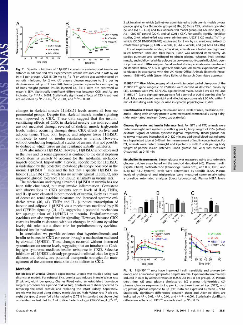
Specific Inhibition of 11βHSD1 with UE2316 Protects Uremic Rats fromInsulin Resistance. Finally, to confirm a specific role for 11βHSD1in uremia-induced insulin resistance and rule out potentialnonspecific CBX effects, we examined the effects of specific11βHSD1 inhibitor (UE2316) (32) on insulin resistance in ure-mic (adenine-fed) rats. In agreement with data obtained fromCBX treatment, UE2316 (20 mg·kg−1·d−1) significantly improvedglucose tolerance and insulin sensitivity in adenine-fed rats (Fig.7 A and B), without affecting parameters of renal failure (TableS6), confirming a role for 11βHSD1 in mediating uremia-in-duced insulin resistance.
11βHSD1−/− Mice Are Protected from Uremic Insulin Resistance. Tofurther establish the role of 11βHSD1 in uremia-induced insulinresistance we used 11βHSD1−/−mice (33). Following consumptionof an adenine-diet, both 11βHSD1−/− and wild-type (WT) micedeveloped similar levels of renal dysfunction (Fig. 8A and TableS3). Similar to the phenotype observed in rats, uremic WT micedeveloped dyslipidemia (raised systemic triglyceride levels), im-paired glucose tolerance, and reduced insulin sensitivity (Fig. 8B–E). In marked contrast, 11βHSD1−/− mice were protectedagainst uremia-induced insulin resistance and dyslipidemia,displaying a lipid profile similar to control animals, as well asimproved glucose tolerance and insulin sensitivity comparablewith that in WT controls.
DiscussionWe report here that elevated hepatic 11βHSD1 mediates impairedglucose tolerance, reduced insulin sensitivity, hyperinsulinemia,and dyslipidemia in two distinct male rat models of CKD. Im-portantly, increased 11βHSD1 elevated hepatic corticosteronecontent without changes in systemic corticosterone, suggestinga key role for hepatic 11β-HSD1, the major source of local corti-costerone regeneration in liver, in development of insulin re-sistance in CKD. Although these studies have yet to be confirmedin female rats, improvements in metabolic profiles following11βHSD1 inhibition or ablation occurred without corrections inrenal dysfunction, indicating that selective inhibitors of 11βHSD1may be a plausible therapeutic approach to insulin resistance andits complications in CKD.Previous studies have reported elevated hepatic glucose pro-
duction in chronically uremic patients (34). Furthermore, increasedhepatic gluconeogenesis in acute experimental uremia is reversed
by the glucocorticoid receptor antagonist RU 38486 (35). Elevatedhepatic gluconeogenesis can cause insulin resistance and hyper-insulinemia (8, 9), whereas knockdown of gluconeogenic genes andcofactors have resulted in the correction of insulin resistance andimprovement in insulin sensitivity in murine models (36–39). Thissuggests that 11βHSD1-mediated increases in hepatic gluconeo-genesis via up-regulation of PCK1 and PGC1α may representa likely cause of hyperinsulinemia and insulin resistance in uremia.Dyslipidemia is also a common complication of CKD (5, 6,
24). We observed abnormally elevated plasma lipids and cho-lesterol in uremic rats, with parallel increases in lipogenic geneand protein expression. 11βHSD1 inhibition corrected systemicdyslipidemia, in association with reduced hepatic lipogenic geneand protein expression. These observations are consistent withglucocorticoid induction of triglyceride synthesis and fatty liverin rats, whereas mice overexpressing hepatic 11βHSD1 display in-creased hepatic lipogenesis (14). However, it is unclear in ourmodel whether increased hepatic lipogenic gene expression occursvia direct induction by glucocorticoids or through elevated insulin-mediated SREBP1c induction, as would be anticipated because ofundesirable metabolic consequence of hyperinsulinemia (28, 29).Although our data points to a crucial role for hepatic 11βHSD1
as a key mediator of insulin resistance in uremia, we also observedincreases in 11βHSD1 levels and parallel impairment of insulinsignaling in adipose tissue, changes which were reversed by CBX.These changes may also contribute to uremia-induced insulin re-sistance, for example via glucocorticoid-mediated increases in cir-culating NEFA levels, as observed in our models, which mayaccount for the impaired insulin signaling observed in uremic liver,through ectopic lipid deposition in liver and skeletal muscle. Thismay be particularly plausible given the lack of increased triglyceridelevels in uremic liver. Interestingly, studies have suggested thatCBX may not act directly on adipose tissue (15), raising thepossibility that a secreted hepatokine produced during hepaticCBX metabolism may have impact on adipose tissue 11βHSD1.Similar to adipose tissue and liver, insulin signaling was alsoimpaired in uremic skeletal muscle, whereas skeletal musclelipid levels are increased, suggesting a potential contributoryrole for muscle ectopic lipid deposition in uremia-induced insulinresistance. However, uremia-induced increases in skeletal muscletriglyceride were not reversed by CBX, while we did not observe
Fig. 5. CBX suppresses total and hepatic lipogenesis in SNx animals. Experi-mental uremia was induced in rats by SNx (n = 8 per group). CBX(50 mg·kg−1·d−1) or vehicle was administered by oral gavage for 2 wk. (A) Plasmacholesterol, (B) plasma triglycerides, (C) plasma NEFA, (D) HMGCRmRNA, (E) ACCmRNA and protein, (F) FAS mRNA and protein, and (G) SREBP1 mRNA. Data areexpressed as mean ± SEM. Statistically significant differences between sham andSNx are indicated by *P < 0.05 and **P < 0.01. Statistically significant effects ofCBX treatment are indicated by #P < 0.05.
Fig. 6. CBX suppresses total and hepatic lipogenesis in Adenine-fed animals.Experimental uremia was induced in rats by Ad (n = 8 per group). CBX (50mg·kg−1·d−1) or vehicle was administered by oral gavage for 2 wk. (A) Plasmacholesterol, (B) plasma triglycerides, (C) plasma NEFA, (D) HMGCR mRNA, (E)ACC mRNA and protein, (F) FAS mRNA and protein, (G) SREBP1 mRNA. Dataare expressed as mean ± SEM. Statistically significant differences betweensham and Ad are indicated by *P < 0.05, **P < 0.01, and ***P < 0.001. Sta-tistically significant effects of CBX treatment are indicated by #P < 0.05, ##P <0.01, and ###P < 0.001.
3820 | www.pnas.org/cgi/doi/10.1073/pnas.1312436111 Chapagain et al.
Dow
nloa
ded
by g
uest
on
Apr
il 8,
202
0
changes in skeletal muscle 11βHSD1 levels across all four ex-perimental groups. Despite this, skeletal muscle insulin signalingwas improved by CBX. These data suggest that the insulin-sensitizing effects of CBX in skeletal muscle are indirect, andare not mediated through reversal of skeletal muscle triglyceridelevels, instead occurring through direct CBX effects on liver andadipose tissue. Thus, both hepatic and adipose tissue 11βHSD1contribute to onset of insulin resistance in uremia. However,without conducting longitudinal studies of uremia, it is not possibleto deduce in which tissue insulin resistance initially manifests.CBX also inhibits 11βHSD2. However, 11βHSD2 is not expressed
in the liver and its renal expression is confined to the distal nephronwhich alone is unlikely to account for the substantial metabolicimpacts observed. Importantly, a crucial, specific role for 11βHSD1is underlined by the protective metabolic phenotype observed in theuremic 11βHSD1−/− mice and the fact that a specific 11βHSD1 in-hibitor (UE2316) (32), which has no activity against 11βHSD2, alsoimproved glucose tolerance and insulin sensitivity in uremic rats.The mechanism underlying elevated 11βHSD1 in uremia has not
been fully elucidated, but may involve inflammation. Consistentwith observations in CKD patients, serum levels of IL-6, TNFα,and IL-1β were elevated in both models of uremia, likely as a resultof decreased renal cytokine clearance and increased systemic oxi-dative stress (40, 41). TNFα and IL-1β induce transcription ofhepatic and adipose 11βHSD1 via a mechanism mediated by p38and C/EBPα signaling (21, 42), suggesting a potential mechanismfor up-regulation of 11βHSD1 in uremia. Proinflammatorycytokines can also impair insulin signaling. However, because CBXcorrects insulin resistance without changes in plasma cytokinelevels, this rules out a direct role for proinflammatory cytokine-induced insulin resistance.In conclusion, we provide evidence that hyperinsulinemia and
insulin resistance in CKD can occur through a mechanism mediatedby elevated 11βHSD1. These changes occurred without increasedsystemic corticosterone levels, suggesting that an intrahepatic Cush-ing-type syndrome mediates insulin resistance in CKD. Selectiveinhibitors of 11βHSD1, already progressed to clinical trials for type 2diabetes and obesity, are potential therapeutic strategies for man-agement of the common metabolic abnormalities in CKD.
MethodsRat Models of Uremia. Chronic experimental uremia was studied using twodistinct rat models. For subtotal SNx, uremia was induced in male Wistar rats(7 wk old, eight per group; Charles River) using an established two-stagesurgical procedure for a period of 4 wk (43). Controls were sham operated byremoving the renal capsule and replacing the intact kidney. Separately,uremia was induced using dietary manipulation. Male Wistar rats (7 wk old,eight per group) were fed a high-adenine (0.75% in standard rat chow) dietor standard rodent diet for 2 wk (Lillico Biotechnology). CBX (50 mg·kg−1·d−1;
2 wk in saline) or vehicle (saline) was administered to both uremic models by oralgavage, giving four SNxmodel groups [(i) SNx, (ii) SNx + CBX, (iii) sham operated(S), and (iv) S + CBX] and four adenine-fed model groups [(i) adeinine (Ad), (ii)Ad + CBX, (iii) control (CON), and (iv) CON + CBX]. For specific 11βHSD1 inhibitorstudies, 2-wk adenine-fed rats were administered UE2316 (20 mg·kg−1·d−1) orvehicle (50:50 DMSO/PEG-400) equivalent for 2 wk by s.c. minipump (Alzet) tocreate three groups [(i) CON + vehicle, (ii) Ad + vehicle, and (iii) Ad + UE2316].
For all experimental models, after 4 wk, animals were fasted overnight andkilled between 0800 and 1000 hours. Blood was obtained immediately viacardiac puncture and centrifuged to obtain plasma, whereas liver, skeletalmuscle, andepididymalwhite adipose tissuewere snap-frozen in liquid nitrogenfor protein andmRNA analyses. For all rodent studies, animals weremaintainedon standard chow on a 12 h light/12 h dark cycle. All animal experiments wereconducted in accordance with the UK Home Office Animals (Scientific Proce-dures), 1986 (44), with Queen Mary Ethics of Research Committee approval.
11βHSD1−/− Mice. Male progeny of mice with targeted global disruption of the11βHSD1−/− gene congenic on C57BL/6J were derived as described previously(33). Controls were WT, C57BL/6J, age-matched males. Adult 8-wk old WT and11βHSD1−/− (six to eight per group) were fed a control or 0.25% adenine diet for4 wk. Mice were fasted overnight and killed at approximately 9:00 AM, within 1min of disturbing each cage, or used in dynamic physiological studies.
Quantification of Renal Injury. Plasma and urine levels of urea, creatinine, Na+,and K+ along with urinary protein were measured commercially using a dry-slide automated analyzer (Idexx Laboratories).
Glucose, Pyruvate, and Insulin Tolerance Test. For GTT and PTT, animals werefasted overnight and injected i.p. with 2 g per kg body weight of 25% (wt/vol)dextrose (Sigma) or sodium pyruvate (Sigma), respectively. Blood glucose (tailvein) was measured (Accucheck) at 0–120min and additional bloodwas collectedin a heparinized tube at 0–45 min for measurement of insulin concentration. ForITT, animals were fasted overnight and injected i.p. with 2 units per kg bodyweight of porcine insulin (Intervet). Blood glucose (tail vein) was measured(Accucheck) at 0–45 min.
Metabolite Measurements. Serum glucose was measured using a colorimetricglucose oxidase assay based on the method described (45). Plasma insulin(Crystal Chem), corticosterone (Cambridge Biosciences), and IL-6, TNFα, andIL-1β (all R&D Systems) levels were determined by specific ELISA. Plasmalevels of cholesterol and triglycerides were measured commercially usinga dry-slide automated analyzer (Idexx Laboratories). Plasma NEFA levels
Fig. 7. Specific inhibition of 11βHSD1 corrects uremia-induced insulin re-sistance in adenine-fed rats. Experimental uremia was induced in rats by Ad(n = 8 per group). UE2316 (20 mg·kg−1·d−1) or vehicle was administered byosmotic minipump for 2 wk. (A) plasma glucose response to 2 g per kgdextrose injected i.p. (GTT) and (B) plasma glucose response to 2 units per kgof body weight porcine insulin injected i.p. (ITT). Data are expressed asmean ± SEM. Statistically significant differences between CON and Ad areindicated by ***P < 0.001. Statistically significant effects of CBX treatmentare indicated by #P < 0.05, ##P < 0.01, and ###P < 0.001.
Fig. 8. 11βHSD1−/− mice have improved insulin sensitivity and glucose tol-erance and a favorable lipid profile despite uremia. Experimental uremia wasinduced in mice by administration of 0.25% Ad (n = 8 per group). (A) Plasmacreatinine, (B) total plasma cholesterol, (C ) plasma triglycerides, (D)plasma glucose response to 2 g per kg dextrose injected i.p. (GTT), and(E) plasma glucose response to i.p. PTT. Data are expressed as mean ± SEM.Statistically significant differences between sham and Adenine diets areindicated by *P < 0.05, **P < 0.01, and ***P < 0.001. Statistically significantdifferences effects of HSD1−/− are indicated by #P < 0.05.
Chapagain et al. PNAS | March 11, 2014 | vol. 111 | no. 10 | 3821
MED
ICALSC
IENCE
S
Dow
nloa
ded
by g
uest
on
Apr
il 8,
202
0

were measured by colorimetric assay kit (ZenBio, Inc.). Liver and skeletalmuscle triglyceride levels were determined using a Fluorometric TriglycerideAssay kit (Cayman Chemical).
Quantitative RT-PCR. All gene expression was measured using quantitativeRT-PCR (qRT-PCR), according to the procedure previously described (46) viaTaqman or Sybr green methodology using specific primers (all Eurogentec;Table S7) designed with Primer Express 2.0 Software (Applied Biosystems).Relative changes in gene expression were determined by standard curvemethodology normalized against 18S RNA (Applied Biosystems).
Immunoblotting. Immunoblotting was conducted as previously described (47).Primary antibodies against 11βHSD1, PCK1, G6Pase (Abcam), ACC, FASN,phospho(Ser473)-AKT, total AKT (Cell Signaling Technologies), and PGC1α(SCBT) were used in this study. Reference protein measurements were madewith mouse monoclonal anti–β-actin (clone AC-15; Sigma).
Hepatic Glucocorticoid Levels and 11βHSD1 Reductase Activity. The Hepaticglucocorticoid levels and 11βHSD1 reductase activity were measured aspreviously described (48). Full methodology is discussed in SI Methods.
Statistical Analysis. Results are expressed as mean ± SEM. Statistical com-parisons were obtained using GraphPad Prism Version 5. Statistical differ-ences were calculated using either an unpaired Student t test or one-wayANOVA followed by a Fisher’s posttest, where appropriate.
ACKNOWLEDGMENTS. The authors thank Dr. S. W. Brouilette (Queen MaryUniversity of London) for his help with qRT-PCR. This work was partly fundedby the Barts and the London National Institute of Health Research Cardio-vascular Biomedical Research Unit. P.W.C. is the recipient of a EuropeanFoundation for the Study of Diabetes/Lilly research fellowship. S.P.W., B.R.W.,and J.R.S. are recipients of a Wellcome Trust Seeding Drug Discovery Award fordevelopment of UE2316 and acknowledge the support of the British HeartFoundation Centre of Research Excellence.
1. Levey AS, Andreoli SP, DuBose T, Provenzano R, Collins AJ (2007) Chronic kidneydisease: Common, harmful, and treatable—World Kidney Day 2007. Clin J Am SocNephrol 2(2):401–405.
2. Kaysen GA (2007) Disorders in high-density metabolism with insulin resistance andchronic kidney disease. J Ren Nutr 17(1):4–8.
3. Stefanovi�c V, Nesi�c V, Stojimirovi�c B (2003) Treatment of insulin resistance in uremia.Int J Artif Organs 26(2):100–104.
4. Fliser D, et al. (1998) Insulin resistance and hyperinsulinemia are already present inpatients with incipient renal disease. Kidney Int 53(5):1343–1347.
5. Shinohara K, et al. (2002) Insulin resistance as an independent predictor of cardio-vascular mortality in patients with end-stage renal disease. J Am Soc Nephrol 13(7):1894–1900.
6. Nishimura M, et al. (2006) Insulin resistance and impaired myocardial fatty acid me-tabolism in dialysis patients with normal coronary arteries. Kidney Int 69(3):553–559.
7. Bodlaj G, Berg J, Pichler R, Biesenbach G (2006) Prevalence, severity and predictors ofHOMA-estimated insulin resistance in diabetic and nondiabetic patients with end-stage renal disease. J Nephrol 19(5):607–612.
8. Valera A, Pujol A, Pelegrin M, Bosch F (1994) Transgenic mice overexpressing phos-phoenolpyruvate carboxykinase develop non-insulin-dependent diabetes mellitus.Proc Natl Acad Sci USA 91(19):9151–9154.
9. Sun Y, et al. (2002) Phosphoenolpyruvate carboxykinase overexpression selectivelyattenuates insulin signaling and hepatic insulin sensitivity in transgenic mice. J BiolChem 277(26):23301–23307.
10. Yoon JC, et al. (2001) Control of hepatic gluconeogenesis through the transcriptionalcoactivator PGC-1. Nature 413(6852):131–138.
11. Wamil M, Seckl JR (2007) Inhibition of 11beta-hydroxysteroid dehydrogenase type 1as a promising therapeutic target. Drug Discov Today 12(13-14):504–520.
12. Bujalska IJ, Kumar S, Stewart PM (1997) Does central obesity reflect “Cushing’s diseaseof the omentum”? Lancet 349(9060):1210–1213.
13. Chapman KE, Seckl JR (2008) 11beta-HSD1, inflammation, metabolic disease and age-related cognitive (dys)function. Neurochem Res 33(4):624–636.
14. Paterson JM, et al. (2004) Metabolic syndrome without obesity: Hepatic over-expression of 11beta-hydroxysteroid dehydrogenase type 1 in transgenic mice. ProcNatl Acad Sci USA 101(18):7088–7093.
15. Livingstone DE, Walker BR (2003) Is 11beta-hydroxysteroid dehydrogenase type 1a therapeutic target? Effects of carbenoxolone in lean and obese Zucker rats.J Pharmacol Exp Ther 305(1):167–172.
16. Walker BR, Connacher AA, Lindsay RM, Webb DJ, Edwards CR (1995) Carbenoxoloneincreases hepatic insulin sensitivity in man: A novel role for 11-oxosteroid reductasein enhancing glucocorticoid receptor activation. J Clin Endocrinol Metab 80(11):3155–3159.
17. Andrews RC, Rooyackers O, Walker BR (2003) Effects of the 11 beta-hydroxysteroiddehydrogenase inhibitor carbenoxolone on insulin sensitivity in men with type 2 di-abetes. J Clin Endocrinol Metab 88(1):285–291.
18. Liu Y, et al. (2008) Reduction of hepatic glucocorticoid receptor and hexose-6-phos-phate dehydrogenase expression ameliorates diet-induced obesity and insulin re-sistance in mice. J Mol Endocrinol 41(2):53–64.
19. Whorwood CB, Sheppard MC, Stewart PM (1993) Licorice inhibits 11 beta-hydrox-ysteroid dehydrogenase messenger ribonucleic acid levels and potentiates glucocor-ticoid hormone action. Endocrinology 132(6):2287–2292.
20. Tomlinson JW, et al. (2004) 11beta-hydroxysteroid dehydrogenase type 1: A tissue-specific regulator of glucocorticoid response. Endocr Rev 25(5):831–866.
21. Ignatova ID, et al. (2009) Tumor necrosis factor-alpha upregulates 11beta-hydrox-ysteroid dehydrogenase type 1 expression by CCAAT/enhancer binding protein-betain HepG2 cells. Am J Physiol Endocrinol Metab 296(2):E367–E377.
22. Escher G, Galli I, Vishwanath BS, Frey BM, Frey FJ (1997) Tumor necrosis factor alphaand interleukin 1beta enhance the cortisone/cortisol shuttle. J Exp Med 186(2):189–198.
23. Li W, et al. (2006) Enhancement of cortisol-induced 11beta-hydroxysteroid de-hydrogenase type 1 expression by interleukin 1beta in cultured human chorionictrophoblast cells. Endocrinology 147(5):2490–2495.
24. D’Apolito M, et al. (2010) Urea-induced ROS generation causes insulin resistance inmice with chronic renal failure. J Clin Invest 120(1):203–213.
25. Shulman GI (2000) Cellular mechanisms of insulin resistance. J Clin Invest 106(2):171–176.
26. Erion DM, Shulman GI (2010) Diacylglycerol-mediated insulin resistance. Nat Med16(4):400–402.
27. Hobbs HH, Russell DW, Brown MS, Goldstein JL (1990) The LDL receptor locus in fa-milial hypercholesterolemia: Mutational analysis of a membrane protein. Annu RevGenet 24:133–170.
28. Horton JD, Goldstein JL, Brown MS (2002) SREBPs: Activators of the complete pro-gram of cholesterol and fatty acid synthesis in the liver. J Clin Invest 109(9):1125–1131.
29. Shimomura I, et al. (1999) Insulin selectively increases SREBP-1c mRNA in the livers ofrats with streptozotocin-induced diabetes. Proc Natl Acad Sci USA 96(24):13656–13661.
30. Yokozawa T, Zheng PD, Oura H, Koizumi F (1986) Animal model of adenine-inducedchronic renal failure in rats. Nephron 44(3):230–234.
31. Kim HJ, Moradi H, Yuan J, Norris K, Vaziri ND (2009) Renal mass reduction results inaccumulation of lipids and dysregulation of lipid regulatory proteins in the remnantkidney. Am J Physiol Renal Physiol 296(6):F1297–F1306.
32. Cobice DF, et al. (2013) Mass spectrometry imaging for dissecting steroid intra-crinology within target tissues. Anal Chem 85(23):11576–11584.
33. Kotelevtsev Y, et al. (1997) 11beta-hydroxysteroid dehydrogenase type 1 knockoutmice show attenuated glucocorticoid-inducible responses and resist hyperglycemia onobesity or stress. Proc Natl Acad Sci USA 94(26):14924–14929.
34. Rubenfeld S, Garber AJ (1978) Abnormal carbohydrate metabolism in chronic renalfailure. The potential role of accelerated glucose production, increased gluconeo-genesis, and impaired glucose disposal. J Clin Invest 62(1):20–28.
35. Schaefer RM, et al. (1990) Normalization of enhanced hepatic gluconeogenesis by theantiglucocorticoid RU 38486 in acutely uraemic rats. Eur J Clin Invest 20(1):35–40.
36. Gómez-Valadés AG, et al. (2008) Pck1 gene silencing in the liver improves glycemiacontrol, insulin sensitivity, and dyslipidemia in db/db mice. Diabetes 57(8):2199–2210.
37. Gómez-Valadés AG, et al. (2006) Overcoming diabetes-induced hyperglycemiathrough inhibition of hepatic phosphoenolpyruvate carboxykinase (GTP) with RNAi.Mol Ther 13(2):401–410.
38. Rodgers JT, Puigserver P (2007) Fasting-dependent glucose and lipid metabolic re-sponse through hepatic sirtuin 1. Proc Natl Acad Sci USA 104(31):12861–12866.
39. Wang Y, et al. (2010) Targeted disruption of the CREB coactivator Crtc2 increasesinsulin sensitivity. Proc Natl Acad Sci USA 107(7):3087–3092.
40. Shen Y, Peake PW, Kelly JJ (2005) Should we quantify insulin resistance in patientswith renal disease? Nephrology (Carlton) 10(6):599–605.
41. Betjes MG (2013) Immune cell dysfunction and inflammation in end-stage renal dis-ease. Nat Rev Nephrol 9(5):255–265.
42. Esteves CL, et al. (2012) Regulation of adipocyte 11β-hydroxysteroid dehydrogenasetype 1 (11β-HSD1) by CCAAT/enhancer-binding protein (C/EBP) β isoforms, LIP andLAP. PLoS ONE 7(5):e37953.
43. Harwood SM, et al. (2003) Calpain is activated in experimental uremia: Is calpaina mediator of uremia-induced myocardial injury? Kidney Int 63(3):866–877.
44. Home Office (2013) Draft Guidance on the Operation of the Animals (Scientific Proce-dures) Act 1986 (as amended). Available at https://www.gov.uk/government/uploads/system/uploads/attachment_data/file/116843/aspa-draft-guidance.pdf. Accessed July 18,2013.
45. Caton PW, Nayuni NK, Murch O, Corder R (2009) Endotoxin induced hyperlactatemiaand hypoglycemia is linked to decreased mitochondrial phosphoenolpyruvate car-boxykinase. Life Sci 84(21-22):738–744.
46. Douthwaite JA, Lees DM, Corder R (2003) A role for increased mRNA stability in theinduction of endothelin-1 synthesis by lipopolysaccharide. Biochem Pharmacol 66(4):589–594.
47. Caton PW, Nayuni NK, Khan NQ, Wood EG, Corder R (2011) Fructose induces gluco-neogenesis and lipogenesis through a SIRT1-dependent mechanism. J Endoncrinol208(3):273–283.
48. Hu A, et al. (2009) Th2 cytokine-induced upregulation of 11beta-hydroxysteroid de-hydrogenase-1 facilitates glucocorticoid suppression of proasthmatic airway smoothmuscle function. Am J Physiol Lung Cell Mol Physiol 296(5):L790–L803.
3822 | www.pnas.org/cgi/doi/10.1073/pnas.1312436111 Chapagain et al.
Dow
nloa
ded
by g
uest
on
Apr
il 8,
202
0