electrooculography: technical standards and … advance...chapter 5 electrooculography: technical...
TRANSCRIPT

Chapter 5
Electrooculography: technical standards and applications
W. Heidea,*, E. Koenigb, P. Trillenberga, c, D. KoÈmpfa and D.S. Zeec
aDepartment of Neurology, Medical University at LuÈbeck, Ratzeburger Allee 160, D-23538 LuÈbeck (Germany)
bDepartment of Neurology, Neurological Hospital, Kolbermoorer Strasse 72, D-83043 Bad Aibling (Germany)cDepartment of Neurology, Johns Hopkins Hospital, Pathology 2-210, 600 North Wolfe Street, Baltimore, MD 21287 (USA)
General description, physiological background,
and comparison of different oculographic
techniques
The term electronystagmography (ENG) refers
to the recording of eye movements and nystagmus
during ®xation and in response to vestibular, visual,
caloric, rotational, or positional stimulation. Typi-
cally, electrooculography (EOG) is the method
used to record eye movements during ENG, with
the exception that the classical EOG uses DC
(direct current coupling) ampli®cation, whereas
ENG in clinical routine often uses condenser-
coupled AC (alternating current coupling) ampli®-
cation with a time constant of 5 or 10 s, resulting in
a high pass ®ltered signal, in which slow baseline
drifts are damped. Other methods for recording eye
movements, especially when used to record ocular
nystagmus, also have been labeled as `electronys-
tagmography', such as the term `video-ENG' for
computer-aided video-based eye movement
recording systems.
Eye movement recording has been in use since
the beginning of this century. Up to the 19th
century, eye movements could only be monitored
by an experimenter who was sitting closely to one
side of the subject and watching his eyes (review:
Carpenter 1988). Alternatively, subjects traced
their own eye movements according to the path of
a moving afterimage. Some of the ®rst mechanical
recording methods that required the suture of a
lever system to the sclera are only of historical
interest. Optical methods using a small mirror
system attached to the sclera were more precise,
but were also uncomfortable for the subjects and
thus were also abandoned. Direct photographic
recording is hardly used, because the evaluation
procedure is time consuming and is presently
replaced by computer assisted analysis of video
images. A breakthrough was achieved in 1922
when the potential difference between the cornea
and the retina, known as the corneo-retinal poten-
tial, was used for the recording of ocular
nystagmus. This technique, called electrooculo-
graphy (EOG), was introduced for diagnostic
purposes in neurology and otology in the 1930s
by R. Jung and others, and it is still the most widely
applied technique for eye movement recording in
clinical routine, used by otolaryngologists and
neurologists alike (review: Jung and Kornhuber
1964). Since the late 1950s, other techniques have
been developed: infrared re¯ection oculography,
photoelectric methods, the magnetic scleral search
coil technique, and videooculography. All these eye
movement recording techniques are still in use,
223
Recommendations for the Practice of Clinical Neurophysiology:
Guidelines of the International Federation of Clinical Physiology (EEG Suppl. 52)
Editors: G. Deuschl and A. Eisen
q 1999 International Federation of Clinical Neurophysiology. All Rights Reserved.
Published by Elsevier Science B.V.
* Correspondence to: Dr. Wolfgang Heide, Klinik fuÈr
Neurologie, Medizinische UniversitaÈt, Ratzeburger Allee
160, D-23538 LuÈbeck (Germany).
indicating that none of these methods is optimal for
all recording purposes. The basic properties, appli-
cations, advantages, and disadvantages of the
different techniques will be outlined in the
following sections, whereas the second part of
this paper (pp. 228±240) will deal only with
EOG/ENG as the standard for clinical routine
(Rottach and Heide 1997).
Electrooculography (EOG)
The technical principle of EOG and ENG is
based on the fact that the eye acts as an electrical
dipole between the positive potential of the cornea
and the negative potential of the retina, maintained
by means of active ion transport within its
pigmented layer. This corneo-retinal potential
difference ranges between 0.4 and 1.0 mV and is
oriented along the line of sight (electrical axis), thus
moving with the eye. In relation to a remote loca-
tion, a skin electrode placed in the vicinity of the
eye becomes more positive when the eye rotates
towards it and less positive when it rotates in the
opposite direction. For binocular horizontal EOG or
ENG recordings, two silver-silver chloride elec-
trodes are attached to the outer canthi of the eyes,
for monocular horizontal recordings at the outer
and inner canthi of each eye, for vertical recordings
above and below one eye. The corneo-retinal poten-
tial leads to a potential difference between these
two skin electrodes that directly depends on eye
position and can be measured by means of a differ-
ential ampli®er (Jung and Kornhuber 1964;
Carpenter 1988). The potential is proportional to
the sine of the angle between the electrical axes
of current eye position and of primary eye position,
showing an approximately linear relationship for
angles up to 308 and amounting to about 15 to 20
mV per degree of eye rotation (for review see Baloh
and Honrubia 1990).
Advantages. Application of surface electrodes is
easy, noninvasive, without discomfort for the
patient, and does not limit the ®eld of view. In
contrast to most other methods, EOG can be used
with the subject wearing glasses and is applicable to
children, poorly cooperative patients, or patients
with ophthalmic disease. Further, it is possible to
record eye movements with eyes closed, or during
free head movements, and EOG permits accurate
recording of a large range of horizontal movements
(^408), with a resolution of about 1±28. Except for
the costs of the equipment (preampli®ers, ampli-
®ers), additional costs for each recording session
are low (electrodes, electrode cream). Up to now,
it is the most practical clinical method to record eye
movements, whereas for many scienti®c purposes,
more accurate methods are needed.
Disadvantages. The amplitude of the corneo-
retinal potential changes with the amount of
ambient light, so illumination has to be kept
constant as much as possible. Further EOG and
ENG is often contaminated by electrical, electroen-
cephalographic, and electromyographic artifacts,
by lid and blink artifacts, and by slow baseline
drifts, caused by changes of skin resistance (see
pp. 229±230).
Bitemporal recordings are sometimes taken,
using electrodes attached to the outer canthi of the
eyes, thus collecting a compound potential differ-
ence resulting from both eyes, i.e. from an
imaginary `cyclopean eye'. However, despite the
advantage of increasing the signal-to-noise ratio,
these bitemporal recordings have the disadvantage
of camou¯aging disconjugate eye movements and
conversely are unreliable in the presence of discon-
jugacy. For monocular recordings of horizontal eye
movements, the lateral tilt of each eye's recording
axis may lead to a false appearance of dissociated
eye movements such as dissociated gaze-evoked
nystagmus on extreme lateral gaze (see p. 230).
Also the vergence angle cannot be determined
exactly with EOG because of these problems.
Finally, EOG shows differences in the speed of
abducting versus adducting saccades (abducting
saccades appear slower) that do not appear with
other methods (in fact abducting saccades are
faster).
Furthermore, EOG recordings of vertical eye
movements are unreliable and require special
con®gurations for quantitative analysis (Peng et
al. 1994). They are often contaminated by large
lid artifacts. EOG does not allow the determination
of the amplitude and direction of vertical eye move-
ments associated with lid closure, as each blink or
eyelid closure leads to a large upward de¯ection of
224
the vertical EOG signal, due to the eyelid move-
ment (see p. 230). To record torsional eye move-
ments (around the line of sight) is practically not
possible by EOG, as eye rotations around its elec-
trical axis do not in¯uence the potential difference
along the horizontal or vertical recording axes.
Infrared re¯ection oculography (IROG) and
photoelectric techniques
These methods rely on the fact that the white
sclera re¯ects more light than the pupil and the
iris. When the eye moves to one side, less infrared
light is re¯ected back to the detector on one side of
the eye than on the other. Visual boundaries
between the different ocular structures are used
for tracking, such as the limbus (border between
iris and sclera) or the edges of the pupil. To avoid
blinding, constriction of the pupils, or perceptual
interference with stationary light sources, most
systems use invisible infrared light to illuminate
the eye. Three different approaches have been
developed. First, focused illumination of the eye
with infrared light and a wide angle photodetector
measuring the re¯ected light; second, diffuse illu-
mination of the eye with infrared light and a photo-
detector with a narrow receiver angle; third, wide-
angle illumination combined with wide-angle photo
detection. The latter methods were improved by
using two light sources for each eye and two sepa-
rate photodetectors equally spaced on either side of
the iris, and by differential ampli®cation, resulting
in an extended linear range (158 in either horizontal
direction from primary position). Further improve-
ments were achieved by miniaturizing the detec-
tors, in order to keep the obstruction of the ®eld
of view at minimum, and by introducing a circuit
for blink control and the chopped emission of
infrared light. Equipment with multiple photodetec-
tors have a larger linear range (^208) and permit
the recording of vertical eye movements within a
limited range of ^5±108 at maximum (Katz et al.
1987; Reulen et al. 1988). As the distance between
the re¯ective surface of the eye and the sensors is
critical for recording and has to be kept exactly
constant, most systems are head-mounted, e.g. on
a spectacle frame, thus positioning light sources
and sensors close to the eye and permitting free
head movements. Alternatively, table-mounted
devices are in use as well (Katz et al. 1987).
Advantages. The resolution for small horizontal
eye movements is very good (0.1±0.58) because of
the high signal-to-noise ratio. The baseline is
usually stable, except for some ¯uctuations elicited
by tears. Both eyes are recorded separately, so that a
comparison of movements between the eyes is
possible. Except for the equipment, the recording
is free of costs. The application is noninvasive,
without discomfort for the patient, except for the
fact that in head-mounted systems the spectacle
frame has to be ®xed tightly to the head and
might exert some pressure.
Disadvantages. The recording device is within
the visual ®eld of the subject and therefore
obstructs the view. Recording is usually restricted
to the horizontal plane, whereas recording of
torsional movements is impossible and recording
of vertical eye movements is limited to ^58 or 88.
Horizontal eye movements are linear up to an
eccentricity of at least ^158, some systems up to
^308. Recording with corrective spectacles or
contact lenses or with closed eyes is not possible,
so spontaneous nystagmus has to be recorded with
eyes open in darkness (which is the best technique
anyway). Blinks of the lids cause large artifacts.
Furthermore, the position between the eye and the
device has to be kept constant, requiring exact posi-
tioning of the detectors by a skilled person. This is a
time consuming procedure and only possible in
cooperative patients. Head movements must be
prevented in table-mounted systems. In head-
mounted systems, the device may slip down during
the recording session, because of its own weight.
This might cause not only baseline drifts, but also
in¯uence the amplitude of the eye position signal.
Tears, partial closure of the eyelids, or an altered
infrared content of the ambient light may change
the calibration factor. DC lighting should be used
during the recordings, as the photodetectors may
pick up the 50/60 Hz of AC lighting.
Videooculography and Purkinje eye-trackers
In contrast to IROG, video-based oculography
(VOG) uses head-mounted miniaturized video
cameras to track the image of the pupil (pupillo-
225

graphic method) or of the light re¯exes (Purkinje
images 1, 2, 3, and 4) generated by the anterior and
posterior surfaces of the cornea (corneal re¯ex
method) and of the lens, or a combination of both
methods. The most prominent is the ®rst Purkinje
image on the anterior surface of the cornea, it can
be detected easily by photographic or video-based
camera systems. Each horizontal or vertical eye
movement causes displacement of this image in
relation to the limbus, the pupil and to the other
Purkinje images, as the center of curvature of the
corneal bulge differs from the center of rotation of
the eye globe. However, in systems that measure
eye rotation by tracking only corneal re¯ections or
only the pupil, movements of the transducer rela-
tive to the subject's head will be interpreted as eye
rotations, thus causing large errors of eye position,
by about 58 or 108 per 1 mm of lateral motion
(Young and Sheena 1975). To overcome this
problem, some video systems measure movement
of re¯ected light from one surface of the eye (e.g.
the corneal image) in conjunction with another (e.g.
the center of the pupil, or the fourth Purkinje image
re¯ected from the posterior surface of the lens). As
these two images move relative to each other only
during eye rotation, but not during translation,
lateral shifts of the eye with respect to the trans-
ducer will not in¯uence the measurement
(DiScenna et al. 1995). Nevertheless, most systems
require a rigid coupling between head, infrared
light source, and camera. There are lightweight
systems that are head-mounted and contain a
video camera for each eye. In addition, some of
them measure head movements, either passively,
e.g. with an ultrasound device, or actively by moni-
toring the visual scene or at least the position of the
visual stimulus generator using a third head-
mounted camera. In other systems a remote camera
is able to track the pupil thus compensating for
small head movements so that a rigid coupling of
light source, head and camera is avoided.
Infrared light is usually used for illumination of
the eye. The video picture is analyzed either on-line
or off-line by digital image processing. The
sampling rate depends on the frame rate of the
video camera, amounting to 30 to 60 Hz in conven-
tional systems, but up to 250 or even 400 Hz in
modern systems. Most systems do not analyze
torsional eye movements. Recently, however,
systems have been developed that perform a
computerized analysis (pattern recognition) of the
whole video image, instead of tracking only the
pupil or corneal light re¯exes. These systems can
additionally measure ocular torsion in terms of
shifts in the radial structure of the iris (Scherer et
al. 1991).
Advantages. In addition to quantitative eye posi-
tion records in two or three dimensions, video
records provide a overall visual impression of a
patient's eye movements, thus being superior for
teaching purposes and for the clinical observation
of eye movement disorders. Further, 3D-VOG is the
only noninvasive method allowing a three dimen-
sional analysis of eye movements. Even in 2D
systems, the linear range for the measurement of
horizontal (up to ^408) and vertical (up to ^308)
eye positions is much larger than with IROG, and
the spatial resolution is almost comparable (about
0.58). In contrast to IROG, most video systems are
easy to handle, and many of them allow head-free
or even fully remote recording.
Disadvantages. In early video systems the
camera obstructed the view of the subject. This
was partially avoided by the use of a semi-translu-
cent mirror in the line of sight or in the periphery.
Recording with closed eyes is not possible, and
most systems cannot measure eye torsion. As
most conventional video systems use frame rates
of 30 or 60 pictures per second, the velocity and
latency of saccadic eye movements can not be
analyzed exactly with these systems. Purkinje-
eye-tracking systems are in¯uenced by lens motion
artifacts which in particular distort saccade wave-
forms. Head-mounted systems have to be ®xed
tightly, thus being quite uncomfortable to wear
for more than 30 min, and may change the inertia
of the head so that studies of active head move-
ments are not feasible. Fast head movements may
cause a slip of the headband and consequently
recording artifacts and a loss of calibration. Finally,
video systems are rather expensive, particularly
3D-systems, systems with high sampling rates,
and those with active on-line compensation of
head movements. The search-coil technique
226
(Section 1.4) is superior to VOG in most respects,
though it is invasive and does not allow for visua-
lization of the eye. Nevertheless, video systems are
being improved constantly and might gain
increasing importance in the near future.
Search coil technique
This technique is based on Faraday's law of
electro-magnetic induction: a voltage is induced
in a moving electric conductor that is oriented
perpendicular to a magnetic ®eld. Robinson
(1963) introduced this method for measuring eye
movements, embedding a small coil of wire (search
coil) in a contact lens and ®xing it on the surface of
the eye by suction. The subject's head is placed in
the center of two oscillating magnetic ®elds,
oriented perpendicular to each other, elicited by
large surrounding coils (Helmholtz coils) that may
either be head-mounted (with a diameter of 30 or 40
cm) of may ®ll the whole room (diameter of 180
cm). These two magnetic ®elds are alternated with a
high frequency, ranging between 50 and 100 kHz,
and with a phase shift of 908 between the horizontal
and the vertical ®eld. They induce an electric
voltage in the small search coil during each hori-
zontal or vertical eye movement. The amplitude of
the induced voltage is proportional to the sine of the
angle between the axes of the search coil and the
magnetic ®eld. A thin wire connects the eye coil to
phase detectors that assign the induced voltages to
horizontal or vertical eye rotations, depending on
the phase of the signal, thus determining eye posi-
tion with the respect to the Helmholtz coils (ampli-
tude detection method). Alternatively, the inducing
horizontal magnetic ®eld may be rotating so that
each horizontal eye rotation leads to a phase shift
between the signal induced in the eye coil and the
signal induced in a stationary reference coil (phase
detection method). This phase shift re¯ects hori-
zontal eye rotation, it is linear even for very large
gaze shifts. Further improvements of the method
were achieved by embedding the search coil in a
¯exible silicone ring self-adhering to the corneal
limbus (Collewijn et al. 1975) and by integrating
a second search coil wound in the sagittal plane for
the recording of ocular torsion (torsion coil)
(Ferman et al. 1987).
Advantages. The search coil technique with
torsion coils allows precise three-dimensional
recording of eye movements without baseline drift
or restrictions in sampling frequency (thus being
able to register fast eye movements accurately),
yet with an excellent signal-to-noise ratio and a
high resolution (in the order of 1 or 2 min of arc),
without obstruction of the visual ®eld. The linear
range is^308 or 408 in each dimension with ampli-
tude detection and unlimited with phase detection.
Calibration of the signal can be performed in vitro,
prior to the recording session, when the eye coil is
mounted on a calibration (gimbal) device, placed at
the same position as the eye of the subject. Head-
free recording is possible within the central portion
of the magnetic ®eld where it is still homogenous;
its size depends on the size of the Helmholtz coils.
On the other hand, larger Helmholtz coils are disad-
vantageous because the homogeneity of the ®eld
may be affected by nearby metal objects.
Disadvantages. In general the application of the
search coil technique requires special technical
knowledge, and the examiner has to develop some
skill for the semi-invasive procedure of mounting
the silicone ring on the anesthetized eye and
removing it at the end. Recording duration is
limited to an hour at maximum, but preferably to
30 min, then the silicone ring must be removed.
Irritation or erosion of the cornea and corneal
edema (inducing blurred vision) can occur with
long recording sessions, but frequent application
of arti®cial tears and encouraging blinking prevents
these complications. These possible side effects
limit the application of this method for clinical
testing, excluding patients with glaucoma or other
ophthalmic disease, young children, and possibly
patients with cardiac pacemakers. As an additional
disadvantage, the Helmholtz coils may obstruct the
visual ®eld and interfere with the projection of
visual stimuli. Further, the thin wires connecting
the search coil with the detection circuit break
easily so that a coil usually works properly for
only three or four recording sessions, even if the
coils are handled carefully. Finally, there may be
some slippage of the coil on the eye, which may
interfere with measures of torsion. As coils and
especially torsion coils are rather expensive, this
227
causes relatively high costs for each recording
session, additionally to the costs of the equipment.
In conclusion, because of its superior recording
properties the search coil technique is the gold stan-
dard in experimental eye movement research. It
should be applied in patients only when other
recording techniques are not appropriate to address
the speci®c question.
Technical requirements
Basic technology and recording equipment
The following sections will concentrate exclu-
sively on EOG as the standard technique for clinical
electronystagmography. EOG requires equipment
for the recording and the preprocessing of the
physiological signal.
For recording, surface silver-silver chloride elec-
trodes available from different companies may be
used. First the skin has to be cleaned with alcohol.
According to our experience it is not necessary to
rub off the super®cial layers of the skin. Then rings
of self-adhesive tape are attached to the outer rim of
the cup electrodes and the cup electrodes are ®lled
with electrode gel. The electrodes are attached
laterally of the outer canthi of both eyes for bino-
cular horizontal recording of an imaginary `cyclo-
pean eye' and above and below one eye for vertical
recording. Placing the horizontal EOG electrodes
more posteriorly toward the temples can help to
reduce artifacts from muscle activity (Young and
Sheena 1975). A ground electrode is placed on the
center of the forehead or on the auricle. For mono-
cular horizontal recordings, electrodes may be
attached to the lateral aspect of the nose at eye
level, thus more anteriorly than the temporal elec-
trodes. The resulting lateral tilt of the monocular
recording axes causes some distortion of the signal
(see p. 230), further these recordings are often
affected by lid or muscle artifacts. Nevertheless,
monocular horizontal recordings are needed to
record disconjugate eye movements. Only if
disconjugacy or misalignment has been excluded,
can the bitemporal binocular horizontal EOG with
its better signal-to-noise ratio be used for analysis.
After the electrodes have been attached, a few
minutes should be allowed for the electrode
cream to soak into the skin until the baseline is
fairly steady.
For preprocessing an ampli®er is needed which is
switchable for DC recording (direct current
coupling) or AC recording (alternate current
coupling; with time constants of 5 or 10 s). In the
DC mode, some amount of drift of the baseline is
inevitable, so the ampli®er should have the option
of an automatic or manual DC reset, if the drift
exceeds a certain voltage. If possible, the horizontal
EOG should be performed in the DC mode in order
to provide a better record of actual eye position,
whereas for the vertical EOG with its multiple arti-
facts the AC mode may be suf®cient in clinical
routine use. To reduce interference with EMG
signals, high-frequency ®ltering should be possible
at different frequencies. A high-frequency ®lter
with the 23 dB point at 300 Hz does not interfere
with fast eye movements. When the patient is not
relaxed, and thus subject to stronger EMG interfer-
ence, or with electronic artifacts around 50 Hz, it
may be necessary to use 100 Hz, 70 Hz, or 30 Hz
®ltering. But high-frequency ®ltering affects the
velocity signal of fast eye movements (saccades,
fast phases of nystagmus) by reducing its peak velo-
city. In general, 30 Hz ®ltering is regarded as suf®-
cient for clinical routine ENG. The ampli®er should
have at least two channels, one for recording hori-
zontal eye movements across both eyes (electrode
placement at the outer canthi of both eyes), and one
for the vertical EOG of one eye (electrodes placed
above and below). The latter is used mainly for
identifying lid artifacts during blinks or eye closure.
Additional channels may be used to record the
monocular horizontal EOG from each eye sepa-
rately, or the vertical EOG from the second eye,
and it is advisable to register the visual or vestibular
stimulus on a separate channel. If EOG recording is
used in combination with a rotating chair it is advi-
sable to use a preampli®er mounted at the chair to
increase the amplitude of the signal, for achieving a
better signal-to-noise ratio before the signal is fed
into the slip rings. It is, however, also possible to
mount the main ampli®er to the chair, particularly if
it has a remote control of the base line signal,
permitting on-line corrections of base line drifts
while the chair is rotating.
228

The EOG signals can be recorded by a pen
recorder on a paper trace, with a paper speed of
50 or 100 mm/s for saccades and of 10 mm/s for
the remaining ENG. Alternatively, the EOG can be
digitized by an analog-to-digital converter and
analyzed with interactive computer programs
(Baloh and Honrubia 1990). By convention, eye
movements to the right are displayed so that they
produce an upward de¯ection of the horizontal
EOG trace and those to the left produce a down-
ward de¯ection. For vertical recordings, upward
and downward eye movements produce upward
and downward de¯ections, respectively.
Stimulation equipment and conditions
Preparation and recording should be done in a
dimly lit room, and patients should be allowed to
adapt to the light conditions for 15 min to achieve a
steady corneo-retinal potential. Ideally, electronys-
tagmography should be performed in a light proof
chamber so that vestibularly-induced eye move-
ments (spontaneous nystagmus, vestibular
nystagmus) can be recorded in total darkness with
eyes open, as EOG recordings with closed eyelids
are often distorted by artifacts. Further, a head-rest
is required to stabilize the head during testing, in
order to prevent interfering eye movements
compensatory to head movements.
The calibration procedure and the recording of
®xation, saccades, and smooth pursuit eye move-
ments require a small visual target (preferably a
laser target, with a diameter of 0.58) that can be
presented at various positions on the horizontal
and vertical meridians of the visual ®eld and can
be moved with constant or sinusoidally modulated
velocity pro®les up to 608/s. The light or laser spot
may be projected onto a screen by means of a
mirror that can be driven by a galvanometer, prefer-
ably in two dimensions. Alternatively, ®xed light-
emitting diodes may be used as visual targets for
calibration and saccades. For the recording of opto-
kinetic nystagmus (OKN), a coherently moving
stimulus covering a large portion of the visual
®eld (preferably the whole visual ®eld) is required,
such as a drum covered with a random black and
white pattern rotating around the patient, or moving
light dots projected onto the inner surface of a
globe, thus covering the patient's visual ®eld. Alter-
natively, the patient can be rotated with constant
chair velocity while viewing the stationary
surround. In this case, the initial 40±60 s of the
record contain a vestibular component to the
nystagmus, but subsequently, the stimulus is predo-
minant visual. Testing of the vestibulo-ocular re¯ex
(VOR) requires a chair that can be rotated at velo-
cities of at least 908/s, preferably up to 2008/s. For
caloric irrigation, a thermostat-controlled heating
device should be used, with a water pump that
provides a constant ¯ow rate. A large syringe
(containing 200 to 300 ml) may also suf®ce (in
this case the syringe should be immersed into the
water used for irrigation at the desired temperature
so that the water does not immediately change
temperature once it is ®lled into the syringe). The
right and the left external auditory canals are alter-
nately irrigated for a ®xed duration (30 or 40 s) with
water of 448C, and with water of 308C, respectively.
Details of the procedure are outlined on p. 235.
Factors affecting the quality of the investigation
Both technical and biological factors in¯uence
the quality of EOG recording.
Electronic artifacts in the frequency range of 50
Hz can be abolished by using a ®lter with a high-
frequency cut-off above 30 Hz. If there are electro-
static artifacts, the grounding should be improved.
The amplitude of the corneo-retinal potential
changes up to 50% with dark adaptation (Henn
1993). Thus repeated calibrations are necessary
after changes of illumination.
The baseline may drift as a result of changes in
skin resistance, especially when the subject is
sweating, which frequently accompanies nausea
and vertigo. Baseline drifts may be reduced by
using condenser-coupled AC recording. The time
constant, however, should be as long as possible
to minimize the distortion of slow eye movements.
According to our experience a time constant of 5 s
is a good compromise to reduce base line drifts
without severely affecting the eye movement
signal.
Surface electrodes pick up other undesired
biopotentials, such as the ECG, the EEG from
229

frontal brain regions, and the EMG from the
temporal and the orbicularis oculi muscles. This
reduces the resolution of the method for small eye
movements which is typically 1±28. High-
frequency ®ltering (30 Hz) reduces these problems,
but may affect the velocity pro®le of saccades (see
p. 228). Immediately (4±12 ms) before large ampli-
tude saccades a negative potential (presaccadic
spike potential) may be picked up, which is
assumed to be due to extraocular muscle activity.
The vertical EOG is often contaminated by
muscle and eyelid artifacts, making an exact quan-
titative analysis almost impossible. Eye blinks are
easily identi®ed because of their peaked waveform
and their short duration; in the horizontal EOG,
however, they can mimic saccades or even
nystagmus (Baloh and Honrubia 1990). Some
patients show a constant lid ¯utter with closed
eyes that resembles nystagmus in both the hori-
zontal and vertical channels. They should be
recorded with eyes open in darkness. Constant
eyelid closure leads to a large tonic upward de¯ec-
tion of the vertical EOG signal, which is caused by
the eyelid movement, whereas a large upward eye
rotation does not occur during normal lid closure.
Search coil recordings have shown that lid closure
is associated with only small vertical eye deviations
either upwards or downwards.
Also monocular horizontal EOG recordings are
often contaminated by muscle or lid artifacts,
though to a lesser extent than is the vertical EOG.
Furthermore, the recording axis of each eye's
monocular horizontal EOG is tilted laterally with
respect to the usual bitemporal axis, as the medial
electrode has to be placed anterior to the eye on the
lateral aspect of the nose. Therefore horizontal eye
movements that are identical in both eyes may look
different in the monocular recordings, and
abducting saccades appear slower than adducting
saccades. Particularly on extreme lateral gaze this
electrode arrangement may lead to a false appear-
ance of dissociated eye movements, e.g. to a larger
amplitude of gaze-evoked nystagmus in the
abducting eye, thus mimicking dissociated gaze-
evoked nystagmus.
Also important is the degree of cooperation and
alertness of the patient, which is critical in ENG.
Patients often fatigue or become inattentive,
because the behavioral context of an ENG is arti®-
cial and the lights are turned down. Also a light or
laser spot is not an interesting target, and instruc-
tions in some parts of the testing are relatively
complicated. Performance of smooth pursuit eye
movements (SPEM) or measures of vestibular
responses in darkness are particularly vulnerable
to decline of attention or vigilance. All instructions
should be as clear and precise as possible. Further-
more, optokinetic, caloric, or rotatory testing can
cause nausea that is uncomfortable and sometimes
intimidating. In this respect, adequate information
will help to keep the patient cooperative. The coop-
eration of the patient is especially important during
calibration, since accurate quantitative measure-
ments depend on it.
Clinical evaluation of eye movements
Prior to recording, a detailed clinical evaluation
of eye movements is always necessary. As has been
pointed out, most eye movement recording techni-
ques in clinical use can not register eye movements
in all three dimensions. Furthermore registration of
eye movements is often limited to one eye or mono-
cular recordings may be distorted by artifacts so
that it is not possible to determine whether both
eyes move conjugately. Thus ocular alignment or
strabismus should be assessed by clinical observa-
tion or by appropriate strabismological tests (cover
test, red glass test, Maddox rod test, etc.). Further,
direct visual inspection is superior to EOG (resolu-
tion 18 or 28) in detecting small-amplitude eye
movements, such as gaze-evoked nystagmus or
saccadic intrusions, with a maximal sensitivity of
approximately 0.18. Last but not least, ENG testing
must usually be performed in a laboratory so that it
is more time consuming than clinical examination
and cannot be performed in severely ill patients.
Clinical testing should include the investigation
of central and eccentric binocular and monocular
visual ®xation. Nystagmus during attempted ®xa-
tion or gaze-evoked nystagmus, saccadic oscilla-
tions, disconjugate eye positions, or a vertical
divergence may be detected. One should look for
pathological head tilts. One should measure ocular
230

motility in horizontal, vertical, and oblique direc-
tions, elicit re¯exive and voluntary saccades to
visual targets, assess sinusoidal smooth pursuit
during tracking of a pendulum swinging with 0.3±
0.5 Hz, and OKN while looking at a moving hand-
held drum. Bedside vestibular testing (Leigh and
Zee 1999) should include the examination of spon-
taneous, head-shaking, hyperventilation-induced,
or positional nystagmus under Frenzel glasses or
during ophthalmoscopy, and an estimation of the
vestibulo-ocular re¯ex (VOR) by looking at gaze
stability during rapid head movements so that
visual tracking re¯exes can not assist gaze stabili-
zation (Halmagyi-Curthoys or head thrust
maneuver, ophthalmoscopy during head shaking,
assessment of dynamic visual acuity).
Protocol of the investigation and design of
procedures
The following sections outline the most common
procedures used in the diagnosis of supranuclear
eye movement disorders. The intention of such a
battery is to test all basic categories of eye move-
ments (®xation, gaze holding, saccades, smooth
pursuit, optokinetic nystagmus, vestibuloocular
re¯ex) and to keep the test as short as possible, as
fatigue interferes with oculomotor performance.
With respect to the neurophysiological basis of
the different oculomotor subsystems, the clinical
syndromes resulting from lesions in these systems,
and their signi®cance for neurological, otological
and ophthalmological diagnosis, the reader is
referred to the standard text books (Carpenter
1988; Baloh and Honrubia 1990; Leigh and Zee
1999).
Calibration of eye movements
Eye movements are usually calibrated by the
performance of visually-guided saccades from
primary position to targets of different visual eccen-
tricities (108, 208, 308) on the horizontal and vertical
meridian. By convention, ampli®cation of the EOG
signal is set to the level at which an eye rotation of
208 causes a pen de¯ection of 10 mm on the poly-
graph recorder. To control the stability of the cali-
bration factor for quantitative analysis, the
calibration procedure should be performed about
every 15 min. If larger eccentricities (408 or 458)
are also used for calibration, the range of linearity
of the signal can be assessed and at the same time
any gaze-evoked nystagmus can be recorded.
Saccades
After the calibration, the same procedure can be
used to record visually-guided saccades, triggered
by centrifugal or centripetal target steps of 108, 208,
and 308. In order to avoid anticipatory saccades
with latencies of less than 80 ms, visual stimulation
should be unpredictable, i.e. the foveal target (LED
or laser spot) should be moved to random locations
at random time intervals. For assessing the correla-
tion between saccadic amplitude, duration, and
peak velocity (the so-called 'main sequence') it is
important to elicit saccades of various amplitudes.
As a prerequisite for the proper analysis of saccadic
latencies and saccadic peak velocities, the high-
frequency cutoff of the ®lter must be at least 30
Hz, preferably above 70 Hz, and the paper speed
of the polygraph recorder should be 50 or 100 mm/
s. The latter is not critical, if the data are digitized
and sampled by a computer program that allows an
appropriate adaptation of the time scale for off-line
analysis later on; the sampling rate, however, must
be at least twice as large as the upper cutoff
frequency of the ®lter.
Depending on the clinical diagnosis, one can
adapt or extend the investigation of saccadic eye
movements accordingly. In cases with double
vision, strabismus, internuclear ophthalmoplegia,
or impaired gaze holding, the monocular horizontal
EOG of both eyes should be recorded in the DC
mode during monocular ®xation, once with the
left and once with the right eye ®xating, while the
other eye is occluded. In patients with cortical
dysfunction or basal ganglia disease, it is useful to
record not only visually-guided re¯exive saccades,
but also speci®c subtypes of voluntary saccades: In
the anti-saccade task, the target appears in one
hemi®eld, and the patient is asked to look into the
opposite hemi®eld. The percentage of erroneous
re¯exive saccades into the wrong ipsilateral hemi-
®eld (towards the target position) is elevated in
patients with Huntington's disease, schizophrenia,
231

or frontal lobe lesions. Patients with Parkinsonian
syndromes or prefrontal lesions show a hypometria
of memory-guided saccades, i.e. saccades
performed in darkness to the remembered position
of a visual target that had been ¯ashed for about 200
ms more than 1 s prior to the saccade.
Spontaneous nystagmus (SPN)
The term `spontaneous nystagmus' refers to a
nystagmus which is present during attempted ®xa-
tion in darkness, without vestibular stimulation.
Most frequently it is horizontal and re¯ects a static
imbalance in the central or peripheral vestibular
system. Its slow-phase velocity may be regarded
as a direct measure of the magnitude of this imbal-
ance. Preferably, it should be tested in complete
darkness with eyes open, as eyelid closure can
lead to an attenuation of nystagmus intensity in
some patients. Further, SPN intensity is dependent
on the state of arousal and may be increased by
mental tasks (called 'mental activation', for
example serial subtractions of 7 from 100, which
is a standard procedure in many labs). In contrast to
clinical observation under Frenzel glasses (with the
room lights turned off to eliminate ®xation
capability), EOG recording of SPN, of course, has
the advantage that it can be used to record eye
movements in total darkness.
Nystagmus during ®xation
After recording the SPN in darkness, the patient
should be recorded during binocular and monocular
visual ®xation in primary position. Typically, SPN
of peripheral vestibular origin is completely or
partially suppressed by ®xation, whereas primary
position nystagmus of central origin and congenital
nystagmus usually increase their intensity and
change their appearance with visual ®xation, thus
being called ®xational nystagmus. EOG registration
during ®xation may reveal such a disorder. Bin-
ocular ®xation should be recorded for at least 3
min in order to exclude periodic alternating
nystagmus. The diagnosis of latent nystagmus (a
form of congenital nystagmus in strabismic
patients) requires occlusion of one eye, as it is
accentuated or brought out during monocular ®xa-
tion. Therefore, when congenital or latent nystag-
mus is suspected clinically, some parts of the ENG
should be repeated with left monocular and with
right monocular ®xation, namely central ®xation,
eccentric ®xation, smooth pursuit, and OKN.
Gaze-evoked nystagmus and rebound nystagmus
De®cits in holding an eccentric gaze position
manifest themselves in a slow centripetal eye drift
towards primary position, followed by a corrective
saccade back to the eccentric target. This sequence
of events leads to a pattern of slow and quick phases
called gaze-evoked nystagmus. The velocity of the
drift increases with larger eccentricities of the
target. Within each slow phase, however, eye velo-
city decreases according to a decaying exponential,
caused by a defective eye-velocity-to-position inte-
grator. To quantify the severity of the gaze-holding
de®cit the patient is tested while ®xating targets of
increasing eccentricities (108, 208, 308, 408, 458) in
both horizontal and vertical directions, for about 8 s
each. Many normal subjects exhibit a weak
nystagmus during extreme lateral gaze at 408 or
458, which is a physiological endpoint nystagmus
and should be differentiated from pathological
gaze-evoked nystagmus. The earlier literature
distinguished between a gaze-paretic nystagmus
(large amplitude, low frequency) and a gaze-
evoked nystagmus (small amplitude and high
frequency). These are not two distinct phenomena,
however, nor are they caused by distinct lesions, but
by a continuum of lesions varying in severity.
Gaze-evoked nystagmus is a prominent feature of
lesions of the vestibular nuclei or vestibulocere-
bellum including drug-induced gaze-holding de®-
cits. It should be noted, however, that a horizontal
SPN of vestibular origin is often enhanced during
upgaze and during gaze into the direction of the fast
component (Alexander's law), thus mimicking
gaze-evoked nystagmus.
When an eccentric gaze position has been held
for about 30 s, gaze-evoked nystagmus may decline
or vanish altogether. When the patient is then asked
to perform a recentering saccade back to primary
position, a nystagmus may occur that beats in the
direction opposite to the previous gaze-evoked
nystagmus. This is called rebound nystagmus
232

being suggestive of a vestibulo-cerebellar de®cit.
The rebound nystagmus decays in a few seconds.
Smooth pursuit eye movements (SPEM)
For recording SPEM, the patient is asked to
pursue the foveal light or laser spot, moving back
and forth predictively at frequencies of 0.2, 0.3, and
0.5 Hz (amplitudes ^158 or 208), with constant or
sinusoidally modulated velocity pro®les. About 6
or 8 cycles should be recorded at each frequency.
To enhance selective attention and to achieve an
optimal SPEM performance, the patient may be
asked to identify numbers or letters located inside
the moving target. Thus even an elderly patient
should be able to pursue a sinusoidal movement
of 0.2 Hz and ^208 amplitude smoothly for at
least two cycles. Stimuli of randomly changing
velocities and directions are more dif®cult to
pursue, but they are hardly used during clinical
ENG, neither stimuli of pursuit initiation that
occurs when a target suddenly starts moving after
the subject had been ®xating (`ramp' or `step-ramp'
stimuli).
Optokinetic nystagmus (OKN)
In a more general sense OKN is used for the
sequence of slow eye movements during which a
moving visual scene is more or less stabilized on
the retina with resetting saccades or quick phases
occurring in between. The stimulus may consist of a
few small objects eliciting repetitive pursuit eye
movements, or at the other extreme, the whole
visual ®eld may be ®lled with contrasts all moving
coherently at the same angular velocity. The latter
stimulus is used to elicit so-called full-®eld OKN,
being associated with the subjective sensation of
visually-induced self rotation (`circular vection',
which is a vestibular sensation). Thus full-®eld
OKN implies the visually-induced activation of
the vestibular system, in addition to the pursuit
system. For clinical testing most laboratories use
a projected large-®eld pattern of dark and light
bars or of random light dots, moving in either the
horizontal or vertical direction at constant angular
velocities of 308, 608, 908, and 1208/s and being
presented for about 15 s each. For one of these
velocities (e.g. 308/s), however, the full-®eld
stimulus should be presented for at least 30 s,
before the lights are turned off, in order to elicit
optokinetic afternystagmus (OKAN), which is a
weak nystagmus into the direction of the previous
OKN that slowly decays in darkness. Horizontal
full-®eld OKN can also be elicited during rotational
testing with a constant-velocity stimulus (see the
next section) since the vestibular stimulus decays
after the chair has been moving at a constant velo-
city for 30±45 s. The stimulus then becomes purely
optokinetic.
Vestibular nystagmus: rotational testing
For rotational testing the patient is seated on a
rotating chair with the head stabilized in a head rest.
To also investigate low-frequency components of
the VOR, many laboratories choose a low chair
acceleration (of 0.98/s2) near threshold (which is
below 18/s2 in normal subjects), up to a ®nal chair
velocity of 908/s in darkness with eyes open or, if a
light proof chamber is not available, with eyes
closed. Per-rotatory nystagmus, if present at all,
usually vanishes before the ®nal chair velocity is
reached. Constant velocity rotation at 908/s may be
used for a test of full-®eld OKN by asking the
patient to open the eyes for 30 s and to watch the
objects moving along.
Again with eyes closed (or in darkness, respec-
tively) and after OKAN has disappeared, the chair
is abruptly stopped and postrotatory nystagmus I
(PRN I) as well as a turning sensation is evoked
into the opposite direction. Its maximum slow-
phase velocity relative to the previous chair velo-
city (VOR gain) re¯ects the VOR response in its
medium or higher frequency range. It shows an
exponential decay with a time constant varying
between 10 and 20 s. Thus postrotatory nystagmus
I as well as the turning sensation vanish within
about 40 s, the turning sensation earlier than the
nystagmus. After this a weak postrotatory
nystagmus II (second phase) might appear for 1
or 2 min, beating in the other horizontal direction.
Therefore EOG registration should be continued up
to 3 min after the stop. Then the same rotational
procedure is performed in the opposite direction to
test for the symmetry of the vestibular response.
Because of the confounding problem of habitua-
233
tion, some laboratories use a velocity step (accel-
eration) from 0 to 908/s within 1 or 2 s, instead of
near-threshold acceleration. Thus, habituation-
induced directional asymmetries will cancel each
other, as they should affect right and left beating
nystagmus (per- and postrotatory) to an equal
extent. To counteract fatigue vestibular nystagmus
may be enhanced by performing a mental task,
carrying on a conversation (`mental activation'),
or by imagining a head-stationary target. Further,
the use of higher chair velocities (for instance 1808/
s or even higher) is preferable with respect to the
detection of asymmetries of high-frequency VOR
gain in patients with unilateral labyrinthine
dysfunction. Alternatively, high-acceleration
stimuli may be applied by a sudden chair displace-
ment generated by a torque motor.
It has to be kept in mind that major parts of the
vestibular system are not tested by the procedure
(i.e. the vertical semicircular canals and the
otoliths), as a rotating chair with the usually upright
head position stimulates primarily the horizontal
semicircular canals. Therefore future developments
will have to take these parts of the vestibular system
into account. A promising way is to use active head
movements in various directions, thereby using
natural stimulus pro®les instead of the arti®cial
stimuli (constant acceleration, constant velocity,
sudden stop) proposed for the rotational testing. A
quantitative test of this kind, however, requires the
possibility to monitor head position and head velo-
city exactly.
Postrotatory nystagmus may be attenuated by an
active head tilt out of the prior (vertical) axis of
rotation; this is re¯ected in a reduction of the
vestibular time constant from 12 or 16 s to about
7 s which is assumed to be the time constant of the
peripheral cupula-endolymph system. This short-
ening of the postrotatory response can be achieved
by asking the patient to tilt his head out of the
vertical 908 forward, shortly (about 4 s) after the
stop from a rotation of 908/s angular velocity. The
forward tilt is the more effective in reducing vestib-
ular nystagmus and is less nauseogenic than lateral
or backward tilts. In monkey experiments it has
been shown that this inhibition relies on the integ-
rity of the midline structures of the vestibulo-cere-
bellum (nodulus, uvula, inferior vermis), a ®nding
that has been con®rmed in patients with lesions of
this region (Leigh and Zee 1999). Thus, tilt inhibi-
tion seems to be a speci®c functional test for the
inferior cerebellar vermis, but is not yet commonly
used in routine evaluation of patients. As tilt
suppression is the result of the static otolith input
interfering with input from the semicircular canals,
it might also be used for testing otolith function
during ENG.
In addition to the application of velocity steps,
rotatory testing can be accomplished with sinu-
soidal stimulation. One can also integrate a test of
®xation suppression of the VOR, an important func-
tion of visual-vestibular interaction. We perform
this test at frequencies of 0.04 and 0.1 Hz and at
an amplitude of ^908/s chair velocity. First the
patient is rotated in darkness to assess his vestibular
response to sinusoidal stimuli (VOR). Then the
patient is rotated in a light surround, the VOR
being supported by visual input (visual
VOR � VVOR). Finally the patient is asked to
®xate a small target that is attached to the chair
and therefore stationary with respect to the patient,
in order to assess the residual VOR during ®xation
suppression (VOR-Fix). Alternatively, a stationary
visual stimulus could be presented during per- or
post-rotatory nystagmus or during caloric
nystagmus. In both paradigms vestibular nystagmus
is evoked and has to be suppressed by ®xation of a
stationary visual stimulus. A normal subject should
be able to suppress the VOR almost completely by
visual ®xation. De®cits in ®xation suppression may
result from lesions at various locations, but are
particularly prominent in patients with vestibulo-
cerebellar lesions who also have de®cits of smooth
pursuit and gaze holding.
Vestibular nystagmus: caloric testing
The caloric response is still the best method for
selective stimulation of one labyrinth. Prior to
caloric testing the patient's eardrums must be
inspected with an otoscope to verify that there is
no perforation. It is advisable to ask the patient
whether there is a history of otitis media, acoustic
trauma or of vertigo during swimming. In doubtful
cases we refer the patient to an E.N.T. specialist as
234

minor perforations of the tympanum may only be
visible when it is inspected with a microscope. For
caloric irrigation the horizontal semicircular canals
should be positioned vertically. As the plane of the
horizontal canal is tilted about 308 upwards at its
rostral end, it suf®ces to lean the patient (at least the
head) 608 backwards or to test the patient in a
supine position with the head elevated by 308. As
visual ®xation or even eye closure can suppress the
induced nystagmus, the recording should be
performed with eyes open in total darkness (Baloh
and Honrubia 1990). It is generally recommended
to perform the `alternate, binaural bithermal caloric
test' (American Academy of Neurology 1996) by
successively irrigating the right and the left external
auditory canal for a ®xed duration (30 or 40 s) with
water of 448C, followed by irrigation of the left and
the right ear with water of 308C, so that the
nystagmus direction alternates with each caloric
stimulus. One must wait a minimum of 5 min
from the end of one response to the next stimulus
to avoid additive effects.
When an ear is unresponsive to bithermal stimu-
lation ice-water irrigation is performed for
maximum thermal stimulation of the labyrinth.
Eye movements should be recorded with the patient
in both the supine position, with the head up 308
relative to the body, and in the prone position. In
this way, a more accurate assessment of vestibular
function can be obtained since the induced
nystagmus should be in opposite directions in the
supine and prone positions (American Academy of
Neurology 1996).
The physiological basis of caloric nystagmus was
assumed to be an upward streaming of endolymph
induced by warm water and a downward streaming
induced by cold water, due to a temperature-depen-
dent gradient of the endolymph's speci®c gravity.
However, this cannot be the only mechanism, as
experiments in microgravity have shown a
preserved caloric response under these conditions.
A direct in¯uence of temperature on the discharge
rate of the afferent nerve may account for this effect
(Baloh and Honrubia 1990). Heat convection
certainly plays a major role, which in turn is in¯u-
enced by the compactness of the bone and the pneu-
matization of the mastoid. Therefore the strength of
the caloric response (as determined by the
maximum slow-phase velocity or the cumulative
amplitude of caloric nystagmus) varies consider-
ably between individuals so that it is problematic
to establish normative values. Consequently the
latter refer mainly to side differences of caloric
excitability.
In the case of a perforation of the tympanum it is
possible to use irrigation with warm and cold air
instead of water. According to our experience this
stimulus is considerably weaker and less reliable
than water stimuli when the same stimulus para-
meters are used (temperature and duration). There-
fore we use a somewhat longer irrigation time (45 s
instead of 30 s) and a somewhat larger temperature
difference for cold air (278 instead of 308 water).
For warm air higher temperatures than 448 elicit
discomfort in many patients and thus must be
avoided. A direct comparison between water and
air caloric stimuli is dif®cult, and side differences
may be mimicked if perforation of one tympanum
results in different conditions for heat convection
on both sides. Therefore the aim of air caloric
testing is mainly to prove that the ear with the
lesioned tympanum has preserved vestibular excit-
ability.
Head shaking nystagmus (HSN)
Testing for head shaking (HSN) is performed by
asking patients to vigorously shake their heads in
the horizontal plane for 20 s (about 2 or 3 Hz,^308
amplitude). HSN is strongest immediately after the
end of head shaking and then slowly decays like
post-rotatory nystagmus. It is usually accompanied
by a turning sensation and may also reverse direc-
tion (secondary phase).
Although HSN has been a well-known phenom-
enon, its signi®cance remained obscure. Mostly it
had been regarded as a method to provoke an other-
wise not apparent spontaneous nystagmus (SPN). In
more recent studies it was shown, however, that
HSN is not a correlate of a static imbalance in the
vestibular system as SPN. Rather, it is a sign of
dynamic vestibular imbalance, which may remain
permanent after peripheral vestibular lesions, inde-
pendent of a SPN. During head rotation, one labyr-
inth is stimulated, and the labyrinth on the opposite
235
side is inhibited. For high velocities of the head, the
stimulation of the excited labyrinth exceeds the
inhibition of the inhibited labyrinth. This is due to
a resting discharge rate of about 90 spikes/s that can
be increased up to 400 spikes/s, whereas the inhib-
ited labyrinth can only be driven down to 0. At a
head velocity of approximately 2008/s this
complete inhibition is reached in the labyrinth
stimulated in the inhibitory direction so that higher
head velocities are only transferred by the labyrinth
stimulated in the excitatory direction (Ewald's
second law). If there is a unilateral lesion the intact
labyrinth is more strongly stimulated in the excita-
tory direction than in the inhibitory direction, if the
critical velocity is exceeded. Therefore it is neces-
sary to shake the head quite vigorously in order to
provoke HSN. The net difference between excita-
tions by rotations to both sides is assumed to sum
over time in a central vestibular velocity storage.
This concept is in accord with the observation that
the initial velocity and the duration of HSN increase
with increasing duration of the head-shaking
maneuver. In peripheral lesions HSN exhibits fast
phases towards the intact labyrinth. HSN can also
arise from an imbalance in the central vestibular
velocity storage for both horizontal directions.
This central vestibular imbalance is further
re¯ected in asymmetric durations and time
constants of per- and postrotatory nystagmus. To
differentiate between both causes of HSN, rotatory
and caloric testing should be performed. Head
shaking is a fast and excellent bedside test for
vestibular asymmetry in the high-frequency
domain. It should not be neglected in a vestibular
test battery, even if rotatory and caloric testing are
done regularly, as these tests are not fully comple-
mentary because they stimulate different frequency
components of the VOR.
Positional testing
If there is a history of positional vertigo, posi-
tional testing may be included in the ENG. It means
the recording of eye movements (with eyes open in
darkness or with eyes closed) while the patient is
(1) supine; (2) supine with the head turned to the
right and (3) left; (4) lying in right-lateral and (5)
left-lateral position. Positional testing might
include the Dix-Hallpike maneuver, for eliciting
benign paroxysmal positional nystagmus (BPPN).
It entails rapid positioning of patients from the
seated to the head-hanging 458 right or head-
hanging 458 left positions. When positional
nystagmus is detected, it is appropriate to assess
the in¯uence of visual ®xation. An inability to
suppress or decrease the slow-phase velocity of
positional nystagmus with visual ®xation suggests
a central abnormality, provided that the patient was
attentive and had normal vision. In general, the
clinical utility of positional testing during ENG is
limited, as the most frequent types of positional
nystagmus such as BPPN beat predominantly in
torsional or vertical direction, thus being not
adequately recorded with EOG. In this respect clin-
ical observation of nystagmus through Frenzel
glasses is of higher diagnostic value.
Typical applications for the clinical practice
Compared to clinical observation of eye move-
ments, ENG recording has the following advan-
tages:
Recording eye movements without visual ®xa-
tion is of primary importance for vestibular testing
(preferably with eyes open in the darkness), if a
peripheral or central vestibular disorder is
suspected in a patient complaining of dizziness,
vertigo, or dysequilibrium. Only for the diagnosis
of positional nystagmus with its vertical or torsional
components clinical observation under Frenzel's
glasses is more appropriate than EOG.
Recording and quantifying eye movements helps
to identify ocular motor disorders that might be
missed during clinical observation, but are of
importance for the diagnosis of neurological or
vestibular disease. Examples include reduced
saccadic peak velocities (slow saccades) in brain-
stem or systemic neurological disease (such as
progressive supranuclear palsy, spinocerebellar
ataxia, Huntington's disease, or multiple system
atrophy), saccadic dysmetria in cerebellar dysfunc-
tion, prolonged saccadic latencies in cerebral hemi-
spheric dysfunction, directional asymmetries of
smooth pursuit or OKN gain in unilateral fronto-
parietal, cerebellar or pontine lesions. Furthermore,
236
most measures of vestibular dysfunction can hardly
be assessed without ENG records, such as direc-
tional asymmetries of VOR gain or time constant,
impaired tilt suppression of post-rotatory
nystagmus, and impaired ®xation suppression of
vestibular nystagmus. The latter two de®cits are
sensitive signs of vestibulo-cerebellar dysfunction
and might, for example, con®rm the presence of a
supraspinal lesion in patients with suspected
multiple sclerosis. Also, the proper diagnosis of
incomplete unilateral labyrinthine dysfunction
requires quantitative ENG records during caloric
testing.
ENG has a better temporal resolution for
recording fast and complex events, which are dif®-
cult to analyze by visual inspection only. This
includes the analysis of spontaneous ocular oscilla-
tions such as congenital or latent nystagmus,
acquired nystagmus during ®xation, ocular ¯utter,
opsoclonus, and square wave jerks.
ENG as a standardized procedure with quantita-
tive records provides the possibility to compare the
actual recording with an earlier one, which is
important for estimating the course of a disease.
The comparison between the ENG record and
visually observed eye movement often reveals
more details of the disorder and trains the doctor's
ability to analyze eye movements by observation.
It should be noted, however, that the usefulness
of ENG testing is highly dependent upon test
administration and test interpretation. In this
respect, there is still variability among different
laboratories, and there is no agency that governs
credentials for persons who administer ENG
testing. For proper ENG administration the experi-
ence and training of the laboratory personnel are
critical, and a proper ENG interpretation is not
possible without knowledge and experience in the
neuroanatomy, physiology, and clinical investiga-
tion of the oculomotor system.
Description and analysis of ENG traces
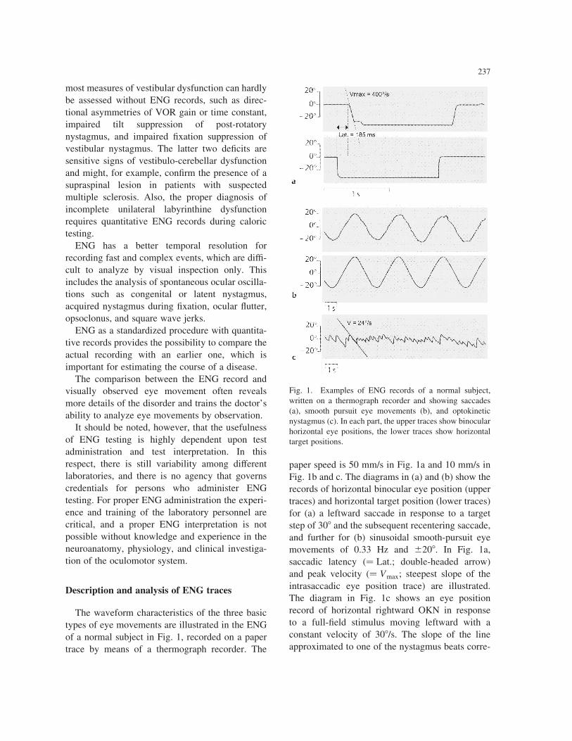
The waveform characteristics of the three basic
types of eye movements are illustrated in the ENG
of a normal subject in Fig. 1, recorded on a paper
trace by means of a thermograph recorder. The
paper speed is 50 mm/s in Fig. 1a and 10 mm/s in
Fig. 1b and c. The diagrams in (a) and (b) show the
records of horizontal binocular eye position (upper
traces) and horizontal target position (lower traces)
for (a) a leftward saccade in response to a target
step of 308 and the subsequent recentering saccade,
and further for (b) sinusoidal smooth-pursuit eye
movements of 0.33 Hz and ^208. In Fig. 1a,
saccadic latency �� Lat.; double-headed arrow)
and peak velocity (� Vmax; steepest slope of the
intrasaccadic eye position trace) are illustrated.
The diagram in Fig. 1c shows an eye position
record of horizontal rightward OKN in response
to a full-®eld stimulus moving leftward with a
constant velocity of 308/s. The slope of the line
approximated to one of the nystagmus beats corre-
237
Fig. 1. Examples of ENG records of a normal subject,
written on a thermograph recorder and showing saccades
(a), smooth pursuit eye movements (b), and optokinetic
nystagmus (c). In each part, the upper traces show binocular
horizontal eye positions, the lower traces show horizontal
target positions.
sponds to slow-phase eye velocity. In each of the
diagrams, upward de¯ection (positive ordinate)
means rightward eye or target movement, down-
ward de¯ection (negative ordinate) means leftward
movement.
In the following sections we will outline the
analysis and normative ranges of ENG results in
various parts of the testing. Most of the proposed
normal values are global ranges taken from the
international standard literature (Baloh and
Honrubia 1990; Henn 1993; Leigh and Zee 1999).
Nevertheless, these values have to be treated with
caution, as due to the different testing conditions
including patient instructions, illumination, stimu-
lation and recording devices, each laboratory
should establish its own normative data. For asses-
sing the normal range, we and others take the
mean^ 2:5 standard deviations.
Spontaneous nystagmus and nystagmus during
®xation
SPN and nystagmus during ®xation are detected
by visual inspection of the eye position traces and
quanti®ed by measuring the maximal eye velocity
during nystagmus slow phases. The velocity is
determined by numerical differentiation carried
out by a computerized analysis program or graphi-
cally as the slope of the eye position trace. This
measurement should not be performed on slow
phases when the vertical EOG shows eyelid arti-
facts. A weak SPN in darkness (up to 4 or 58/s
slow-phase velocity) has been found in about
20% of normal subjects and thus cannot be
regarded as pathological per se, if there are no
other signs of vestibular dysfunction. The same
normal range refers to head-shaking or positional
nystagmus: slow-phase velocities of 58/s or less are
still within normal limits. The presence of
nystagmus during central ®xation is always patho-
logical. It should be noted, whether such a
nystagmus depends on gaze direction. Gaze-
evoked nystagmus is considered pathological if it
occurs at eccentricities of less than 408. Disruption
of ®xation by saccadic intrusions (square wave
jerks, saccadic oscillations, ocular ¯utter, opso-
clonus) is also abnormal.
Saccadic tests
The latency of saccades (with respect to the
stimulus movement) should be between 100 ms
and 300 ms. The maximal eye velocity (visible as
the maximal slope of the position trace, as shown in
Fig. 1a, or obtained by digital differentiation of the
eye position signal) during saccades should be
determined. Normal values for saccade duration
and peak velocity depend on the amplitude of the
saccade, according to their `main sequence', e.g. for
a 208-saccade the peak velocity amounts to
420^ 708/s, and a velocity below 2508/s is consid-
ered as pathologic. For the assessment of saccade
metrics it should be noted whether the patient
reaches the target with one saccade, or whether
corrective saccades are needed to compensate for
either an overshoot or undershoot. An overshoot is
usually pathologic, often indicating cerebellar
dysfunction, whereas a mild undershoot (see Fig.
1a) is normal. For quanti®cation, many investiga-
tors calculate the saccadic amplitude gain (i.e. the
ratio between the amplitude of the saccade and the
amplitude of target displacement), which on
average amounts to about 90% in normal subjects.
Smooth-pursuit tests
It is dif®cult to give exact normal values as
conditions change slightly between laboratories,
but even an elderly patient should be able to pursue
a sinusoidal movement of 0.2 Hz and ^208 ampli-
tude smoothly for at least two cycles. In a young
subject this may be possible up to 0.5 Hz. With
higher frequencies smooth eye velocity lags behind
target velocity, and more and more catch-up
saccades occur to foveate the moving target. In
pathologic conditions these catch up saccades
occur also at lower frequencies and amplitudes,
resulting in a saccadic or `cogwheel'-like pursuit.
If stimuli of randomly changing velocities and
directions are pursued, catch-up saccades occur at
much lower frequencies than during predictive
pursuit. Reduced attention during pursuit might
lead to anticipatory saccades, that move the eye
off the target by anticipating the target trajectory.
The critical measure of smooth pursuit performance
is its velocity gain, i.e. the ratio of smooth eye
velocity and stimulus velocity. If a computer
238
program is used for the analysis of the test, a larger
interval free of saccades can be selected to assess
mean eye velocity. Alternatively, eye velocity can
be inferred as the slope of the eye position trace. In
general, gain decreases with age, inattention,
certain drugs (sedatives, antiepileptics, neurolep-
tics), and with any brain disease. More important
for clinical diagnosis and for the localization of
lesions is a direction-speci®c reduction of pursuit
gain. With a sinusoidal stimulus of ^208 and 0.2
Hz, a gain above 0.8 should be reached even by
elderly subjects.
Optokinetic nystagmus
Slow-phase eye velocity during optokinetic
stimulation should be assessed either by computer
analysis or as the slope of the eye position trace (of
at least the 5 steepest slow-phase segments). OKN
gain is calculated as the quotient of smooth eye and
stimulus velocity. Usually the maximum OKN gain
is calculated by averaging slow-phase velocities of
the 5 steepest slow-phase segments. OKN gain
decreases with increasing stimulus velocities; for
908/s it should be above 0.35. More important, the
OKN response is considered pathologically asym-
metric, if the quotient �vr 2 vl�=�vr 1 vl� exceeds
20% (where vr and vl denote slow-phase velocity
to the right and to the left, respectively).
Rotational testing
The rotating chair is a good tool to determine the
threshold for perrotatory vestibular nystagmus and
the turning sensation, furthermore the gain of the
VOR (i.e. the maximum slow-phase velocity of
postrotatory nystagmus after the stop divided by
chair velocity before the stop) and the time course
of the decline of nystagmus velocity after the stop
(usually a nearly exponential decay is assumed and
a single exponential may be ®tted to this velocity
function thereby determining a `vestibular time
constant'). The decline of nystagmus velocity,
however, is almost linear after stops from low velo-
cities. After stops from high velocities nystagmus
changes direction 30±60 s following the stop
(secondary postrotatory nystagmus ± PRN II).
Thus the calculation of a time constant for PRN I
remains problematic, but is nevertheless widely
used because it helps to assess the symmetry of
vestibular velocity storage. VOR gain of per- or
postrotatory nystagmus should be between 0.3 and
1.05, and a directional asymmetry of VOR gain or
time constant of more than 20% (as assessed for
OKN) is pathological (Baloh and Honrubia 1990).
For clinical purposes the VOR time constant may
be roughly estimated as one third of the duration of
PRN I. Normal values range between 10 and 20 s.
Head tilts performed 4 s after the stop should reduce
the time constant to below 10 s.
Caloric nystagmus
As described for VOR and OKN, maximal slow-
phase velocities induced by stimulation with water
of 308C and 448C temperature are determined. The
symmetry of the labyrinth responses is assessed by
the formula
��vR;44 1 vR;30�2 �vL;44 1 vL;30��=��vR;44 1 vR;30�
1 �vL;44 1 vL;30��;
where R and L denote responses to right and left ear
stimulation, respectively, and 30 and 44 denote the
temperature of stimulation. If this quotient (index
of asymmetry) is above 25%, unilateral labyr-
inthine dysfunction (vestibular paresis) is diag-
nosed. Furthermore, asymmetry with respect to
nystagmus direction can be assessed as
��vL;44 1 vR;30�2 �vR;44 1 vL;30��=��vL;44 1 vR;30�
1 �vR;44 1 vL;30��:
An index above 30% can be considered as patholo-
gical, in terms of a directional preponderance.
As was mentioned earlier, several software
programs are available for automatic computerized
analysis of eye movement parameters (Baloh and
Honrubia 1990). When relying on such computer
measurements it has to be kept in mind that the
programs may have dif®culties to cope with the
various artifacts and a poor signal-to-noise ratio,
thus possibly producing misleading results. It is
therefore necessary to scrutinize the original eye
position trace and to perform the analysis interac-
tively. For the experienced examiner it is usually
possible to detect abnormalities just by visual
inspection of the eye position record. This proce-
239
dure is fast, but does not provide quantitative
results. Thus inspection of the record and manual
measurements (with paper and pencil) of the most
important parameters (maximum velocity of
saccades and pursuit, maximum slow-phase velo-
city of optokinetic, post-rotatory, and aloric
nystagmus as well as the duration of postrotatory
and caloric nystagmus) may still be a good compro-
mise.
How to report the results
The report of an ENG investigation should be
divided in three parts:
1. Description of recording and stimulation para-
meters, such as AC or DC recording, applied
®lters, number of recording channels, placement
and connections of electrodes, sampling rate of
data, type and magnitude of visual and vestibular
stimuli, short outline of the ENG protocol.
2. Description of qualitative and quantitative results,
as outlined on pp. 237±239. This includes reports
of spontaneous, ®xation, head-shaking, and gaze-
evoked nystagmus, saccadic latencies, metrics and
peak velocities, gain and directional asymmetries
of smooth pursuit and OKN, duration of OKAN,
gain and time constant of rotatory vestibular
nystagmus, with or without ®xation suppression
or head tilt suppression, further the index of vestib-
ular asymmetry and the maximal slow-phase velo-
cities of caloric nystagmus. Artifacts should be
mentioned as far as they limit the reliability of
the results, as well as the alertness and coopera-
tivity of the patient.
3. Summary of abnormalities, cerebral localization of
disorders, and diagnosis or at least discussion of
results in the clinical context.
References
American Academy of Neurology. Report of the Therapeutics and
Technology Assessment Subcommittee. Assessment: electro-
nystagmography. Neurology, 1996, 46: 1763±1766.
Baloh, R.W. and Honrubia, V. (Eds.) Clinical Neurophysiology of
the Vestibular System, 2nd Edition. Davis, Philadelphia, PA,
1990: 130±173.
Carpenter, R.H.S. (Ed.). Movements of the Eyes, 2nd Edition. Pion,
London, 1988: 405±426.
Collewijn, H., van der Mark, F., and Jansen, T.C. Precise recording
of human eye movements. Vision Res., 1975, 15: 447±450.
DiScenna, A.O., Das, V., Zivotofski, A.Z., Seidman, S.H. and
Leigh, R.J. Evaluation of a video tracking device for measure-
ment of horizontal and vertical eye rotations during locomotion.
J. Neurosci. Methods, 1995, 58: 89±94.
Ferman, L., Collewijn, H., Jansen, T.C. and van den Berg, A.V.
Human gaze stability in the horizontal, vertical and torsional
direction during voluntary head movements, evaluated with a
three-dimensional scleral induction coil technique. Vision Res.,
1987, 27: 811±828.
Henn, V. Electronystagmography: the answers one might get. In:
L.R. Caplan and H.C. Hopf (Eds.), Brain-Stem Localization and
Function. Springer, Berlin, 1993: 93±99.
Jung, R. and K.ornhuber, H.H. Results of electronystagmography in
man: The value of optokinetic, vestibular, and spontaneous
nystagmus for neurologic diagnosis and research. In: M.B.
Bender (Ed.), The Oculomotor System. Harper and Row, New
York, 1964: 428±488.
Katz, B., MuÈller, K. and Helmle, H. Binocular eye movement
recording with CCD arrays. Neuroophthalmology, 1987, 7:
81±91.
Leigh, R.J. and Zee, D.S. (Eds.). The Neurology of Eye Movements/
it>,, 3rd Edition. Oxford, New York, 1999.
Peng, G.C., Baker, J.F. and Peterson, B.W. Dynamics of directional
plasticity in the human vertical vestibulo-ocular re¯ex. J. Vestib.
Res., 1994, 4: 453±460.
Reulen, J., Marcus, J., Koops, D., de Vries, F., Tiesinga, G.,
Boshuizen, K. and Bos, J. Precise recording of eye movement:
the IRIS technique, part 1. Med. Biol. Eng. Comput., 1988, 26:
20±26.
Robinson, D.A. A method of measuring eye movement using a
search coil in a magnetic ®eld. IEEE Trans. Bio.-Med. Electr.,
1963, 10: 137±145.
Rottach, K. and Heide W. Elektrookulographie. In: A. Huber and D.
KoÈmpf (Eds.), Klinische Neuroophthalmologie. Thieme, Stutt-
gart, 1997: 191±199.
Scherer, H., Teiwes, W. and Clarke, A. Measuring three dimensions
of eye movements in dynamic situations by means of videoo-
culography. Acta Oto-Laryngol., 1991, 111: 182±187.
Young, L.R. and Sheena, D. Survey of eye movement recording
methods. Behav. Res. Methods Instrum., 1975, 7: 397±429.
240