electronic delocalization, hypochromism in the uv ... nvidia graphical processing units (gpus) using...
TRANSCRIPT

Loughborough UniversityInstitutional Repository
Electronic delocalization,charge transfer and
hypochromism in the UVabsorption spectrum of
polyadenine unravelled bymultiscale computations andquantitative wavefunction
analysis
This item was submitted to Loughborough University's Institutional Repositoryby the/an author.
Citation: NOGUEIRA, J.J., PLASSER, F. and GONZALEZ, L., 2017. Elec-tronic delocalization, charge transfer and hypochromism in the UV absorptionspectrum of polyadenine unravelled by multiscale computations and quantita-tive wavefunction analysis. Chemical Science, 8(8), pp. 5682-5691.
Additional Information:
• This is an Open Access Article. It is published by royal Society ofChemistry under the Creative Commons Attribution-NonCommercial 3.0Unported (CC BY-NC). Full details of this licence are available at:http://creativecommons.org/licenses/by-nc/3.0/
Metadata Record: https://dspace.lboro.ac.uk/2134/32128
Version: Published
Publisher: c© Royal Society of Chemistry
Rights: This work is made available according to the conditions of the CreativeCommons Attribution-NonCommercial 3.0 Unported (CC BY-NC 3.0) licence.

Full details of this licence are available at: http://creativecommons.org/licenses/by-nc/3.0/
Please cite the published version.
2

Electronic delocalization, charge transfer andhypochromism in the UV absorption spectrum ofpolyadenine unravelled by multiscalecomputations and quantitative wavefunctionanalysis†
Juan J. Nogueira, ‡* Felix Plasser ‡* and Leticia Gonzalez *
The characterization of the electronically excited states of DNA strands populated upon solar UV light
absorption is essential to unveil light-induced DNA damage and repair processes. We report
a comprehensive analysis of the electronic properties of the UV spectrum of single-stranded
polyadenine based on theoretical calculations that include excitations over eight nucleobases of the
DNA strand and environmental effects by a multiscale quantum mechanics/molecular mechanics
scheme, conformational sampling by molecular dynamics, and a meaningful interpretation of the
electronic structure by quantitative wavefunction analysis. We show that electronic excitations are
extended mainly over two nucleobases with additional important contributions of monomer-like
excitations and excitons delocalized over three monomers. Half of the spectral intensity derives from
locally excited and Frenkel exciton states, while states with partial charge-transfer character account for
the other half and pure charge-transfer states represent only a minor contribution. The hypochromism
observed when going from the isolated monomer to the strand occurs independently from
delocalization and charge transfer and is instead explained by long-range environmental perturbations of
the monomer states.
1 Introduction
Absorption of UV light by DNA initiates a series of photo-chemical events that can lead to lethal genetic modications.1,2
Although it is challenging, the characterization of the elec-tronically excited states involved in these photoinduced eventsis unavoidable to understand the mechanisms of DNA photo-damage. In particular, the question of whether the UV absorp-tion spectrum of DNA is dominated by monomer-likeexcitations or by collective excitations has intrigued researchersfor over 50 years.3–9 The initial extent of the exciton over the DNAstrand is of fundamental signicance as it decides whether theearly electronically excited-state dynamics of DNA is dominatedby delocalized excitons undergoing intraband scattering andenergy transfer,10 by dimer excitations paving the way for exci-mer formation11–13 and dimerization,14–17 or by monomer-likeprocesses.18,19 Furthermore, delocalization of UV energy over
several nucleobases has been invoked as a self-protectionmechanism of DNA against radiation.20,21 The initial discus-sion of electronic delocalization in DNA strands was dominatedby two seemingly contradictory observations:22 on the one hand,the strong hypochromism observed upon helix formation wasinterpreted in terms of delocalized states;8 on the other hand,the fact that no signicant energy shis were observed was seenas an indication of strictly localized states.5 To resolve thisparadox, the extent of electronic delocalization along stackednucleobases has been further investigated in the last decadewith involved spectroscopic experiments3,6,20,23 and theoreticalcalculations.9,24–32 However, no consensus has been reached sofar as quite different electronic degrees of delocalizationranging from localized states23 to delocalization over more thansix bases27 have been reported.
Another intriguing question intensively discussed in theliterature is the role of charge-transfer (CT) states in the UVabsorption spectrum. These states are relevant to DNA damagebecause their radical character may induce photochemicalreactions aer Franck–Condon excitation.17 Additionally, theyplay a crucial role in some DNA repair mechanisms.33 However,the energetic position of these states is under dispute. Someauthors argue that CT states constitute the red tail28,32,34 of thespectrum, which is absent in the monomer, while other authors
Institute of Theoretical Chemistry, Faculty of Chemistry, University of Vienna,
Wahringer Straße 17, 1090 Vienna, Austria. E-mail: nogueira.perez.juanjose@univie.
ac.at; [email protected]; [email protected]
† Electronic supplementary information (ESI) available. See DOI:10.1039/c7sc01600j
‡ Authors contributed equally to this work.
Cite this: Chem. Sci., 2017, 8, 5682
Received 10th April 2017Accepted 9th June 2017
DOI: 10.1039/c7sc01600j
rsc.li/chemical-science
5682 | Chem. Sci., 2017, 8, 5682–5691 This journal is © The Royal Society of Chemistry 2017
ChemicalScience
EDGE ARTICLE
Ope
n A
cces
s A
rtic
le. P
ublis
hed
on 1
3 Ju
ne 2
017.
Dow
nloa
ded
on 0
1/03
/201
8 12
:23:
04.
Thi
s ar
ticle
is li
cens
ed u
nder
a C
reat
ive
Com
mon
s A
ttrib
utio
n-N
onC
omm
erci
al 3
.0 U
npor
ted
Lic
ence
.
View Article OnlineView Journal | View Issue
contend that all CT states are at least similar in energy or higherthan the bright electronic states.31,35–38 Additional complicationsarise from the mixing between CT and local states. Such mixingis not only important from a methodological viewpoint, as itdetermines whether a Frenkel exciton model is appropriate todescribe the absorption spectrum, but it can also give a rstindication whether interconversion between local and CTstates17 plays a role in the dynamical processes following UVabsorption.
Discrepancies between different delocalization lengths andthe position of CT states originate from the difficulty to studythese phenomena experimentally and computationally. In therst case, the challenge is due to the fact that electronic delo-calization3,6,20,23 and CT11 can only be deduced indirectly. Thedifficulty in the calculations comes from the extended systemsize, the importance of environmental interactions and struc-tural disorder, as well as the fact that the resulting wave-functions have to be analyzed in a meaningful and consistentway.22,39
This paper is the rst calculation of the lowest-energy UVabsorption band of adenosine monophosphate (AMP) andsingle stranded polyadenine (dA)20 including eight nucleobasesat quantum mechanical level and taking into account confor-mational sampling and environmental effects. Through a skil-led wavefunction analysis we also provide the most rigorous todate quantitative characterization of the electronic excitationsclassied in terms of delocalization length and CT character.Further, we clarify the origin of the hypochromism when goingfrom the monomer to the polymer and unveil the nature of theexcitations involved in the red tail of the polymer spectrum.
2 Theory
Our computational protocol is based on a multiscale approachthat combines molecular dynamics simulations with extensivequantum mechanics/molecular mechanics (QM/MM) calcula-tions and a comprehensive wavefunction analysis.
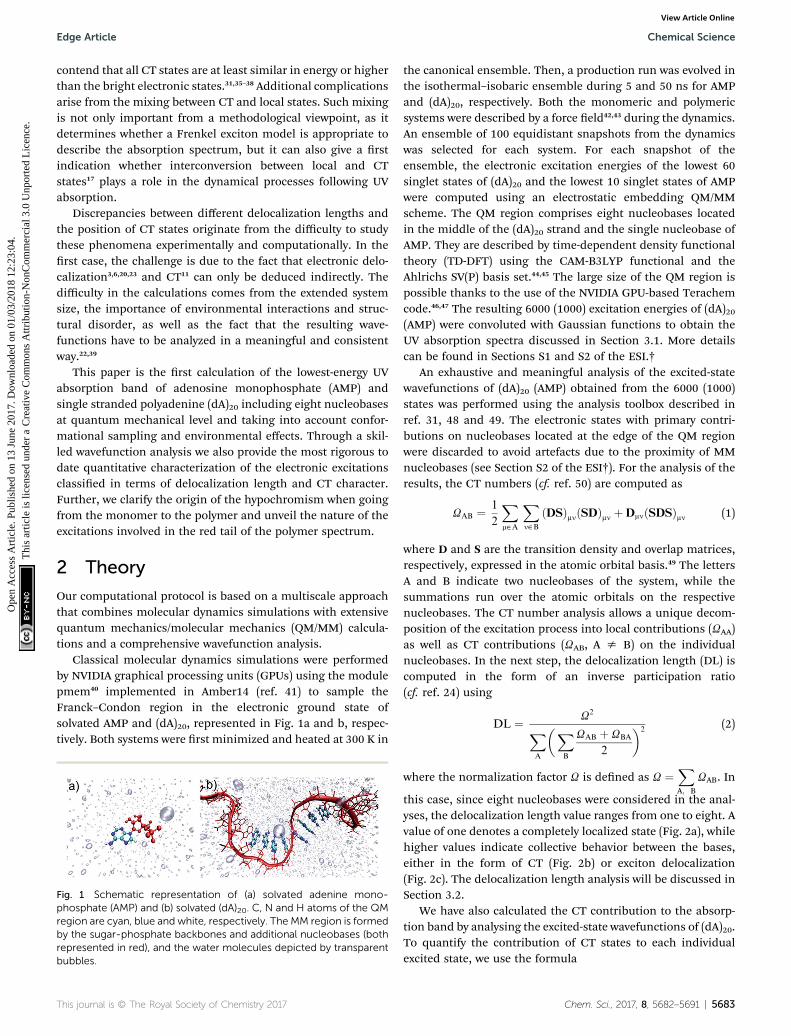
Classical molecular dynamics simulations were performedby NVIDIA graphical processing units (GPUs) using the modulepmem40 implemented in Amber14 (ref. 41) to sample theFranck–Condon region in the electronic ground state ofsolvated AMP and (dA)20, represented in Fig. 1a and b, respec-tively. Both systems were rst minimized and heated at 300 K in
the canonical ensemble. Then, a production run was evolved inthe isothermal–isobaric ensemble during 5 and 50 ns for AMPand (dA)20, respectively. Both the monomeric and polymericsystems were described by a force eld42,43 during the dynamics.An ensemble of 100 equidistant snapshots from the dynamicswas selected for each system. For each snapshot of theensemble, the electronic excitation energies of the lowest 60singlet states of (dA)20 and the lowest 10 singlet states of AMPwere computed using an electrostatic embedding QM/MMscheme. The QM region comprises eight nucleobases locatedin the middle of the (dA)20 strand and the single nucleobase ofAMP. They are described by time-dependent density functionaltheory (TD-DFT) using the CAM-B3LYP functional and theAhlrichs SV(P) basis set.44,45 The large size of the QM region ispossible thanks to the use of the NVIDIA GPU-based Terachemcode.46,47 The resulting 6000 (1000) excitation energies of (dA)20(AMP) were convoluted with Gaussian functions to obtain theUV absorption spectra discussed in Section 3.1. More detailscan be found in Sections S1 and S2 of the ESI.†
An exhaustive and meaningful analysis of the excited-statewavefunctions of (dA)20 (AMP) obtained from the 6000 (1000)states was performed using the analysis toolbox described inref. 31, 48 and 49. The electronic states with primary contri-butions on nucleobases located at the edge of the QM regionwere discarded to avoid artefacts due to the proximity of MMnucleobases (see Section S2 of the ESI†). For the analysis of theresults, the CT numbers (cf. ref. 50) are computed as
UAB ¼ 1
2
Xm˛A
Xn˛B
ðDSÞmnðSDÞmn þDmnðSDSÞmn (1)
where D and S are the transition density and overlap matrices,respectively, expressed in the atomic orbital basis.49 The lettersA and B indicate two nucleobases of the system, while thesummations run over the atomic orbitals on the respectivenucleobases. The CT number analysis allows a unique decom-position of the excitation process into local contributions (UAA)as well as CT contributions (UAB, A s B) on the individualnucleobases. In the next step, the delocalization length (DL) iscomputed in the form of an inverse participation ratio(cf. ref. 24) using
DL ¼ U2
XA
�XB
UAB þ UBA
2
�2(2)
where the normalization factor U is dened as U ¼XA; B
UAB. In
this case, since eight nucleobases were considered in the anal-yses, the delocalization length value ranges from one to eight. Avalue of one denotes a completely localized state (Fig. 2a), whilehigher values indicate collective behavior between the bases,either in the form of CT (Fig. 2b) or exciton delocalization(Fig. 2c). The delocalization length analysis will be discussed inSection 3.2.
We have also calculated the CT contribution to the absorp-tion band by analysing the excited-state wavefunctions of (dA)20.To quantify the contribution of CT states to each individualexcited state, we use the formula
Fig. 1 Schematic representation of (a) solvated adenine mono-phosphate (AMP) and (b) solvated (dA)20. C, N and H atoms of the QMregion are cyan, blue and white, respectively. The MM region is formedby the sugar-phosphate backbones and additional nucleobases (bothrepresented in red), and the water molecules depicted by transparentbubbles.
This journal is © The Royal Society of Chemistry 2017 Chem. Sci., 2017, 8, 5682–5691 | 5683
Edge Article Chemical Science
Ope
n A
cces
s A
rtic
le. P
ublis
hed
on 1
3 Ju
ne 2
017.
Dow
nloa
ded
on 0
1/03
/201
8 12
:23:
04.
Thi
s ar
ticle
is li
cens
ed u
nder
a C
reat
ive
Com
mon
s A
ttrib
utio
n-N
onC
omm
erci
al 3
.0 U
npor
ted
Lic
ence
.View Article Online

CT ¼ U�1 XBsA
UAB (3)
i.e. a summation over all off-diagonal elements of the U-matrixis performed. The CT contribution is equal to one fora completely charge-separated state (Fig. 2b) while it is zero fora locally excited state (Fig. 2a) or a Frenkel exciton (Fig. 2c). Anextensive analysis of the CT character of the lowest absorptionband of (dA)20 is presented in Section 3.3.
3 Results and discussion3.1 Absorption spectra of AMP and (dA)20
The calculated and experimental32 absorption spectra for bothsolvated AMP and (dA)20 are plotted in Fig. 3a and b, respec-tively. The experimental spectrum of (dA)20 (black line) peaks at4.85 eV.32,51 For ease of comparison, the maximum of thecalculated spectrum for (dA)20 obtained from the ensemble of100 geometries has been red-shied by 0.70 eV. This shi canbe attributed to the basis set, as it is known52 that larger basissets place the spectrum at lower energies. Unfortunately, theuse of eight nucleobases in the QM region and the large numberof snapshots precludes the use of more extended basis sets.However, a larger basis set does not change signicantly thecharacter of the excited-state wavefunctions (see Section S3 ofthe ESI†).
The experimental absorption spectrum of the polynucleotide(black line in Fig. 3b) presents three important differences withrespect to the spectrum of the mononucleotide (red line). Uponformation of a stacked helix, the most dramatic effect is thehypochromism observed7,8 – the integrated absorption coeffi-cient decreases by 35%. Furthermore, the spectrum is blue-shied by 0.04 eV and a low-intensity red tail appears.51 Thecalculations based on the ensemble (solid lines in Fig. 3a)reproduce properly the hypochromism of the polymer (inte-grated intensity decreases by 36%) and the red tail of thepolymer spectrum, which crosses with the AMP spectrum at
4.5 eV. The blue shi of 0.04 eV when going from AMP to (dA)20is not described by the calculations; instead, a redshi of0.03 eV is obtained –similar to the redshi of 0.05 eV obtainedin previous TD-DFT calculations.9 Whereas this erroneousbehavior has been attributed to the underestimation of excita-tion energies of CT states in TD-DFT,53 we want to point out herethat such a small change in energy is well beyond the accuracyof TD-DFT (and any current quantum mechanical method forexcited states) and any agreement is due to fortuitous coinci-dence. Overall, we conclude that our calculations provide a gooddescription of the experimental absorption spectrum of AMPand (dA)20, which is a prerequisite for the forthcoming analysis.
For methodological reasons we found interesting to analyzethe effect of conformational sampling on the absorptionspectra. Thus, excited-state calculations were also performed ona single geometry, i.e. the optimized geometries of AMP and(dA)20 (dashed lines in Fig. 3a). The geometry optimization wasperformed classically using the same force eld employed in thedynamics42,43 (more details in Section S3†), and the sameredshi of 0.70 eV was applied. Conspicuously, the spectra ofAMP and (dA)20 for the optimized geometries are narrowedagainst those from the ensembles due temperature effects (0 Kfor optimized geometries vs. 300 K for molecular dynamicsensembles). In addition, the maximum of the spectra for theoptimized monomer and polymer are blue-shied by 0.15 eVwith respect to the ensemble spectra. Intra- and intermolecularmotion thus decreases the energy of the absorption band
Fig. 2 Example excited states occurring in a stack of DNA bases: (a)locally excited states, (b) charge-transfer (CT) states, and (c) delo-calized Frenkel excitons. Delocalization length (DL) and CT contribu-tion are indicated. Cyan rectangles and red lines depict thenucleobases and sugar-phosphate backbones, respectively. The redand blue circles connected by a black arrow represent the hole andelectron generated after excitation.
Fig. 3 (a) Calculated lowest-energy band of the UV absorptionspectrum for AMP (red line) and (dA)20 (black line) considering 100geometries (solid lines) or the optimized geometry (dashed lines). Thespectra are red shifted by 0.70 eV to facilitate the comparison withexperiments. (b) Experimental UV absorption spectra of AMP (red line)and (dA)20 (black line) taken from ref. 32.
5684 | Chem. Sci., 2017, 8, 5682–5691 This journal is © The Royal Society of Chemistry 2017
Chemical Science Edge Article
Ope
n A
cces
s A
rtic
le. P
ublis
hed
on 1
3 Ju
ne 2
017.
Dow
nloa
ded
on 0
1/03
/201
8 12
:23:
04.
Thi
s ar
ticle
is li
cens
ed u
nder
a C
reat
ive
Com
mon
s A
ttrib
utio
n-N
onC
omm
erci
al 3
.0 U
npor
ted
Lic
ence
.View Article Online

providing a closer agreement with the experiment. Fair enough,the calculations on the optimized geometries reproduce wellthe appearance of the red tail when going from the monomer tothe polymer. Moreover, the hypochromism is reasonably welldescribed, with a 30% decrease of the integrated absorptioncoefficient vs. the 35% and 36% decrease obtained experimen-tally and with the ensemble calculations, respectively. However,as it will be seen below, conformational sampling is mandatoryto properly describe other crucial electronic features of the UVabsorption band.
3.2 Delocalization length
One of the most intensively debated questions related to theelectronically excited states of DNA strands is the number ofnucleobases, or delocalization length, involved in Franck-Condon excitations.3,9,20,24–32 Exciton theory calculations ondouble strands (dA)20(dT)20 and (dAdT)10(dAdT)10 concludedthat electronically excited states are delocalized over the wholelength of the helix when the model relies on an idealized B-DNAgeometry,24 over four to eight nucleobases when conformationalmotion is considered by molecular dynamics,25 or over only oneto two nucleobases when the exciton Hamilton matrix isrened.26 The results obtained by exciton theory are also quitedependent on the approximation employed to compute theelectronic coupling between nucleobases; thus, dependingwhether the ideal dipole or the transition density cube approx-imation is used electronic delocalization lengths of 4.5 to 7.1 or3.7 to 8.2 nucleobases have been obtained for the double strand(dA)12(dT)12.27 Quantum mechanical calculations have also beenemployed to investigate the size of exciton states.9,28,29,31 TD-DFTcomputations on an adenine trimer (A)3 with a geometryrestrained to mimic an idealized B-DNA conformation showeddelocalization over the three nucleobases.28 The same result wasobtained when conformational motion was taken into accountfor a single strand (dA)4.9 However, a larger degree of delocal-ization of ve to six nucleobases was reported when the single-stranded (dA)9 and (dA)11 oligomers were built by consideringa B-DNA arrangement.9 The impact of conformational samplingwas also discussed based on semiempirical calculationscombined with a QM/MM approach, in which the double strand(A)6(T)6 was included in the QM region. Delocalization lengths oftwo to three and four to ve nucleobases were computed formolecular dynamics structures and an idealized B-DNA geom-etry, respectively.29 Higher-level QM/MM calculations, using thesecond-order algebraic diagrammatic construction (ADC(2)) tocompute electronic excitations of four stacked bases, (TA)2 and(CG)2, concluded that most electronically excited states aremonomer-like excitations or are delocalized over two mono-mers.31 Experimental measurements also dissent about elec-tronic delocalization. The general importance of excited-statecollectivity was illustrated by transient uorescence anisotropyexperiments.6 Femtosecond time-resolved broadband spectra ofa series of single-stranded (dA)n and double-stranded (dA)n(dT)noligomers invoked delocalization lengths of three nucleobasesfor the single strand, while exciton extensions larger than fournucleobases were attributed to the double strand.3 Circular
dichroism experiments on single strands (dA)n showed that onlynearest neighbor interactions play a crucial role on the excita-tion for excitation wavelengths above 200 nm, and thus theexciton is extended over only two monomers.20 In contrast, theresults of Kerr-gated time-resolved uorescence and transientabsorption experiments were interpreted in terms of localabsorbing states.23
In order to provide a clear-cut answer on the delocalizationlength, the rst band of the UV absorption spectrum, calculatedfor the ensemble of 100 (dA)20 geometries has been decom-posed according to eqn (2). The different delocalization lengthcontributions are displayed in Fig. 4a. The delocalization lengthcan acquire any real value from 1 to 8. In order to simplify theanalysis, and directly relate delocalization with the number ofnucleobases, the computed delocalization length of each elec-tronic state was rounded to the closest integer value.
The analyses show that most of electronic transitions aredelocalized over two nucleobases (47.9%). Delocalization lengthof three nucleobases (22.8%) and monomer-like excitations(22.7%) also contribute signicantly to the absorption band.Excited states delocalized over four nucleobases are much lessrelevant, but not negligible (5.0%). In conclusion, virtually thewhole UV lowest-energy band (98.4%) is composed by excita-tions involving at most four monomers. The average delocal-ization length is 2.2 nucleobases, which agrees well with thedelocalization lengths of two and three nucleobases obtainedfrom circular dichroism20 and femtosecond time-resolved broadband spectroscopies,3 respectively.
Fig. 4 Decomposition of the lowest-energy band of the UV absorp-tion spectrum for (dA)20 into different delocalization length (DL)contributions calculated for (a) an ensemble of 100 geometries and (b)the optimized geometry. The insets of both plots show the DLdecomposition in the region of the red tail. The numbers given inparentheses indicate the intensity contribution to the total spectrum.
This journal is © The Royal Society of Chemistry 2017 Chem. Sci., 2017, 8, 5682–5691 | 5685
Edge Article Chemical Science
Ope
n A
cces
s A
rtic
le. P
ublis
hed
on 1
3 Ju
ne 2
017.
Dow
nloa
ded
on 0
1/03
/201
8 12
:23:
04.
Thi
s ar
ticle
is li
cens
ed u
nder
a C
reat
ive
Com
mon
s A
ttrib
utio
n-N
onC
omm
erci
al 3
.0 U
npor
ted
Lic
ence
.View Article Online

It is intriguing that the shape of the spectrum of monomer-like excitations in the polymer is very different from that inAMP: the pronounced maximum in the center of the band inAMP (solid red line in Fig. 3a) is attened in the polymer (blueline in Fig. 4a). This attening indicates that the formation ofexciton states in the polymer occurs by combination of mono-meric states that lie mostly in the center of the absorption band.The inspection of the density of states of AMP (Fig. S1†)corroborates that a larger number of energetically degeneratedstates, which are prone to form exciton states in the polymer, isfound in the center of the band. As a consequence themonomer-like spectrum of the strand is depleted in this area.
In order to appreciate the impact of structural disorder onthe absorbing states, the contributions of different delocaliza-tion lengths were also calculated for the optimized single-stranded geometry. As seen from Fig. 4b, the delocalizationlength is dramatically overestimated when sampling is omitted.Electronic excitations distributed over three nucleobases are themost signicant ones (60.2%), and delocalization over venucleobases still contributes notably (8.1%) to the absorptionband. Moreover, monomer-like excitations are erroneouslyunderestimated, with a contribution of only 3.9%. This over-estimation of the delocalization length for the optimized (andunrealistic) structure is a consequence of the stronger stackingbetween neighboring nucleobases that favors electronic delo-calization over the strand. Contrary, when thermal motion isconsidered, structural disorder is introduced, leading to local-ization of the excited states. The decrease of delocalizationlength induced by conformational sampling has been previ-ously shown by exciton theory25 and quantum mechanicalcalculations9,29 performed on geometrical ensembles andidealized B-DNA structures. It has been pointed out that therelevant degrees of freedom responsible for this effect areintramolecular motions rather than large scale uctuations.29
The hyperchromism observed in the low-energy region of theUV absorption band is a singular feature of DNA strands. Asdiscussed above, both the calculations on the optimizedgeometry and the ensemble correctly reproduce this red tail. Acloser inspection of the delocalization length decompositionaround the low-energy tail of the ensemble spectrum (inset ofFig. 4a) reveals that the electronically excited states in thisenergy range are mainly monomer-like excitations, while thecontribution of excitations delocalized over two or morenucleobases is signicantly reduced when compared to theremaining spectrum. Specically, the average delocalizationlength of the states located in the red tail is 1.4, i.e., it is smallerthan the delocalization length of 2.2 computed for the wholespectrum. Thus, we are led to conclude that the energy loweringof these states is mainly caused by electrostatic interactions andpolarization effects with the rest of the strand, while excited-state delocalization plays a minor role. This means that theseelectronic states can be seen as perturbed monomer-like exci-tations. Our analysis stands in contrast to previous theoreticalstudies that concluded that the low-energy states derive fromexciton coupling.38,54 However, in these studies,38,54 the conclu-sion could be strongly inuenced by the choice of a small QMregion, which only included two adenine residues. Moreover,
a statistically signicant statement can only be made aerperforming sampling. It has also been argued that the elec-tronic states that are involved in the red tail are CT excita-tions.28,34 Although a comprehensive CT analysis is discussed inthe next section, we anticipate here that CT states are not rele-vant in the red tail since only one nucleobase is involved inmostof excitations. The delocalization length analysis around the redtail performed on the optimized structure (inset of Fig. 4b) leadsmistakenly to the conclusion that the red tail for the optimizedstrand is completely dominated by excitations involving twonucleobases, instead of one.
Finally, it is interesting to discuss our results in the contextof time-resolved experimental measurements. A delocalizationlength of two nucleobases for the initial states serves asa natural precursor for excimer formation as has been invokedto explain the experimental transients of interacting adeninemolecules.11 More specically, the dominance of delocalizationover two nucleobases observed here, agrees with the observa-tion that the bleach recovery signals for ApA are basicallyidentical to those of longer adenine strands.11 Our results alsounderline the importance of collective excited-state behavior.The degree of delocalization extracted from the calculations,together with a general high density of states, is certainlyconsistent with rapid energy transfer occurring during theinitial dynamics, as was observed experimentally.6
3.3 Charge transfer states
A proper characterization of CT electronic states aer Franck–Condon excitation is vital to understand DNA damage andrepair.17,33 The formation of CT states between two nucleobasesinvolves charge separation, generating cation and anion radi-cals. On the one hand, these radical species can initiate a seriesof reactions leading to DNA damage; for example, CT statesbetween two pyrimidines may result in the formation of 6–4adducts.17 On the other hand, nucleobases with radical char-acter may play a role in DNA repair mechanisms. It has recentlybeen shown that cyclobutane pyrimidine dimer lesions can berepaired when a CT state in a guanine-adenine stacked pairadjacent to the lesion is generated.33 The formation of cyclo-butane pyrimidine dimers can be quenched by the formation ofa CT state as a result of electron transfer from guanine to one ofthe reactive thymines.55,56 Furthermore, mixed local and CTstates have been connected to long-lived high-energy uores-cence in DNA.57
Despite the importance of CT states in the photophysics ofDNA strands, the extent and energy range in which CT statescontribute to Franck–Condon excitations has generatedcontroversy in the literature. While several studies argued thatlow-energy CT states are present in the UV absorption band ofDNA strands, including in the red tail,28,32,34 others haveconcluded that CT states are higher or at least similar in energyto the bright electronic states.31,35–38
Fig. 5a shows the contribution of CT electronic states to therst absorption band calculated for the ensemble of (dA)20. Inagreement with a previous study on alternating duplexes,31 wend that only about half of the spectral intensity (51%) derives
5686 | Chem. Sci., 2017, 8, 5682–5691 This journal is © The Royal Society of Chemistry 2017
Chemical Science Edge Article
Ope
n A
cces
s A
rtic
le. P
ublis
hed
on 1
3 Ju
ne 2
017.
Dow
nloa
ded
on 0
1/03
/201
8 12
:23:
04.
Thi
s ar
ticle
is li
cens
ed u
nder
a C
reat
ive
Com
mon
s A
ttrib
utio
n-N
onC
omm
erci
al 3
.0 U
npor
ted
Lic
ence
.View Article Online

from states with CT character below 0.1. These states can bejustiably termed pure Frenkel excitons or pure local excita-tions. The remaining half of the spectral intensity is carried bystates with non-negligible CT admixture. The strong interac-tions between CT and Frenkel states can be explained58 in termsof orbital overlap between the different nucleobases. It has beenindeed concluded from experiment that orbital overlap plays animportant role in the excited-state dynamics.59 Furthermore, thestrong mixing between Frenkel and CT states stands in agree-ment with the hypothesis of interconversion between thesetypes of states during the dynamical processes following UVabsorption17 and the formation of exciplexes with CTcharacter.11,13,32,56,60,61
The CT decomposition of the density of states, in which theintensity of the electronic transitions is not considered so thatevery state contributes equally, is analysed and plotted inFig. 5b. As can be seen, pure Frenkel excitons and pure localexcitations represent almost half of the states (44.8%), as in the
absorption spectrum, and the other half is composed of stateswith CT admixture. The most notable difference between theabsorption spectrum and the density of states is the appearanceof a shoulder in the blue side of the latter. This shoulder can beascribed to a high density of states with signicant CT character(CT > 0.3). These states are rather dark and, thus, do notcontribute signicantly to the initial photon absorption. Due totheir high energy it is also not expected that they play animportant role in the early dynamics. Additionally, it is inter-esting to analyse whether CT occurs only between nearestneighbour nucleobases or whether long-range CT takes place.This can be analysed by computing the net CT distance(CTnet),48 which is dened as the difference between the centerof charge of the electron and the center of charge of the holeinvolved in the process. A value of CTnet lower than 1.5 indicatesthat CT takes place between adjacent nucleobases, while a valuelarger than 1.5 indicates long-range CT. According to this de-nition only 1.3% of the CT states in the rst absorption band arelong-range CT states and in all other cases nearest neighbourinteractions dominate. This fact can be understood by consid-ering that long-range CT imposes a severe energetic penaltythrough the Coulomb energy that is needed for separating thecharges.
The effect of conformational sampling on the CT character ofthe absorption band is reected in Fig. 5c, where only theoptimized geometry is considered. As in the delocalizationlength analysis, the differences on the CT decomposition areextreme. Using a single geometry leads to incorrectly inter-preting the excitation band as a combination of Frenkel andmonomer-like excitations, since the majority of electronic states(91.9%) has insignicant CT contribution (lower than 0.1). Onlya small amount (3.2%) of electronic states with important CTcontribution (higher than 0.3) appears in the blue tail of theabsorption band. Therefore, the lack of conformationalsampling clearly underestimates the contribution of CT states.This indicates that anisotropy of the environment as well asintra- and intermolecular motion of the DNA strand change theamount of mixing between CT, Frenkel and local states.
The contribution of electronic states with different amountof CT character in the energy range of the red tail can be seen inthe insets of Fig. 5a and c for the thermal ensemble and opti-mized geometry, respectively. In case of the ensemble (Fig. 5a),most of electronic states do not present an important CTcontribution. This was already expected considering that thered tail is dominated by monomer-like excitations, as was dis-cussed in Section 3.2. The absence of CT states in the red tail ofthe spectrum is in contradiction to results reported by previoustheoretical studies; therefore, it is worth investigating thesediscrepancies in more detail. The initial claim28 of the presenceof low-energy CT states derives from calculations using thePBE0 functional on an adenine trimer (A)3 restrained to theidealized B-DNA conformation. Later, QM/MM calculations onsolvated adenine and thymine monomers and dimers usingPBE0, long-range corrected density functionals andwavefunction-based methods on B-DNA structures showed36
that PBE0 is not capable of describing CT states properly due tothe absence of long-range Hartree–Fock exchange. In a second
Fig. 5 Decomposition of the lowest-energy band of the (a) UVabsorption spectrum and (b) density of states (DOS) for (dA)20 intodifferent charge-transfer (CT) contributions calculated for anensemble of 100 geometries. (c) CT decomposition of the absorptionspectrum calculated for the optimized geometry. The insets of plots (a)and (c) show the charge-transfer decomposition in the region of thered tail.
This journal is © The Royal Society of Chemistry 2017 Chem. Sci., 2017, 8, 5682–5691 | 5687
Edge Article Chemical Science
Ope
n A
cces
s A
rtic
le. P
ublis
hed
on 1
3 Ju
ne 2
017.
Dow
nloa
ded
on 0
1/03
/201
8 12
:23:
04.
Thi
s ar
ticle
is li
cens
ed u
nder
a C
reat
ive
Com
mon
s A
ttrib
utio
n-N
onC
omm
erci
al 3
.0 U
npor
ted
Lic
ence
.View Article Online

study, CT states were found to be above the bright states, whilethey were transferred to the red tail only through the applicationof inhomogeneous broadening to the computed energies ofoptimized stacked adenine clusters of different sizes.32 Many-body Green function theory calculations performed fordifferent solvated single-stranded adenine and thymine oligo-mers, and double-stranded adenine-thymine pair oligomerspredicted low-energy CT states.34 It seems therefore that theemployed computational model and method strongly affectsthe position of CT states, which can be then found in the redtail, close to the band maximum or in the high-energy region ofthe band. Clearly, a denitive conclusion cannot be drawnbased on the available literature results as these previouscalculations28,32,34 used small cluster models without includingthe effect of a DNA strand. This means that explicit watermolecules34 or the continuum solvent model28,32 are in directcontact with the excited nucleobases, and thus solvation effectsare likely overestimated. Additionally, conformational samplingwas not performed, and the lack of vibrational motion stronglyaffects the computations, as was discussed above. All theseissues have been considered in the present study and, thus, ourconclusions rely on a more realistic theoretical model.
For completeness, the CT decomposition of the red tail of thespectrum for the optimized geometry is shown in the inset ofFig. 5c. Since the overall absorption band does not presentsignicant CT character, so is found in the red tail. However, itis curious to note that electronic states with CT contributionsmaller than 0.1 only appear in the high-energy part of the redtail. Thus, while the contribution of CT states to the entireabsorption band is underestimated without conformationalsampling, it is overestimated in the red tail, leading once moreto an incorrect interpretation of the spectrum.
3.4 Hypochromism
The most remarkable ngerprint of the UV absorption spectraof DNA strands, when compared to the spectra of isolatednucleobases, is a pronounced hypochromism of the rst UVabsorption band7,8 (cf. Fig. 3b). It was early recognized that thishypochromism stands in contrast to a “zeroth-order” excitonmodel.7 Therefore, a number of different quite involved modelsfor hypochromism were developed, including orientation-specic dipole interactions,8 dispersion–force interactions,7
local eld effects,62 and mutual shielding of chromophores.63
More recently, CT admixture due to orbital overlap interactionswas also invoked to explain hypochromism.57,64,65
The basic starting point for the present discussion is theFrenkel exciton model.22,24,66 In its standard form,24 this modelis based on two elementary steps. First, the zeroth-order func-tions are constructed as products of monomer wavefunctions.Second, linear combinations of the zeroth-order functions areformed to construct delocalized exciton wavefunctions. Stepone is based on two important restrictions: (i) there is no orbitaloverlap between the monomer wavefunctions and (ii) thewavefunctions of the monomers are not perturbed by environ-mental interactions. Under these assumptions, it is assured thatthe squares of the transition dipole moments are conserved
upon polymer formation.24 Unless there is a signicant shi inenergy, this means that also the total oscillator strength and,hence, the spectral intensity is unaffected. Thus, a Frenkelexciton model cannot explain uniform hypochromism but canonly account for a redistribution of intensity between differentparts of the spectrum. Hypochromism in one band could occuronly if “one band steals intensity from the other”.7 Interestingly,evidence for such a second band with increased intensity hasnever been provided.
The occurrence of uniform hypochromism is thus incontradiction to the Frenkel exciton model described above.Orbital overlap interactions58 and consequent interactionsbetween Frenkel and CT states have been considered as onepossible reason for hypochromism.57,64,65 As we showed inSection 3.3, mixing between CT states and Frenkel excitonscertainly plays an important role in UV absorption, accountingfor about half of the total spectral intensity. However, it is notclear whether this mixing can be responsible for thepronounced hypochromism present in DNA. Therefore, here wealso want to evaluate a different hypothesis that has notreceived much attention so far: the perturbation of monomer-like excitations by environmental effects. This endeavour ismotivated by the well-known fact that the surrounding mediumcan have a strong impact on the excited-state properties ofchromophores, including hypochromism.67
To summarize the above discussion, there are three differenthypotheses to explain DNA hypochromism: (i) redistribution ofthe oscillator strength to higher energy bands, (ii) orbital over-lap interactions, and (iii) perturbation of monomer-like excita-tions. We shall investigate rst hypothesis (i). To that aim, wecalculated the lowest six bands of the absorption spectrum of(dA)20 using only four nucleobases in the QM region of theoptimized solvated (dA)20 geometry and 200 states. Unfortu-nately, the calculation of the six bands is unaffordable using theprevious level of theory and setup. Although the employedmodel is not quantitative, it is sufficient to obtain a qualitativepicture of the intensity of the absorption bands, see Fig. 6. Forcomparison, the lowest six absorption bands of solvated AMPcalculated from the lowest 20 excited states were also computed
Fig. 6 Calculated six lowest-energy bands of the absorption spectrumfor AMP (red line) and (dA)20 (black line) at the optimized geometries.
5688 | Chem. Sci., 2017, 8, 5682–5691 This journal is © The Royal Society of Chemistry 2017
Chemical Science Edge Article
Ope
n A
cces
s A
rtic
le. P
ublis
hed
on 1
3 Ju
ne 2
017.
Dow
nloa
ded
on 0
1/03
/201
8 12
:23:
04.
Thi
s ar
ticle
is li
cens
ed u
nder
a C
reat
ive
Com
mon
s A
ttrib
utio
n-N
onC
omm
erci
al 3
.0 U
npor
ted
Lic
ence
.View Article Online

for the optimized geometry. A general hypochromism along theentire spectrum when going from AMP to (dA)20 can be appre-ciated. Only the h band is more intense in the strand than inthe monomer, but clearly the hyperchromism of this band doesnot compensate the hypochromism of the remaining bands. Weconclude therefore, that the hypochromism of the lowest-energyUV absorption band is not a consequence of absorption inten-sity transfer between different absorption bands due to forma-tion of exciton states.
To elucidate then the factors leading to hypochromism, weconstruct a model that allows discriminating between hypoth-esis (ii) and (iii). For this purpose, additional calculations forthe adenine dimer (A)2 in the gas phase were carried out. Thissimplied model, which was constructed following argumentsmade in ref. 68, retains the main physics behind electronicexcitation while it allows for a clearer picture of the electronicproperties. In order to partially retain the effect of conforma-tional sampling, the dimer was built based on two structuresrandomly selected from the thermal ensemble of AMP (seemore details in Section S4 of the ESI†). The variation of excita-tion energy, absorption intensity, delocalization length and CTcontribution of the two lowest absorption bands with theinterbase separation was calculated and plotted in Fig. 7. Theseparation between the two adenine monomers goes from 30 A,where interactions between monomers are negligible, to 3.6 A,which is the average interbase separation obtained from theclassical molecular dynamics simulation.
The average excitation energy (Fig. 7a) of both absorptionbands is virtually constant with the interbase distance, withonly a small red shi of 0.05 and 0.1 eV for bands 1 and 2,respectively, when the separation between monomers is lowerthan 4.5 A. The oscillator strength (Fig. 7b) shows a strikinglydifferent behaviour. The intensity starts decreasing for both
bands already at large distances and decreases signicantlywhen the nucleobases approach each other. The oscillatorstrength of the lowest-energy band goes from 0.52 to 0.41, i.e.,the absorption intensity decreases by 21.2%. As discussed inSection 3.1, the hypochromism of the absorption spectrumcalculated for the thermal ensemble of (dA)20 is 36%. Thus, theemployed dimer model already accounts for more than half ofthe hypochromism of the strand. Similar results were obtainedin a previous theoretical study,38 which concluded that adeninedimer takes into account half of the hypochromism of thepolymer. In a more general sense Fig. 7a and b reect the effectof helix formation in the total absorption spectrum shown inFig. 3a: whereas interbase interactions induce only small shison the energies, the oscillator strengths are affected dramati-cally. This indicates that the dimer model is a reasonablequalitative model to describe the hypochromism.
To investigate whether orbital overlap and/or collectiveexcitation character is involved in the observed hypochromismof the two lowest absorption bands, the delocalization lengthand CT contribution (Fig. 7c and d) are analysed next. We startwith the discussion of band 1. The important observation is thatneither delocalization nor CT play any important role until thetwo bases approach each other quite closely. Taken, as anexample, a separation of 5.0 A, it is observed that all states of therst band are completely localized with a maximum delocal-ization length of 1.07 bases and a maximum CT of 0.003 a.u.Nonetheless, at this geometry already a hypochromism of 13.6%is observed. This shows that long-range interactions betweenthe nucleobases are the main factor responsible for the hypo-chromism and that collective excitation character is notrequired. An analysis of band 2 is somewhat more involved dueto the presence of CT states at this energy range. However, alsoin this case it is apparent that the onset of hypochromismlargely happens without either delocalization or CT. Note thatthe spike in the values occurring at 5.0 A derives from an acci-dental degeneracy between a local state and a CT state and hasno further physical implication.
We therefore conclude that hypochromism can be explainedby the perturbation of monomer-like excitations, induced bylong-range interactions between nucleobases, and it does notrequire delocalization or participation of CT states. Thisobservation opens a number of new questions regarding theproperties of the perturbed wavefunctions, the nature of therelevant long-range interactions (electrostatic, induced dipoles,dispersion), and the cumulative effect when severalsurrounding nucleobases are considered. In addition, it wouldbe of interest to investigate the cumulative effect of monomer-state perturbations and exciton or CT effects. The answer tothese detailed mechanistic questions can be the subject offuture investigations.
4 Conclusions
In summary, we have shown that conformational sampling bymolecular dynamics simulations and a QM/MM scheme thattakes into account environmental effects and includes eightnucleobases at quantum mechanical level is able to resolve
Fig. 7 Variation of (a) excitation energy, (b) oscillator strength, (c)delocalization length, and (d) charge-transfer contribution for the twolowest absorption bands of an adenine dimer with the separationbetween monomers. Properties in (a), (c) and (d) are calculated as anoscillator-strength-weighted average for each band. Panel (b) showsthe sum of oscillator strengths of the electronic states contributing toeach band.
This journal is © The Royal Society of Chemistry 2017 Chem. Sci., 2017, 8, 5682–5691 | 5689
Edge Article Chemical Science
Ope
n A
cces
s A
rtic
le. P
ublis
hed
on 1
3 Ju
ne 2
017.
Dow
nloa
ded
on 0
1/03
/201
8 12
:23:
04.
Thi
s ar
ticle
is li
cens
ed u
nder
a C
reat
ive
Com
mon
s A
ttrib
utio
n-N
onC
omm
erci
al 3
.0 U
npor
ted
Lic
ence
.View Article Online

a number of open questions regarding the electronic propertiesof the lowest-energy UV absorption band of single-strandedpolyadenine. Specically, electronic delocalization, chargetransfer and hypochromism have been investigated and thefollowing conclusions are obtained. (i) Exciton states delo-calized over two nucleobases represent about half of the lowestUV absorption band, while the remaining intensity is almostequally distributed between monomer-like excitations andstates delocalized over three monomers. The red tail present inthe absorption spectrum of the strand, but absent in themonomer one, is formed by states with a reduced delocalizationdegree as opposed to the spectral maximum. It is dominated bymonomer-like excitations, which are perturbed by interactionswith the rest of the strand. (ii) The occurrence of CT states onlyhappens in the blue side of the spectrum and represents a verysmall contribution to the intensity. Thus, direct population ofCT states upon UV radiation is unlikely. However, we note thata signicant fraction of the states do possess non-negligible CTadmixture and only about 50% of the spectral intensity can beexplained by pure Frenkel excitons and local excitations. (iii)Finally, the hypochromism of the lowest-energy absorptionband observed when going from the monomer to the polymer isexplained by perturbed monomer-like excitations induced bylong-range interactions between the nucleobases.
Our study also shows unequivocally that conformationalsampling is crucial to describe the electronic properties of theabsorption band. A model based on a single optimized geom-etry is able to qualitatively describe the hypochromism and theappearance of the red tail of the spectrum, but the lack ofsampling strongly overestimates electronic delocalization andunderestimates the contribution of CT states.
This theoretical work thus provides a clear-cut picture onthree excited-state properties largely debated in the DNAcommunity: electronic delocalization, charge transfer, andhypochromism. The conclusions drawn here for polyadeninehave important consequences on the current notions ofphotoinduced DNA damage and repair. The dominance ofdelocalization over two adjacent bases highlights the impor-tance of collective excitation character but also suggests thatmonomer-like processes play an important role in the earlydynamics. Pure CT states do not play an important role in theinitial absorption process. However, due to strong mixingbetween locally excited and CT states the formation of chargedexciplexes and radicals in the ensuing dynamical processes canbe expected. This study also sheds new light on hypochromism,the most prominent spectral signature of the absorption spec-trum of the stack. As opposed to previous speculations, hypo-chromism does not require either collective excitation characteror CT, but it occurs through the perturbation of monomericstates. Generalization of these conclusions needs furthercalculations on additional single and double DNA strands withdifferent nucleobase sequences.
Acknowledgements
We thank Philipp Schilling for performing preliminary calcu-lations. This material is based upon work supported by the VSC
Research Center funded by the Austrian Federal Ministry ofScience, Research and Economy (bmwfw). We also thank theUniversity of Vienna.
Notes and references
1 A. Besaratinia, T. W. Synold, H. H. Chen, C. Chang, B. Xi,A. D. Riggs and G. P. Pfeifer, Proc. Natl. Acad. Sci. U. S. A.,2005, 102, 10058–10063.
2 R. P. Sinha and D. P. Hader, Photochem. Photobiol. Sci., 2002,1, 225–236.
3 I. Buchvarov, Q. Wang, M. Raytchev, A. Trifonov andT. Fiebig, Proc. Natl. Acad. Sci. U. S. A., 2007, 104, 4794–4797.
4 C. E. Crespo-Hernandez, B. Cohen and B. Kohler, Nature,2005, 436, 1141–1144.
5 J. Eisinger and R. G. Shulman, Science, 1968, 161, 1311–1319.6 D. Markovitsi, D. Onidas, T. Gustavsson, F. Talbot andE. Lazzarotto, J. Am. Chem. Soc., 2005, 127, 17130–17131.
7 W. Rhodes, J. Am. Chem. Soc., 1961, 83, 3609–3617.8 I. Tinoco Jr, J. Am. Chem. Soc., 1960, 82, 4785–4790.9 S. Tonzani and G. C. Schatz, J. Am. Chem. Soc., 2008, 130,7607–7612.
10 D. Onidas, T. Gustavsson, E. Lazzarotto and D. Markovitsi,Phys. Chem. Chem. Phys., 2007, 9, 5143–5148.
11 T. Takaya, C. Su, K. De La Harpe, C. E. Crespo-Hernandezand B. Kohler, Proc. Natl. Acad. Sci. U. S. A., 2008, 105,10285–10290.
12 G. Olaso-Gonzalez, M. Merchan and L. Serrano-Andres, J.Am. Chem. Soc., 2009, 131, 4368–4377.
13 F. Plasser and H. Lischka, Photochem. Photobiol. Sci., 2013,12, 1440–1452.
14 W. J. Schreier, T. E. Schrader, F. O. Koller, P. Gilch,C. E. Crespo-Hernandez, V. N. Swaminathan, T. Carell,W. Zinth and B. Kohler, Science, 2007, 315, 625–629.
15 M. Boggio-Pasqua, G. Groenhof, L. V. Schafer,H. Grubmuller and M. A. Robb, J. Am. Chem. Soc., 2007,129, 10996–10997.
16 C. Rauer, J. J. Nogueira, P. Marquetand and L. Gonzalez, J.Am. Chem. Soc., 2016, 138, 15911–15916.
17 D. Markovitsi, Photochem. Photobiol., 2016, 92, 45–51.18 M. Barbatti, A. J. A. Aquino, J. J. Szymczak, D. Nachtigallova,
P. Hobza and H. Lischka, Proc. Natl. Acad. Sci. U. S. A., 2010,107, 21453–21458.
19 I. Conti, P. Altoe, M. Stenta, M. Garavelli and G. Orlandi,Phys. Chem. Chem. Phys., 2010, 12, 5016–5023.
20 U. Kadhane, A. I. S. Holm, S. V. Hoffmann and S. B. Nielsen,Phys. Rev. E: Stat., Nonlinear, So Matter Phys., 2008, 77,021901.
21 H. H. Ritze, P. Hobza and D. Nachtigallova, Phys. Chem.Chem. Phys., 2007, 9, 1672–1675.
22 F. Plasser, A. J. A. Aquino, H. Lischka and D. Nachtigallova,Top. Curr. Chem., 2015, 356, 1–38.
23 W.M. Kwok, C. Ma and D. L. Phillips, J. Am. Chem. Soc., 2006,128, 11894–11905.
24 B. Bouvier, T. Gustavsson, D. Markovitsi and P. Millie, Chem.Phys., 2002, 275, 75–92.
5690 | Chem. Sci., 2017, 8, 5682–5691 This journal is © The Royal Society of Chemistry 2017
Chemical Science Edge Article
Ope
n A
cces
s A
rtic
le. P
ublis
hed
on 1
3 Ju
ne 2
017.
Dow
nloa
ded
on 0
1/03
/201
8 12
:23:
04.
Thi
s ar
ticle
is li
cens
ed u
nder
a C
reat
ive
Com
mon
s A
ttrib
utio
n-N
onC
omm
erci
al 3
.0 U
npor
ted
Lic
ence
.View Article Online

25 B. Bouvier, J. P. Dognon, R. Lavery, D. Markovitsi, P. Millie,D. Onidas and K. Zakrzewska, J. Phys. Chem. B, 2003, 107,13512–13522.
26 E. Emanuele, D. Markovitsi, P. Millie and K. Zakrzewska,ChemPhysChem, 2005, 6, 1387–1392.
27 A. Czader and E. R. Bittner, J. Chem. Phys., 2008, 128, 035101.28 F. Santoro, V. Barone and R. Improta, Proc. Natl. Acad. Sci. U.
S. A., 2007, 104, 9931–9936.29 A. A. Voityuk, Photochem. Photobiol. Sci., 2013, 12, 1303–
1309.30 Z. Benda and P. G. Szalay, Phys. Chem. Chem. Phys., 2016, 18,
23596–23606.31 F. Plasser, A. J. A. Aquino, W. L. Hase and H. Lischka, J. Phys.
Chem. A, 2012, 116, 11151–11160.32 A. Banyasz, T. Gustavsson, D. Onidas, P. Changenet-Barret,
D. Markovitsi and R. Improta, Chem.–Eur. J., 2013, 19,3762–3774.
33 D. B. Bucher, C. L. Kufner, A. Schlueter, T. Carell andW. Zinth, J. Am. Chem. Soc., 2016, 138, 186–190.
34 H. Yin, Y. Ma, J. Mu, C. Liu and M. Rohlng, Phys. Rev. Lett.,2014, 112, 228301.
35 E. B. Starikov, G. Cuniberti and S. Tanaka, J. Phys. Chem. B,2009, 113, 10428–10435.
36 A. W. Lange and J. M. Herbert, J. Am. Chem. Soc., 2009, 131,3913–3922.
37 A. J. A. Aquino, D. Nachtigallova, P. Hobza, D. G. Truhlar,C. Hattig and H. Lischka, J. Comput. Chem., 2011, 32,1217–1227.
38 V. A. Spata and S. Matsika, J. Phys. Chem. A, 2014, 118, 12021–12030.
39 P. Marquetand, J. J. Nogueira, S. Mai, F. Plasser andL. Gonzalez, Molecules, 2017, 22, 49.
40 R. Salomon-Ferrer, A. W. Gotz, D. Poole, S. Le Grand andR. C. Walker, J. Chem. Theory Comput., 2013, 9, 3878–3888.
41 D. A. Case, J. T. Berryman, R. M. Betz, D. S. Cerutti,T. E. Cheatham III, T. A. Darden, R. E. Duke, T. J. Giese,H. Gohlke and A. W. Goetz, et al., AMBER 2015, Universityof California, San Francisco, 2015.
42 W. L. Jorgensen, J. Chandrasekhar, J. D. Madura,R. W. Impey and M. L. Klein, J. Chem. Phys., 1983, 79, 926–935.
43 J. A. Maier, C. Martinez, K. Kasavajhala, L. Wickstrom,K. E. Hauser and C. Simmerling, J. Chem. Theory Comput.,2015, 11, 3696–3713.
44 A. Schafer, H. Horn and R. Ahlrichs, J. Chem. Phys., 1992, 97,2571–2577.
45 T. Yanai, D. P. Tew and N. C. Handy, Chem. Phys. Lett., 2004,393, 51–57.
46 I. S. Umtsev and T. J. Martinez, J. Chem. Theory Comput.,2009, 5, 2619–2628.
47 TeraChem v. 1.9, PetaChem, LLC, 2015.48 F. Plasser and H. Lischka, J. Chem. Theory Comput., 2012, 8,
2777–2789.49 F. Plasser, M. Wormit and A. Dreuw, J. Chem. Phys., 2014,
141, 024106.50 A. V. Luzanov and O. A. Zhikol, Int. J. Quantum Chem., 2010,
110, 902–924.51 A. Banyasz, I. Vaya, P. Changenet-Barret, T. Gustavsson,
T. Douki and D. Markovitsi, J. Am. Chem. Soc., 2011, 133,5163–5165.
52 P. Caruso, M. Causa, P. Cimino, O. Crescenzi, M. D'Amore,R. Improta, M. Pavone and N. Rega, Theor. Chem. Acc.,2012, 131, 1–12.
53 L. Hu, Y. Zhao, F. Wang, G. Chen, C. Ma, W. M. Kwok andD. L. Phillips, J. Phys. Chem. B, 2007, 111, 11812–11816.
54 R. R. Ramazanov, D. A. Maksimov and A. I. Kononov, J. Am.Chem. Soc., 2015, 137, 11656–11665.
55 W. Lee and S. Matsika, Phys. Chem. Chem. Phys., 2015, 17,9927–9935.
56 V. A. Spata, W. Lee and S. Matsika, J. Phys. Chem. Lett., 2016,7, 976–984.
57 I. Vaya, J. Brazard, M. Huix-Rotllant, A. K. Thazhathveetil,F. D. Lewis, T. Gustavsson, I. Burghardt, R. Improta andD. Markovitsi, Chem.–Eur. J., 2016, 22, 4904–4914.
58 G. D. Scholes and K. P. Ghiggino, J. Phys. Chem., 1994, 98,4580–4590.
59 J. Chen and B. Kohler, J. Am. Chem. Soc., 2014, 136, 6362–6372.
60 R. Improta and V. Barone, Angew. Chem., Int. Ed., 2011, 50,12016–12019.
61 F. Santoro, V. Barone and R. Improta, J. Am. Chem. Soc., 2009,131, 15232–15245.
62 A. D. McLachlan and M. A. Ball,Mol. Phys., 1964, 8, 581–595.63 N. L. Vekshin, J. Biol. Phys., 1999, 25, 339–354.64 D. Markovitsi, T. Gustavsson and F. Talbot, Photochem.
Photobiol. Sci., 2007, 6, 717–724.65 A. Banyasz, S. Karpati, E. Lazzarotto, D. Markovitsi and
T. Douki, J. Phys. Chem. C, 2009, 113, 11747–11750.66 J. Frenkel, Phys. Rev., 1931, 37, 1276–1294.67 A. B. Myers and R. R. Birge, J. Chem. Phys., 1980, 73, 5314–
5321.68 G. D. Scholes, J. Phys. Chem., 1996, 100, 18731–18739.
This journal is © The Royal Society of Chemistry 2017 Chem. Sci., 2017, 8, 5682–5691 | 5691
Edge Article Chemical Science
Ope
n A
cces
s A
rtic
le. P
ublis
hed
on 1
3 Ju
ne 2
017.
Dow
nloa
ded
on 0
1/03
/201
8 12
:23:
04.
Thi
s ar
ticle
is li
cens
ed u
nder
a C
reat
ive
Com
mon
s A
ttrib
utio
n-N
onC
omm
erci
al 3
.0 U
npor
ted
Lic
ence
.View Article Online