electron microscope observations on synaptic vesi- · electron microscope observations on synaptic...
TRANSCRIPT

ELECTRON MICROSCOPE OBSERVATIONS ON SYNAPTIC VESI- CLES IN SYNAPSES OF THE RETINAL RODS AND CONES*
BY EDUARDO DE ROBERTIS, M.D., AND CARLOS NI. FRANCHI
(From Departamenlo de Ultraestructura Celular, Instituto de Investigaci6n de Cierwias Biol6gicaz, Montevideo, Uruguay)
PLATES 64 TO 67
(Received for publication, October 31, 1955)
The study of the submicroscopic organization of the synapse is of partic- ular importance in view of the special physiological and biochemical properties attributed to this region of the nervous tissue. In synapses of the sympa- thetic ganglia of the frog and of the neuropile of the earthworm a submicro- scopic vesicular component was described by De Robertis and Bennett (1, 2) under the heading of "synapfic vesicles." Similar observations were made in synapses of the central nervous system of mammals (3-5), the neuromus- cular junction (3), the neuropile of arthropods (4), and in the synapses be- tween retinal rods and bipolar cells (5). Furthermore it was demonstrated that the synaptic vesicles undergo an early and rapid degeneration after destruction of the afferent neurone in the ventral acoustic ganglion (6).
The synapses of the retina are particularly fitted for an electron micro- scope investigation because they are localized in two well defined zones--the so called plexiform layers--and because they have been thoroughly studied with classical microscopical methods (7). In a previous electron microscope study of the retinal rod of the guinea pig Sj6strand has defined some of the most striking characteristics of the rod synapses, namely the fact that the expansions from the bipolar cells penetrate and digitate into the terminal spherule of the rod cell. In the synaptic cytoplasm Sj~strand mentioned minute granules which probably correspond to the "synaptic vesicles."
This paper is primarily intended to describe the synaptic vesicles of the rods and cones of the rabbit and their relationship with the synaptic mem- brane. In addition some preliminary observations of changes of the synaptic vesicles after prolonged maintenance of the animal in complete darkness will be mentioned. Furthermore a special type of synapse in which the postsyn- aptic dendritic expansion makes a direct contact with the cell body of the rod will be described.
Techniztue,
A total of 10 young albino rabbits were used in this study; five of them were maintained in complete darkness for periods of 24 hours, 46 hours, or 9 days. In the cases of the rabbits
* Work supported by a grant from The Rockefeller Foundation. 307
J. BIOI'HYSIC. AND BIOCIt:~. CYTOL., 1956, Vol. 2, No. 3

308 SYNAPTIC VESICLES STUDIED BY ELECTRON MICROSCOPE
kept in darkness, dissection of the retina and fixation were carried out in a dark room with a mln~mum of red illumination. The eyeballs were exposed and the sclera sectioned at the level of the ora serrata. Following separation of the vitreous the fixative was directly flooded over the retina. After a few minutes of fixation pieces of retina were detached and sectioned into small pieces for further fixation for 1 to 4 hours. As fixative minor modifications of Palade's (9) buffered osmium tetroxide were used. The pH of the solution, which included Ringer with a double concentration of Ca ++, was 7.4. The material was dehydrated, embedded in metha- crylate and sectioned with a Porter and Binm (10) microtome. Proper orientation of the blocks was arranged so as to provide sections normal, tangential, or oblique to the surface, as desired. The sections were selected for thinness on the basis of interference reflection colors. The micro- graphs were taken with an RCA ENIU 2C electron microscope with a compensated objective lens at magnifications ranging between 5,000 and 10,000 diameters.
OBSERVATIONS
Rod synapse.--The so called "external plexiform layer" of the retina is the synaptic field in which the rod and cone synapses are found. In order to sim- plify interpretation of the electron micrographs a diagram of the organization of the outer plexiform and part of the granular layer is shown in Text-fig. 1. The rod synapses marked Rsy 1, 2, 3, 4, represent the most common type in which the rod fiber connecting with the rod cell (Re) expands into a ter- minal spherule. In addition, the rod cells which are located in the neighbor- hood of the plexiform layer make a special type of synapse in which the post- synaptic dendritic expansion enters directly into the cytoplasm (Rsy 5, 6). A cone synapse located a little deeper in the plexiform layer is also repre- sented (Csy). This synapse is connected to the cone cell (generally located into the outer part of the granulous layer and not shown in the diagram) by a long and thick cone fiber (Cf).
Rod synapses of animals submitted to the normal light environment are illustrated in Figs. 1 and 4. Fig. 1 shows a section normal to the surface of the retina in which there is a relatively large rod terminal spherule. The con- nection of this spherule with the rod cell (not seen in the figure) is by means of a fiber which measures about 0.5 ~ in diameter and is similar in structure to an unmyelinated nerve fiber. This fiber is surrounded by a double mem- brane and shows, in addition to a more or less homogeneous matrix, long parallel neuroprotofibrils of about 200 A in diameter (11). At a definite locus of the internal or vitreous surface of the spherule the postsynaptic expansion of the bipolar cell penetrates into the spherule and divides into two main branches.
In tangential sections (Fig. 4) the three dimensional complexity of this synaptic relationship--as first shown by Sj~strand--is better illustrated. The penetrating nerve expansion digitates into 2 to 5 or more branches Which are always separated from the presynaptic rod cell spherule by a double surface membrane which constitutes the synaptic membrane. The following submicroscopic components can be described in the rod synapses: (a) the

]~. DE ROBERTIS AND C. ~I. FRANCHI 309
membrane of the terminal or sphernle; (b) the synaptic membrane; (c) the presynaptic cytoplasm containing the synaptic vesicles, large vacuoles of the endoplasmic reticulum and the matrix; (d) the postsynaptic cytoplasm.
. i
6
i TEXT-FIG. 1. Schematic representation of the structure of the outer plexiform layer (O.PI.L)
and the adjacent part of the outer granular layer (O.G.L.) of the rabbit's retina as observed with the electron microscope. Re, rod cell; Rf, rod fiber; Rsy 1, 2, 3, 4: rod synapses in which the dendritic postsynaptic fibers penetrate into a spherule or terminal of the axonic rod fiber. Rsy 5 and 6: special rod synapses in which there is contact between the cell body of a rod cell and the postsynaptic dendrite. Csy cone synapse (see further description in the text).
Membrane of the Terminal or Spkerule.--As shown in Figs. 2, 4, and Text- fig. 1 the spherules or synaptic terminals of the rod cells are surrounded by a dense double membrane with a total thickness of about 220 A. Each single membrane has a thickness of about 50 to 60 A and the intervening space, of lower density, about 100 A. Attentive observation of the disposition of the two components of the membrane of the terminal suggests that the internal membrane corresponds to the rod cell proper while the external belongs to glial elements which surround the terminals. In fact, as can be observed in

310 SYNAPTIC VESICLES STUDIED BY ELECTRON MICROSCOPE
Fig. 4 (marked with arrows), at definite points, the external membrane de- taches from one terminal and traverses the intervening space toward another rod terminal, thus limiting the cytoplasm of two glial processes. This fact is also indicated in the diagram of Text-fig. 1. The cytoplasm of the glial cells appears to be composed of an amorphous matrix material in which some dense granules and fine filaments are also observed.
Synaptic Membrane.--The bizarre profiles of the synapfic membrane are best illustrated in Fig. 4. This membrane is also composed of two layers but in some places its structure is obscured by the intimate relationship with the synaptic vesicles. Of the two single layers (or membranes) the external belongs to the terminal or presynaptic cytoplasm and for this reason/it will be also called presynaptic membrane. The internal one belongs to the pene- trating dendrite of the bipolar cell and will be considered to be postsynaptic. The total thickness separating the pre- from the post-synaptic protoplasm is difficult to ascertain with certainty because of the complex morphology, but distances varying between 180 to 300 or more A have been measured. Both the internal and external layers are of about 50 to 60 A in thickness and the intervening space of less density varies between 80 and 200 A. It is interesting to observe the fact that the external (presynaptic membrane) has in some places an increased density and thickness as if some denser material is adher- ent to its outer surface (see especially Fig. 2). Very frequently at these denser regions the synaptic vesicles are attached to the membranes. Another re- markable feature of the synaptic membrane of the rods and cones is the pres- ence of straight dense lines projecting from the membrane. These projections probably arise from infoldings of the presynaptic or external membrane and are generally surrounded by a great number of synapfic vesicles (see Fig. 2). Here they will be designated as processes of the synaptic membrane (Psm) (see Figs. 2 to 4).
Synaptic Vesicles.--The most conspicuous component of the presynaptic or terminal cytoplasm is represented by the synaptic vesicles. This component is very similar to that previously described in a variety of other synapses (1, 2, 5). They appear more or less uniformly distributed throughout the spher- ule but with a tendency to be concentrated around the synaptic membrane and its processes (Fig. 2). The synaptic vesicles are spherical or oval in shape with a content slightly denser than the surrounding matrix. They have a dense limiting membrane of only 40 to 50 A in thickness. Measurements of more than two hundred synaptic vesicles of the rod synapses from eyes of animals exposed to light are shown in Text-fig. 2 A. The long diameter varies between 200 and 650 A with a high peak between 350 and 400 A, the mean of all measurements being 386 A.
Endoplasmic Reticulum.--A few larger vacuoles (800 to 1200 A or more) are found within the spherules which are localized mainly at the external end near

E. DE ROBERTIS AND C. M. PRANCHI 311
the connection with the rod fiber (see Fig. 2). Some of these vacuoles may even be found within the nerve fiber itself. These vacuoles are frequently connected by tubular junctions (Figs. 2 and 4) and have a resemblance to the endoplasmic reticulum of Porter (12) observed in other cell types. The pos- sibility of a relationship between this endoplasmic reticulum and the synap- tic vesicles is suggested by the presence of intermediate forms but this fact cannot be demonstrated with certainty. A similar association has been pre- viously suggested in the synapses of the earthworm (1, 2).
Matrix.--All the intervening space between the synaptic vesicles and the endoplasmic reticulum is occupied by a homogeneous matrix in which, in some high resolution micrographs, it is possible to observe a very fine (less than 50 A thick) filamentous component (Fig. 2). Before ending this descrip- tion of the presynaptic cytoplasm of the spherule it is interesting to note the fact that mitochondria are completely absent from this synapse, both inside the spherule and in the postsynaptic fiber. The mitochondria of the rod cells are all localized in the inner segment of the rod cell at a considerable distance from the synaptic membrane.
The Postsynaptic Cytoplasm.--The penetrating expansion which consti- tutes the postsynaptic cytoplasm appears as a round or oval region 200 to 250 m/~ in diameter, with an electron density that is generally lower than that of the presynaptic cytoplasm (Fig. 4). Within it there is a matrix that is practically homogeneous and shows very few details of structure. However, in some cases there are indications of the presence of a few vesicles that re- semble slightly the presynaptic ones.
A Special Rod Synapse.--Fig. 3 illustrates a special type of rod synapse which can be observed in the retina of the rabbit. In this case the postsynap- tic expansion, instead of penetrating into a spherule or rod terminal, enters directly into contact with the body of the rod cell. This type of synapse, which in line with Cajal's classification could be considered as a somatodendritic junction (i.e. between the cell body and a dendrite), is found in the layer of rod cells immediately adjacent to the plexiform layer (Text-fig. 1). The rod cells in the other layers make their contact by means of an expansion (axon) ending into the spherule. In this special synapse there are the same com- ponents described in the other most common type of synapse (see Fig. 3). One finds a postsynaptic fiber penetrating and dividing into the presynap- tic cytoplasm, a double synaptic membrane with projecting processes, and the synaptic vesicles concentrated around the membrane and dispersed throughout the rest of the cytoplasm. In Fig. 3 the compact nucleus of the rod cell with a double nuclear membrane and some large cytoplasmic vacu- oles are also apparent.
The Action of Prolonged Darkness upon the Rod Synapses.--Some prelim- inary results will be presented here relating to changes in the synaptlc vesi-
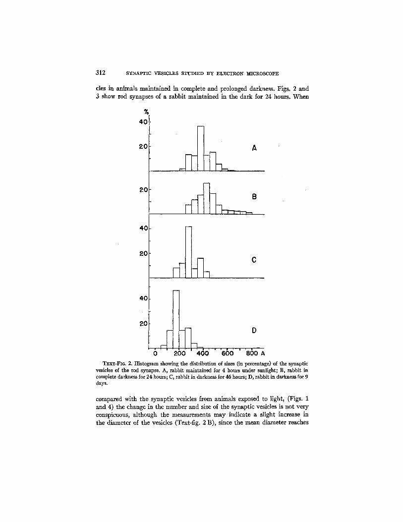
312 SYNAPTIC VESICLES STUDIED BY ELECTRON MICROSCOPE
cles in animals maintained in complete and prolonged darkness. Figs. 2 and 3 show rod synapses of a rabbit maintained in the dark/or 24 hours. When
% 40
20 A
20
40
20
40
20
0
~ ~ _ B
C
• | • • ,
200 400 660 ' BSOA TEXT-FIG. 2. Histogram showing the distribution of sizes (in percentage) of the synaptic
vesicles of the rod synapse. A, rabbit maintained for 4 hours under sunlight; B, rabbit in complete darkness for 24 hours; C, rabbit in darkness for 46 hours; D, rabbit in darkness for 9 days.
compared with the synaptic vesicles from animals exposed to light, (Figs. 1 and 4) the change in the number and size of the synaptic vesicles is not very conspicuous, although the measurements may indicate a slight increase in the diameter of the vesicles (Text-fig. 2 B), since the mean diameter reaches

E . D E I~OB~I~TIS AND C. ~ . F R A N C H I 313
444 A. The only fact that seems to be significant is the accumulation of a great number of synaptic vesicles around the synapfic membrane and its processes (Figs. 2 and 4). Although this accumulation is also present in the animals under sunlight (Figs. 1 and 4), here it is more general and conspic- uous. For example, the entire outer surface of the synaptic membrane of Fig. 3 is covered by a tight lining of vesicles. The process of the synaptic mem- brane of Fig. 2 is covered by 2 and even 3 layers of tightly packed vesicles.
Some measurements made on the diameter of synaptic vesicles after 46 hours of darkness indicated a slight decrease as compared with those of ani- mals exposed normally to light, the mean being 281 A (Text-fig. 2 C).
The changes in the vesicles are particularly striking after 9 days of dark- ness (Fig. 5). The main difference is in the size of the vesicles, which have diminished considerably (Text-fig. 2 D). Some of the vesicles are so small that they are difficult to measure. The diameters range between 50 and 400 A, with a mean of 195 A, as compared with the mean of 396 A, in the con- trols. There are considerable variations from one synapse to another regard- ing the number of vesicles. Thus in some the number is probably the same as in the controls, while in others it is considerably diminished and the ves- icles have disappeared from large areas of the spherule (Fig. 4). Finally, in other synapses the concentration of vesicles may even be augmented (see Fig. 7). However, in all cases the size of the vesicles is much below the nor- mal. In Fig. 5 it is also apparent that there are fewer vesicles in the neigh- borhood of the synaptic membrane. This fact is particularly striking if one compares it with the synapses after 24 hours of darkness (Figs. 2 and 3).
Cone Synapses under Illumination and Prolonged Darkness.--Figs. 6 and 7 illustrate some of the features of the cone synapses of the rabbit. In a section normal to the surface of the retina the profile of the cone terminal is gen- erally triangular with an internal base of 4 to 6 or more microns and an apex which continues with the thick cone fiber. The synapses of the cones are generally situated more internally with respect to the rod synapses within the so called "external plexiform layer" of the retina (Text-fig. 1). The rela- tionships between the terminal and the postsynaptic fibers are much more complex than in the rod synapse, although they follow a similar pattern in the sense that the postsynaptic expansion penetrates and digitates deeply into the cone terminal. This disposition is clearly seen in Fig. 7, in which the internal edge of the terminal is penetrated by several fibers which di- verge and branch within the body of the cone terminal. In Fig. 6, which cor- responds to a more oblique section, the large number and complex relation- ship of the penetrating postsynaptic fibers can be observed. To avoid repe- tition we will not describe in detail the individual constituents of the cone synapses (the membrane of the terminal and the synaptic membrane, the synaptic vesicles, the matrix of the terminal, and the postsynaptic fiber), as they resemble those of the rod synapses.

3!4 SYNAPTIC VESICLES STUDIED BY ELECTRON R[ICROSCOPE
Only a few observations have been made on the action of complete dark- ness on the synaptic vesicles of the cone. The changes are particularly striking after 9 days. Comparing Figs. 6 and 7, one can see a great reduction in size of the vesicles after prolonged darkness. This difference makes the whole terminal much less dense than in the normal control (Fig. 6). Measurements in the diameter of the vesicles clearly reflect that change. Thus, in the nor- mal synapse the vesicles vary in diameter between 150 and 550 A with a mean of 338 A, while in the cone synapse of animals after 9 days of darkness the
%
20
A
0 A I ~) 200 400 A
TEXT-Fro. 3. Histogram showing the distribution of sizes (in percentage) of the synapfic vesicles of the cone synapse. A, rabbit m ,aintained under sunlight for 4 hours; B, rabbit in darkness for 9 days.
diameters vary between 50 and 400 A, with a mean diameter of 236 A (Text- fig. 3).
DISCUSSION
The observations reported here extend to synapses between two receptors and the corresponding neurones, the finding of a vesicular submicroscopic component previously observed in interneuronal synapses (1, 2). In the rod and cone synapses the synaptic vesicles are localized preferentially in the presynapti c side, filling the enlarged terminal of the rod and cone fibers. Only very seldom is it possible, to find in the postsynaptic fibers a few profiles of what might be synaptic vesicles. However, in this case they appear as dis- torted and ghost-like outlines, resembling those observed in synapses of the neuropile of the earthworm (1, 2).
This preferential distribution of the vesicles on the presynapfic side im-

E. DE ROBEP~TIS AND C, M. IrRANCHI 315
plies a polarized submicroscopic organization which may be of importance in synaptic function. In these retinal synapses the polarity is striking because of the lack of mitochondria in the terminal. The apparent accumulation of mitochondria in certain synapses, as observed by different authors (see ref- erence 13), suggested to Bodian the possibility of a relationship between mitochondria and the local secretion of acetylcholine and cholinesterase. It was also suggested that they might be concerned with more general metabolic processes which could modify the electrical properties of the membrane (13). This hypothesis can not be maintained in the case of the rod and cone synap- ses when there are no nearby mitochondria at all, but when the mitochondria are located a considerable distance away in the receptor.
Furthermore, in other synapses the mitochondria that may be observed bear no close relationship with the synaptic membrane (5). In contrast, the localization of the synaptic vesicles and their immediate contact with the synaptic membrane suggest that this might be a more important component in the biochemical phenomena of synaptlc transmission.
At the moment we do not have any direct information about the biochem- ical nature of the synaptlc vesicles and of their possible physiological role. Yet one might mention the fact that the retina has a very high cholinesterase activity (see reference 14), and that this is preferentially concentrated in the synaptic layers (15). Moreover the diphosphopyridine nucleotide is found in particularly high concentration in the synaptic regions. This fact is of interest in view of the role of this compound in the formation of acetylcholine (15).
We may also speculate about the finding of a special rod synapse in which the postsynaptic fiber penetrates directly into the cytoplasm of the rod cell and which, to our knowledge, has not been described before. Following the classical classification of synapses into axosomatic, axodendritic, axoaxonic and so forth, this type of synapse could be considered as a somatodendritic one, since the cell body of the rod cell is probably presynaptic with respect to the dendritic expansion of the bipolar cell. I t is interesting to observe the fact that in all these types of synapses the most striking submicroscopic characteristic is that the synaptic vesicles are localized on the presynaptic side of the synaptic membrane (see Fig. 2). One should not exclude the pos- sibility of dendrodendritic junctions as recently emphasized by Estable (17). The presence of polarized synapses between homologous elements (axon- axon, dendrite-dendrite) might be explained on the basis of the differential submicroscopic organization of the synaptic region. If the synaptic vesicles play a role in synaptic transmission, one may hypothesize that this activity can take place between any part of the presynaptic neurone (cell body or expansion) in contact with any part of the postsynaptic neurone, as long as this submicroscopic vesicular component is localized preferentially on one side of the contacting membranes. However, this generalization cannot be

316 SYNAPTIC VESICLES STUDIED BY ELECTRON ~[ICROSCOPE
made for the moment and this hypothesis can only be applied to the few types of synapses which have been studied so far (1, 2, 4, 5).
The preliminary nature of the results obtained in the study of the action of complete darkness warrant only a few remarks. One fact which seems evident is that after 24 hours of dark adaptation the synaptic vesicles are concentrated around the synaptic membrane as compared with animals adapted to light. However the most striking result is the finding of a definite reduction in size of the vesicles both in the rod and in the cone synapses after prolonged action of darkness (9 days). I t seems possible that this fact may be a consequence of functional disuse of the retina. However, further ex- periments on this line should be performed.
The observations reported here on the complex tridimensional configura- tion of the synaptic membrane both in the rod and cone junctions are con- firmatory of those previously described by Sj6strand (8) in the guinea pig. This author applied the name of intracellular to the rod synapse to account for the penetration and branching of the postsynaptic expansion into the rod terminal. This name may however be misleading ff it is wrongly inter- preted as indicating the actual mixing of the pre- and postsynaptic proto- plasms. This is actually not the case since both are separated by a continuous double membrane (7), Furthermore this type of synapsis in which the post- synaptic expansion indents into the presynaptic one is not uncommon, par- ticularly in the invertebrates (2).
SUId[~ARY
The submicroscopic organization of the rod and cone synapses of the albino rabbit has been investigated with the use of the electron microscope. The most common rod synapse consists of an enlarged expansion of the rod fiber (the so called sphcrnle) into which the dendritic postsynaptic fiber of the bipolar cell penetrates and digitates. The membrane surrounding the ter- minal consists of a double layer, the external of which is interpreted as be- longing to the intervening glial cells. The synaptic membrane has a pre- and a postsynaptic layer with a total thickness of 180 to 300 A. The presynaptic layer is frequently denser and is intimately associated with the adjacent synaptic vesicles. The synaptic membrane shows processes constituted by foldings of the presynaptic layer. The entire spherule is filled with synaptic vesicles varying in diameter between 200 and 650 A with a mean of 386 A. In addition, the spherule contains a few large vacuoles near the rod fiber, interpreted as endoplasmic reticulum, and a matrix in which with high reso- lution a fine filamentous material can be observed. The postsynaptic fiber is homogeneous and usually does not show synaptic vesicles.
In animals maintained in complete darkness for 24 hours vesicles appear to accumulate near the synaptic membrane and its processes. After 9 days there is a sharp decrease in size of the synaptic vesicles.
E. DE ROBERTIS AND C. M, FRANCHI 317
A special rod synapse in which the dendritic postsynaptic expansion pen- etrates directly into the rod cell body has been identified. In line with Ca- jal's classification this type of synapse could be considered as a somatoden- dritic one.
The cone synapse has a much larger terminal with a more complex rela- tionship with the postsynaptic fiber. However, the same components recog- nized in the rod synapse can be observed. In animals maintained for 9 days in complete darkness there is also a considerable diminution in size of the synaptic vesicles.
The authors are deeply indebted to Professor Clemente Estable for advice on the surgical technique and for his criticism and unfailing interest in the work.
BIBLIOGRAPHY
1. De Robertis, E., and Bennett, H. S., Fed. Proc., 1954, 13, 35. 2. De Roberfis, E., and Bennett, H. S., J. Biophysic. and Biochem. Cytol. 1955,
1, 47. 3. Palade, G. E., Anat. Rec., 1954, 118, 335. 4. De Robertis, E., and Franchi, C. M., J. Appl. Physics, 1954, 25, 1162. 5. De Robertis, E., Acta Neurol. Latinoam., 1955, 1, 1. 6. De Robertis, E., Anat. Rec., 1955, 1219 284. 7. Polyak, S. L., The Retina, The University of Chicago Press, 1941. 8. Sj~strand, F. S., J. Appl. Physics, 1953, 24, 1422. 9. Palade, G. E., J. Exp. Med. 1952, 95, 295.
10. Porter, K. R., and Blum, J., Anat. Rec. 1953, 117,685. 11. De Roberfis, E., and Franchi, C. M., J. Exp. Med. 1953, 98, 269. 12. Porter, K. R., J. Exp. Med, 1953, 97, 727. 13. Bodian, D., Physiol. Rev., 1942, 22, 146. 14. Nachmansohn, D., in The Hormones, (G. Pincus and K. U. Thimann, editors),
New York, Academic Press, Inc., 1950, 2,515. 15. Anfinsen, C. B., J. Biol. Chem., 1944, 152, 267. 16. Anfinsen, C. B., J. Biol. Chem., 1944, 152,283. 17. Estable, C., Symposium on the Synapse, Montevideo, 1953, to be published.

318 SYNAPTIC VESICLES STUDIED BY ELECTRON MICROSCOPE
EXPLANATION OF PLATES
Description of Figures
Csy, cone synapse. er, endoplasmic reticulum. G, glial ceil m~, or tin, membrane of the terminal. rim, nuclear membrane. Pse, postsynapfie cytoplasm.
Psm, process of synaptic membrane. Rf, rod fiber. Rn, rod nucleus. Rsy, rod synapse. sin, synaptic membrane. sv, syrmptic vesicles.
PLATE 64
FIG. i. Electron micrograph of the external plexiform layer of the retina of a rabbit showing a rod fiber ending in a sphemle or synaptic terminal (Rsy). The postsynaptic expansion (Psc) is seen to enter and divide into the spherule being separated from the presynaptic cytoplasm by the synapfic membrane (sin). The spherule is filled with synaptic vesicles (sv). (See further description in the text.) Specimen maintained under sunlight for 4 hours. X 38,800.

THE JOURNAL OF BIOPHYSICAL AND BIOCHEMICAL
CYTOLOGY
PLATE 64 VOL. 2
(De Robertis and Franchi: Synaptic vesicles studied by electron microscope)

PLATE 65
Fie.. 2. Rod synapse of rabbit maintained in complete darkness for 24 hours. The synaptic vesicles are accumulated in great number around the synaptic membrane (sin) and particularly around the process of the synaptic membrane (Psm). Note that the synaptic membrane is double with the external or presynaptic layer of higher density. The membrane of the terminal (~m) is also double. The endoplasmic reticulum (er) is seen in the top end of the spherule (see further description in the text). >( 36,200. Inset at higher magnification showing the close relationship of the synaptic membrane with the vesicles. N 58,000.
FIG. 3. Special rod synapse of a rabbit maintained in darkness for 24 hours. The postsynaptic fiber (Psc) penetrates directly into the cytoplasm of the rod cell. The synaptic vesicles accumulate around the synaptic membrane and its process (Psm) (see further description in the text). >( 36,000.

THE JOURNAL OF BIOPHYSICAL AND BIOCHEMICAL
CYTOLOGY
PLATE 65 VOL. 2
(De Robertis and Franchi: Synaptic vesicles studied by electron microscope)

PLATE 66
FIG. 4. Subtangential section of the external layer of the retina of a rabbit main- tained under sunlight for 4 hours. Several rod terminals with the complex disposition of the synapfic membrane are seen. At points marked with arrows the outer layer of the membrane of the terminal detaches and crosses the intervening space, probably limiting two glial cell processes. Note the size of the synaptic vesicles and compare with those of Fig. 5. In one terminal several vacuoles and tubules of the endoplasmic reticulum are seen (see further description in the text). X 44,500.
FIG. S. Figure similar to that of Fig. 4 but from a rabbit maintained in the dark fol 9 days. Arrows mark places in which the outer membrane of the terminal detaches from it and crosses the intervening space to reach another spherule. Note the great diminution in size of the synaptic vesicles as compared with the control of Fig. 4 (see further description in the text). X 41,500.

THE JOURNAL OF BIOPHYSICAL AND BIOCHEMICAL
CYTOLOGY
PLATE 66 VOL. 2
(De Robertis and Franchi: Synaptic vesicles studied by electron microscope)

PLATE 67
FIG. 6. Cone synapse of a rabbit under sunlight illumination. Because the section is subtangential the postsynaptic fibers (Psc) are seen embedded in the cytoplasm of the terminal and surrounded by the synaptic membrane (Sin). Note the size of the synaptic vesicles as compared with the specimen of Fig. 7 (see further description in the text). >( 33,200.
FIG. 7. Cone synapse of a rabbit maintained in the dark for 9 days. Section normal to the retina. In the lower end note the entrance of the postsynaptic fibers (Psc) surrounded by the synaptic membrane. The entire cone terminal is filled with synaptic vesicles which are much smaller than in the control specimen of Fig. 6. The rod synapses (Rsy) contain a great number of very small vesicles (see further description in the text). )< 31,700.

THE JOURNAL OF BIOPHYSICAL AND BIOCHEMICAL
CYTOLOGY
PLATE 67 VOL. 2
(De Robertis and Franchi: Synaptic vesicles studied by electron microscope)