electroencephalography for the diagnosis of brain death · received: april 27, 2017 revised: may...
TRANSCRIPT

Received: April 27, 2017
Revised: May 31, 2017
Accepted: June 7, 2017
Correspondence to
The Korean Society of Clinical NeurophysiologyDaeil Building 1111, 12 Insadong-gil, Jongno-gu, Seoul 03163, KoreaTel: +82-2-2291-2290Fax: +82-2-737-6531E-mail: [email protected]
http://www.e-acn.org
pISSN 2508-691X eISSN 2508-6960
Copyright © 2017 The Korean Society of Clinical NeurophysiologyThis is an Open Access article distributed under the terms of the Creative Commons Attribution Non-Commercial License (http://creativecommons.org/licenses/by-nc/4.0) which permits unrestricted non-commercial use, distribution, and reproduction in any medium, provided the original work is properly cited.
Electroencephalography for the diagnosis of brain deathSeo-Young Lee1, Won-Joo Kim2, Jae Moon Kim3, Juhan Kim4, Soochul Park5; and on behalf of the Korean Society of Clinical Neurophysiology Education Committee1Department of Neurology, Kangwon National University School of Medicine, Chuncheon, Korea 2Department of Neurology, Gangnam Severance Hospital, Yonsei University College of Medicine, Seoul, Korea 3Department of Neurology, Chungnam National University Hospital, Daejeon, Korea 4Department of Neurology, Hanyang University Seoul Hospital, Seoul, Korea 5Department of Neurology, Yonsei University College of Medicine, Seoul, Korea
Electroencephalography (EEG) is frequently used to assist the diagnosis of brain death. Howev-er, to date there have been no guidelines in terms of EEG criteria for determining brain death in Korea, despite EEG being mandatory. The purpose of this review is to provide an update on the evidence and controversies with regarding to the utilization of EEG for determining brain death and to serve as a cornerstone for the development of future guidelines. To determine brain death, electrocerebral inactivity (ECI) should be demonstrated on EEG at a sensitivity of 2 μV/mm using double-distance electrodes spaced 10 centimeters or more apart from each other for at least 30 minutes, with intense somatosensory or audiovisual stimuli. ECI should be also verified by checking the integrity of the system. Additional monitoring is needed if extracerebral potentials cannot be eliminated. Interpreting EEG at high sensitivities, which is required for the diagnosis of brain death, can pose a diagnostic challenge. Furthermore, EEG is affected by physiologic variables and drugs. However, no consensus exists as to the minimal requirements for blood pressure, oxygen saturation, and body temperature during the EEG recording itself, the minimal time for observation after the brain injury or rewarming from hypothermia, and how to determine brain death when the findings of ECI is equivocal. There-fore, there is a strong need to establish detailed guidelines for performing EEG to determine brain death.
Key words: Electroencephalography; Brain death; Electrocerebral inactivity
ANNALS OF CLINICAL NEUROPHYSIOLOGY
Ann Clin Neurophysiol 2017;19(2):118-124https://doi.org/10.14253/acn.2017.19.2.118
THE KOREAN SOCIETY OF CLINICAL NEUROPHYSIOLOGY
SPECIAL ARTICLE

119http://www.e-acn.org https://doi.org/10.14253/acn.2017.19.2.118
Seo-Young Lee, et al. EEG for the diagnosis of brain death
Electroencephalography for the diagnosis of brain deathSeo-Young Lee1, Won-Joo Kim2, Jae Moon Kim3, Juhan Kim4, Soochul Park5; and on behalf of the Korean Society of Clinical Neurophysiology Education Committee1Department of Neurology, Kangwon National University School of Medicine, Chuncheon, Korea 2Department of Neurology, Gangnam Severance Hospital, Yonsei University College of Medicine, Seoul, Korea 3Department of Neurology, Chungnam National University Hospital, Daejeon, Korea 4Department of Neurology, Hanyang University Seoul Hospital, Seoul, Korea 5Department of Neurology, Yonsei University College of Medicine, Seoul, Korea
Electroencephalography (EEG) is frequently used to assist the diagnosis of brain death. Howev-er, to date there have been no guidelines in terms of EEG criteria for determining brain death in Korea, despite EEG being mandatory. The purpose of this review is to provide an update on the evidence and controversies with regarding to the utilization of EEG for determining brain death and to serve as a cornerstone for the development of future guidelines. To determine brain death, electrocerebral inactivity (ECI) should be demonstrated on EEG at a sensitivity of 2 μV/mm using double-distance electrodes spaced 10 centimeters or more apart from each other for at least 30 minutes, with intense somatosensory or audiovisual stimuli. ECI should be also verified by checking the integrity of the system. Additional monitoring is needed if extracerebral potentials cannot be eliminated. Interpreting EEG at high sensitivities, which is required for the diagnosis of brain death, can pose a diagnostic challenge. Furthermore, EEG is affected by physiologic variables and drugs. However, no consensus exists as to the minimal requirements for blood pressure, oxygen saturation, and body temperature during the EEG recording itself, the minimal time for observation after the brain injury or rewarming from hypothermia, and how to determine brain death when the findings of ECI is equivocal. There-fore, there is a strong need to establish detailed guidelines for performing EEG to determine brain death.
Key words: Electroencephalography; Brain death; Electrocerebral inactivity
‘Brain death’ is a term that implies irreversible loss of func-tion of entire brain, including brainstem and hemisphere.1,2 The concept of brain death first emerged in the 1950s, driven by progress in critical care medicine such as cardiopulmonary resuscitation and the development of mechanical ventila-tion.3,4
Since the ad hoc Committee of Harvard Medical School defined the characteristics of irreversible coma as 1) unrecep-tivity and unresponsiveness, 2) the absence of movement and breathing, 3) the absence of brainstem reflexes, and 4) flat electroencephalogram,5 a myriad of criteria for determin-ing brain death have been suggested and evolved. However, a recent review reported that there was significant variation among the criteria and protocols to determine brain death according to countries and institutions, although most coun-tries have legal provisions on brain death. Particularly, ancillary tests to confirm brain death are used in variation. The most frequently required test is an electroencephalography (EEG), which is mandatory in 28% and optional in 47% of countries.3 In Korea, standards for determining brain death were first es-tablished in 1983 by the ‘Committee for defining death’ of the Korean Medical Association. On Feb 8, 1999 ‘Laws on organ transplantation’ have been established, defining brain death only for the purpose of organ transplantation and requiring a flat EEG for at least 30 min.6 The most recent Korean stan-dards for determining brain death have been stated in Article 21 of the enforcement decree of the Organ Transplant Act.7
Despite the fact that number of annual cases requiring de-termination of brain death reached 537 cases in 2015,8 to date no specific guidelines or consensus with regard to performing and interpreting EEGs for the diagnosis of brain death have been established in Korea. The goal of this review is to provide an update on the role of EEG for determining brain death and the potential confounding factors, for development of future guidelines.
EEG FINDINGS REPRESENTATIVE OF BRAIN DEATH
The consensus is that EEG potentials should be absent to declare brain death. A survey by the American EEG Society’s ad hoc Committee on the EEG Criteria for the Determination of Cerebral Death in the late 1960s revealed that, three out
of 2,650 adult cases of coma with presumably “isoelectric” EEGs met the committee’s criteria showed any recovery of cerebral function. These three cases were due to massive overdoses of nervous system depressants.9 Non-physiologic terms such as “electrocerebral silence”, “isoelectric,” “linear,” and “flat” were replaced in the 1970s with the term “electro-cerebral inactivity (ECI)”, which appeared in the Glossary of the International Federation of Clinical Neurophysiology.10
ECI was defined as absence over all regions of head of iden-tifiable electrical activity or cerebral origin, whether sponta-neous or induced by physiologic stimuli and pharmacological agents.11 The widely-accepted working definition of ECI is the absence of non-artifactual electrical activity with a peak to peak amplitude above 2 μV when recording from scalp elec-trode pairs that were 10 cm or further apart.10
CLINICAL SETTING AND CONFOUNDING FACTORS
ECI is required to confirm cessation of brain function, but this doesn’t ensure either the irreversibility or loss of whole brain function. EEG as well as the clinical examination is af-fected by physiologic variables and medication. The Korean law lists the following as a prerequisite for determining brain death: A. The absence of any treatable drug toxicities (such as due to anesthetics, hypnotics, sedatives, muscle relaxants, or poisons); B. The absence of any treatable endocrinological disorders (such as hepatic coma, uremic coma, hypogly-cemic encephalopathy); C. The absence of hypothermia (defined as a rectal temperature of 32℃ or lower); D. The absence of a shock-like state.7 If sedative medications were used before the diagnosis of brain death, the French guide-lines recommend using the techniques based on the study of intracerebral blood flow such as cerebral angiography, which are not influenced by the medications, rather than EEG.12
American Clinical Neurophysiology Society (ACNS) guide-lines 2016 newly addressed the item that blood pressure and oxygen saturation should be recorded.10 The French guide-lines on EEG recommended maintaining the patient’s body temperature above 34℃ and that the EEG should be per-formed at least 12 hours after cardiorespiratory arrest.12 Partic-ularly, determining brain death needs caution in the setting of
120 http://www.e-acn.org https://doi.org/10.14253/acn.2017.19.2.118
Annals of Clinical Neurophysiology Volume 19, Number 2, July 2017
therapeutic hypothermia.There had been a case of a patient who had regained cough reflex and spontaneous respiration in the operating room for organ procurement 48 hours after rewarming from therapeutic hypothermia.13
The guidelines as to when the EEG should be recorded after the brain injury, as well as blood pressure, oxygen satura-tion, body temperature, and the minimal time required after rewarming from hypothermia should be further developed. Moreover, ECI may be seen in cases where subcortical or brainstem function remains intact as EEG represents cortical activity.14 This may be seen in hypoxic ischemic encephalopa-thy as the cerebral cortex is more susceptible to ischemia.15
REQUIREMENTS FOR EEG ACQUISITION
1. Electrodes and montageA full set of scalp electrodes over all major brain areas us-ing conventional 10-20 system should be used. In certain cases, recording with a full set of conventional 10-20 scalp locations may not be feasible such as due to head trauma or recent surgery. In these cases, the electrode positions may be moved as necessary, as long as careful documentation is made and the inter-electrode distances remain at least 10 centimeters.10
The French guidelines stated that a minimum of 8 elec-trodes should be placed, whereas the American Academy of Neurology (AAN) recommend at least 16 channels.1 French guidelines recommended the placement of FP2, C4, O2, T4, FP1, C3, O1, T3 and Cz electrodes. The ACNS guidelines main-tained that midline electrodes (Fz, Cz, Pz) should be included because these electrodes are useful for the detection of re-sidual low-voltage physiologic activity, and are relatively free from artifacts.10
Although a ground electrode should be added in most cases, a ground electrode should not be used if grounds from other electrical equipment are already attached to the pa-tient, which often happens in intensive care units because of safety issue according to ACNS guidelines 2006.9 This recom-mendation was replaced with the requirement of an ‘isolated’ ground electrode in ACNS guidelines 2016.10
Inter-electrode impedance should be balanced between 100 and 10,000 Ohms as a routine.10,16 High impedance results in more artifacts and can be decreased by cleansing the scalp.
It is also affected by the quality of the electrodes. Impedances below 100 Ohms may be a possible reason for a false-positive ECI recording. It is essential that excess electrode paste does not spread from one electrode to another, creating a shunt or short circuit, which may also attenuate the signal.10 Electrode impedance should be rechecked during the recording when any type of artifact appears. normal impedance in an elec-trode demonstrating noise may still necessitate changing or modifying that electrode.16
The use of needle electrodes and “electrode caps” should be avoided.10 Not only are there concerns such as patient dis-comfort and potential risk of injury to the staff, but these elec-trodes have higher impedance than cup electrodes, resulting in potentially higher levels of noise.16 When using electrode caps, excess jelly spreading from one electrode to another can create a short circuit, which may attenuate the signal.9
2. EEG recordingA sampling rate of at least 256 Hz is recommended ac-
cording to the French guidelines.12 The sensitivity must be increased to a maximum of 2 μV/mm. The calibration voltage should be either 2 or 5 μV, which differs from routine EEG re-cording.10
With regard to filter settings, the high-frequency (low pass) filters should not be set below 30 Hz, and the low-frequency (high pass) filters should not be set above 1 Hz.10 The French recommendations are more strict; 70 Hz or higher for the low pass filter and 0.53 Hz for the high pass filter.12 The 50 or 60-Hz notch filter can be used with care, but there should be segments of EEG that are recorded without this filter for com-parison.10
If the EEG suggests ECI, the integrity of the system should be tested by gently touching or shaking each electrode of the montage to create an artifact potential. This verifies that the electrode board is connected to the recording device and that the montage settings match the electrode place-ment.10,12 At least 30 minutes of EEG recording is recommend-ed by Korean law as described in most guidelines,10,12 based on the observation that intermittent cortical activity with amplitudes as low as 2 μV may appear at intervals of up to 20 minutes during continuous recording for up to 8 hours.17
Auditory and bilateral somatosensory stimuli (touch and pain) should be repeatedly performed, and be clearly marked

121http://www.e-acn.org https://doi.org/10.14253/acn.2017.19.2.118
Seo-Young Lee, et al. EEG for the diagnosis of brain death
on the recording (i.e., the point where the somatosensory stimulus was applied).10,12 The technician must document all the events that occur during the test on the EEG tracings.
The Korean law mandates that EEG should be performed once after the second clinical examination and the apnea test in patients aged one year or above, and twice (before and after the second clinical examination) in infants between ages two months and one year. Although it is stated to perform a repeat clinical examination 48 hours later in those less than 1 year of age, it is unclear as to what the time intervals between the two EEGs should be.7 The French guidelines recommend-ed a repeat EEG in all age groups. The time interval between the two EEGs was recommended to be at least 4 hours (the starting point being the beginning of each EEG recording) for those more than 1 year of age; 24 hours between the ages of 1 month and 1 year; 48 hours between the ages of 7 days and 1 month.12 The American guidelines state that one EEG should be sufficient1 but added the following comments: In the event that technical or other difficulties lead to uncer-tainty in the evaluation of ECI, the entire procedure should be repeated after an interval. This may be as short as 6 hours in adult patients, but in neonates and children the interval should be at least 24 hours. Consideration should be given to other confirmatory tests if technical limitations are unlikely to be overcome in subsequent recordings, based on the opin-ions of the treating physicians.10
The basis as to why a single EEG is sufficient is described in the American EEG guidelines as the following; in the Collabo-rative Study of Cerebral Death, no patients survived for more than a short period after an EEG showed ECI, excluding the cases which were due to overdosing with central nervous sys-tem (CNS) depressants. This provides evidence that a single EEG demonstrating ECI is a highly reliable tool for determining cortical death.10
3. Evaluation of extracerebral potentials The technician should recognize the common sources of
artifact, either internal or external, such as electrical artifact originating from electric beds, IV drips, blood warmers, or other electrical devices. Electric potentials of extracerebral or-igin often cannot be avoided at high sensitivities in intensive care unit (ICU) and responsible for the disagreement in the interpretation. Therefore, additional monitoring is required to differentiate cerebral from extracerebral potentials. The ACNS
guidelines recommend the following (modified);10 1) Electrocardiogram monitoring is essential. This may be
monitored by using two precordial disc electrodes.2) Continuous video recording is strongly recommended to
help identify any artifact in the EEG recording.3) Respiration artifact must be documented with specific
notations on the record or monitored by a transducer if this cannot be eliminated. Brief disconnection of the respirator should allow definitive identification of the artifact, if clinically feasible.
4) Motion monitors should be placed if intermittent move-ments are noted in a limb.
5) An additional monitor is often needed for other artifacts emanating from the patient or the local environment. The most convenient for this purpose is a pair of electrodes on the dorsum of the hand separated by about 6 to 7 cm.
6) Machine noise, thermal noise, and electrical interference
Fig. 1. An example of a dummy patient with a three-lead device (Grass Technologies, West Warwick, RI, USA). This can be used to measure machine noise and external interference which enters to the recording system.

122 http://www.e-acn.org https://doi.org/10.14253/acn.2017.19.2.118
Annals of Clinical Neurophysiology Volume 19, Number 2, July 2017
entering the recording system from the jack box to the am-plifiers may be measured on a “dummy patient” (Fig. 1), i.e., placing a 10,000-Ohm resistor between input terminal 1 (G1) and input terminal 2 (G2) of one of the channels, as long as either G1 or G2 is shorted to the reference electrode.
7) If electromyogram potentials are of such amplitude as to obscure the tracing, it may be necessary to reduce or eliminate them by using a short-term neuromuscular block-ing agent. As this may interfere with the neurologic exam-ination and cause other problems, neuromuscular blockade should be performed under the guidance of an experienced physician familiar using medications in critically ill patients.
INTERPRETATION
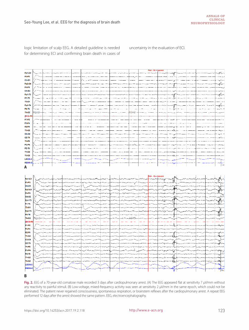
An estimate must be made of whether the amplitudes ex-ceed 2 μV. The interpretation should be carried out using long distance montages such as longitudinal or transverse bipolar montages with double distance electrodes. Filters should be avoided.12 It is actually very difficult to acquire EEG without artifacts higher than 2 μV/mm in ICU (Fig. 2). Hence, determining ECI creates diagnostic challenges, and significant inter- and intra-rater disagreement has been reported.18 In practice, it is necessary to tolerate artifacts, as long as it can be ensured that they are artifacts. Determining whether the signals shown in EEG are cerebral or not can be done cautiously based on the results of the additional monitoring as indicated in III-3. Reviewing an accompanying video recording can also be useful.
When a lack of confidence exists, the EEG report must reflect such uncertainty, and the record cannot be stated as demonstrating ECI.10 In Korea, ECI is mandatory for declaring brain death by law, and no guidelines exist for determining brain death when it is uncertain whether the EEG truly shows ECI or not. According to statistics from Korea Organ Dona-tion Agency since 2010, only one patient was diagnosed with brain death without the EEG results clearly demonstrat-ing ECI due to difficulties in performing the EEG itself as a result of craniectomy (personal communication). Details of the EEG reports for the diagnosis of brain death which were suggested in French recommendation are as follows (modi-fied);12
1. Administrative data: the administrative data for routine EEG as well as the location and exact times indicating the begin-ning and end of the recording must be specified.
2. Clinical information: the circumstances and origin of the coma, especially the presumed date of the onset of the coma; the time lapse from the event which resulted in brain death; imaging data; clinical criteria for brain death; the presence of interfering factors: toxicological or metabolic; drugs administered in the past 24 hours; body temperature; blood pressure; oxygen saturation; specific recording con-ditions (e.g. scalp condition, environment).
3. EEG description: read-out on long-distance montages; the duration of the interpretable recording; background activi-ty: indicate the maximum amplitude measured aside from the artifacts; results from auditory, somatosensory tactile and nociceptive stimulations; describe an eventual diffusion of the electrocardiogram and its topography (diffuse, diffu-sion only on certain derivations).
4. Conclusion: Absence of all brain electrical activity under the stated conditions of the EEG recording.
PERSONNEL
The recording should be performed by a specialized techni-cian, qualified to identify and eliminate artifacts, implement the required settings, perform standard activation proce-dures and document all relevant data in the recording. The report should only be created by a physician specializing in EEG or Neurophysiology.10,12
CONCLUSIONS
To determine brain death, ECI should be demonstrated on scalp EEG which is recorded as following criteria. 1. The sen-sitivity of 2 mV/mm; 2. Inter-electrode distances more than 10 centimeters; 3. Covering over all major brain areas includ-ing midline area 4. Recording for at least 30 minutes; 5. Giv-ing intense somatosensory or audiovisual stimuli. ECI should be also verified by checking the integrity of the system. Eval-uation of extracerebral potentials is required, because such potentials are often unavoidable at high sensitivity in ICU. Physicians should keep in mind the anatomical and physio-
123http://www.e-acn.org https://doi.org/10.14253/acn.2017.19.2.118
Seo-Young Lee, et al. EEG for the diagnosis of brain death
A
B
Fig. 2. EEG of a 70-year-old comatose male recorded 3 days after cardiopulmonary arrest. (A) The EEG appeared flat at sensitivity 7 μV/mm without any reactivity to painful stimuli. (B) Low-voltage, mixed-frequency activity was seen at sensitivity 2 μV/mm in the same epoch, which could not be eliminated. The patient never regained consciousness, spontaneous respiration, or brainstem reflexes after the cardiopulmonary arrest. A repeat EEG performed 12 days after the arrest showed the same pattern. EEG, electroencephalography.
logic limitation of scalp EEG. A detailed guideline is needed for determining ECI and confirming brain death in cases of
uncertainty in the evaluation of ECI.
124 http://www.e-acn.org https://doi.org/10.14253/acn.2017.19.2.118
Annals of Clinical Neurophysiology Volume 19, Number 2, July 2017
AcknowledgementsWe would like to thank Ms. Miran Kong, EEG technician of Kangwon National University Hospital, and Mr. Daejun Lee, Director of YoungWoo Meditech, Co., Ltd., for providing their knowledge and expertise on the technical aspects of EEG ma-chinery and recording.
REFERENCES
1. Practice parameters for determining brain death in adults (sum-
mary statement). The Quality Standards Subcommittee of the
American Academy of Neurology. Neurology 1995;45:1012-1014.
2. Wijdicks EF. Brain death worldwide: accepted fact but no global
consensus in diagnostic criteria. Neurology 2002;58:20-25
3. Wahlster S, Wijdicks EF, Patel PV, Greer DM, Hemphill JC 3rd,
Carone M, et al. Brain death declaration: practices and perceptions
worldwide. Neurology 2015;84:1870-1879.
4. Kramer AH. Ancillary testing in brain death. Semin Neurol
2015;35:125-138.
5. A definition of irreversible coma. Report of the Ad Hoc Committee
of the Harvard Medical School to examine the definition of brain
death. JAMA 1968;205:337-340.
6. Nam SO. Brain death and organ transplantation. Korean J Pediatr
2009;52:856-861.
7. Korean Ministry of Gevernment Legislation Pages©. Enforcement
decree of the organ transplant act [Internet]. Sejong, Korea: Kore-
an Ministry of Gevernment Legislation; c2016 [accessed 2016 Oct
28]. Available from: http://www.law.go.kr.
8. Korea Organ Donation Agency Pages©. KODA Annual Report 2015
[Internet]. Seoul, Korea: Korea Organ Donation Agency; c2015 [ac-
cessed 2016 Oct 28]. Available from: http://fliphtml5.com/down-
load-pdf-file/590915.
9. American Clinical Neurophysiology Society. Guideline 3: Minimum
technical standards for EEG recording in suspected cerebral death.
J Clin Neurophysiol 2006;23:97-104.
10. Stecker MM, Sabau D, Sullivan L, Das RR, Selioutski O, Drislane FW,
et al. American Clinical Neurophysiology Society Guideline 6: Min-
imum technical standards for EEG recording in suspected cerebral
death. J Clin Neurophysiol 2016;33:324-327.
11. A glossary of terms most commonly used by clinical electro-
encephalographers. Electroencephalogr Clin Neurophysiol
1974;37:538-548.
12. André-Obadia N, Lamblin MD, Sauleau P. French recommenda-
tions on electroencephalography. Neurophysiol Clin 2015;45:1-17.
13. Webb AC, Samuels OB. Reversible brain death after cardiopulmo-
nary arrest and induced hypothermia. Crit Care Med 2011;39:1538-
1542.
14. Young GB, Lee D. A critique of ancillary tests for brain death. Neur-
ocrit Care 2004;1:499-508.
15. Brierley JB, Graham DI, Adams JH, Simpsom JA. Neocortical death
after cardiac arrest. A clinical, neurophysiological, and neuropatho-
logical report of two cases. Lancet 1971;2:560-565.
16. Sinha SR, Sullivan L, Sabau D, San-Juan D, Dombrowski KE, Halford
JJ, et al. American Clinical Neurophysiology Society Guideline 1:
Minimum technical requirements for performing clinical electro-
encephalography. J Clin Neurophysiol 2016;33:303-307.
17. Jorgensen EO. Technical contribution. Requirements for record-
ing the EEG at high sensitivity in suspected brain death. Electro-
encephalogr Clin Neurophysiol 1974;36:65-69.
18. Buchner H, Schuchardt V. Reliability of electroencephalogram in
the diagnosis of brain death. Eur Neurol 1990;30:138-141.